IPF- ERS Statement
37
American Thoracic Society Documents An Official ATS/ERS/JRS/ALAT Statement: Idiopathic Pulmonary Fibrosis: Evidence-based Guidelines for Diagnosis and Management Ganesh Raghu, Harold R. Collard, Jim J. Egan, Fernando J. Martinez, Juergen Behr, Kevin K. Brown, Thomas V. Colby, Jean-Franc xois Cordier, Kevin R. Flaherty, Joseph A. Lasky, David A. Lynch, Jay H. Ryu, Jeffrey J. Swigris, Athol U. Wells, Julio Ancochea, Demosthenes Bouros, Carlos Carvalho, Ulrich Costabel, Masahito Ebina, David M. Hansell, Takeshi Johkoh, Dong Soon Kim, Talmadge E. King, Jr., Yasuhiro Kondoh, Jeffrey Myers, Nestor L. Mu ¨ller, Andrew G. Nicholson, Luca Richeldi, Moise ´s Selman, Rosalind F. Dudden, Barbara S. Griss, Shandra L. Protzko, and Holger J. Schu ¨nemann, on behalf of the ATS/ERS/JRS/ALAT Committee on Idiopathic Pulmonary Fibrosis THIS OFFICIAL STATEMENT OF THE AMERICAN THORACIC SOCIETY (ATS), THE EUROPEAN RESPIRATORY SOCIETY (ERS), THE JAPANESE RESPIRATORY SOCIETY (JRS), AND THE LATIN AMERICAN THORACIC ASSOCIATION (ALAT) WAS APPROVED BY THE ATS BOARD OF DIRECTORS,NOVEMBER 2010, THE ERS EXECUTIVE COMMITTEE,SEPTEMBER 2010, THE JRS BOARD OF DIRECTORS,DECEMBER 2010, AND THE ALAT EXECUTIVE COMMITTEE,NOVEMBER 2010 THIS STATEMENT HAS BEEN FORMALLY ENDORSED BY THE SOCIETY OF THORACIC RADIOLOGY AND BY THE PULMONARY PATHOLOGY SOCIETY CONTENTS Introduction Objective Methods Committee Composition Disclosure of Conflicts of Interest Committee Meetings and Evidence Review Process Document Preparation Document Structure Formulation of the Topic Sections and Questions Literature Review and Preparation of Evidence Profiles Quality of Evidence and Strength of Recommendations External Review Process Significance of Evidence-based Recommendations to Clinicians for the Management of IPF Summary Conclusions and Treatment Recommendations Conclusions Treatment Recommendations Definition and Epidemiology Definition Clinical Presentation Incidence and Prevalence Potential Risk Factors Genetic Factors Definition Of UIP Pattern UIP Pattern: HRCT Features UIP Pattern: Histopathology Features Diagnosis Diagnostic Criteria Exclusion of Other Known Causes Bronchoalveolar Lavage Cellular Analysis Transbronchial Lung Biopsy Serological Testing for Connective Tissues Disease Multidisciplinary Discussion Natural History of IPF Acute Exacerbation of IPF Vital Statistics Staging and Prognosis Demographics Dyspnea Physiology HRCT Features Composite Scoring Systems Six-Minute-Walk Testing Histopathology Pulmonary Hypertension Emphysema Serum and Bronchoalveolar Lavage Biomarkers Treatment Pharmacologic Therapies Nonpharmacologic Therapies Selected Complications and Comorbid Conditions Palliative Care Monitoring the Clinical Course of Disease Monitoring for Progressive Disease Monitoring for Worsening Symptoms Monitoring for Worsening Oxygenation Monitoring for Complications and Comorbidities Summary of Clinical Management of IPF Future Directions This document is an international evidence-based guideline on the diagnosis and management of idiopathic pulmonary fibrosis, and is a collaborative effort of the American Thoracic Society, the European Respiratory Society, the Japanese Respiratory Society, and the Latin American Thoracic Association. It represents the current state of knowledge regarding idiopathic pulmonary fibrosis (IPF), and con- tains sections on definition and epidemiology, risk factors, diagnosis, natural history, staging and prognosis, treatment, and monitoring disease course. For the diagnosis and treatment sections, pragmatic GRADE evidence-based methodology was applied in a question- based format. For each diagnosis and treatment question, the committee graded the quality of the evidence available (high, moderate, low, or very low), and made a recommendation (yes or no, strong or weak). Recommendations were based on majority This document has an online supplement, which is accessible from this issue’s table of contents at www.atsjournals.org This version of the Statement contains the corrections outlined online at http:// ajrccm.atsjournals.org/cgi/content/full/183/6/788/DC2. Am J Respir Crit Care Med Vol 183. pp 788–824, 2011 DOI: 10.1164/rccm.2009-040GL Internet address: www.atsjournals.org
-
Upload
vikramsobti -
Category
Documents
-
view
23 -
download
2
description
IPF- ERS Statement
Transcript of IPF- ERS Statement

American Thoracic Society Documents
An Official ATS/ERS/JRS/ALAT Statement: IdiopathicPulmonary Fibrosis: Evidence-based Guidelines forDiagnosis and Management
Ganesh Raghu, Harold R. Collard, Jim J. Egan, Fernando J. Martinez, Juergen Behr, Kevin K. Brown,Thomas V. Colby, Jean-Francxois Cordier, Kevin R. Flaherty, Joseph A. Lasky, David A. Lynch, Jay H. Ryu,Jeffrey J. Swigris, Athol U. Wells, Julio Ancochea, Demosthenes Bouros, Carlos Carvalho, Ulrich Costabel,Masahito Ebina, David M. Hansell, Takeshi Johkoh, Dong Soon Kim, Talmadge E. King, Jr., Yasuhiro Kondoh,Jeffrey Myers, Nestor L. Muller, Andrew G. Nicholson, Luca Richeldi, Moises Selman, Rosalind F. Dudden,Barbara S. Griss, Shandra L. Protzko, and Holger J. Schunemann, on behalf of the ATS/ERS/JRS/ALAT Committeeon Idiopathic Pulmonary Fibrosis
THIS OFFICIAL STATEMENT OF THE AMERICAN THORACIC SOCIETY (ATS), THE EUROPEAN RESPIRATORY SOCIETY (ERS), THE JAPANESE
RESPIRATORY SOCIETY (JRS), AND THE LATIN AMERICAN THORACIC ASSOCIATION (ALAT) WAS APPROVED BY THE ATS BOARD OF
DIRECTORS, NOVEMBER 2010, THE ERS EXECUTIVE COMMITTEE, SEPTEMBER 2010, THE JRS BOARD OF DIRECTORS, DECEMBER 2010, AND
THE ALAT EXECUTIVE COMMITTEE, NOVEMBER 2010
THIS STATEMENT HAS BEEN FORMALLY ENDORSED BY THE SOCIETY OF THORACIC RADIOLOGY AND BY THE PULMONARY PATHOLOGY SOCIETY
CONTENTS
IntroductionObjectiveMethods
Committee CompositionDisclosure of Conflicts of InterestCommittee Meetings and Evidence Review ProcessDocument PreparationDocument StructureFormulation of the Topic Sections and QuestionsLiterature Review and Preparation of Evidence ProfilesQuality of Evidence and Strength of RecommendationsExternal Review Process
Significance of Evidence-based Recommendations to Cliniciansfor the Management of IPF
Summary Conclusions and Treatment RecommendationsConclusionsTreatment Recommendations
Definition and EpidemiologyDefinitionClinical PresentationIncidence and PrevalencePotential Risk FactorsGenetic Factors
Definition Of UIP PatternUIP Pattern: HRCT FeaturesUIP Pattern: Histopathology Features
DiagnosisDiagnostic CriteriaExclusion of Other Known CausesBronchoalveolar Lavage Cellular AnalysisTransbronchial Lung BiopsySerological Testing for Connective Tissues Disease
Multidisciplinary DiscussionNatural History of IPFAcute Exacerbation of IPFVital Statistics
Staging and PrognosisDemographicsDyspneaPhysiologyHRCT FeaturesComposite Scoring SystemsSix-Minute-Walk TestingHistopathologyPulmonary HypertensionEmphysemaSerum and Bronchoalveolar Lavage Biomarkers
TreatmentPharmacologic TherapiesNonpharmacologic TherapiesSelected Complications and Comorbid Conditions
Palliative CareMonitoring the Clinical Course of Disease
Monitoring for Progressive DiseaseMonitoring for Worsening SymptomsMonitoring for Worsening OxygenationMonitoring for Complications and Comorbidities
Summary of Clinical Management of IPFFuture Directions
This document is an international evidence-based guideline on thediagnosis and management of idiopathic pulmonary fibrosis, and isa collaborativeeffortof theAmericanThoracicSociety, theEuropeanRespiratory Society, the Japanese Respiratory Society, and the LatinAmerican Thoracic Association. It represents the current state ofknowledge regarding idiopathic pulmonary fibrosis (IPF), and con-tainssectionsondefinitionandepidemiology, risk factors,diagnosis,natural history, staging and prognosis, treatment, and monitoringdisease course. For the diagnosis and treatment sections, pragmaticGRADE evidence-based methodology was applied in a question-based format. For each diagnosis and treatment question, thecommittee graded the quality of the evidence available (high,moderate, low, or very low), and made a recommendation (yes orno, strong or weak). Recommendations were based on majority
This document has an online supplement, which is accessible from this issue’s
table of contents at www.atsjournals.org
This version of the Statement contains the corrections outlined online at http://
ajrccm.atsjournals.org/cgi/content/full/183/6/788/DC2.
Am J Respir Crit Care Med Vol 183. pp 788–824, 2011DOI: 10.1164/rccm.2009-040GLInternet address: www.atsjournals.org

vote. It is emphasized that cliniciansmust spend adequate timewithpatients to discuss patients’ values and preferences and decide onthe appropriate course of action.
Keywords: idiopathic pulmonary fibrosis; usual interstitial pneumonia;
evidence-based medicine, diagnosis, therapeutics; tomography, X-ray
computed
Idiopathic pulmonary fibrosis (IPF) is defined as a specific formof chronic, progressive fibrosing interstitial pneumonia of un-known cause, occurring primarily in older adults, and limitedto the lungs. It is characterized by progressive worsening ofdyspnea and lung function and is associated with a poorprognosis. The American Thoracic Society and European Re-spiratory Society (ATS/ERS), in collaboration with the Amer-ican College of Chest Physicians (ACCP), published aninternational consensus statement in 2000 on the diagnosisand management of IPF (1). Importantly, the statement recog-nized IPF as a distinct clinical entity associated with the his-tologic appearance of usual interstitial pneumonia (UIP), andprovided specific recommendations for clinicians regarding itsdiagnosis and management. Since the publication of the 2000ATS/ERS statement, studies have used the ATS/ERS statementrecommendations to further our understanding of the clinicalmanifestations and course of IPF. The accumulated data andobservations made in these studies allow us to provide newguidelines for the diagnosis and management of IPF based onthe best available evidence using ATS/ERS methodology.
OBJECTIVE
This document is an international evidence-based guideline onthe diagnosis and management of IPF. The purpose of theseguidelines is to analyze the additional evidence accumulatedsince the publication of the 2000 ATS/ERS consensus statementand to provide evidence-based recommendations for manage-ment, with an emphasis on diagnosis and treatment. Thisdocument is intended to replace the previous ATS/ERS IPFconsensus statement, and will be updated when appropriate inaccordance with the policy of the sponsoring societies.
The primary objective of this document is to providerecommendations based on a thorough review of the evidencepublished to date using the GRADE methodology (see below)to clinicians in a transparent manner. It is intended to empowerclinicians to interpret these recommendations in the context ofindividual patient values and preferences, and to make appro-priate decisions regarding all aspects of disease management,tailored to the patient with typical IPF.
METHODS
Committee Composition
This guideline is a collaborative effort between the ATS, ERS, JapaneseRespiratory Society (JRS), and Latin American Thoracic Association(ALAT). The project chair (G.R.) nominated two co-chairs (J.J.E. andF.J.M.) and a group of experts in IPF and/or evidence-based method-ology from North America, Europe, Asia, and South America. Thisgroup consisted of clinicians with recognized expertise in IPF andinterstitial lung diseases (24 pulmonologists, 4 radiologists, and 4pathologists), 1 methodologist (also a general pulmonologist), and 1chief librarian, assisted by 2 librarians experienced with literaturesearches for pulmonary diseases. This group was approved by andrepresented the membership of the four sponsoring societies.
Disclosure of Conflicts of Interest
Panel members disclosed all potential conflicts of interest. The chairdiscussed and resolved all potential conflicts of interest with committeemembers. All potential conflicts of interest (including those of the chair
and co-chairs) were discussed with the chair of the Ethics and Conflictof Interest Committee of the ATS.
During all deliberations, members with perceived conflicts of interestabstained from voting on specific recommendations related to theconflict of interest. Furthermore, members were reminded to considertheir own and other members’ potential conflicts of interest whendiscussing and voting on recommendations. In addition, other potentialconflict of interest, if any (e.g., academic conflicts of interest), that werenot apparent in the formal disclosures were left to be resolved byindividual committee members based on their own conscience, judgment,and discretion in making recommendations (i.e., voting). The referencelibrarians did not participate in voting for any of the recommendations.
Committee Meetings and Evidence Review Process
The committee was divided into subgroups, and each subgroup wasprovided with articles relevant to their respective sections and/orquestions. The subgroups were tasked with reviewing the literature,developing relevant questions, and developing preliminary sectiondrafts. Four face-to-face meetings were held in which the subgroupdrafts were reviewed. For certain sections, evidence-based recommen-dations were discussed, voted on, and finalized by the entire committee.
Document Preparation
The chair and a member of the committee (H.R.C.) integrated the draftsections and voting results into a preliminary document that wascirculated among the committee members for further input. Input fromthe committee members was incorporated into the document which wasread and edited further by an editing committee (G.R., H.R.C., J.H.R.,J.B., M.E., K.R.F., and H.J.S.) via live webinar-teleconference. A finaldraft document was reviewed by the full committee, finalized, approved,and submitted to the ATS and ERS for peer review. The document wasrevised to incorporate the pertinent comments suggested by the externalreviewers and the input provided by the editor of the ATS documenta-tion and implementation committee. The drafted revised document wasread and edited via webinar-teleconference (G.R., J.J.E., F.J.M., H.R.C.,and H.J.S.) and circulated to the entire committee for further input. Apre-final draft of the revised document was subsequently finalized viawebinar-teleconference (G.R., J.J.E., F.J.M., H.R.C., and H.J.S.). Con-cerns raised by some committee members regarding the choice of mostappropriate words to convey the significance of recommendations wereresolved by consensus reached by all concerned, which included the chair(G.R.), co-chairs (F.J.M. and J.J.E.), and committee members (H.J.S.,H.R.C., A.U.W., U.C., and J.B.), and incorporated in the document. Onecommittee member (R.D.B.) requested not to be a co-author of the finaldocument due to his concerns regarding the methodology used for thetreatment section. Since he participated in voting and documentpreparation, he is listed as a committee member. The revised documentwas reviewed by the authors, finalized, approved, and submitted to theeditor of the ATS documentation and implementation committee.
Document Structure
This document is structured to provide an evidence-based review ofthe current state of knowledge regarding IPF, and contains guidelinesfor the management of IPF that include definition and epidemiology;risk factors; natural history; staging and prognosis; monitoring diseasecourse; future directions. For the diagnosis and treatment sections,pragmatic GRADE evidence-based methodology was applied (2, 3).These sections were organized around specific questions as describedbelow. The committee performed a complete systematic review of theliterature for the questions focused on treatment. The literaturesearches and assessment of the evidence followed the GRADE ap-proach to rate the quality of evidence and strength of the recommen-dations for all questions in the diagnosis and treatment sections. Theremaining sections were written after a thorough review of theavailable literature in a narrative review format.
Formulation of the Topic Sections and Questions
Relevant section topics and questions were identified by committeemembers. Additional input was sought from general pulmonologists inthe community and at academic centers.
American Thoracic Society Documents 789

Literature Review and Preparation of Evidence Profiles
An evidence profile was created for each question using the GRADEmethodology (2, 3). A MEDLINE search from 1996 to December 2006was performed at the beginning of the committee’s work, with periodicupdates during document development and finalization through May31, 2010. Searching the literature before 1996 was not done systemat-ically, since it had been searched extensively for the 2000 ConsensusStatement (1). The current search was augmented by searches ofEMBASE and committee member files. The literature search waslimited to manuscripts published in the English language and Englishabstracts available from articles published in other languages. For thesection on IPF treatment, we utilized the methodology of systematicreview, which included meta-analysis of studies where appropriate (4–7). This review examined all relevant studies including randomizedcontrolled trials, cohort studies, case-control studies, and cross-sectionalstudies. A few studies were not included in this question-baseddocument due to the preliminary nature of their observations (8–11).For details of the literature search methodology and results, please seethe online supplement.
Quality of Evidence and Strength of Recommendations
The quality of evidence was determined according to the ATSGRADE criteria (3) (Tables 1 and 2). The GRADE approachidentifies all outcomes that are of importance to patients and differ-entiates the critical outcomes from the important but not critical ones.Recommendations depend on the evidence for all patient-importantoutcomes and the quality of evidence for each of those outcomes.GRADE evidence profiles are tabulated in this document for random-ized controlled trials (see TREATMENT below). For each question, thecommittee graded the quality of the evidence available (high, moder-ate, low, or very low), and made a recommendation for or against.Recommendations were decided on the basis of majority vote. Therewere 31 voting members of the committee (the reference librarianswere not voting members). The number of votes for, against, abstain-ing, and absent are reported for all treatment votes. Recommendationswere either ‘‘strong’’ or ‘‘weak.’’ The strength of a recommendationreflects the extent to which one can, across the range of patients forwhom the recommendation is intended, be confident that desirableeffects outweigh undesirable effects (3).
All recommendations were made after face-to-face, detailed dis-cussions of the evidence profile and quality by committee memberspresent at the face-to-face discussions. While the recommendation onthe use of pirfenidone had been made by the committee membersduring the face-to-face discussions, the question was revisited becauseof the subsequent release of substantial additional scientific evidence.The ATS and ERS also recommended including the additional
scientific data from just-completed clinical trials of pirfenidone thathad been released to the scientific and public domain in the commit-tee’s recommendation. This new evidence, including a meta-analysis ofthe available pirfenidone data, was sent to all members of thecommittee electronically, and the final voting for pirfenidone wasmade by e-mail. Thus, the total number of votes for the pirfenidonequestion reflects all the voting members of the committee; that is, itincluded the votes of the members who were not present during theprior face-to-face discussions of pirfenidone and other topics.
Newer data published subsequent to the final formal face-to-facevoting was not considered for evidence-based recommendations becausethere was not sufficient time for a thorough review and consideration ofthe data by the committee members. These newer data that were notsubjected to formal face-to-face discussion are provided as a summarizednarrative in the text of the document. These and all other new pertinentpublished data will be considered for formal evidence-based recommen-dations in future updates of this document.
External Review Process
This document was subjected to review by the ATS Board of Directorsand ERS Science Committee as well as external peer review. The finaldocument met the approval of the governing bodies of the ATS, ERS,JRS, and ALAT.
SIGNIFICANCE OF EVIDENCE-BASEDRECOMMENDATIONS TO CLINICIANS FORTHE MANAGEMENT OF IPF
Over the last decade, there has been an increasing body ofevidence pertinent to the clinical management of IPF. Thiscommittee has reviewed the extensive literature published todate, and recommendations are provided based on a robust andtransparent methodology. Since the process is transparent, therecommendations provided empower the clinician confrontedwith the patient with typical IPF to make the most appropriatedecisions tailored to the patient’s values and preferences.
Clinicians need guidance to interpret evidence-based rec-ommendations, in particular the direction and strength of arecommendation (Table 3). Recommendations against certaininterventions are particularly important if an expert committee(guideline panel) is concerned that current practice needs tochange and if the evidence indicates that there may be moreharm than benefit from an intervention that is frequently used.It should be emphasized that evidence-based recommendations
TABLE 1. QUALITY OF EVIDENCE DETERMINATION
Quality of Evidence Study Design Lower If: Higher If:
High Randomized controlled trial d Limitation in study quality
d Indirectness
d Important inconsistency
d Sparse or imprecise data
d High probability of publication bias
d Strong association, no plausible confounders
d Evidence of a dose–response gradient
d Plausible confounders would have reduced
the effect
Moderate Downgraded randomized controlled trial
or upgraded observational study
Low Well done observational study with control groups
Very low Any other evidence (e.g., case reports, case series)
TABLE 2. QUALITY OF THE EVIDENCE RATING AND IMPLICATIONS
Quality of the Evidence (GRADE) The quality of the evidence is a judgment about the extent to which we can be confident that the estimates of effect are
correct. These judgments are made using the GRADE system, and are provided for each outcome. The judgments are based
on the type of study design (randomized trials versus observational studies), the risk of bias, the consistency of the results
across studies, and the precision of the overall estimate across studies. For each outcome, the quality of the evidence is rated
as high, moderate, low, or very low using the following definitions:
High (4444) Further research is very unlikely to change our confidence in the estimate of effect.
Moderate (444s) Further research is likely to have an important impact on our confidence in the estimate of effect and may change the estimate.
Low (44ss) Further research is very likely to have an important impact on our confidence in the estimate of effect and is likely to change
the estimate.
Very low (4sss) We are very uncertain about the estimate. (For more information about the GRADE system, see: www.gradeworkinggroup.org)
790 AMERICAN JOURNAL OF RESPIRATORY AND CRITICAL CARE MEDICINE VOL 183 2011

are for typical patients. For individual patients, the best decisionmay sometimes not be the one recommended by evidence-based guidelines. Factors that influence such decisions areprimarily related to patients’ values and preferences. Somepatients may be willing to accept possible adverse consequenceseven if expected benefits are small; others may not.
The strength of the recommendations is either strong orweak based on the quality of evidence and the voting of thecommittee members. When the recommendation is for the useof a specific treatment (or a specific question), it is denoted asa ‘‘YES,’’ and when the recommendation is against the use ofthe specific treatment (or a specific question), it is denoted asa ‘‘NO.’’ Thus the recommendations are either (1) STRONG–YES, (2) STRONG–NO, (3) WEAK–YES, or (4) WEAK–NO.
A strong recommendation implies that most patients wouldwant the recommended course of action. A weak recommendationimplies that the majority of patients would want the intervention,but many would not. Specifically, a weak negative recommendationimplies that the majority of patients would not want the intervention,but many would. In the case of a weak recommendation, cliniciansare especially required to spend adequate time with patients todiscuss patients’ values and preferences. Such an in-depth discus-sion is necessary for the patient to make the best decision. Thismay lead a significant proportion of patients to choose analternative approach. Fully informed patients are in the bestposition to make decisions that are consistent with the bestevidence and patients’ values and preferences.
The committee recognizes that regulatory agencies reviewapplications seeking their approval for use of specific drugs fortreatment of IPF, and decisions regarding approval are madeaccording to set policies and procedures of the agencies.
SUMMARY CONCLUSIONS AND TREATMENTRECOMMENDATIONS
Conclusions
1. IPF is defined as a specific form of chronic, progressivefibrosing interstitial pneumonia of unknown cause, occur-ring primarily in older adults, limited to the lungs, andassociated with the histopathologic and/or radiologicpattern of UIP.
2. The diagnosis of IPF requires:
a. Exclusion of other known causes of interstitial lungdisease (ILD) (e.g., domestic and occupational envi-ronmental exposures, connective tissue disease, anddrug toxicity).
b. The presence of a UIP pattern on high-resolutioncomputed tomography (HRCT) in patients not sub-jected to surgical lung biopsy.
c. Specific combinations of HRCT and surgical lungbiopsy pattern in patients subjected to surgical lungbiopsy.The major and minor criteria proposed in the 2000 ATS/
ERS Consensus Statement have been eliminated.
3. The accuracy of the diagnosis of IPF increases withmultidisciplinary discussion between pulmonologists, ra-diologists, and pathologists experienced in the diagnosisof ILD.
4. IPF is a fatal lung disease; the natural history is variableand unpredictable:
a. Most patients with IPF demonstrate a gradual worsen-ing of lung function over years; a minority of patientsremains stable or declines rapidly.
b. Some patients may experience episodes of acute re-spiratory worsening despite previous stability.
5. Disease progression is manifested by increasing respira-tory symptoms, worsening pulmonary function test re-sults, progressive fibrosis on HRCT, acute respiratorydecline, or death.
6. Patients with IPF may have sub-clinical or overt co-morbid conditions including pulmonary hypertension,gastroesophageal reflux, obstructive sleep apnea, obesity,and emphysema. The impact of these conditions on theoutcome of patients with IPF is unclear.
Treatment Recommendations
The recommendations detailed below are based on theGRADE approach outlined in the introductory section (3).The committee felt the preponderance of evidence to datesuggests that pharmacologic therapy for IPF is without defini-tive, proven benefit. For this reason, the committee has chosento make recommendations of varying strength against mosttherapies.
Treatment recommendations for specific therapies are thefollowing (the quality of evidence is in parenthesis, presented asone to four plus signs, with zeroes as place holders where thereare fewer than four plus signs):
1. The recommendation against the use of the followingagents for the treatment of IPF is strong:
a. Corticosteroid monotherapy (4sss)b. Colchicine (4sss)c. Cyclosporine A (4sss)d. Combined corticosteroid and immune-modulator ther-
apy (44ss)
TABLE 3. IMPLICATIONS OF RECOMMENDATIONS FOR PATIENTS, CLINICIANS, AND POLICY MAKERS
Strong Weak
‘‘Strong Yes’’ ‘‘Strong No’’ ‘‘Weak Yes’’ ‘‘Weak No’’
Patients Most people in this
situation would want
the intervention and only a
small proportion would not
Most people in this situation
would not want the intervention
and only a small proportion would
The majority of people in this
situation would want the
intervention, but many
would not
The majority of people in this
situation would not want the
intervention, but many would
Clinicians Most patients should receive the recommended
course of action
Be more prepared to help patients to make a decision that is consistent
with the patient’s own values
Policy Makers The recommendation can be adopted as a policy in
most situations
There is a need for substantial debate and involvement of stakeholders
American Thoracic Society Documents 791
e. Interferon g 1b (4444)f. Bosentan (444s)g. Etanercept (444s)
2. The recommendation against the use of the followingagents for the treatment of IPF is weak; that is, thesetherapies should not be used in the majority of patientswith IPF, but may be a reasonable choice in a minority:
a. Combined acetylcysteine and azathioprine and predni-sone (44ss)
b. Acetylcysteine monotherapy (44ss)c. Anticoagulation (4sss)d. Pirfenidone (44ss)
3. The recommendation for long-term oxygen therapy inpatients with IPF and clinically significant resting hypox-emia is strong (4sss).
4. The recommendation for lung transplantation in appro-priate patients with IPF is strong (4sss).
5. The recommendation against mechanical ventilation inpatients with respiratory failure due to IPF is weak; thatis, mechanical ventilation should not be used in themajority of patients with IPF, but may be a reasonablechoice in a minority (44ss).
6. The recommendation for pulmonary rehabilitation inpatients with IPF is weak; that is, pulmonary rehabilita-tion should be used in the majority of patients with IPF,but not using pulmonary rehabilitation may be a reason-able choice in a minority (44ss).
7. The recommendation for corticosteroids in patients withacute exacerbation of IPF is weak; that is, corticosteroidsshould be used in the majority of patients with acuteexacerbation of IPF, but not using corticosteroids may bea reasonable choice in a minority (4sss).
8. The recommendation against the treatment of pulmonaryhypertension associated with IPF is weak; that is, pulmo-nary hypertension should not be treated in the majority ofpatients with IPF, but treatment may be a reasonablechoice in a minority (4sss).
9. The recommendation for the treatment of asymptomaticgastroesophageal reflux in patients with IPF is weak; that is,asymptomatic gastroesophageal reflux should be treated inthe majority of patients with IPF, but not treating asymp-tomatic gastroesophageal reflux may be a reasonable choicein a minority (4sss).
Based on the evidence published to date, there is no provenpharmacological therapy for IPF. While a few studies havesuggested potential benefits from some pharmacologic agents,the recommendations made by the committee for these agentswere ‘‘weak no.’’ For the well-informed patient who stronglydesires pharmacologic treatment, it is suggested that the choice ofagent may be made from therapies that received a weak recom-mendation against their use (‘‘weak no’’).Continued, concerted efforts should be made by physicians,
patients, and sponsors to pursue well-designed clinical trials aimedat improving outcomes, including quality of life, in patients withIPF. The committee recognizes the need to update treatmentrecommendation when new and pertinent high-quality evidence
regarding the use of other treatment becomes available for scientificreview.
DEFINITION AND EPIDEMIOLOGY
Definition
IPF is defined as a specific form of chronic, progressive fibrosinginterstitial pneumonia of unknown cause, occurring primarily inolder adults, limited to the lungs, and associated with thehistopathologic and/or radiologic pattern of UIP defined below(1, 12, 13). The definition of IPF requires the exclusion of otherforms of interstitial pneumonia including other idiopathic in-terstitial pneumonias and ILD associated with environmentalexposure, medication, or systemic disease (1, 12).
Clinical Presentation
IPF should be considered in all adult patients with unexplainedchronic exertional dyspnea, and commonly presents with cough,bibasilar inspiratory crackles, and finger clubbing (14–16). Theincidence of the disease increases with older age, with pre-sentation typically occurring in the sixth and seventh decades(16–19). Patients with IPF aged less than 50 years are rare; suchpatients may subsequently manifest overt features of an un-derlying connective tissue disease that was subclinical at thetime IPF was diagnosed (20, 21). More men have been reportedwith IPF than women, and the majority of patients havea history of cigarette smoking (14–17, 22, 23).
Incidence and Prevalence
There are no large-scale studies of the incidence or prevalenceof IPF on which to base formal estimates. The incidence of IPFwas estimated at 10.7 cases per 100,000 per year for men and 7.4cases per 100,000 per year for women in a population-basedstudy from the county of Bernalillo, New Mexico (23). A studyfrom the United Kingdom reported an overall incidence rate ofonly 4.6 per 100,000 person-years, but estimated that theincidence of IPF increased by 11% annually between 1991and 2003 (16). This increase was not felt to be attributableto the aging of the population or increased ascertainment ofmilder cases. A third study from the United States estimatedthe incidence of IPF to be between 6.8 and 16.3 per 100,000persons using a large database of healthcare claims in a healthplan (20).
Prevalence estimates for IPF have varied from 2 to 29 casesper 100,000 in the general population (17, 22–25). The wide rangein these numbers is likely explained by the previous lack ofuniform definition used in identifying cases of IPF, as well as bydifferences in study designs and populations. A recent analysisbased on healthcare claims data of a large health plan in theUnited States yielded a prevalence estimate of between 14.0 and42.7 per 100,000 persons depending on the case definition used(20). It is unknown if the incidence and prevalence of IPF areinfluenced by geographic, ethnic, cultural, or racial factors.
Potential Risk Factors
Although idiopathic pulmonary fibrosis is, by definition, a dis-ease of unknown etiology, a number of potential risk factorshave been described.
Cigarette smoking. Smoking is strongly associated with IPF,particularly for individuals with a smoking history of more than20 pack-years (22, 26–31). This applies to familial as well assporadic IPF (29).
Environmental exposures. Increased risk for IPF has beenfound to be associated with a variety of environmental expo-sures (22, 26, 27, 30, 32–34). A significantly increased risk hasbeen observed after exposure to metal dusts (brass, lead, andsteel) and wood dust (pine) (26, 30, 33) Farming, raising birds,
792 AMERICAN JOURNAL OF RESPIRATORY AND CRITICAL CARE MEDICINE VOL 183 2011

hair dressing, stone cutting/polishing, and exposure to livestockand to vegetable dust/animal dust have also been associatedwith IPF (27). Supporting an environmental etiology, increasednumbers of inorganic particles have been detected in lymphnodes of patients with pulmonary fibrosis in autopsy studies(35). These observations must be interpreted with great caution,since epidemiologic studies of environmental risk factors aresubject to a variety of biases and limitations.
Microbial agents. Several studies have investigated the pos-sible role of chronic viral infection in the etiology of IPF (30,36–52). Most research has been focused on Epstein-Barr virus(EBV) (38, 40, 41, 44–46, 48, 50, 52) and hepatitis C (30, 36, 37,39, 42, 47, 49). Both the protein and the DNA of EBV havebeen identified in lung tissue of patients with IPF, usually in thealveolar epithelial cells (38, 44). EBV genome rearrangement,which is associated with productive EBV replication, was foundin 11 of 18 EBV DNA-positive IPF biopsies (48). Tang andcoworkers tested for the presence of eight herpesviruses,including EBV, in lung specimens from 33 patients with IPF,and found that one or more herpesviruses were detected inalmost all IPF lungs compared with one-third of the controllungs (50). The positive viruses include EBV, cytomegalovirus,human herpesvirus (HHV)-7, and HHV-8. However, negativeassociation studies have also been reported (41, 52). Variableresults have emerged from studies of hepatitis C (30, 36, 37, 39,42, 47, 49). Elevation of serum antibodies to cytomegalovirushas been reported (43), while associations with other viruses,including BK and JC polyomaviruses, have not been found(51).
The evaluation of putative associations of virus, and othermicrobes, with IPF is hindered by confounding factors: patientswere likely receiving immunosuppressive therapy, making in-fection a potential complication of therapy (40); the prevalenceof EBV in general population is high: in one study, EBV DNAwas detected in 96% of patients with IPF, but also in 100% offibrotic lungs secondary to systemic sclerosis and in 71% ofcontrol lungs (45). Despite the large number of studies to date,definitive conclusions about the role of infection in IPF cannot bemade.
Gastroesophageal reflux. Several studies have suggested thatabnormal acid gastroesophageal reflux (GER), through its pre-sumed association with microaspiration, is a risk factor for IPF.Abnormal GER is common in patients with IPF (19, 53, 54). Ina Veterans Administration case-control study, GER-associatederosive esophagitis was linked with a number of respiratorydiseases, including pulmonary fibrosis (55). GER is clinicallysilent in the majority of patients with IPF (19, 53), and the typicalsymptoms of heartburn and regurgitation do not distinguishbetween those with and without GER (54). GER is frequent inthe normal population as well as in patients with other advancedlung diseases such as lung fibrosis associated with scleroderma(56). Since abnormal GER may have nonacid components,alkaline GER may also be important in patients with IPF. It isunknown if changes in intrathoracic pressure, as a result of poorlycompliant lung, lead to abnormal GER. Nevertheless, theputative role of GER in IPF warrants further study.
Other risk factors such as diabetes mellitus have beenrecently described (57).
Genetic Factors
Familial pulmonary fibrosis. Although accounting for less than5% of total patients with IPF, familial forms of IPF (i.e., thoseaffecting two or more members of the same primary biologicalfamily) have been reported (58–64). The criteria used to defineIPF in familial and sporadic cases are the same; familial IPF andsporadic IPF are clinically and histologically indistinguishable
(59, 60), although familial forms may develop at an earlier age(59, 60, 64) and seem to have different patterns of genetranscription (65). The evidence of a ‘‘founder effect’’ (i.e.,a significant geographical clustering of cases) of familial pul-monary fibrosis in the Finnish population supports the rele-vance of genetic factors in the development of pulmonaryfibrosis (60). The results of a recent genome-wide search bythe same authors suggest that ELMOD2, a gene of unknownbiological function located on chromosome 4q31, may bea susceptibility gene for familial IPF (66). Many studies ofapparent ‘‘familial IPF’’ are actually studies of familial pulmo-nary fibrosis, since at least half of the pedigrees demonstrate thepresence of more than one type of idiopathic interstitialpneumonia (IIP) (e.g., IPF, nonspecific interstitial pneumonia[NSIP], cryptogenic organizing pneumonia [COP], unclassifiedILD) (29).
The most likely mode of genetic transmission of pulmonaryfibrosis in familial cases is autosomal-dominant with variablepenetrance (29, 61, 62, 67, 68). A linkage with chromosome 14has been suggested (68). More strong associations with familialidiopathic interstitial pneumonia have been found with muta-tions in the surfactant protein C gene (69), but this associationhas not been found in patients with the sporadic form of thedisease (70–72). Rare mutations in the gene encoding anothersurfactant protein, A2 (SFTPA2), have been associated withfamilial pulmonary fibrosis and lung cancer (73); the locus wasdiscovered by genetic linkage in a large pedigree and two raremutations were discovered by sequencing candidate geneswithin the linked interval.
Recent reports by several investigators have documented thatgenetic variants within the human telomerase reverse transcrip-tase (hTERT) or human telomerase RNA (hTR) components ofthe telomerase gene are associated with familial pulmonary fibro-sis and are present in some patients with sporadic IPF. These raremutations can be found in up to 15% of familial pulmonary fibro-sis kindreds and 3% of sporadic IIP cases (74–78), and result intelomere shortening that ultimately causes apoptosis of cells,including the alveolar epithelial cell.
Genetic factors in sporadic cases of IPF. Polymorphisms ofgenes encoding for cytokines (interleukin [IL]-1 a, tumornecrosis factor-a, lymphotoxin a, IL-4, IL-6, IL-8, IL-10, andIL-12 [79–88]), enzymes (a1-antitrypsin [89, 90] and angiotensin-converting enzyme [91]), profibrotic molecules (transforminggrowth factor-b1 [92]), coagulation pathway genes (plasmino-gen activator inhibitors-1 and -2), genes for surfactant protein-Aand -B (70), immunomodulatory genes (complement receptor 1,NOD2/CARD15 [93]), and matrix metalloproteinase (MMP)-1(94) have been reported to have increased frequencies inpatients with sporadic IPF. Many of these have also beenrelated to disease progression. However, none of these findingshas been validated in subsequent studies. Human leukocyteantigen (HLA) class I and class II allele haplotypes have askewed distribution among patients with IPF (95), and ethnicbackground might have a role in determining clinical outcome(96). Recent data from a Mexican population suggests a re-lationship between MHC class I chain–related gene A (MICA)and IPF (94). These association studies need to be investigatedin larger cohorts; at present there are no genetic factors that areconsistently associated with sporadic IPF. Microarray analysesof gene expression will contribute to our understanding ofpathogenesis, refinement of classification, and the targeting ofcandidates for therapy, but these are currently in an early phaseof development (97).
While genetic studies in familial pulmonary fibrosis haveprovided useful insights into the pathogenesis of IPF, more func-tional studies that confirm their significance and studies in-
American Thoracic Society Documents 793
vestigating other mutations, associations, and gene–environmentrelationships are needed. In our present state of understand-ing, the committee does not recommend genetic testing inpatients with either familial or sporadic IPF as part of clinicalevaluation.
DEFINITION OF UIP PATTERN
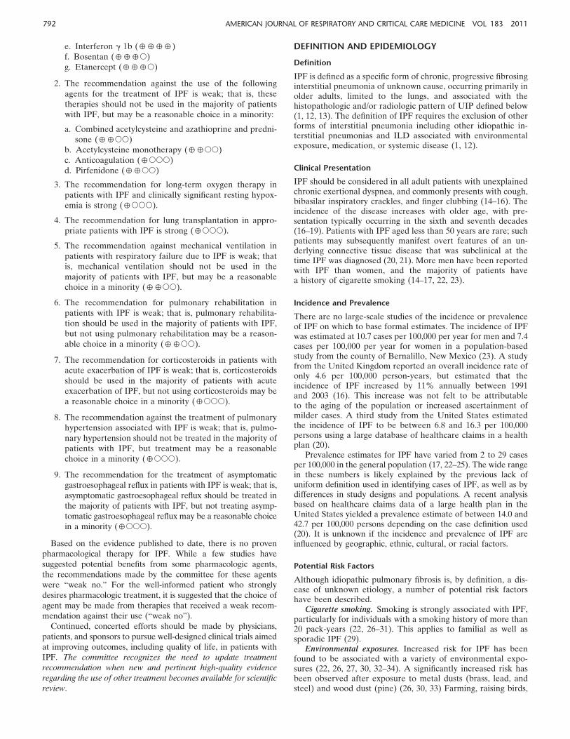
UIP Pattern: HRCT Features
HRCT is an essential component of the diagnostic pathway inIPF (Table 4, Figure 1). The optimal HRCT technique forevaluation of ILD is provided in the online supplement (seeTable E6). UIP is characterized on HRCT by the presence ofreticular opacities, often associated with traction bronchiectasis(98, 99). Honeycombing is common, and is critical for makinga definite diagnosis. Honeycombing is manifested on HRCT asclustered cystic airspaces, typically of comparable diameters onthe order of 3–10 mm but occasionally as large as 2.5 cm. It isusually subpleural and is characterized by well-defined walls(100). Ground glass opacities are common, but usually lessextensive than the reticulation. The distribution of UIP on
HRCT is characteristically basal and peripheral, though oftenpatchy. The presence of coexistent pleural abnormalities (e.g.,pleural plaques, calcifications, significant pleural effusion) sug-gests an alternative etiology for UIP pattern. Micronodules, airtrapping, nonhoneycomb cysts, extensive ground glass opacities,consolidation, or a peribronchovascular-predominant distribu-tion should lead to consideration of an alternative diagnosis.Mild mediastinal lymph node enlargement (usually , 1.5 cmin short axis) can be seen (101, 102). The chest radiograph isless useful than HRCT in evaluating patients with suspectedIPF (103).
Several studies have documented that the positive predictivevalue of a HRCT diagnosis of UIP is 90 to 100% (103–108). Thesestudies are affected by selection bias because they only includedpatients with biopsy-proven diagnoses. Nonetheless, a UIP pat-tern on HRCT is highly accurate for the presence of UIP patternon surgical lung biopsy. If honeycombing is absent, but theimaging features otherwise meet criteria for UIP, the imagingfeatures are regarded as representing possible UIP, and surgicallung biopsy is necessary to make a definitive diagnosis. In patientswhose HRCT does not demonstrate a UIP pattern, the surgicallung biopsy may still demonstrate UIP pattern on histopathology.
TABLE 4. HIGH-RESOLUTION COMPUTED TOMOGRAPHY CRITERIA FOR UIP PATTERN
UIP Pattern (All Four Features) Possible UIP Pattern (All Three Features) Inconsistent with UIP Pattern (Any of the Seven Features)
d Subpleural, basal predominance
d Reticular abnormality
d Honeycombing with or without traction
bronchiectasis
d Absence of features listed as inconsistent with
UIP pattern (see third column)
d Subpleural, basal predominance
d Reticular abnormality
d Absence of features listed as inconsistent with
UIP pattern (see third column)
d Upper or mid-lung predominance
d Peribronchovascular predominance
d Extensive ground glass abnormality (extent .
reticular abnormality)
d Profuse micronodules (bilateral, predominantly
upper lobes)
d Discrete cysts (multiple, bilateral, away from areas
of honeycombing)
d Diffuse mosaic attenuation/air-trapping (bilateral,
in three or more lobes)
d Consolidation in bronchopulmonary segment(s)/lobe(s)
Definition of abbreviation: UIP 5 usual interstitial pneumonia.
Figure 1. High-resolution com-
puted tomography (HRCT) im-
ages demonstrating usualinterstitial pneumonia (UIP)
pattern and possible UIP pat-
tern. (A and B) UIP pattern,
with extensive honeycomb-ing: axial and coronal HRCT
images show basal predomi-
nant, peripheral predominantreticular abnormality with
multiple layers of honeycomb-
ing (arrows). (C and D) UIP
pattern, with less severe hon-eycombing: axial and coronal
CT images show basal pre-
dominant, peripheral predom-
inant reticular abnormalitywith subpleural honeycomb-
ing (arrows). (E and F ) Possible
UP pattern: axial and coronalimages show peripheral pre-
dominant, basal predominant
reticular abnormality with
a moderate amount of groundglass abnormality, but without
honeycombing.
794 AMERICAN JOURNAL OF RESPIRATORY AND CRITICAL CARE MEDICINE VOL 183 2011

UIP Pattern: Histopathology Features
The histopathologic hallmark and chief diagnostic criterion isa heterogeneous appearance at low magnification in which areasof fibrosis with scarring and honeycomb change alternate withareas of less affected or normal parenchyma (1, 12) (Table 5,Figure 2). These histopathologic changes often affect thesubpleural and paraseptal parenchyma most severely. Inflam-mation is usually mild and consists of a patchy interstitialinfiltrate of lymphocytes and plasma cells associated withhyperplasia of type 2 pneumocytes and bronchiolar epithelium.The fibrotic zones are composed mainly of dense collagen,although scattered convex subepithelial foci of proliferatingfibroblasts and myofibroblasts (so-called fibroblast foci) area consistent finding. Areas of honeycomb change are composedof cystic fibrotic airspaces that are frequently lined by bronchi-olar epithelium and filled with mucus and inflammatory cells.Smooth muscle metaplasia in the interstitium is commonly seenin areas of fibrosis and honeycomb change.
The differential diagnosis for UIP pattern on pathology isrelatively short, especially when strict criteria for UIP aremaintained. The major differential diagnostic considerationsinclude UIP in other clinical settings such as connective tissuediseases, chronic hypersensitivity pneumonitis (extrinsic allergicalveolitis), and pneumoconioses (especially asbestosis).
Some biopsies may reveal a pattern of fibrosis that does notmeet the above criteria for UIP pattern (1). These biopsies maybe termed ‘‘nonclassifiable fibrosis.’’ In the absence of histologicfeatures diagnostic of an alternative condition (e.g., hypersen-sitivity pneumonitis, sarcoidosis, etc.), such biopsies may beconsistent with the diagnosis of IPF (Tables 5 and 6) in theappropriate clinical and radiologic setting and after carefulmultidisciplinary discussion.
DIAGNOSIS
Diagnostic criteria and schema for adult patients with ILD andsuspected IPF are presented in Figure 3 and Table 6. Carefulexclusion of alternative etiologies through multidisciplinary dis-cussion between pulmonologists, radiologists, and pathologistsexperienced in the diagnosis of ILD is of the utmost importanceto an accurate diagnosis. In situations in which multidisciplinary
discussion is not feasible, it is recommended that patients bereferred to experienced clinical experts in ILD for consultation.
The diagnostic criteria for IPF presented in this documenthave been significantly modified from those stated in theprevious ATS/ERS Statement (1). Given the high-qualityevidence regarding HRCT specificity for the recognition ofhistopathologic UIP pattern, surgical lung biopsy is not essential(104, 105, 109, 110). In the appropriate clinical setting (asdescribed in the clinical presentation section above; this in-cludes a thorough medical, occupational/environmental andfamily history, physical examination, physiological testing, andlaboratory evaluation), the presence of a UIP pattern on HRCTis sufficient for the diagnosis of IPF. Thus, the major and minorcriteria for the clinical (i.e., nonpathologic) diagnosis of IPFhave been eliminated.
Diagnostic Criteria
The diagnosis of IPF requires the following:
1. Exclusion of other known causes of ILD (e.g., domesticand occupational environmental exposures, connectivetissue disease, and drug toxicity).
2. The presence of a UIP pattern on HRCT in patients notsubjected to surgical lung biopsy (see Table 4).
3. Specific combinations of HRCT and surgical lung biopsypattern in patients subjected to surgical lung biopsy (seeTables 4, 5, and 6).
Thus, the accuracy of diagnosis of IPF increases with clinical,radiologic, and histopathologic correlation and can be accom-plished with a multidisciplinary discussion among experiencedclinical experts in the field of ILDs (111). This is particularlyrelevant in cases in which the radiologic and histopathologicpatterns are discordant (e.g., HRCT is inconsistent with UIP andhistopathology is UIP). An HRCT or pathologic UIP pattern isnot 100% specific to IPF (1, 12, 112–114). Discordant histologicpatterns on surgical lung biopsy specimens obtained fromdifferent segments have been described. Cases with coexistingUIP pattern and fibrotic NSIP pattern (discordant UIP) appearto behave similarly to those with UIP pattern in all lobes(concordant UIP) (115, 116). This supports the obtainment of
TABLE 5. HISTOPATHOLOGICAL CRITERIA FOR UIP PATTERN
UIP Pattern (All Four Criteria) Probable UIP Pattern
Possible UIP Pattern
(All Three Criteria)
Not UIP Pattern
(Any of the Six Criteria)
d Evidence of marked fibrosis/
architectural distortion, 6
honeycombing in a
predominantly subpleural/
paraseptal distribution
d Presence of patchy
involvement of lung
parenchyma by fibrosis
d Presence of fibroblast foci
d Absence of features
against a diagnosis
of UIP suggesting
an alternate diagnosis
(see fourth column)
d Evidence of marked fibrosis /
architectural distortion, 6
honeycombing
d Absence of either patchy
involvement or fibroblastic
foci, but not both
d Absence of features against a
diagnosis of UIP suggesting
an alternate diagnosis
(see fourth column)
OR
d Honeycomb changes only‡
d Patchy or diffuse
involvement of lung
parenchyma by
fibrosis, with or without
interstitial inflammation
d Absence of other criteria
for UIP (see UIP
PATTERN column)
d Absence of features
against a diagnosis
of UIP suggesting an
alternate diagnosis
(see fourth column)
d Hyaline membranes*
d Organizing pneumonia*†
d Granulomas†
d Marked interstitial
inflammatory cell
infiltrate away from
honeycombing
d Predominant airway
centered changes
d Other features
suggestive of an
alternate diagnosis
Definition of abbreviations: HRCT 5 high-resolution computed tomography; UIP 5 usual interstitial pneumonia.
* Can be associated with acute exacerbation of idiopathic pumonary fibrosis.† An isolated or occasional granuloma and/or a mild component of organizing pneumonia pattern may rarely be coexisting in lung biopsies with an otherwise UIP
pattern.‡ This scenario usually represents end-stage fibrotic lung disease where honeycombed segments have been sampled but where a UIP pattern might be present in other
areas. Such areas are usually represented by overt honeycombing on HRCT and can be avoided by pre-operative targeting of biopsy sites away from these areas using HRCT.
American Thoracic Society Documents 795

surgical lung biopsies from multiple lobes in patients withsuspected IPF.
Several studies have compared VATS to open thoracotomy(117–120). The diagnostic yield from surgical lung biopsiesobtained from VATS and open thoracotomy are similar. WhileVATS may be associated with lower morbidity and length ofstay than open thoracotomy, the decision on which procedure toperform should be based on individual patient characteristicsand surgical expertise. In patients with severe physiologicimpairment or substantial comorbidity, the risks of surgicallung biopsy may outweigh the benefits of establishing a securediagnosis of IPF. The final decision regarding whether or not topursue a surgical lung biopsy must be tailored to the clinicalsituation of the individual patient.
Exclusion of Other Known Causes
The exclusion of other known causes of ILD is a broad andinherently subjective criterion, but several specific pointsshould be made. A careful history and physical examinationfocusing on comorbidities, medication use, environmental ex-
posures, and family history is essential, and physicians shouldutilize a standardized approach. While there are no validatedtools for this, a template, such as the one available through theAmerican College of Chest Physicians (http://www.chestnet.org/memberResources/downloads/networks/IDLDquestionnaire.pdf),may be of use. It is of particular importance to evaluate patientsthoroughly for possible chronic hypersensitivity pneumonitis,since such patients may mimic IPF. The inciting antigen maynot be identifiable in some patients despite a thorough search(121); bronchoalveolar lavage (BAL) showing a lymphocytosisof 40% or greater may suggest occult hypersensitivity pneumo-nitis in this setting, prompting further investigations for envi-ronmental exposures, and possibly a surgical lung biopsy.Patients who meet established criteria for connective tissuedisease do not have IPF. Younger patients, especially women,without clinical or serologic features at presentation may sub-sequently manifest clinical features of connective tissue disease.Therefore, the index of suspicion for connective tissue diseasein younger patients (under the age of 50 yr) should be high.
j Question: Should BAL cellular analysis be performed in thediagnostic evaluation of suspected IPF?
Cellular analyses of BAL can be useful in the diagnosis ofcertain forms of ILD. In the evaluation of patients withsuspected IPF, the most important application of BAL is inthe exclusion of chronic hypersensitivity pneumonitis;prominent lymphocytosis (. 40%) should suggest thediagnosis. Recent retrospective data suggest that 8% ofpatients with an HRCT UIP pattern may have BALfindings suggestive of an alternative diagnosis (122). It isunclear whether BAL adds significant diagnostic specificityto a careful exposure history and clinical evaluation.
Recommendation: BAL cellular analysis should not beperformed in the diagnostic evaluation of IPF in themajority of patients, but may be appropriate in a minority(weak recommendation, low-quality evidence).
Values: This recommendation places a high value on theadditional risk and cost of BAL in patients with IPF anda low value on possible improved specificity of diagnosis.
Remarks: This recommendation is only for BAL differentialcell count (‘‘cellular analysis’’). It does not refer to the useof BAL in the evaluation of infection, malignancy, etc. Atpresent, BAL cellular analysis should be considered inthe evaluation of patients with IPF at the discretion of thetreating physician based on availability and experience attheir institution/regional laboratory. (Vote: 4 for the useof BAL, 18 against the use of BAL, 1 abstention, 8 absent.)
j Question: Should transbronchial lung biopsy be used in theevaluation of suspected IPF?
Transbronchial lung biopsy is useful in the evaluation ofselected conditions (e.g., granulomatous disorders such assarcoidosis). A UIP pattern on HRCT makes theseconditions unlikely (104, 105, 109). In cases requiringhistopathology, the specificity and positive predictivevalue of UIP pattern identified by transbronchial biopsyhas not been rigorously studied. While transbronchialbiopsy specimens may show all the histologic features ofUIP (123), the sensitivity and specificity of this approachfor the diagnosis for UIP pattern is unknown. It is alsounknown how many and from where transbronchialbiopsies should be obtained.
Figure 2. Surgical lung biopsy specimens demonstrating UIP pattern. (A)
Scanning power microscopy showing a patchy process with honeycombspaces (thick arrow), some preserved lung tissue regions (thin arrow), and
fibrosis extending into the lung from the subpleural regions. (B) Adjacent
to the regions of more chronic fibrosis (thick arrow) is a fibroblast focus
(asterisk), recognized by its convex shape and composition of edematousfibroblastic tissue, suggestive of recent lung injury.
796 AMERICAN JOURNAL OF RESPIRATORY AND CRITICAL CARE MEDICINE VOL 183 2011
Recommendation: Transbronchial biopsy should not be usedin the evaluation of IPF in the majority of patients, butmay be appropriate in a minority (weak recommendation,low-quality evidence).
Values: This recommendation places a high value on the addi-tional morbidity of transbronchial lung biopsy in patientswith IPF who will subsequently undergo surgical lungbiopsy and low value on possible diagnostic specificity.
Remarks: (Vote: none for the use of transbronchial biopsy, 23against the use of transbronchial biopsy, no abstentions, 8absent.)
j Question: Should serologic testing for connective tissuesdisease be used in the evaluation of suspected IPF?
There are no reliable data on the role of screening serologiesin patients with suspected IPF. Connective tissue diseasecan present with a UIP pattern (124), and ILD has beendescribed as the sole clinical manifestation of theseconditions and can precede the overt manifestation ofa specific connective tissue disease (125).
Recommendation: Serologic testing for connective tissue dis-ease should be performed in the evaluation of IPF in themajority of patients, but may not be appropriate in a minor-ity (weak recommendation, very low-quality evidence).
Values: This recommendation places a high value ondistinguishing connective tissue disease from IPF andlow value on cost.
Remarks: Serologic evaluation should be performed even inthe absence of signs or symptoms of connective tissuedisease, and should include rheumatoid factor, anti-cyclic
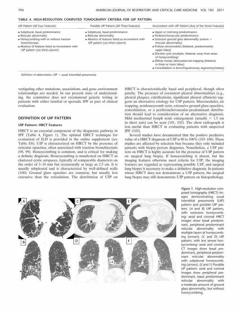
TABLE 6. COMBINATION OF HIGH-RESOLUTION COMPUTED TOMOGRAPHY AND SURGICAL LUNG BIOPSY FOR THEDIAGNOSIS OF IPF (REQUIRES MULTIDISCIPLINARY DISCUSSION)
HRCT Pattern* Surgical Lung Biopsy Pattern* (When Performed) Diagnosis of IPF?†
UIP UIP YES
Probable UIP
Possible UIP
Nonclassifiable fibrosis‡
Not UIP No
Possible UIP UIP YES
Probable UIP
Possible UIP Probablex
Nonclassifiable fibrosis
Not UIP No
Inconsistent with UIP UIP Possiblex
Probable UIP No
Possible UIP
Nonclassifiable fibrosis
Not UIP
Definition of abbreviations: HRCT 5 high-resolution computed tomography; IPF 5 idiopathic pulmonary fibrosis; UIP 5 usual interstitial pneumonia.
Bold type indicates combinations of HRCT and surgical lung biopsy patterns that correspond with a diagnosis of IPF (a YES in the far right column). The combination of
UIP HRCT and probable UIP or possible UIP or Nonclassifiable fibrosis (surgical lung biopsy patterns) (for example) equals a diagnosis of IPF; the combination of UIP
HRCT and Not UIP (surgical lung biopsy pattern) does not make the diagnosis of IPF.
* Patterns as described in Tables 4 and 5.‡ Nonclassifiable fibrosis: Some biopsies may reveal a pattern of fibrosis that does not meet the above criteria for UIP pattern and the other idiopathic interstitial
pneumonias (1) (see text). These biopsies may be termed ‘‘nonclassifiable fibrosis.’’† The accuracy of the diagnosis of IPF increases with multidisciplinary discussion (MDD). This is particularly relevant in cases in which the radiologic and histopathologic
patterns are discordant (e.g., HRCT is inconsistent with UIP and histopathology is UIP). There are data to suggest that the accuracy of diagnosis is improved with MDD among
interstitial lung disease experts compared to clinician-specialists in the community setting (126); timely referral to interstitial lung disease experts is encouraged.x Multidisciplinary discussion should include discussions of the potential for sampling error and a re-evaluation of adequacy of technique of HRCT. NOTE: In cases with an
‘‘inconsistent with UIP’’ HRCT pattern and a ‘‘UIP’’ surgical lung biopsy pattern, the possibility of a diagnosis of IPF still exists and clarification by MDD among interstitial lung
disease experts is indicated.
Figure 3. Diagnostic algorithm for idiopathic pulmonary fibrosis (IPF).Patients with suspected IPF (i.e., patients with unexplained dyspnea on
exertion and/or cough with evidence of interstitial lung disease [ILD])
should be carefully evaluated for identifiable causes of ILD. In the
absence of an identifiable cause for ILD, an HRCT demonstrating UIPpattern is diagnostic of IPF. In the absence of UIP pattern on HRCT, IPF
can be diagnosed by the combination of specific HRCT and histopath-
ological patterns. The accuracy of the diagnosis of IPF increases with
multidisciplinary discussion (MDD) among ILD experts. *Refer to Table4 for definitions. †Refer to Table 5 for definitions.
gg
gg
American Thoracic Society Documents 797

citrullinated peptide, and anti-nuclear antibody titer andpattern. The routine use of other serological tests such asantisynthetase antibodies (e.g., Jo-1), creatine kinase andaldolase, Sjogren’s antibodies (SS-A, SS-B), and sclero-derma antibodies (scl-70, PM-1) is of unclear benefit, butmay be helpful in selected cases. Patients with IPF mayhave a mildly positive antinuclear antibody titer and/orrheumatoid factor level without any other clinical fea-tures of connective tissue. Such patients should becarefully screened for signs and symptoms of connectivetissues disease (e.g., arthritis, Raynaud’s phenomenon,skin changes, abnormal esophageal motility). In theabsence of additional serologic or clinical evidence tosupport a connective tissues diagnosis, the diagnosis ofIPF is appropriate. Repeat serologic and clinical evalua-tion during follow up may subsequently confirm thedevelopment of a connective tissue disease; in such cases,the diagnosis should be revised. (Vote: 23 for the use ofserologic testing, none against the use of serologic testing,no abstentions, 8 absent.)
j Question: Should a multi-disciplinary discussion be used inthe evaluation of suspected IPF?
The diagnosis of IPF is, by definition, multidisciplinary,drawing on the expertise of experienced clinicians, radiol-ogists, and pathologists. Proper communication betweenthe various disciplines involved in the diagnosis of IPF(pulmonary, radiology, pathology) has been shown toimprove inter-observer agreement among experiencedclinical experts as to the ultimate diagnosis (111, 126).
Recommendation: We recommend that a multi-disciplinarydiscussion should be used in the evaluation of IPF (strongrecommendation, low-quality evidence).
Values: This recommendation places a high value on theaccurate diagnosis of IPF and a low value on the access toand availability of experts for multidisciplinary discussion.
Remarks: It is recognized that a formal multidisciplinarydiscussion (MDD) between the treating pulmonologist,radiologist, and pathologist is not possible for manypractitioners. Effort should be made, however, to pro-mote verbal communication between specialties duringthe evaluation of the case. There are data to suggest thatthe accuracy of diagnosis is improved through MDDamong ILD experts compared with MDD among special-ists in the community setting (126); timely referral to ILDexperts is encouraged. (Vote: 23 for the use of multidis-ciplinary discussion, none against the use of multidisci-plinary discussion, no abstentions, 8 absent.)
NATURAL HISTORY OF IPF
The natural history of IPF has been described as a progressivedecline in subjective and objective pulmonary function untileventual death from respiratory failure or complicating comor-bidity (127–129). Available longitudinal studies do not allowa clear assessment of median survival in IPF. Several retrospec-tive longitudinal studies suggest a median survival time from2 to 3 years from the time of diagnosis (130–134). However,recent data from clinical trials of patient with preserved pulmo-nary function suggest this may be an underestimate (135–137).
There appear to be several possible natural histories forpatients with IPF (Figure 4) (138). For a given patient, the
natural history is unpredictable at the time of the diagnosis. Themajority of patients demonstrate a slow, gradual progressionover many years. Some patients remain stable while others havean accelerated decline (139). Some patients may experienceepisodes of acute respiratory worsening. It is unknown if thesedifferent natural histories represent distinct phenotypes of IPFor if the natural history is influenced by geographic, ethnic,cultural, racial, or other factors. Other comorbid conditionssuch as emphysema and pulmonary hypertension may impactthe disease course (140–142).
Acute Exacerbation of IPF
Recent observations have suggested that acute respiratoryworsening occurs in a small minority of patients with IPFannually (approximately 5–10%) (137, 143, 144). These epi-sodes may occur secondary to common conditions such aspneumonia, pulmonary embolism, pneumothorax, or cardiacfailure (145, 146). When a cause cannot be identified for theacute respiratory decline, the term acute exacerbation of IPFhas been used (144, 145, 147–157). It is presently unclear if acuteexacerbation of IPF is simply a manifestation of an unidentifiedrespiratory complication (such as pulmonary emboli, infection)contributing to an acute worsening in a patient with IPF orrepresents an inherent acceleration in the pathobiological pro-cesses involved in IPF. Recent data from gene expressionprofiling of patients with acute exacerbation of IPF do notsuggest an infectious etiology (158).
Historically, criteria for acute exacerbation of IPF haveincluded an unexplained worsening of dyspnea within 1month, evidence of hypoxemia as defined by worsened orseverely impaired gas exchange, new radiographic alveolarinfiltrates, and an absence of an alternative explanation suchas infection, pulmonary embolism, pneumothorax, or heartfailure (143). Acute exacerbation can occur at any point in
Figure 4. Natural history of IPF. There appear to be several possiblenatural histories for patients with IPF. The majority of patients experi-
ence a slow but steady worsening of their disease (‘‘Slow progression’’).
Some patients remain stable (‘‘Stable’’), while others have an acceler-
ated decline (‘‘Rapid progression’’). A minority of patients mayexperience unpredictable acute worsening of their disease (lightning
bolt), either from a secondary complication such as pneumonia, or for
unrecognized reasons. This event may be fatal or may leave patientswith substantially worsened disease. The relative frequency of each of
these natural histories is unknown.
798 AMERICAN JOURNAL OF RESPIRATORY AND CRITICAL CARE MEDICINE VOL 183 2011

the course of IPF and occasionally can be its presentingmanifestation (149, 153, 159, 160). Worsened cough, fever,and/or increased sputum have been observed (148, 149, 153).While there are no known risk factors for acute exacerbationof IPF, there have been reports of acute respiratory de-compensation after thoracic surgery (161–165) and bron-choalveolar lavage (149, 166). It is unclear whether or notthese events represent true acute exacerbations or complica-tions of the respective procedures.
Acute exacerbation of IPF histologically manifests as acuteor organizing diffuse alveolar damage (DAD), or, less com-monly, organizing pneumonia in zones of relatively preservedlung tissue away from the most fibrotic regions (143). Anecdotalexperience indicates that sampling issues in some patients mayresult in specimens demonstrating only uncomplicated UIP orthe organizing phase of DAD without histologic evidence ofunderlying UIP in the sample evaluated (153).
Vital Statistics
Deaths from pulmonary fibrosis increase with increasing age(18, 167). In addition, there is evidence to suggest increasingmortality from pulmonary fibrosis over the past two decades(18, 167). A recent analysis of the death certificate data in theUnited States noted a significant increase in mortality frompulmonary fibrosis from 1992 to 2003 (167). When the mostrigorous definition of IPF was applied, the mortality rate in theUnited States in 2003 was 61.2 deaths per 1,000,000 in men and54.5 per 1,000,000 in women (167). In Japan, the mortality ratefor IPF was estimated to be 33 per 1,000,000 in men and 24 per1,000,000 in women (22). The mortality burden attributable toIPF is higher than that of some cancers (168). Recent evidencesuggests that mortality from IPF in the United States is greaterin the winter months (169). The most common cause of death isprogressive lung disease (60% of deaths) (146, 167). Additionalcauses of morbidity and mortality in patients with IPF includecoronary artery disease (170), pulmonary embolism, and lungcancer.
STAGING AND PROGNOSIS
The extent of disease and the severity of functional impairmentof patients with IPF at the time of diagnosis are variable. Thereasons for this are thought to be variation in subjectiveperception of symptoms and differences in providers’ aware-ness. Recent studies have clarified predictors of survival in IPF.However, the accuracy of these predictors is limited by the
retrospective nature of some of these studies and variations instudy design.
Terms such as ‘‘mild,’’ ‘‘moderate,’’ ‘‘severe,’’ ‘‘early,’’ and‘‘advanced’’ have been suggested for staging disease. Proposedstages are commonly based on resting pulmonary function testmeasurements and/or extent of radiologic abnormalities. How-ever, it is unknown if these staging approaches are relevant toclinical decision making. The committee recognizes the impor-tance of identifying patients with increased risk for mortalitywithin 2 years to prompt consideration for lung transplantation.Limited data suggest selected features commonly observed inclinical practice are associated with increased mortality (seebelow and Table 7). Because of variability in the natural historyof IPF, it is unknown if the presence of one of more of thesefeatures identifies a subpopulation of patients with ‘‘advanced’’or ‘‘end-stage’’ IPF.
Demographics
Patients that are older and male have been reported as havingworse prognosis in some but not all studies (15, 131, 171–177).The effect of smoking has been shown to be associated withboth increased (134, 178) and decreased (131) risk of sub-sequent mortality. The prognostic value of geographic, ethnic,cultural, and racial factors is unknown.
Dyspnea
Baseline dyspnea has been shown to correlate with quality oflife and survival in several studies (15, 179–182). A variety ofdifferent metrics for dyspnea have been used, including themedical research council scale, baseline dyspnea index, qualityof life (QoL) measurement tools with respiratory question-naires, Borg scale, University of California San Diego shortnessof breath questionnaire, and the clinical-radiological-physiolog-ical dyspnea score (183–185). It remains unclear which dyspneametric is most predictive of outcome in patients with IPF.Change in dyspnea over time has also been shown to predictsurvival (186).
Physiology
Baseline pulmonary function test values have shown mixedassociations with survival in IPF. This may be due, in part, tocomorbid conditions such as emphysema, pulmonary vasculardisease, and obesity, or technical differences in testing. BaselineFVC is of unclear predictive value (15, 173, 175, 177, 180, 186–189). Diffusing capacity for carbon monoxide (DLCO, singlebreath, hemoglobin corrected) is more reliably predictive ofsurvival at baseline, and a threshold of approximately 40percent predicted has been associated with an increased riskof mortality (186, 187, 190, 191). Limited data suggest thatbaseline total lung capacity (TLC) and alveolar-arterial oxygendifference in partial pressures (P(A-a)O2
) may be predictive ofsurvival, but no clear threshold exists (186). Baseline cardio-pulmonary exercise testing (maximal oxygen uptake) has beensuggested to predict survival (192).
Longitudinal change in physiology is clearly an importantpredictor of mortality in IPF. A decline in FVC over 6 or 12months has been reliably associated with decreased survival(177, 186, 187, 191, 193). Recent data indicate that in IPF,declines in FVC of 5–10% may be predictive of mortality. Adecline in DLCO has also been associated with decreasedsurvival, although less consistently (186, 187, 191, 193).Greater than 15 mm Hg change in P(A-a)O2
after 12 monthshas been shown to be predictive of survival (187). Six-monthchange in TLC and P(A-a)O2
may also be predictive of survival(186).
TABLE 7. SELECTED FEATURES ASSOCIATED WITH INCREASEDRISK OF MORTALITY IN IDIOPATHIC PULMONARY FIBROSIS
Baseline factors*
Level of dyspnea†
DLCO , 40% predicted
Desaturation < 88% during 6MWT
Extent of honeycombing on HRCT†
Pulmonary hypertension
Longitudinal factors
Increase in level of dyspnea†
Decrease in Forced Vital Capacity by > 10% absolute value
Decrease in DLCO by > 15% absolute value
Worsening of fibrosis on HRCT†
Definition of abbreviations: 6MWT 5 6-minute-walk test; DLCO 5 diffusion
capacity for carbon monoxide; HRCT 5 high-resolution computed tomography.
* Baseline forced vital capacity is of unclear predictive value.† Currently, there is no uniformity in approach to quantification.
American Thoracic Society Documents 799

HRCT Features
HRCT features of fibrosis and honeycombing are stronglycorrelated with FVC and DLCO measurements (194). Severalgroups have demonstrated that the extent of fibrosis andhoneycombing on HRCT are predictive of survival in IPF(109, 195–198).
Composite Scoring Systems
Composite scoring systems have been developed utilizingphysiological and radiographic variables in an attempt to pro-vide more accurate prognostic information. A composite phys-iologic index (CPI) has been developed that uses values fromFEV1, FVC, and DLCO to predict the extent of disease on HRCT(141, 191). This CPI was a stronger predictor of mortality thanindividual measures of lung function such as FEV1, FVC, DLCO,TLC, PaO2
, the clinical-radiographic-physiological scoring system(CRP) (183) or new CRP scoring systems (15). However, thiscomposite approach has not been tested in any prospectiveclinical trials to date and its clinical utility is unknown.
Six-Minute-Walk Testing
Although the 6-minute-walk test (6MWT) is widely used inclinical practice, its prognostic value is limited due to lack ofstandardization of the procedure in patients with IPF. Somestudies have suggested that desaturation (i.e., a decline inoxygen saturation to below 88%) during 6MWT is a markerfor increased risk of mortality (188, 199, 200). Shorter walkdistance and delayed heart-rate recovery after walk testing havebeen associated with an increased risk of subsequent mortality(188, 201–203). However, it is unclear if desaturation, distancewalked, and other variables measured during 6MWT in thispopulation are reproducible (204). A steady-state 6-minuteexercise test using a walking treadmill has been used in patientswith IPF in a recent clinical trial in Japan, but the clinical utilityof this unvalidated test is unclear (144).
Histopathology
Varied histopathologic patterns can be found within individualpatients when multiple biopsies are obtained. A pattern of UIPand NSIP has been identified in 12 to 26% of patients withmultiple lobe biopsies (115, 116), highlighting the importance ofobtaining biopsies from multiple lobes. The prognosis forpatients with discordant UIP (pattern of UIP and NSIP withinthe same patient) appears to be similar to that of patients withconcordant UIP (UIP in all lobes biopsied) (115, 116).
An increased number of fibroblast foci has been associatedwith an increased risk of mortality in some studies (134, 175,205–207). A higher profusion of fibroblast foci has also beenassociated with a decline in FVC and DLCO over 6 and 12months of follow-up (205). The utility of detailed histopatho-logic scoring systems in the day-to-day clinical management ofpatients with IPF has not been evaluated.
Pulmonary Hypertension
The majority of data regarding the presence and significance ofpulmonary hypertension come from patients with IPF under-going evaluation for lung transplantation. The presence ofpulmonary hypertension (defined as a mean pulmonary arterypressure of . 25 mm Hg at rest) has been associated withincreased risk of mortality for patients with IPF (140, 142, 176).In a separate series of 70 patients with IPF, receiver operatorcharacteristic (ROC) analysis suggested a mean pulmonaryartery pressure of 17 mm Hg as the best discriminator ofmortality (189). These data need to be validated. Echocar-diographic estimation of pulmonary artery systolic pressures
does not correlate well with right heart catheterization (208–210). Increased pulmonary vascular resistance has also beenlinked to worse survival (211). It is not clear if IPF withpulmonary hypertension represents a distinct clinical pheno-type (IPF–PH).
Emphysema
Recent retrospective data suggest that patients with IPF andcoexisting emphysema have a poorer outcome than thosewithout emphysema (140, 212). Patients with coexisting IPFand emphysema may require treatment for both conditions.Limited data suggest that patients with IPF and emphysema arelikely to require long-term oxygen therapy and may havesignificant pulmonary hypertension. When controlling for thesedifferences, the presence of emphysema was not significantlypredictive of survival (140). Thus, it is not clear if IPF withcoexisting emphysema represents a distinct clinical phenotype(combined pulmonary fibrosis and emphysema) with a distinctprognosis or whether emphysema in these cases is simplya comorbidity.
Serum and BAL Biomarkers
There are limited retrospective data on the predictive value ofserum and BAL biomarkers in IPF. However, these are largelyunavailable for routine clinical use. Krebs von den Lungen-6(KL-6) is a high-molecular-weight glycoprotein, classified ashuman MUC1 mucin, that is produced by regenerating type IIpneumocytes (213). Serum levels of KL-6 have been shown tobe elevated in patients with IPF, and these levels may correlatewith increased risk of subsequent mortality (214, 215). Serumlevels of surfactant protein A and D are also elevated inpatients with IPF and are predictive of survival (216–218).Recent data demonstrate a relationship between serumCCL18, other chemokines, and serum brain natiuretic peptidelevels and mortality (219–223). Studies of plasma and BALmatrix metalloproteinase (MMP) levels suggest that MMP1 andMMP7 are increased in patients with IPF, and MMP7 levelsmay correlate with disease severity (224). BAL levels of SP-Aappear predictive of survival (225, 226). Cellular analysis ofBAL is of unclear predictive value in IPF (226, 227). Pre-liminary evidence suggests that the presence of circulatingfibrocytes (mesenchymal progenitor cells) is associated withworse short-term survival (228).
TREATMENT
Pharmacological Therapies
The committee did not find sufficient evidence to support theuse of any specific pharmacologic therapy for patients with IPF.However, clinical trials of some agents have suggested a possiblebenefit. The recommendations detailed below are based on theevidence-based approach outlined in the introductory section;these recommendations may change if additional and/or newdata become available in publications subjected to peer review.The number of votes for, against, abstaining, and absent arereported for all treatment votes. Most abstentions were a resultof panel members withholding from voting on questions withwhich they felt they had a potential conflict of interest.
The strength of a recommendation reflects the extent towhich the committee was confident that desirable effects ofa therapy outweighed its undesirable effects (3). The recom-mendations against most treatment therapies are strong; thereis insufficient evidence to support the routine use of thesetherapies. Other treatment recommendations were weak, re-flecting the need for better quality data and uncertainty
800 AMERICAN JOURNAL OF RESPIRATORY AND CRITICAL CARE MEDICINE VOL 183 2011

regarding the benefits and risks of therapy. The strength ofa recommendation has important implications for patients,clinicians, and policy makers (Table 3).
Therapies with a weak recommendation against their use maystill be appropriate in selected patients. Clinicians should beprepared to help patients make an appropriate decision regardingwhether or not to use a specific treatment regimen with weakrecommendation that is consistent with their own goals and values.For the well-informed patient who strongly desires pharmacologictreatment, it is suggested that the choice of agent be made fromtherapies receiving a weak recommendation against their use.
j Question: Should patients with IPF be treated with cortico-steroid monotherapy?
No randomized controlled trials have been conducted with
corticosteroid monotherapy (229, 230). Retrospective
uncontrolled studies have reported no survival benefits,
but have suggested that a minority of patients treated
with corticosteroid monotherapy improve their pulmo-
nary function (179, 231, 232); controlled data have found
no survival benefit (14, 233). There is substantial morbid-
ity from long-term corticosteroid therapy (231).
Recommendation: We recommend that patients with IPFshould not be treated with corticosteroid monotherapy
(strong recommendation, very low-quality evidence).Values: This recommendation places a high value on preven-
ting treatment-related morbidity and a low value on
potential improvement in pulmonary function as based
on very low-quality evidence.Remarks: (Vote: none for use, 21 against use, 2 abstentions,
8 absent.)
j Question: Should patients with IPF be treated with colchicine?
Colchicine has been shown to inhibit fibroblast proliferation
and collagen synthesis in vitro (234), and early studies in
patients with IPF suggested a potential benefit (235).
Several prospective clinical trials have compared colchi-
cine to various treatment regimens showing no difference
in clinical outcomes (8, 236–238). None of these studies
contained a ‘‘no therapy’’ arm. A retrospective study of
487 patients with IPF compared survival as a function of
treatment program (14). Compared with no therapy, col-
chicine had no impact on survival (see Table 8).
Recommendation: We recommend that patients with IPF
should not be treated with colchicine (strong recommen-
dation, very low-quality evidence).Values: This recommendation places a high value on the
very low-quality evidence, suggesting no benefit.Remarks: (Vote: none for use, 21 against use, 2 abstentions,
8 absent.)
j Question: Should patients with IPF be treated with cyclo-sporin A?
There are limited data on the use of cyclosporin A in the
English language literature. Early reports in small, un-
controlled groups of patients with IPF suggested a possi-
ble benefit (239, 240). More recently, a retrospective
study of 10 patients with IPF showed no apparent benefit
to cyclosporine treatment (241). Two studies of small
groups of post–lung transplant patients with IPF treated
with cyclosporine-containing immunosuppressive regi-
mens have shown progression of disease in the nativelung (242, 243).
Recommendation: We recommend that patients with IPFshould not be treated with cyclosporine A (strong rec-ommendation, very low quality evidence).
Values: This recommendation places a high value on pre-venting side effects and cost and a low value on very low-quality evidence showing discordant results.
Remarks: (Vote: none for use, 18 against use, 4 abstentions,9 absent.)
j Question: Should patients with IPF be treated with combina-tion corticosteroid and immunomodulator therapy (e.g.,azathioprine or cyclophosphamide)?
A retrospective study suggested a potential benefit of
treatment with azathioprine plus prednisone in a small
case series (244). A small randomized trial of corticoste-
roid versus corticosteroid and azathioprine showed a trend
toward a survival benefit with combination therapy with
corticosteroid and azathioprine (245) (Table 9). Cortico-
steroid and cyclophosphamide was compared with corti-
costeroid alone, and a survival benefit with cyclophospha-
mide was demonstrated (246). The results of this trial are
confounded by the inclusion of patients that do not meet
recent diagnostic criteria for IPF (1). Two retrospective,
controlled studies of cyclophosphamide have been pub-
lished. The first compared corticosteroid and cyclophos-
phamide therapy to no therapy in 164 patients, and found
no survival difference (172). The second compared corti-
costeroid and cyclophosphamide therapy to corticosteroid
alone in 82 patients, and found a survival benefit with
combination therapy (247).
Recommendation: We recommend that patients with IPFshould not be treated with combination corticosteroidand immunomodulator therapy (strong recommendation,low-quality evidence).
Values: This recommendation places a high value on preven-ting treatment-related morbidity and on recent data suggest-ing that the addition of acetylcysteine to this regimen slowedthe decline in pulmonary function (see below). It places alower value on possible improvement in pulmonary function.
Remarks: The evidence was low quality and there wasa variable degree of uncertainty about the balance ofbenefits and harms. (Vote: none for use, 21 against use, 2abstentions, 8 absent.) The committee was not unanimousregarding the strength of this recommendation; themajority voted for a strong recommendation.
j Question: Should patients with IPF be treated with combina-tion corticosteroid, azathioprine, and acetylcysteine therapy?
Acetylcysteine is a precursor to the antioxidant glutathione,
which may be reduced in the lungs of patients with IPF
(248, 249). A randomized controlled trial comparing the
effect of high-dose acetylcysteine versus placebo in patients
receiving prednisone plus azathioprine has been completed
(250) (Table 10). In this study, the 12-month declines in
vital capacity and diffusing capacity were significantly less
in the acetylcysteine-containing arm (vital capacity: 0.18
liter difference; 95% confidence interval [CI], 0.03–0.32;
P 5 0.02; diffusion capacity: 0.75 mmol/min/kilopascal
difference; 95% CI, 0.27–1.23; P 5 0.003). There was no
American Thoracic Society Documents 801
Vikrams1
Highlight
Vikrams1
Highlight
Vikrams1
Highlight

observed difference in mortality or other secondary end-points including dyspnea, quality of life, exercise physiol-ogy, or radiographic appearance. Limitations of this studyinclude substantial drop-out (approximately 30%), the un-clear clinical significance of the observed treatment effect,and the lack of a true ‘‘no treatment’’ arm (251, 252).
Recommendation: Themajority of patientswith IPF should notbe treated with combination corticosteroid, azathioprine,and acetylcysteine therapy, but this therapymaybe a reason-able choice in a minority (weak recommendation, low-quality evidence).
Values: This recommendation places a high value on pre-venting treatment-related morbidity and a low value onlow-quality data, including the absence of a true ‘‘no-therapy’’ arm.
Remarks: There was considerable debate about this recom-mendation. All committee members agreed more data areneeded to definitively address this question (vote: 3 for use,17 against use, 3 abstentions, 8 absent). This treatment maybe appropriate in patients who are willing to accept possibleadverse consequences even if expected benefits are small.Fully informed patients are in the best position to makedecisions that are consistent with the best evidence and thatpatient’s values and preferences.
j Question: Should patients with IPF be treated with acetylcys-teine monotherapy?
The most recent data for acetylcysteine monotherapy comesfrom a randomized controlled trial discussed in the corti-
costeroid, azathioprine, and oral acetylcysteine sectionabove (250). In this study, the addition of oral acetylcys-teine to corticosteroid and azathioprine was associatedwith a significantly smaller decline in pulmonary function.A previous uncontrolled study of 18 patients treated withoral acetylcysteine for 12 weeks also demonstrated im-provements in lung function indices including vital capac-ity, diffusion capacity, and capillary PaO2
(249). Anotherprevious study randomized 30 patients to aerosolizedacetylcysteine or placebo for 12 months and documentedsignificant improvement in the extent of ground glass oncomputed tomography and reduction in KL-6 levels (253)(Table 11). No differences in physiologic measurements orwalk distance were found.
Recommendation: The majority of patients with IPF shouldnot be treated with acetylcysteine monotherapy, but thistherapy may be a reasonable choice in a minority (weakrecommendation, low-quality evidence).
Values: This recommendation places a high value on thepotential cost of therapy and a low value on low-qualitydata, including the absence of a true ‘‘no therapy’’ arm,and indirect evidence of a potential benefit.
Remarks: There was considerable debate about this recom-mendation. All committee members agreed more dataare needed to definitively address this question. Thecommittee recognizes that there is a lack of standardiza-tion in the preparation of acetylcysteine available to thepublic in some countries, and the route of delivery isdifferent between studies (oral versus aerosolized), which
TABLE 8. COLCHICINE GRADE EVIDENCE PROFILE*
Quality Assessment
No. of Studies Design Limitations Inconsistency Indirectness Imprecision
Other
Considerations
Mortality (follow-up median 1.5 yr) 1 Randomized trials No serious
limitations‡No serious
inconsistency
Seriousx Very serious{ None
Pulmonary Function (Better indicated
by higher values)
1 Randomized trials Serious1 No serious
inconsistency
Serious†† No serious
imprecision
None
Summary of Findings
No. of Patients Effect
Colchicine No colchicine Relative (95% CI) Absolute Quality Importance†
Mortality (follow-up median 1.5 yr) 10/14
(71.4%)
10/12
(83.3%)kRR, 0.86
(0.56–1.30)
117 fewer per 1,000
(from 367 fewer
to 250 more)**
4sss Very low Critical
Pulmonary Function (Better indicated
by higher values)
14‡‡ 12 — xx 44ss Low Important
Data are from Reference 238.
* The overall quality of evidence rating is listed in the first row and is the one used in the text of the document. The quality rating for outcomes listed in other rows may
differ. How these additional outcomes are rated in terms of quality does not influence the final quality rating as they are to inform, but not to make, decisions.† Importance rating: the relative importance of the outcome for decision making. The rating ‘‘critical’’ indicates making recommendations on choice of testing and
treatment strategies. The rating ‘‘important’’ indicates that the outcome is important but not critical for making recommendations.‡ The use of colchicine in the prednisone arm for less than 2 weeks, and of prednisone at less than 20 mg/day for less than 2 weeks in the colchicine arm was permitted
for reasons other than as treatment of idiopathic pulmonary fibrosis. This would blur treatment effects.x The comparison in this trial is corticosteroids, leaving the single effect of colchicine versus no treatment somewhat uncertain.{ Only 26 patients were enrolled in this trial.k This data was abstracted from the figure provided in the publication.
** Please note that the baseline mortality risk is high in this trial which would produce large absolute effects based on apparent small relative effects.†† The patient importance of the pulmonary function measures is questionable.‡‡ Summary estimates are not provided here, but all differences were not significant.xx FVC (percent predicted) was not significantly different for prednisone- versus colchicine-treated subjects (26.9 and 25.1, respectively, P 5 0.385), although both
treatment groups experienced a significant decline from baseline (P 5 0.012 and P 5 0.027, respectively). The change from baseline DLCO (ml/min/mm Hg) was not
significantly different for prednisone- versus colchicine-treated subjects (22.0 and 21.1, P5 0.529); however, both treatment groups experienced a significant decrease
from baseline (P 5 0.031 and P 5 0.017, respectively).
802 AMERICAN JOURNAL OF RESPIRATORY AND CRITICAL CARE MEDICINE VOL 183 2011
Vikrams1
Highlight
Vikrams1
Highlight
Vikrams1
Highlight
Vikrams1
Highlight
Vikrams1
Highlight
Vikrams1
Highlight

may affect the mechanism of action. (Vote: 5 for use, 15against use, 3 abstentions, 8 absent.) This treatmentmay be appropriate in patients who are willing to acceptpossible adverse consequences even if expected benefitsare small. Fully informed patients and clinicians are inthe best position to make decisions that are consistentwith the best evidence and that patient’s values andpreferences.
j Question: Should patients with IPF be treated with inter-feron-g 1b?
Interferon-g 1b (IFN-g) is an agent with antifibrotic and
immunomodulatory properties that has been evaluated in
two large clinical trials after a pilot study suggested
benefit (254) (Table 12). The first clinical trial evaluated
the time to clinical worsening or death in 330 patients
with IPF, randomized 1:1 to receive IFN-g 200 mg three
times a week subcutaneously or placebo, with low-dose
prednisone being allowed as concomitant medication in
both groups (135). The primary endpoint was not differ-
ent between groups; post hoc analysis suggested a trend
toward improved survival with IFN-g in a subgroup of
patients with less severe physiological disease at baseline.
A subsequent open-label study of IFN-g compared with
colchicine in patients with less severe physiology alsosuggested a possible benefit (237). A recent, definitive trialtested this hypothesis in more than 800 patients withphysiologically mild disease and demonstrated there wasno difference in overall mortality (14.5% in the IFN-ggroup compared with 12.7% in the placebo arm) (136).
Recommendation: We recommend that patients with IPFshould not be treated with IFN-g (strong recommenda-tion, high-quality evidence).
Values: This recommendation places a high value on thepotential risks and cost of therapy.
Remarks: (Vote: none for use, 17 against use, 6 abstentions,8 absent.)
j Question: Should patients with IPF be treated with bosentan?
Endothelin-1 (ET-1) is a powerful vasoconstrictor and-growth factor that is involved in the pathogenesis ofpulmonary hypertension and potentially of IPF. Elevatedendothelin levels in serum and BAL, and exaggeratedexpression of endothelin receptors and ET-1 in lungtissue have been observed in patients with IPF (255).Bosentan, a dual endothelin receptor A and B antagonist,was tested in a phase II randomized controlled trial, using
TABLE 9. AZATHIOPRINE GRADE EVIDENCE PROFILE*
Quality Assessment
No. of Studies Design Limitations Inconsistency Indirectness Imprecision
Other
Considerations
Mortality (follow-up mean 1 yr;
directly assessed)
1 Randomized trials Serious‡ No serious inconsistency No serious
indirectness
Seriousx None
Adverse Effects (follow-up
mean 1 yr; study follow up)
1 Randomized trials No serious
limitations
No serious inconsistency No serious
indirectness
No serious
imprecision
None
Forced vital capacity in percent
predicted (follow-up mean 1 yr;
measured with: percent predicted
FVC; Better indicated by
higher values)
1 Randomized trials Very serious** No serious inconsistency Very serious†† No serious
imprecision
None
Summary of Findings
No. of Patients Effect
Azathioprine No Azathioprine Relative (95% CI) Absolute Quality Importance†
Mortality (follow-up mean 1 yr;
directly assessed)
4/14 (28.6%) 4/13 (30.8%) HR 1.0 (not provided){ Not calculated–not
statistically significant
44ss Low Critical
Adverse Effects (follow-up
mean 1 yr; study follow up)
28/14k (200%) 25/13k (192.3%) 1 — 4444 High Critical
Forced vital capacity in percent
predicted (follow-up mean 1 yr;
measured with: percent predicted
FVC; Better indicated by
higher values)
14 13 — Mean 4.8 higher‡‡ 4sss Very low Important
Data are from Reference 245.
* The overall quality of evidence rating is listed in the first row and is the one used in the text of the document. The quality rating for outcomes listed in other rows may
differ. How these additional outcomes are rated in terms of quality does not influence the final quality rating as they are to inform, but not to make, decisions.† Importance rating: the relative importance of the outcome for decision making. The rating critical indicates making recommendations on choice of testing and
treatment strategies. The rating important indicates that the outcome is important but not critical for making recommendations.‡ No effect measure was calculated. The RR is approximately 1.x Only 27 patients were randomized. The confidence intervals were very wide.{ Based on follow-up after 1 year, four patients died in each group. The HR after age-adjustment and follow up for up to 9 yr was 0.26 (0.08-0.88).k Patients had more than one event.
** Patients crossed over (n 5 3) from one to the other group because of ‘‘clinical deterioration.’’ Data were available only for patients who did not die in the first year.†† It is not clear how important a change in FVC% is for patients.‡‡ P value 0.87.
American Thoracic Society Documents 803
Vikrams1
Highlight

the change of the modified 6MWD as the primaryendpoint (137) (Table 13). The primary endpoint wasnot reached. There were trends favoring bosentan ina predefined endpoint of time to disease progression ordeath, dyspnea, and quality of life. A post hoc analysis
suggested that in patients who underwent surgical lungbiopsy for diagnosis of IPF, bosentan had a beneficialeffect on the predefined endpoint of time to diseaseprogression or death and quality of life (256). The natureof these analyses limits the ability to interpret the results.
TABLE 10. COMBINED CORTICOSTEROIDS, AZATHIOPRINE, AND ACETYLCYSTEINE GRADE EVIDENCE PROFILE*
Quality Assessment
No. of Studies Design Limitations Inconsistency Indirectness Imprecision
Other
Considerations
Mortality (follow-up 12 mo;
study follow-up)
1 Randomized trials Serious‡ No serious
inconsistency
No serious
indirectness
Seriousx None
Adverse Outcomes 1 Randomized trials** Serious†† No serious
inconsistency
Serious‡‡ No serious imprecision None
FVC (follow-up 12 mo{{;measured with: liters;
Better indicated by higher
values)
1 Randomized trials Serious{{ No serious
inconsistency
Serious‡‡,kk No serious imprecision*** None†††
DLCO (follow-up 12 mo{{;measured with:
mmol/min/kPa; Better
indicated by higher values)
1 Randomized trials Serious‡‡‡ No serious
inconsistency
Serious‡‡, kk No serious imprecision*** None†††
Summary of Findings
No. of Patients
EffectCorticosteroids and
Azathioprine and
Acetylcysteine
Azathioprine and
Corticosteroids Relative (95% CI) Absolute Quality Importance†
Mortality (follow-up 12 mo;
study follow-up)
7/80 (8.8%) 8/75 (10.7%) RR, 0.82 (0.31–2.15){ 19 fewer per 1,000
(from 74 fewer
to 123 more)k
44ss Low Critical
Adverse Outcomes 322/80 (402.5%)xx 303/75 (404%)xx Approximately RR 1.0 — 44ss Low Critical
FVC (follow-up 12 mo{{;measured with: liters;
Better indicated by higher
values)
71 68 — Mean 0.18 higher
(0.03 to 0.32 higher)
44ss Low Critical
DLCO (follow-up 12 mo{{;measured with:
mmol/min/kPa; Better
indicated by higher values)
71 68 — Mean 0.75 higher
(0.27 to 1.23 higher)
44ss Low Important
Data are from Reference 250.
* The overall quality of evidence rating is listed in the first row and is the one used in the text of the document. The quality rating for outcomes listed in other rows may
differ. How these additional outcomes are rated in terms of quality does not influence the final quality rating as they are to inform, but not to make, decisions.† Importance rating: the relative importance of the outcome for decision making. The rating ‘‘critical’’ indicates making recommendations on choice of testing and
treatment strategies. The rating ‘‘important’’ indicates that the outcome is important but not critical for making recommendations.‡ Drop out from this study was high. Furthermore, reevaluation of inclusion of patients on the basis of lack of confirmation of usual interstitial pneumonia pattern and
histological findings led to exclusion to patients after randomization. Of 180 randomized, only 155 were included in the mortality analysis.x No explanation was provided.{ The P value was 0.69 for this comparison.k No absolute effect was calculated because the relative estimate has very wide confidence intervals and is not statistically significant.
** We have not evaluated observational studies that compare this triple therapy with no therapy or azathioprine and corticosteroids with no therapy in this
comparison.†† Similar concerns regarding drop out or lack of inclusion apply to the accounting for adverse outcomes.‡‡ This study compared a combination of acetylcysteine plus corticosteroids plus azathioprine to corticosteroids plus azathioprine alone. It is possible that
acetylcysteine did not act on the actual disease progression of IPF but rather prevented toxicity from what was considered standard treatment (which was not evaluated
in well-done clinical studies).xx Note that many patients had more than one adverse event.{{ There was important drop out of patients in this study. At 1-year follow-up approximately 30% of initially randomized patients were available for follow-up. A total
of 71 and 68 patients in the acetylcysteine and placebo group, respectively, provided data for the 1-year follow-up results, with the last observation carried forward in the
FVC analyses. A total of 53 and 51 patients in the two groups actually provided FVC data after 1-year follow-up. In part, deaths in the two groups are responsible for the
use of the last observation carried forward.kk There also is concern about how direct the outcomes FVC and DLCO related to patient important outcomes, such as quality of life, function, and mortality.
*** It is not clear how important the observed difference in pulmonary function in the analysis based on the last observation carried forward is. However, we did not
downgrade further.††† It is unlikely that additional trials have been performed but were not published.‡‡‡ There was important dropout of patients in this study. At 1-year follow-up, approximately 30% of initially randomized patients were available for follow-up. A total
of 68 and 63 patients in the acetylcysteine and placebo group, respectively, provided data for the 1-year follow-up results, with the last observation carried forward in the
DLCO analyses. A total of 48 and 47 patients in the two groups actually provided DLCO data after 1-year follow-up. In part, deaths in the two groups are responsible for the
use of the last observation carried forward.
804 AMERICAN JOURNAL OF RESPIRATORY AND CRITICAL CARE MEDICINE VOL 183 2011

Importantly, a successor study is ongoing to investigatewhether bosentan benefits patients with IPF who haveundergone surgical lung biopsy.
Recommendation: We recommend that patients with IPFshould not be treated with bosentan (strong recommen-dation, moderate-quality evidence).
Values: This recommendation places a high value on thepotential risks and cost of therapy and a low value ontrends in secondary outcome measures.
Remarks: The evidence was moderate quality and there wasa variable degree of uncertainty about the balance ofbenefits and harms. (Vote: none for use, 10 against use, 13abstentions, 8 absent.) The committee was not unanimousregarding the strength of this recommendation; themajority voted for a strong recommendation.
j Question: Should patients with IPF be treated with etanercept?
Etanercept is a recombinant soluble human tumor necrosisfactor (TNF) receptor that binds to TNF and neutralizesits activity in vitro (257). TNF has been implicated in thepathogenesis of pulmonary fibrosis (258, 259). A recentrandomized controlled study of etanercept for patientswith IPF failed to show a difference in the primaryendpoint of change in FVC over 48 weeks, although thestudy was underpowered. Nonsignificant trends wereobserved in DLCO, 6MWT parameters, or patient-cen-tered outcomes (260) (Table 14).
Recommendation: We recommend that patients with IPFshould not be treated with etanercept (strong recommen-dation, moderate-quality evidence).
TABLE 11. AEROSOLIZED ACETYLCYSTEINE MONOTHERAPY GRADE EVIDENCE PROFILE*
Quality Assessment
No. of
Studies Design Limitations Inconsistency Indirectness Imprecision
Other
Considerations
Quality of Life (follow-up 12 mo;
measured with SF36; better
indicated by higher values)
1 Randomized trials No serious limitations No serious inconsistency No serious
indirectness
Serious‡ None
Exercise Capacity (follow-up
12 mo; measured with
6-minute-walk test; better
indicated by higher values)
1 Randomized trials No serious limitations No serious inconsistency Serious{ Serious‡ None
Adverse Effects (follow-up
12 mo; better indicated by
lower values)
1 Randomized trials Seriousk No serious
inconsistency
No serious
indirectness
Serious‡ None
Vital Capacity (% of predicted)
(follow-up 12 mo; better
indicated by lower values)
1 Randomized trials No serious limitations No serious
inconsistency
Serious** Serious‡ None
Oxygen Saturation (follow-up
12 mo; measured with: SaO2;
better indicated by higher values)
1 Randomized trials No serious limitations No serious
inconsistency
Serious{ Serious‡ None
Summary of Findings
No. of Patients Effect
N-acetylcysteine Bromhexine Hydrocholoride Relative (95% CI) Absolute Quality Importance†
Quality of Life (follow-up 12 mo;
measured with SF36; better
indicated by higher values)
10 12 — —x 444s Moderate Critical
Exercise Capacity (follow-up
12 mo; measured with
6-minute-walk test; better
indicated by higher values)
10 12 — MD 66.4 higher
(37.9 lower to
170.7 higher)
44ss Low Critical
Adverse Effects (follow-up
12 mo; better indicated by
lower values)
10 12 — — 44ss Low Critical
Vital Capacity (% of predicted)
(follow-up 12 mo; better
indicated by lower values)
10 12 — MD 2.4 higher
(6.9 lower to
11.7 higher)
44ss Low Important
Oxygen Saturation (follow-up
12 mo; measured with: SaO2;
better indicated by higher values)
10 12 — MD 7.1 higher
(5.45 to
8.75 higher)
44ss Low Important
Data are from Reference 253.
* The overall quality of evidence rating is listed in the first row and is the one used in the text of the document. The quality rating for outcomes listed in other rows may
differ. How these additional outcomes are rated in terms of quality does not influence the final quality rating as they are to inform, but not to make, decisions.† Importance rating: the relative importance of the outcome for decision making. The rating ‘‘critical’’ indicates making recommendations on choice of testing and
treatment strategies. The rating ‘‘important’’ indicates that the outcome is important but not critical for making recommendations.‡ Small sample size with wide confidence intervals leading to uncertainty about the actual magnitude of any effect.x No signficant differences on the SF36 and direction of change inconsistent among instruments indic.{ Oxygen saturation is a surrogate for patient important outcomes and the direct relation unclear.k Unclear if adverse events were specifically measured, however, authors report that none were observed.
** Pulmonary function is a surrogate for patient important outcomes and the direct relation unclear.
American Thoracic Society Documents 805

Values: This recommendation places a high value on thepotential risks and cost of therapy and a low value onpossible improvement in secondary outcome measures.
Remarks: The committee recognizes that due to the un-derpowered nature of this trial, no definitive conclusionregarding efficacy can be drawn. (Vote: none for use, 18against use, 4 abstentions, 9 absent.)
j Question: Should patients with IPF be treated with anticoag-ulants?
Anticoagulation therapy has been evaluated for the treat-ment of IPF in a Japanese unblinded, randomized con-trolled trial that compared corticosteroids plus anticoagu-lation (unfractionated or low-molecular-weight heparinduring follow-up when re-hospitalized and warfarin duringoutpatient treatment) to corticosteroids alone (152) (Table15). A survival benefit in the anticoagulation arm was
demonstrated and felt to be due to reduced mortalityduring hospitalization for acute exacerbations or diseaseprogression. Significant limitations of the study includedthe absence of blinding, differential drop-out rates, failureto exclude pulmonary embolism as a potential cause ofdeterioration, and suboptimal documentation of the qual-ity of anticoagulation during outpatient phases.
Recommendation: The majority of patients with IPF shouldnot be treated with anticoagulants, but this therapy maybe a reasonable choice in a minority (weak recommen-dation, very low-quality evidence).
Values: This recommendation places a high value on thepotential risks and cost of therapy and a low value on verylow quality data showing a benefit.
Remarks: There was considerable debate about this recom-mendation. The evidence was very low quality and therewas a variable degree of uncertainty about the balance of
TABLE 12. INTERFERON-GAMMA 1B GRADE EVIDENCE PROFILE*
Quality Assessment
No. of Studies Design Limitations Inconsistency Indirectness Imprecision
Other
Considerations
Mortality (follow-up
48-96 wk; Study follow up)
3‡ Randomized trials No serious
limitationsxNo serious
inconsistency
No serious
indirectness
No serious
imprecision{None
Influenza-like Illness
(follow-up 48–96 wk)
3 Randomized trials No serious
limitations
No serious
inconsistency
No serious
indirectness
No serious
imprecision
None
Fever (follow-up 48–96 wk) 3 Randomized trials No serious
limitations
No serious
inconsistency
No serious
indirectness
No serious
imprecision
None
Fatigue (follow-up 48–96 wk) 3 Randomized trials No serious
limitations
No serious
inconsistency
No serious
indirectness
No serious
imprecision
None
Any Adverse Events (follow-up
48–96 wk)
3 Randomized trials No serious
limitations
No serious
inconsistency
No serious
indirectness††No serious
imprecision
None
Summary of Findings
No. of Patients Effect
IFN-g No IFN-g Relative (95% CI) Absolute Quality Importance†
Mortality (follow-up
48-96 wk;
Study follow up)
95/722 (13.2%) 63/452 (13.9%) RR, 0.94 (0.69–1.28)k 8 fewer per 1,000
(from 43 fewer
to 39 more)
4444 High Critical
Influenza-like Illness
(follow-up 48–96 wk)
232/713 (32.5%) 57/443 (12.9%) RR, 2.31 (1.78–3.01)** 169 more per 1,000
(from 100 more
to 259 more)
4444 High Critical
Fever (follow-up 48–96 wk) 209/713 (29.3%) 41/443 (9.3%) RR, 3.22 (2.35–4.41)** 205 more per 1,000
(from 125 more
to 316 more)
4444 High Critical
Fatigue (follow-up 48–96 wk) 221/713 (31%) 98/443 (22.1%) RR, 1.35 (1.10–1.67)** 77 more per 1,000
(from 22 more
to 148 more)
4444 High Critical
Any Adverse Events (follow-up
48–96 wk)
710/722 (98.3%) 439/452 (97.1%) RR, 1.01 (0.99–1.03) 10 more per 1,000
(from 10 fewer
to 29 more)
4444 High Important
Data are from References 135, 136, and 254.
* overall quality of evidence rating is listed in the first row and is the one used in the text of the document. The quality rating for outcomes listed in other rows may
differ. How these additional outcomes are rated in terms of quality does not influence the final quality rating as they are to inform, but not to make, decisions.† Importance rating: The relative importance of the outcome for decision making. The rating ‘‘critical’’ indicates making recommendations on choice of testing and
treatment strategies. The rating ‘‘important’’ indicates that the outcome is important but not critical for making recommendations.‡ Studies had different length of follow-up.x In the study by Ziesche and coworkers the methods of randomization, concealment, and other study characteristics were not well described. However, we did not
downgrade the quality of evidence, because the results had little impact on the overall results.{ The panel did not downgrade for imprecision, although the confidence intervals remain wide despite the two larger studies that have been conducted. One of the
underlying reasons for not downgrading is that in the context of the downsides of therapy the still-conceivable benefit (based on the confidence intervals) of therapy
likely does not outweigh the harms.k No patient (a total of 18 patients were reported) in the study by Ziesche and colleagues had died after 1 year of follow-up. These data were pooled using a fixed
effect model with data from the studies by King and coworkers and by Raghu and colleagues. Ziesche and coworkers reported no death in either group.
** The study by Ziesche and colleagues was not included as this outcome was not reported separately.†† It is questionable whether counting of any adverse event is direct enough for decision making. It can lead to blurring the effect of important adverse effects.
806 AMERICAN JOURNAL OF RESPIRATORY AND CRITICAL CARE MEDICINE VOL 183 2011

benefits and harms. (Vote: 1 for use, 20 against use, 2abstentions, 8 absent.) This treatment may be appropriatein patients who are willing to accept possible adverseconsequences even if expected benefits are small. Fullyinformed patients are in the best position to make decisionsthat are consistent with the best evidence and that patient’svalues and preferences.
j Question: Should patients with IPF be treated with pirfeni-done?
Pirfenidone is a pyridone compound with pleiotropic, anti-inflammatory, antifibrotic, and antioxidant properties,with antagonism of TGF-b1 effects. Pilot studies sug-gested benefit to this drug (261, 262). A subsequentrandomized controlled trial (RCT) from Japan compar-ing pirfenidone with placebo was stopped prematurelyafter a secondary endpoint—acute exacerbation—wasfound significantly more frequently in the placebo groupas compared with the active treatment arm (144) (seeTable 16). Although the data set was incomplete due topremature interruption of the trial, there was a sugges-tion of beneficial treatment effect on oxygen saturationduring 6-minute steady-state exercise test (which wasthe primary endpoint) and a significantly diminisheddecline of vital capacity in the active treatment com-pared with the placebo arm. A second RCT from Japancomparing pirfenidone to placebo found a reduction inthe rate of decline in vital capacity over 52 weeks in thepirfenidone arm (290 ml vs. 2160 ml, P value 0.04)(263). There was also a difference in progression-freesurvival (defined by death or . 10% decline in vitalcapacity) favoring the pirfenidone group (P 5 0.03).However, there were significant limitations to this trial.These included highly selective enrolment of patientswho demonstrated desaturation on an unvalidated exer-cise study. In addition, the primary endpoint of the study(that was the rationale for the selected patient popula-tion) was changed before unblinding.
The results of two additional international RCTs ofpirfenidone have been recently reviewed by the UnitedStates Food and Drug Administration (USFDA), anda detailed report is available (264–267). One trial (PIPF-004) met the primary endpoint of absolute change frombaseline in percent-predicted FVC with an effect size of4.4% favoring pirfenidone over placebo. The other trial(PIPF-006) did not meet this same primary endpoint.Some secondary efficacy variables were numericallysupportive, but inconsistent between trials. A survivalbenefit was not established for all-cause on-treatmentmortality. Pirfenidone was associated with significantgastrointestinal adverse events, liver laboratory abnor-malities, photosensitivity, and rash.
Recommendation: The majority of patients with IPF shouldnot be treated with pirfenidone, but this therapy may bea reasonable choice in a minority (weak recommendation,low- to moderate-quality evidence)
Values: This recommendation places a high value on sideeffects and cost and a lower value on the possible smallreduction in pulmonary function decline.
Remarks: The number of votes for this recommendationreflects the voting of the entire committee membership(other than the librarians) because the pirfenidone votingwas updated subsequent to the face-to-face meeting elec-tronically as detailed in METHODS. Thus the total number ofvotes for this recommendation is more than the votes madefor other recommendations by participants present at theface-to-face meetings (electronic vote: 4 for use, 10 againstuse, 17 abstentions, none absent). This treatment may beappropriate in patients who are willing to accept possibleadverse consequences even if expected benefits are small.Fully informed patients are in the best position to makedecisions that are consistent with the best evidence andthat patient’s values and preferences. The committeeacknowledges that the methodology used in makingrecommendations for or against the use of therapies in
TABLE 13. BOSENTAN GRADE EVIDENCE PROFILE*
Quality Assessment
No. of Studies Design Limitations Inconsistency Indirectness Imprecision
Other
Considerations
Mortality or Disease Progression 1 Randomized trials No serious limitations No serious
inconsistency
No serious
indirectness
Serious‡ None
Exercise Capacity (follow-up
median 12 mo; in meters)
1 Randomized trials No serious limitations No serious
inconsistency
Seriousx No serious
imprecision
None
Summary of Findings
No. of Patients Effect
Bosentan No Bosentan Relative (95% CI) Absolute Quality Importance†
Mortality or Disease Progression 38/71 (53.5%) 43/83 (51.8%) RR, 1.03 (0.77–1.39) 26 more per 1,000 (from 518
fewer to 518 fewer)
444s Moderate Critical
Exercise Capacity (follow-up
median 12 mo; in meters)
71 83 — Mean 18 meters higher{ 444s Moderate Critical
Data are from Reference 137.
* The overall quality of evidence rating is listed in the first row and is the one used in the text of the document. The quality rating for outcomes listed in other rows may
differ. How these additional outcomes are rated in terms of quality does not influence the final quality rating as they are to inform, but not to make, decisions.† Importance rating: the relative importance of the outcome for decision making. The rating ‘‘critical’’ indicates making recommendations on choice of testing and
treatment strategies. The rating ‘‘important’’ indicates that the outcome is important but not critical for making recommendations.‡ We downgraded for imprecision because of few events and wide confidence intervals overall for this outcome.x Six-minute-walk distance is of questionable importance to patients.{ Confidence interval for difference not provided in the study.
American Thoracic Society Documents 807

this evidence-based guideline differs from that used byregulatory agencies.
Therapies without Recommendations: Newer Data Published
Subsequent to Final Formal Face-to-Face Discussions
(see METHODS)
Sildenafil. Sildenafil (an oral phosphodiesterase 5 inhibitorthat has been shown to safely reduce pulmonary vascularpressures in patients with IPF [268]) has been studied in smallcohorts of patients with IPF and pulmonary hypertension, andhas demonstrated improved walk distance and pulmonaryhemodynamics over 8 to 12 weeks (269, 270). Patients withIPF and a severely reduced DLCO are at increased risk forpulmonary vascular disease (271). Based on these observa-tions, a phase III randomized controlled trial of sildenafil inpatients with IPF and a severely reduced DLCO (, 35%predicted) has recently be completed and published online(272) (Table 17). One hundred eighty subjects were random-ized to sildenafil (20 mg three times daily) or placebo for 12weeks, with a subsequent 12-week open label phase in whichall patients received active drug. The primary endpoint wascategorical change of 20% in 6-minute-walk distance at 12
weeks. Key secondary endpoints were dyspnea, quality of life,gas exchange (e.g., PaO2
) and pulmonary function (e.g., DLCO).There was no difference in the primary endpoint betweenactive therapy and placebo (10.1% versus 6.6%, P 5 0.39).There were statistically significant differences in the change indyspnea, PaO2
, DLCO, and quality of life favoring sildenafil.There were no differences in serious adverse events ormortality over 24 weeks. A second study randomized 29subjects with IPF to sildenafil or placebo (273). Unlike thefirst study, there was no requirement for advanced disease; theaverage DLCO in this study group was 42% predicted. Theprimary endpoint was the change in 6-minute-walk distance at6 months. There was no significant difference in the primaryendpoint. There was also no difference in change in exertionaldyspnea.
Imatinib. Imatinib mesylate is a tyrosine kinase inhibitorwith activity against platelet-derived growth factor receptors.This has led to the investigation of imatinib as an antiprolfer-ative agent in several diseases, including IPF. In a phase IIrandomized controlled trial of imatinib, 121 patients withrecently diagnosed (, 36 mo), progressive disease with pre-served pulmonary function (FVC . 55%, DLCO . 35%) wererandomized to oral imatinib (600 mg once daily) or placebo for
TABLE 14. ETANERCEPT GRADE EVIDENCE PROFILE*
Quality Assessment
No. of Studies Design Limitations Inconsistency Indirectness Imprecision
Other
Considerations
Death or Disease Progression
(follow-up 48 wk; study follow up)
1 Randomized trials No serious
limitations‡,xNo serious
inconsistency
Serious{ No serious
imprecisionkNone
Pulmonary function (follow-up 48 wk;
better indicated by higher values)
1 Randomized trials No serious
limitations†No serious
inconsistency
Very serious** No serious
imprecision
None
Adverse outcomes (follow-up 48 wk) 1 Randomized trials No serious
limitations‡‡No serious
inconsistency
Very seriousxx No serious
imprecision
None
Summary of Findings
No. of Patients Effect
Ertanercept No Ertanercpt Relative (95% CI) Absolute Quality Importance†
Death or Disease Progression
(follow-up 48 wk; study follow up)
23/45 (51.1%) 22/40 (55%) RR, 0.91 (0.61–1.36) 49 fewer per 1,000
(from 214 fewer
to 198 more)
444s Moderate Critical
Pulmonary function (follow-up
48 wk; better indicated by higher values)
45 40 — MD 0.1 lower†† 44ss Low Important
Adverse outcomes (follow-up 48 wk) 42/46 (91.3%) 37/41 (90.2%) RR, 1.01 (0.88–1.16) 9 more per 1,000
(from 108 fewer
to 144 more)
44ss Low Critical
Data are from Reference 260.
* The overall quality of evidence rating is listed in the first row and is the one used in the text of the document. The quality rating for outcomes listed in other rows may
differ. How these additional outcomes are rated in terms of quality does not influence the final quality rating as they are to inform, but not to make, decisions.† Importance rating: the relative importance of the outcome for decision making. The rating ‘‘critical’’ indicates making recommendations on choice of testing and
treatment strategies. The rating ‘‘important’’ indicates that the outcome is important but not critical for making recommendations.‡ Of 88 patients randomized, three were excluded from the anlaysis before unblinding. One of those three patients did not receive any study medication, and two
were excluded because of issues related to study conduct at one site.x The overall quality of evidence is based on the quality of the outcome death or disease progression. The other outcomes are equally showing no important benefit
and thus are going in the same direction.{ We did not separate death from disease progression, although death is clearly a more important outcome than disease progression, and there is indirectness related
to the outcome disease progression (defined as decline in FVC% of more than 10%).k We did not downgrade for imprecision given that the lack of benefit on all outcomes suggests no overall benefit against either the potential harms or benefits for the
outcome death or disease progression.
** FVC% is very indirect with respect to patient importance.†† No confidence interval around the mean difference was reported. At 48 weeks of follow-up, patients in the etanercept group experienced a mean decline of 0.1 (SD,
0.3) L from baseline actual FVC, compared with a mean decline of 0.2 (SD, 0.3) L experienced by patients in the placebo group (P 5 0.1076). For the outcome DLCOHb,
atients in the etanercept group experienced a mean decline of 0.9 (SD 2.6) ml/min/mm Hg from baseline in absolute measures of DLCOHb, compared with a mean
decline of 1.7 (SD 2.9) ml/min/mm Hg experienced by patients in the placebo group (P 5 0.158).‡‡ This outcome is reported for 87 patients, including the 2 patients who were withdrawn from the assessment of the other outcomes.xx The outcomes are mixed, including very mild to very serious events.
808 AMERICAN JOURNAL OF RESPIRATORY AND CRITICAL CARE MEDICINE VOL 183 2011

up to 96 weeks (274). The primary endpoint was a compositemeasure of disease progression defined by a greater than 10%decline in FVC or death. No difference in this endpoint wasobserved (hazard ratio, 1.05; 95% CI, 0.56–1.96; P 5 0.89).There were no meaningful differences in secondary endpoints.Imatinib was associated with a higher incidence of adverseevent–related dropouts (22% versus 10%).
Nonpharmacologic Therapies
The committee recommends the use of several nonpharmaco-logic therapies in appropriate patients with IPF.
j Question: Should patients with IPF and resting hypoxemiareceive long-term oxygen therapy?
There are no data that directly inform the use of long-termoxygen therapy in patients with IPF. One study hasretrospectively compared survival in a cohort of patientswith IPF, many of whom (27%) received oxygen therapy(14). In multivariate analysis, no survival benefit wasdemonstrated with oxygen use. This study was limited byits retrospective design. There is limited evidence demon-strating improvement in exercise capacity in patients withresting hypoxemia using oxygen (275). Indirect evidencefrom two large randomized trials in obstructive lung diseasehas demonstrated a clear survival benefit with long-termoxygen therapy (276, 277). Variable definitions of hypox-emia were used in these studies (PaO2
of 55–65 mm Hg).
Recommendation: We recommend that patients with IPFand clinically significant resting hypoxemia should betreated with long-term oxygen therapy (strong recom-mendation, very low-quality evidence).
Values: This recommendation places a high value on evi-dence from other chronic lung diseases and a low value oninconvenience to patients and cost.
Remarks: The committee was divided over the strength ofthis recommendation. There was a degree of uncertaintyabout the balance of benefits and inconvenience topatients/cost. The strong recommendation was driven byphysiological rationale, ethical concern over withholdingsupplemental oxygen in a patient demonstrating clinically
significant resting hypoxemia (commonly defined bya resting SpO2
of , 88%), and extrapolation from datain COPD. The committee is not able to specify a PaO2
cutoff for use of supplemental oxygen; for now this mustbe determined at the discretion of the treating physician.It is unknown if supplemental long-term oxygen therapyin patients who demonstrate only exertional hypoxemiaimproves survival. However, limited data suggest im-proved walk distance with supplemental oxygen in thesepatients (188). (Vote: 18 for use, none against use, 4abstentions, 9 absent.)
j Question: Should appropriate patients with IPF undergo lungtransplantation?
Five-year survival rates after lung transplantation in IPF areestimated at 50 to 56% (278, 279). A single-center study of46 patients referred for lung transplantation with IPFdemonstrated a reduced risk of death at 5 years in patientsreceiving lung transplantation (280). Additional evidencesuggests that patients with pulmonary fibrosis undergoinglung transplantation have favorable long-term survivalcompared with other disease indications (279). There areno clear data to guide precise timing of transplantation,although criteria have been proposed based on diffusioncapacity and or the presence of progressive disease (190). Itis unclear if the survival benefit is different in single- versusdouble-lung transplant recipients (281).
Recommendation: We recommend that appropriate patientswith IPF should undergo lung transplantation (strongrecommendation, low-quality evidence).
Values: This recommendation places a high value on low-quality evidence showing a survival benefit and lowervalue on cost and procedural risk.
Remarks. The committee recognizes that there is variabilityamong lung transplantation programs regarding eligibilityand timing of listing for transplantation. There are majorlimitations to the published retrospective studies of lungtransplantation for IPF. Most importantly, the patientpopulations in these studies include patients with otherforms of fibrotic lung disease. Discussion of lung trans-
TABLE 15. ANTICOAGULATION GRADE EVIDENCE PROFILE*
Quality Assessment
No. of Studies Design Limitations Inconsistency Indirectness Imprecision
Other
Considerations
Mortality (follow-up
median 12 mo)
1 Randomized trials Very serious‡ No serious inconsistency No serious indirectness Seriousx None
Summary of Findings
No. of Patients Effect
Anticoagulation No Anticoagulation Relative (95% CI) Absolute Quality Importance†
Mortality (follow-up
median 12 mo)
5/23 (21.7%) 20/33 (60.6%) RR, 0.36 (0.16–0.82) 388 fewer per 1,000
(from 109 fewer
to 509 fewer)
4sss Very low Critical
Data are from Reference 152.
* The overall quality of evidence rating is listed in the first row and is the one used in the text of the document. The quality rating for outcomes listed in other rows may
differ. How these additional outcomes are rated in terms of quality does not influence the final quality rating as they are to inform, but not to make, decisions.† Importance rating: the relative importance of the outcome for decision making. The rating ‘‘critical’’ indicates making recommendations on choice of testing and
treatment strategies. The rating ‘‘important’’ indicates that the outcome is important but not critical for making recommendations.‡ No explanation was provided.x The confidence intervals are wide. In the context of bleeding risk and other burden from anticoagulant therapy, the panel downgraded for imprecision.
American Thoracic Society Documents 809

TABLE 16. PIRFENIDONE GRADE EVIDENCE PROFILE*
Quality Assessment
No. of
Studies Design Limitations Inconsistency Indirectness Imprecision
Other
Considerations
Mortality (follow-up 72 wk) 4 Randomized trials No serious
limitations
No serious
inconsistency
No serious
indirectness‡Seriousx None{
Acute Exacerbation (follow-up 72 wkk) 4 Randomized trials Serious** No serious
inconsistency
No serious
indirectness
Seriousx None{
Vital Capacity (follow-up 72 wk; measured
with: SMD based on FVC% predicted,
VC and FVC; better indicated by
higher values)
4 Randomized trials No serious
limitations††No serious
inconsistency
Very serious‡‡ No serious
imprecision
None{
Photosensitivity (follow-up 72 wk) 4 Randomized trials No serious
limitations
No serious
inconsistency
No serious
indirectness
No serious
imprecision
None{
Anorexia 4 Randomized trials No serious
limitations
No serious
inconsistency
No serious
indirectness
No serious
imprecision
None{
Fatigue 3 Randomized trials No serious
limitations
No serious
inconsistency
No serious
indirectnesskkNo serious
imprecision
None{
Stomach Discomfort 4 Randomized trials No serious
limitations
No serious
inconsistency
Serious*** No serious
imprecision
None{
DLCO (better indicated by lower values) 4 Randomized trials No serious
limitations
No serious
inconsistency
Serious††† No serious
imprecision***
None
Oxygen Saturation (follow-up 9 mo;
better indicated by higher values)
2 Randomized trials No serious
limitations
No serious
inconsistency
Seriousxxx No serious
imprecision
None
Summary of Findings
No. of Patients Effect
Pirfenidone No Pirfenidone Relative(95% CI) Absolute Quality Importance
Mortality (follow-up 72 wk) 30/526 (5.7%) 39/486 (8%) RR, 0.77 (0.49–1.21) 18 fewer per 1,000
(from 41 fewer
to 17 more)
444s Moderate Critical
Acute Exacerbation (follow-up 72 wkk) 10/526 (1.9%) 14/486 (2.9%) RR, 0.69 (0.2–2.42) 9 fewer per 1,000
(from 23 fewer
to 41 more)
44ss Low Critical
Vital Capacity (follow-up 72 wk;
measured with: SMD based on
FVC% predicted, VC and FVC;
better indicated by higher
values)
521 485 — SMD 0.23 higher
(0.06 to 0.41 higher)
44ss Low Importantxx
Photosensitivity (follow-up 72 wk) 130/526 (24.7%) 30/489 (6.1%) RR, 5.3 (1.46–19.24) {{ 264 more per 1,000
(from 28 more
to 1,119 more)
4444 High Important
Anorexia 78/526 (14.8%) 18/489 (3.7%) RR, 3.57 (2.15–5.93) {{ 95 more per 1,000
(from 42 more
to 181 more)
4444 High Important
Fatigue 120/417 (28.8%){{ 72/382 (18.8%) RR, 2.54 (0.53–12.18) 290 more per 1,000
(from 89 fewer
to 2,107 more)
4444 High Important
Stomach Discomfort 54/526 (10.3%) 10/489 (2%) RR, 4.2 (2.17–8.11) 65 more per 1,000
(from 24 more
to 145 more)
444s Moderate Important
DLCO (better indicated by
lower values)
526 486‡‡‡ — Not pooled 444s Moderate Important
Oxygen Saturation (follow-up
9 mo; better indicated by
higher values)
171 135 — MD 0.53 higher
(1.01 lower
to 2.06 higher)
444s Moderate Important
Data are from References 144 and 263–267.
* The overall quality of evidence rating is listed in the first row and is the one used in the text of the document. The quality rating for outcomes listed in other rows may
differ. How these additional outcomes are rated in terms of quality does not influence the final quality rating as they are to inform, but not to make, decisions.† Importance rating: the relative importance of the outcome for decision making. The rating ‘‘critical’’ indicates making recommendations on choice of testing and
treatment strategies. The rating ‘‘important’’ indicates that the outcome is important but not critical for making recommendations.‡ The studies used slightly different doses of pirfenidone.x There are sparse data, leading to imprecison. The number of events and patients in the studies is too small to show an effect or exclude with confidence that no
important effect on mortality is achieved. The confidence intervals are wide.{ The number of studies is small and publication bias is difficult to detect given the small number of studies.k Two trials (004 and 006) with follow-up of 72 weeks.
** One trial (Azuma and colleagues) stopped early because of perceived benefit in relation to exacerbations.†† Data were imputed in studies 004 and 006.‡‡ It is not clear how important a change in FVC% is for patients. In one trial vital capacity and not FVC was measured.xx The study period data for PFT data were used. The SMD was used because there were no standard deviations given for the absoulte difference in FVC (only for FVC in
percent predicted). The FVC data used here are derived from the prespecified imputed data in the FDA document for studies 004 and 006 (Table 8).{{ Data for 004 and 006 were not provided separately and for the meta-analysis it was assumed that this is one study for this outcome.kk It is not clear whether this outcome was continuous and how severe it was (whether it was sporadic or transient).
*** No explanation was provided.††† DLCO is not a patient-important outcome.‡‡‡ It is not clear which patients had DLCO measured and the data provided in the publications do not allow for pooling of the results.xxx The importance of this outcome measure for patients and the relation to patient-important outcomes is uncertain.
810 AMERICAN JOURNAL OF RESPIRATORY AND CRITICAL CARE MEDICINE VOL 183 2011

TABLE 17. SILDENAFIL GRADE EVIDENCE PROFILE*
Quality Assessment
No. of Studies Design Limitations Inconsistency Indirectness Imprecision
Other
Considerations
Mortality (Copy) (follow-up 12–24 wk) 2 Randomized trials No serious
limitations
No serious
inconsistency‡
No serious
indirectness
Very seriousx None
Exacerbations (follow-up mean 12 wk) 1{ Randomized trials No serious
limitations
No serious
inconsistency
No serious
indirectness
Very seriousk None
Quality of Life (SGRQ) (follow-up mean
12 wk; better indicated by lower values)
1 Randomized trials No serious
limitations
No serious
inconsistency
No serious
indirectness
Serious** None
FVC (follow-up 12–24 wk; better indicated
by lower values)
2 Randomized trials No serious
limitations
No serious
inconsistency
Serious†† Serious** None
Dyspnea Change Scores Borg (follow-up
12–24 wk; better indicated by lower
values)
2 Randomized trials No serious
limitations
No serious
inconsistency
No serious
indirectness‡‡Serious** None
DLCO (better indicated by lower values) 2 Randomized trials No serious
limitations
No serious
inconsistency
Seriousxx Serious** None
Six-Minute-Walk Distance (better indicated
by lower values)
2 Randomized trials No serious
limitations
No serious
inconsistency
Serious{{ Serious** None
Oxygen Saturation (better indicated by
lower values)
2 Randomized trials No serious
limitations
No serious
inconsistency
Seriouskk Serious** None
Summary of Findings
No. of Patients Effect
Sildenafil No Sildenafil Relative (95% CI) Absolute Quality Importance††
Mortality (Copy) (follow-up 12–24 wk) 2/103 (1.9%) 4/106 (3.8%) RR 0.51 (0.1–2.72) 18 fewer per 1,000
(from 34 fewer to
65 more)
44ss Low Critical
Exacerbations (follow-up mean 12 wk) 1/89 (1.1%) 3/91 (3.3%) RR 0.34 (0.04–3.22) 22 fewer per 1,000
(from 32 fewer to
73 more)
44ss Low Critical
3.3% 22 fewer per 1,000
(from 32 fewer to
73 more)
Quality of Life (SGRQ) (follow-up mean
12 wk; better indicated by lower values)
89 91 — MD 4.09 lower
(7.31 to 0.87
lower)
444s Moderate Critical
FVC (follow-up 12–24 wk; better indicated
by lower values)
103 106 — SMD 0.07 higher
(0.2 lower to
0.34 higher)
44ss Low Critical
Dyspnea Change Scores Borg (follow-up
12–24 wk; better indicated by lower
values)
103 106 — MD 0.18 lower
(0.61 lower to
0.25 higher)
444s Moderate Important
DLCO (better indicated by lower values) 103 106 — SMD 0.01 lower
(0.33 lower to
0.31 higher)
44ss Low Important
Six-Minute-Walk Distance (better indicated
by lower values)
103 106 — MD 2.75 lower
(50.99 lower to
45.5 higher)
44ss Low Important
Oxygen Saturation (better indicated by
lower values)
103 106 — SMD 0.04 lower
(0.82 lower to
0.74 higher)
44ss Low Important
Data are from References 272 and 273.
* The overall quality of evidence rating is listed in the first row and is the one used in the text of the document. The quality rating for outcomes listed in other rows may
differ. How these additional outcomes are rated in terms of quality does not influence the final quality rating as they are to inform, but not to make, decisions.† Importance rating: the relative importance of the outcome for decision making. The rating ‘‘critical’’ indicates making recommendations on choice of testing and
treatment strategies. The rating ‘‘important’’ indicates that the outcome is important but not critical for making recommendations.‡ No events in the trial by Jackson.x Only six events in total.{ Reported in only one of the two trials. The follow-up period was very short.k Only four events observed in an overall relatively small sample size.
** There are very few patients in these trials—the continuous outcome measure may mask that there are few patients.†† It is not clear how important a change in FVC% or FVC is for patients.‡‡ Dyspnea is a fairly direct outcome, and this outcome measure has been validated.xx DLCO is not a patient important outcome.{{ There is some question whether 6-minute-walk distance is a patient-important outcome or not.kk The importance of this outcome measure for patients and the relation to patient important outcomes is uncertain.
American Thoracic Society Documents 811

plantation is encouraged in appropriate patients at thetime of diagnosis, and detailed evaluation for lung trans-plantation should occur in a timely manner at the firstsign of objective deterioration. (Vote: 21 for use, noneagainst use, 1 abstention, 9 absent.)
j Question: Should patients with respiratory failure due to IPFreceive mechanical ventilation?
There are several small studies of mechanical ventilation inpatients with IPF and respiratory failure, all of which showa high hospital mortality rate (149, 282–291). The inclusioncriteria varied among studies, with some only includingpatients with respiratory failure of unknown etiology (149,287). A representative study of 23 patients with IPF andrespiratory failure who required mechanical ventilationreported a hospital mortality rate of 96% (286). The onlysurvivor underwent lung transplantation 6 hours after in-tubation. A systematic review of mechanical ventilation inpatients with IPF and respiratory failure reports a similarlypoor hospital mortality of 87% among the 135 reportedcases (292).
Recommendation: The majority of patients with respiratoryfailure due to IPF should not receive mechanical ventila-tion, but mechanical ventilation may be a reasonableintervention in a minority (weak recommendation, low-quality evidence).
Values: This recommendation places a high value on thehigh mortality observed in this patient population and onreducing unnecessary suffering.
Remarks: Clinicians need to evaluate each patient carefullybefore making the decision to not pursue mechanicalventilation. Given the high mortality associated with me-chanical ventilation in IPF, this therapy should only be usedafter discussion with patients and their caregivers regardinggoals of care. Whether or not to receive mechanicalventilation in this situation is a value-laden decision thatis best made by the patient, clinician, and family ahead oftime (ideally during a previous clinic visit). Noninvasivepositive pressure ventilation may be appropriate in somepatients. In rare circumstances, mechanical ventilation maybe appropriate as a bridge to lung transplantation. (Vote: 2for use, 19 against use, 2 abstentions, 8 absent.)
j Question: Should patients with IPF receive pulmonary re-habilitation?
Pulmonary rehabilitation programs involve aerobic condi-tioning, strength and flexibility training, educational lec-tures, nutritional interventions, and psychosocial support.Pulmonary rehabilitation has recently been studied inpatients with ILD. Two controlled trials of pulmonaryrehabilitation in IPF have demonstrated an improvementin walk distance and symptoms or quality of life (293, 294).Other uncontrolled studies have found similar findings(295–298). The beneficial effects of pulmonary rehabilita-tion may be more pronounced in patients with worsebaseline functional status (295).
Recommendation: The majority of patients with IPF shouldbe treated with pulmonary rehabilitation, but pulmonaryrehabilitation may not be reasonable in a minority (weakrecommendation, low-quality evidence).
Values: This recommendation places a high value onmoderate-quality data demonstrating improvement in
functional status and patient-centered outcomes anda low value on cost and uncertainty regarding durationof benefit.
Remarks: The long-term benefit of pulmonary rehabilitationremains unclear. The committee recognizes that thecomponents of pulmonary rehabilitation may need to betailored to this patient population. (Vote: 19 for use, noneagainst use, 3 abstentions, 9 absent.)
TREATMENT OF SELECTED COMPLICATIONS ANDCOMORBID CONDITIONS
There is an increasing awareness of complications and comorbidconditions frequently associated with IPF. These include acuteexacerbation of IPF, pulmonary hypertension, gastroesophagealreflux disease, obesity, emphysema, and obstructive sleep apnea(299). It is unknown if treating these comorbidities influencesclinical outcomes. There are no data on which to makerecommendations for treatment of obesity, emphysema, andobstructive sleep apnea in the setting of IPF.
j Question: Should patients with acute exacerbation of IPF betreated with corticosteroids?
Although high-dose corticosteroids are commonly prescribedfor the treatment of acute exacerbation of IPF (143, 144,147–149, 152, 153, 155, 157, 300), there are no controlledtrials on which to judge efficacy. Cyclosporin A andanticoagulation have also been used without conclusiveresults (152, 241, 301).
Recommendation: The majority of patients with acuteexacerbation of IPF should be treated with corticoste-roids, but corticosteroids may not be reasonable in aminority (weak recommendation, very low-quality evi-dence).Values: This recommendation places a high valueon anecdotal reports of benefit and the high mortality ofacute exacerbation of IPF.
Remarks: Specific recommendations regarding the dose, route,and duration of corticosteroid therapy cannot be made.Intravenous corticosteroids up to a gram per day have beenreported in a few case series. There was consensus thatsupportive care is the mainstay of therapy for acuteexacerbation of IPF. (Vote: 14 for use, 5 against use, 1abstention, 11 absent.)
j Question: Should pulmonary hypertension be treated inpatients with IPF?
There are limited data on the treatment of pulmonaryhypertension (generally defined by the presence of a meanpulmonary artery pressure of . 25 mm Hg on right heartcatheterization) in patients with IPF. A single dose trial ofintravenous and aerosolized epoprostenol in eight patientswith ILD and pulmonary hypertension (one had IPF)demonstrated improved pulmonary hemodynamics butworsened shunt flow and oxygenation (302). A retrospec-tive study of long-term therapy with intravenous epopros-tenol or oral bosentan in 19 patients with ILD andpulmonary hypertension (eight with IPF) suggested im-provement in 6-minute-walk distance and quality of lifeover 6 months (303). A single dose of sildenafil has beenshown to improve pulmonary hemodynamics without in-creasing shunt flow or worsening oxygenation (268). Twosmall, uncontrolled prospective studies of sildenafil in
812 AMERICAN JOURNAL OF RESPIRATORY AND CRITICAL CARE MEDICINE VOL 183 2011

patients with IPF and pulmonary hypertension demon-strated improved walk distance and pulmonary hemody-namics over 8 to 12 weeks (269, 270).
Recommendation: Pulmonary hypertension should not betreated in the majority of patients with IPF, but treatmentmay be a reasonable choice in a minority (weak recom-mendation, very low-quality evidence).
Values: This recommendation places a high value on cost andthe potential for drug-related morbidity, and a low valueon very low-quality data suggesting a possible benefit inselected patients.
Remarks: In patients with moderate to severe pulmonaryhypertension documented by right heart catheterization(i.e., mean pulmonary artery pressure . 35 mm Hg), inline with the interpretation of a weak recommendation,a trial of vasomodulatory therapy may be indicated. Thecommittee recognizes the need for clinical trials of vaso-modulatory therapies in this patient population. (Vote: 8for use, 14 against use, 1 abstention, 8 absent.)
j Question: Should asymptomatic gastroesophageal reflux dis-ease be medically treated in patients with IPF?
Abnormal acid gastroesophageal reflux (GER) is highlyprevalent in patients with IPF, and up to one half ofpatients are asymptomatic (19, 53, 304). Abnormal GER isa risk factor for aspiration, which is a known cause ofpneumonitis, and may contribute to chronic airways in-flammation and fibrosis (305–307). Two retrospective caseseries describe stabilization of pulmonary function andoxygen requirements with medical and surgical manage-ment of gastroesophageal reflux (19, 308).
Recommendation: Asymptomatic gastroesophageal refluxdisease should be medically treated in the majority ofpatients with IPF, but treatment may not be reasonablein a minority (weak recommendation, very low-qualityevidence).
Values: This recommendation places a high value onvery low-quality evidence suggesting a possible benefit,and a low value on cost and potential increased riskof pneumonia and osteoporosis with acid suppressiontherapy.
Remarks: This recommendation does not extend to thepotential treatment of non–acid reflux and surgical treat-ment with fundoplication. Treatment of abnormal GER inpatients with IPF warrants further studies and clinical trials.(Vote: 15 for use, 8 against use, no abstentions, 8 absent.)
PALLIATIVE CARE
Palliative care focuses on reducing symptoms and providingcomfort to patients, rather than treating patients’ disease.Specific goals for palliative care include relief from physicaland emotional suffering and consideration for psychological andspiritual support for patients and caregivers. Such care will needto be individualized. Palliative care should be considered anadjunct to disease-focused care.
Worsening of symptoms such as cough and dyspnea arecommon and difficult to treat. Limited data suggest thatcorticosteroids and thalidomide may be beneficial for chroniccough in IPF (309, 310). Chronic opioids may be used for severedyspnea and cough; careful monitoring for side effects shouldbe performed (10). Advanced directives and end-of-life careissues should be addressed in the ambulatory setting in all
patients with IPF, particularly those with severe physiologicimpairment and comorbid conditions. In patients who are bed-bound due to IPF, hospice care should be considered.
MONITORING THE CLINICAL COURSE OF DISEASE
Monitoring of patients with IPF is necessary to proactivelyidentify patients with progressive disease, to appreciate wors-ening of symptoms and oxygenation, and to detect the de-velopment of disease or treatment complications. In addition,careful assessment of the clinical course is useful in helpingpatients understand their disease course and in initiating timely,appropriate therapeutic interventions, including considerationof lung transplantation.
Monitoring for Progressive Disease
Disease progression may be manifested by increasing respiratorysymptoms, worsening pulmonary function test results, progres-sive fibrosis on HRCT, or acute respiratory decline.
In the absence of another identifiable cause, the presence ofany of the following changes is consistent with progressive disease:
d Progressive dyspnea (objectively assessed)d Progressive, sustained decrease from baseline in absolute
FVCd Progressive, sustained decrease from baseline in absolute
DLCO (corrected for hemoglobin)d Progression of fibrosis from baseline on HRCTd Acute exacerbationd Death from respiratory failure
These parameters were developed based on data from clinicaltrials (see STAGING AND PROGNOSIS). While progressive dyspnea isan important subjective variable, objective assessment of dyspneais encouraged (e.g., with dysnpea scores, assessment of dyspneaby validated tools such as University of California San Diegoshortness of breath questionnaire). Further studies are needed tovalidate the inclusion of dyspnea assessment and other variables.Evidence from several clinical cohorts to date confirms thata change in absolute FVC of 10% (with or without a concomitantchange in DLCO) or a change in absolute DLCO of 15% (with orwithout a concomitant change in FVC) is a surrogate marker ofmortality and is evidence of, in the absence of an alternativeexplanation, disease progression (177, 186, 187, 191, 193). Smaller(5–10%) but progressive, sustained changes in FVC may alsorepresent progression of disease (311). The committee wasunable to specify the absolute minimum magnitude of FVCand DLCO change required for determination of disease pro-gression, but isolated changes of less than 5% in FVC and lessthan 10% in DLCO should be interpreted with caution. Changesin this range are more likely to overlap with the intrinsicvariability of the test (312–316). On average, progression ofdisease is monitored over periods of 3 to 6 months, but sustainedchanges in symptoms, physiology, and radiology over shorterperiods of time may also identify disease progression.
Of the above parameters, pulmonary function testing pro-vides the most standardized approach to objective monitoringand quantification of disease progression. Evidence suggeststhat progressive fibrosis leads to gradual decline in pulmonaryfunction and worsening symptoms (317). The placebo arms ofseveral large, randomized controlled treatment trials in IPFhave suggested an average annual decline in FVC of approxi-mately 0.2 liters in the overall population of patients with IPFwith mild to moderate pulmonary function abnormalities at thetime of enrollment (135, 137, 144, 260). The rate of decline inindividual patients is widely variable.
American Thoracic Society Documents 813

A decline in absolute DLCO in the absence of an alternativeexplanation is consistent with progressive disease, although sucha decline may also reflect changes in the pulmonary vasculatureand coexistent pulmonary hypertension. Using our currenttechniques, longitudinal measurement of other clinical andphysiological variables (e.g., TLC, P(A-a)O2
) and 6-MWT vari-ables have significant limitations and are not recommended forroutine use in monitoring for disease progression at this time.Monitoring for desaturation during 6MWT is useful, however,in patients with significant exercise intolerance to assess theneed for supplemental oxygen.
The physiological effect of comorbidities such as coexistingemphysema on the predictive values of serial changes inpulmonary function is unclear, but is likely to be a confoundingfactor (318). The committee recognizes that the presence ofsignificant emphysema impacts FVC measurement, and thuschanges in FVC alone may not be as reliable an indicator ofdisease progression in these circumstances (319–321). Underthese circumstances, a combination of FVC and DLCO may beuseful in assessing progression of disease.
The committee recommends that FVC and DLCO measure-ments be performed during routine monitoring in accordancewith ATS/ERS standards to follow trends (312–316). While it isappropriate to routinely monitor disease course with FVC andDLCO measurements at 3- to 6-month intervals, a subset ofpatients with rapid progression or acute worsening may not havedemonstrated progression during the preceding interval (145).The optimal time interval for repetition of FVC and DLCO hasnot been formally investigated. A flexible approach to monitor-ing for disease progression is required with a lower threshold forearlier repetition of FVC and DLCO in the presence of pro-gressive dyspnea or other features of a more rapidly progressivecourse.
Monitoring for Worsening Symptoms
Identifying patients with worsening respiratory symptoms (e.g.,dyspnea) has important management implications. Patientsexperiencing worsening respiratory symptoms require evalua-tion for progressive disease, assessment of oxygenation at restand with exertion, and prompt detection of secondary compli-cations (e.g., development of deep venous thrombosis andpulmonary embolus). In addition, some patients may benefitfrom symptom-based therapies. There are several research toolsavailable for the quantification of dyspnea. It is unclear if any ofthese tools have clinical utility.
Monitoring for Worsening Oxygenation
Oxygen saturation by pulse oximetry should be measured at restand with exertion in all patients regardless of symptoms to assureadequacy of oxygenation and identify the need for supplementaloxygen at baseline and during follow-up evaluation. Carefulattention to the pulse oximetry tracing and signal is required toovercome potential problems related to poor circulation andinadequate signal quality. Generally, desaturation below 88%during a formal 6MWT or equivalent has been used to prescribesupplemental oxygen (322). Such measurements should beperformed at baseline and during follow up at 3- to 6-monthintervals. Formal cardiopulmonary exercise testing does not havea defined role and is not recommended for routine monitoring.
Monitoring for Complications and Comorbidities
Comorbidities including pulmonary hypertension, pulmonaryembolism, lung cancer, and coronary artery disease are knownto occur in IPF. While the development of these comorbiditiesmay influence survival, the role of routine screening to identifysuch complications in patients with IPF (e.g., annual HRCT for
Figure 5. Schematic path-
way for clinical managementof patients with IPF. Clini-
cians are required to spend
adequate time with patients
to discuss patients’ values,preferences, and prognosis.
All patients should be made
aware of available clinical tri-
als for possible enrollment.Patient at increased risk of
mortality should be consid-
ered for lung transplanta-tion. Pharmacologic treatment
should be limited to a carefully
selected minority of patients
who are willing to accept pos-sible adverse consequences
even if expected benefits are
small. See text for specific rec-
ommendations of pharmaco-logical therapies. Oxygen
supplementation (if hypox-
emic) and pulmonary rehabil-itation are recommended
treatments (strong yes and
weak yes, respectively). All pa-
tients should be monitored fordisease progression and iden-
tification of complications at 4 to 6 months or sooner as clinically indicated. Corticosteroids are an appropriate treatment option for acuteexacerbation (weak yes). Mechanical ventilation is not recommended for the majority of patients with respiratory failure due to progression of
their disease (weak no). Symptom control (palliative care) focuses on reducing symptoms (e.g., cough and dyspnea) and providing comfort to
patients, rather than treating patients’ disease. Advanced directives must be discussed in the ambulatory setting. See text for additional details.
814 AMERICAN JOURNAL OF RESPIRATORY AND CRITICAL CARE MEDICINE VOL 183 2011

lung cancer surveillance) is unknown. Thus, a recommendationfor routine screening cannot be made. In patients demonstratingprogressive disease, the identification of pulmonary hyperten-sion may impact consideration for lung transplantation ineligible patients, and evaluation is indicated. Echocardiographyis inaccurate in estimating pulmonary hemodynamics in patientswith fibrotic lung disease and should not be relied upon to assessthe presence and severity of pulmonary hypertension (208, 210,271). Brain natiuretic peptide levels have been shown tocorrelate with the presence of moderate to severe pulmonaryhypertension, but have not been thoroughly validated asa screening tool (220, 221). A clinical prediction model has alsobeen proposed but requires independent validation (323). Atthe present time, right heart catheterization is required toconfirm the presence of pulmonary hypertension.
Since some patients with connective tissue disease (e.g.,younger women) may present with isolated pulmonary abnor-malities characteristic of IPF prior to overt manifestations ofsystemic disease, appropriate serological monitoring for connec-tive tissue disease should be considered in such patients whensymptoms arise.
For patients manifesting acute respiratory worsening, thepossibility of acute exacerbation of IPF should be entertained,and prompt evaluation for alternative etiologies of acuteworsening such as pulmonary embolus, pneumothorax, respira-tory infection, or aspiration should be undertaken.
Monitoring for complications associated with pharmacologictherapy will need to be tailored to the known side effect profilesof the specific treatment regimen.
SUMMARY OF CLINICAL MANAGEMENT OF IPF
The committee has integrated its recommendations into a sche-matic pathway for clinical management based on these consid-erations (Figure 5).
FUTURE DIRECTIONS
This document represents the current state of the art in theevidence-based management of IPF. It will require amendmentas new evidence emerges. Since evidence-based recommenda-tions were only feasible for the diagnosis and treatmentsections, future research needs to focus on the other areas inparticular (e.g., natural history, biomarkers, monitoring).
The definition and diagnosis of IPF may require modificationas more is learned about the pathogenesis and biology of thedisease. For example, the measurement of differential geneexpression using microarrays might provide insights on noveldisease-specific patterns that will help to improve diagnosticspecificity or identify distinct phenotypes within IPF that haveclinically relevant differences.
Prognosis in IPF remains difficult and is limited by the lack ofcomprehensive approaches to identifying multivariable predictivemodels. Such an effort is critically important and would informissues such as staging of disease and identification of additionalsurrogate endpoints (i.e., biomarkers) for clinical trials. Thecommittee encourages the use of large, well-described cohortsfor this purpose. For example, the combination of small changesin multiple physiological endpoints (e.g., FVC and DLCO) mayprove useful measures of disease progression. The clinicalsignificance of changes in small magnitudes of pulmonaryfunction tests needs to be determined in future studies. Variablesassessed during the 6MWT (e.g., walk distance, walk speed, timeto desaturation) warrant additional study.
Additional high-quality, prospective, controlled, clinical tri-als of new therapies for IPF are required. The committeebelieves that successful treatment of IPF will require a combi-nation of therapies targeting multiple pathways involved in
fibroproliferation. Future clinical trials should incorporateendpoints of proven clinical value, utilize sophisticated studydesign and statistical methodology, investigate the impact ofpotential preventive measures (e.g., treatment of gastroesoph-ageal reflux), and consider combinations of promising therapiesthat work through distinct mechanisms. Although improvedsurvival is an important endpoint in clinical trials, mortality isnot the only appropriate outcome measure in the committee’sopinion. Endpoints for future clinical trials should be carefullychosen based on the clinical characteristics of the study popula-tion (e.g., extent and severity of disease, presence of emphysema,pulmonary hypertension, etc.) and the target of therapy. Adiscussion among clinical investigators and regulatory agenciesis needed to reach a consensus on clinically significant andmeaningful endpoints in clinical trials of IPF.
Finally, it is hoped that with continued collaboration be-tween basic and clinical scientists, the goals of finding thecause(s) of IPF, detecting disease in preclinical and early stages,improving outcomes and quality of life, prolonging survival,and, ultimately, curing IPF will be realized. Genetic studies andpreventive and regenerative strategies, including stem celltransplant research and gene therapy, hold promise and shouldbe aggressively pursued.
This statement was prepared by the ATS/ERS/JRS/ALATCommittee on Idiopathic Pulmonary Fibrosis.
Members of the ATS/ERS/JRS/ALAT Committee on Idio-pathic Pulmonary Fibrosis:
GANESH RAGHU, M.D. (Chair)JIM J. EGAN, M.D. (Co-Chair)FERNANDO J. MARTINEZ, M.D., M.S. (Co-Chair)
PulmonaryJULIO ANCOCHEA, M.D.JUERGEN BEHR, M.D.DEMOSTHENES BOUROS, M.D., PH.D.KEVIN K. BROWN, M.D.CARLOS CARVALHO, M.D.HAROLD R. COLLARD, M.D.JEAN-FRANcxOIS CORDIER, M.D.ULRICH COSTABEL, M.D.ROLAND DU BOIS, M.D.*KEVIN R. FLAHERTY, M.D.DONG SOON KIM, M.D.TALMADGE E. KING, JR., M.D.YASUHIRO KONDOH, M.D.JOSPEH A. LASKY, M.D.LUCA RICHELDI, M.D., PH.D.JAY H. RYU, M.D.MOISES SELMAN, M.D.JEFFREY J. SWIGRIS, D.O., M.S.ATHOL U. WELLS, M.D.
Pulmonary and Evidence-based Approaches, Epidemiologyand Biostatistics
HOLGER J. SCHUNEMANN, M.D., PH.D., M.Sc.
RadiologyDAVID M. HANSELL, M.D.TAKESHI JOHKOH, M.D., PH.D.DAVID A. LYNCH, M.D.NESTOR L. MULLER, M.D. PH.D.
PathologyTHOMAS V. COLBY, M.D.MASAHITO EBINA, M.D.
American Thoracic Society Documents 815

JEFFREY MYERS, M.D.ANDREW G. NICHOLSON, F.R.C.PATH., D.M.
Reference LibrariansROSALIND F. DUDDEN, M.L.S.BARBARA S. GRISS, M.L.S.SHANDRA L. PROTZKO, M.L.S.
* Dr. du Bois was a member of the Committee, but requested that he not belisted as an author of this Document (see DOCUMENT PREPARATION in text).
Author Disclosure: G.R. reported consultancies with Actelion ($10,001–$50,000),Amgen ($1,001–$5,000), Amira ($1,001–$5,000), Bayer ($1,001–$5,000),Boehringer Ingelheim ($5,001–$10,000), Celgene (anticipated: $5,001–$10,000), Centocor/Johnson & Johnson ($1,001–$5,000), Genzyme ($10,001–$50,000), Gilead Sciences ($1,001–$5,000), Oncothyreon ($1,001–$5,000), andStromedix ($1001–$5,000); lecture fees from Actelion ($1,001–$5,000); non-governmental research support from Actelion ($10,001–$50,000). H.R.C.reported consultancies with Actelion ($5,001–$10,000), Amira ($1,001–$5,000), Bayer (up to $1,000), and with CV Therapeutics, Fibrogen, Genzyme,Gilead, and Nektar ($1,001–$5,000 each). He received a development awardfrom the ASP/CHEST Foundation ($50,001–$100,000). J.J.E. served on a Wyethadvisory committee ($1,001–$5,000). F.J.M. consulted with Adelphi, Common-health, Decision Resources, Eidetics, MedMark, Mpex, and Travelex (up to$1,000 each), and with Genzyme ($1,001–$5,000) and Novartis ($10,001–$50,000). He served on advisory committees of Altana/Nycomed ($10,001–$50,000), Astra Zeneca ($1,001–$5,000), Comgenix ($5,001–$10,000), Dey($1,001–$5,000), Forest ($5,001–$10,000), Fusion MD ($1,001–$5,000), Glaxo-SmithKline ($10,001–$50,000), Johnson & Johnson ($5,001–$10,000), Novar-tis ($1,001–$5,000), Sanofi ($1,001–$5,000), Sepracor ($1,001–$5,000),Schering ($10,001–$50,000) and Talecris ($1,001–$5,000). He received lecturefees from American Health Education ($10,001–$50,000), Astra Zeneca($10,001–$50,000), Boehringer Ingelheim ($10,001–$50,000), GlaxoSmithKline($100,001 or more), Pfizer ($10,001–$50,000), Schering ($5,001–$10,000), andWebMD ($5,001–$10,000). He received research support from Actelion($50,001–$10,000) and Boehringer Ingelheim ($10,001–$50,000), and royaltiesfrom Associates in Medical Marketing ($10,001–$50,000), Castle Connolly (up to$1,000), FB Associates ($1,001–$5,000) and HHC (up to $1,000). J.B. consultedwith Actelion ($5,001–$10,000), Bayer Schering Pharma ($5,001–$10,000), andGenzyme, Intermune, and Pari Pharma ($1,001–$5,000 each); served onadvisory committees of Bayer Schering Pharma ($5,001–$10,000), Gilead($1,001–$5,000), GlaxoSmithKline ($1,001–$5,000), and Lilly (up to $1,000);received lecture fees from Actelion ($10,001–$50,000), Bayer Schering Pharma($5,001–$10,000), Boehringer Ingelheim ($5,001–$10,000), GlaxoSmithKline($1,001–$5,000), and Pfizer ($1,001–$5,000); received nongovernmental re-search support from Bayer Schering Pharma ($10,001–$50,000) and Pari Pharma($10,001–50,000). K.K.B. consulted with Actelion ($10,001–$50,000), Amgen($1,001–$5,000), Celgene ($1001–5000), Elan ($1,001–$5,000), Fibrogen($1,001–$5,000), Genzyme ($10,001–$50,000), MondoBiotech ($1,001–$5,000), Pacific Therapeutics ($1,001–$5,000), Phillips (up to $1,000) andStromedix ($1,001–$5,000). He served on advisory committees of BoehringerIngelheim, Centocor, Gilead, and Novartis ($5001–$10,000 each); receivedlecture fees from Biogen ($1001–$5,000); and nongovernmental researchsupport from Actelion ($100,001 or more), Amgen ($50,001–$100,000),Genzyme ($50,001–$100,000), Gilead ($50,001–$100,000), and Novartis($50,001–$100,000). T.V.C. reported employment by Gilead for microscopicslide review ($10,001–$50,000), and book royalties from various publishers (upto $1,000). J-F.C. consulted with Actelion, GlaxoSmithKline and Pfizer (up to$1,000 each); served on advisory committees of Actelion, GlaxoSmithKline, andPfizer (up to $1,000 each); and received lecture fees from Actelion, GlaxoSmith-Kline, and Pfizer (up to $1,000 each). K.R.F. consulted with BoehringerIngelheim, Fibrogen, GlaxoSmithKline, Gilead, and Neopharm ($1,001–$5,000each), and received nongovernmental research support from Intermune andJohnson & Johnson ($50,001–$100,000 each). J.A.L. served on advisory com-mittees of Actelion, Boehringer Ingelheim, and Centocor ($1,001–$5,000 each),and received nongovernmental research support from Intermune ($50,001–$100,000) and Novartis ($100,000 or more). D.A.L. consulted with Centocor,Gilead, Intermune, Novartis, and Perceptive Imaging ($1,001–$5,000 each);served on an Actelion advisory committee ($10,001–$50,000), and receivednongovernmental research support from Siemens ($10,001–$50,000; institu-tional grant, $50,001–$100,000). J.J.S. consulted with Actelion ($1,001–$5,000)and was a site principal investigator for drug trials of Actelion, Intermune, andNovartis (payment amounts not noted). A.U.W. consulted with Actelion($5,001–$10,000); served on advisory committees of Actelion, Centocor, Ency-sive, and Genzyme ($1,001–$5,000 each); and received lecture fees fromActelion ($1,001–$5,000). J.A. served on a Boehringer Ingelheim advisorycommittee ($5,001–$10,000); received lecture fees from Boehringer Ingelheim,GlaxoSmithKline, and Pfizer ($1,001–$5,000 each); and nongovernmental re-search support from GlaxoSmithKline ($10,001–$50,000). U.C. consulted withActelion ($1,001–$5,000), Bayer (up to $1,000), Boehringer Ingelheim($10,001–$50,000), Centocor ($5,001–$10,000) and Intermune ($10,001–$50,000). He served on a Serono advisory committee ($10,001–$50,000);received lecture fees from Astra Zeneca ($1,001–$5,000); and nongovernmentalresearch support from Actelion and Astra Zeneca ($10,001–$50,000 each),Boehringer Ingelheim ($100,001 or more), Gilead ($10,001–$50,000), and
Intermune ($100,001 or more). D.M.H. consulted with Astra Zeneca ($1,001–$5,000). D.S.K. consulted with Boehringer Ingelheim ($5,001–$10,000) andreceived nongovernmental research support from Actelion ($10,001–$50,000).T.E.K., Jr. consulted with Boehringer Ingelheim ($10,001–$50,000); served onadvisory committees of Actelion ($10,001–$50,000), CV Therapeutics (up to$1,000), ImmuneWorks ($5,001–$10,000) and Intermune ($10,001–$50,000);and received lecture fees from Actelion (up to $1,000). Y.K. received lecture feesfrom Astra Zeneca, Eizai, and Shionogi (up to $1,000 each). J.M. reporteda patent with the University of Michigan for a modular system of trays formanaging microscopic slides and paraffin blocks, and book royalties fromBlackwell and Thieme publishers (up to $1,000 each). N.L.M. consulted withActelion and Bayer Japan ($5,001–$10,000 each), and received nongovernmen-tal research support from GlaxoSmithKline ($100,000 or more). A.G.N. consultedwith Actelion ($10,001–50,000), Astra Zeneca ($1,001–$5,000) and BoehringerIngelheim ($10,001–$50,000). L.R. consulted with Cellestis and Oxford Immu-notec ($1,001–$5,000 each); served on advisory committees of BoehringerIngelheim ($5,001–$10,000), Celgene ($1,001–$5,000), Gilead ($1,001–$5,000), and Novartis ($5,001–$10,000); received lecture fees from Cellestis($1,001–$5,000); and nongovernmental research support from BoehringerIngelheim ($10,001–$50,000), Gilead ($5,001–$10,000), and Intermune($10,001–$50,000). M.S. consulted with Boehringer Ingelheim ($5,001–$10,000). R.F.D. reported book royalties from Neal-Schuman Publishers($1,001–$5,000). H.J.S. consulted with Pfizer ($1,001–$5,000); received royal-ties from GlaxoSmithKline and United Biosource ($1,001–$5,000 each) fordevelopment of a quality of life instrument; and nongovernmental researchsupport from Barilla ($100,001 or more) and the World Allergy Organization($100,001 or more). J.H.R., D.B., C.C., M.E., T.J., B.S.G., and S.L.P each reportedno commercial interests or nongovernmental, noncommercial interests relevantto subject matter.
Acknowledgment: The committee acknowledges the American Thoracic Society,European Respiratory Society, Japanese Respiratory Society, and Latin AmericanThoracic Association for supporting this project; the staff of University College,Dublin, Ireland, and University of Modena and Reggio Emilia, Italy, for assistanceswith face-to-face meetings; Ms. Judy Corn, Mr. Lance Lucas and the ATS staff foradministrative assistance; members of the ATS Documentation and Implementa-tion Committee, and the many peer reviewers and community providers forproviding input and recommendations during the development of this docu-ment.
References
1. American Thoracic Society; European Respiratory Society. Idiopathic
pulmonary fibrosis: diagnosis and treatment: international consensusstatement. Am J Respir Crit Care Med 2000;161:646–664.
2. Guyatt G, Gutterman D, Baumann MH, Addrizzo-Harris D, Hylek
EM, Phillips B, Raskob G, Lewis SZ, Schunemann H. Gradingstrength of recommendations and quality of evidence in clinicalguidelines: report from an American College of Chest PhysiciansTask Force. Chest 2006;129:174–181.
3. Schunemann HJ, Jaeschke R, Cook DJ, Bria WF, El-Solh AA, Ernst
A, Fahy BF, Gould MK, Horan KL, Krishnan JA, et al. An officialATS statement: grading the quality of evidence and strength ofrecommendations in ATS guidelines and recommendations. Am JRespir Crit Care Med 2006;174:605–614.
4. Cochrane Handbook for Systematic Reviews of Interventions 5.0.2.
Oxford: The Cochrane Collaboration, 2009. [online] 2009. [accessed20 June 2010]; Available from: http://www.cochrane.org/resources/handbook/.
5. McGowan J, Sampson M. Systematic reviews need systematic
searchers. J Med Libr Assoc 2005;93:74–80.6. Harris MR. The librarian’s roles in the systematic review process:
a case study. J Med Libr Assoc 2005;93:81–87.7. McKibbon KA. Systematic reviews and librarians. Libr Trends 2006;55:
202–215.8. Selman M, Carrillo G, Salas J, Padilla RP, Perez-Chavira R, Sansores
R, Chapela R. Colchicine, D-penicillamine, and prednisone in thetreatment of idiopathic pulmonary fibrosis: a controlled clinical trial.Chest 1998;114:507–512.
9. Yoshida M, Taguchi O, Gabazza EC, Yasui H, Kobayashi T, Kobayashi
H, Maruyama K, Adachi Y. The effect of low-dose inhalation of nitricoxide in patients with pulmonary fibrosis. Eur Respir J 1997;10:2051–2054.
10. Allen S, Raut S, Woollard J, Vassallo M. Low dose diamorphine
reduces breathlessness without causing a fall in oxygen saturation inelderly patients with end-stage idiopathic pulmonary fibrosis. PalliatMed 2005;19:128–130.
11. Nadrous HF, Ryu JH, Douglas WW, Decker PA, Olson EJ. Impact of
angiotensin-converting enzyme inhibitors and statins on survival inidiopathic pulmonary fibrosis. Chest 2004;126:438–446.
816 AMERICAN JOURNAL OF RESPIRATORY AND CRITICAL CARE MEDICINE VOL 183 2011

12. American Thoracic Society, European Respiratory Society. American
Thoracic Society/European Respiratory Society International Mul-tidisciplinary Consensus Classification of the Idiopathic InterstitialPneumonias. Am J Respir Crit Care Med 2002;165:277–304.
13. Visscher DW, Myers JL. Histologic spectrum of idiopathic interstitial
pneumonias. Proc Am Thorac Soc 2006;3:322–329.14. Douglas WW, Ryu JH, Schroeder DR. Idiopathic pulmonary fibrosis:
Impact of oxygen and colchicine, prednisone, or no therapy onsurvival. Am J Respir Crit Care Med 2000;161:1172–1178.
15. King TE Jr, Tooze JA, Schwarz MI, Brown KR, Cherniack RM.
Predicting survival in idiopathic pulmonary fibrosis: scoring systemand survival model. Am J Respir Crit Care Med 2001;164:1171–1181.
16. Gribbin J, Hubbard RB, Le Jeune I, Smith CJ, West J, Tata LJ.
Incidence and mortality of idiopathic pulmonary fibrosis and sar-coidosis in the UK. Thorax 2006;61:980–985.
17. Scott J, Johnston I, Britton J. What causes cryptogenic fibrosing
alveolitis? A case-control study of environmental exposure to dust.BMJ 1990;301:1015–1017.
18. Mannino DM, Etzel RA, Parrish RG. Pulmonary fibrosis deaths in the
United States, 1979–1991: an analysis of multiple-cause mortalitydata. Am J Respir Crit Care Med 1996;153:1548–1552.
19. Raghu G, Freudenberger TD, Yang S, Curtis JR, Spada C, Hayes J,
Sillery JK, Pope CE II, Pellegrini CA. High prevalence of abnormalacid gastro-oesophageal reflux in idiopathic pulmonary fibrosis. EurRespir J 2006;27:136–142.
20. Raghu G, Weycker D, Edelsberg J, Bradford WZ, Oster G. Incidence
and prevalence of idiopathic pulmonary fibrosis. Am J Respir CritCare Med 2006;174:810–816.
21. Nadrous HF, Myers JL, Decker PA, Ryu JH. Idiopathic pulmonary fi-
brosis in patients younger than 50 years.Mayo Clin Proc 2005;80:37–40.22. Iwai K, Mori T, Yamada N, Yamaguchi M, Hosoda Y. Idiopathic
pulmonary fibrosis: epidemiologic approaches to occupational ex-posure. Am J Respir Crit Care Med 1994;150:670–675.
23. Coultas DB, Zumwalt RE, Black WC, Sobonya RE. The epidemiology
of interstitial lung diseases. Am J Respir Crit Care Med 1994;150:967–972.
24. von Plessen C, Grinde O, Gulsvik A. Incidence and prevalence of
cryptogenic fibrosing alveolitis in a Norwegian community. RespirMed 2003;97:428–435.
25. Karakatsani A, Papakosta D, Rapti A, Antoniou KM, Dimadi M,
Markopoulou A, Latsi P, Polychronopoulos V, Birba G, Ch L, et al.;Hellenic Interstitial Lung Diseases Group. Epidemiology of in-terstitial lung diseases in Greece. Respir Med 2009;103:1122–1129.
26. Hubbard R, Lewis S, Richards K, Johnston I, Britton J. Occupational
exposure to metal or wood dust and aetiology of cryptogenicfibrosing alveolitis. Lancet 1996;347:284–289.
27. Baumgartner KB, Samet JM, Stidley CA, Colby TV, Waldron JA.
Cigarette smoking: a risk factor for idiopathic pulmonary fibrosis.Am J Respir Crit Care Med 1997;155:242–248.
28. Enomoto T, Usuki J, Azuma A, Nakagawa T, Kudoh S. Diabetes
mellitus may increase risk for idiopathic pulmonary fibrosis. Chest2003;123:2007–2011.
29. Steele MP, Speer MC, Loyd JE, Brown KK, Herron A, Slifer SH,
Burch LH, Wahidi MM, Phillips JA III, Sporn TA, et al. Clinical andpathologic features of familial interstitial pneumonia. Am J RespirCrit Care Med 2005;172:1146–1152.
30. Miyake Y, Sasaki S, Yokoyama T, Chida K, Azuma A, Suda T, Kudoh
S, Sakamoto N, Okamoto K, Kobashi G, et al. Occupational andenvironmental factors and idiopathic pulmonary fibrosis in Japan.Ann Occup Hyg 2005;49:259–265.
31. Taskar VS, Coultas DB. Is idiopathic pulmonary fibrosis an environ-
mental disease? Proc Am Thorac Soc 2006;3:293–298.32. Johnston ID, Prescott RJ, Chalmers JC, Rudd RM. British Thoracic
Society study of cryptogenic fibrosing alveolitis: current presentation andinitial management. Fibrosing Alveolitis Subcommittee of the ResearchCommittee of the British Thoracic Society. Thorax 1997;52:38–44.
33. Hubbard R, Cooper M, Antoniak M, Venn A, Khan S, Johnston I,
Lewis S, Britton J. Risk of cryptogenic fibrosing alveolitis in metalworkers. Lancet 2000;355:466–467.
34. Gustafson T, Dahlman-Hoglund A, Nilsson K, Strom K, Tornling G,
Toren K. Occupational exposure and severe pulmonary fibrosis.Respir Med 2007;101:2207–2212.
35. Kitamura H, Ichinose S, Hosoya T, Ando T, Ikushima S, Oritsu M,
Takemura T. Inhalation of inorganic particles as a risk factor foridiopathic pulmonary fibrosis: elemental microanalysis of pulmonary
lymph nodes obtained at autopsy cases. Pathol Res Pract 2007;203:575–585.
36. Ueda T, Ohta K, Suzuki N, Yamaguchi M, Hirai K, Horiuchi T,
Watanabe J, Miyamoto T, Ito K. Idiopathic pulmonary fibrosis andhigh prevalence of serum antibodies to hepatitis C virus. Am RevRespir Dis 1992;146:266–268.
37. Irving WL, Day S, Johnston ID. Idiopathic pulmonary fibrosis and
hepatitis C virus infection. Am Rev Respir Dis 1993;148:1683–1684.38. Egan JJ, Stewart JP, Hasleton PS, Arrand JR, Carroll KB, Woodcock
AA. Epstein-Barr virus replication within pulmonary epithelial cellsin cryptogenic fibrosing alveolitis. Thorax 1995;50:1234–1239.
39. Meliconi R, Andreone P, Fasano L, Galli S, Pacilli A, Miniero R,
Fabbri M, Solforosi L, Bernardi M. Incidence of hepatitis C virusinfection in Italian patients with idiopathic pulmonary fibrosis.Thorax 1996;51:315–317.
40. Kuwano K, Nomoto Y, Kunitake R, Hagimoto N, Matsuba T,
Nakanishi Y, Hara N. Detection of adenovirus E1A DNA inpulmonary fibrosis using nested polymerase chain reaction. EurRespir J 1997;10:1445–1449.
41. Wangoo A, Shaw RJ, Diss TC, Farrell PJ, du Bois RM, Nicholson AG.
Cryptogenic fibrosing alveolitis: lack of association with Epstein-Barr virus infection. Thorax 1997;52:888–891.
42. Yamaguchi S, Kubo K, Fujimoto K, Honda T, Sekiguchi M, Sodeyama
T. Analysis of bronchoalveolar lavage fluid in patients with chronichepatitis C before and after treatment with interferon alpha. Thorax1997;52:33–37.
43. Yonemaru M, Kasuga I, Kusumoto H, Kunisawa A, Kiyokawa H,
Kuwabara S, Ichinose Y, Toyama K. Elevation of antibodies tocytomegalovirus and other herpes viruses in pulmonary fibrosis. EurRespir J 1997;10:2040–2045.
44. Stewart JP, Egan JJ, Ross AJ, Kelly BG, Lok SS, Hasleton PS,
Woodcock AA. The detection of Epstein-Barr virus DNA in lungtissue from patients with idiopathic pulmonary fibrosis. Am J RespirCrit Care Med 1999;159:1336–1341.
45. Tsukamoto K, Hayakawa H, Sato A, Chida K, Nakamura H, Miura K.
Involvement of Epstein-Barr virus latent membrane protein 1 indisease progression in patients with idiopathic pulmonary fibrosis.Thorax 2000;55:958–961.
46. Lok SS, Stewart JP, Kelly BG, Hasleton PS, Egan JJ. Epstein-Barr
virus and wild p53 in idiopathic pulmonary fibrosis. Respir Med 2001;95:787–791.
47. Idilman R, Cetinkaya H, Savas I, Aslan N, Sak SD, Bastemir M,
Sarioglu M, Soykan I, Bozdayi M, Colantoni A, et al. Bronchoal-veolar lavage fluid analysis in individuals with chronic hepatitis C. JMed Virol 2002;66:34–39.
48. Kelly BG, Lok SS, Hasleton PS, Egan JJ, Stewart JP. A rearranged
form of Epstein-Barr virus DNA is associated with idiopathicpulmonary fibrosis. Am J Respir Crit Care Med 2002;166:510–513.
49. Arase Y, Ikeda K, Tsubota A, Saitoh S, Suzuki Y, Kobayashi M, Suzuki
F, Someya T, Akuta N, Hosaka T, et al. Usefulness of serum KL-6for early diagnosis of idiopathic pulmonary fibrosis in patients withhepatitis C virus. Hepatol Res 2003;27:89–94.
50. Tang YW, Johnson JE, Browning PJ, Cruz-Gervis RA, Davis A,
Graham BS, Brigham KL, Oates JA Jr, Loyd JE, Stecenko AA.Herpesvirus DNA is consistently detected in lungs of patients withidiopathic pulmonary fibrosis. J Clin Microbiol 2003;41:2633–2640.
51. Procop GW, Kohn DJ, Johnson JE, Li HJ, Loyd JE, Yen-Lieberman B,
Tang YW. BK and JC polyomaviruses are not associated withidiopathic pulmonary fibrosis. J Clin Microbiol 2005;43:1385–1386.
52. Zamo A, Poletti V, Reghellin D, Montagna L, Pedron S, Piccoli P,
Chilosi M. HHV-8 and EBV are not commonly found in idiopathicpulmonary fibrosis. Sarcoidosis Vasc Diffuse Lung Dis 2005;22:123–128.
53. Tobin RW, Pope CE II, Pellegrini CA, Emond MJ, Sillery J, Raghu G.
Increased prevalence of gastroesophageal reflux in patients withidiopathic pulmonary fibrosis. Am J Respir Crit Care Med 1998;158:1804–1808.
54. Patti MG, Tedesco P, Golden J, Hays S, Hoopes C, Meneghetti A,
Damani T, Way LW. Idiopathic pulmonary fibrosis: how often is itreally idiopathic? J Gastrointest Surg 2005;9:1053–1056.
55. el-Serag HB, Sonnenberg A. Comorbid occurrence of laryngeal or
pulmonary disease with esophagitis in United States military vet-erans. Gastroenterology 1997;113:755–760.
56. D’Ovidio F, Singer LG, Hadjiliadis D, Pierre A, Waddell TK, de Perrot
M, Hutcheon M, Miller L, Darling G, Keshavjee S. Prevalence of
American Thoracic Society Documents 817

gastroesophageal reflux in end-stage lung disease candidates for lungtransplant. Ann Thorac Surg 2005;80:1254–1260.
57. Gribbin J, Hubbard R, Smith C. Role of diabetes mellitus and gastro-
oesophageal reflux in the aetiology of idiopathic pulmonary fibrosis.Respir Med 2009;103:927–931.
58. Bitterman PB, Rennard SI, Keogh BA, Wewers MD, Adelberg S,
Crystal RG. Familial idiopathic pulmonary fibrosis: evidence of lunginflammation in unaffected family members. N Engl J Med 1986;314:1343–1347.
59. Marshall RP, Puddicombe A, Cookson WO, Laurent GJ. Adult
familial cryptogenic fibrosing alveolitis in the UK. Thorax 2000;55:143–146.
60. Hodgson U, Laitinen T, Tukiainen P. Nationwide prevalence of
sporadic and familial idiopathic pulmonary fibrosis: evidence offounder effect among multiplex families in Finland. Thorax 2002;57:338–342.
61. Allam JS, Limper AH. Idiopathic pulmonary fibrosis: is it a familial
disease? Curr Opin Pulm Med 2006;12:312–317.62. Lee H, Ryu JH, Wittmer MH, Hartman TE, Lymp JF, Tazelaar HD,
Limper AH. Familial idiopathic pulmonary fibrosis: clinical featuresand outcome. Chest 2005;127:2034–2041.
63. Raghu G, Hert R. Interstitial lung diseases: genetic predisposition
and inherited interstitial lung diseases. Sem Respir Med 1993;14:323–332.
64. Mageto YN, Raghu G. Genetic predisposition of idiopathic pulmonary
fibrosis. Curr Opin Pulm Med 1997;3:336–340.65. Yang IV, Burch LH, Steele MP, Savov JD, Hollingsworth JW,
McElvania-Tekippe E, Berman KG, Speer MC, Sporn TA, BrownKK, et al. Gene expression profiling of familial and sporadicinterstitial pneumonia. Am J Respir Crit Care Med 2007;175:45–54.
66. Hodgson U, Pulkkinen V, Dixon M, Peyrard-Janvid M, Rehn M,
Lahermo P, Ollikainen V, Salmenkivi K, Kinnula V, Kere J, et al.ELMOD2 is a candidate gene for familial idiopathic pulmonaryfibrosis. Am J Hum Genet 2006;79:149–154.
67. Rosas IO, Ren P, Avila NA, Chow CK, Franks TJ, Travis WD, Philip
MJ Jr, May RM, Wu H, Nguyen DM, et al. Early interstitial lungdisease in familial pulmonary fibrosis. Am J Respir Crit Care Med2007;176:698–705.
68. Musk AW, Zilko PJ, Manners P, Kay PH, Kamboh MI. Genetic studies
in familial fibrosing alveolitis. Possible linkage with immunoglobulinallotypes (Gm). Chest 1986;89:206–210.
69. Thomas AQ, Lane K, Phillips J III, Prince M, Markin C, Speer M,
Schwartz DA, Gaddipati R, Marney A, Johnson J, et al. Heterozy-gosity for a surfactant protein C gene mutation associated with usualinterstitial pneumonitis and cellular nonspecific interstitial pneumo-nitis in one kindred. Am J Respir Crit Care Med 2002;165:1322–1328.
70. Selman M, Lin H, Montano M, Jenkins AL, Estrada A, Lin Z, Wang G,
DiAngelo SL, Guo X, Umstead TM, et al. Surfactant protein A andB genetic variants predispose to idiopathic pulmonary fibrosis. HumGenet 2003;113:542–550.
71. Lawson WE, Grant SW, Ambrosini V, Womble KE, Dawson EP, Lane
KB, Markin C, Renzoni E, Lympany P, Thomas AQ, et al. Geneticmutations in surfactant protein C are a rare cause of sporadic casesof IPF. Thorax 2004;59:977–980.
72. Markart P, Ruppert C, Wygrecka M, Schmidt R, Korfei M, Harbach H,
Theruvath I, Pison U, Seeger W, Guenther A, et al. Surfactantprotein C mutations in sporadic forms of idiopathic interstitialpneumonias. Eur Respir J 2007;29:134–137.
73. Wang Y, Kuan PJ, Xing C, Cronkhite JT, Torres F, Rosenblatt RL,
DiMaio JM, Kinch LN, Grishin NV, Garcia CK. Genetic defects insurfactant protein A2 are associated with pulmonary fibrosis andlung cancer. Am J Hum Genet 2009;84:52–59.
74. Armanios MY, Chen JJ, Cogan JD, Alder JK, Ingersoll RG, Markin C,
Lawson WE, Xie M, Vulto I, Phillips JA III, et al. Telomerasemutations in families with idiopathic pulmonary fibrosis. N Engl JMed 2007;356:1317–1326.
75. Tsakiri KD, Cronkhite JT, Kuan PJ, Xing C, Raghu G, Weissler JC,
Rosenblatt RL, Shay JW, Garcia CK. Adult-onset pulmonaryfibrosis caused by mutations in telomerase. Proc Natl Acad SciUSA 2007;104:7552–7557.
76. Cronkhite JT, Xing C, Raghu G, Chin KM, Torres F, Rosenblatt RL,
Garcia CK. Telomere shortening in familial and sporadic pulmonaryfibrosis. Am J Respir Crit Care Med 2008;178:729–737.
77. Alder JK, Chen JJ, Lancaster L, Danoff S, Su SC, Cogan JD, Vulto I,
Xie M, Qi X, Tuder RM, et al. Short telomeres are a risk factor for
idiopathic pulmonary fibrosis. Proc Natl Acad Sci USA 2008;105:13051–13056.
78. Mushiroda T, Wattanapokayakit S, Takahashi A, Nukiwa T, Kudoh S,
Ogura T, Taniguchi H, Kubo M, Kamatani N, Nakamura Y;Pirfenidone Clinical Study Group. A genome-wide association studyidentifies an association of a common variant in TERT with suscep-tibility to idiopathic pulmonary fibrosis. J Med Genet 2008;45:654–656.
79. Renzoni E, Lympany P, Sestini P, Pantelidis P, Wells A, Black C,
Welsh K, Bunn C, Knight C, Foley P, et al. Distribution of novelpolymorphisms of the interleukin-8 and CXC receptor 1 and 2 genesin systemic sclerosis and cryptogenic fibrosing alveolitis. ArthritisRheum 2000;43:1633–1640.
80. Whyte M, Hubbard R, Meliconi R, Whidborne M, Eaton V, Bingle C,
Timms J, Duff G, Facchini A, Pacilli A, et al. Increased risk offibrosing alveolitis associated with interleukin-1 receptor antagonistand tumor necrosis factor-alpha gene polymorphisms. Am J RespirCrit Care Med 2000;162:755–758.
81. Freeburn RW, Kendall H, Dobson L, Egan J, Simler NJ, Millar AB.
The 39 untranslated region of tumor necrosis factor-alpha is highlyconserved in idiopathic pulmonary fibrosis (IPF). Eur Cytokine Netw2001;12:33–38.
82. Pantelidis P, Fanning GC, Wells AU, Welsh KI, Du Bois RM. Analysis
of tumor necrosis factor-alpha, lymphotoxin-alpha, tumor necrosisfactor receptor II, and interleukin-6 polymorphisms in patients withidiopathic pulmonary fibrosis. Am J Respir Crit Care Med 2001;163:1432–1436.
83. Hutyrova B, Pantelidis P, Drabek J, Zrkova M, Kolek V, Lenhart K,
Welsh KI, Du Bois RM, Petrek M. Interleukin-1 gene clusterpolymorphisms in sarcoidosis and idiopathic pulmonary fibrosis.Am J Respir Crit Care Med 2002;165:148–151.
84. Latsi P, Pantelidis P, Vassilakis D, Sato H, Welsh KI, du Bois RM.
Analysis of IL-12 p40 subunit gene and IFN-gamma G5644A poly-morphisms in Idiopathic Pulmonary Fibrosis. Respir Res 2003;4:6.
85. Whittington HA, Freeburn RW, Godinho SIH, Egan J, Haider Y,
Millar AB. Analysis of an IL-10 polymorphism in idiopathicpulmonary fibrosis. Genes Immun 2003;4:258–264.
86. Riha RL, Yang IA, Rabnott GC, Tunnicliffe AM, Fong KM, Zimmerman
PV. Cytokine gene polymorphisms in idiopathic pulmonary fibrosis.Intern Med J 2004;34:126–129.
87. Vasakova M, Striz I, Slavcev A, Jandova S, Dutka J, Terl M, Kolesar L,
Sulc J. Correlation of IL-1alpha and IL-4 gene polymorphisms andclinical parameters in idiopathic pulmonary fibrosis. Scand J Immu-nol 2007;65:265–270.
88. Vasakova M, Striz I, Slavcev A, Jandova S, Kolesar L, Sulc J. Th1/Th2
cytokine gene polymorphisms in patients with idiopathic pulmonaryfibrosis. Tissue Antigens 2006;67:229–232.
89. Geddes DM, Webley M, Brewerton DA, Turton CW, Turner-Warwick
M, Murphy AH, Ward AM. alpha 1-antitrypsin phenotypes infibrosing alveolitis and rheumatoid arthritis. Lancet 1977;2:1049–1051.
90. Hubbard R, Baoku Y, Kalsheker N, Britton J, Johnston I. Alpha1-
antitrypsin phenotypes in patients with cryptogenic fibrosing alveo-litis: a case-control study. Eur Respir J 1997;10:2881–2883.
91. Morrison CD, Papp AC, Hejmanowski AQ, Addis VM, Prior TW.
Increased D allele frequency of the angiotensin-converting enzymegene in pulmonary fibrosis. Hum Pathol 2001;32:521–528.
92. Xaubet A, Marin-Arguedas A, Lario S, Ancochea J, Morell F, Ruiz-
Manzano J, Rodriguez-Becerra E, Rodriguez-Arias JM, Inigo P,Sanz S, et al. Transforming growth factor-beta1 gene polymorphismsare associated with disease progression in idiopathic pulmonaryfibrosis. Am J Respir Crit Care Med 2003;168:431–435.
93. Zorzetto M, Ferrarotti I, Campo I, Trisolini R, Poletti V, Scabini R,
Ceruti M, Mazzola P, Crippa E, Ottaviani S, et al. NOD2/CARD15gene polymorphisms in idiopathic pulmonary fibrosis. SarcoidosisVasc Diffuse Lung Dis 2005;22:180–185.
94. Checa M, Ruiz V, Montano M, Velazquez-Cruz R, Selman M, Pardo
A. MMP-1 polymorphisms and the risk of idiopathic pulmonaryfibrosis. Hum Genet 2008;124:465–472.
95. Falfan-Valencia R, Camarena A, Juarez A, Becerril C, Montano M,
Cisneros J, Mendoza F, Granados J, Pardo A, Selman M. Majorhistocompatibility complex and alveolar epithelial apoptosis inidiopathic pulmonary fibrosis. Hum Genet 2005;118:235–244.
96. Lederer DJ, Arcasoy SM, Barr RG, Wilt JS, Bagiella E, D’Ovidio F,
Sonett JR, Kawut SM. Racial and ethnic disparities in idiopathicpulmonary fibrosis: A UNOS/OPTN database analysis. Am J Trans-plant 2006;6:2436–2442.
818 AMERICAN JOURNAL OF RESPIRATORY AND CRITICAL CARE MEDICINE VOL 183 2011
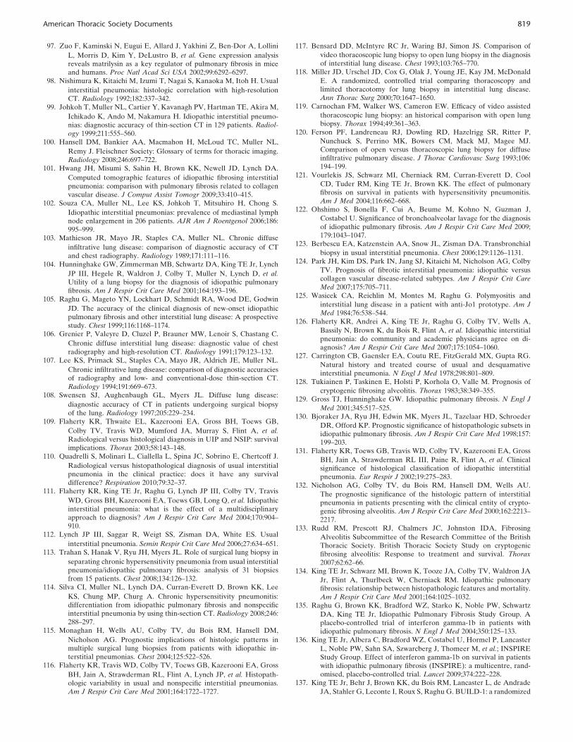
97. Zuo F, Kaminski N, Eugui E, Allard J, Yakhini Z, Ben-Dor A, Lollini
L, Morris D, Kim Y, DeLustro B, et al. Gene expression analysisreveals matrilysin as a key regulator of pulmonary fibrosis in miceand humans. Proc Natl Acad Sci USA 2002;99:6292–6297.
98. Nishimura K, Kitaichi M, Izumi T, Nagai S, Kanaoka M, Itoh H. Usual
interstitial pneumonia: histologic correlation with high-resolutionCT. Radiology 1992;182:337–342.
99. Johkoh T, Muller NL, Cartier Y, Kavanagh PV, Hartman TE, Akira M,
Ichikado K, Ando M, Nakamura H. Idiopathic interstitial pneumo-nias: diagnostic accuracy of thin-section CT in 129 patients. Radiol-ogy 1999;211:555–560.
100. Hansell DM, Bankier AA, Macmahon H, McLoud TC, Muller NL,
Remy J. Fleischner Society: Glossary of terms for thoracic imaging.Radiology 2008;246:697–722.
101. Hwang JH, Misumi S, Sahin H, Brown KK, Newell JD, Lynch DA.
Computed tomographic features of idiopathic fibrosing interstitialpneumonia: comparison with pulmonary fibrosis related to collagenvascular disease. J Comput Assist Tomogr 2009;33:410–415.
102. Souza CA, Muller NL, Lee KS, Johkoh T, Mitsuhiro H, Chong S.
Idiopathic interstitial pneumonias: prevalence of mediastinal lymphnode enlargement in 206 patients. AJR Am J Roentgenol 2006;186:995–999.
103. Mathieson JR, Mayo JR, Staples CA, Muller NL. Chronic diffuse
infiltrative lung disease: comparison of diagnostic accuracy of CTand chest radiography. Radiology 1989;171:111–116.
104. Hunninghake GW, Zimmerman MB, Schwartz DA, King TE Jr, Lynch
JP III, Hegele R, Waldron J, Colby T, Muller N, Lynch D, et al.Utility of a lung biopsy for the diagnosis of idiopathic pulmonaryfibrosis. Am J Respir Crit Care Med 2001;164:193–196.
105. Raghu G, Mageto YN, Lockhart D, Schmidt RA, Wood DE, Godwin
JD. The accuracy of the clinical diagnosis of new-onset idiopathicpulmonary fibrosis and other interstitial lung disease: A prospectivestudy. Chest 1999;116:1168–1174.
106. Grenier P, Valeyre D, Cluzel P, Brauner MW, Lenoir S, Chastang C.
Chronic diffuse interstitial lung disease: diagnostic value of chestradiography and high-resolution CT. Radiology 1991;179:123–132.
107. Lee KS, Primack SL, Staples CA, Mayo JR, Aldrich JE, Muller NL.
Chronic infiltrative lung disease: comparison of diagnostic accuraciesof radiography and low- and conventional-dose thin-section CT.Radiology 1994;191:669–673.
108. Swensen SJ, Aughenbaugh GL, Myers JL. Diffuse lung disease:
diagnostic accuracy of CT in patients undergoing surgical biopsyof the lung. Radiology 1997;205:229–234.
109. Flaherty KR, Thwaite EL, Kazerooni EA, Gross BH, Toews GB,
Colby TV, Travis WD, Mumford JA, Murray S, Flint A, et al.Radiological versus histological diagnosis in UIP and NSIP: survivalimplications. Thorax 2003;58:143–148.
110. Quadrelli S, Molinari L, Ciallella L, Spina JC, Sobrino E, Chertcoff J.
Radiological versus histopathological diagnosis of usual interstitialpneumonia in the clinical practice: does it have any survivaldifference? Respiration 2010;79:32–37.
111. Flaherty KR, King TE Jr, Raghu G, Lynch JP III, Colby TV, Travis
WD, Gross BH, Kazerooni EA, Toews GB, Long Q, et al. Idiopathicinterstitial pneumonia: what is the effect of a multidisciplinaryapproach to diagnosis? Am J Respir Crit Care Med 2004;170:904–910.
112. Lynch JP III, Saggar R, Weigt SS, Zisman DA, White ES. Usual
interstitial pneumonia. Semin Respir Crit Care Med 2006;27:634–651.113. Trahan S, Hanak V, Ryu JH, Myers JL. Role of surgical lung biopsy in
separating chronic hypersensitivity pneumonia from usual interstitialpneumonia/idiopathic pulmonary fibrosis: analysis of 31 biopsiesfrom 15 patients. Chest 2008;134:126–132.
114. Silva CI, Muller NL, Lynch DA, Curran-Everett D, Brown KK, Lee
KS, Chung MP, Churg A. Chronic hypersensitivity pneumonitis:differentiation from idiopathic pulmonary fibrosis and nonspecificinterstitial pneumonia by using thin-section CT. Radiology 2008;246:288–297.
115. Monaghan H, Wells AU, Colby TV, du Bois RM, Hansell DM,
Nicholson AG. Prognostic implications of histologic patterns inmultiple surgical lung biopsies from patients with idiopathic in-terstitial pneumonias. Chest 2004;125:522–526.
116. Flaherty KR, Travis WD, Colby TV, Toews GB, Kazerooni EA, Gross
BH, Jain A, Strawderman RL, Flint A, Lynch JP, et al. Histopath-ologic variability in usual and nonspecific interstitial pneumonias.Am J Respir Crit Care Med 2001;164:1722–1727.
117. Bensard DD, McIntyre RC Jr, Waring BJ, Simon JS. Comparison of
video thoracoscopic lung biopsy to open lung biopsy in the diagnosisof interstitial lung disease. Chest 1993;103:765–770.
118. Miller JD, Urschel JD, Cox G, Olak J, Young JE, Kay JM, McDonald
E. A randomized, controlled trial comparing thoracoscopy andlimited thoracotomy for lung biopsy in interstitial lung disease.Ann Thorac Surg 2000;70:1647–1650.
119. Carnochan FM, Walker WS, Cameron EW. Efficacy of video assisted
thoracoscopic lung biopsy: an historical comparison with open lungbiopsy. Thorax 1994;49:361–363.
120. Ferson PF, Landreneau RJ, Dowling RD, Hazelrigg SR, Ritter P,
Nunchuck S, Perrino MK, Bowers CM, Mack MJ, Magee MJ.Comparison of open versus thoracoscopic lung biopsy for diffuseinfiltrative pulmonary disease. J Thorac Cardiovasc Surg 1993;106:194–199.
121. Vourlekis JS, Schwarz MI, Cherniack RM, Curran-Everett D, Cool
CD, Tuder RM, King TE Jr, Brown KK. The effect of pulmonaryfibrosis on survival in patients with hypersensitivity pneumonitis.Am J Med 2004;116:662–668.
122. Ohshimo S, Bonella F, Cui A, Beume M, Kohno N, Guzman J,
Costabel U. Significance of bronchoalveolar lavage for the diagnosisof idiopathic pulmonary fibrosis. Am J Respir Crit Care Med 2009;179:1043–1047.
123. Berbescu EA, Katzenstein AA, Snow JL, Zisman DA. Transbronchial
biopsy in usual interstitial pneumonia. Chest 2006;129:1126–1131.124. Park JH, Kim DS, Park IN, Jang SJ, Kitaichi M, Nicholson AG, Colby
TV. Prognosis of fibrotic interstitial pneumonia: idiopathic versuscollagen vascular disease-related subtypes. Am J Respir Crit CareMed 2007;175:705–711.
125. Wasicek CA, Reichlin M, Montes M, Raghu G. Polymyositis and
interstitial lung disease in a patient with anti-Jo1 prototype. Am JMed 1984;76:538–544.
126. Flaherty KR, Andrei A, King TE Jr, Raghu G, Colby TV, Wells A,
Bassily N, Brown K, du Bois R, Flint A, et al. Idiopathic interstitialpneumonia: do community and academic physicians agree on di-agnosis? Am J Respir Crit Care Med 2007;175:1054–1060.
127. Carrington CB, Gaensler EA, Coutu RE, FitzGerald MX, Gupta RG.
Natural history and treated course of usual and desquamativeinterstitial pneumonia. N Engl J Med 1978;298:801–809.
128. Tukiainen P, Taskinen E, Holsti P, Korhola O, Valle M. Prognosis of
cryptogenic fibrosing alveolitis. Thorax 1983;38:349–355.129. Gross TJ, Hunninghake GW. Idiopathic pulmonary fibrosis. N Engl J
Med 2001;345:517–525.130. Bjoraker JA, Ryu JH, Edwin MK, Myers JL, Tazelaar HD, Schroeder
DR, Offord KP. Prognostic significance of histopathologic subsets inidiopathic pulmonary fibrosis. Am J Respir Crit Care Med 1998;157:199–203.
131. Flaherty KR, Toews GB, Travis WD, Colby TV, Kazerooni EA, Gross
BH, Jain A, Strawderman RL III, Paine R, Flint A, et al. Clinicalsignificance of histological classification of idiopathic interstitialpneumonia. Eur Respir J 2002;19:275–283.
132. Nicholson AG, Colby TV, du Bois RM, Hansell DM, Wells AU.
The prognostic significance of the histologic pattern of interstitialpneumonia in patients presenting with the clinical entity of crypto-genic fibrosing alveolitis. Am J Respir Crit Care Med 2000;162:2213–2217.
133. Rudd RM, Prescott RJ, Chalmers JC, Johnston IDA, Fibrosing
Alveolitis Subcommittee of the Research Committee of the BritishThoracic Society. British Thoracic Society Study on cryptogenicfibrosing alveolitis: Response to treatment and survival. Thorax2007;62:62–66.
134. King TE Jr, Schwarz MI, Brown K, Tooze JA, Colby TV, Waldron JA
Jr, Flint A, Thurlbeck W, Cherniack RM. Idiopathic pulmonaryfibrosis: relationship between histopathologic features and mortality.Am J Respir Crit Care Med 2001;164:1025–1032.
135. Raghu G, Brown KK, Bradford WZ, Starko K, Noble PW, Schwartz
DA, King TE Jr, Idiopathic Pulmonary Fibrosis Study Group. Aplacebo-controlled trial of interferon gamma-1b in patients withidiopathic pulmonary fibrosis. N Engl J Med 2004;350:125–133.
136. King TE Jr, Albera C, Bradford WZ, Costabel U, Hormel P, Lancaster
L, Noble PW, Sahn SA, Szwarcberg J, Thomeer M, et al.; INSPIREStudy Group. Effect of interferon gamma-1b on survival in patientswith idiopathic pulmonary fibrosis (INSPIRE): a multicentre, rand-omised, placebo-controlled trial. Lancet 2009;374:222–228.
137. King TE Jr, Behr J, Brown KK, du Bois RM, Lancaster L, de Andrade
JA, Stahler G, Leconte I, Roux S, Raghu G. BUILD-1: a randomized
American Thoracic Society Documents 819

placebo-controlled trial of bosentan in idiopathic pulmonary fibrosis.Am J Respir Crit Care Med 2008;177:75–81.
138. Raghu G. Idiopathic pulmonary fibrosis: a rational clinical approach.
Chest 1987;92:148–154.139. Selman M, Carrillo G, Estrada A, Mejia M, Becerril C, Cisneros J,
Gaxiola M, Perez-Padilla R, Navarro C, Richards T, et al. Acceler-ated variant of idiopathic pulmonary fibrosis: clinical behavior andgene expression pattern. PLoS ONE 2007;2:e482.
140. Mejia M, Carrillo G, Rojas-Serrano J, Estrada A, Suarez T, Alonso D,
Barrientos E, Gaxiola M, Navarro C, Selman M. Idiopathic pulmo-nary fibrosis and emphysema: decreased survival associated withsevere pulmonary arterial hypertension. Chest 2009;136:10–15.
141. Wells AU, Desai SR, Rubens MB, Goh NSL, Cramer D, Nicholson
AG, Colby TV, du Bois RM, Hansell DM. Idiopathic pulmonaryfibrosis: a composite physiologic index derived from disease extentobserved by computed tomography. Am J Respir Crit Care Med2003;167:962–969.
142. Lettieri CJ, Nathan SD, Barnett SD, Ahmad S, Shorr AF. Prevalence
and outcomes of pulmonary arterial hypertension in advancedidiopathic pulmonary fibrosis. Chest 2006;129:746–752.
143. Collard HR, Moore BB, Flaherty KR, Brown KK, Kaner RJ, King TE
Jr, Lasky JA, Loyd JE, Noth I, Olman MA, et al.; IdiopathicPulmonary Fibrosis Clinical Research Network Investigators. Acuteexacerbations of idiopathic pulmonary fibrosis. Am J Respir CritCare Med 2007;176:636–643.
144. Azuma A, Nukiwa T, Tsuboi E, Suga M, Abe S, Nakata K, Taguchi Y,
Nagai S, Itoh H, Ohi M, et al. Double-blind, placebo-controlled trialof pirfenidone in patients with idiopathic pulmonary fibrosis. Am JRespir Crit Care Med 2005;171:1040–1047.
145. Martinez FJ, Safrin S, Weycker D, Starko KM, Bradford WZ, King TE
Jr, Flaherty KR, Schwartz DA, Noble PW, Raghu G, et al.; IPFStudy Group. The clinical course of patients with idiopathic pulmo-nary fibrosis. Ann Intern Med 2005;142:963–967.
146. Panos RJ, Mortenson RL, Niccoli SA, King TE Jr. Clinical deteriora-
tion in patients with idiopathic pulmonary fibrosis: causes and assess-ment. Am J Med 1990;88:396–404.
147. Akira M, Hamada H, Sakatani M, Kobayashi C, Nishioka M, Yama-
moto S. CT findings during phase of accelerated deterioration inpatients with idiopathic pulmonary fibrosis. AJR Am J Roentgenol1997;168:79–83.
148. Ambrosini V, Cancellieri A, Chilosi M, Zompatori M, Trisolini R,
Saragoni L, Poletti V. Acute exacerbation of idiopathic pulmonaryfibrosis: report of a series. Eur Respir J 2003;22:821–826.
149. Kim DS, Park JH, Park BK, Lee JS, Nicholson AG, Colby T. Acute
exacerbation of idiopathic pulmonary fibrosis: frequency and clinicalfeatures. Eur Respir J 2006;27:143–150.
150. Kondoh Y, Taniguchi H, Kawabata Y, Yokoi T, Suzuki K, Takagi K.
Acute exacerbation in idiopathic pulmonary fibrosis: analysis ofclinical and pathologic findings in three cases. Chest 1993;103:1808–1812.
151. Kondoh Y, Taniguchi H, Yokoi T, Nishiyama O, Ohishi T, Kato T,
Suzuki K, Suzuki R. Cyclophosphamide and low-dose prednisolonein idiopathic pulmonary fibrosis and fibrosing nonspecific interstitialpneumonia. Eur Respir J 2005;25:528–533.
152. Kubo H, Nakayama K, Yanai M, Suzuki T, Yamaya M, Watanabe M,
Sasaki H. Anticoagulant therapy for idiopathic pulmonary fibrosis.Chest 2005;128:1475–1482.
153. Parambil JG, Myers JL, Ryu JH. Histopathologic features and outcome
of patients with acute exacerbation of idiopathic pulmonary fibrosisundergoing surgical lung biopsy. Chest 2005;128:3310–3315.
154. Tiitto L, Bloigu R, Heiskanen U, Paakko P, Kinnula VL, Kaarteenaho-
Wiik R. Relationship between histopathological features and thecourse of idiopathic pulmonary fibrosis/usual interstitial pneumonia.Thorax 2006;61:1091–1095.
155. Rice AJ, Wells AU, Bouros D, du Bois RM, Hansell DM, Polychro-
nopoulos V, Vassilakis D, Kerr JR, Evans TW, Nicholson AG.Terminal diffuse alveolar damage in relation to interstitial pneumo-nias: an autopsy study. Am J Clin Pathol 2003;119:709–714.
156. Churg A, Muller NL, Silva CIS, Wright JL. Acute exacerbation (acute
lung injury of unknown cause) in UIP and other forms of fibroticinterstitial pneumonias. Am J Surg Pathol 2007;31:277–284.
157. Kondo A, Saiki S. Acute exacerbation in idiopathic interstitial pneu-
monia (IIP). In: Harasawa M, Fukuchi Y, Morinari H, editors.Interstitial pneumonia of unknown etiology. Tokyo: University ofTokyo Press; 1989. pp. 33–42.
158. Konishi K, Gibson KF, Lindell KO, Richards TJ, Zhang Y, Dhir R,
Bisceglia M, Gilbert S, Yousem SA, Song JW, et al. Gene expressionprofiles of acute exacerbations of idiopathic pulmonary fibrosis. AmJ Respir Crit Care Med 2009;180:167–175.
159. Sakamoto K, Taniguchi H, Kondoh Y, Ono K, Hasegawa Y, Kitaichi
M. Acute exacerbation of idiopathic pulmonary fibrosis as the initialpresentation of the disease. Eur Respir Rev 2009;18:129–132.
160. Kondoh Y, Taniguchi H, Kataoka K, Kato K, Suzuki R, Ogura T,
Johkoh T, Yokoi T, Wells AU, Kitaichi M; Tokai Diffuse LungDisease Study Group. Prognostic factors in rapidly progressiveinterstitial pneumonia. Respirology 2010;15:257–264.
161. Kumar P, Goldstraw P, Yamada K, Nicholson AG, Wells AU, Hansell
DM, Dubois RM, Ladas G. Pulmonary fibrosis and lung cancer: riskand benefit analysis of pulmonary resection. J Thorac CardiovascSurg 2003;125:1321–1327.
162. Yuksel M, Ozyurtkan MO, Bostanci K, Ahiskali R, Kodalli N. Acute
exacerbation of interstitial fibrosis after pulmonary resection. AnnThorac Surg 2006;82:336–338.
163. Utz JP, Ryu JH, Douglas WW, Hartman TE, Tazelaar HD, Myers JL,
Allen MS, Schroeder DR. High short-term mortality following lungbiopsy for usual interstitial pneumonia. Eur Respir J 2001;17:175–179.
164. Zegdi R, Azorin J, Tremblay B, Destable MD, Lajos PS, Valeyre D.
Videothoracoscopic lung biopsy in diffuse infiltrative lung diseases:a 5-year surgical experience. Ann Thorac Surg 1998;66:1170–1173.
165. Kondoh Y, Taniguchi H, Kitaichi M, Yokoi T, Johkoh T, Oishi T,
Kimura T, Nishiyama O, Kato K, du Bois RM. Acute exacerbationof interstitial pneumonia following surgical lung biopsy. Respir Med2006;100:1753–1759.
166. Hiwatari N, Shimura S, Takishima T, Shirato K. Bronchoalveolar
lavage as a possible cause of acute exacerbation in idiopathicpulmonary fibrosis patients. Tohoku J Exp Med 1994;174:379–386.
167. Olson AL, Swigris JJ, Lezotte DC, Norris JM, Wilson CG, Brown KK.
Mortality from pulmonary fibrosis increased in the United Statesfrom 1992 to 2003. Am J Respir Crit Care Med 2007;176:277–284.
168. Jemal A, Siegel R, Ward E, Murray T, Xu J, Thun MJ. Cancer
statistics, 2007. CA Cancer J Clin 2007;57:43–66.169. Olson AL, Swigris JJ, Raghu G, Brown KK. Seasonal variation:
mortality from pulmonary fibrosis is greatest in the winter. Chest2009;136:16–22.
170. Hubbard RB, Smith C, Le Jeune I, Gribbin J, Fogarty AW. The
association between idiopathic pulmonary fibrosis and vasculardisease: a population-based study. Am J Respir Crit Care Med2008;178:1257–1261.
171. Nadrous HF, Pellikka PA, Krowka MJ, Swanson KL, Chaowalit N,
Decker PA, Ryu JH. Pulmonary hypertension in patients withidiopathic pulmonary fibrosis. Chest 2005;128:2393–2399.
172. Collard HR, Ryu JH, Douglas WW, Schwarz MI, Curran-Everett D,
King TE Jr, Brown KK. Combined corticosteroid and cyclophos-phamide therapy does not alter survival in idiopathic pulmonaryfibrosis. Chest 2004;125:2169–2174.
173. Hubbard R, Venn A, Smith C, Cooper M, Johnston I, Britton J.
Exposure to commonly prescribed drugs and the etiology ofcryptogenic fibrosing alveolitis: a case-control study. Am J RespirCrit Care Med 1998;157:743–747.
174. Schwartz DA, Van Fossen DS, Davis CS, Helmers RA, Dayton CS,
Burmeister LF, Hunninghake GW. Determinants of progression inidiopathic pulmonary fibrosis. Am J Respir Crit Care Med 1994;149:444–449.
175. Enomoto N, Suda T, Kato M, Kaida Y, Nakamura Y, Imokawa S, Ida
M, Chida K. Quantitative analysis of fibroblastic foci in usualinterstitial pneumonia. Chest 2006;130:22–29.
176. Nadrous HF, Pellikka PA, Krowka MJ, Swanson KL, Chaowalit N,
Decker PA, Ryu JH. The impact of pulmonary hypertension onsurvival in patients with idiopathic pulmonary fibrosis. Chest 2005;128:616S–617S.
177. Jegal Y, Kim DS, Shim TS, Lim C, Do Lee S, Koh Y, Kim WS, Kim
WD, Lee JS, Travis WD, et al. Physiology is a stronger predictor ofsurvival than pathology in fibrotic interstitial pneumonia. Am JRespir Crit Care Med 2005;171:639–644.
178. Wells AU, Hogaboam CM. Update in diffuse parenchymal lung disease
2007. Am J Respir Crit Care Med 2008;177:580–584.179. Turner-Warwick M, Burrows B, Johnson A. Cryptogenic fibrosing
alveolitis: response to corticosteroid treatment and its effect onsurvival. Thorax 1980;35:593–599.
820 AMERICAN JOURNAL OF RESPIRATORY AND CRITICAL CARE MEDICINE VOL 183 2011

180. Schwartz DA, Helmers RA, Galvin JR, Van Fossen DS, Frees KL,
Dayton CS, Burmeister LF, Hunninghake GW. Determinants ofsurvival in idiopathic pulmonary fibrosis. Am J Respir Crit Care Med1994;149:450–454.
181. Nishiyama O, Taniguchi H, Kondoh Y, Kimura T, Ogawa T, Watanabe
F, Nishimura K. Health-related quality of life in patients withidiopathic pulmonary fibrosis: what is the main contributing factor?Respir Med 2005;99:408–414.
182. Nishiyama O, Taniguchi H, Kondoh Y, Kimura T, Kato K, Kataoka K,
Ogawa T, Watanabe F, Arizono S. A simple assessment of dyspneaas a prognostic indicator in idiopathic pulmonary fibrosis. Eur RespirJ 2010;36:1067–1072.
183. Watters LC, King TE Jr, Schwarz MI, Waldron JA, Stanford RE,
Cherniack RM. A clinical, radiographic, and physiologic scoringsystem for the longitudinal assessment of patients with idiopathicpulmonary fibrosis. Am Rev Respir Dis 1986;133:97–103.
184. Witek TJ Jr, Mahler DA. Minimal important difference of the
transition dyspnoea index in a multinational clinical trial. Eur RespirJ 2003;21:267–272.
185. Kupferberg DH, Kaplan RM, Slymen DJ, Ries AL. Minimal clinically
important difference for the UCSD Shortness of Breath Question-naire. J Cardiopulm Rehabil 2005;25:370–377.
186. Collard HR, King TE Jr, Bartelson BB, Vourlekis JS, Schwarz MI,
Brown KK. Changes in clinical and physiologic variables predictsurvival in idiopathic pulmonary fibrosis. Am J Respir Crit Care Med2003;168:538–542.
187. King TE Jr, Safrin S, Starko KM, Brown KK, Noble PW, Raghu G,
Schwartz DA. Analyses of efficacy end points in a controlled trial ofinterferon-gamma1b for idiopathic pulmonary fibrosis. Chest 2005;127:171–177.
188. Hallstrand TS, Boitano LJ, Johnson WC, Spada CA, Hayes JG, Raghu
G. The timed walk test as a measure of severity and survival inidiopathic pulmonary fibrosis. Eur Respir J 2005;25:96–103.
189. Hamada K, Nagai S, Tanaka S, Handa T, Shigematsu M, Nagao
T, Mishima M, Kitaichi M, Izumi T. Significance of pulmonaryarterial pressure and diffusion capacity of the lung as prognosti-cator in patients with idiopathic pulmonary fibrosis. Chest 2007;131:650–656.
190. Egan JJ, Martinez FJ, Wells AU, Williams T. Lung function estimates
in idiopathic pulmonary fibrosis: the potential for a simple classifi-cation. Thorax 2005;60:270–273.
191. Latsi PI, du Bois RM, Nicholson AG, Colby TV, Bisirtzoglou D,
Nikolakopoulou A, Veeraraghavan S, Hansell DM, Wells AU.Fibrotic idiopathic interstitial pneumonia: the prognostic value oflongitudinal functional trends. Am J Respir Crit Care Med 2003;168:531–537.
192. Fell CD, Liu LX, Motika C, Kazerooni EA, Gross BH, Travis WD,
Colby TV, Murray S, Toews GB, Martinez FJ, et al. The prognosticvalue of cardiopulmonary exercise testing in idiopathic pulmonaryfibrosis. Am J Respir Crit Care Med 2009;179:402–407.
193. Flaherty KR, Mumford JA, Murray S, Kazerooni EA, Gross BH, Colby
TV, Travis WD, Flint A, Toews GB, Lynch JP III, et al. Prognosticimplications of physiologic and radiographic changes in idiopathicinterstitial pneumonia. Am J Respir Crit Care Med 2003;168:543–548.
194. Lynch DA, Godwin JD, Safrin S, Starko KM, Hormel P, Brown KK,
Raghu G, King TE Jr, Bradford WZ, Schwartz DA, et al., IdiopathicPulmonary Fibrosis Study Group. High-resolution computed tomog-raphy in idiopathic pulmonary fibrosis: diagnosis and prognosis. AmJ Respir Crit Care Med 2005;172:488–493.
195. Best AC, Meng J, Lynch AM, Bozic CM, Miller D, Grunwald GK,
Lynch DA. Idiopathic pulmonary fibrosis: physiologic tests, quanti-tative CT indexes, and CT visual scores as predictors of mortality.Radiology 2008;246:935–940.
196. Jeong YJ, Lee KS, Muller NL, Chung MP, Chung MJ, Han J, Colby
TV, Kim S. Usual interstitial pneumonia and non-specific interstitialpneumonia: serial thin-section CT findings correlated with pulmo-nary function. Korean J Radiol 2005;6:143–152.
197. Shin KM, Lee KS, Chung MP, Han J, Bae YA, Kim TS, Chung MJ.
Prognostic determinants among clinical, thin-section CT, and histo-pathologic findings for fibrotic idiopathic interstitial pneumonias:tertiary hospital study. Radiology 2008;249:328–337.
198. Sumikawa H, Johkoh T, Colby TV, Ichikado K, Suga M, Taniguchi H,
Kondoh Y, Ogura T, Arakawa H, Fujimoto K, et al. Computedtomography findings in pathological usual interstitial pneumonia:relationship to survival. Am J Respir Crit Care Med 2008;177:433–439.
199. Lama VN, Flaherty KR, Toews GB, Colby TV, Travis WD, Long Q,
Murray S, Kazerooni EA, Gross BH, Lynch JP III, et al. Prognosticvalue of desaturation during a 6-minute walk test in idiopathicinterstitial pneumonia. Am J Respir Crit Care Med 2003;168:1084–1090.
200. Enright PL. The six-minute walk test. Respir Care 2003;48:783–785.201. Lederer DJ, Arcasoy SM, Wilt JS, D’Ovidio F, Sonett JR, Kawut SM.
Six-minute-walk distance predicts waiting list survival in idiopathicpulmonary fibrosis. Am J Respir Crit Care Med 2006;174:659–664.
202. Swigris JJ, Swick J, Wamboldt FS, Sprunger D, du Bois R, Fischer A,
Cosgrove GP, Frankel SK, Fernandez-Perez ER, Kervitsky D, et al.Heart rate recovery after 6-min walk test predicts survival in patientswith idiopathic pulmonary fibrosis. Chest 2009;136:841–848.
203. Caminati A, Bianchi A, Cassandro R, Mirenda MR, Harari S. Walking
distance on 6-MWT is a prognostic factor in idiopathic pulmonaryfibrosis. Respir Med 2009;103:117–123.
204. Eaton T, Young P, Milne D, Wells AU. Six-minute walk, maximal
exercise tests: reproducibility in fibrotic interstitial pneumonia. Am JRespir Crit Care Med 2005;171:1150–1157.
205. Nicholson AG, Fulford LG, Colby TV, du Bois RM, Hansell DM,
Wells AU. The relationship between individual histologic featuresand disease progression in idiopathic pulmonary fibrosis. Am JRespir Crit Care Med 2002;166:173–177.
206. Flaherty KR, Colby TV, Toews GB, Travis WD, Flint A, Gay SE,
Strawderman RL III, Jain A, Lynch JP III, Martinez FJ. Differentialpresence of fibroblastic foci in UIP patients with or withoutconntective tissue disease. Am J Respir Crit Care Med 2001;163:A983.
207. Hanak V, Ryu JH, de Carvalho E, Limper AH, Hartman TE, Decker
PA, Myers JL. Profusion of fibroblast foci in patients with idiopathicpulmonary fibrosis does not predict outcome. Respir Med 2008;102:852–856.
208. Arcasoy SM, Christie JD, Ferrari VA, Sutton MS, Zisman DA,
Blumenthal NP, Pochettino A, Kotloff RM. Echocardiographicassessment of pulmonary hypertension in patients with advancedlung disease. Am J Respir Crit Care Med 2003;167:735–740.
209. Nathan SD, Shlobin OA, Barnett SD, Saggar R, Belperio JA, Ross DJ,
Ahmad S, Saggar R, Libre E, Lynch JP III, et al. Right ventricularsystolic pressure by echocardiography as a predictor of pulmonaryhypertension in idiopathic pulmonary fibrosis. Respir Med 2008;102:1305–1310.
210. Fisher MR, Forfia PR, Chamera E, Housten-Harris T, Champion HC,
Girgis RE, Corretti MC, Hassoun PM. Accuracy of Dopplerechocardiography in the hemodynamic assessment of pulmonaryhypertension. Am J Respir Crit Care Med 2009;179:615–621.
211. Corte TJ, Wort SJ, Gatzoulis MA, Macdonald P, Hansell DM, Wells
AU. Pulmonary vascular resistance predicts early mortality inpatients with diffuse fibrotic lung disease and suspected pulmonaryhypertension. Thorax 2009;64:883–888.
212. Cottin V, Nunes H, Brillet PY, Delaval P, Devouassoux G, Tillie-
Leblond I, Israel-Biet D, Court-Fortune I, Valeyre D, Cordier JF;Groupe d’Etude et de Recherche sur les Maladies OrphelinesPulmonaires (GERM O P). Combined pulmonary fibrosis andemphysema: a distinct underrecognised entity. Eur Respir J 2005;26:586–593.
213. Stahel RA, Gilks WR, Lehmann HP, Schenker T. Third International
Workshop on Lung Tumor and Differentiation Antigens: overview ofthe results of the central data analysis. Int J Cancer Suppl 1994;8:6–26.
214. Kohno N, Kyoizumi S, Awaya Y, Fukuhara H, Yamakido M, Akiyama
M. New serum indicator of interstitial pneumonitis activity: sialy-lated carbohydrate antigen KL-6. Chest 1989;96:68–73.
215. Yokoyama A, Kondo K, Nakajima M, Matsushima T, Takahashi T,
Nishimura M, Bando M, Sugiyama Y, Totani Y, Ishizaki T, et al.Prognostic value of circulating KL-6 in idiopathic pulmonaryfibrosis. Respirology 2006;11:164–168.
216. Greene KE, King TE Jr, Kuroki Y, Bucher-Bartelson B, Hunninghake
GW, Newman LS, Nagae H, Mason RJ. Serum surfactant proteins-A and -D as biomarkers in idiopathic pulmonary fibrosis. Eur RespirJ 2002;19:439–446.
217. Kinder BW, Brown KK, McCormack FX, Ix JH, Kervitsky A, Schwarz
MI, King TE Jr. Serum surfactant protein-A is a strong predictor ofearly mortality in idiopathic pulmonary fibrosis. Chest 2009;135:1557–1563.
218. Takahashi H, Fujishima T, Koba H, Murakami S, Kurokawa K,
Shibuya Y, Shiratori M, Kuroki Y, Abe S. Serum surfactant proteinsA and D as prognostic factors in idiopathic pulmonary fibrosis and
American Thoracic Society Documents 821

their relationship to disease extent. Am J Respir Crit Care Med 2000;162:1109–1114.
219. Prasse A, Probst C, Bargagli E, Zissel G, Toews GB, Flaherty KR,
Olschewski M, Rottoli P, Muller-Quernheim J. Serum CC-chemo-kine ligand 18 concentration predicts outcome in idiopathic pulmo-nary fibrosis. Am J Respir Crit Care Med 2009;179:717–723.
220. Leuchte HH, Baumgartner RA, Nounou ME, Vogeser M, Neurohr C,
Trautnitz M, Behr J. Brain natriuretic peptide is a prognosticparameter in chronic lung disease. Am J Respir Crit Care Med2006;173:744–750.
221. Leuchte HH, Neurohr C, Baumgartner R, Holzapfel M, Giehrl W,
Vogeser M, Behr J. Brain natriuretic peptide and exercise capacityin lung fibrosis and pulmonary hypertension. Am J Respir Crit CareMed 2004;170:360–365.
222. Song JW, Song JK, Kim DS. Echocardiography and brain natriuretic
peptide as prognostic indicators in idiopathic pulmonary fibrosis.Respir Med 2009;103:180–186.
223. Shinoda H, Tasaka S, Fujishima S, Yamasawa W, Miyamoto K, Nakano
Y, Kamata H, Hasegawa N, Ishizaka A. Elevated CC chemokinelevel in bronchoalveolar lavage fluid is predictive of a poor outcomeof idiopathic pulmonary fibrosis. Respiration 2009;78:285–292.
224. Rosas IO, Richards TJ, Konishi K, Zhang Y, Gibson K, Lokshin AE,
Lindell KO, Cisneros J, Macdonald SD, Pardo A, et al. MMP1 andMMP7 as potential peripheral blood biomarkers in idiopathicpulmonary fibrosis. PLoS Med 2008;5:e93.
225. Phelps DS, Umstead TM, Mejia M, Carrillo G, Pardo A, Selman M.
Increased surfactant protein-A levels in patients with newly di-agnosed idiopathic pulmonary fibrosis. Chest 2004;125:617–625.
226. McCormack FX, King TE Jr, Bucher BL, Nielsen L, Mason RJ.
Surfactant protein A predicts survival in idiopathic pulmonaryfibrosis. Am J Respir Crit Care Med 1995;152:751–759.
227. Kinder BW, Brown KK, Schwarz MI, Ix JH, Kervitsky A, King TE Jr.
Baseline BAL neutrophilia predicts early mortality in idiopathicpulmonary fibrosis. Chest 2008;133:226–232.
228. Moeller A, Gilpin SE, Ask K, Cox G, Cook D, Gauldie J, Margetts PJ,
Farkas L, Dobranowski J, Boylan C, et al. Circulating fibrocytes arean indicator of poor prognosis in idiopathic pulmonary fibrosis. Am JRespir Crit Care Med 2009;179:588–594.
229. Davies HR, Richeldi L, Walters EH. Immunomodulatory agents for
idiopathic pulmonary fibrosis. Cochrane Database Syst Rev 2003; (3):CD003134.
230. Richeldi L, Davies HR, Ferrara G, Franco F. Corticosteroids for
idiopathic pulmonary fibrosis. Cochrane Database Syst Rev 2003;(3):CD002880.
231. Gay SE, Kazerooni EA, Toews GB, Lynch JP III, Gross BH, Cascade
PN, Spizarny DL, Flint A, Schork MA, Whyte RI, et al. Idiopathicpulmonary fibrosis: predicting response to therapy and survival. AmJ Respir Crit Care Med 1998;157:1063–1072.
232. Flaherty KR, Toews GB, Lynch JP III, Kazerooni EA, Gross BH,
Strawderman RL, Hariharan K, Flint A, Martinez FJ. Steroids inidiopathic pulmonary fibrosis: a prospective assessment of adversereactions, response to therapy, and survival. Am J Med 2001;110:278–282.
233. Nagai S, Kitaichi M, Hamada K, Nagao T, Hoshino Y, Miki H, Izumi T.
Hospital-based historical cohort study of 234 histologically provenJapanese patients with IPF. Sarcoidosis Vasc Diffuse Lung Dis 1999;16:209–214.
234. Entzian P, Schlaak M, Seitzer U, Bufe A, Acil Y, Zabel P. Antiin-
flammatory and antifibrotic properties of colchicine: implications foridiopathic pulmonary fibrosis. Lung 1997;175:41–51.
235. Peters SG, McDougall JC, Douglas WW, Coles DT, DeRemee RA.
Colchicine in the treatment of pulmonary fibrosis. Chest 1993;103:101–104.
236. Fiorucci E, Lucantoni G, Paone G, Zotti M, Li BE, Serpilli M,
Regimenti P, Cammarella I, Puglisi G, Schmid G. Colchicine,cyclophosphamide and prednisone in the treatment of mild-moderateidiopathic pulmonary fibrosis: comparison of three currently avail-able therapeutic regimens. Eur Rev Med Pharmacol Sci 2008;12:105–111.
237. Antoniou KM, Nicholson AG, Dimadi M, Malagari K, Latsi P, Rapti
A, Tzanakis N, Trigidou R, Polychronopoulos V, Bouros D. Long-term clinical effects of interferon gamma-1b and colchicine inidiopathic pulmonary fibrosis. Eur Respir J 2006;28:496–504.
238. Douglas WW, Ryu JH, Swensen SJ, Offord KP, Schroeder DR, Caron
GM, DeRemee RA; Lung Study Group. Colchicine versus predni-
sone in the treatment of idiopathic pulmonary fibrosis: a randomizedprospective study. Am J Respir Crit Care Med 1998;158:220–225.
239. Alton EW, Johnson M, Turner-Warwick M. Advanced cryptogenic
fibrosing alveolitis: preliminary report on treatment with cyclosporinA. Respir Med 1989;83:277–279.
240. Moolman JA, Bardin PG, Rossouw DJ, Joubert JR. Cyclosporin as
a treatment for interstitial lung disease of unknown aetiology.Thorax 1991;46:592–595.
241. Homma S, Sakamoto S, Kawabata M, Kishi K, Tsuboi E, Motoi N,
Yoshimura K. Cyclosporin treatment in steroid-resistant and acutelyexacerbated interstitial pneumonia. Intern Med 2005;44:1144–1150.
242. Grgic A, Lausberg H, Heinrich M, Koenig J, Uder M, Sybrecht GW,
Wilkens H. Progression of fibrosis in usual interstitial pneumonia:serial evaluation of the native lung after single lung transplantation.Respiration 2008;76:139–145.
243. Wahidi MM, Ravenel J, Palmer SM, McAdams HP. Progression of
idiopathic pulmonary fibrosis in native lungs after single lung trans-plantation. Chest 2002;121:2072–2076.
244. Winterbauer RH, Hammar SP, Hallman KO, Hays JE, Pardee NE,
Morgan EH, Allen JD, Moores KD, Bush W, Walker JH. Diffuseinterstitial pneumonitis: clinicopathologic correlations in 20 patientstreated with prednisone/azathioprine. Am J Med 1978;65:661–672.
245. Raghu G, Depaso WJ, Cain K, Hammar SP, Wetzel CE, Dreis DF,
Hutchinson J, Pardee NE, Winterbauer RH. Azathioprine combinedwith prednisone in the treatment of idiopathic pulmonary fibrosis:a prospective double-blind, randomized, placebo-controlled clinicaltrial. Am Rev Respir Dis 1991;144:291–296.
246. Johnson MA, Kwan S, Snell NJ, Nunn AJ, Darbyshire JH, Turner-
Warwick M. Randomised controlled trial comparing prednisolonealone with cyclophosphamide and low dose prednisolone in combi-nation in cryptogenic fibrosing alveolitis. Thorax 1989;44:280–288.
247. Pereira CAC, Malheiros T, Coletta EM, Ferreira RG, Rubin AS,
Otta JS, Rocha NS. Survival in idiopathic pulmonary fibrosis-cytotoxic agents compared to corticosteroids. Respir Med 2006;100:340–347.
248. Cantin AM, Hubbard RC, Crystal RG. Glutathione deficiency in the
epithelial lining fluid of the lower respiratory tract in idiopathicpulmonary fibrosis. Am Rev Respir Dis 1989;139:370–372.
249. Behr J, Maier K, Degenkolb B, Krombach F, Vogelmeier C. Anti-
oxidative and clinical effects of high-dose N-acetylcysteine infibrosing alveolitis: adjunctive therapy to maintenance immunosup-pression. Am J Respir Crit Care Med 1997;156:1897–1901.
250. Demedts M, Behr J, Buhl R, Costabel U, Dekhuijzen R, Jansen HM,
MacNee W, Thomeer M, Wallaert B, Laurent F, et al.; IFIGENIAStudy Group. High-dose acetylcysteine in idiopathic pulmonaryfibrosis. N Engl J Med 2005;353:2229–2242.
251. Hunninghake GW. Antioxidant therapy for idiopathic pulmonary
fibrosis. N Engl J Med 2005;353:2285–2287.252. Wells AU. Antioxidant therapy in idiopathic pulmonary fibrosis: hope
is kindled. Eur Respir J 2006;27:664–666.253. Tomioka H, Kuwata Y, Imanaka K, Hashimoto K, Ohnishi H, Tada K,
Sakamoto H, Iwasaki H. A pilot study of aerosolized N-acetylcys-teine for idiopathic pulmonary fibrosis. Respirology 2005;10:449–455.
254. Ziesche R, Hofbauer E, Wittmann K, Petkov V, Block LH. A
preliminary study of long-term treatment with interferon gamma-1b and low-dose prednisolone in patients with idiopathic pulmonaryfibrosis. N Engl J Med 1999;341:1264–1269.
255. Thannickal VJ, Flaherty KR, Martinez FJ, Lynch JP III. Idiopathic
pulmonary fibrosis: emerging concepts on pharmacotherapy. ExpertOpin Pharmacother 2004;5:1671–1686.
256. Raghu G, King TE Jr, Behr J, Brown KK, du Bois RM, Leconte I,
Roux S, Swigris J. Quality of life and dyspnoea in patients treatedwith bosentan for idiopathic pulmonary fibrosis (BUILD-1). EurRespir J 2010;35:118–123.
257. Mohler KM, Torrance DS, Smith CA, Goodwin RG, Stremler KE,
Fung VP, Madani H, Widmer MB. Soluble tumor necrosis factor(TNF) receptors are effective therapeutic agents in lethal endotox-emia and function simultaneously as both TNF carriers and TNFantagonists. J Immunol 1993;151:1548–1561.
258. Sime PJ, Marr RA, Gauldie D, Xing Z, Hewlett BR, Graham FL,
Gauldie J. Transfer of tumor necrosis factor-alpha to rat lunginduces severe pulmonary inflammation and patchy interstitial fibro-genesis with induction of transforming growth factor-beta1 andmyofibroblasts. Am J Pathol 1998;153:825–832.
822 AMERICAN JOURNAL OF RESPIRATORY AND CRITICAL CARE MEDICINE VOL 183 2011

259. Piguet PF, Ribaux C, Karpuz V, Grau GE, Kapanci Y. Expression and
localization of tumor necrosis factor-alpha and its mRNA inidiopathic pulmonary fibrosis. Am J Pathol 1993;143:651–655.
260. Raghu G, Brown KK, Costabel U, Cottin V, du Bois RM, Lasky JA,
Thomeer M, Utz JP, Khandker RK, McDermott L, et al. Treatment ofidiopathic pulmonary fibrosis with etanercept: an exploratory, pla-cebo-controlled trial. Am J Respir Crit Care Med 2008;178:948–955.
261. Raghu G, Johnson WC, Lockhart D, Mageto Y. Treatment of
idiopathic pulmonary fibrosis with a new antifibrotic agent, pirfeni-done: results of a prospective, open-label Phase II study. Am JRespir Crit Care Med 1999;159:1061–1069.
262. Nagai S, Hamada K, Shigematsu M, Taniyama M, Yamauchi S, Izumi
T. Open-label compassionate use one year-treatment with pirfeni-done to patients with chronic pulmonary fibrosis. Intern Med 2002;41:1118–1123.
263. Taniguchi H, Ebina M, Kondoh Y, Ogura T, Azuma A, Suga M,
Taguchi Y, Takahashi H, Nakata K, Sato A, et al., PirfenidoneClinical Study Group in Japan. Pirfenidone in idiopathic pulmonaryfibrosis. Eur Respir J 2010;35:821–829.
264. Seymour S. Briefing Information for the March 9, 2010 Meeting of
the Pulmonary-Allergy Drugs Advisory Committee. NDA 22–535.Washington, D.C.: US Food and Drug Administration, 2010.[accessed 2010 May 10.] Available from: http://www.fda.gov/downloads/AdvisoryCommittees/CommitteesMeetingMaterials/Drugs/Pulmonary-AllergyDrugsAdvisoryCommittee/UCM203081.pdf.
265. Seymour S. Division Summary: Overview of the FDA background
materials for New Drug Application (NDA) 22-535, Esbriet (pirfe-nidone) for the treatment of patients with idiopathic pulmonaryfibrosis (IPF) to reduce the decline in lung function. In: Seymour S,editor. FDA Briefing Information for the March 9, 2010 Meeting ofthe Pulmonary-Allergy Drugs Advisory Committee (PADAC).NDA 22–535. Washington, D.C.: US Food and Drug Administration;2010. pp. 2–20. [accessed 2010 May 10.] Available from: http://www.fda.gov/downloads/AdvisoryCommittees/CommitteesMeetingMaterials/Drugs/Pulmonary-AllergyDrugsAdvisoryCommittee/UCM203081.pdf.
266. Karimi-Shah B. Esbriet (pirfenidone) 2403 mg/day to reduce the
decline in lung function in patients with idiopathic pulmonaryfibrosis (Clinical Briefing Document). In: Seymour S, editor. FDABriefing Information for the March 9, 2010 Meeting of the Pulmo-nary-Allergy Drugs Advisory Committee (PADAC).NDA 22–535.Washington, D.C.: US Food and Drug Administration; 2010. pp. 21–109. [accessed 2010 May 10.] Available from: http://www.fda.gov/downloads/AdvisoryCommittees/CommitteesMeetingMaterials/Drugs/Pulmonary-AllergyDrugsAdvisoryCommittee/UCM203081.pdf.
267. Zhou F. Pirfenidone capsules [three 267-mg capsules TID] for treat-
ment of patients with idiopathic pulmonary fibrosis to reduce declinein lung function (Statistical Briefing Document). In: Seymour S,editor. FDA Briefing Information for the March 9, 2010 Meeting ofthe Pulmonary-Allergy Drugs Advisory Committee (PADAC).NDA 22–535. Washington, D.C.: US Food and Drug Administration;2010. p. 110–158. [accessed 2010 May 10.] Available from: http://www.fda.gov/downloads/AdvisoryCommittees/CommitteesMeetingMaterials/Drugs/Pulmonary-AllergyDrugsAdvisoryCommittee/UCM203081.pdf.
268. Ghofrani HA, Wiedemann R, Rose F, Schermuly RT, Olschewski H,
Weissmann N, Gunther A, Walmrath D, Seeger W, Grimminger F.Sildenafil for treatment of lung fibrosis and pulmonary hypertension:a randomised controlled trial. Lancet 2002;360:895–900.
269. Madden BP, Allenby M, Loke T, Sheth A. A potential role for
sildenafil in the management of pulmonary hypertension in patientswith parenchymal lung disease. Vascul Pharmacol 2006;44:372–376.
270. Collard HR, Anstrom KJ, Schwarz MI, Zisman DA. Sildenafil im-
proves walk distance in idiopathic pulmonary fibrosis. Chest 2007;131:897–899.
271. Nathan SD, Noble PW, Tuder RM. Idiopathic pulmonary fibrosis and
pulmonary hypertension: connecting the dots. Am J Respir Crit CareMed 2007;175:875–880.
272. The Idiopathic Pulmonary Fibrosis Clinical Research Network. A
controlled trial of sildenafil in advanced idiopathic pulmonaryfibrosis. N Engl J Med 2010;363:620–628.
273. Jackson RM, Glassberg MK, Ramos CF, Bejarano PA, Butrous G,
Gomez-Marin O. Sildenafil therapy and exercise tolerance inidiopathic pulmonary fibrosis. Lung 2010;188:115–123.
274. Daniels CE, Lasky JA, Limper AH, Mieras K, Gabor E, Schroeder
DR; Imatinib-IPF Study Investigators. Imatinib treatment for idio-pathic pulmonary fibrosis: randomized placebo-controlled trial re-sults. Am J Respir Crit Care Med 2010;181:604–610.
275. Morrison DA, Stovall JR. Increased exercise capacity in hypoxemic
patients after long-term oxygen therapy. Chest 1992;102:542–550.276. Nocturnal Oxygen Therapy Trial Group. Continuous or nocturnal
oxygen therapy in hypoxemic chronic obstructive lung disease:a clinical trial. Ann Intern Med 1980;93:391–398.
277. Longterm domiciliary oxygen therapy in chronic hypoxic cor pulmonale
complicating chronic bronchitis and emphysema: report of theMedical Research Council Working Party. Lancet 1981;1:681–686.
278. Mason DP, Brizzio ME, Alster JM, McNeill AM, Murthy SC, Budev
MM, Mehta AC, Minai OA, Pettersson GB, Blackstone EH. Lungtransplantation for idiopathic pulmonary fibrosis. Ann Thorac Surg2007;84:1121–1128.
279. Keating D, Levvey B, Kotsimbos T, Whitford H, Westall G, Williams
T, Snell G. Lung transplantation in pulmonary fibrosis: challengingearly outcomes counterbalanced by surprisingly good outcomesbeyond 15 years. Transplant Proc 2009;41:289–291.
280. Thabut G, Mal H, Castier Y, Groussard O, Brugiere O, Marrash-
Chahla R, Leseche G, Fournier M. Survival benefit of lung trans-plantation for patients with idiopathic pulmonary fibrosis. J ThoracCardiovasc Surg 2003;126:469–475.
281. Thabut G, Christie JD, Ravaud P, Castier Y, Dauriat G, Jebrak G,
Fournier M, Leseche G, Porcher R, Mal H. Survival after bilateralversus single-lung transplantation for idiopathic pulmonary fibrosis.Ann Intern Med 2009;151:767–774.
282. Blivet S, Philit F, Sab JM, Langevin B, Paret M, Guerin C, Robert D.
Outcome of patients with idiopathic pulmonary fibrosis admitted tothe ICU for respiratory failure. Chest 2001;120:209–212.
283. Molina-Molina M, Badia JR, Marin-Arguedas A, Xaubet A, Santos
MJ, Nicolas JM, Ferrer M, Torres A. Outcomes and clinicalcharacteristics of patients with pulmonary fibrosis and respiratoryfailure admitted to an intensive care unit: a study of 20 cases. MedClin (Barc) 2003;121:63–67.
284. Saydain G, Islam A, Afessa B, Ryu JH, Scott JP, Peters SG. Outcome
of patients with idiopathic pulmonary fibrosis admitted to theintensive care unit. Am J Respir Crit Care Med 2002;166:839–842.
285. Nava S, Rubini F. Lung and chest wall mechanics in ventilated patients
with end stage idiopathic pulmonary fibrosis. Thorax 1999;54:390–395.
286. Stern JB, Mal H, Groussard O, Brugiere O, Marceau A, Jebrak G,
Fournier M. Prognosis of patients with advanced idiopathic pulmo-nary fibrosis requiring mechanical ventilation for acute respiratoryfailure. Chest 2001;120:213–219.
287. Al-Hameed FM, Sharma S. Outcome of patients admitted to the
intensive care unit for acute exacerbation of idiopathic pulmonaryfibrosis. Can Respir J 2004;11:117–122.
288. Fumeaux T, Rothmeier C, Jolliet P. Outcome of mechanical ventilation
for acute respiratory failure in patients with pulmonary fibrosis.Intensive Care Med 2001;27:1868–1874.
289. Pitsiou G, Ioannis Trigonis I, Tsiata E, Kontou P, Manolakoglou N,
Stanopoulos I, Mavrofridis E, Argyropoulou P. Outcome of patientswith pulmonary fibrosis admitted to the ICU for acute respiratoryfailure. Eur Respir J Suppl 2006;28:E650.
290. Mollica C, Paone G, Conti V, Ceccarelli D, Schmid G, Mattia P,
Perrone N, Petroianni A, Sebastiani A, Cecchini L, et al. Mechanicalventilation in patients with end-stage idiopathic pulmonary fibrosis.Respiration 2010;79:209–215.
291. Rangappa P, Moran JL. Outcomes of patients admitted to the intensive
care unit with idiopathic pulmonary fibrosis. Crit Care Resusc 2009;11:102–109.
292. Mallick S. Outcome of patients with idiopathic pulmonary fibrosis
(IPF) ventilated in intensive care unit. Respir Med 2008;102:1355–1359.
293. Holland AE, Hill CJ, Conron M, Munro P, McDonald CF. Short term
improvement in exercise capacity and symptoms following exercisetraining in interstitial lung disease. Thorax 2008;63:549–554.
294. Nishiyama O, Kondoh Y, Kimura T, Kato K, Kataoka K, Ogawa T,
Watanabe F, Arizono S, Nishimura K, Taniguchi H. Effects ofpulmonary rehabilitation in patients with idiopathic pulmonaryfibrosis. Respirology 2008;13:394–399.
295. Ferreira A, Garvey C, Connors GL, Hilling L, Rigler J, Farrell S,
Cayou C, Shariat C, Collard HR. Pulmonary rehabilitation ininterstitial lung disease: benefits and predictors of response. Chest2009;135:442–447.
296. Ferreira G, Feuerman M, Spiegler P. Results of an 8-week, outpatient
pulmonary rehabilitation program on patients with and without
American Thoracic Society Documents 823
chronic obstructive pulmonary disease. J Cardiopulm Rehabil 2006;26:54–60.
297. Jastrzebski D, Gumola A, Gawlik R, Kozielski J. Dyspnea and quality
of life in patients with pulmonary fibrosis after six weeks ofrespiratory rehabilitation. J Physiol Pharmacol 2006;57:139–148.
298. Naji NA, Connor MC, Donnelly SC, McDonnell TJ. Effectiveness of
pulmonary rehabilitation in restrictive lung disease. J CardiopulmRehabil 2006;26:237–243.
299. Lancaster LH, Mason WR, Parnell JA, Rice TW, Loyd JE, Milstone
AP, Collard HR, Malow BA. Obstructive sleep apnea is common inidiopathic pulmonary fibrosis. Chest 2009;136:772–778.
300. Kim DS, Collard HR, King TE Jr. Classification and natural history of the
idiopathic interstitial pneumonias. Proc Am Thorac Soc 2006;3:285–292.
301. Sakamoto S, Homma S, Miyamoto A, Kurosaki A, Fujii T, Yoshimura
K. Cyclosporin A in the treatment of acute exacerbation ofidiopathic pulmonary fibrosis. Intern Med 2010;49:109–115.
302. Olschewski H, Ghofrani HA, Walmrath D, Schermuly R, Temmesfeld-
Wollbruck B, Grimminger F, Seeger W. Inhaled prostacyclin andiloprost in severe pulmonary hypertension secondary to lung fibrosis.Am J Respir Crit Care Med 1999;160:600–607.
303. Minai OA, Sahoo D, Chapman JT, Mehta AC. Vaso-active therapy
can improve 6-min walk distance in patients with pulmonary hyper-tension and fibrotic interstitial lung disease. Respir Med 2008;102:1015–1020.
304. Sweet MP, Patti MG, Leard LE, Golden JA, Hays SR, Hoopes C,
Theodore PR. Gastroesophageal reflux in patients with idiopathicpulmonary fibrosis referred for lung transplantation. J ThoracCardiovasc Surg 2007;133:1078–1084.
305. Marik PE. Aspiration pneumonitis and aspiration pneumonia. N Engl J
Med 2001;344:665–671.306. Barnes TW, Vassallo R, Tazelaar HD, Hartman TE, Ryu JH. Diffuse
bronchiolar disease due to chronic occult aspiration.Mayo Clin Proc2006;81:172–176.
307. Davis RD Jr, Lau CL, Eubanks S, Messier RH, Hadjiliadis D, Steele
MP, Palmer SM. Improved lung allograft function after fundo-plication in patients with gastroesophageal reflux disease un-dergoing lung transplantation. J Thorac Cardiovasc Surg 2003;125:533–542.
308. Linden PA, Gilbert RJ, Yeap BY, Boyle K, Deykin A, Jaklitsch MT,
Sugarbaker DJ, Bueno R. Laparoscopic fundoplication in patientswith end-stage lung disease awaiting transplantation. J ThoracCardiovasc Surg 2006;131:438–446.
309. Hope-Gill BDM, Hilldrup S, Davies C, Newton RP, Harrison NK. A
study of the cough reflex in idiopathic pulmonary fibrosis. Am JRespir Crit Care Med 2003;168:995–1002.
310. Horton MR, Danoff SK, Lechtzin N. Thalidomide inhibits the intracta-
ble cough of idiopathic pulmonary fibrosis. Thorax 2008;63:749.
311. Zappala CJ, Latsi PI, Nicholson AG, Colby TV, Cramer D, RenzoniEA, Hansell DM, du Bois RM, Wells AU. Marginal decline inforced vital capacity is associated with a poor outcome in idiopathicpulmonary fibrosis. Eur Respir J 2010;35:830–836.
312. Miller MR, Crapo R, Hankinson J, Brusasco V, Burgos F, Casaburi R,Coates A, Enright P, van der Grinten CP, Gustafsson P, et al.; ATS/ERS Task Force. General considerations for lung function testing.Eur Respir J 2005;26:153–161.
313. Miller MR, Hankinson J, Brusasco V, Burgos F, Casaburi R, Coates A,Crapo R, Enright P, van der Grinten CP, Gustafsson P, et al.; ATS/ERS Task Force. Standardisation of spirometry. Eur Respir J 2005;26:319–338.
314. Wanger J, Clausen JL, Coates A, Pedersen OF, Brusasco V, Burgos F,Casaburi R, Crapo R, Enright P, van der Grinten CP, et al. Stand-ardisation of the measurement of lung volumes. Eur Respir J 2005;26:511–522.
315. Macintyre N, Crapo RO, Viegi G, Johnson DC, van der Grinten CP,Brusasco V, Burgos F, Casaburi R, Coates A, Enright P, et al.Standardisation of the single-breath determination of carbon mon-oxide uptake in the lung. Eur Respir J 2005;26:720–735.
316. Pellegrino R, Viegi G, Brusasco V, Crapo RO, Burgos F, Casaburi R,Coates A, van der Grinten CP, Gustafsson P, Hankinson J, et al.Interpretative strategies for lung function tests. Eur Respir J 2005;26:948–968.
317. Selman M, King TE Jr, Pardo A; American Thoracic Society, EuropeanRespiratory Society, American College of Chest Physicians. Idio-pathic pulmonary fibrosis: prevailing and evolving hypotheses aboutits pathogenesis and implications for therapy. Ann Intern Med 2001;134:136–151.
318. Akagi T, Matsumoto T, Harada T, Tanaka M, Kuraki T, Fujita M,Watanabe K. Coexistent emphysema delays the decrease of vitalcapacity in idiopathic pulmonary fibrosis. Respir Med 2009;103:1209–1215.
319. Mura M, Zompatori M, Pacilli AMG, Fasano L, Schiavina M, Fabbri M.The presence of emphysema further impairs physiologic function inpatients with idiopathic pulmonary fibrosis. Respir Care 2006;51:257–265.
320. Cottin V, Cordier JF. Combined pulmonary fibrosis and emphysema:an experimental and clinically relevant phenotype. Am J Respir CritCare Med 2005;172:1605–1606.
321. Wiggins J, Strickland B, Turner-Warwick M. Combined cryptogenicfibrosing alveolitis and emphysema: the value of high resolution com-puted tomography in assessment. Respir Med 1990;84:365–369.
322. ATS Committee on Proficiency Standards for Clinical PulmonaryFunction Laboratories. ATS statement: guidelines for the six-minutewalk test. Am J Respir Crit Care Med 2002;166:111–117.
323. Zisman DA, Karlamangla AS, Kawut SM, Shlobin OA, Saggar R, RossDJ, Schwarz MI, Belperio JA, Ardehali A, Lynch JP III, et al.Validation of a method to screen for pulmonary hypertension inadvanced idiopathic pulmonary fibrosis. Chest 2008;133:640–645.
824 AMERICAN JOURNAL OF RESPIRATORY AND CRITICAL CARE MEDICINE VOL 183 2011