INVESTIGATION OF NEW CILIOPATHY GENES
63
INVESTIGATION OF NEW CILIOPATHY GENES A THESIS SUBMITTED TO THE DEPARTMENT OF BIOENGINEERING AND THE GRADUATE SCHOOL OF ENGINEERING AND SCIENCE OF ABDULLAH GUL UNIVERSITY IN PARTIAL FULFILLMENT OF THE REQUIREMENTS FOR THE DEGREE OF MASTER OF SCIENCE By Ferhan YENİSERT May 2021 FERHAN YENİSERT A Master’s Thesis AGU 2021
Transcript of INVESTIGATION OF NEW CILIOPATHY GENES

Name Surname
INVESTIGATION OF NEW CILIOPATHY
GENES
A THESIS
SUBMITTED TO THE DEPARTMENT OF BIOENGINEERING
AND THE GRADUATE SCHOOL OF ENGINEERING AND SCIENCE
OF ABDULLAH GUL UNIVERSITY
IN PARTIAL FULFILLMENT OF THE REQUIREMENTS
FOR THE DEGREE OF
MASTER OF SCIENCE
By
Ferhan YENİSERT
May 2021
FE
RH
AN
YE
NİS
ER
T A
Master’s T
hesis
AG
U 2
021

INVESTIGATION OF NEW CILIOPATHY
GENES
A THESIS
SUBMITTED TO THE DEPARTMENT OF BIOENGINEERING
AND THE GRADUATE SCHOOL OF ENGINEERING AND SCIENCE OF
ABDULLAH GUL UNIVERSITY
IN PARTIAL FULFILLMENT OF THE REQUIREMENTS
FOR THE DEGREE OF
MASTER OF SCIENCE
By
Ferhan YENİSERT
May 2021

SCIENTIFIC ETHICS COMPLIANCE
I hereby declare that all information in this document has been obtained in accordance
with academic rules and ethical conduct. I also declare that, as required by these rules and
conduct, I have fully cited and referenced all materials and results that are not original to
this work.
Name-Surname: FERHAN YENİSERT
Signature :

REGULATORY COMPLIANCE
M.Sc. thesis titled INVESTIGATION OF NEW CILIOPATHY GENE: CEP41
has been prepared in accordance with the Thesis Writing Guidelines of the Abdullah Gül
University, Graduate School of Engineering & Science.
Prepared By Advisor
FERHAN YENİSERT Dr. OKTAY İSMAİL KAPLAN
Signature Signature
Head of the Bioengineering Graduate Program
Prof. Dr. SEVİL DİNÇER İŞOĞLU

ACCEPTANCE AND APPROVAL
M.Sc. thesis titled INVESTIGATION OF NEW CILIOPATHY GENE: CEP41 and
prepared by FERHAN YENİSERT has been accepted by the jury in the Bioengineering
Graduate Program at Abdullah Gül University, Graduate School of Engineering &
Science.
……….. /……….. / ………..
JURY:
Advisor : Dr. Oktay İsmail Kaplan
Member: Assoc. Prof. Müşerref Duygu Saçar Demirci
Member: Assoc. Prof. Şükrü Sadık Öner
APPROVAL:
The acceptance of this M.Sc. thesis has been approved by the decision of the Abdullah
Gül University, Graduate School of Engineering & Science, Executive Board dated …..
/….. / ……….. and numbered .…………..……. .
……….. /……….. / ………..
(Date)
Graduate School Dean
Prof. Dr. Hakan Usta

i
ABSTRACT
INVESTIGATION OF NEW CILIOPATHY GENES
Ferhan YENİSERT
MSc. in Bioengineering
Advisor: Dr. Oktay İsmail Kaplan
May 2021
Cilia consist of microtubules in its internal structure and evolutionarily conserved an
antenna-like organelle. The disease caused by defects in the cilia structure is called
ciliopathy and Joubert syndrome is one of the ciliopathies. Patients display a range of
symptoms, such as delayed intellectual and language development, hypotonia, ataxia,
mental retardation, liver cyst, retinal defect/degeneration, genital defect, and cystic
kidney. As a result of recent studies, 38 different genes have been associated with Joubert
syndrome. In 2012, CEP41, an evolutionarily conserved gene, was associated with JS,
one of the diseases of ciliopathy, but that study did not investigate the molecular
mechanism of CEP41. In this study, the effect of ceph-41 mutation on the structure and
function of cilia was investigated by using C. elegans, which is widely used as a model
system in cilia studies.
Keywords: CEP41, Joubert syndrome, Ciliopathy,

ii
ÖZET
YENİ SİLYOPATİ GENLERİNİN ARAŞTIRILIMASI
Ferhan YENİSERT
Biyomühendislik Anabilim Dalı Yüksek Lisans
Tez Danışmanı: Dr. Oktay İsmail Kaplan
Mayıs 2021
Kirpikler, iç yapısında mikrotübüllerden oluşur ve evrimsel olarak anten benzeri bir
organel korunmuştur. Kirpik yapısındaki bozuklukların neden olduğu hastalığa siliyopati
adı verilir ve Joubert sendromu siliyopatilerden biridir. Hastalar, gecikmiş zihinsel ve dil
gelişimi, hipotoni, ataksi, zeka geriliği, karaciğer kisti, retina kusuru / dejenerasyonu,
genital kusur ve kistik böbrek gibi bir dizi semptom gösterir. Son çalışmaların bir sonucu
olarak, 38 farklı gen Joubert sendromu ile ilişkilendirilmiştir. 2012 yılında, evrimsel
olarak korunmuş bir gen olan CEP41, silyopati hastalıklarından biri olan JS ile
ilişkilendirildi, ancak bu çalışma CEP41'in moleküler mekanizmasını araştırılmadı. Bu
çalışmada, cilia çalışmalarında yaygın olarak model sistem olarak kullanılan C. elegans
kullanılarak ceph-41 mutasyonunun kirpiklerin yapısı ve işlevi üzerindeki etkisi
araştırılmıştır.
Anahtar Kelimeler: CEP41, Joubert sendromu, Silyopati

iii
Acknowledgements
I would like to first thank my supervisor Dr Oktay Kaplan, for his guidance and support
during my M.Sc. I would like to thank Dr. Sebiha Çevik Kaplan for her support. I would
like to thank all members of Kaplan lab for their help in my experiments, including Atiye
Zorluer ve Merve Gül Turan.

iv
Table of Contents
1. INTRODUCTION…………………………………………………………....…….1
1.1. CILIA AND FLAGELLA……………………………………………................1
1.1.1Basal body…………………………………….………..…………………....3
1.1.2.Transition zone ……………………………………………………………...3
1.2. CILIOPATHY……..…………….……….…………………………………..…3
1.2.1Polycystic kidney disease(PKD)…………..….…………...…………..….......... 5
1.2.2Joubert syndrome.....………..……………………………….……………..... 6
1.3. INTRAFLAGELLAR TRANSPORT (IFT)………………………………..…. 7
1.4. MODEL ORGANISM: CAENORHABDITIS ELEGANS……………….......…10
1.5. CEP41, A JOUBERT SYNDROME GENE, REGULATES CILIA
BIOGENESIS AND POLYGLUTAMYLATION……………………..……...13
2. MATERIALS AND METHODS……..…………….……………………..….......14
2.1. MATERIALS…………….....……………….……………………….…….….14
2.1.1. Strains……………………..……….………………………….………....14
2.1.2. Primers and Plasmids……………..……………..………………..…….….15
2.2. METHODS…………….…………………………………………….…….......15
2.2.1. Dye Filling Assay…………………………………………….…….…..…..15
2.2.2. Cross System……………………………………………….….……..……16
2.2.3. Crispr/Cas9……………………………………………...………………..17
2.2.4. Microscope analysis……….………………….………………….………...18
3. RESULTS………………………………………………...…….…….…………....19
3.1. CEP41 IS AN EVOLUTIONARILY CONSERVED CILIARY GENE……...19
3.2. CEPH-41 SPECIFICALLY LOCALIZE TO THE MIDDLE SEGMENT OF
CILIA IN C. ELEGANS …………………………………………………….....21
3.3. THE NULL CEPH-41 MUTANT DOES NOT POSSESS THE CILIARY
STRUCTURE DEFECT ……………………………………………………....25
3.4. INVESTIGATION OF LOCALIZATION OF IFT PROTEINS IN CEPH-41
MUTANT …………………………………………………………….…….…28
3.5. CEP41 MUTATION DOES NOT AFFECT IFT ………………………….….29
4. DISCUSSION…………………………………………...……………..………......31
5. CONCLUSIONS AND FUTURE PROSPECTS………………………......…....34
5.1. CONCLUSIONS…………………………….………..…………..….….….…34
5.2. SOCIAL IMPACT AND CONTRIBUTION….…………………………...….34
5.3. FUTURE PROSPECTS….…………………………………………..………...35

v
List of Figures
Figure 1.1 A difference between the structure of motile cilia and primary cilia..….……...2
Figure 1.2 Demonstration of ciliopathies and affected part of human body…………..….5
Figure 1.3 Intraflagellar transport in C. elegans.………………………………………....9
Figure 1.4 The life cycle of C. elegans at 22ºC…………………………………….......11
Figure 3.1 CEP41 gene is evolutionarily conserved in unicellular and multicellular
organisms…………………………………………...……………………….....….21
Figure 3.2 CEP41 in C. elegans is localized middle segment of cilia.…...………….…23
Figure 3.3 [cep-41pCEPH-41::wrmScarlet + tmem-145::TMEM-145::GFP]
colocalization in middle segment of cilia…………………………………….….24
Figure 3.4 ceph-41 did not affect sutructure of cilia……...………………………….... 26
Figure 3.5 ceph-41 did not effect AWB, AWC, ASE, and PHA/PHB cilia structure........27
Figure 3.6 ceph-41 did not effect IFT protein localization……………………..….........29
Figure 3.7 ceph-41 double mutant did not affect IFT protein.…………………………. 30

vi
List of Tables
Table 1.1 Orthologous variant of 38 genes associated with Joubert
syndrome.…………...…………………………………………………………….6

vii
List of Abbreviations
BBS Bardet-Biedl syndrome
BB Basal Body
C. elegans Caenorhabditis elegans
DS Distal Segment
IFT Intraflagellar Transport
JS Joubert syndrome
MKS Meckel-Gruber syndrome
MS Middle Segment
PKD Polycystic kidney disease
TZ Transition Zone

To Kaplan lab member and my family
1
Chapter 1
1. Introduction
1.1 Cilia and Flagella
Projecting as the cellular antenna into the extracellular space, cilia and flagella are present
in unicellular species such as Chlamydomonas reinhardtii and Tetrahymena thermophila,
as well as multicellular organisms such as Caenorhabditis elegans, Drosophila
melanogaster, and mammals, such as mouse and human. The discovery of cilia was dated
back to the 17th century because Anthony van Leeuwenhoek described them as “little
feet” in 1674. Otto Muller named these “little feet” as “cilium” (eyelash in Latin) in 1786
[1,5]. Scientists used electron microscopy to examine the ultrastructure of cilia and
flagella, which revealed a 9+0 or 9+2 microtubule doublet in a ring form surrounded by
the plasma membrane [4,6].
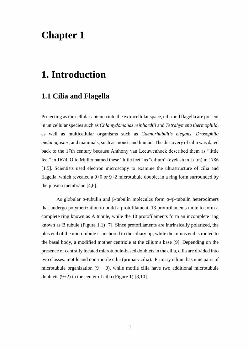
As globular α-tubulin and β-tubulin molecules form α-/β-tubulin heterodimers
that undergo polymerization to build a protofilament, 13 protofilaments unite to form a
complete ring known as A tubule, while the 10 protofilaments form an incomplete ring
known as B tubule (Figure 1.1) [7]. Since protofilaments are intrinsically polarized, the
plus end of the microtubule is anchored to the ciliary tip, while the minus end is rooted to
the basal body, a modified mother centriole at the cilium's base [9]. Depending on the
presence of centrally located microtubule-based doublets in the cilia, cilia are divided into
two classes: motile and non-motile cilia (primary cilia). Primary cilium has nine pairs of
microtubule organization (9 + 0), while motile cilia have two additional microtubule
doublets (9+2) in the center of cilia (Figure 1) [8,10].

2
Figure 1.1 A difference between the structure of motile cilia and primary cilia.
The cross-sections of motile and primary cilia are illustrated in the diagrams [3].
Despite the fact that motile cilia were discovered in the 17th century, scientists
such as Paul Langerhans, Alexander Ecker, Aleksandr Kowalevsky, Albert Kolliker, and
Karl Zimmermann were the first to recognize primary cilium (non-motile cilia) in a large
variety of vertebrate cells in the 19th century. The primary cilium is unable to move,
which may be due to the absence of centrally located microtubule doublets, as research
in the unicellular organism Chlamydomonas reinhardtii revealed that mutants without the
central pair of microtubules are unable to move [7,12,13]. Furthermore, the molecular
motor dynein, which is needed for cilia movement, does not appear to be present in the
primary cilium.
Furthermore, accumulating evidence over the years proved that motile and
primary cilia have distinct functions. For example, mucus removal is mediated by motile
cilia on the surfaces of cells in the respiratory tract and the middle ear, and sperm cell
movement is facilitated by motile cilia [14]. Interestingly, the correct left-right
asymmetry in embryo development needs the nodal cilia (motile cilia) in the mammalian
embryo. Primary cilium, on the other hand, is located on the cell surfaces of most
mammalian cells, including olfactory epithelium, brain cells, fibroblasts, mesenchymal
cells, and retinal photoreceptors, where they perform a variety of sensory roles such as
mechanosensory and chemosensory. The primary cilium of retinal pigment cells, for
example, contains rhodopsin molecules, which are required for vision [11,14]. Primary
cilium on the cells surrounding the kidney nephron is needed for detection of the flow of
fluid; however, if the fluid flow sensing is disrupted, cysts form in the kidneys, resulting

3
in polycystic kidney disease (PKD). PKD was the first disease that was linked to primary
cilium [7].
In vertebrates, the primary cilium acts as a signaling organelle that transmits a
variety of signaling pathways that regulate embryo development, such as Hh, Wnt
[16,17], Notch [19], Hippo [20] Platelet-Derived Growth Factor Subunit A
(PDGFa)[18], and mTOR [21,22] pathways[15]. Furthermore, cilia are implicated in
neuronal migration [23,24] and axonal pathway development in differentiated neurons
[25].
1.1.1 Basal Body
Cilia are divided into several subcompartments, such as basal body (BB),
transition zone (TZ). BB, the modified mother centriole, is a barrel of nine three-fold
microtubules, sub-distal extensions, and nine strut-like structures known as distal
extensions or transition fibers, attached to the membrane at the base of the cilium
[26,27]. During cilia formation, the mother centriole is connected to the ciliary vesicle
or plasma membrane through distal extensions, which then become transitional fibers
(Williams CL). Protein entry into cilia and departure from cilia are thought to be regulated
by BB and TZ at the ciliary base, and there is a concept of a diffuse barrier or gate that
prevents unspecified membrane protein movement into and out of the cilium [28].
1.1.2 Transition Zone
The transition zone (TZ) arises at the BB's distal end and consists of a collection
of transition fibers that extend from the B-tube to the C-end tubes and are embedded
inside the periciliary plasma membrane. In the main body of the TZ, there are multiple
rows of Y-shaped connectors projecting from the outer pairs and connecting to the ciliary
membrane [29]. The TZ layout is then formed by nine outer microtubular pairs with a 9-
bladed propeller base structure [30]. TZ ends distally with the final Y-linker chain. As
mentioned above, TZ works as a gatekeeper by blocking the entry of non-ciliary proteins
into cilia and assists by working on vesicle trafficking of other TZ proteins such as
CC2D2A [31].
1.2 Ciliopathy

4
The motile cilia were first related to a human pathology named primary ciliary
dyskinesia (PCD) in 1976 [32], but later years proved that structural and functional
defects in the primary cilium, in addition to motile cilia, contribute to a human disorder
[33]. Since then, the list of cilia-related diseases has expanded to include Joubert
syndrome (JBTS) [34], Senior-Loken syndrome (SLS), Nephronophthisis (NPHP)[36],
Bardet-Biedl syndrome (BBS)[37], Meckel-Gruber syndrome (MKS) [35], Alström
syndrome (AS), autosomal dominant polycystic kidney disease (ADPKD) [33],
Orofaciodigital syndrome type I (OFD), and Cranioectodermal dysplasia (CED), they are
collectively called ciliopathy. While Cornillie FJ and colleagues first-named ciliopathy in
1984, it gained popularity after Ansley and colleagues discovered that a mutation in BBS8
caused BBS in 2003 [38,39]. Ciliopathy shares a considerable number of clinical
symptoms, including mental retardation, liver cyst, retinal defect/degeneration, sterility
or genital defect, cystic kidney, hydrocephalus, craniofacial defect, rib/thoracic defect,
polydactyly, pelvic bone defect, pancreatic cyst, lung or airway defect, cardiac defect, but
not all symptoms are present in the patient (Figure 1.2).
Our lab has collected the list of genes causing ciliopathy (unpublished data) and
the number of ciliopathy-causing genes has reached over 200. Most proteins encoded by
ciliopathy-causing genes localize to cilia and cilia sub-compartments, while mutations in
genes encoding non-ciliary proteins can cause a ciliopathy.

5
Figure 1.2. Demonstration of ciliopathies and affected part of human body.
1.2.1 Polycystic kidney disease (PKD)
Polycystic kidney disease (PKD) was first identified as kidney disease in the 16th
century, with the first recorded case being Polish king Stefan Bathory, who experienced
weakness (fatigue) and chest pain. They found that the king's kidney is "as big as those
of a bull, with an uneven and bumpy appearance" after he died [40]. After 350 years,
Krakow historians and clinicians concluded that the cause of death was most likely uremia
and autosomal dominant PKD. The polycystic term was firstly used in 1888.
The name "PKD" refers to two different forms of the disease, each with varying
degrees of phenotypic difference and genetic cause: autosomal recessive polycystic
kidney disease (ARPKD) and autosomal dominant polycystic kidney disease (ADPKD)
[41]. ADPKD is a genetically heterogeneous disorder in which two genes, PKD1
(16p13.3) and PKD2 (16p13.3), have been involved [42]. On the other hand, ARPKD is
the less frequent of the two forms of PKD related to the PKHD1 gene [43]. These three
genes encode proteins that localize within cilia. Pkd displays a range of symptoms
including urine blood, elevated blood pressure, headache, stomach pain, and frequent
urination.

6
1.2.2 Joubert syndrome
Joubert syndrome (JBTS; OMIM 213300) is a rare ciliopathy disease affecting the
cerebellum, which regulates balance and coordination. The following are the signs and
symptoms of Joubert syndrome: abnormal eye movements, fast breathing, delayed
intellectual and language development, hypotonia, ataxia, mental retardation, liver cyst,
retinal defect/degeneration, genital defect, and cystic kidney [44]. Number of genes
implicated in JBTS have increased and there are currently 38 genes that cause Joubert
syndrome and related cerebellar diseases, and the identified genes are : TMEM216 [45],
AHI1 [46], NPHP1 [47], CEP290 [48], TMEM67 [49], RPGRIP1L [50], ARL13B [51],
CC2D42A [52], CXORF5 [53], TTC21B[54], KIF7 [55], TCTN1 [56], TMEM237[57],
CEP41[58], TMEM138 [59], C5ORF42 [60], TCTN3 [61], ZNF423[62], TMEM231
[63], CSPP1 [64], PDE6D [65], KIAA0586[66], TCTN2 [67], CEP104 [68], KIAA0556
[69], B9D1[70], MKS1[70], TMEM107[71], ARMC9[72], CEP120[73], SUFU[74],
PIBF1[75], B9D2[76], ARL3[77], FAM149B1[78], TOGARAM1[79], and IFT74[80]
(Table 1). Accumulating evidence revealed that these JBTS genes encode proteins that
are found in the cilia or basal body or transition zone. Cilia formation and/or function are
impaired as a result of mutations in either of these JBTS genes. In this thesis, we
investigated the function of CEP41 in the nematode Caenorhabditis elegans.
Human Gene
Name
OMIM ID C. elegans Orthologous Gene
Name
Wormbase ID
References
TMEM216 613277 mks-2 WBGene00194710 [45]
AHI1 608894 NA NA [46]
NPHP1 607100 nphp-1 WBGene00010898 [47]
CEP290 610142 cep-290 WBGene00012121 [48]
TMEM67 609884 mks-3 WBGene00018042 [49]
RPGRIP1L 610937 mks-5 WBGene00007490 [50]
ARL13B 608922 arl-13 WBGene00021349 [51]
CC2D2A 612013 mks-6 WBGene00010642 [52]
CXORF5 300170 NA NA [53]
TTC21B 612014 ift-139 WBGene00022696 [54]
KIF7 611254 klp-12 WBGene00002223 [55]

7
TCTN1 609863 tctn-1 WBGene00017120 [56]
TMEM237 614423 tmem-237 NA [57]
CEP41 610523 ceph-41 WBGene00249817 [58]
TMEM138 614459 tmem-138 WBGene00008643 [59]
C5ORF42 614571 hpo-40 WBGene00014113 [60]
TCTN3 613847 tctn-1 WBGene00017120 [61]
ZNF423 604577 lin-13 WBGene00003002 [62]
TMEM231 614949 tmem-231 WBGene00020825 [63]
CSPP1 611654 NA NA [64]
PDE6D 602676 pdl-1 WBGene00003966 [65]
KIAA0586 610178 talp-3 NA [66]
TCTN2 613846 NA NA [67]
CEP104 616690 c40h1.3 WBGene00008039 [68]
KATNIP 616650 k04f10.2 NA [69]
B9D1 614144 mksr-1 WBGene00019364 [70]
MKS1 609883 mksr-2 WBGene00021416 [70]
TMEM107 616183 tmem-107 NA [71]
ARMC9 617612 F59G1.4 WBGene00019128 [72]
CEP120 613446 NA NA [73]
SUFU 607035 NA NA [74]
PIBF1 607532 NA NA [75]
B9D2 611951 mksr-2 WBGene00021416 [76]
ARL3 604695 arl-3 WBGene00000188 [77]
FAM149B1 618413 NA NA [78]
TOGARAM1 617618 che-12 WBGene00000491 [79]
IFT74 ift74
WBGene00016005 [80]
Table 1.1. Orthologous variant of 38 genes associated with Joubert syndrome.
38 JS gene and C. elegans orthologous of these genes are shown in the table.
1.3 Intraflagellar transport (IFT)

8
Joel Rosenbaum and his lab discovered that intraflagellar transport (IFT)
undergoes bidirectional motility along the cilia of Chlamydomonas reinhardtii, a
unicellular organism, in 1993, and two years later, his lab biochemically isolated the
multi-protein IFT complex [81]. The multi-protein IFT complex consists of three essential
components; (1) IFT proteins divided into two sub-complexes: IFT complex B and IFT
complex A, (2) anterograde motor protein heterotrimeric Kinesin II, and (3) retrograde
motor protein cytoplasmic Dynein. Genes in these two complexes named according to
their molecular weights are as follows: IFT A complex IFT144, IFT140, IFT139, IFT122,
IFT121, and IFT43 and IFT B complex IFT172, IFT88, IFT81, IFT80, IFT74, IFT72,
IFT57, IFT52, IFT46, IFT20, IFT27[82], IFT70/Dyf-1 [83], IFT25 [84] IFT54/Elipsa
[85] and IFT22/IFTA-2/Rab5 [86]. In later years, it was discovered that IFT is necessary
for cilia formation and flagella maintenance. However, IFT is responsible for the transport
of ciliary constituents and signal molecules, such as PKD and Smoothen receptor SMO.
Depending on the organism, the heterodimeric kinesin-II motor protein or the
heterodimeric kinesin-II motor protein in combination with the homodimeric OSM-3
(Human KIF17) kinesin motor protein transports the IFT complex from the basal body to
the cilia end (anterograde) [86].
Another protein complex that undergoes IFT is the BBSome made up of eight
distinct proteins, including BBS1, BBS2, BBS4, BBS5, BBS8, BBS7, BBS9, and
BBS18/BBIP10. There are several different functions attributed to bbs genes. For
example, work from C. elegans revealed that the BBSome keeps IFT motors and IFT sub-
complexes A and B together. The BBSome assembly requires the BBS/CCT complex
activity that includes the three chaperonin-like BBS proteins (BBS10, BBS6, and BBS12)
and the chaperonin containing TCP-1 (CCT) / T-complex protein-1 ring complex (TriC)
family chaperonins [87].

9
Figure 1.3 Intraflagellar transport in C. elegans. Intraflagellar transport in C.
elegans. Components of the IFT machinery and ciliary cargo assemble at or near the
transition zone (basal body), and heterotrimeric kinesin-II, and homodimeric OSM-3-
kinesin, separately bind IFT particle subcomplexes A and B in order and transport them
along the middle segment in the anterograde (+) direction with IFT-dynein and cargo.
OSM-3-kinesin carries IFT particles and dynein/cargo alone in the distal segment. The
BBS proteins work to keep the motors and IFT particle subcomplexes A and B together.
The IFT-dynein molecular motor recycles components of the IFT machinery, possibly
other ciliary molecules, back to the cilium's foundation. In conjunction with the
microtubular schematics of the cross-specifications, the lengths of Transition Zone (1
mc), Medium Section (4 mc), and Distal (2,5 mc) areas are presented (for cilia amphid);
(on top) [98].
IFT proteins travel at a certain speed along the cilia (Figure 1.3). The first IFT
speed study was performed in Chlamydomonas, which revealed that the heterodimeric
kinesin II protein carries the anterograde complex IFT at a speed of 2.5 μm/s, whereas the
dynein protein carries the retrograde IFT at a speed of 4 μm/s [84]. In human cell culture,
the situation is somewhat different. Human cells have a slower IFT motility than
Chlamydomonas. In mammals, the IFT complex is transported at speeds ranging from 0.3
to 0.7 µm/s in both anterograde and retrograde directions [85]. In this regard, the IFT
velocity of Caenorhabditis elegans might be closer to the slower IFT speed of a mammal
than that of Chlamydomonas. In C. elegans, there are two kinesin motor proteins
heterodimeric Kinesin II and homodimeric kinesin OSM-3 (human KIF17). These two
kinesin motors transport the IFT/BBSome complex along the middle segment in an
anterograde direction at 0.7 m/s, while the homodimeric kinesin motor OSM-3/KIF17
10
drives them along the distal segment at 1.2 m/s. The return of the IFT/BBSome complex
from the ciliary tip/middle part of the cilia is achieved by the retrograde cytoplasmic
dynein motor protein that moves at a rate of 1.3 µm/s.
1.4 Model Organism: Caenorhabditis elegans
Caenorhabditis elegans is a free-living nematode approximately 1 mm long and 65
µm thick. Caenorhabditis elegans have 2 sex: hermaphrodite has XX chromosomes and
male has one X chromosome. Hermaphrodites are living things that can reproduce their
eggs and sperm. It can produce 300 eggs in 3-4 days. There is a noticeably short
generation span, like 2-3 days. Therefore, this gives the chance to see the phenotype
desired to be looked after in the next generation in a noticeably short time. A short lifetime
provides a great advantage especially in studies such as embryogenesis and development
(Figure 1.4). Each adult hermaphrodite always contains exactly 959, and each adult male
contains 1031 somatic cell nuclei. The first multicellular organ with a full genome
sequence was C. elegans [88].
C. elegans is widely used in cilia research because of its ease of use and functional
similarity with human studies [89]. Many evolutionarily conserved ciliary genes were
first identified as model organisms using C. elegans. The cilia functions of IFTA-1 (IFT

11
A sub-complex gene),
Figure 1.4 The life cycle of C. elegans at 22ºC.
It is fertilized for 0 minutes. The blue numbers on the arrows indicate the time the animal
spends at a certain stage. The primitive ends about 40 divisions after fertilization. The
eggs are released approximately 150 minutes after fertilization and gastrulation are
glazed. The length of the animal in each stage is marked in micrometers (μm) next to the
stage name [99].
DYF-1, DYF-2, DYF-13 (IFT B sub-complex genes) were first identified in C.
elegans and later it has been found that these genes are involved in forming the cilia
structure in other animals, including humans [90]. Cilia functions of the evolutionarily
conserved gene K04F10.2 were first demonstrated in C. elegans [91]. DYF-19 (FBF1),
RAB-28, AP-2 complex, AP-1 are examples of genes whose functions have been shown
for the first time in C. elegans [70]. The cilia association of the Joubert syndrome gene
ARL13B was first shown in mice in 2008 [63]. ARL13B study showed that ARL13B was
localized in cilia and found that its deficiency caused structural defects in microtubule
pairs in cilia [63]. Later, the role of this gene was characterized using C. elegans [89].
The study carried out in C. elegans has demonstrated that ARL13B is localized in cilia as
well as structural defects in microtubules of cilia in an ARL-13 mutant study. In addition

12
to mammalian studies, in 2010 a study found that ARL13b is localized in the cilia
membrane and regulates IFT by using C. elegans [89]. This all proves that C. elegans
may be used as an excellent model system for studies of cilia.
The usage advantages/features of C. elegans, one of the most preferred model
systems by scientists, are listed.
It is easy and cheap. They live in the laboratory on agar curtains containing E. coli.
Millions of worms can live in a petri dish. C. elegans can be rapidly frozen and stored in
a refrigerator (- 80 degrees).
C. elegans in Japan (http://www.shigen.nig.ac.jp/c.elegans/index.jsp) and America
(http://www.cbs.umn.edu/CGC/) there are mutant centers. These centers have more than
40% mutants of all genes. Requesting scientists can obtain mutants for a small fee.
Since C. elegans is a multicellular organism, it undergoes complex developmental
processes such as embryogenesis, morphogenesis, and reaching maturity, as is the case
with higher organisms. All cells (neuron, muscle, and intestine, etc.) starting from the
first cell until adulthood have been characterized by scientists. Specific markers are
available for these cells.
Many molecular studies, which are very laborious, time-consuming, and expensive,
can be easily performed in C. elegans. For example, programmed cell division (apoptosis)
was first found in C. elegans. However, studies such as gene expression, studying the
molecular bases of axon and dendrite, cell division, and cell differentiation can be easily
performed in C. elegans. In addition to all these, C. elegans is a widely used model
organism in lifespan studies. Functional studies of genes associated with the disease can
be done.
The fundamental structure of non-motile cilia is at the dendritic ends of neuronal cells
(60 of 302 neurons). These primary cilia found in C. elegans form a grouped structure
called sensilla (amphid and phasmid sensilla), which has a sensory task. C. elegans is
surrounded by cuticles (the structure composed of collagen surrounding C. elegans).
Amphid and phasmid cilia open to the environment through two pores and communicate
with the environment. These are the only structures that communicate with the
environment. In this project, using the advantages of C. elegans, the role of CEP41, which

13
is the Joubert Syndrome gene, in cilia will be investigated for the first time in this
organism.
1.5 CEP41, a Joubert syndrome gene, regulates cilia
biogenesis and polyglutamylation
It was first identified by Joseph G. Gleeson that CEP41, encoding a protein of 41-
kilodalton (kDa), was mutated in patients with Joubert syndrome in 2012 [Lee, J.E]. Since
Joubert syndrome is a disease associated with cilia, it was assumed that CEP41 may have
a role in cilia. In the 2012 study, CEP41 localization was examined, which revealed that
it localized in cilia and centrioles in mouse and human cells [58]. In mRNA expression
studies performed in zebrafish, it was found that CEP41 was expressed in the ciliated
organs Kupffer's vesicles, ear, heart, nerves, and kidneys [58]. When the CEP41 gene was
silenced with morpholino (antisense oligonucleotides), phenotypes associated with
ciliopathy (hydrocephalus, abnormal ear otolith formation, peripheral heart edema, tail
defects) were also repeated in zebrafish [58]. Likewise, when CEP41 was mutated in
mice, cilia-related abnormalities were observed, and the mice died before completing
embryo development, so their functional studies were limited to these studies [58].
Another important function of CEP41 is its involvement in tubulin glutamylation.
In the 2012 stud., it was found that tubulin glutamylation was reduced in fibroblast cells
obtained from patients with Joubert syndrome with CEP41 gene mutations [Lee, J.E].
Simultaneously, the protein complex immunoprecipitation (Co-IP) assay showed that
CEP41 is precipitated by TTLL enzymes [58]. Although the 2012 study states that CEP41
is a part of TTLL enzymes, CEP41 does not have a ligase-like enzyme activity in its
protein structure. The main role of CEP41 in tubulin polyglutamate is currently unknown.
Although only the 2012 article showed that the mutation of CEP41 leads to phenotypes
associated with ciliopathy, it is not yet known how the deficiency of this gene leads to
these phenotypes. My thesis aimed to define the role of CEP41 in C. elegans.

14
Chapter 2
2. Materials and Methods
2.1 Materials
2.1.1 Strains
The strains used in this work:
N2
N2; [Pstr1::ODR10::GFP]
N2; [Pstr1promoter::GFP]
N2; ntIs1 [gcy-5p::GFP + lin-15(+)] V; integration of adEx1262 [gcy-5p::GFP + lin-
15(+)].
N2; gmls13 [srb-
6p::GFP+pRF4)
N2; [OSM-3::GFP+PRF4]
N2; Is [OSM6::GFP]
N2; casIs586(KAP-1::GFP)
N2; jhuEx [CHE-11::GFP+pRF4]
The following strains were generated in this work:
cep41 (tur001)
cep41 (tur001) ;;[Pstr1::ODR10::GFP]
cep41 (tur001) :;[Pstr1promoter::GFP]
cep41 (tur001); ntIs1 [gcy-5p::GFP + lin-15(+)] V; integration of adEx1262 [gcy-
5p::GFP + lin-15(+)].
cep41 (tur001); gmls13 [srb-6p::GFP+pRF4]
cep41 (tur001); Is [OSM6::GFP]
bbs-5 [OSM-6::GFP]

15
cep-41(tur001); bbs-5 [OSM-6::GFP]
cep41 (tur001); jhuEx [CHE-11::GFP+pRF4]
cep41 (tur001); casIs586(KAP-1::GFP)
cep-41(tur001) IV;Ex[OSM-3::GFP++pRF4]
cep41 (tur001); Ex [CHE-11::GFP +pRF4]
him-5(e1490) V; myIs1 [PKD-2::GFP + Punc-122::GFP] IV.; cep-
41promoter_CEP41(F42G8.19)_wrmscarlet (25 ng)
him-5(e1490) V; myIs1 [PKD-2::GFP + Punc-122::GFP] IV.; cep-
41promoter:CEP41(F42G8.19)::-wrmscarlet
N2 [cep-41promoter_CEP41(F42G8.19)::wrmscarlet ;str-1p::GFP]
N2 [cep-41promoter: CEP41(F42G8.19)::wrmscarlet ;str-1p::GFP]
2.1.2 Primers and Plasmids
F42G8.19_sgRNA1_for TCTTGTCAATATCAATAGCCGAATG
F42G8.19_sgRNA1_rev AAACCATTCGGCTATTGATATTGAC
F42G8.19_sgRNA2_for TCTTGCTCAATCCGAGCAAACAAG
F42G8.19_sgRNA2_rev AAACCTTGTTTGCTCGGATTGAGC
F42G8.19_sgRNA3_for TCTTGCTGTATTTGGAGGTCGTCTG
F42G8.19_sgRNA3_rev AAACCAGACGACCTCCAAATACAGC
pRB1017 was a gift from Andrew Fire (Addgene plasmid # 59936;
http://n2t.net/addgene:59936; RRID:Addgene_59936).
The following primers were used in polymer chain reaction (PCR).
F42G8.19_CRISPR_for GCTTCCTACGACTTTCTCTG
F42G8.19_CRISPR_rev GTTTCCTAGATTGGCTCGTTG
2.2. Methods
2.2.1 Dye Filling Assay

16
Dye filling tests are an efficient and straightforward way of testing the
morphological durability of cilia and sensory neurons [93]. When live worms are put into
a solution with fluorescent dyes including the FITC, DiI, DiO, and DiD, exposed ciliate
ends will fulfill the head of the amphibian sensory neurons and phasmid sensory neurons
in the tail. DiI will distinguish amphid neurons ASI, ADL, ASK, AWB, ASH, and ASJ,
and phasmid neurons PHA and PHB as one of the carbocyanine dyes [94]. Normally
when worms are exposed to fluorescence dye like Dil, wild-type worms take up to amphid
and phasmid cilia. If there is a defect in cilia that cannot take dye in cilia is called dyf
(Dye-filling defective).
C. elegans in nematode growth medium (NGM) were collected with M9 solution.
C. elegans were washed with the prepared M9 solution from the petri dish to the
Eppendorf tube and rotated for 1 minute at 2000-3000 rpm in the tabletop centrifuge.
After centrifugation, the supernatant portion of the fluid was discarded and 1 ml of new
M9 is added on top of the bottomed-out C. elegans to transfer worms from Petri to
Eppendorf. This process is repeated till removing bacteria. After that M9 was removed as
possible to achieve a 200: 1 ratio of M9 - dye solution. The lipophilic fluorescent dye is
briefly prepared as follows: 199 µl M9 solution is added to 1.5 ml autoclaved Eppendorf
tube and then 1 µl of lipophilic fluorescent dye is applied on the top (200: 1 ratio) and the
mixture is vortexed. This mixture is added to the washed C. elegans and it remains at
room temperature for 45 minutes. After this procedure, C. elegans is washed twice with
M9 to remove the non-specific lipophilic dye. After washing, it is analyzed under a
microscope (Leica DM6000 and Andor EMCCD Camera system) in a red filter at 20X.
2.2.2 Cross system
The crossing is done to add another mutant and/or transgenic to a mutant or
transgenic according to the Mendelian principles. Hermaphrodite C. elegans, which can
produce offspring by itself, ensures the continuity of the cross product. In this project, we
generally produced single mutant ceph-41(tur001) with transgenic and double mutant
with ceph-41(tur001) transgenic. The single mutant cep 41 with transgenic marker
production is as follows. Firstly, male worms’ mate with hermaphrodites expressing
fluorescent markers. Next, males containing fluorescent markers pair with
hermaphrodites containing the ceph-41(tur001) mutant. At the end of mating, fluorescent
F1 hermaphrodites will be present/absent (+ / -) for the ceph-41 (tur001) mutant, and

17
these hermaphrodites will be left to produce offspring on their own. After 3 - 4 days at
20°C, 16 F2 worms containing fluorescence markers from these offspring are transferred
to individual agar plates. After 2 days, the hermaphrodite worms in each Petri are tested
by Polymerase Chain Reaction (PCR) to look at the genomic status of the ceph-
41(tur001) mutant. The expected genomic probabilities for the ceph-41(tur001) mutant
at step F2 are: + / +, +/-, - / -. The desired crossing product for ceph-41(tur001) is obtained
by selecting the hermaphrodites with - / -.
2.2.3 CRISPR/Cas9
CRISPR (Clustered regularly spaced short palindromic repeat DNA sequences),
was found by Dr. Nakata et al. in 1987 as a defense mechanism in bacteria in E. coli [95].
In 2013, Feng Zhang and colleagues developed the CRISPR-Cas9 technology to achieve
double-strand breakage in the genome of mouse and human cells [96]. CRISPR-Cas9 is
engineered with guide RNA, which has a complementary RNA in the target DNA
sequence to attach and break to a particular double-strand DNA in the genome. The Cas9
enzyme follows the guide RNA to the relevant location in the DNA sequence and cuts
both strands of DNA. At this stage, the cell realizes that the DNA has been damaged and
tries to repair it. It can make changes in one or more of the genomes of the cell using the
DNA repair mechanism.
The unique 3 different single guide RNAs (sgRNA) for knockout of specific genes
in the genome were designed using the SYNTHEGO CRISPR tool (design.synthego.com)
and ordered from Macrogen. In this experiment, an empty vector with a Kanamycin
selection site and gRNA scaffold was selected for cloning the sgRNA (Addgene: #
59936). In the cloning process, the Golden Gate assembly technique has been applied to
the clone. First, forward and reverse primers (1 μl each) were combined in a mixture of 2
μl 10x T4 ligase buffer (Thermo) and 6 μl ddH2O; It is incubated at 95°C for 5 minutes,
then allowed to cool slowly to room temperature using Thermo Scientific Thermocycler.
0.5 μl BsaI restriction enzyme, 0.5 μl T4 DNA ligase, 2 μl T4 DNA ligase buffer, 1 μg
pRB1017 vector, and ddH2O were added to the annealed primers until a total of 20 μl
and this mixture was incubated for 1 hour at 37°C, 5 minutes at 50°C and 20 minutes at
65°C. The cloned plasmid was transferred to dh5 alpha strain of E. coli subjected to CaCl2
treatment to induce competence to express. Competent cell and ligation products were
incubated on ice for 30 minutes. Then the bacterias were incubated in a water bath at

18
42°C for 2 minutes and then incubated on ice for 2 minutes. The competent cell was then
grown for 30 minutes at 37°C to express the protein. Finally, bacteria were transferred to
agar plates containing kanamycin and incubated overnight at 37°C. With the colony PCR
method, single colonies are checked whether sgRNA has been successfully added to the
plasmid. selected single colonies are grown in LB broth at 37°C overnight. Plasmids
containing SgRNAs were isolated using the TransGen EasyPure Plasmid MiniPrep kit.
2.2.4 Microscope analysis For microscope analysis, 2% agarose solution is melted in water and its continuity
is maintained in a 65 ° C water bath. 2% agarose is dripped onto the slide with a pipette,
and a slide is covered on it. Gently press to avoid bubbles and leave to freeze. C. elegans
transfer into a drop of anesthetic solution such as 1-2 ul 50 mM sodium azide or 1-2 ul
50/100 mM levamisole depends on the experiment. Then the cover slide is gently closed
on it. Anesthetized worms stop moving after 1-2 minutes. These worms are placed on the
microscope and are pictured with the fluorescent as a Z stack or movie.

19
Chapter 3
3. Result
3.1. CEP41 is an evolutionarily conserved ciliary gene
The orthologs of CEP41, one of the 38 μb genes, were found to be F42G8.19 in
C. elegans by the blast. We also checked if CEP41 is evolutionarily conserved in
unicellular and multicellular organisms, by using multiple sequence alignment (MSA)
for cep41 gen with H. sapiens, C. lupus, D. rerio, C. elegans, P. troglodytes, B taurus,
M. mulatta, M musculus, R norvegicus, G. gallus, X. tropicalis,. The protein sequence
of human CEP41 retrieved from NCBI detected the orthologues of this gene in the
following species by BLASTp: H. sapiens, G. gallus, P. troglodytes, M. mulatta, C.
lupus, B taurus, M musculus, X. tropicalis, D. rerio, S.pharaonia, R norvegicus, C.
elegans. Here are orthologous protein sequences: C_elegans
F42G8.19(NP_001294206.1), H_sapiens CEP41 (NP_061188.1), P_troglodytes
TSGA14 (XP_001157373.1), M_mulatta TSGA14 (NP_001253767.1), C_familiaris
CEP41 (XP_005628432.1), B_taurus CEP41 (NP_001179050.1), M_musculus
Cep41 (NP_114387.1), R_norvegicus Cep41 (NP_001020941.1), G_gallus CEP41
(NP_001186322.1), X_tropicalis cep41 (NP_001016937.1), D_rerio cep41
(NP_001002194.1) We saw that CEP41 is evolutionarily conserved in unicellular
and multicellular organisms and has Rhodanese Like Domain, which is known as
intracellular transport and regulatory pathway(Figure 3.1 A) [97].
We created a phylogenetic tree to calculate similarities for the CEP41 gene
between these organisms. Firstly, all protein sequences are loaded into the Geneious
software (Geneious version 2020.2) to generate the consensus phylogenetic tree and
remove it with Geneious Alignment, free-end gaps alignment type, and global
alignment selection with other options. After the alignment was created, the Geneious
Tree Builder tool was selected with Neighbor-Combine as the tree generation model
and Jukes-Cantor as the genetic distance model with all default options (Figure 3.1
B).

20
Then the consensus support (%) to create the consensus tree, showing the
percentage of the node inside. The scale bar represents the length of its branches in
consensus as 0.125. To find conserved regions in all these orthologous sequences, the
language classification tool InterPro (Version 5.46-81.0) for protein families is used.
All protein sequences are individually loaded into the scanner to see predictable
domain types and positioned sequences in the gene of interest. These data used in the
Figure show similarity. Jalview version 2.11.1.3 is used to generate multiple sequence
alignment (MSA) by using Clustal Omega (Figure 3.1 B). Consequently, the organism
that most closely resembles the human CEP41 gene is TSGA14 gene in P. troglodytes
and the most distant ones are ceph-41 in C. elegans and CEP41 in S. pharonis
Since CEP41 is one of the ciliopathy genes, it has been estimated that CEP41 may
have a role in cilia. The Xbox sequence is an evolutionarily conserved sequence found
in all cilia genes. To prove the evolutionarily conserved CEP41's relationship with
cilia, we used known cilia genes, including che-2, bbs-1, bbs-5, xbx-1, osm-5, osm-6,
to see if cep41 had the Xbox sequence. We find that the CEP41 is an Xbox sequence,
as with other known cilia genes (Figure 3.1 C).

21
Figure 3.1 CEP41 gene is evolutionarily conserved in unicellular and multicellular
organisms.
A) Multiple sequence alignment (MSA) shows conserved amino acids colored according
to the 50% conservation level (green represents fully preserved, pink represents 9 points
preserved and blue represents 8 points). B) This data indicate similarity between organism
for CEP41 gene. C) Comparison of the Xbox sequence conserved in cilia genes with
ceph-41.
3.2. CEPH-41 specifically localize to the middle segment
of cilia in C. elegans

22
We generated a plasmid that contains the 1000 bp promoter of ceph-41 and exons and
introns of CEPH-41, wrmScarlet was added to the C terminus of CEPH-41(Figure 3.2 A).
The resulting plasmid ceph-41p::CEPH-41::wrmScarlet was then injected at 1 n/g into
C. elegans to examine whether CEPH-41 is expressed in the ciliated sensory neurons. A
number of independent transgenic strains expressing ceph-41p::CEPH-41::wrmScarlet
were obtained, followed by confocal microscope analysis. Our confocal microscopy
images revealed that CEPH-41 is exclusively expressed in the ciliated sensory neurons
and it localizes to the cilia, but it does not localize to the entire cilium, instead it does not
enter into the distal part of cilium (Figure 3.2 D). Similar to mammals, our study revealed
that C. elegans CEPH-41 localizes to the cilium, indicating localization of CEPH-41 is
evolutionary conserved.

23
Figure 3.2 CEP41 in C. elegans is localized middle segment of cilia.

24
A) Sheme of ceph-41p::CEPH-41::wrmScarlet plasmid. B) Sheme of an adult
hermafrodite C. elegans. C) Sheme of amphid and phasmid cilia. D) ceph-41p::CEPH-
41::wrmScarlet localized middle segment of cilia.
As a further investigation of localization of CEP41 in cilia, we co-injected
wrmScarlet tagged CEPH-41 plasmid with GFP tagged TMEM-145 plasmid. TMEM-
145 is a new ciliary protein discovered by our lab (unpublished data). Our data from our
lab suggests that TMEM-145 is a membrane protein that localizes in the middle segment
of cilia in C. elegans. Therefore, we wanted to compare the exact localization of CEPH-
41 with TMEM-145 gene. We generated two independent transgenic strains expressing
[cep-41pCEPH-41::wrmScarlet + tmem-145::TMEM-145::GFP], and we imaged both
of them with the confocal microscope. Our confocal microscopy images revealed that the
localization of CEPH-41 is restricted to the middle segment of cilia (Figure 3.3).
Figure 3.3 [cep-41pCEPH-41::wrmScarlet + tmem-145::TMEM-145::GFP]
colocalization in middle segment of cilia
Localization of [cep-41pCEPH-41::wrmScarlet + tmem-145::TMEM-145::GFP] in
amphid and phasmid cilia.

25
3.3. The null ceph-41 mutant does not possess the ciliary
structure defect
Having established that CEPH-41 is a ciliary protein in C. elegans, we next wanted
to investigate the role of ceph-41 gene in the ciliogenesis initiation and defining cilia
morphology. Before I joined the Kaplan lab, the lab already generated a null ceph-41
mutant, which removes the entire gene including all exons and introns (1173bp), using
CRISPR/Cas9 (Figure 3.4 A).

26
Figure 3.4 ceph-41 did not affect sutructure of cilia.
A) A non-allelic mutant of ceph-41 has a deletion of 1790 bp. B) Shape of C. elegans. C)
Dye fill test was examined for wild type and ceph-41.
With this null mutant, we next examined whether the ceph-41(tur001) influenced
the structure of the cilia. We performed the dye filling assay, and this assay is one of most
basic techniques that can be used to assess the cilia structure. In the dye filling assay, we
included wild type as a control, followed by imaging fluorescence microscope, and wild

27
type always fills up their ciliated cells with red fluorescence dye via their cilia. We found
no dye filling defects in ceph-41(tur001) mutants as compared to the wild type,
suggesting that there is no major defect in the formation of amphid and phasmid cilia in
ceph-41(tur001) mutants (Figure 3.4 C).
We next wanted to image individual cilia of ceph-41(tur001) mutants in C.
elegans because maybe there might be some subtle defect in ceph-41(tur001) mutants.
Fluorescent markers for ASE, AWA, AWB sensory nerve cells in the amphid and
PHA/PHB sensory nerve cells in the phasmid provide information about the cilia structure
in addition to the fluorescent staining experiment. To investigate the structure of the ceph-
41 mutant, we used the following fluorescence markers: AWB (str-1), AWC (odr-10),
ASE (gcy-5), and PHA / PHB (srb-6) sensory nerve cells. Here are the transgenic strains:
N2; [odr-10p::odr-10:gfp], N2; ntIs1[gcy-5p::gfp+ lin-15(+)], N2;gmIs13[srb-
6pr::gfp+rfp4], [str-1p::gfp]. We genetically crossed these markers into ceph-41(tur001)
mutants and confirmed them with genotyping (PCR base strategy). We obtained ceph-41;
[odr-10p::odr-10:gfp], ceph-41;[str-1p::gfp], ceph-41; NTIS1 [gcy-5p::gfp+ lin-15(+)],
ceph-41;gmIs13[srb-6pr::gfp+rfp4]. As a result of microscopic analysis, we realized that
the cilia structure in the ceph-41 mutant was not different from the wild type. Our data
shows that ceph-41 does not affect the structure of the cilia in C. elegans ((Figure 3.5).
Figure 3.5 ceph-41 did not affect AWB, AWC, ASE, and PHA/PHB cilia structure. N2;[odr-10p::odr-10:gfp], N2;ntIs1[gcy-5p::gfp+ lin-15(+)], N2;gmIs13[srb-
6pr::gfp+rfp4], N2;[str-1p::gfp] and ceph-41;[odr-10p::odr-10:gfp], ceph-41;[str-

28
1p::gfp], ceph-41; NTIS1 [gcy-5p::gfp+ lin-15(+)], ceph-41;gmIs13[srb-6pr::gfp+rfp4]
are analyzed.
3.4. Investigation of Localization of IFT Proteins in
CEPH-41 Mutant
We next examined the IFT protein localization in ceph-41(tur001), and we
crossed fluorescence tagged IFT proteins, including N2; Is [OSM6::GFP],
N2;casIs586 [KAP-1::GFP], N2; [OSM-3::GFP+PRF4], N2; jhuEx [CHE-
11::GFP+pRF4] into ceph-41(tur001). Because maybe ceph-41(tur001) may affect
the localization of IFT proteins. We used the confocal microscopy to image IFT-A
(che-11) protein, IFT B (osm-6), one heterodimeric kinesin protein (kap-1), and one
homodimeric kinesin protein (osm-3). Confocal microscopic examination of the wild
type and ceph-41 mutant expressing GFP labelled IFT proteins markers revealed that
depletion of ceph-41 did not impair the localization of IFT proteins and kinesin
motors ((Figure 3.6).

29
Figure 3.6 ceph-41 did not affect IFT protein localization.
ceph-41(tur001) mutant crossed with N2; Is [OSM6::GFP], N2;casIs586 [KAP-1::GFP],
N2; [OSM-3::GFP+PRF4], N2; jhuEx [CHE-11::GFP+pRF4] to analyzed IFT protein
localization.
3.5. CEP41 mutation does not affect IFT
Given that ceph-41 mutants do not display defects in cilia structure and IFT protein
localization, we proposed that another gene may compensate for the loss of ceph-41. We
therefore generated a number of double mutants with ceph-41, including ceph-41; bbs-5.
We crossed N2; [osm-6::gfp] (IFT protein) into bbs-5, and ceph-41;bbs-5 mutants. We
used the confocal microscopy to investigate the cilia structure in bbs-5, and ceph-41; bbs-
5, and found that the length of cilia and IFT protein localization remain unchanged in
ceph-41; bbs-5 double mutants, suggesting that no synthetic genetic interaction between
these two genes ((Figure 3.7 A).
When we calculate speed of the protein transport with the osm6 gene during protein
transport, no effect of ceph-41 was observed in single ceph-41 and double ceph-41; bbs-

30
5 mutants (Figure 3.7 B). Likewise, when we look at the length of the cilia, we saw that
the ceph-41 does not affect the length of the cilia (Figure 3.7 C).
Figure 3.7 ceph-41 double mutant did not affect IFT protein localization and lengthy
of the cilia.
A) For further genetic research of A ceph-41, we obtained the double mutant with bbs-5.
B) ceph-41 did not affect IFT protein localization and speed of IFT. C) ceph-41 was found
to not affect cilia length

31
Chapter 4
4. Discussion
Joubert syndrome is a ciliopathy disease that affects about one in every 100,000
people. It is also categorized as a neurodegenerative condition since it causes
developmental problems in the cerebellum and brainstem in JS sufferers. Symptoms of
abnormal eye movements, fast breathing, delayed intellectual and language development,
hypotonia, ataxia, mental retardation, liver cyst, retinal defect/degeneration, genital
defect, and cystic kidney might be present in JS patients. [44]. There are now 38 genes
known to cause Joubert syndrome, and these genes are given in Table 1 as Human Gene
Name, OMIM Gene ID, Ortholog Gene Name, and Wormbase ID. CEP41 is one of the
38 genes that cause JS.
Cells extend an antenna-like organelle called cilia, which is made up of
microtubules. The structure consisting of microtubules surrounded by a membrane is
called an axoneme. [10] Extending from virtually any cell surface, cilia can act as a sensor
and allow the cell to regulate embryo development and move by harboring signaling
pathways. Cilia receptors (such as rhodopsin) localized on the ciliary membrane surface
allow the environment to be perceived and the cell to respond to them. The transition
zone and the basal body of the cilia controlling the entrance and exit of proteins into the
cilia are located at the + end of the cilia [26]. The evolutionarily conserved CEP41 gene
encoding the 41 kilo-Dalton protein was found to cause JS and was the first and only
study to show the link in 2012 [58]. In that study, CEP41 was found to be localized in
cilia and JS-like abnormalities were shown in the generated mouse and zebrafish JS
models. In 2020, loss of CEP41 was reported by Ki et al to have caused vascular
impairment in zebrafish and human cell lines and thought that CEP41 could have pro-
angiogenic roles [100]. They discovered that CEP41 is necessary for tubulin
glutamylation during cilia disassembly and has a function in endothelial cell dynamics
like tubulogenesis and migration. However, the molecular mechanism by which CEP41
causes JS is not explained in those studies.

32
Our lab was interested to understand to the functions of CEP41 in cilia. Our lab
uses C. elegans as a model system to study cilia biogenesis, and my supervisor identified
the C. elegans orthologue of CEP41 using BLASP analysis. C. elegans F42G8.19 name
emerged as the ortholog of human CEP41 and was named CEPH-41. I performed multiple
sequence alignment (MSA) of CEP41 protein sequences from a range of organisms, such
as H. sapiens, C. lupus, D. rerio, C. elegans, P. troglodytes, B. taurus, S.pharaonia, M.
musculus, R norvegicus, G. gallus, X. tropicalis, M. mulatta, by using multiple sequence
alignment (MSA). (Figure 3.1 A). As a result, CEP41 seems to be an evolutionarily
conserved gene, and has a rhodanese like domain known to play a role in intracellular
transport. A phylogenetic tree was created to calculate its similarity of the CEP41 gene
to H. sapiens, C. lupus, D. rerio, C. elegans, P. troglodytes, B. taurus, S.pharaonia, M.
musculus, R norvegicus, G. gallus, X. tropicalis, and M. mulatta. As a result, P.
troglodytes most closely resembles the human CEP41 gene, and the most distant ones are
C. elegans and S. pharonis. (Figure 3.1B). My supervisor discovered the presence of
potential X-box motif sequence in the promoter of ceph-41. The X-box motif present in
the many ciliary genes may indicate that this may be expressed in the ciliated cells, and
the presence of a possible X-box motif suggests that it is likely expressed in the ciliated
sensory neurons (Figure 3.1 C).
To look at the specific localization of the CEP41, ceph-41p:: CEPH-41::
wrmScarlet plasmid was designed (Figure 3.2 A). As a result of the analysis, it was
determined that ceph-41 was localized specifically in the middle segment of the cilia
(Figure 3.2 D). For further investigation, ceph-41p:: CEPH-41:: wrmScarlet is co-
injected tmem-145p:TMEM-145 :: GFP localized in the middle segment, and [ceph-
41p:: CEPH-41:: wrmScarlet + tmem-145TMEM-145 :: GFP] was obtained . In the
analysis of [ceph-41p:: CEPH-41:: wrmScarlet+ tmem-145TMEM-145 :: GFP], ceph-41
was conclusively proven to be localized in the middle segment (Figure 3.3).
To examine the ceph-41 (tur001) mutant how it causes JS, we used the ceph-41
mutant, which was created in our laboratory and created by the CRISPR / Cas9 technique.
PCR results showed that while the wild type was 1790bp, ceph-41 (tur001) remained 617
bp (Figure 3.4 A). We performed the dye filing assay to examine the cilia structure. As a
result, when we compared the wild type (positive control) and the ceph-41 mutant, we
did not find any structural defects in cilia (Figure 3.4 C)

33
To better examine the effect of ceph-41 on cilia structure, GFP labeled sensory
cilia genes (AWB (str-1), AWC (str-2), AWA (str-1), ASE (gcy-5) and PHA / PHB (srb
-6) We looked at the localization in the ceph-41 (tur001) mutant. We found that the ceph-
41 (tur001) mutant had no effect on the hair structure (Figure 3.5).
To analyze the effect of ceph-41 (tur001) mutant on IFT protein localization, we
used GFP-tagged IFT-A (che-11) protein, IFT B (osm-6), one heterodimeric kinesin
protein (kap-1), and one homodimeric kinesin protein (osm-3). When the ceph-41 mutant
and wild type were compared, no defects in IFT protein localizations were observed
(Figure 3.6).
We suggested that in the ceph-41 mutants, another gene may compensate for the
loss of ceph-41, since the single ceph-41 mutants have no effect on protein localization
and cilia structure. Therefore, by using the [osm-6:: gfp] (IFT protein) marker, we
generated a double mutant with ceph-41, including bbs-5. We found that IFT protein
localization and cilia length remained unchanged in ceph-41; bbs-5 double mutants
(Figure 3.7 B and C). There is no synthetic genetic interaction between these two genes
(Figure 3.7 A).

34
Chapter 5
5. Conclusions and Future Prospects
5.1 Conclusions
In this study, we tried to understand the molecular mechanism of the CEP41 gene
that causes JS. We found that CEP41 is evolutionarily conserved and has a Rhodanese
Like Domain. It was found by the dye filing assay that the ceph-41 mutant produced by
CRISPR / Cas9 did not affect the cilia structure in C. elegans. Furthermore, the structure
of AWB, AWC, AWA, ASE, and PHA / PHB cilia was examined in the ceph-41(tur001)
mutant, no structural defect was found. There were no defects in the localization of the
IFT proteins examined in the ceph-41(tur001) mutant. When we examine the effect of
ceph-41 on the localization of IFT proteins by creating mutants with other genes, it was
found that ceph-41 has no effect.
5.2 Social Impact and Contribution to Global
Sustainability
Joubert syndrome is an inherited disease with an autosomal recessive character.
Because this syndrome occurs in one in 100,000 births, it is classified as a rare disease.
Patients did not develop an area called "vermis" that runs through the middle of the
cerebellum and controls balance and coordination. Deformation occurs in the brainstem
that controls breathing and swallowing. Since these areas are affected, abnormal
breathing is observed in patients from childhood. The group of diseases that cause
structural and functional impairments in cilia, including Joubert syndrome, is called
"ciliopathy". Motile cilia are involved in the removal of mucus from the respiratory tract
and the movement of the cell in the sperm. Nodal cilia in mammalian embryos play a role
in correct left-right asymmetry in embryo development. Immobile eyelashes, which are

35
found on almost all cell surfaces but seen as an unusable structure for a long time, exhibit
more mechanical, sensory, and chemical functions. Ciliopathy is a rare and orphan
disease, so there is no cure for the patient. We want to contribute to this by investigating
the molecular mechanism of the CEP41 gene, which is one of the 38 genes that cause
Joubert syndrome. JS, which shows delayed intellectual and language development,
hypotonia, ataxia, mental retardation, liver cyst, retinal defect / degeneration, genital
defect, and cystic kidney symptoms in patients, causes many difficulties and reduces the
quality of human life.
CEP41, encoding 41 kilodalton protein (kDa), was found to be associated with
Joubert syndrome in 2012 [58]. Since Joubert syndrome is a disease associated with cilia,
it was predicted that CEP41 may have a role in cilia, but the study of this gene's function
in cilia was limited to the 2012 study. Using the C elegans model as an organism, we
found that the ceph-41 gene localized specifically the middle segment of cilia. In our
studies, we found that the ceph-41 mutant in C elegans did not cause any structural
defects. Also, we tried to understand the role of cilia by doing functional analysis.
5.3 Future Prospects
Since the function of cilia is a special for cell structure, protein entry into cilia is
tightly controlled. JS genes TMEM67, RPGRIP1L, CC2D2A, TCTN1 are involved in this
construct that controls protein entry into cilia in the "transition region". In the absence of
one of these genes, TRAM1A (transmembrane protein) and retinitis pigmentosa 2 (RP2),
which were not previously localized to the cilia, may enter the cilia [86]. Transgenics
expressing N2 [RPI-2 :: GFP] and N2 [TRAM1A :: tdTomato] will be crossed with ceph-
41(tur001) to investigate the function of ceph-41 in the transition zone.
To investigate whether CEP41 is affected in the transition zone and controls
protein entry into cilia, a search can be made with 2 different protein complexes: the NPH
module and the MKS module. While the falsoirst protein complex contains the NPHP1
and NPHP4 proteins, this complex is called the NPH module. The other protein complex
has been found to contain the proteins TMEM17, B9D1 / MKSR1, B9D2 / MKSR2,
CC2D2A / MKS6, TCTN1, and TMEM231, and this complex is called the MKS module
[86].

36
Also, it will investigate whether ceph-41 affects the localization of TTLL enzymes
and affects CCP5 enzymes and causes tubulin glutamate dysfunction.

37
BIBLIOGRAPHY
[1] Lane, N., 2015. "The unseen world: reflections on Leeuwenhoek “Concerning little
animals. " Philos. Trans. R. Soc. Lond. B. Biol. Sci. 370. (1677)
[2] Christensen, S.T., Pedersen, L.B., Schneider, L., Satir, P. "Sensory cilia and
integration of signal transduction in human health and disease. " Traffic Cph. Den. 8, 97–
109. (2007)
[3] Deane, J.A., Ricardo, S.D. "Chapter six- Emerging Roles for Renal Primary Cilia in
Epithelial Repair, in: Jeon, K.W. (Ed.), International Review of Cell and Molecular
Biology. " Academic Press, pp. 169–193. (2012)
[4] Dishinger, J.F., Kee, H.L., Verhey, K.J. "Analysis of Ciliary Import, in: Methods in
Enzymology. " Elsevier, pp. 75–89. (2013)
[5] Liu, S., Trupiano, M.X., Simon, J., Guo, J., Anton, E.S. "The essential role of primary
cilia in cerebral cortical development and disorders. " Curr. Top. Dev. Biol. 142, 99–146.
(2021)
[6] Nogales, E., Whittaker, M., Milligan, R.A., Downing, K.H. "High-Resolution Model
of the Microtubule. " Cell 96, 79–88. (1999)
[7] Pazour, G.J., Witman, G.B. "The vertebrate primary cilium is a sensory organelle. "
Curr. Opin. Cell Biol. 15, 105–110. (2003)
[8] Reiter, J.F., Blacque, O.E., Leroux, M.R. "The base of the cilium: roles for transition
fibres and the transition zone in ciliary formation, maintenance and
compartmentalization. " EMBO Rep. 13, 608–618. (2012)
[9] Satir, P., Christensen, S.T. "Overview of structure and function of mammalian cilia. "
Annu. Rev. Physiol. 69, 377–400. (2007)
[10] Sorokin, S.P. "Reconstructions of centriole formation and ciliogenesis in mammalian
lungs. " J. Cell Sci. 3, 207–230. (1968)
[11] Sung CH, Davenport CM, Hennessey JC, Maumenee IH, Jacobson SG, Heckenlively
JR, Nowakowski R, Fishman G, Gouras P, Nathans J. "Rhodopsin mutations in autosomal
dominant retinitis pigmentosa". Proc Natl Acad Sci U S A. 1;88(15):6481-5 (1991)
[12] Satir P, Christensen ST. " Overview of structure and function of mammalian cilia.
Annu Rev Physiol. 69:377-400 (2007)
[13] Vieira OV, Gaus K, Verkade P, Fullekrug J, Vaz WL, Simons K. "FAPP2, cilium
formation, and compartmentalization of the apical membrane in polarized Madin-Darby
canine kidney (MDCK) cells. " Proc Natl Acad Sci U S A. 5;103(49):18556-61(2006)
[14] Eggenschwiler JT, Anderson KV. " Cilia and developmental signaling." Annu Rev
Cell Dev Biol. 23:345-73 (2007)
[15] Andreu-Cervera A, Catala M, Schneider-Maunoury S. "Cilia, ciliopathies and
hedgehog-related forebrain developmental disorders. " Neurobiol Dis. Mar; 150:105236.
(2020)
[16] Gerdes JM, Katsanis N. "Ciliary function and Wnt signal modulation. " Curr Top
Dev Biol. ; 85:175-95. (2008)
[17] Wallingford JB, Mitchell B. "Strange as it may seem: the many links between Wnt
signaling, planar cell polarity, and cilia. " Genes Dev. 1;25(3):201-13 (2011)
[18] L. Schneider, C.A. Clement, S.C. Teilmann, G.J. Pazour, E.K. Hoffmann, P. Satir,
et al. "PDGFR alpha signaling is regulated through the primary cilium in fibroblasts"
Curr. Biol., 15 (20), pp. 1861-1866 (2005)

38
[19] E.J. Ezratty, N. Stokes, S. Chai, A.S. Shah, S.E. Williams, E. Fuchs "A role for the
primary cilium in notch signaling and epidermal differentiation during skin development"
Cell, 145 (7), pp. 1129-1141 (2011)
[20] S. Habbig, M.P. Bartram, R.U. Müller, R. Schwarz, N. Andriopoulos, S. Chen, et
al. "NPHP4, a cilia-associated protein, negatively regulates the hippo pathway" J. Cell
Biol., 193 (4) , pp. 633-642 (2011)
[21] C. Boehlke, F. Kotsis, V. Patel, S. Braeg, H. Voelker, S. Bredt, et al. "Primary cilia
regulate mTORC1 activity and cell size through Lkb1" Nat. Cell Biol., 12 (11), pp. 1115-
1122 (2010)
[22] P. Foerster, M. Daclin, S. Asm, M. Faucourt, A. Boletta, A. Genovesio, et al.
"mTORC1 signaling and primary cilia are required for brain ventricle morphogenesis"
Development, 144 (2), pp. 201-210 (2017)
[23] H. Higginbotham, T.Y. Eom, L.E. Mariani, A. Bachleda, J. Hirt, V. Gukassyan, et
al. " Arl13b in primary cilia regulates the migration and placement of interneurons in the
developing cerebral cortex" Dev. Cell, 23 (5), pp. 925-938 (2012)
[24] J.P. Baudoin, L. Viou, P.S. Launay, C. Luccardini, S. Espeso Gil, V. Kiyasova, et
al. "Tangentially migrating neurons assemble a primary cilium that promotes their
reorientation to the cortical plate" Neuron, 76 (6), pp. 1108-1122(2012)
[25] J. Guo, J.M. Otis, S.K. Suciu, C. Catalano, L. Xing, S. Constable, et al. "Primary
cilia signaling promotes axonal tract development and is disrupted in Joubert syndrome-
related disorders models" Dev. Cell, 51 (6), pp. 759-774.e5(2019)
[26] Sorokin S. "Centrioles and the formation of rudimentary cilia by fibroblasts and
smooth muscle cells. " J Cell Biol. Nov;15(2):363-77. (1962)
[27] Hurtado H, de Paermentier F, van den Bosch de Aguilar P. "Effet toxique du liquide
accumulé dans une chambre de régénération en silicone sur la survie et la prolifération de
fibroblastes provenant du coeur de rats nouveau-nés [Toxic effect of fluid accumulated
within silicone regeneration chambers on the survival and proliferation of fibroblasts of
the heart of newborn rats]. " C R Acad Sci III.;307(3):129-32.(1988)
[28] Hu Q, Nelson WJ. "Ciliary diffusion barrier: the gatekeeper for the primary cilium
compartment. " Cytoskeleton (Hoboken). Jun;68(6):313-24. (2011)
[29] Gibbons IR, Grimstone AV. On flagellar structure in certain flagellates. J Biophys
Biochem Cytol. Jul;7(4):697-716. (1960)
[30] Anderson RG. "The three-dimensional structure of the basal body from the rhesus
monkey oviduct. " J Cell Biol. Aug;54(2):246-65. (1972)
[31] Hsiao Y.C., Tong Z.J., Westfall J.E., Ault J.G., Page-McCaw P.S., Ferland R.J. Ahi1,
" Whose human ortholog is mutated in Joubert syndrome, is required for Rab8a
localization, ciliogenesis and vesicle trafficking. " Hum Mol Genet.18:3926–3941.(2009)
[32] Afzelius BA. "A human syndrome caused by immotile cilia. " Science. Jul
23;193(4250):317-9. (1976)
[33] Pazour GJ, Dickert BL, Vucica Y, Seeley ES, Rosenbaum JL, Witman GB, Cole
DG. "Chlamydomonas IFT88 and its mouse homologue, polycystic kidney disease gene
tg737, are required for assembly of cilia and flagella. " J Cell Biol. Oct 30;151(3):709-
18. (2000)
[34] Gleeson JG, Keeler LC, Parisi MA, Marsh SE, Chance PF, Glass IA, Graham JM Jr,
Maria BL, Barkovich AJ, Dobyns WB. " Molar tooth sign of the midbrain-hindbrain
junction: occurrence in multiple distinct syndromes. " Am J Med Genet A. Mar
1;125A(2):125-34; discussion 117.(2004)
[35] Meckel JF. "Beschreibung zweier, durch sehr ahnliche bildungsabweichungen
entstelter geschwister. " Dtsch ArchPhysiol 7:99-172.(1822)

39
[36] Fanconi G, Hanhart E, von Albertini A, Uhlinger E, Dolivo G, Prader A. " Die
familiäre juvenile Nephronophthise (die idiopathische parenchymatöse Schrumpfniere)
[Familial, juvenile nephronophthisis (idiopathic parenchymal contracted kidney)]. " Helv
Paediatr Acta. Feb;6(1):1-49. (1951)
[37] Ansley SJ, Badano JL, Blacque OE, Hill J, Hoskins BE, Leitch CC, Kim JC, Ross
AJ, Eichers ER, Teslovich TM, Mah AK, Johnsen RC, Cavender JC, Lewis RA, Leroux
MR, Beales PL, Katsanis N. " Basal body dysfunction is a likely cause of pleiotropic
Bardet-Biedl syndrome. " Nature. Oct 9;425(6958):628-33. (2003)
[38] Cornillie FJ, Lauweryns JM, Corbeel L. "Atypical bronchial cilia in children with
recurrent respiratory tract infections. A comparative ultrastructural study. " Pathol Res
Pract.;178:595–604.(1984)
[39] Ansley SJ, Badano JL, Blacque OE, Hill J, Hoskins BE, Leitch CC, Kim JC, Ross
AJ, Eichers ER, Teslovich TM, Mah AK, Johnsen RC, Cavender JC, Lewis RA, Leroux
MR, Beales PL, Katsanis N. " Basal body dysfunction is a likely cause of pleiotropic
Bardet-Biedl syndrome. " Nature.;425(6958):628–633.(2003)
[40] Balat A. "Tear drops of kidney: a historical overview of Polycystic Kidney Disease.
" G Ital Nefrol. Feb;33 Suppl 66:33. S66.21.(2016)
[41] Barr MM, Sternberg PW. "A polycystic kidney-disease gene homologue required for
male mating behaviour in C. elegans. " Nature. Sep 23;401(6751):386-9. (1999)
[42] Veldhuisen B, Breuning MH, Wesby-van Swaay E, Boersma J, Peters DJ. "Analysis
of a large family with the second type of autosomal dominant polycystic kidney disease.
" Nephrol Dial Transplant. 11 Suppl 6:13-7. (1966)
[43] Adeva M, El-Youssef M, Rossetti S, Kamath PS, Kubly V, Consugar MB, Milliner
DM, King BF, Torres VE, Harris PC. "Clinical and molecular characterization defines a
broadened spectrum of autosomal recessive polycystic kidney disease (ARPKD). "
Medicine (Baltimore). Jan;85(1):1-21. (2006)
[44] Bachmann-Gagescu R, Dempsey JC, Bulgheroni S, Chen ML, D'Arrigo S, Glass IA,
Heller T, Héon E, Hildebrandt F, Joshi N, Knutzen D, Kroes HY, Mack SH, Nuovo S,
Parisi MA, Snow J, Summers AC, Symons JM, Zein WM, Boltshauser E, Sayer JA,
Gunay-Aygun M, Valente EM, Doherty D. " Healthcare recommendations for Joubert
syndrome. " Am J Med Genet A. Jan;182(1):229-249. (2020)
[45] Edvardson, S., Shaag, A., Zenvirt, S., Erlich, Y., Hannon, G.J., Shanske, A.L.,
Gomori, J.M., Ekstein, J., Elpeleg, O., " Joubert Syndrome 2 (JBTS2) in Ashkenazi Jews
Is Associated with a TMEM216 Mutation. The American Journal of Human Genetics"
Cell Press, 86, 93–97 (2010)
[46] Utsch, B., Sayer, J.A., Attanasio, M., Pereira, R.R., Eccles, M., Hennies, H.-C., Otto,
E.A., Hildebrandt, F., "Identification of the first AHI1 gene mutations in
nephronophthisis-associated Joubert syndrome. " Pediatr Nephrol 21, 32–35 (2006)
[47] Parisi, M.A., Bennett, C.L., Eckert, M.L., Dobyns, W.B., Gleeson, J.G., Shaw,
D.W.W., McDonald, R., Eddy, A., Chance, P.F., Glass, I.A., " The NPHP1 Gene Deletion
Associated with Juvenile Nephronophthisis Is Present in a Subset of Individuals with
Joubert Syndrome. " The American Journal of Human Genetics 75, 82–91 (2004)
[48] Valente, E.M., Silhavy, J.L., Brancati, F., Barrano, G., Krishnaswami, S.R., Castori,
M., Lancaster, M.A., Boltshauser, E., Boccone, L., Al-Gazali, L., Fazzi, E., Signorini, S.,
Louie, C.M., Bellacchio, E., Related Disorders Study Group, I.J.S., Bertini, E.,
Dallapiccola, B., Gleeson, J.G., "Mutations in CEP290, which encodes a centrosomal
protein, cause pleiotropic forms of Joubert syndrome. " Nat Genet 38, 623–625 (2006)
[49] Jensen VL, Carter S, Sanders AA, Li C, Kennedy J, Timbers TA, Cai J, Scheidel N,
Kennedy BN, Morin RD, Leroux MR, Blacque OE. " Whole-Organism Developmental

40
Expression Profiling Identifies RAB-28 as a Novel Ciliary GTPase Associated with the
BBSome and Intraflagellar Transport. " PLoS Genet. 12(12): e1006469 (2016)
[50] Delous, M., Baala, L., Salomon, R., Laclef, C., Vierkotten, J., Tory, K., Golzio, C.,
Lacoste, T., Besse, L., Ozilou, C., Moutkine, I., Hellman, N.E., Anselme, I., Silbermann,
F., Vesque, C., Gerhardt, C., Rattenberry, E., Wolf, M.T.F., Gubler, M.C., Martinovic, J.,
Encha-Razavi, F., Boddaert, N., Gonzales, M., Macher, M.A., Nivet, H., Champion, G.,
Berthélémé, J.P., Niaudet, P., McDonald, F., Hildebrandt, F., Johnson, C.A., Vekemans,
M., Antignac, C., Rüther, U., Schneider-Maunoury, S., Attié-Bitach, T., Saunier, S., "The
ciliary gene RPGRIP1L is mutated in cerebello-oculo-renal syndrome (Joubert syndrome
type B) and Meckel syndrome. "Nat Genet 39, 875–881 (2007)
[51] Thomas, S., Cantagrel, V., Mariani, L., Serre, V., Lee, J.-E., Elkhartoufi, N., de
Lonlay, P., Desguerre, I., Munnich, A., Boddaert, N., Lyonnet, S., Vekemans, M., Lisgo,
S.N., Caspary, T., Gleeson, J., Attié-Bitach, T., "Identification of a novel ARL13B variant
in a Joubert syndrome-affected patient with retinal impairment and obesity. " Eur J Hum
Genet 23, 621–627 (2015)
[52] Gorden, N.T., Arts, H.H., Parisi, M.A., Coene, K.L.M., Letteboer, S.J.F., van
Beersum, S.E.C., Mans, D.A., Hikida, A., Eckert, M., Knutzen, D., Alswaid, A.F.,
Özyurek, H., Dibooglu, S., Otto, E.A., Liu, Y., Davis, E.E., Hutter, C.M., Bammler, T.K.,
Farin, F.M., Dorschner, M., Topçu, M., Zackai, E.H., Rosenthal, P., Owens, K.N.,
Katsanis, N., Vincent, J.B., Hildebrandt, F., Rubel, E.W., Raible, D.W., Knoers,
N.V.A.M., Chance, P.F., Roepman, R., Moens, C.B., Glass, I.A., Doherty, D., "CC2D2A
Is Mutated in Joubert Syndrome and Interacts with the Ciliopathy-Associated Basal Body
Protein CEP290. " The American Journal of Human Genetics 83, 559–571 (2008)
[53] Coene, K.L.M., Roepman, R., Doherty, D., Afroze, B., Kroes, H.Y., Letteboer,
S.J.F., Ngu, L.H., Budny, B., van Wijk, E., Gorden, N.T., Azhimi, M., Thauvin-Robinet,
C., Veltman, J.A., Boink, M., Kleefstra, T., Cremers, F.P.M., van Bokhoven, H., de
Brouwer, A.P.M., "OFD1 Is Mutated in X-Linked Joubert Syndrome and Interacts with
LCA5-Encoded Lebercilin. " The American Journal of Human Genetics 85, 465–481
(2009)
[54] NISC Comparative Sequencing Program, Davis, E.E., Zhang, Q., Liu, Q., Diplas,
B.H., Davey, L.M., Hartley, J., Stoetzel, C., Szymanska, K., Ramaswami, G., Logan,
C.V., Muzny, D.M., Young, A.C., Wheeler, D.A., Cruz, P., Morgan, M., Lewis, L.R.,
Cherukuri, P., Maskeri, B., Hansen, N.F., Mullikin, J.C., Blakesley, R.W., Bouffard,
G.G., Gyapay, G., Rieger, S., Tönshoff, B., Kern, I., Soliman, N.A., Neuhaus, T.J.,
Swoboda, K.J., Kayserili, H., Gallagher, T.E., Lewis, R.A., Bergmann, C., Otto, E.A.,
Saunier, S., Scambler, P.J., Beales, P.L., Gleeson, J.G., Maher, E.R., Attié-Bitach, T.,
Dollfus, H., Johnson, C.A., Green, E.D., Gibbs, R.A., Hildebrandt, F., Pierce, E.A.,
Katsanis, N., " TTC21B contributes both causal and modifying alleles across the
ciliopathy spectrum. "Nat Genet 43, 189–196 (2011)
[55] Dafinger, C., Liebau, M.C., Elsayed, S.M., Hellenbroich, Y., Boltshauser, E.,
Korenke, G.C., Fabretti, F., Janecke, A.R., Ebermann, I., Nürnberg, G., Nürnberg, P.,
Zentgraf, H., Koerber, F., Addicks, K., Elsobky, E., Benzing, T., Schermer, B., Bolz, H.J.,
" Mutations in KIF7 link Joubert syndrome with Sonic Hedgehog signaling and
microtubule dynamics. " J. Clin. Invest. 121, 2662–2667 (2011)
[56] Garcia-Gonzalo, F.R., Corbit, K.C., Sirerol-Piquer, M.S., Ramaswami, G., Otto,
E.A., Noriega, T.R., Seol, A.D., Robinson, J.F., Bennett, C.L., Josifova, D.J., García-
Verdugo, J.M., Katsanis, N., Hildebrandt, F., Reiter, J.F., " A transition zone complex
regulates mammalian ciliogenesis and ciliary membrane composition. " Nat Genet 43,
776–784 (2011)

41
[57] Huang, L., Szymanska, K., Jensen, V.L., Janecke, A.R., Innes, A.M., Davis, E.E.,
Frosk, P., Li, C., Willer, J.R., Chodirker, B.N., Greenberg, C.R., McLeod, D.R., Bernier,
F.P., Chudley, A.E., Müller, T., Shboul, M., Logan, C.V., Loucks, C.M., Beaulieu, C.L.,
Bowie, R.V., Bell, S.M., Adkins, J., Zuniga, F.I., Ross, K.D., Wang, J., Ban, M.R.,
Becker, C., Nürnberg, P., Douglas, S., Craft, C.M., Akimenko, M.-A., Hegele, R.A.,
Ober, C., Utermann, G., Bolz, H.J., Bulman, D.E., Katsanis, N., Blacque, O.E., Doherty,
D., Parboosingh, J.S., Leroux, M.R., Johnson, C.A., Boycott, K.M., " TMEM237 Is
Mutated in Individuals with a Joubert Syndrome Related Disorder and Expands the Role
of the TMEM Family at the Ciliary Transition Zone. " The American Journal of Human
Genetics 89, 713–730 (2011)
[58] Lee, J.H., Silhavy, J.L., Lee, J.E., Al-Gazali, L., Thomas, S., Davis, E.E., Bielas,
S.L., Hill, K.J., Iannicelli, M., Brancati, F., Gabriel, S.B., Russ, C., Logan, C.V., Sharif,
S.M., Bennett, C.P., Abe, M., Hildebrandt, F., Diplas, B.H., Attie-Bitach, T., Katsanis,
N., Rajab, A., Koul, R., Sztriha, L., Waters, E.R., Ferro-Novick, S., Woods, C.G.,
Johnson, C.A., Valente, E.M., Zaki, M.S., Gleeson, J.G., "Evolutionarily Assembled cis-
Regulatory Module at a Human Ciliopathy Locus. " Science 335, 966–969 (2012)
[59] Srour, M., Hamdan, F.F., Schwartzentruber, J.A., Patry, L., Ospina, L.H., Shevell,
M.I., Désilets, V., Dobrzeniecka, S., Mathonnet, G., Lemyre, E., Massicotte, C., Labuda,
D., Amrom, D., Andermann, E., Sébire, G., Maranda, B., Consortium, F.C., Rouleau,
G.A., Majewski, J., Michaud, J.L. " Mutations in TMEM231 cause Joubert syndrome in
French Canadians. " J Med Genet 49, 636–641 (2012)
[60] Lee, J.E., Silhavy, J.L., Zaki, M.S., Schroth, J., Bielas, S.L., Marsh, S.E., Olvera, J.,
Brancati, F., Iannicelli, M., Ikegami, K., Schlossman, A.M., Merriman, B., Attié-Bitach,
T., Logan, C.V., Glass, I.A., Cluckey, A., Louie, C.M., Lee, J.H., Raynes, H.R., Rapin,
I., Castroviejo, I.P., Setou, M., Barbot, C., Boltshauser, E., Nelson, S.F., Hildebrandt, F.,
Johnson, C.A., Doherty, D.A., Valente, E.M., Gleeson, J.G. " CEP41 is mutated in Joubert
syndrome and is required for tubulin glutamylation at the cilium. " Nat Genet. 44, 193–
199 (2012)
[61]Thomas, S., Legendre, M., Saunier, S., Bessières, B., Alby, C., Bonnière, M.,
Toutain, A., Loeuillet, L., Szymanska, K., Jossic, F., Gaillard, D., Yacoubi, M.T.,
Mougou-Zerelli, S., David, A., Barthez, M.-A., Ville, Y., Bole-Feysot, C., Nitschke, P.,
Lyonnet, S., Munnich, A., Johnson, C.A., Encha-Razavi, F., Cormier-Daire, V., Thauvin-
Robinet, C., Vekemans, M., Attié-Bitach, T. " TCTN3 Mutations Cause Mohr-Majewski
Syndrome. The American Journal of Human Genetics 91, 372–378 (2012)
[62] Froese, A., Breher, S.S., Waldeyer, C., Schindler, R.F.R., Nikolaev, V.O., Rinné, S.,
Wischmeyer, E., Schlueter, J., Becher, J., Simrick, S., Vauti, F., Kuhtz, J., Meister, P.,
Kreissl, S., Torlopp, A., Liebig, S.K., Laakmann, S., Müller, T.D., Neumann, J., Stieber,
J., Ludwig, A., Maier, S.K., Decher, N., Arnold, H.-H., Kirchhof, P., Fabritz, L., Brand,
T. " Popeye domain containing proteins are essential for stress-mediated modulation of
cardiac pacemaking in mice. " J. Clin. Invest. 122, 1119–1130 (2012)
[63] Y. Ishino, H. Shinagawa, K. Makino, M. Amemura, and A. Nakata, “Nucleotide
Sequence of the iap Gene, Responsible for Alkaline Phosphatase Isozyme Conversion in
Escherichia coli, and Identification of the Gene Product,” Journal of bacteriology
169(12): 5429–5433. (1987)
[64] Tuz, K., Bachmann-Gagescu, R., O’Day, D.R., Hua, K., Isabella, C.R., Phelps, I.G.,
Stolarski, A.E., O’Roak, B.J., Dempsey, J.C., Lourenco, C., Alswaid, A., Bönnemann,
C.G., Medne, L., Nampoothiri, S., Stark, Z., Leventer, R.J., Topçu, M., Cansu, A.,
Jagadeesh, S., Done, S., Ishak, G.E., Glass, I.A., Shendure, J., Neuhauss, S.C.F.,
Haldeman-Englert, C.R., Doherty, D., Ferland, R.J. " Mutations in CSPP1 Cause Primary

42
Cilia Abnormalities and Joubert Syndrome with or without Jeune Asphyxiating Thoracic
Dystrophy. " The American Journal of Human Genetics 94, 62–72 (2014)
[65] Thomas, S., Wright, K.J., Corre, S.L., Micalizzi, A., Romani, M., Abhyankar, A.,
Saada, J., Perrault, I., Amiel, J., Litzler, J., Filhol, E., Elkhartoufi, N., Kwong, M.,
Casanova, J.-L., Boddaert, N., Baehr, W., Lyonnet, S., Munnich, A., Burglen, L.,
Chassaing, N., Encha-Ravazi, F., Vekemans, M., Gleeson, J.G., Valente, E.M., Jackson,
P.K., Drummond, I.A., Saunier, S., Attié-Bitach, T. " A Homozygous PDE6D Mutation
in Joubert Syndrome Impairs Targeting of Farnesylated INPP5E Protein to the Primary
Cilium." Human Mutation 35, 137–146 (2014)
[66] Bachmann-Gagescu, Ruxandra, Phelps, I.G., Dempsey, J.C., Sharma, V.A., Ishak,
G.E., Boyle, E.A., Wilson, M., Marques Lourenço, C., Arslan, M., University of
Washington Center for Mendelian Genomics, Shendure, J., Doherty, D. " KIAA0586 is
Mutated in Joubert Syndrome." Human Mutation 36, 831–835 (2015)
[67] Huppke, P., Wegener, E., Böhrer-Rabel, H., Bolz, H.J., Zoll, B., Gärtner, J.,
Bergmann, C. " Tectonic gene mutations in patients with Joubert syndrome. " Eur J Hum
Genet 23, 616–620 (2015)
[68] Srour, M., Hamdan, F.F., McKnight, D., Davis, E., Mandel, H., Schwartzentruber,
J., Martin, B., Patry, L., Nassif, C., Dionne-Laporte, A., Ospina, L.H., Lemyre, E.,
Massicotte, C., Laframboise, R., Maranda, B., Labuda, D., Décarie, J.-C., Rypens, F.,
Goldsher, D., Fallet-Bianco, C., Soucy, J.-F., Laberge, A.-M., Maftei, C., Boycott, K.,
Brais, B., Boucher, R.-M., Rouleau, G.A., Katsanis, N., Majewski, J., Elpeleg, O.,
Kukolich, M.K., Shalev, S., Michaud, J.L. "Joubert Syndrome in French Canadians and
Identification of Mutations in CEP104. " The American Journal of Human Genetics 97,
744–753 (2015)
[69] Sanders, A.A.W.M., de Vrieze, E., Alazami, A.M., Alzahrani, F., Malarkey, E.B.,
Sorusch, N., Tebbe, L., Kuhns, S., van Dam, T.J.P., Alhashem, A., Tabarki, B., Lu, Q.,
Lambacher, N.J., Kennedy, J.E., Bowie, R.V., Hetterschijt, L., van Beersum, S., van
Reeuwijk, J., Boldt, K., Kremer, H., Kesterson, R.A., Monies, D., Abouelhoda, M.,
Roepman, R., Huynen, M.H., Ueffing, M., Russell, R.B., Wolfrum, U., Yoder, B.K., van
Wijk, E., Alkuraya, F.S., Blacque, O.E. "KIAA0556 is a novel ciliary basal body
component mutated in Joubert syndrome." Genome Biol 16, 293 (2015)
[70] Romani, M., Micalizzi, A., Kraoua, I., Dotti, M., Cavallin, M., Sztriha, L., Ruta, R.,
Mancini, F., Mazza, T., Castellana, S., Hanene, B., Carluccio, M., Darra, F., Máté, A.,
Zimmermann, A., Gouider-Khouja, N., Valente, E. "Mutations in B9D1 and MKS1 cause
mild Joubert syndrome: expanding the genetic overlap with the lethal ciliopathy Meckel
syndrome." Orphanet J Rare Dis 9, 72 (2014)
[71] Lambacher, N.J., Bruel, A.-L., van Dam, T.J.P., Szymańska, K., Slaats, G.G., Kuhns,
S., McManus, G.J., Kennedy, J.E., Gaff, K., Wu, K.M., van der Lee, R., Burglen, L.,
Doummar, D., Rivière, J.-B., Faivre, L., Attié-Bitach, T., Saunier, S., Curd, A., Peckham,
M., Giles, R.H., Johnson, C.A., Huynen, M.A., Thauvin-Robinet, C., Blacque, O.E.
"TMEM107 recruits ciliopathy proteins to subdomains of the ciliary transition zone and
causes Joubert syndrome. Nat Cell Biol 18, 122–131(2016)
[72] Van De Weghe, J.C., Rusterholz, T.D.S., Latour, B., Grout, M.E., Aldinger, K.A.,
Shaheen, R., Dempsey, J.C., Maddirevula, S., Cheng, Y.-H.H., Phelps, I.G., Gesemann,
M., Goel, H., Birk, O.S., Alanzi, T., Rawashdeh, R., Khan, A.O., Bamshad, M.J.,
Nickerson, D.A., Neuhauss, S.C.F., Dobyns, W.B., Alkuraya, F.S., Roepman, R.,
Bachmann-Gagescu, R., Doherty, D. "Mutations in ARMC9 , which Encodes a Basal
Body Protein, Cause Joubert Syndrome in Humans and Ciliopathy Phenotypes in
Zebrafish." The American Journal of Human Genetics 101, 23–36 (2017)

43
[73] Roosing, S., Romani, M., Isrie, M., Rosti, R.O., Micalizzi, A., Musaev, D., Mazza,
T., Al-gazali, L., Altunoglu, U., Boltshauser, E., D’Arrigo, S., De Keersmaecker, B.,
Kayserili, H., Brandenberger, S., Kraoua, I., Mark, P.R., McKanna, T., Van Keirsbilck,
J., Moerman, P., Poretti, A., Puri, R., Van Esch, H., Gleeson, J.G., Valente, E.M. "
Mutations in CEP120 cause Joubert syndrome as well as complex ciliopathy phenotypes.
" J Med Genet 53, 608–615 (2016)
[74] De Mori, R., Romani, M., D’Arrigo, S., Zaki, M.S., Lorefice, E., Tardivo, S., Biagini,
T., Stanley, V., Musaev, D., Fluss, J., Micalizzi, A., Nuovo, S., Illi, B., Chiapparini, L.,
Di Marcotullio, L., Issa, M.Y., Anello, D., Casella, A., Ginevrino, M., Leggins, A.S.,
Roosing, S., Alfonsi, R., Rosati, J., Schot, R., Mancini, G.M.S., Bertini, E., Dobyns,
W.B., Mazza, T., Gleeson, J.G., Valente, E.M., 2017. Hypomorphic Recessive Variants
in SUFU Impair the Sonic Hedgehog Pathway and Cause Joubert Syndrome with Cranio-
facial and Skeletal Defects. The American Journal of Human Genetics 101, 552–563
(2017)
[75] UK10K Consortium, University of Washington Center for Mendelian Genomics,
Wheway, G., Schmidts, M., Mans, D.A., Szymanska, K., Nguyen, T.-M.T., Racher, H.,
Phelps, I.G., Toedt, G., Kennedy, J., Wunderlich, K.A., Sorusch, N., Abdelhamed, Z.A.,
Natarajan, S., Herridge, W., van Reeuwijk, J., Horn, N., Boldt, K., Parry, D.A., Letteboer,
S.J.F., Roosing, S., Adams, M., Bell, S.M., Bond, J., Higgins, J., Morrison, E.E.,
Tomlinson, D.C., Slaats, G.G., van Dam, T.J.P., Huang, L., Kessler, K., Giessl, A.,
Logan, C.V., Boyle, E.A., Shendure, J., Anazi, S., Aldahmesh, M., Al Hazzaa, S., Hegele,
R.A., Ober, C., Frosk, P., Mhanni, A.A., Chodirker, B.N., Chudley, A.E., Lamont, R.,
Bernier, F.P., Beaulieu, C.L., Gordon, P., Pon, R.T., Donahue, C., Barkovich, A.J., Wolf,
L., Toomes, C., Thiel, C.T., Boycott, K.M., McKibbin, M., Inglehearn, C.F., Stewart, F.,
Omran, H., Huynen, M.A., Sergouniotis, P.I., Alkuraya, F.S., Parboosingh, J.S., Innes,
A.M., Willoughby, C.E., Giles, R.H., Webster, A.R., Ueffing, M., Blacque, O., Gleeson,
J.G., Wolfrum, U., Beales, P.L., Gibson, T., Doherty, D., Mitchison, H.M., Roepman, R.,
Johnson, C.A. " An siRNA-based functional genomics screen for the identification of
regulators of ciliogenesis and ciliopathy genes. " Nat Cell Biol 17, 1074–1087 (2015)
[76] Bachmann-Gagescu, R, Dempsey, J.C., Phelps, I.G., O’Roak, B.J., Knutzen, D.M.,
Rue, T.C., Ishak, G.E., Isabella, C.R., Gorden, N., Adkins, J., Boyle, E.A., de Lacy, N.,
O’Day, D., Alswaid, A., Ramadevi A, R., Lingappa, L., Lourenço, C., Martorell, L.,
Garcia-Cazorla, À., Ozyürek, H., Haliloğlu, G., Tuysuz, B., Topçu, M., University of
Washington Center for Mendelian Genomics, Chance, P., Parisi, M.A., Glass, I.A.,
Shendure, J., Doherty, D. " Joubert syndrome: a model for untangling recessive disorders
with extreme genetic heterogeneity. " J Med Genet 52, 514–522 (2015)
[77] Alkanderi, S., Molinari, E., Shaheen, R., Elmaghloob, Y., Stephen, L.A., Sammut,
V., Ramsbottom, S.A., Srivastava, S., Cairns, G., Edwards, N., Rice, S.J., Ewida, N.,
Alhashem, A., White, K., Miles, C.G., Steel, D.H., Alkuraya, F.S., Ismail, S., Sayer, J.A.
" ARL3 Mutations Cause Joubert Syndrome by Disrupting Ciliary Protein Composition.
" The American Journal of Human Genetics 103, 612–620 (2018)
[78] Shaheen, R., Jiang, N., Alzahrani, F., Ewida, N., Al-Sheddi, T., Alobeid, E., Musaev,
D., Stanley, V., Hashem, M., Ibrahim, N., Abdulwahab, F., Alshenqiti, A., Sonmez, F.M.,
Saqati, N., Alzaidan, H., Al-Qattan, M.M., Al-Mohanna, F., Gleeson, J.G., Alkuraya, F.S.
" Bi-allelic Mutations in FAM149B1 Cause Abnormal Primary Cilium and a Range of
Ciliopathy Phenotypes in Humans. " The American Journal of Human Genetics 104, 731–
737 (2019)
[79] Das A, Dickinson DJ, Wood CC, Goldstein B, Slep KC. Crescerin uses a TOG
domain array to regulate microtubules in the primary cilium. Mol Biol Cell. Nov
15;26(23):4248-64. (2015)

44
80-Luo M, Lin Z, Zhu T, Jin M, Meng D, He R, Cao Z, Shen Y, Lu C, Cai R, Zhao Y,
Wang X, Li H, Wu S, Zou X, Luo G, Cao L, Huang M, Jiao H, Gao H, Sui R, Zhao C,
Ma X, Cao M. "Disrupted intraflagellar transport due to IFT74 variants causes Joubert
syndrome. " Genet Med. (2021)
[81] Kozminski KG, Beech PL, Rosenbaum JL. "The Chlamydomonas kinesin-like
protein FLA10 is involved in motility associated with the flagellar membrane. " J Cell
Biol.Dec;131(6 Pt 1):1517-27. (1995)
[82] Behal RH, Miller MS, Qin H, Lucker BF, Jones A, Cole DG. "Subunit interactions
and organization of the Chlamydomonas reinhardtii intraflagellar transport complex A
proteins. " J Biol Chem. Apr 6;287(15):11689-703. (2012)
[83] Fan ZC, Behal RH, Geimer S, Wang Z, Williamson SM, Zhang H, Cole DG, Qin H.
"Chlamydomonas IFT70/CrDYF-1 is a core component of IFT particle complex B and is
required for flagellar assembly. " Mol Biol Cell. Aug 1;21(15):2696-706. (2010)
[84] Ou G, Blacque OE, Snow JJ, Leroux MR, Scholey JM. "Functional coordination of
intraflagellar transport motors. " Nature. Jul 28;436(7050):583-7. (2005)
[85] Follit JA, Tuft RA, Fogarty KE, Pazour GJ. "The intraflagellar transport protein
IFT20 is associated with the Golgi complex and is required for cilia assembly. " Mol Biol
Cell. Sep;17(9):3781-92. (2006)
[86] Cole DG, Diener DR, Himelblau AL, Beech PL, Fuster JC, Rosenbaum JL.
"Chlamydomonas kinesin-II-dependent intraflagellar transport (IFT): IFT particles
contain proteins required for ciliary assembly in Caenorhabditis elegans sensory neurons.
" J Cell Biol. May 18;141(4):993-1008. (1998)
[87] Seo S, Baye LM, Schulz NP, Beck JS, Zhang Q, Slusarski DC, Sheffield VC. "BBS6,
BBS10, and BBS12 form a complex with CCT/TRiC family chaperonins and mediate
BBSome assembly. " Proc Natl Acad Sci U S A. Jan 26;107(4):1488-93. (2010)
[88] C. elegans Sequencing Consortium. "Genome sequence of the nematode C. elegans:
a platform for investigating biology. " Science. Dec 11;282(5396):2012-8. (1998)
[89] Kaplan OI, Doroquez DB, Cevik S, Bowie RV, Clarke L, Sanders AA, Kida K,
Rappoport JZ, Sengupta P, Blacque OE. " Endocytosis genes facilitate protein and
membrane transport in C. elegans sensory cilia. " Curr Biol. 22(6):451-60 (2012)
[90] Blacque OE, Perens EA, Boroevich KA, Inglis PN, Li C, Warner A, Khattra J, Holt
RA, Ou G, Mah AK, McKay SJ, Huang P, Swoboda P, Jones SJ, Marra MA, Baillie DL,
Moerman DG, Shaham S, Leroux MR. "Functional genomics of the cilium, a sensory
organelle. " Curr Biol. (10):935-41 (2005)
[91] Wei Q, Xu Q, Zhang Y, Li Y, Zhang Q, Hu Z, Harris PC, Torres VE, Ling K, Hu J.
" Transition fibre protein FBF1 is required for the ciliary entry of assembled intraflagellar
transport complexes. " Nat Commun. 4:2750 (2013)
[92] Kaplan OI, Molla-Herman A, Cevik S, Ghossoub R, Kida K, Kimura Y, Jenkins P,
Martens JR, Setou M, Benmerah A, Blacque OE. " The AP-1 clathrin adaptor facilitates
cilium formation and functions with RAB-8 in C. elegans ciliary membrane transport. " J
Cell Sci. 123(Pt 22):3966-77 (2010)
[93] Tong YG, Bürglin TR. Conditions for dye-filling of sensory neurons in
Caenorhabditis elegans. J Neurosci Methods. Apr 30;188(1):58-61. (2010)
[94] Collet J, Spike CA, Lundquist EA, Shaw JE, Herman RK. Analysis of osm-6, a gene
that affects sensory cilium structure and sensory neuron function in Caenorhabditis
elegans. Genetics. Jan;148(1):187-200. (1998)
[95] Cong L, Ran FA, Cox D, Lin S, Barretto R, Habib N, Hsu PD, Wu X, Jiang W,
Marraffini LA, Zhang F. " Multiplex genome engineering using CRISPR/Cas systems.
“Science. 339(6121):819-23 (2013)

45
[96] Gorden, N.T., Arts, H.H., Parisi, M.A., Coene, K.L.M., Letteboer, S.J.F., van
Beersum, S.E.C., Mans, D.A., Hikida, A., Eckert, M., Knutzen, D., Alswaid, A.F.,
Özyurek, H., Dibooglu, S., Otto, E.A., Liu, Y., Davis, E.E., Hutter, C.M., Bammler, T.K.,
Farin, F.M., Dorschner, M., Topçu, M., Zackai, E.H., Rosenthal, P., Owens, K.N.,
Katsanis, N., Vincent, J.B., Hildebrandt, F., Rubel, E.W., Raible, D.W., Knoers,
N.V.A.M., Chance, P.F., Roepman, R., Moens, C.B., Glass, I.A., Doherty, D. "CC2D2A
Is Mutated in Joubert Syndrome and Interacts with the Ciliopathy-Associated Basal Body
Protein CEP290. “The American Journal of Human Genetics 83, 559–571 (2008)
[97] Williams CL, Li C, Kida K, Inglis PN, Mohan S, Semenec L, Bialas NJ, Stupay RM,
Chen N, Blacque OE, Yoder BK, Leroux MR. " MKS and NPHP modules cooperate to
establish basal body/transition zone membrane associations and ciliary gate function
during ciliogenesis. " J Cell Biol. 192(6):1023-41 (2011)
[98] Intraflagellar transport in C. elegans. Intraflagellar transport in C. elegans
http://www.wormbook.org/chapters/www_ciliumbiogenesis.2/ciliumbiogenesis.html)
(05.04.2021)
[99] The life cycle of C. elegans at 22ºC.
https://www.wormatlas.org/dauer/introduction/DIntroframeset.html). (20.04.2021)
[100] Ki SM, Kim JH, Won SY, Oh SJ, Lee IY, Bae YK, Chung KW, Choi BO, Park B,
Choi EJ, Lee JE. "CEP41-mediated ciliary tubulin glutamylation drives angiogenesis
through AURKA-dependent deciliation. " EMBO Rep. 2020 Feb 5;21(2):e48290. (2019)

46
APPENDIX
Detailed List of
Materials/Instruments Used in
Experiments
Material / Reagent /
Instrument / Software
Company Order number / Model
Compound microscope LEICA LEICA DM6 B
Andor iXon Ultra EMCCD ANDOR 253.3.5/16/14996
Andor iQ 3.6.2 Software ANDOR
ZEISS LSM 900 with Airyscan
2
ZEISS
ZEISS ZEN 3.0 (blue edition) ZEISS
Carl Zeiss microscope CARL ZEISS Axio Vert.A1
Pressure Supply Port NARISHIGE IM-400
Oil Hydraulic Micromanipulator NARISHIGE MMO-4
Heater NARISHIGE PC-100
Pure nitrogen tank Gazsan GA-K2099096
Fluorescence stereo microscope LEICA LEICA M205 FA
Stereo microscopes LEICA LEICA S9I
Cooled incubator Panasonic MIR-554-PA
Ultra-Low Temperature (UTL)
freezer
Haier DW-86L62B
Autoclave (steam sterilizer) Tuttnauer 3850ELC-D
Nüve steam Art OT 90L
Laminar flow hood Nucleon Laboratory
Instruments
Class II Biosafety cabinet
PCR thermal cyclers Thermo Fisher Scientific ProFlex PCR System

47
Bio-Rad C1000 Touch Thermal
Cycler
Waterbath Thermo Scientific™ Precision™GP 02
Thermo Scientific™ Precision™ GP 10
Electrophoresis Thermo Scientific™ Owl EasyCast™ B2
Thermo Scientific™ Owl EasyCast™ B1
Bio-Rad Sub-Cell Model 96
Molecular Imager® Bio-Rad Gel
Doc™ XR+ System
Spectrophotometer Thermo Scientific™ NanoDrop™ 2000
Centrifuge Thermo Scientific™ MicroCL 21R
Microcentrifuge
Thermo Scientific™ MicroCL 21
Microcentrifuge
Micro centrifuges GYROZEN KGZS23518120872
Electronic balance Precisa LS 1200C SCS
SHIMADZU ATX 224
Vortex mixer Stuart Biocote SA8
Magnetic stirrers Heidolph MR HEI-TEC
Microwave oven Vestel MD 20 MB
Ice system Scotsman AF 80 AS 230/50/1
Water purification system Merck ZRQSVP800
Agar-Agar, Kobe I CARL ROTH 5210.2 - 1 kg
Bacto™ Peptone BD Bioscience™ 211677 - 500 gr
Sodium chloride (NaCl) ISOLAB 969.033.1000 - 1 kg
Cholesterin CARL ROTH 8866.1 - 100 gr
Nystatin RPI (Research Products
International)
N82020-10.0
MgSO4.7H2O CARLO ERBA
REAGENTS
10034-99-8
CaCl2 CARLO ERBA
REAGENTS
10043-52-4
KH2PO4 Merck 104873.1000 - 1 kg
K2HPO4 Merck 105101.1000 - 1 kg
LB Broth, Miller Formulation VWR Life Science J106 - 1 kg
Proteinase K Sigma-ALDRICH SLBQ1035V
KCl Merck 7447-40-7
Tris base Sigma-ALDRICH T1503 - 1 kg

48
MgCl2 Merck 7786-30-3
10X Easy Taq buffer TRANSBIO N21106
High Pure dNTPs TRANSBIO AD101-02
Primers MACROGEN
Easy Taq DNA polymerase TRANSBIO AP111-01
Ultra pure water Tekkim Kimya TK.911010.0
Agarose Prona Agarose Biomax D00216PR
Ethidium bromide BioShop ETB444.1
Tris base Sigma-ALDRICH T1503 - 1 kg
Glacial acetic acid ISOLAB 64-19-7
EDTA CARLO ERBA
REAGENTS
6381-92-6
100bp Plus II DNA ladder TRANSBIO BM321-01
6X Loading buffer TRANSBIO GH101-01
Bromophenol blue AMRESCO 115-39-9
Immersion oil Sigma-ALDRICH 56822 - 50 mL
Sodium azide SERVA 30175.01
Halocarbon oil Sigma-ALDRICH H8898 - 50 mL
HEPES BioShop 7365-45-9
Ethanol ALKOKİM 01012018-IR.01
Injection neddle WPI TW100F-4
Sterile syringe (0.5mL) AYSET Tıbbi Ürünler KD8354-00-10/17
Sterile syringe (10mL) Helmed 20160802
Sterile filter unit with MF-
Millipore, 0.22 μm
Millex® Syringe Filters SLGS033SS
Microscope slide ISOLAB I.075.05.003
Cover glass ISOLAB 075.00.004
Parafilm Bemis PM-996
Pipettes
0.2-2 μl
1-10 μl
2-20 μl
10-100 μl
20-200 μl
100-1000 μl"
Thermo Scientific PH79581
JH97441
PH79581
JH97441
JH95162
JH95573
Pipettes
10-100 μl
100-1000 μl
SCILOGEX YM5D071264
YM5G082883

49
Pipettes
0.5-10 μl
2-20 μl
10-100 μl
NICHIRYO Nichepet EX
II
J15809081
J16317571
J16101431
Multi-channel pipettes
1-10 μl
10-100 μl
Thermo Scientific OH22524 LJ02605
Pipette tips
10 μl
200 μl
10000 μl
ISOLAB ISOLAB
Biosigma
005.01.001
005.01.002
17A0845
S1 pipet filler Thermo Scientific 187550
Serological pipettes
10 mL
25 mL
Biosigma N403346
N403347
Individual tubes (0.2 mL) Thermo Scientific AB-0620
Cryogen tube (2 mL) Biosigma CL2ARBEPSTS
Eppendorf tubes
1.5 ml
2 ml
LAMTEK ISOLAB LT1003098
MTPPN6020008
Centrifuge tubes
15 mL
50 mL
ISOLAB ISOLAB CTPPA7015002
CTPPA9050002
Petri dishes
60*15 mm
90*15 mm
FIRATMED FIRATMED
8870000046
8870000011
Weighing boats; 30 mL
100 mL
ISOLAB WBPSN7030001
WBPSN7100001

50
CURRICULUM VITAE
2013 – 2017 Molecular Biology and Genetic, Necmettin ERBAKAN Universty,
Konya, TURKEY
2017 – 2021 Present M.Sc., Bioengineering, Abdullah GÜL Universty, Kayseri,
TURKEY
SELECTED PUBLICATIONS AND PRESENTATIONS
There is no publication and presentation.