Are Activated Proto-onc Genes Cancer Genes?€¦ · Are Activated Proto-onc Genes Cancer Genes? *...
19
Haematology and Blood Transfusion V<;>1. 29 Modern Trends in Human LeukemIa VI Edited by Neth, Gallo, 9rea,:es, Janka © Springer-Verlag Berlin HeIdelberg 1985 Are Activated Proto-onc Genes Cancer Genes? * P.H. DuesbergI, M. Nunn 2, Nancy Kan 3 , D. Watson 3 , P.H. Seeburg\ and T. Papas 3 A. Introduction The main objective of cancer biology is to identity cancer genes. fierce efforts, this objective has still not been met [1-3]. As yet the only known can- cer genes are the one of retroviruses. Typically these VIruses InItiate and maintain cancers with autonomous transforming genes that in susceptible cells [5]. The of gene determinants of cancer In retroVIruses has become a precedent that has infected cancer gene research. It has made retroviral one genes the favorite models of cellular oncogenes, although of single-gene models to VIrus-negative tu- mors is as yet unknown. Fortunately, one genes are either detrimental or at least use- less to the viability of the virus and thus are not maintained by retroviruses. They are the products of rare, genetic generated by illegitimate recombmatIOns between retroviruses and cellular genes, * This lecture was also presented at the "In- ternational Conference on RNA Tumor Viruses in Human Cancer," Denver, Colorado, United States, 10-14 June, 1984. A portion of this lecture will also be printed as part of a review in Science, May 10, 1985 I Department of Molecular Biology, University of California, Berkeley, CA 94720, USA 2 The Salk Institute, P. O. Box 85800, San Diego, CA 92138-9216, USA 3 Laboratory of Molecular Oncology, National Cancer Institute, Frederick Cancer Research Facility, Frederick, MD 21701, USA 4 Genentech, Inc., 460 Point San Bruno Bou- levard, South San Francisco, CA 90007, USA termed proto-one genes. About 20 different proto-one genes corresponding to 20 dif- ferent retroviral one genes are known [5]. At this time the normal function of proto- one genes has not yet been determined. One of them is structurally related to a growth factor, another is related to a growth factor receptor [6], and two appear to be yeast cell cycle genes [6, 7]. It is now widely believed that, upon tran- scriptional or mutational "activation," proto-one genes function like viral one genes. Activation is assumed to be the con- version of a nononcogenic proto-one gene into a carcinogenic variant. Indeed, mu- tationally altered or transcriptionally ac- tivated proto-one genes have been found in certain tumors. However, the known mu- tationally or transcriptionally altered proto- one genes are structurally different from viral one genes and have not been shown to be the causes of tumors. Consistent with the single gene models set by retroviral one genes, it has been pro- posed, recently, that molecularly defined or cloned DNA species from some tumors are autonomous cancer genes, because these DNAs are capable of transforming the morphology of certain preneoplastic lines [4]. Despite the populanty of thIS view, there is no convincing eVIdence to date that these DNA species can also trans- form normal cells in culture or that they are the causes of tumors in animals (see be- low). Circumstantial evidence suggests that most cancers are not caused by single genes but are the products of multiple have been formally divided Into mItIatIOn 9
Transcript of Are Activated Proto-onc Genes Cancer Genes?€¦ · Are Activated Proto-onc Genes Cancer Genes? *...
Haematology and Blood Transfusion V<;>1. 29 Modern Trends in Human LeukemIa VI Edited by Neth, Gallo, 9rea,:es, Janka © Springer-Verlag Berlin HeIdelberg 1985
Are Activated Proto-onc Genes Cancer Genes? * P.H. DuesbergI, M. Nunn 2, Nancy Kan 3
, D. Watson 3, P.H. Seeburg\ and T. Papas 3
A. Introduction
The main objective of cancer molecu~ar biology is to identity cancer genes. ~espite fierce efforts, this objective has still not been met [1-3]. As yet the only known cancer genes are the transformin~ one g~n.e~ of retroviruses. Typically these VIruses InItiate and maintain cancers with autonomous transforming genes that ~re domina~t in susceptible cells [5]. The dIsc~very of ~Ingle gene determinants of cancer In retroVIruses has become a precedent that has infected cancer gene research. It has made retroviral one genes the favorite models of cellular oncogenes, although th~ releva~ce of single-gene models to VIrus-negative tumors is as yet unknown. Fortunately, one genes are either detrimental or at least useless to the viability of the virus and thus are not maintained by retroviruses. They are the products of rare, genetic ac.cid~nts, generated by illegitimate recombmatIOns between retroviruses and cellular genes,
* This lecture was also presented at the "International Conference on RNA Tumor Viruses in Human Cancer," Denver, Colorado, United States, 10-14 June, 1984. A portion of this lecture will also be printed as part of a review in Science, May 10, 1985
I Department of Molecular Biology, University of California, Berkeley, CA 94720, USA
2 The Salk Institute, P. O. Box 85800, San Diego, CA 92138-9216, USA
3 Laboratory of Molecular Oncology, National Cancer Institute, Frederick Cancer Research Facility, Frederick, MD 21701, USA
4 Genentech, Inc., 460 Point San Bruno Boulevard, South San Francisco, CA 90007, USA
termed proto-one genes. About 20 different proto-one genes corresponding to 20 different retroviral one genes are known [5]. At this time the normal function of protoone genes has not yet been determined. One of them is structurally related to a growth factor, another is related to a growth factor receptor [6], and two appear to be yeast cell cycle genes [6, 7].
It is now widely believed that, upon transcriptional or mutational "activation," proto-one genes function like viral one genes. Activation is assumed to be the conversion of a nononcogenic proto-one gene into a carcinogenic variant. Indeed, mutationally altered or transcriptionally activated proto-one genes have been found in certain tumors. However, the known mutationally or transcriptionally altered protoone genes are structurally different from viral one genes and have not been shown to be the causes of tumors.
Consistent with the single gene models set by retroviral one genes, it has been proposed, recently, that molecularly defined or cloned DNA species from some tumors are autonomous cancer genes, because these DNAs are capable of transforming the morphology of certain preneoplastic ce~l lines [4]. Despite the populanty of thIS view, there is no convincing eVIdence to date that these DNA species can also transform normal cells in culture or that they are the causes of tumors in animals (see below).
Circumstantial evidence suggests that most cancers are not caused by single genes but are the products of multiple g~~e~ t~at have been formally divided Into mItIatIOn
9
and maintenance genes [1-3]. Retroviruses without one genes (leukemia viruses) and DNA viruses are thought to function either as initiation or as maintenance genes in multigene carcinogenesis because these viruses enhance the cancer risk of infected animals.
Recently it has been proposed that activated proto-one genes play a role in multigene carcinogenesis, rather than being autonomous cancer genes. Here the evidence for the views that activated proto-one genes are sufficient (one gene-one cancer hypothesis) or at least necessary (multigene-one cancer hypothesis) is reviewed. It is conduded, that there is as yet no adequate functional evidence for oncogenicity and no consistent correlation between any proto-one alteration and a certain tumor. To date viral one genes are the only proven examples of "activated" proto-one genes.
B. Retroviral one Genes and Normal Proto-one Genes
Retroviruses with one genes are the fastestacting, obligatory carcinogens known to date. Such viruses have only been isolated from animals with neoplasms, while all other retroviruses and all DNA viruses with oncogenic potential are regularly isolated from animals without neoplasms. This is consistent with single-gene carcinogenesis by retroviruses with one genes and possible multigene carcinogenesis with all other viruses. Indeed, retroviral one genes are the only genes known that initiate and maintain cancers per se. That they are necessary for transformation has been proven genetically with temperature-sensitive (ts) mutants of Rous (RSV) [8], Kirsten (KiSV) [9], and Fujinami sarcoma viruses [10, 11]; with avian erythroblastosis virus [12]; and with deletion mutants of these and other retroviruses [13-19]. The most convincing argument, that they are also sufficient to initiate and maintain neoplastic transformation, is that all susceptible cells infected by retroviruses with one genes become transformed as soon as they are infected. This high transformation efficiency virtually excludes selection of preneoplastic cells initiated by another gene.
10
The structural characteristic of retroviral one genes is a specific sequence that is unrelated to the three essential virion genes gag, pol, and env. This one-specific sequence of retroviruses is related to one or several proto-one genes. Typically the onespecific sequence replaces essential virion genes and thus renders the virus replication-defective, or it is added to the essential genes as in the case of RSV and is readily deleted [5, 13, 14, 20]. Since one sequences are parasitic and have no survival value for the virus, one genes are readily lost by spontaneous deletion [5, 20]. Therefore, viruses with one genes are subject to extinction unless maintained in laboratories.
About 17 of the 20 known viral one genes are hybrids of coding regions from protoone genes linked to coding regions from essential retroviral genes [20]. The remaining viral one genes consist of coding regions from proto-one genes linked to retroviral control elements. The identification of hybrid one genes provided the first unambiguous dues that viral one genes and corresponding cellular proto-one genes are different, since proto-one genes are neither related to nor linked in the cell to elements of essential retrovirus genes [21, 22]. Sequence comparisons of cloned genes have since confirmed and extended that all proto-one genes and corresponding viral one genes are not isogenic [5, 20]. The known viral one genes are subsets of proto-one genes linked to regulatory and coding elements of VIrIOn genes.
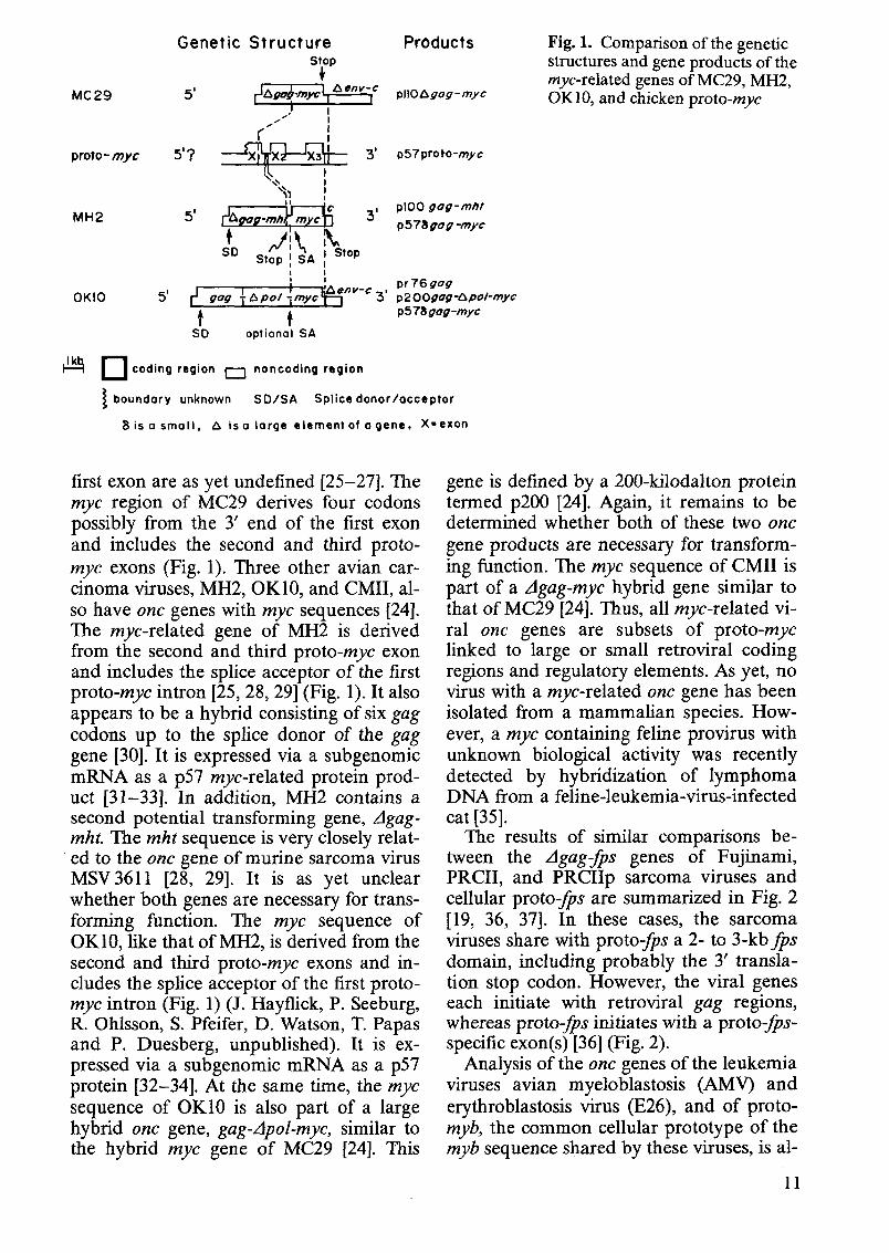
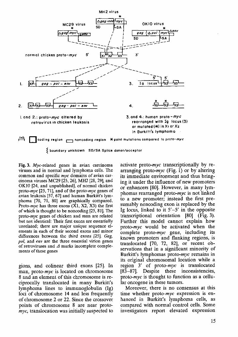
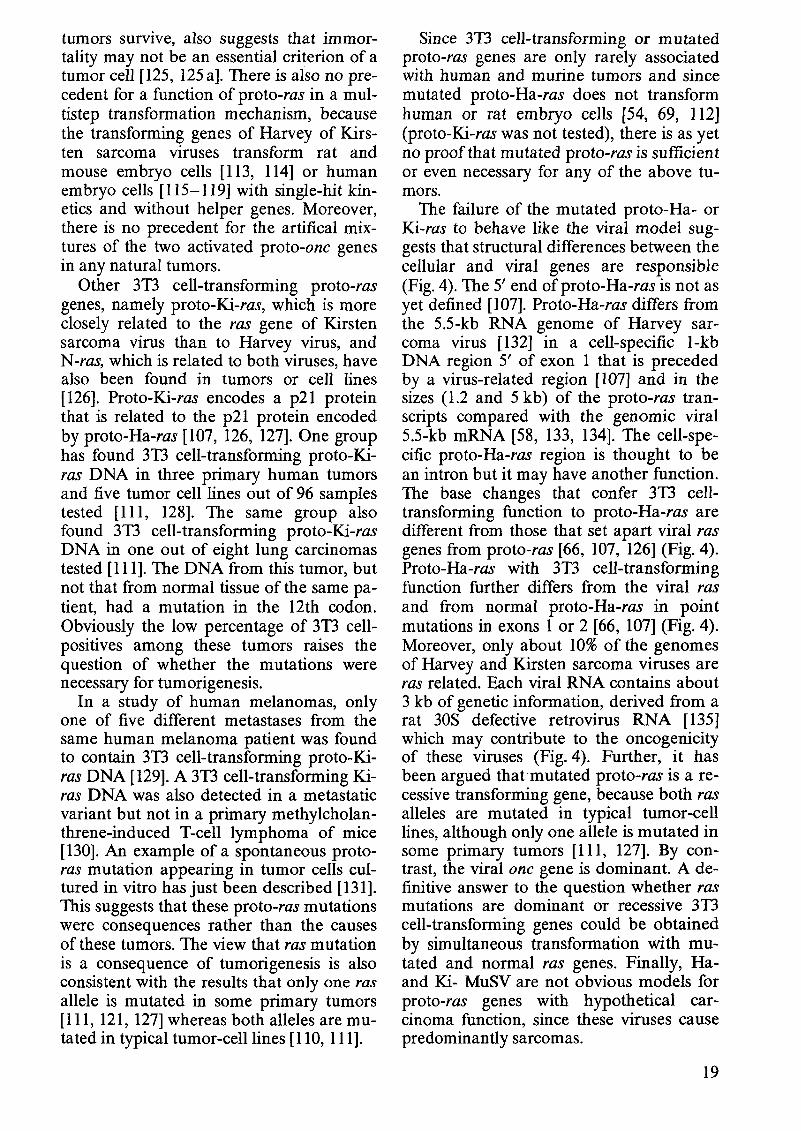
In our laboratories we are studying the structural and functional relationships between viral one genes and corresponding proto-one genes, with particular emphasis on the one genes of the following avian carcinoma, sarcoma, and leukemia viruses. The one gene of avian carcinoma virus MC29 was the first among viral one genes to be diagnosed as a hybrid gene [21, 23] (Fig. 1). About one-half of its information (1.5 kb) is derived from the gag gene of retroviruses; the other half (1.6 kb), termed mye, is derived from the proto-mye gene [22]. The gene is defined by a 110-kilodalton L1gag-mye protein, termed pllO [21, 24]. The proto-mye gene of the chicken has at least three exons. The boundaries of the
MC 29
proto-myc
MH2
OKlO
Genetic Structure Products stop
4
X,' I I
f " I
5'7 3' p57 proto-myc
"\L,\ I 11 !
plOO gag-mht p 5 7 8 g o g -myc
Stop ! SA I I I
I nv-c p r 7 6 g o g 5' ,-J gog fapo/grnyc 3' p 2 oogag-APO/-myc
t 1 ~ 5 7 % gog -myc
i*9 coding region [=1 noncoding region
boundary unknown S D I S A Splice donorlacceptoi
8 is a srnail, A is a iarge elernent of a gene, X-exon
first exon are as yet undefined [25-271. The myc region of MC29 derives four codons possibly from the 3' end of the first exon and includes the second and third proto- myc exons (Fig. 1). Three other avian car- cinoma viruses, MH2, OK10, and CMII, al- so have onc genes with myc sequences [24]. The myc-related gene of MH2 is derived from the second and third proto-myc exon and includes the splice acceptor of the first proto-myc intron [25,28,29] (Fig. 1). It also appears to be a hybrid consisting of six gag codons up to the splice donor of the gag gene [30]. It is expressed via a subgenomic mRNA as a p57 myc-related protein prod- uct [31-331. In addition, MH2 contains a second potential transforming gene, Agag- mht. The mht sequence is very closely relat- ed to the onc gene of murine sarcoma virus MSV 361 1 [28, 291. It is as yet unclear whether both genes are necessary for trans- forming function. The myc sequence of OK10, like that of MH2, is derived from the second and third proto-myc exons and in- cludes the splice acceptor of the first proto- myc intron (Fig. 1) (J. Hayflick, P. Seeburg, R. Ohlsson, S. Pfeifer, D. Watson, T. Papas and P. Duesberg, unpublished). It is ex- pressed via a subgenomic mRNA as a p57 protein [32-341. At the Same time, the myc sequence of OKlO is also Part of a large hybrid onc gene, gag-Apol-myc, similar to the hybrid myc gene of MC29 [24]. This
Fig. 1. Comparison of the genetic structures and gene products of the myc-related genes of MC29, MH2, OK10, and chicken proto-myc
gene is defined by a 200-kilodalton protein terrned p200 [24]. Again, it remains to be detennined whether both of these two onc gene products are necessary for transform- ing function. The myc sequence of CM11 is Part of a Agag-myc hybrid gene similar to that of MC29 [24]. Thus, all myc-related vi- ral onc genes are subsets of proto-myc linked to large or small retroviral coding regions and regulatory elements. As yet, no virus with a myc-related onc gene has been isolated from a mammalian species. How- ever, a myc containing feline provirus with unknown biological activity was recently detected by hybridization of lymphoma DNA from a feline-leukemia-virus-infected cat [35].
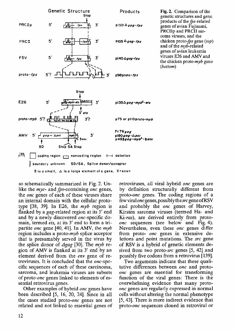
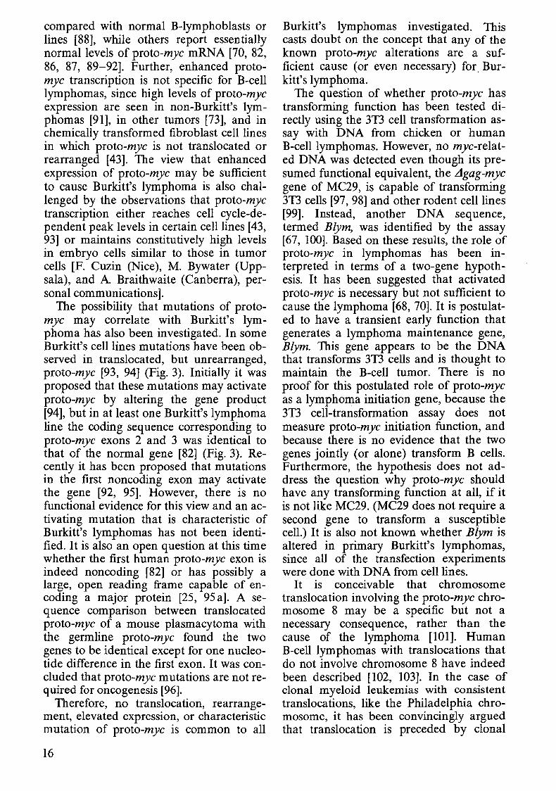
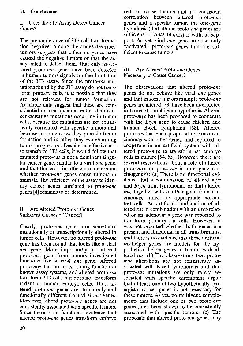
The results of similar comparisons be- tween the Agag-fps genes of Fujinami, PRCII, and PRCIIp sarcoma viruses and cellular proto-fps are summarized in Fig. 2 [19, 36, 371. In these cases, the sarcoma viruses share with proto-fps a 2- to 3-kb fps domain, including probably the 3' transla- tion stop codon. However, the viral genes each initiate with retroviral gag regions, whereas proto-fps initiates with a proto-fps- specific exon(s) [36] (Fig. 2).
Analysis of the onc genes of the leukemia viruses avian myeloblastosis (AMV) and erythroblastosis virus (E26), and of proto- myb, the common cellular prototype of the myb sequence shared by these viruses, is al-
Genetic Structure Stop
• PRcnp 5' P 3'
PRcn 5'
FSV 5'
proto-fps
Stop
+ E26 5' rv~myt-.,s~ 3'
.... -- " t ('.... (3' ets
proto-myb 5'7~ 3'7 .............. -....... I --
AMV 5' gag- Opal 3'
t SO Stop SA Stop
Products
pl506gog-lps
PIOS 6 gog-Ips
pl40l:.gog-lpS
p98proto- Ips
p75 or pllOproto-myb
Pr76gog pl80gog-6pol p488gog-mybA -8env
Fig. 2. Comparison of the genetic structures and gene products of the .IPs-related genes of avian Fujinami, PRCIIp and PRCII sarcoma viruses, and the chicken proto:fPs gene (top) and of the myb-related genes of avian leukemia viruses E26 and AMVand the chicken proto-myb gene (bottom)
~ D coding region 0 noncoding region ~ deletion
} boundary unknown SO/SA. Splice donor/acceptor
8 is a small, I:. is 0 large element of a gene, X"exon
so schematically summarized in Fig. 2. Unlike the mye- and iPs-containing one genes, the one genes of each of these viruses share an internal domain with the cellular prototype [38, 39]. In E26, the myb region is flanked by a gag-related region at its 5' end and by a newly discovered one-specific domain, termed ets, at its 3' end to form a tripartite one gene [40, 41]. In AMY, the myb region includes a proto-myb splice acceptor that is presumably served in the virus by the splice donor of LJgag [30]. The myh region of AMV is flanked at its 3' end by an element derived from the env gene of retroviruses. It is concluded that the one-specific sequences of each of these carcinoma, sarcoma, and leukemia viruses are subsets of proto-one genes linked to elements of essential retrovirus genes.
Other examples of hybrid one genes have been described [5, 16, 20, 24]. Since in all the cases studied proto-one genes are not related and not linked to essential genes of
12
retroviruses, all viral hybrid one genes are by definition structurally different from proto-one genes. The coding regions of a few viral one genes, possiblythesre gene ofRSV and probably the one genes of Harvey, Kirsten sarcoma viruses (termed Ha- and Ki-ras) , are derived entirely from protoone sequences (see below and Fig. 4). Nevertheless, even these one genes differ from proto- one genes in extensive deletions and point mutations. The sre gene of RSV is a hybrid of genetic elements derived from two proto-sre genes [5, 42] and possibly five codons from a retrovirus [139].
Two arguments indicate that these qualitative differences between one and protoone genes are essential for transforming function of the viral genes: There is the overwhelming evidence that many protoone genes are regularly expressed in normal cells without altering the normal phenotype [5,43]. There is more indirect evidence that proto-one sequences cloned in retroviral or
plasmid vectors do not transform normal, diploid cells. For example, phage or plasmid vectors carrying the viral src-related region, but not a complete complement of the major proto-src gene [44-47] or protofos, the precursor of the transforming gene of FBJ (Finkel-Biskis-Jinkins) murine os-teosarcoma virus [48J, or proto-fpslfes, the precursors of avian Fujinami and feline sarcoma viruses [491 (W.-H. Lee and P. H. Duesberg, unpublished), or proto-myc, the precursor of avian MC29 virus (T. Robins, P. Duesberg, and G. Vande Woude, unpublished), do not transform cells in culture. The src-related region of the major protosre gene also fails to transform in a RSV vector [50J. Further, proto-sre and protoHa-ras (the precursor of Ha-MuSV) fail to transform in a reticuloendotheliosis virus vector while the corresponding viral one genes have transforming function [51].
Apparent exceptions are proto-mos and proto-ras which, after ligation to retroviral promoters, transform the preneoplastic NIH 3D cell line [52, 531. The proto-mos and ras regions used in these constructions are essentially the same as those found in Moloney and Harvey sarcoma viruses but are not complete proto-one genes (see below and Fig. 2). Conceivably, the proto-one regions that were not included into these constructions and are not in the viruses might in the cell suppress transforming potential of the complete proto-one genes. Moreover, it will be detailed below that transforming function in 3D cells is not a reliable measure of transforming function in diploid embryo cells or in the animal. Neither the proto-ras nor the proto-mos construction were found to transform diploid embryo cells [54, 55] (G. Vande Woude, personal communication). Thus, normal proto-one genes and viral one genes are related, but are structurally and functionally different. The question is now whether there are conditions under which proto-one genes can cause cancer.
C. The Search for Activation of Proto-onc Genes to Cancer Genes
The only clear, although indirect, proof for activation of proto-one genes to cancer
genes is based on the rare cases in which proto-one genes functioned as accidental parents of retroviral one genes. It has been deduced from structural analyses of retroviral genes and proto-one genes that viral one genes were generated by transduction of specific domains from proto-one genes [5, 20J. Because no significant sequence homology exists between retroviruses and proto-one genes, such transductions must procede via two rare, nonhomologous recombinations [5, 25]5. It is probably for this reason that viral transductions or "activations" are extremely rare, even though all cells contain proto- one genes and many animal species contain retroviruses without one genes. Only 50-100 sporadic cancers from which retroviruses with one genes were isolated have been reported and no experimentally reproducible system of transduction has ever been described [56-58]. Thus, retroviruses with one genes are the causes of rare, natural tumors
. rather than laboratory artifacts. Their role as accidental progenitors of
viral one genes has made proto-one genes the focus of the search for cellular cancer genes. Their possible function in cancer was initially tested in many laboratories in view of a "one gene-one cancer" and more recently in view of a "multigene-one cancer" hypothesis. The one gene-one cancer hypothesis is similar to postulates that activation of inactive cellular oncogenes is sufficient to cause cancer the oncogene hypothesis of Huebner and Todaro [59]. Some investigators have postulated that activation is the result of increased dosage of a given proto-one gene product. This view, termed the quantitative model, received support from early experiments which suggested that the sre gene of RSV or the myc gene of MC29 and the corresponding proto-one genes were equivalents [60-64J. In the meantime, significant structural and functional differences between these genes have been found [5, 43, 44-47, 50] (see above).
5 In addition, it appears that only a few cellular genes are proto-one genes or can function as progenitors of viral one genes since the same proto-one sequences have been found in different isolates [29]
13
Others have suggested that proto-one genes are activated by mutations or rearrangements in the primary DNA sequence [65, 66J. This view is termed the qualitative model [5].
The multigene-one cancer hypothesis postulates that an activated proto-one gene is necessary, but unlike the corresponding viral gene, not sufficient to cause cancer. A quantitatively or qualitatively activated proto-one gene is postulated to function either as initiation or as maintenance gene together with another proto-one gene, in a multistep process [54, 55, 67-73J. This hypothesis fits the view of how virus-negative tumors are thought to arise in general and provides identifiable candidates to test the hypothesis. However, since retroviral one genes have yet to be dissociated into initiation and maintenance functions, this hypothesis is without viral precedent.
Two kinds of assays have been performed to test these hypotheses. One assay correlates transcriptional activation and mutation of proto-one genes with cancer; the other directly measures transforming function of proto-one genes upon transfection into certain recipient cells, typically the preneoplastic mouse NIH 31'3 cell line [4, 54, 55]. Such experiments have most frequently linked cancers with alterations of proto-mye and proto-ras.
I. Is Proto-mye Activation the Cause ofB-Cell Lymphomas?
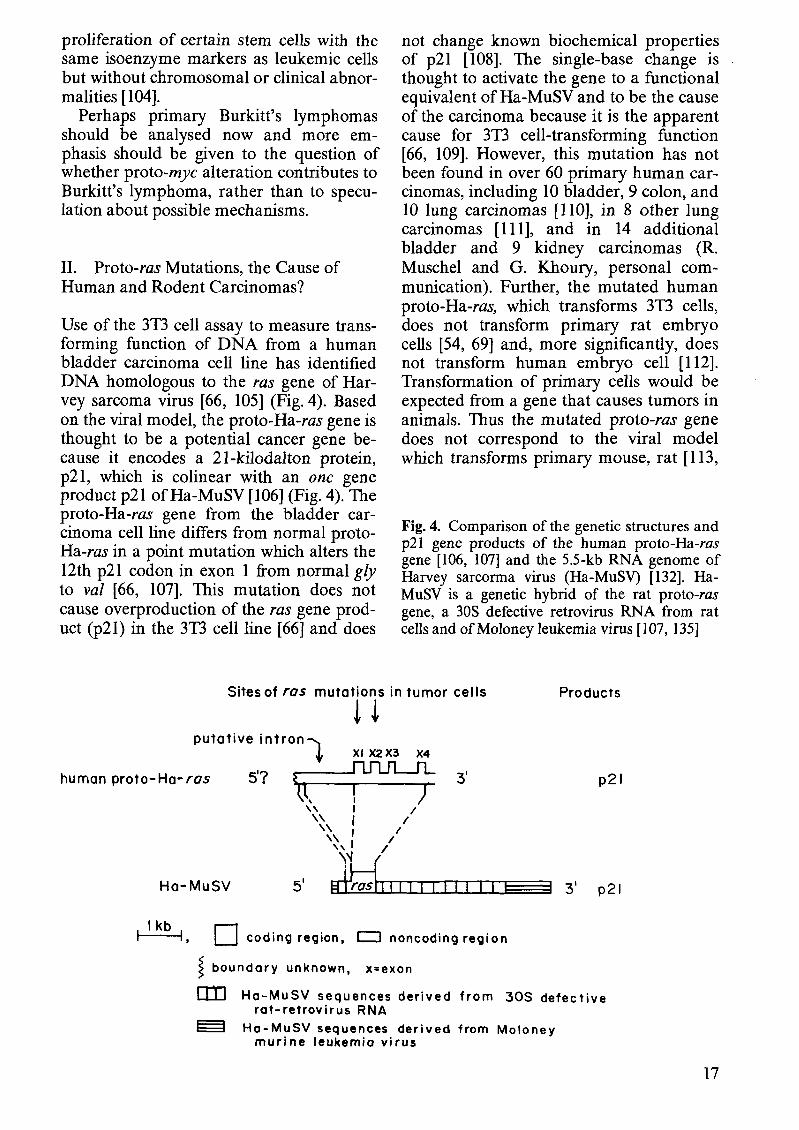
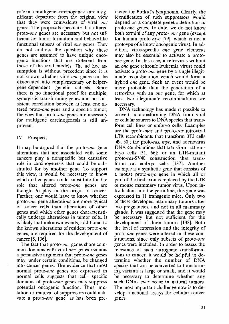
Based on the observation that transcription of the cellular proto-mye is enhanced in retroviral lymphomas of chicken, it has been postulated that transcriptional activation of proto-mye is the cause of B-cell lymphoma [64, 74]. Chicken B-cell lymphoma is a clonal cancer that appears in a small fraction of animals infected by one of the avian leukosis viruses (which have no one genes) after latent periods of over 6 months [58]. The hypothesis, termed downstream promotion, postulates that the gene is activated by the promoter of a retrovirus integrated upstream (Fig. 3) and that activated proto-mye functions like the transforming gene of MC29 [64]. Subsequently, samples were found in which the retrovirus is in-
14
tegrated 3' of proto-mye or 5' in the opposite transcriptional direction. In these cases, the virus is thought to function like an enhancer of proto-myc [74] (Fig. 3).
However, proto-mye differs structurally from the 3-kb L1gag-mye gene of MC29 as diagrammed in Figs. 1 and 3 [25, 26]. Further, it has been argued previously [5] that the hypothesis fails to explain the origin of about 20% of viral lymphomas in which proto-mye is not activated [64J; the discrepancies between the phenotype of the disease and the cancers caused by MC29; the clonality of the tumors, defined by a single integration site of the retrovirus with regard to proto-mye; and the long latent period of the disease. Given about 106 kb of chicken DNA and activation of protomye by retrovirus integration within about 5 kb ofproto-mye [27, 74J, one in 2 X 10⁵ infections should generate the first tumor cell. Since the chicken probably has over 107 uncommitted B cells and many more virus particles, the critical carcinogenic integration event should occur after a short latent period. The tumor should also not be clonal, since integration by retroviruses is not site specific and there could be numerous infections during the latent period of about 6 months. Further, the model has not been confirmed in murine [75, 76], feline [35], and bovine [77] leukemia. Instead, the high percentage of virus-negative feline [35] and bovine [78] lymphomas indicates that a retrovirus is not even necessary for the disease.
Recently, it was suggested that a mutation, rather than a virus, may have activated avian proto-mye because mutations have been observed in viral lymphoma [79]. However, the proto-mye mutations have not been shown to be the cause of the viral lymphoma.
Activation of proto-mye has also been postulated to cause the retrovirus-negative, human Burkitt's lymphomas, and mouse plasmacytomas. In these cases, chromosome translocation has been proposed as a mechanism of activating proto-mye function [70, 71, 80, 81]. The human proto-mye is related to that of the chicken from which carcinoma viruses have been derived (Fig. 3). The two genes have unique first exons, similar second exons with unique re-
MH2 virus *
OKlO virus
normal chicken proto-myc 5'
I. a n d 2.: proto-myc a l t e r e d by re t rov i rus in chicken leukosis
3. and 4.: human proto -myC rearranged with Ig locus (3) or mutated (36) in XI or X2
in Burkitt's lymphoma I K ~ H codinp region noncodinp region X point mutotions sornpored to proto-myc
3 boundory unknown SDISA Splice donorlocceptor
Fig. 3. Myc-related genes in avian carcinoma viruses and in normal and lymphoma cells. The common and specific myc domains of avian car- cinoma viruses MC29 [25, 261, MH2 [28, 291, and OKlO [24, and unpublished], of normal chicken proto-myc [25,71], and of the proto-myc genes of avian leukosis [57, 671 and human Burkitt's lym- phoma [70, 71, 801 are graphically compared. Proto-myc has three exons (XI, X2, X3) the first of which is thought to be noncoding [25, 811. The proto-myc genes of chicken and man are related but not identical: Their first exons are essentially unrelated; there are major unique sequence el- ements in each of their second exons and minor differences between the third exons [25]. Gag, pol, and env are the three essential virion genes of retroviruses and A marks incomplete comple- ments of these genes
gions, and colinear third exons [25] . In man, proto-myc is located on chromosome 8 and an element of this chromosome is re- ciprocally translocated in many Burkitt's lymphoma lines to immunoglobulin (Ig) loci of chromosome 14 and less frequently of chromosome 2 or 22. Since the crossover points of chromosome 8 are near proto- myc, translocation was initially suspected to
activate proto-myc transcriptionally by re- arranging proto-myc (Fig. 1) or by altering its immediate environment and thus bring- ing it under the influence of new promoters or enhancers [80]. However, in many lym- phomas rearranged proto-myc is not linked to a new promoter; instead the first pre- sumably noncoding exon is replaced by the Ig locus, linked to it 5'-5' in the opposite transcriptional orientation [80] (Fig. 3). Further this model cannot explain how proto-myc would be activated when the complete proto-myc gene, including its known promoters and flanking regions, is translocated [70, 72, 821, or recent ob- servations that in a significant minority of Burkitt's lymphomas proto-myc remains in its original chromosomal location while a region 3' of proto-myc is translocated [83-871, Despite these inconsistencies, proto-myc is thought to function as a cellu- lar oncogene in these tumors.
Moreover, there is no Consensus at this time whether proto-myc expression is en- hanced in Burkitt's lymphoma cells, as compared with normal control cells. Some investigators report elevated expression
compared with normal B-lymphoblasts or lines [88], while others report essentially normal levels of proto-myc mRNA [70, 82, 86, 87, 89-92]. Further, enhanced protomyc transcription is not specific for B-cell lymphomas, since high levels of proto-myc expression are seen in non-Burkitt's lymphomas [91], in other tumors [73], and in chemically transformed fibroblast cell lines in which proto-myc is not translocated or rearranged [43]. The view that enhanced expression of proto-myc may be sufficient to cause Burkitt's lymphoma is also challenged by the observations that proto-myc transcription either reaches cell cycle-dependent peak levels in certain cell lines [43, 93] or maintains constitutively high levels in embryo cells similar to those in tumor cells [F. Cuzin (Nice), M. Bywater (Uppsala), and A. Braithwaite (Canberra), personal communications].
The possibility that mutations of protomyc may correlate with Burkitt's lymphoma has also been investigated. In some Burkitt's cell lines mutations have been observed in translocated, but unrearranged, proto-myc [93, 94] (Fig. 3). Initially it was proposed that these mutations may activate proto-myc by altering the gene product [94], but in at least one Burkitt's lymphoma line the coding sequence corresponding to proto-myc exons 2 and 3 was identical to that of the normal gene [82] (Fig. 3). Recently it has been proposed that mutations in the first noncoding exon may activate the gene [92, 95]. However, there is no functional evidence for this view and an activating mutation that is characteristic of Burkitt's lymphomas has not been identified. It is also an open question at this time whether the first human proto-myc exon is indeed noncoding [82] or has possibly a large, open reading frame capable of encoding a major protein [25, 95 a]. A sequence comparison between translocated proto-myc of a mouse plasmacytoma with the germline proto-myc found the two genes to be identical except for one nucleotide difference in the first exon. It was concluded that proto-myc mutations are not required for oncogenesis [96].
Therefore, no translocation, rearrangement, elevated expression, or characteristic mutation of proto-myc is common to all
16
Burkitt's lymphomas investigated. This casts doubt on the concept that any of the known proto-myc alterations are a sufficient cause (or even necessary) for, Burkitt's lymphoma.
The question of whether proto-myc has transforming function has been tested directly using the 3T3 cell transformation assay with DNA from chicken or human B-celllymphomas. However, no myc-related DNA was detected even though its presumed functional equivalent, the L1gag-myc gene of MC29, is capable of transforming 3T3 cells [97, 98] and other rodent cell lines [99]. Instead, another DNA sequence, termed Blym, was identified by the assay [67, 100]. Based on these results, the role of proto-myc in lymphomas has been interpreted in terms of a two-gene hypothesis. It has been suggested that activated proto-myc is necessary but not sufficient to cause the lymphoma [68, 70]. It is postulated to have a transient early function that generates a lymphoma maintenance gene, Blym. This gene appears to be the DNA that transforms 3T3 cells and is thought to maintain the B-cell tumor. There is no proof for this postulated role of proto-myc as a lymphoma initiation gene, because the 3T3 cell-transformation assay does not measure proto-myc initiation function, and because there is no evidence that the two genes jointly (or alone) transform B cells. Furthermore, the hypothesis does not address the question why proto-myc should have any transforming function at all, if it is not like MC29. (MC29 does not require a second gene to transform a susceptible cell.) It is also not known whether Blym is altered in primary Burkitt's lymphomas, since all of the transfection experiments were done with DNA from cell lines.
It is conceivable that chromosome translocation involving the proto-myc chromosome 8 may be a specific but not a necessary consequence, rather than the cause of the lymphoma [101]. Human B-cell lymphomas with translocations that do not involve chromosome 8 have indeed been described [102, 103]. In the case of clonal myeloid leukemias with consistent translocations, like the Philadelphia chromosome, it has been convincingly argued that translocation is preceded by clonal
proliferation of certain stem cells with the same isoenzyme markers as leukemic cells but without chromosomal or clinical abnormalities [104].
Perhaps primary Burkitt's lymphomas should be analysed now and more emphasis should be given to the question of whether proto-mye alteration contributes to Burkitt's lymphoma, rather than to speculation about possible mechanisms.
II. Proto-ras Mutations, the Cause of Human and Rodent Carcinomas?
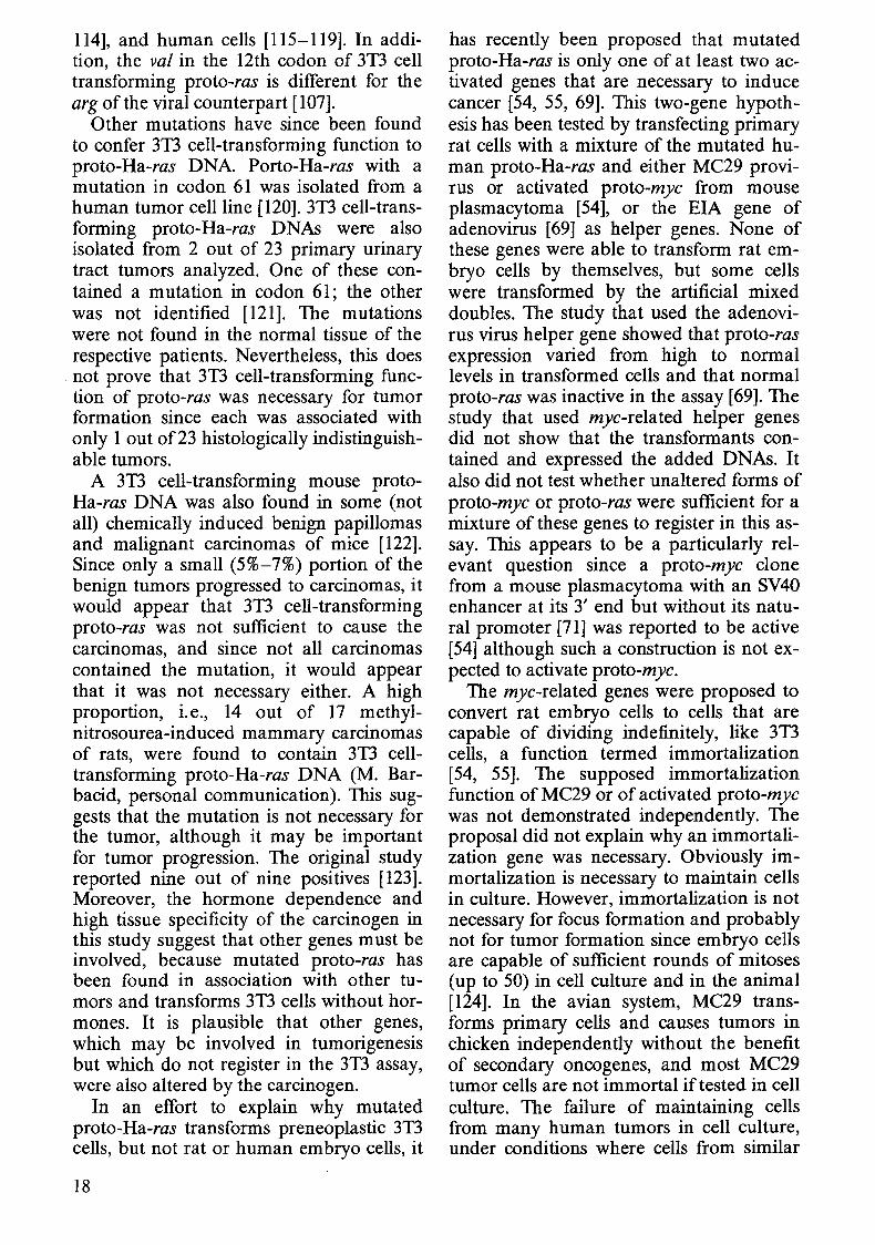
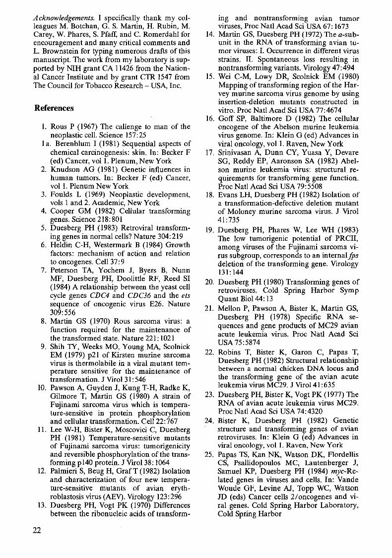
Use of the 31'3 cell assay to measure transforming function of DNA from a human bladder carcinoma cell line has identified DNA homologous to the ras gene of Harvey sarcoma virus [66, 105] (Fig. 4). Based on the viral model, the proto-Ha-ras gene is thought to be a potential cancer gene because it encodes a 2l-kilodalton protein, p2l, which is colin ear with an one gene product p21 of Ha-MuSV [106] (Fig. 4). The proto-Ha-ras gene from the bladder carcinoma cell line differs from normal protoHa-ras in a point mutation which alters the 12th p2l codon in exon 1 from normal gly to val [66, 107]. This mutation does not cause overproduction of the ras gene product (p21) in the 31'3 cell line [66] and does
not change known biochemical properties of p2l [108]. The single-base change is thought to activate the gene to a functional equivalent of Ha-MuSV and to be the cause of the carcinoma because it is the apparent cause for 31'3 cell-transforming function [66, 109]. However, this mutation has not been found in over 60 primary human carcinomas, including 10 bladder, 9 colon, and 10 lung carcinomas [110], in 8 other lung carcinomas [111], and in 14 additional bladder and 9 kidney carcinomas (R. Muschel and G. Khoury, personal communication). Further, the mutated human proto-Ha-ras, which transforms 31'3 cells, does not transform primary rat embryo cells [54, 69] and, more significantly, does not transform human embryo cell [112]. Transformation of primary cells would be expected from a gene that causes tumors in animals. Thus the mutated proto-ras gene does not correspond to the viral model which transforms primary mouse, rat [113,
Fig. 4. Comparison of the genetic structures and p21 gene products of the human proto~Ha~ras gene [106, 107] and the 5.5-kb RNA genome of Harvey sarcorma virus (Ha~MuSV) [132]. HaMuSV is a genetic hybrid of the rat proto-ras gene, a 30S defective retrovirus RNA from rat cells and of Moloney leukemia virus (107, 135]
Sites of ras mutations in tumor cells Products
! ~ putative i ntron""'l
'" XI X2X3 X4
human prola-Ha-ros 5'? ~ ~ 3' ~\ 1
Ha-MuSV
, 'kb I, 0
'" I '~, I I \,' / " , /
5' Josflll I I I I I I I I I
coding region, 0 noncoding region
~ boundary unknown, x-exon
p21
3' p21
[!]J Ha-Mu5V sequences derived from 305 defective rat-retrovirus RNA
E§ Ha - Mu5V sequences derived from Moloney muri ne leukemia vi rus
17
114], and human cells [115-119]. In addition, the val in the 12th codon of 3T3 cell transforming proto-ras is different for the arg of the viral counterpart [107].
Other mutations have since been found to confer 3T3 cell-transforming function to proto-Ha-ras DNA. Porto-Ha-ras with a mutation in codon 61 was isolated from a human tumor cell line [120]. 3T3 cell-transforming proto-Ha-ras DNAs were also isolated from 2 out of 23 primary urinary tract tumors analyzed. One of these contained a mutation in codon 61; the other was not identified [121]. The mutations were not found in the normal tissue of the respective patients. Nevertheless, this does not prove that 3T3 cell-transforming function of proto-ras was necessary for tumor formation since each was associated with only lout of 23 histologically in distinguish -able tumors.
A 3T3 cell-transforming mouse protoHa-ras DNA was also found in some (not all) chemically induced benign papillomas and malignant carcinomas of mice [122]. Since only a small (5%-7%) portion of the benign tumors progressed to carcinomas, it would appear that 3T3 cell-transforming proto-ras was not sufficient to cause the carcinomas, and since not all carcinomas contained the mutation, it would appear that it was not necessary either. A high proportion, i.e., 14 out of 17 methylnitrosourea-induced mammary carcinomas of rats, were found to contain 3T3 celltransforming proto-Ha-ras DNA (M. Barbacid, personal communication). This suggests that the mutation is not necessary for the tumor, although it may be important for tumor progression. The original study reported nine out of nine positives [123]. Moreover, the hormone dependence and high tissue specificity of the carcinogen in this study suggest that other genes must be involved, because mutated proto-ras has been found in association with other tumors and transforms 3T3 cells without hormones. It is plausible that other genes, which may be involved in tumorigenesis but which do not register in the 3T3 assay, were also altered by the carcinogen.
In an effort to explain why mutated proto-Ha-ras transforms preneoplastic 3T3 cells, but not rat or human embryo cells, it
18
has recently been proposed that mutated proto-Ha-ras is only one of at least two activated genes that are necessary to induce cancer [54, 55, 69]. This two-gene hypothesis has been tested by transfecting primary rat cells with a mixture of the mutated human proto-Ha-ras and either MC29 provirus or activated proto-myc from mouse plasmacytoma [54], or the EIA gene of adenovirus [69] as helper genes. None of these genes were able to transform rat embryo cells by themselves, but some cells were transformed by the artificial mixed doubles. The study that used the adenovirus virus helper gene showed that proto-ras expression varied from high to normal levels in transformed cells and that normal proto-ras was inactive in the assay [69]. The study that used myc-related helper genes did not show that the transformants contained and expressed the added DNAs. It also did not test whether unaltered forms of proto-myc or proto-ras were sufficient for a mixture of these genes to register in this assay. This appears to be a particularly relevant question since a proto-myc clone from a mouse plasmacytoma with an SV40 enhancer at its 3' end but without its natural promoter [71] was reported to be active [54] although such a construction is not expected to activate proto-myc.
The myc-related genes were proposed to convert rat embryo cells to cells that are capable of dividing indefinitely, like 3T3 cells, a function termed immortalization [54, 55]. The supposed immortalization function of MC29 or of activated proto-myc was not demonstrated independently. The proposal did not explain why an immortalization gene was necessary. Obviously immortalization is necessary to maintain cells in culture. However, immortalization is not necessary for focus formation and probably not for tumor formation since embryo cells are capable of sufficient rounds of mitoses (up to 50) in cell culture and in the animal [124]. In the avian system, MC29 transforms primary cells and causes tumors in chicken independently without the benefit of secondary oncogenes, and most MC29 tumor cells are not immortal if tested in cell culture. The failure of maintaining cells from many human tumors in cell culture, under conditions where cells from similar
tumors survive, also suggests that immortality may not be an essential criterion of a tumor cell [125, 125 a1. There is also no precedent for a function of proto-ras in a multistep transformation mechanism, because the transforming genes of Harvey of Kirsten sarcoma viruses transform rat and mouse embryo cells [113, 1141 or human embryo cells [115-1191 with single-hit kinetics and without helper genes. Moreover, there is no precedent for the artifical mixtures of the two activated proto-one genes in any natural tumors.
Other 3T3 cell-transforming proto-ras genes, namely proto-Ki-ras, which is more closely related to the ras gene of Kirsten sarcoma virus than to Harvey virus, and N-ras, which is related to both viruses, have also been found in tumors or cell lines [126]. Proto-Ki-ras encodes a p21 protein that is related to the p21 protein encoded by proto-Ha-ras [107, 126, 127]. One group has found 3T3 cell-transforming proto-Kiras DNA in three primary human tumors and five tumor cell lines out of 96 samples tested [111, 128]. The same group also found 3T3 cell-transforming proto-Ki-ras DNA in one out of eight lung carcinomas tested [111]. The DNA from this tumor, but not that from normal tissue of the same patient, had a mutation in the 12th codon. Obviously the low percentage of 3T3 cellpositives among these tumors raises the question of whether the mutations were necessary for tumorigenesis.
In a study of human melanomas, only one of five different metastases from the same human melanoma patient was found to contain 3T3 cell-transforming proto-Kiras DNA [129]. A 3T3 cell-transforming Kiras DNA was also detected in a metastatic variant but not in a primary methy1cholanthrene-induced T-cell lymphoma of mice [130]. An example of a spontaneous protoras mutation appearing in tumor cells cultured in vitro has just been described [131]. This suggests that these proto-ras mutations were consequences rather than the causes of these tumors. The view that ras mutation is a consequence of tumorigenesis is also consistent with the results that only one ras allele is mutated in some primary tumors [Ill, 121, 127] whereas both alleles are mutated in typical tumor-cell lines [110, III].
Since 31'3 cell-transforming or mutated proto-ras genes are only rarely associated with human and murine tumors and since mutated proto-Ha-ras does not transform human or rat embryo cells [54, 69, 112] (proto-Ki-ras was not tested), there is as yet no proof that mutated proto-ras is sufficient or even necessary for any of the above tumors.
The failure of the mutated proto-Ha- or Ki-ras to behave like the viral model suggests that structural differences between the cellular and viral genes are responsible (Fig. 4). The 5' end ofproto-Ha-ras is not as yet defined [107]. Proto-Ha-ras differs from the 5.5-kb RNA genome of Harvey sarcoma virus [132] in a cell-specific I-kb DNA region 5' of exon I that is preceded by a virus-related region [107] and in the sizes (1.2 and 5 kb) of the proto-ras transcripts compared with the genomic viral 5.5-kb mRNA [58, 133, 134]. The cell-specific proto-Ha-ras region is thought to be an intron but it may have another function. The base changes that confer 3T3 celltransforming function to proto-Ha-ras are different from those that set apart viral ras genes from proto-ras [66, 107, 126] (Fig. 4). Proto-Ha-ras with 31'3 cell-transforming function further differs from the viral ras and from normal proto-Ha-ras in point mutations in exons I or 2 [66, 107] (Fig. 4). Moreover, only about 10% of the genomes of Harvey and Kirsten sarcoma viruses are ras related. Each viral RNA contains about 3 kb of genetic information, derived from a rat 30S defective retrovirus RNA [135] which may contribute to the oncogenicity of these viruses (Fig. 4). Further, it has been argued that mutated proto-ras is a recessive transforming gene, because both ras alleles are mutated in typical tumor-cell lines, although only one allele is mutated in some primary tumors [111, 127]. By contrast, the viral one gene is dominant. A definitive answer to the question whether ras mutations are dominant or recessive 31'3 cell-transforming genes could be obtained by simultaneous transformation with mutated and normal ras genes. Finally, Haand Ki- MuSV are not obvious models for proto-ras genes with hypothetical carcinoma function, since these viruses cause predominantly sarcomas.
19
D. Conclusions
I. Does the 3T3 Assay Detect Cancer Genes?
The preponderance of 3T3 cell-transformation negatives among the above-described tumors suggests that either no genes have caused the negative tumors or that the assay failed to detect them. That only ras-related proto-one genes have been detected in human tumors signals another limitation of the 3T3 assay. Since the proto-ras mutations found by the 3T3 assay do not transform primary cells, it is possible that they are not relevant for tumor formation. Available data suggest that these are coincidential or consequential rather than cancer causative mutations occurring in tumor cells, because the mutations are not consistently correlated with specific tumors and because in some cases they precede tumor formation and in other they evolve during tumor progression. Despite its effectiveness to transform 3T3 cells, it would follow that mutated proto-ras is not a dominant singular cancer gene, similar to a viral one gene, and that the test is insufficient to determine whether proto-one genes cause tumors in animals. The efficiency of the assay to identify cancer genes unrelated to proto-one genes [4] remains to be determined.
II. Are Altered Proto-one Genes Sufficient Causes of Cancer?
Clearly, proto-one genes are sometimes mutationally or transcriptionally altered in tumor cells. However, no altered proto-one gene has been found that looks like a viral one gene. More importantly, no altered proto-one gene from tumors investigated functions like a viral one gene. Altered proto-mye has no transforming function in known assay systems, and altered proto-ras transform 3T3 cells but does not transform rodent or human embryo cells. Thus, altered proto-one genes are structurally and functionally different from viral one genes. Moreover, alterd proto-one genes are not consistently associated with specific tumors. Since there is no functional evidence that altered proto-one genes transform embryo
20
cells or cause tumors and no consistent correlation between altered proto-one genes and a specific tumor, the one-gene hypothesis (that altered proto-one genes are sufficient to cause tumors) is without support. As yet, viral one genes are the only "activated" proto-one genes that are sufficient to cause tumors.
III. Are Altered Proto-one Genes Necessary to Cause Cancer?
The observations that altered proto-one genes do not behave like viral one genes and that in some tumors multiple proto-one genes are altered [73] have been interpreted in terms of a multi gene hypothesis. Altered proto-mye has been proposed to cooperate with the Blym gene to cause chicken and human B-cell lymphoma [68]. Altered proto-ras has been proposed to cause carcinomas with other genes, and reported to cooperate in an artificial system with altered proto-mye to transform rat embryo cells in culture [54, 55]. However, there are several reservations about a role of altered proto-mye or proto-ras in multigene carcinogenesis: (a) There is no functional evidence that a combination of altered mye and Blym from lymphomas or that altered ras, together with another gene from carcinomas, transforms appropriate normal test cells. An artificial combination of altered ras in combination with an mye-related or an adenovirus gene was reported to transform primary rat cells. However, it was not reported whether both genes are present and functional in all transformants, and there is no evidence that these artificial ras-helper genes are models for the hypothetical helper genes in tumors with altered ras. (b) The observations that protomye alterations are not consistently associated with B-cell lymphomas and that proto-ras mutations are only rarely associated with specific carcinomas argue that at least one of two hypothetically synergistic cancer genes is not necessary for these tumors. As yet, no multigene complements that include one or two proto-one genes have been shown to be consistently associated with specific tumors. (c) The proposals that altered proto-one genes play
role in a multi gene carcinogenesis are a significant departure from the original view that they were equivalents of viral one genes. The proposals speculate that altered proto-one genes are necessary but not sufficient for tumor formation and behave like functional subsets of viral one genes. They do not address the question why these genes are assumed to have unique oncogenic functions that are different from those of the viral models. The ad hoc assumption is without precedent since it is not known whether viral one genes can be dissociated into complementary or helpergene-dependent genetic subsets. Since there is no functional proof for multiple, synergistic transforming genes and no consistent correlation between at least one altered proto-one gene and a specific tumor, the view that proto-one genes are necessary for multigene carcinogenesis is still unproven.
IV. Prospects
It may be argued that the proto-one gene alterations that are associated with some cancers play a nonspecific but causative role in carcinogenesis that could be substituted for by another gene. To support this view, it would be necessary to know which other genes could substitute for the role that altered proto-one genes are thought to play in the origin of cancer. Further, one would have to know whether proto-one gene alterations are more typical of cancer cells than alterations of other genes and which other genes characteristically undergo alterations in tumor cells. It is likely that unknown events, additional to the known alterations of resident proto-one genes, are required for the development of cancer [5, 136].
The fact that proto-one genes share common domains with viral one genes remains a persuasive argument that proto-one genes may, under certain conditions, be changed into cancer genes. The evidence that most normal proto-one genes are expressed in normal cells suggests that cell- specific domains of proto-one genes may suppress potential oncogenic function. Thus, mutation or removal of suppressors could activate a proto-one gene, as has been pre-
dicted for Burkitt's lymphoma. Clearly, the identification of such suppressors would depend on a complete genetic definition of proto-one genes. To date, we do not know both termini of any proto- one gene (except for human proto-mye [79], which is not a protoype of a know oncogenic virus). In addition, virus-specific one gene elements may also be essential to activate a protoone gene. In this case, a retrovirus without an one gene (chronic leukemia virus) could activate a proto-one gene by a single illegitimate recombination which would form a hybrid one gene. Such an event would be more probable than the generation of a retrovirus with an one gene, for which at least two illegitimate recombinations are necessary.
DNA technology has made it possible to convert nontransforming DNA from viral or cellular sources to DNA species that transform cell lines or embryo cells. Examples are the proto-mos and proto-ras retroviral L 1R recombinants that transform 3D cells [49, 50]; the proto-ras, mye, and adenovirus DNA combinations that transform rat embryo cells [51, 66]; or an L1R-mutant proto-ras-SV40 construction that transforms rat embryo cells [137]. Another example is a synthetic gene that consists of a mouse proto-mye gene in which all or part of the first exon is replaced by the L 1R of mouse mammary tumor virus. Upon introduction into the germ line, this gene was expressed in 11 transgenic mice. Only two of these developed mammary tumors after two pregnancies, and not in all mammary glands. It was suggested that the gene may be necessary but not sufficient for the development of these tumors [138]. Both the level of expression and the integrity of proto-one genes were altered in these constructions, since only subsets of proto-one genes were included. In order to assess the relevance of such iatrogenic transformations to cancer, it would be helpful to determine whether the number of DNA species that can be converted to transforming variants is large or small, and it would be necessary to determine whether any such DNAs ever occur in natural tumors. The most important challenge now is to develop functional assays for cellular cancer genes.
21
Acknowledgements. I specifically thank my colleagues M. Botchan, G. S. Martin, H. Rubin, M. Carey, W. Phares, S. Pfaff, and C. Romerdahl for encouragement and many critical comments and L. Brownstein for typing numerous drafts of this manuscript. The work from my laboratory is supported by NIH grant CA 11426 from the National Cancer Institute and by grant CTR 1547 from The Council for Tobacco Research - USA, Inc.
References
1. Rous P (1967) The callenge to man of the neoplastic cell. Science 157: 25
1 a. Berenblum I (1981) Sequential aspects of chemical carcinogenesis: skin. In: Becker F (ed) Cancer, vol I. Plenum, New York
2. Knudson AG (1981) Genetic influences in human tumors. In: Becker F (ed) Cancer, vol 1. Plenum New York
3. Foulds L (1969) Neoplastic development, vols 1 and 2. Academic, New York
4. Cooper GM (1982) Cellular transforming genes. Science 218: 801
5. Duesberg PH (1983) Retroviral transforming genes in normal cells? Nature 304:219
6. Heldin C-H, Westermark B (1984) Growth factors: mechanism of action and relation to oncogenes. Cell 37: 9
7. Peterson TA, Yochem J, Byers B, Nunn MF, Duesberg PH, Doolittle RF, Reed SI (1984) A relationship between the yeast cell cycle genes CDC4 and CDC36 and the ets sequence of oncogenic virus E26. Nature 309:556
8. Martin GS (1970) Rous sarcoma virus: a function required for the maintenance of the transformed state. Nature 221: 1021
9. Shih 1Y, Weeks MO, Young MA, Scolnick EM (1979) p21 of Kirsten murine sarcoma virus is thermolabile in a viral mutant temperature sensitive for the maintenance of transformation. J Virol 31: 546
10. Pawson A, Guyden J, Kung T-H, Radke K, Gilmore T, Martin GS (1980) A strain of Fujinami sarcoma virus which is temperature-sensitive in protein phosphorylation and cellular transformation. Cell 22: 767
11. Lee W-H, Bister K, Moscovici C, Duesberg PH (1981) Temperature-sensitive mutants of Fujinami sarcoma virus: tumorigenicity and reversible phosphorylation of the transforming p140 protein. J Virol38: 1064
12. Palmieri S, Beug H, GrafT (1982) Isolation and characterization of four new temperature-sensitive mutants of avian erythroblastosis virus (AEV). Virology 123:296
13. Duesberg PH, Vogt PK (1970) Differences between the ribonucleic acids of transform-
22
ing and nontransforming avian tumor viruses, Proc Natl Acad Sci USA 67: 1673
14. Martin GS, Duesberg PH (1972) The a-subunit in the RNA of transforming avian tumor viruses: I. Occurrence in different virus strains. II. Spontaneous loss resulting in nontransforming variants. Virology 47: 494
15. Wei C-M, Lowy DR, Scolnick EM (1980) Mapping of transforming region of the Harvey murine sarcoma virus genome by using insertion-deletion mutants constructed in vitro. Proc Natl Acad Sci USA 77:4674
16. Goff SP, Baltimore D (1982) The cellular oncogene of the Abelson murine leukemia virus genome. In: Klein G (ed) Advances in viral oncology, vol I. Raven, New York
17. Srinivasan A, Dunn CY, Yuasa Y, Devare SG, Reddy EP, Aaronson SA (1982) Abelson murine leukemia virus: structural requirements for transforming gene function. Proc Nat! Acad Sci USA 79:5508
18. Evans LH, Duesberg PH (1982) Isolation of a transformation-defective deletion mutant of Moloney murine sarcoma virus. J Virol 41 :735
19. Duesberg PH, Phares W, Lee WH (1983) The low tumorigenic potential of PRCll, among viruses of the Fujinami sarcoma virus subgroup, corresponds to an internalfps deletion of the transforming gene. Virology 131: 144
20. Duesberg PH (1980) Transforming genes of retroviruses. Cold Spring Harbor Symp Quant Bioi 44: 13
21. Mellon P, Pawson A, Bister K, Martin GS, Duesberg PH (1978) Specific RNA sequences and gene products of MC29 avian acute leukemia virus. Proc Nat! Acad Sci USA 75:5874
22. Robins T, Bister K, Garon C, Papas T, Duesberg PH (1982) Structural relationship between a normal chicken DNA locus and the transforming gene of the avian acute leukemia virus MC29. J Virol41 :635
23. Duesberg PH, Bister K, Vogt PK (1977) The RNA of avian acute leukemia virus MC29. Proc Nat! Acad Sci USA 74:4320
24. Bister K, Duesberg PH (1982) Genetic structure and transforming genes of avian retroviruses. In: Klein G (ed) Advances in viral oncology, vol I. Raven, New York
25. Papas TS, Kan NK, Watson DK, Flordellis CS, Psallidopoulos MC, Lautenberger J, Samuel KP, Duesberg PH (1984) myc-Related genes in viruses and cells. In: Vande Woude OF, Levine AJ, Topp WC, Watson JD (eds) Cancer cells 2/oncogenes and viral genes. Cold Spring Harbor Laboratory, Cold Spring Harbor
26. Watson DK, Reddy EP, Duesberg PH, Papas TS (1983) Nucleotide sequence analysis of the chicken c-mye gene reveals homologous and unique coding regions by comparison with the transforming gene of avian myelocytomatosis virus MC29, L1gag-mye. Proc Nat! Acad Sci USA 80:2146
27. Shih C-K, Linial M, Goodenow MM, Hayward WS (1984) Nucleotide sequence 5' of the chicken c-mye coding region: localization of a noncoding exon that is absent from mye transcripts in most avian leukosis virus-induced lymphomas. Proc Natl Acad Sci USA 81 : 4697
28. Kan NC, Flordellis CS, Mark GE, Duesberg PH, Papas TS (1984) Nucleotide sequence of avian carcinoma virus MH2: two potential one genes, one related to avian virus MC29, the other to murine sarcoma virus 3611. Proc Natl Acad Sci USA 81:3000
29. Kan NC, Flordellis CS, Mark GE, Duesberg PH, Papas TS (1984) A common one gene sequence transduced by avian carcinoma virus MH2 and by murine sarcoma virus 3611. Science 223: 813
30. Schwartz DE, Tizard R, Gilberg W (1983) Nucleotide sequence of Rous sarcoma virus. Cell 32: 853
31. Pachl C, Biegalke B, Linial M (1983) RNA and protein encoded by MH2 virus: evidence for sub genomic expression of v-mye. 1 Viro145: 133
32. Hann SR, Abrams HD, Rohrschneider LR, Eisenman RN (1983) Proteins encoded by v-myc and c-mye oncogenes: identification and localization in acute leukemia virus transformants and bursal lymphoma cell lines. Cell 34:781
33. Alitalo K, Ramsay G, Bishop 1M, Pfeiffer SO, Colby WW, Levinson AD (1983) Identification of nuclear proteins encoded by viral and cellular mye oncogenes. Nature 306:274
34. Chiswell Dl, Ramsey G, Hayman Ml (1981) Two virus-specific RNA species are present in cells transformed by defective leukemia virus OKlO. 1 Viro140:301
35. Levy LS, Gardner MB, Casey lW (1984) Isolation of a feline leukaemia provirus containing the oncogene mye from a feline lymphosarcoma. Nature 308: 853
36. Seeburg PH, Lee W-H, Nunn MF, Duesberg PH (1984) The 5' ends of the transforming gene of Fujinami sarcoma virus and of the cellular proto:fps gene are not colinear. Virology 133:460
37. Lee W-H, Phares W, Duesberg PH (1983) Structural relationship between chicken DNA locus, proto-fps, and the transforming
gene of Fujinami sarcoma virus (L1gag-fps). Virology 129: 79
38. Rushlow KE, Lautenberger lA, Papas TS, Baluda MA, Perbal B, Chirikjian IG, Reddy EP (1982) Nucleotide sequence of the transforming gene of avian myeloblastosis virus. Science 216: 1421
39. Klempnauer K-H, Gonda TS, Bishop JM (1982) Nucleotide sequence of the retroviral leukemia gene v-myb and its progenitor c-myb: the architecture of a transduced oncogene. Cell 31: 453
40. Nunn MF, See burg PH, Moscovici C, Duesberg PH (1983) Tripartite structure of the avian erythroblastosis virus E26 transforming gene. Nature 306:391
41. Nunn M, Weiher H, Bullock P, Duesberg PH (1984) Avian erythroblastosis virus E26: nucleotide sequence of the tripartite one gene and of the L1R, and analysis of the cellular prototype of the viral ets sequence. Virology 139, 330: 339
42. Takeya T, Hanafusa H (1983) Structure and sequence of the cellular gene homologous to the sre gene of RSV and the mechanism of the generation of the viral transforming gene. Cell 32:881
43. Campisi J, Gray HE, Bardee AB, Dean M, Sonenshein GE (1984) Cell-cycle control of c-mye but not c-ras expression is lost following chemical transformation. Cell 36:241
44. Takeya T, Hanafusa H (1982) DNA sequence of the viral and cellular sre gene of chickens. II. Comparison of the sre genes of two strains of avian sarcoma virus and of the cellular homolog. J Viro144: 12
45. Parker RC, Varmus HE, Bishop JM (1984) Expression of v-src and chicken c-src in rat cells demonstrates qualitative differences between pp60 v-src and pp60 c-sr~ Cell 37: 131
46. Parsons JT, Bryant D, Wilkerson V, Gilmartin G, Parsons SJ (1984) Site-directed mutagenesis ofRous sarcoma virus pp60 src
:
identification of functional domains required for transformation, In: Vande Woude GF, Levine AI, Topp WC, Watson ID (eds) Cancer cells 2/0ncogenes and viral genes. Cold Spring Harbor Laboratory, Cold Spring Harbor
47. Shalloway D, Coussens PM, Yaciuk P (1984) e-src and sre homolog overexpression in mouse cells. In: Vande Woude GF, Levine AI, Topp WC, Watson JD (eds) Cancer cells 21 oncogenes and viral genes. Cold Spring Harbor Laboratory, Cold Spring Harbor
48. Miller AD, Curran T, Verma 1M (1984) c:fos Protein can induce cellular transfor-
23
mation: a novel mechanism of activation of a cellular oncogene. Cell 36: 51
49. Sodroski JG, Goh WC, Haseltine WA (1984) Transforming potential of a human protooncogene (c-fpslfes) locus. Proc Natl Acad Sci USA 81: 3039
50. Iba H, Takeya T, Cross FR, Hanafusa T, Hanafusa H (1984) Rous sarcoma virus variants that carry the cellular sre gene instead of the viral sre gene cannot transform chicken embryo fibroblasts. Proc Natl Acad Sci USA 81:4424
51. Wilhelmsen KC, Tarpley WG, Temin HM (1984) Identification of some of the parameters governing transformation by oncogenes in retroviruses. In: Vande Woude GF, Levine AJ, Topp WC, Watson JD (eds) Cancer cells 2/0ncogenes and viral genes. Cold Spring Harbor Laboratory, Cold Spring Harbor
52. Blair DG, Oskarsson M, Wood TG, McClements WC, Fischinger PJ, Vande Woude GF (1981) Activation of the transforming potential of a normal cell sequence: a molecular model for oncogenesis. Science 212:941
53. Chang EH, Furth ME, Scolnick EM, Lowy DR (1982) Tumorigenic transformation of mammalian cells induced by a normal human gene homologous to the oncogene of Harvey murine sarcoma virus. Nature 297:479
54. Land H, Parada LF, Weinberg RA (1983) Tumorigenic conversion of primary embryo fibroblasts requires at least two cooperating oncogenes. Nature 304: 596
55. Land H, Parada LF, Weinberg RA (1983) Cellular oncogenes and multistep carcinogenesis. Science 222:771
56. Gross L (1970) Oncogenic viruses. Pergamon, New York
57. Tooze J (ed) (1973) The molecular biOlogy of tumour viruses. Cold Spring Harbor Laboratory, Cold Spring Harbor
58. Weiss RA, Teich NM, Varmus H, Coffin JM (eds) (1982) Molecular biology of tumor viruses: RNA tumor viruses. Cold Spring Harbor Laboratory, Cold Spring Harbor
59. Huebner RJ, Todaro GJ (1969) Oncogenes of RNA tumor viruses as determinants of cancer. Proc Natl Acad Sci USA 64: 1087
60. Bishop JM, Courtneidge SA, Levinson AD, Oppermann H, Quintrell N, Sheiness DK, Weiss SR, Varmus HE (1980) Origin and function of avian retrovirus transforming genes. Cold Spring Harbor Symp Quant Bioi 44:919
61. Bishop JM (1981) Enemies within: the genesis of retrovirus oncogenes. Cell 23:5
24
62. Wang L-H, Snyder P, Hanafusa T, Moscovici C, Hanafusa H (1980) Comparative analysis of cellular and viral sequences related to sarcomagenic cell transformation. Cold Spring Harbor Symp Quant Bioi 44:766
63. Karess RE, Hayward WS, Hanafusa H (1980) Transforming proteins encoded by the cellular information of recovered avian sarcoma viruses. Cold Spring Harbor Symp Quant Bioi 44: 765
64. Hayward WS, Nee! BG, Astrin SM (1981) Activation of a cellular one gene by promoter insertion in AL V-induced lymphoid leukosis. Nature 290:475
65. Klein G (1981) The role of gene dosage and genetic transposition in carcinogenesis. Nature 294: 313
66. Tabin CJ, Bradley SM, Bargmann CI, Weinberg RA, Papageorge AG, Scolnick EM, Dhar R, Lowy DR, Chang EH (1982) Mechanism of activation of a human oncogene. Nature 300: 143
67. Cooper GM, Neiman PE (1981) Two distinct candidate transforming genes of lymphoid leukosis virus-induced neoplasms. Nature 292: 857
68. Diamond A, Cooper GM, Ritz J, Lane M-A (1983) Identification and molecular cloning of the human Blym transforming gene activated in Burkitt's lymphomas. Nature 305:112
69. Ruley HE (1983) Adenovirus early region I A enables viral and cellular transforming genes to transform primary cells in culture. Nature 304: 602
70. Leder P, Battey J, Lenoir G, Moudling C, Murphy W, Potter H, Stewart T, Taub R (1983) Translocations among antibody genes in human cancer. Science 222:765
71. Adams JM, Gerondakis S, Webb E, Carcoran LM, Cory S (1983) Cellular mye oncogene is altered by chromosome translocation to an immunoglobulin locus in murine plasmacytoma and is rearranged similarly in human Burkitt lymphomas. Proc Natl Acad Sci USA 80: 1982
72. Klein G, Klein E (1984) Oncogene activation and tumor progression. Carcinogenesis 5:429
73. Slam on DJ, deKernion JB, Verma 1M, Cline MJ (1984) Expression of cellular oncogenes in human malignancies. Science 224:256
74. Payne GS, Bishop JM, Varmus HE (1982) Multiple arrangements of viral DNA and an activated host oncogene in bursal lymphomas. Nature 295: 209
75. Tsichlis PN, Strauss PG, Hu LF (1983) A common region for proviral DNA integra-
tion in MoMuLV-induced rat thymic lymphomas. Nature 302:445
76. Yoshimura FK, Levine KL (1983) AKR thymic lymphomas involving mink cell focus-inducing leukemia viruses have a common region of provirus integration. J Virol 45:576
77. Kettmann R, Deschamps J, Cleuter Y, Couez D, Burny A, Marbaix G (1982) Leukemogenesis by bovine leukemia virus: proviral DNA integration and lack of RNA expression of viral long terminal repeat and 3' proximate cellular sequences. Proc Natl Acad Sci USA 79: 2465
78. Miller JM, Miller LD, Olson C, Gillette KS (1969) Virus-like particles in phytohemagglutinin-stimulated lymphocyte cultures with reference to bovine lymphosarcoma. J Natl Cancer Inst 43: 1297
79. Westaway D, Payne G, Varmus HE (1984) Proviral deletions and oncogene base-substitutions in insertionally mutagenized c-mye alleles may contribute to the progression of avian bursal tumors. Proc Natl Acad Sci USA 81: 843
80. Klein G (1983) Specific chromosomal translocations and the genesis of B-cell-derived tumors in mice and men. Cell 32: 311-
81. Rowley JD (1983) Human oncogene locations and chromosome aberrations. Nature 301: 290
82. Battey J, Moulding C, Taub R, Murphy W, Stewart T, Potter H, Lenoir G, Leder P (1983) The human c-mye oncogene: structural consequences of translocation into the IgH locus in Burkitt lymphoma. Cell 34:779
83. Gelmann E, Psallidopoulos MC, Papas TS, Dalla-Favera R (1983) Identification of reciprocal translocation sites within the c-mye oncogene and immunoglobulin # locus in a Burkitt lymphoma. Nature 306:799
84. Croce CM, Thierfelder W, Erikson J, Nishikura K, Finan J, Lenoir GM, Nowell PC (1983) Transcriptional activation of an unrearranged and untranslocated c-mye oncogene by translocation of a Cit locus in Burkitt lymphoma cells. Proc Natl Acad Sci USA 80:6922
85. Erikson J, Ar-Rushidi A, Drwinga HL, Nowell PC, Croce CM (1983) Transcriptional activation of the translocated c-mye oncogene in Burkitt lymphoma. Proc Natl Acad Sci USA 80: 820
86. Hollis GF, Mitchell KF, Battey J, Potter H, Taub R, Lenoir GM, Leder P (1984) A variant translocation places the it immunoglobulin genes 3' to the c-mye oncogene in Burkitt's lymphoma. Nature 307:752
87. Davis M, Malcolm S, Rabbitts rn (1984) Chromosome translocation can occur on either side of the c-mye oncogene in Burkitt lymphoma cells. Nature 308:286
88. Erikson J, Nishikura K, Ar-Rushdi A, Finan J, Emanuel B, Lenoir G, Nowell PC, Croce CM (1983) Translocation of an immunoglobulin '" locus to a region 3' of an unrearranged c-myc oncogene enhances c-mye transcription. Proc Natl Acad Sci USA 80:7581
89. Westin EH, Wong-Staal F, Gelmann EP, Dalla Favera R, Papas TS, Lautenberger JA, Eva A, Reddy EP, Tronick SR, Aaronson SA, Gallo RC (1982) Expression of cellular homologues of retroviral one genes in human hematopoietic cells. Proc Natl Acad Sci USA 79:2490
90. Maguire RT, Robins TS, Thorgersson SS, Heilman CA (1983) Expression of cellular mye and mos genes in undifferentiated B-cell lymphomas of Burkitt and non-Burkitt types. Proc Natl Acad Sci USA 80: 1947
91. Hamlyn PH, Rabbitts rn (1983) Translocation joins c-mye and immunoglobulin yl genes in a Burkitt lymphoma revealing a third ex on in the c-myc oncogene. Nature 304: 135
92. Taub R, Moulding C, Battey J, Murphy W, Vasicek T, Lenoir GM, Leder P (1984) Activation and somatic mutation of the translocated c-mye gene in Burkitt lymhoma cells. Cell 36: 339
93. Kelly K, Cochran BH, Stiles CD, Leder P (1983) Cell-specific regulation of the c-myc gene by lymphocyte mitogens and platelet derived growth factor. Cell 35: 603
94. Rabbitts rn, Hamlyn PH, Baer R (1983) Altered nucleotide sequences of a translocated c-mye gene in Burkitt lymphoma. Nature 306:760
95. Rabbitts ill, Forster A, Hamlyn P, Baer R (1984) Effect of somatic mutation within translocated c-myc genes in Burkitt's lymphoma. Nature 309: 593
95 a. Gazin C, Dupont de Dinechin S, Hampe A, Masson J-M, Martin P, Stehelin D, Galibert F (1984) Nucleotide sequence of the human c-myc locus: provocative open reading frame within the first exon. EMBO, J 3:383
96. Stanton LW, Fahrlander PD, Tesser PM, Marcu KB (1984) Nucleotide sequence comparison of normal and translocated murine m-myc genes. Nature 310:423
97. Copeland NG, Cooper GM (1980) Transfection by DNAs of avian erythroblastosis virus and avian myelocytomatosis virus strain MC29. J Viro133: 1199
25
98. Lautenberger JA, Schulz RA, Garon CF, Tsichlis PH, Papas TS (1981) Molecular cloning of avian myeloblastosis virus (MC29) transforming sequences. Proc Natl Acad Sci USA 78: 1518
99. Quade K (1979) Transformation of mammalian cells by avian myelocytomatosis virus and avian erythroblastosis virus. Virology 98:461
100. Goubin G, Goldman DS, Luce J, Neiman PE, Cooper GM (1983) Molecular cloning and nucleotide sequence of a transforming gene detected by transfection of chicken B-celllymphoma DNA. Nature 302: 114
101. Rubin H (1984) Chromosome aberrations and oncogenes: cause or consequence in cancer. Nature 309: 518
102. Erikson J, Finan J, Tsujimoto Y, Nowell PC, Croce C (1984) The chromosome 14 breakpoint in neoplastic B cells with the t (11; 14) translocation involves the immunoglobulin heavy chain locus. Proc Natl Acad Sci USA 81 :4144
103. Yunis 11, Ok en MD, Kaplan ME, Ensurd KM, Howe RR, Theologides A (1982) Distinctive chromosomal abnormalities in histologic subtypes of non-Hodgkin's lymphoma. N Engl J Med 307: 1231
104. Fialkow RJ, Singer JW (1984) Tracing development and cell lineages in human hemopoietic neoplasia. In: Proceedings of the Dahlem Workshop on Leukemia. Springer, Berlin Heidelberg New York Tokyo (in press)
105. Der JC, Krontiris TG, Cooper GM (1982) Transforming genes of human bladder and lung carcinoma cell lines are homologous to the ras genes of Harvey and Kirsten sarcoma viruses. Proc Natl Acad Sci USA 79:3637
106. Ellis RW, Lowy DR, Scolnick EM (1982) The viral and cellular p21 (ras) gene family. In: Klein G (ed) Advances in viral oncology, vol 1. Raven, New York
107. Capon DJ, Chen EY, Levinson AD, Seeburg PH, Goeddel DV (1983) Complete nucleotide sequences of the T24 human bladder carcinoma oncogene and its normal homologue. Nature 302: 33
108. Finkel T, Channing JD, Cooper GM (1984) Activation of ras genes in human tumors does not affect localization, modification, or nucleotide binding properties of p21. Cell 37: 151
109. Reddy EP, Reynolds RK, Santos E, Barbacid M (1982) A point mutation is responsible for the acquisition of transforming properties by the T24 human bladder carcinoma oncogene. Nature 300: 149
26
110. Feinberg AP, Vogelstein B, Droller MJ, Baylin SB, Nelkin BD (1983) Mutation affecting the 12th amino acid of the c-Ha-ras oncogene product occurs infrequently in human cancer. Science 220: 1175
111. Santos E, Martin-Zanca D, Reddy EP, Pierotti MA, Della Porta G, Barbacid M (1984) Malignant activation of a K-ras oncogene in lung carcinoma but not in normal tissue of the same patient. Science 223:661
112. Sager R, Tanaka K, Lau CC, Ebina Y, Anisowicz A (1983) Resistance of human cells to tumorigenesis induced by cloned transforming genes. Proc Natl Acad Sci USA 80:7601
113. Harvey 11, East J (1971) The murine sarcoma virus (MSV). Int Rev Exp Pathol 10:265
114. Levy JA (1973) Demonstration of differences in murine sarcoma virus foci formed in mouse and rat cells under a soft agar overlay. J Natl Cancer Inst 46: 1001
115. Aaronson SA, Todaro GI (1970) Transformation and virus growth by murine sarcoma virus in human cells. Nature 225:458
116. Aaronson SA, Weaver CA (1971) Characterization of murine sarcoma virus (Kirsten) transformation of mouse and human cells. J Gen Virol 13: 245
117. Klement V, Friedman M, McAllister R, Nelson-Rees W, Huebner RJ (1971) Differences in susceptibility of human cells to mouse sarcoma virus. J Natl Cancer Inst 47:65
118. Pfeffer LM, Kopeolvich L (1977) Differential genetic susceptibility of cultured human skin fibroblasts to transformation by Kirsten murine sarcoma virus. Cell 10:313
119. Levy JA (1975) Host range of murine xenotropic virus: replication in avian cells. Nature 253: 140
120. Yuasa Y, Srivastava SK, Dunn CY, Rhim JS, Reddy EP, Aaronson SA (1983) Acquisition of transforming properties by alternative point mutations within c-bas/ has human proto-oncogene. Nature 303:775
121. Fujita J, Yoshida 0, Yuasa Y, Rhim JS, Hatanaka M, Aaronson SA (1984) Ha-ras oncogenes are activated by somatic alterations in human urinary tract tumors. Nature 309:464
122. Balmain A, Ramsden M, Bowden GT, Smith J (1984) Activation of the mouse cellular Harvey-ras gene in chemically induced benign skin papillomas. Nature 307:658
123. Sukumar S, Notario V, Martin-Zanca D, Barbacid M (1983) Induction of mammary
carcinomas in rats by nitroso-methylurea involves malignant activation of H-ras-l locus by single point mutations. Nature 306:658
124. Holliday R (1983) Cancer and cell senescence. Nature 306:742
125. Fogh J (ed) (1975) Human tumor cells in vitro. Plenum, New York
125 a. Salmon SE (1980) Cloning of human tumor stem cells. Alan R Liss, NY
126. Wigler M, Fasano 0, Taparowsky E, Powers S, Kataoka T, Brinbaum D, Shimizu KF, Goldfarb M (1984) Structure and activation of ras genes. In: Vande Woude GF, Levine AJ, Topp WC, Watson JD (eds) Cancer cells 2/0ncogenes and viral genes. Cold Spring Harbor Laboratory, Cold Spring Harbor
127. Capon Dl, Seeburg PH, McGrath IP, Hayflick IS, Edman U, Levinson AD, Goeddel DV (1983) Activation of Ki-ras 2 gene in human colon and lung carcinomas by two different point mutations. Nature 304: 507
128. Pulciani S, Santos E, Lauver AV, Long LK, Aaronson SA, Barbacid M (1982) Oncogenes in solid human tumors. Nature 300:539
129. Albino AP, Le Strange R, Oliff AI, Furth ME, Old U (1984) Transforming ras genes from human melanoma: a manifestation of tumor heterogeneity? Nature 308:69
130. Vousden KM, Marshall Cl (1984) Three different activated ras genes in mouse tumours; evidence for oncogene activation during progression of a mouse lymphoma. EMBOJ 3:913
131. Tainsky MA, Cooper CS, Giovanella BC, Vande Woude GF (1984) An activated ras N
gene: detected in late but not early passage human Pa 1 teratocarcinoma cells. Science 225:643
132. Maisel 1, Klement V, Lai MMC, Ostertag W, Duesberg PH (1973) Ribonucleic acid components of murine sarcoma and leukemia viruses. Proc Nat! Acad Sci USA 70:3536
133. Ellis RW, Defeo D, Furth ME, Scolnick EM (1982) Mouse cells contain two distinct ras gene mRNA species that can be translated into a p2l one protein. Mol Cell Bioi 2: 1339
134. Parada LF, Tabin C, Shih C, Weinberg RA (1982) Human El bladder carcinoma oncogene is homologue of Harvey sarcoma ras gene. Nature 297:474 .
135. Scolnick EM, Vass WC, Howk, Duesberg PH (1979) Defective retrovirus-like 30S RNA species of rat and mouse cells are infectious if packaged by Type C helper virus. 1 Virol9: 964
136. Temin HM (1983) We still don't understand cancer. Nature 302:656
137. Spandidos DA, Wilkie NM (1984) Malignant transformation of early passage rodent cells by a single mutated human oncogene. Nature 310:469
138. Stewart TA, Pattengale PK, Leder P (1984) Spontaneous mammary adenocarcinoma in transgenic mice that carry and express M1Vlmyc fusion genes. Cell 38:627-637
139. Bizub D, Katz RA, Skalka AM (1984) Nucleotide sequence of noncoding regions in Rous-associated Virus-2: comparisons delineate conserved regions important in republication and oncogenesis. 1 Virol 49:557-565
27