Investigation and production of biodegredable nanofibers and their properties
40
INVESTIGATION AND PRODUCTION OF BIODEGREDABLA NANOFIBERS AND THEIR PROPERTIES FALL TERM MASTER PROJECT Submitted by: Aybala OZCAN Submitted to:Prof.Dr.Ali KIRECCI
-
Upload
kevser-carpet -
Category
Education
-
view
300 -
download
2
Transcript of Investigation and production of biodegredable nanofibers and their properties

INVESTIGATION AND PRODUCTION OF
BIODEGREDABLA NANOFIBERS AND
THEIR PROPERTIES
FALL TERM MASTER PROJECT
Submitted by: Aybala OZCAN
Submitted to:Prof.Dr.Ali KIRECCI

APPENDIX
• Introduction
• Biodegradable Nanofibers
• Usage Areas Of Biodegradable Nanofibers
• Biodegradable Polymers
• Synthetic Biodegradable Polymers
• Natural Biodegradable Polymers
• Techniques
• Usage Areas
• Conclusion

INTRODUCTION
The development of a viable
nanotechnology passes through the
development of processing
techniques, which can be used in
desired production scales with
reproducibility, especially in the
cases where the final product is still
in the nanoscale.

Biodegradable Nanofibers
Biodegradable nanofibers ;
contains the absorbent particules which are containing
antibodies to numerous biohazards and chemicals
and they are very compatible with human body,cell or
tissue due to consist of biopolymers such as does not
harmfull the biological and environmental system ,
widely known for its wound healing, anti-tumor,
antioxidant, and anti-inflammatory properties, were
prepared via electrospinning method.

Usage Areas of Biodegradable Nanofibers
- tissue engineering
neural repair
cell-based therapeutics
- medical and pharmaceutical applications
- composites and resins
- agriculture applications
- air filtration

Synthetics
Polycaprolactone(PCL)
Polylactic acid (PLA)
Polyglycolic acid (PGA)
PGA-PLA
Polydioxanone (PDO)
Polycaprolactone-Polylactic Acid
(PCL-PLA)
Polydioxanone-Polycaprolactone
(PDO-PCL)
Naturals
Elastin
- Gelatin collagen
- Fibrillar collagen
- Collagen blends
- Fibrinogen
-Polysaccharides
BIODEGRADABLE POLYMERS:
Both of the synthetics and natural polymers may be referred
to use for production of biodegradable nanofibers.

SYNTHETIC POLYMERS
POLYGLYCOLIC ACID (PGA)
- biocompatible
- consistent mechanical properties
hydrophilic
predictable bioabsorption (2-4 weeks)
- electrospinning yields diameters ~ 200 nm

Random fiber collection (L), aligned collection (R)

POLYLACTIC ACID (PLA)
SYNTHETIC POLYMERS
- aliphatic polyester
- methyl group decreases hydrophilicity
- predictable bioabsorption, slower than PGA (30 wks)
- half-life ideal for drug delivery
(POLYGLYCOLIC ACID+ POLYLACTIC ACID)
PGA+PLA = PLGA
-tested composition at 25-75, 50-50, 75-25
ratios
- degradation rate proportional to composition
- hydrophilicity proportional to composition

Thickness controlled by electrospin solvent
Both fibers randomly collected(PLA)

SYNTHETIC POLYMERS
POLYDIOXANONE (PDO)
- crystalline (55%)
- degradation rate between PGA/PLA
- shape memory
POLYCAPROLACTONE (PCL)
-highly elastic
- slow degradation rate (1-2 yrs)
- similar stress capacity to PDO,
higher elasticity
•Advantages
- overall better for cardiac tissue – no
shape retention.

SYNTHETIC POLYMERS
POLYDIOXANONE-POLYCAPROLACTONE
(PDO-PCL)
- PGA high stress tolerance
- PCL high elasticity
- optimized combination PGA/PCL ~ 3/1
- bioabsorption at least 3 months.
POLYCAPROLACTONE-POLYLACTIC ACİD
( PCL-PLA)
PLA highly biocompatible (natural by products)
- PCL high elasticity
- more elastic than PGA/PCL
- strain limit increases 8x with just 5% PCL

NATURAL POLYMERS
ELASTIN
- highly elastic biosolid (benchmark for PDO)
- hydrophobic
- present in:vascular walls,skin
COLLAGENS: GELATIN
highly soluble, biodegradable (very rapid)
- current emphasis on increasing lifespan

NATURAL POLYMERS
COLLAGENS: FIBRIL FORMING
• Type I
- 100 nm (not consistent)
- almost identical to native collagen (TEM)
- present is most tissues
• Type II
- 100-120 nm (consistent)
- found in cartilage
- pore size and fiber diameter easily controlled by dilution

NATURAL POLYMERS
COLLAGENS BLENDS
In context: vasculature
- intima – collagen type IV + elastin
- media – thickest, elastin, collagen I, III, SMC
- adventia – collagen I

NATURAL POLYMERS
FIBRINOGEN
- smallest diameter (both synthetic and bio)
80, 310, 700 nm fibers possible
-high surface area to volume ratio
-increase surface interaction used in clot formation
HEMOGLOBIN
hemoglobin mats
- clinical applications:
drug delivery
hemostatic bandages
- fiber sizes 2-3 um
- spun with fibrinogen for clotting/healing
- high porosity = high oxygenation

NATURAL POLYMERS
Biodegradable nanofibers microscope allignment based on
hemoglobin.

NATURAL POLYMERS
POLYSACCHARIDES
Cellulose acetate (CA), a derivative of cellulose, has
also been electrospun into ultrafine fibers using
acetone or acetone/water as solvent.
Additionally, electrospun CA fibers have also been
used in cosmetics ,drug delivery ,protein detection,
bactericide and bio-scaffolding applications.
Chitin is the second most abundant naturally
occurring polysaccharide after celluose, and it can be
readily obtained from the shells of arthropods, such as
crabs and insects.

TECHNIQUES
Three methods are available to produce biodegradable
nanofibers which are electrospinning,self-assembly and
Phase seperation method.
The availability of a wide range of natural and
synthetic biomaterials has broadened the scope for
development of nanofibrous scaffolds, especially
using the electrospinning technique.

1-Electrospinning Method
Electrospinning represents
an attractive technique for
the processing of polymeric
biomaterials into nanofibers.
This technique also offers
the opportunity for control
over thickness and
composition of the nanofibers
along with porosity of the
nanofiber meshes using a
relatively simple experimental
setup.
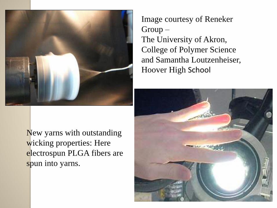
Image courtesy of Reneker
Group –
The University of Akron,
College of Polymer Science
and Samantha Loutzenheiser,
Hoover High School
New yarns with outstanding
wicking properties: Here
electrospun PLGA fibers are
spun into yarns.

• Parameters influencing on electrospinningprocess
-Solution properties, such as concentration
viscosity,elasticity,conductivity,volatility of the solvent,
and surface tension.
-Processing parameters,such as applied voltage,tip
collector distance,electric field strength,needle tip
design,collector composition and geometry ,and flow-rate.
-Ambient parameters,such as temperature,humidity,and
air velocity.
-Aligned fibers.
-Rotating mandrels.

2-Self-Assembly Method
Definition: spontaneous organization into stable structure without
covalent bonds
Biologically relevant processes
- DNA, RNA, protein organization
- can achieve small diameter
Drawbacks: more complex in vitro limited to
1) several polymers and
2) hydrophobic/philic interactions
Example: peptide-amphiphiles
- hydrophobic tail
- cysteine residues disulfide bonds

Schematic illustration of the self-assembly process of peptide-
amphiphiles functionalized to form a nanofiber 7.6 ± 1 nm in
diameter.

3-PHASE SEPERATİON METHOD
Definition: thermodynamic separation of polymer solution into
polymer-rich/poor layers
- similar to setting a gel
- control over macroporous architecture using porogens, microbeads,
salts 98% porosity achieved.
- consistent
Drawbacks:
- limited to several polymers
- small production scale

Process Advantages Limitations
Self -Assembly Achieves fiber diameters o
lowest scale (5-8 nm)
• Only short fibers can be
created.(<1 nm)
•Low yield.
•Matrix directly fabricated.
•Limited to a few polymers.
Phase Seperation • Tailorable mechanical
properties,pore size and
interconnectivity.
•Batch-to-batch
consistency.
• Low yield
•Matrix directly fabricated
•Limited to a few polymers
Electrospinning • Cost effective
•Long continuous
nanofibers
•Production of aligned
nanofibers
•Tailorable mechanical
porpertiesiszeishape
•Plethore of polymers may
be used
• Large nanometer to
micron scale fibers
•Use of organic solvents
•No control over 3D pore
structure

OVERVIEW
- Electrospinning viable for both synthetic and biological scaffolds/mats
- Wide range of fiber sizes necessary and possible
ECM ideally 150-500 nm
cell mats 2-3 um
- Hybridizing polymers can, but not necessarily, lead to hybrid properties
Specifics:
- PGA, PLA, PLGA most commonly used scaffold materials
- PDO exhibits elastin+collagen functionality in 1 synthetic polymer
BUT inhibited by “shape memory”
- PCL most elastic synthetic – frequently mixed with other synthetics

Tissue Engineering
Tissue engineering approaches make use of
biomaterials, cells, and factors either alone or in
combination to restore or regenerate, maintain, or improve
tissue function.
The scaffold gradually degrades with time to be
replaced by newly grown tissue from the seeded cells
(Langer and Vacanti 1993).

Biodegradable nanofibers, irrespective of their
method of synthesis, have been used as scaffolds for
musculoskeletal tissue engineering (including bone,
cartilage,ligament,and skeletal muscle), skin tissue
engineering, vascular tissue engineering,neural tissue
engineering, and as carriers for the controlled delivery
of drugs, proteins, and DNA.
Genetically engineered cells can be used as therapeutics.
using of biodegradable nanofibers are becoming rising stars
in Cell-based therapeutics for tissue engineering and cell-
replacement therapy because of their pluripotency ,self-
renewal capability and compatible with body.

Tissue Engineering
-Techniques and Polymers
Both self-asemmbly and phase seperation techniques
have been used succesfully to fabricate nanofibers for tissue
engineering.
However,in comparison,electrospinning is widely used by
researchers because of the simplicity,diversity and control
over scaffold geometries and mechanical characteristics,the
easily scaling-up property.

Tissue Scaffolding:
Fibroblast cells grown
on PLGA nanofibers
Image by Amy Liu, Hoover High School Student
Both synthetic polymers,such as polyglycoli acid(PGA),polylactic
acid(PLA),polycaprolactone (PCL) and their blends of
copolymers,and natural polymers such as elastin and collagens,have
been exploited.
Techniques And Polymers
Biodegradable polymeric nanofibers are of great
interest as scaffolds for tissue engineering and
drug delivery due to their extremely high surface
area, high aspect ratio and in structure to the
extracellular matrix (ECM) is the meaning of structural
material between cells and referred as connective tissue.

Agriculture Applications
Electrospinned biodegradable nanofibers from
different biodegradabla polymers,as PVA and
PLA are used,and titaniumdioxide is also used to
improve antibacterial and catalytic activities of
nanofibers in agriculture applications.

- Medical And Pharmaceutical Applications
The biodegradable nanofibers which are formed by
electrospinning fibers of biodegradable fiberizable
material,comprise a composite of different biodegradable
fibers.
These nanofibers having special medical uses include an
adhesion-reducing barrier and a controlled delivery system.
The methods include methods for reducing surgical
adhesions,controlled delivery of a medicinal agent and
porviding controlled tissue healing.

CONCLUSION
Usage area of biodegradable nanofiber is especially a rising
star of tissue engineering and drug delivery technlogy.
Biodegradable polymers such as PLGA and PCL have
already been electrospun into nanofibers, and nerve
guidance conduits have been fabricated using these
materials.
The ability of scaffolds to support biodegradable
nanofibers combined with good mechanical
prperties,biocompatibility and tuneable biodegradable
properties of the scaffold suggest their potential use for
tissue engineering.

References
1-W. J. Li, C. T. Laurencin, E. J. Caterson, R. S. Tuan, and F. K. Ko. J. Biomed. Mater. Res.60 [41,613. (2002).
2-L.S.Nair, S. Bhattacharyya, J.D. Bender, Y.E.Greish, P.W. Brown, H.R. Allcock, and
C.T.Laurencin. Biomacromolecules 5, 2212-2220 (2004).
3-J. Groll, W. Haubensak, T. Ameringer, M. Moeller, Ultrathin coatings from isocyanate star PEG prepolymers: patterning of proteins on the layers., Langmuir, 2005, 21(7), 3076-3083.
4-Biodegradable Cell-Seeded Nanofiber Scaffolds forNeuralRepair-Dong Han&Karen C. Cheung-Polymers 2011, 3, 1684-1733; doi:10.3390/polym3041684
5-Biodegradabla Nanofibers and Implementations theory-US 2012/0135234 A1,May 31 2012

6-Biodegradable and/or Bioabsorbable Fibrous Articles and Methodsfor
Using The Articles for Medical Applications,Us 7.172,765
B2,Feb.6,2007( The Research Foundation of State University of New
York,Stony Brook,NY(S) .)
7-Development of Biodegradable Polyphosphazene-
Nanohydroxyapatite Composite Nanofibers Via Electrospinning
Department of Chemical Engineering, The University of Virginia,
Charlottesville, VA-22904 6Department of Biomedical Engineering,
The University of Virginia, Charlottesville, VA-22908.(2005)
8-Biodegradable Nanofiber Mesh for Tissue Engineering
Erica Brown 1, Margaret W. Frey 2, Mary Rebovich 2
Department of Chemical, Biological, and Materials
Engineering, University of Oklahoma, Norman, OK 73019 1
Department of Textiles and Apparel, Cornell University, Ithaca,
NY 14853.

9-Biomedical Pathces With Aligned Fibers-Wo 2011/159889
A2,Washington University,One Brookings
Drive,St.Louis,MO 63130(US).
10-Biodegradable polyesters reinforced with triclosan loaded
polylactide micro/nanofibers:Properties, release and
biocompatibility L. J. del Valle,* A. Díaz, M. Royo, A.
Rodríguez-Galán, J. Puiggalí
11-role of Nanomedicines in Cell-Based Therapeutics-
Zhaoyang Ye & Ram I Mahato,the Johns Hopkins
University,Department of biomedical
engineering,Baltimore,USA.
12-Biodegradable Polymers:Past,Present,and Future. M.
Kolybaba1, L.G. Tabil 1, S. Panigrahi1, W.J. Crerar1, T.
Powell1, B. Wang1Department of Agricultural and
Bioresource Engineering University of Saskatchewan.

13-Nanofiber Technology: Designing The Next Generation Of
Tissue Engineering Scaffolds. C.P. Barnes, S.A. Sell, E.D.
Boland, D.G. Simpson, G.L. Bowlin
Department of Biomedical Engineering, Department of
Anatomy and Neurobiology Virginia Commonwealth
University, Richmond, VA.

Thanks for your interest