Investigation and evaluation of acute and sub-acute dermal...
16
Int. J. Curr. Res. Med. Sci. (2017). 3(5): 84-99 84 International Journal of Current Research in Medical Sciences ISSN: 2454-5716 P-ISJN: A4372-3064, E -ISJN: A4372-3061 www.ijcrims.com Original Research Article Volume 3, Issue 5 -2017 Investigation and evaluation of acute and sub-acute dermal toxicity studies of ethanolic leaves extract of Melastoma malabathricum in Sprague Dawley rats Ali Khairullah Zahi 1 , Hazilawati Hamzah 1 , Mohd Rosly Shaari 2 , Riyanto Teguh Widodo 3 , Lucy Johnny 4 , Noordin M.M 1 , and Shanmugavelu Sithambaram 2 1 Department of Pathology and Microbiology, Faculty of Veterinary Medicine, University Putra Malaysia, 43400 UPM Serdang, Selangor, Malaysia. 2 Department of Deputy Director, Strategic Livestock Research Centre, Malaysia Agricultural Research and Development Institute, 43400 Serdang, Selangor, Malaysia. 3 Department of Pharmacy, Faculty of Medicine, University of Malaya, 50603 Kuala Lumpur, Malaysia. 4 Sunway College KL, 46150 Bandar Sunway, Selangor, Malaysia *Corresponding author: [email protected] Abstract Melastoma malabathricum is an important plant commonly used in traditional medicine. Until recently, the dermal toxicity profile of M. malabathricum remained unknown. The objective of this study is to investigate the in vivo acute and sub-acute dermal toxicity of ethanolic leaves extract of M. malabathricum in rats. In acute experiment, a total of 20 female rats were divided into four groups, each group had five rats. While, a total of 30 male rats were divided into five groups, each group consisted of six rats in sub-acute experiment. Single doses of the extract at 2000 and 5000 mg/kg of body weight failed to produce treatment-related signs of toxicity or mortality during the 14-day observation period. In a repeated dose 28-day study, 500, 1000 and 2000 mg/kg of body weight/day applications of leaf extract lead to no significant change (p >0.05) in bodyweight or haematological and biochemical parameters compared with the control group. Similarly, gross pathology and histopathology examinations of liver, spleen, kidneys, and skin did not reveal any morphological alteration. Results indicate that the close application of M. malabathricum leaves extract had no critically dangerous effect on the rats tested. Therefore, the concentrate may be used pharmaceutically. Keywords: Melastoma malabathricum, acute toxicity, sub-acute toxicity, histopathology. Introduction Melastomataceae plants originate in tropic and subtropical regions. There is a total of more than 4000 species worldwide. In just South-East Asia region alone, the genus Melastoma comprises 22 species, 2 subspecies, and 3 varieties (Rajenderan, 2010). Malaysia, particularly, with a tropical DOI: http://dx.doi.org/10.22192/ijcrms.2017.03.05.013
Transcript of Investigation and evaluation of acute and sub-acute dermal...

Int. J. Curr. Res. Med. Sci. (2017). 3(5): 84-99
84
International Journal of Current Research inMedical Sciences
ISSN: 2454-5716P-ISJN: A4372-3064, E -ISJN: A4372-3061
www.ijcrims.com
Original Research Article Volume 3, Issue 5 -2017
Investigation and evaluation of acute and sub-acutedermal toxicity studies of ethanolic leaves extract ofMelastoma malabathricum in Sprague Dawley rats
Ali Khairullah Zahi1, Hazilawati Hamzah1, Mohd Rosly Shaari2,Riyanto Teguh Widodo3, Lucy Johnny4, Noordin M.M1, and
Shanmugavelu Sithambaram2
1Department of Pathology and Microbiology, Faculty of Veterinary Medicine, University Putra Malaysia,43400 UPM Serdang, Selangor, Malaysia.
2Department of Deputy Director, Strategic Livestock Research Centre, Malaysia Agricultural Research andDevelopment Institute, 43400 Serdang, Selangor, Malaysia.
3Department of Pharmacy, Faculty of Medicine, University of Malaya, 50603 Kuala Lumpur, Malaysia.4Sunway College KL, 46150 Bandar Sunway, Selangor, Malaysia
*Corresponding author: [email protected]
Abstract
Melastoma malabathricum is an important plant commonly used in traditional medicine. Until recently, the dermaltoxicity profile of M. malabathricum remained unknown. The objective of this study is to investigate the in vivo acuteand sub-acute dermal toxicity of ethanolic leaves extract of M. malabathricum in rats. In acute experiment, a total of20 female rats were divided into four groups, each group had five rats. While, a total of 30 male rats were divided intofive groups, each group consisted of six rats in sub-acute experiment. Single doses of the extract at 2000 and 5000mg/kg of body weight failed to produce treatment-related signs of toxicity or mortality during the 14-day observationperiod. In a repeated dose 28-day study, 500, 1000 and 2000 mg/kg of body weight/day applications of leaf extractlead to no significant change (p >0.05) in bodyweight or haematological and biochemical parameters compared withthe control group. Similarly, gross pathology and histopathology examinations of liver, spleen, kidneys, and skin didnot reveal any morphological alteration. Results indicate that the close application of M. malabathricum leaves extracthad no critically dangerous effect on the rats tested. Therefore, the concentrate may be used pharmaceutically.
Keywords: Melastoma malabathricum, acute toxicity, sub-acute toxicity, histopathology.
Introduction
Melastomataceae plants originate in tropic andsubtropical regions. There is a total of more than4000 species worldwide. In just South-East Asia
region alone, the genus Melastoma comprises 22species, 2 subspecies, and 3 varieties (Rajenderan,2010). Malaysia, particularly, with a tropical
DOI: http://dx.doi.org/10.22192/ijcrms.2017.03.05.013

Int. J. Curr. Res. Med. Sci. (2017). 3(5): 84-99
85
climate, is home to at least 12 species, many ofwhich are used by natives in folk medicine. Oneof the plants within the Melastomataceae familywith a status in Malay folklore belief isMelastoma malabathricum L., which has beenknown to comprise two subspecies, namelyM. malabathricum L. ssp. malabathricum andM. malabathricum Linn ssp. normale (Meyer,2001). The characteristics of M. malabathricuminclude its average height of (0.5-1 m) high butmay occasionally grow up to 5 m long (Zakariaand Mohd, 1994). Melastoma is Greek for “blackmouth”, a name appreciated by generations ofchildren who have eaten the berries.M. malabathricum has evergreens and flowersthroughout the year (Koay, 2008).
In addition, M. malabathricum leaves, flowers,seeds, and roots are used in traditional medicineto treat a variety of ailments as skin diseases,venereal diseases and diabetes as well as having aworkable and neat anti-inflammatory and anti-microbial effects (Grosvenor et al., 1995;Sunilson et al., 2008). Hazardous chemicals maylead to adverse health effects in humans. Plantsused in alternative medicine may contain toxiccompounds. Toxicity refers to chemicals withinthe plant being poisonous to cells. Typically,exposure to toxic compounds leads to signs ofacute toxic effects. Proper evaluation of theseeffects protects the public’s health. One form oftoxicological assessment is animal testing ofpotential remedies. OECD guidelines allowtesting on animals such as rabbits, monkeys,guinea pigs, and dogs. In practical terms,evaluations must consider acute, sub-acute, sub-chronic, chronic, carcinogenic, and reproductiveeffects (Asante-Duah, 2002).
This study examines the use of this extract intopical applications. Both acute and sub-acutedermal toxicity studies are performed. Acutetoxicity is measured using a single large dose todetermine both immediate toxic effects and LD50.The sub-acute toxicity test uses sub-lethal dosesover 15 to 30 days to determine the no observedadverse effect level or NOAEL (Lipnick et al.,1995). This study examines the toxic effects ofethanolic leaf extract of M. malabathricum inSprague Dawley rats at dosages of 2000 and 5000
mg/kg body weight for a period of 14 days foracute toxicity study per OECD 402 guidelines,and at dosages of 2000, 1000 and 500 mg/kg bodyweight for a period of 28 days for sub-acutetoxicity study per OECD 410 guidelines.
Methods
Experimental animals:
Male and female Sprague Dawley rats with abody weight ranging from 200 to 250 g werepurchased from a local supplier at eight weeksold. Upon arrival, the rats were weighed andassigned randomly in polypropylene plastic cages,where one rat was placed in each cage with woodchips for bedding and housed in an animal roomwith controlled conditions involving theseparameters; temperature (22±2°C), humidity(55±10%) and lighting (12 hours light/dark) in theanimal house at the Malaysian AgriculturalResearch and Development Institute (MARDI),Serdang, Selangor.
Preparation of Melastoma malabathricum(Senduduk) leaf extracts
Fresh, whole plants were obtained from Jerantut,Pahang, Malaysia. After separating the leaves,they were dried at temperature between 25°C to30°C for three to five days. The dried leaves werethen ground into powder via a grinder and placedin a refrigerator at 4°C. After that, the powderedleaves were macerated in a flask with 97%ethanol. The mixture underwent a water bath attemperature between 55°C to 60°C for one day inorder for the chemicals in the leaves to becomefully dissolved in the ethanol solution. Theethanol was isolated from the mixture viafiltration followed by evaporation using a rotorpaper evaporator. The remaining elements werethen dried via a freeze dryer for one day. Thediluted solutions were stored in a refrigerator forlater use. The leaf extract was mixed with whitesoft paraffin (10%).
Skin preparation for dermal toxicity studies
The skin at the dorsal thoracic area of the rats wasclipped under general ketamine (50 mg/kg) andxylazine (5 mg/kg) anaesthesia with an electricclipper. This was followed by manual shaving

Int. J. Curr. Res. Med. Sci. (2017). 3(5): 84-99
86
using a razor blade. Based on OECD guidelines402 and 410, not less than 10% of the bodysurface area was cleared for the application of thetest substance. The extract was applied to dorsumarea.
Experimental design
To determine acute and sub-acute dermal toxicity,the toxic effects of the ethanolic leaf extract ofM. malabathricum in Sprague Dawley rats wereascertained at dosages of 2000 and 5000 mg/kgbody weight for a period of 14 days (acute dermaltoxicity), and at dosages of 2000, 1000 and 500mg/kg body weight for a period of 28 days (sub-acute dermal toxicity) per OECD guidelines 402and 410, respectively. Different doses were usedto ensure that this extract is not toxic and safe indifferent concentrations. Base on OECDguidelines, female rats are more sensitive to toxicsubstances than males in acute toxicity studies.The application of extract in acute toxicity studyis once but in sub-acute dermal toxicity is daily,thus female rats used in acute dermal toxicity andmale used in sub-acute dermal toxicity. In acutedermal toxicity, a total number of 20 eight-weekold female Sprague Dawley rats were divided intofour groups (n=5) namely Groups 1, 2, 3 and 4.Duration of this study was 14 days. Each groupwas applied with the extract once at day 1 andsacrificed at day 14 of the experimental period.
On the other hand, in sub-acute dermal toxicity atotal of 30 eight-week old male Sprague Dawleyrats were divided into 5 groups (n=6) namelyGroups 1, 2, 3, 4 and 5. This study ran for 28 daysand each group underwent extract applicationonce daily for 28 days, and all were sacrificed atday 28 of the experimental period.
Necropsy
All rats were humanely scarified by completeexsanguinations under general anaesthesia with amixture of 75 mg/kg Ketamine and 10 mg/kgXylazine.
Gross pathology
Complete gross examination was conducted todetect any gross changes especially skin necrosis.
The liver and kidneys were blotted dry andweighed immediately after necropsy.
Histopathology
Skin, liver, and kidneys samples were collectedand fixed in 10% formalin for 48 hours. Sampleswere sliced to 0.5 cm thickness and placed inplastic cassettes for dehydration using anautomated processor (Leica ASP300, Germany).After this step, they were embedded in paraffin(Leica EG1160, Germany). The tissue sampleswere then trimmed and sectioned at 4 μmthicknesses (Leica RM2155, Germany). Mountingwas carried out on glass slides with a hot plate(Leica HI1220, Germany). Deparaffinization wasthen carried out via xylene exchange, after whichrehydration using different ethanol dilutions(100%, 90% and 70%) was then performed for 2minutes each, respectively. The tissue sectionswere then further rinsed in tap water and stainedwith Harris’s haematoxylin and eosin (H&E)stain. A light microscope at 40x, 100x, 200x,400x and 1000x magnification levels was used forobservation.
Blood biochemistry
The blood samples were centrifuged (Hettichzent-EBA20, Germany) at 5000 rpm for 5 minutes.The serum was then placed into micro centrifugetubes and kept at -20°C. Serum samples wereanalysed using an automatic biochemistryanalyser (TRX 7070, Biorex, Germany). Levels ofaspartate transaminase (AST), lactatedehydrogenase (LDH), alanine transaminase(ALT), total protein, albumin, creatinine, uricacid, urea, and bilirubin were measured. Globulinlevels were determined via the formula (totalprotein – albumin).
Haematology
Blood samples were taken from the posterior venacava of each rat. Samples were then placed intoethylene diamine tetra acetic acid (EDTA)vacuumed blood collection tubes and gentlymixed with EDTA-anticoagulant material for bothautomatic and manual haematology analyses.

Int. J. Curr. Res. Med. Sci. (2017). 3(5): 84-99
87
Automatic haematology
Blood samples were analysed via an automatichaematology analyser (Cell Dyn, 3700, Abbot,USA) for the total number of white blood cell(WBC), red blood cell (RBC) and haemoglobinconcentration.
Manual haematology
EDTA blood was placed via capillary action intoa capillary microhematocrit tube to about a levelof about three-fourths of its length. The dry end ofmicrohematocrit tubes was sealed by melting,then placed in a micro-centrifuge machine(HettichHaematokrit 210, Germany). It was thencentrifuged at 10,000 rpm for 5 minutes toseparate the RBCs from plasma. The plasma roseto the top of the microhematocrit tube while theRBCs went to the bottom. The centrifugedmicrohematocrit tubes were used fordetermination of packed cell volume (PCV),icteric index and plasma protein concentration.For the PCV, the microhematocrit tube wasplaced in holder slots of the microhematocrit tubereader (Hawksley), where the base of red bloodcell was intersected with base line of reader andthe top of plasma was intersected with the top lineof the reader by moving the holder left or right,before the middle line of the reader was adjustedto intersect with the top of the RBCs and themeasuring ruler. PCV results were obtained fromthe middle line and the measuring ruler point(e.g.: 24% is equal to 0.24 L). Icteric index resultswere obtained by comparing plasma colour in themicrohaematocrit tube with the icteric indexstandard board colour degree. While the bloodplasma protein concentration was obtained bydropping the plasma on the refractometer glass(Atago T2-NE, Japan) to obtain the result fromthe measuring ruler and the plasma unit was readin protein in gram divided by plasma in litre (e.g.:6.2 % is equal to 62 g/L).
Peripheral blood smear
A peripheral blood smear was performed. First, asmall blood drop was placed on a glass slide.Then, a drop smeared using a cover slip on theslide surface. This was left at room temperature
for 20 minutes to dry. The blood smear wasstained via Wright’s stain method. The bloodsmear slide underwent examination with a lightmicroscope at 100x, 200x, 400x, and 1000xmagnifications.
A manual WBC differential count was carried outfor one hundred WBCs. The values for each WBCtype were converted to a percentage. Then, thesevalues were multiplied by the automated totalWBC count to obtain absolute WBC differentialcounts (× 109/L).
Statistical analysis
The data obtained were statistically analysed byusing Statistical Package for Social Science(SPSS) software version 20. The values wereexpressed as mean ± standard deviation (SD) fordifferent parameters. Repeated measurements ofanalysis of variance (ANOVA) tests were done tocompare the differences of data between andwithin the groups. A Duncan test was conductedfor post hoc analysis. The level of statisticalsignificance was set at P < 0.05.
Results
Acute Toxicity Study
General sign and behaviour of the rats
M. malabathricum ethanolic extracts’ effects onappearance and general behavioural patterns areshown in Table 1 and Table 2, respectively. Notoxic signs or mortality were observed in anyanimals, which survived up to 14 days afterapplying of the extracts once on the first day atsingle dose level of 5000 and 2000 mg/kg bodyweight. Behavioural patterns were observed bothfor the first 6 hours and 14 hours after extractapplication. The animals in both vehicle-treatedand extract-treated groups were normal and didnot display significant changes in behaviour, skineffects, breathing, impairment in food intake andwater consumption, postural abnormalities andhair loss. Treated groups showed a rapid heartbeatfor the first 6 hours, but soon normalized. This islikely due to stresses endured during handling.

Int. J. Curr. Res. Med. Sci. (2017). 3(5): 84-99
88
Table 1: Mortality rate of rats after applied with topical ethanolic extracts of M. malabathricum at5000 mg/kg and 2000 mg/kg, once, for the acute toxicity study
Group Mortality rate*(%)G1 0G2 0G3 0G4 0
* Mortality rate is number of dead rats divided by total number of rats per group.G1: No treatment,G2: Paraffin, G3: M. malabathricum 5000 mg/kg, G4: M. malabathricum 2000 mg/kg
Table 2: Behavioural patterns and general appearance of rats in all groups
Abnormal sign Controlgroup:
G1 (6 h)
Control group:G1 (14 h)
Treatmentgroups:
G2-G4 (6 h)
Treatmentgroups:
G2-G4 (14 h)Skin and fur Normal Normal Normal Normal
Eyes Normal Normal Normal NormalMucous membrane Normal Normal Normal Normal
Behavioural patterns Normal Normal Tachycardia NormalSalivation Normal Normal Normal NormalLethargy Normal Normal Normal Normal
Sleep Normal Normal Normal NormalDiarrhea Normal Normal Normal Normal
Coma NO NO NO NOTremors NO NO NO NO
G1: No treatment, G2: Paraffin, G3: M. malabathricum 5000 mg/kg, G4: M. malabathricum2000 mg/kg. NO: Not observed
Organ and body weight
Body weights and weights of liver and kidneysare shown in Figure 1 and Table 3. No significantchanges in the body weight were found. All ratshad shown a normal increment in the body weight
which was not significantly (p > 0.05) differentbetween both control and treated groups. As withbody weight, no significant (p > 0.05) differencesin the changes of relative organ weights betweengroups were noted.
Table 3: Organ relative weights of rats in all groups
Group Liver (g) Kidneys (g)G1 0.024 ± 0.005 0.005 ± 0.0005G2 0.020 ± 0.007 0.006 ± 0.0004G3 0.026 ± 0.005 0.005 ± 0.0005G4 0.024 ± 0.008 0.005 ± 0.0008
Values are expressed as mean ± SD ( = 5 for each group). Relative organ weight was calculated by organweight/body weight × 100%. G1: No treatment, G2: Paraffin, G3: M. malabathricum 5000 mg/kg,
G4: M. malabathricum 2000 mg/kg

Int. J. Curr. Res. Med. Sci. (2017). 3(5): 84-99
89
Fig. 1: Mean body weight (g) of rats in all groups. Data collected were recorded and presented as mean ±standard error of mean. G1: No treatment; G2: Paraffin; G3: M. malabathricum 2000 mg/kg;G4: M. malabathricum 5000 mg/kg. None of the numbers were seriously variant at p>0.05
Haematology evaluation
All erythron parameters were normal in all rats asshown in Table 4. Results were also normal for
the leukon parameters, which are depicted inTable 5.
Table 4: Erythron parameters and plasma protein concentration of rats in all groups
Parameter Unit G1(Mean±SD)
G2(Mean±SD)
G3(Mean±SD)
G4(Mean±SD)
RBC x1012/L 8.25±0.28 8.56±0.61 8.66±0.60 8.43±0.83Hb g/L 171±7.91 166±7.79 178±8.90 169±16.69
PCV L/L 0.45±0.00 0.45±0.01 0.45±0.00 0.45±0.02MCV fl 58.2±0.83 57.8±1.30 56.6±1.51 56.6±1.51
PP g/L 75.4±2.07 77.8±1.92 77.0±3.16 75.4±1.14Values are expressed as mean ± SD ( = 5 for each group). G1: No treatment; G2: Paraffin;
G3:M. malabathricum 5000 mg/kg; G4: M. malabathricum 2000 mg/kg
Table 5: Leukon and thrombon parameters of rats in all groups
Parameter Unit G1(Mean±SD)
G2(Mean±SD)
G3(Mean±SD)
G4(Mean±SD)
WBC 109/L 7.82±0.87 7.68±0.93 7.80±1.05 7.52±1.02Neutrophils 109/L 1.45±1.14 1.47±0.83 1.43±0.89 1.36±0.83
Lymphocytes 109/L 5.66±1.14 5.66±1.64 5.89±1.67 5.56±1.22Monocytes 109/L 0.31±1.00 0.32±1.30 0.29±0.83 0.36±0.83Esoinophils 109/L 0.21±0.83 0.12±0.89 0.09±0.83 0.12±0.54Basophils 109/L 0.07±0.70 0.09±0.83 0.07±0.70 0.10±1.14Platelets 109/L 869±40.70 913±28.10 890±39.20 888±33.60
Values are expressed as mean ± SD ( = 5 for each group). G1: No treatment; G2: Paraffin;G3:M. malabathricum 5000 mg/kg; G4: M. malabathricum 2000 mg/kg

Int. J. Curr. Res. Med. Sci. (2017). 3(5): 84-99
90
Blood biochemistry
The serum biochemical parameters of liver andmuscle enzymes are shown below. Table 6depicts the kidney parameters, while Table 7
shows protein concentration and Table 8 containsinformation for the control female rats andM. malabathricum extract groups. No significant(p > 0.05) changes were observed for anybiochemical parameter.
Table 6: Serum biochemical parameters of liver and muscle enzymes of rats in all groups
Parameter Unit G1(Mean±SD)
G2(Mean±SD)
G3(Mean±SD)
G4(Mean±SD)
ALT (U/L) 41.2 ±1.30 43.4 ±1.14 41.6±1.14 43.4±2.07ALP (U/L) 74.0 ±2.73 73.2 ±2.58 74.2±1.64 72.0±2.34AST (U/L) 134.6 ±2.96 139.0 ±2.23 139.2±2.38 138.2±2.77CK (U/L) 172.8 ±2.68 174.6 ±1.81 174.2±2.16 173.8±2.16
Values are expressed as mean ± SD ( = 5 for each group). G1: No treatment; G2: Paraffin;G3: M. malabathricum 5000 mg/kg; G4: M. malabathricum 2000 mg/kg
Table 7: Serum biochemical parameters of kidney of rats in all groups
Parameter Unit G1(Mean±SD)
G2(Mean±SD)
G3(Mean±SD)
G4(Mean±SD)
Urea (mmol/L) 6.44 ±0.25 6.30 ±0.30 6.70±0.15 6.40±0.27Creatinine (μmol/L) 36.5 ±1.35 37.5 ±1.46 37.6±1.84 36.3±2.86
Values are expressed as mean ± SD ( = 5 for each group). G1: No treatment; G2: Paraffin;G3: M. malabathricum 5000 mg/kg; G4: M. malabathricum 2000 mg/kg
Table 8: Serum biochemical parameters of protein concentration of rats in all groups
Parameter Unit G1(Mean±SD)
G2(Mean±SD)
G3(Mean±SD)
G4(Mean±SD)
TP (g/L) 72.8 ±1.92 73.8 ±1.92 73.4±1.14 73.0±2.54Albumin (g/L) 37.7 ±0.97 38.1 ±0.79 38.1±0.73 38.5±0.80Globulin (g/L) 32.7 ±0.72 33.1 ±0.75 32.1±0.43 34.2±0.66A/G ratio (g/L) 1.14 ±0.03 1.14 ±0.03 1.18±0.03 1.12±0.02
Values are expressed as mean ± SD ( = 5 for each group). G1: No treatment; G2: Paraffin;G3: M. malabathricum 5000 mg/kg; G4: M. malabathricum 2000 mg/kg
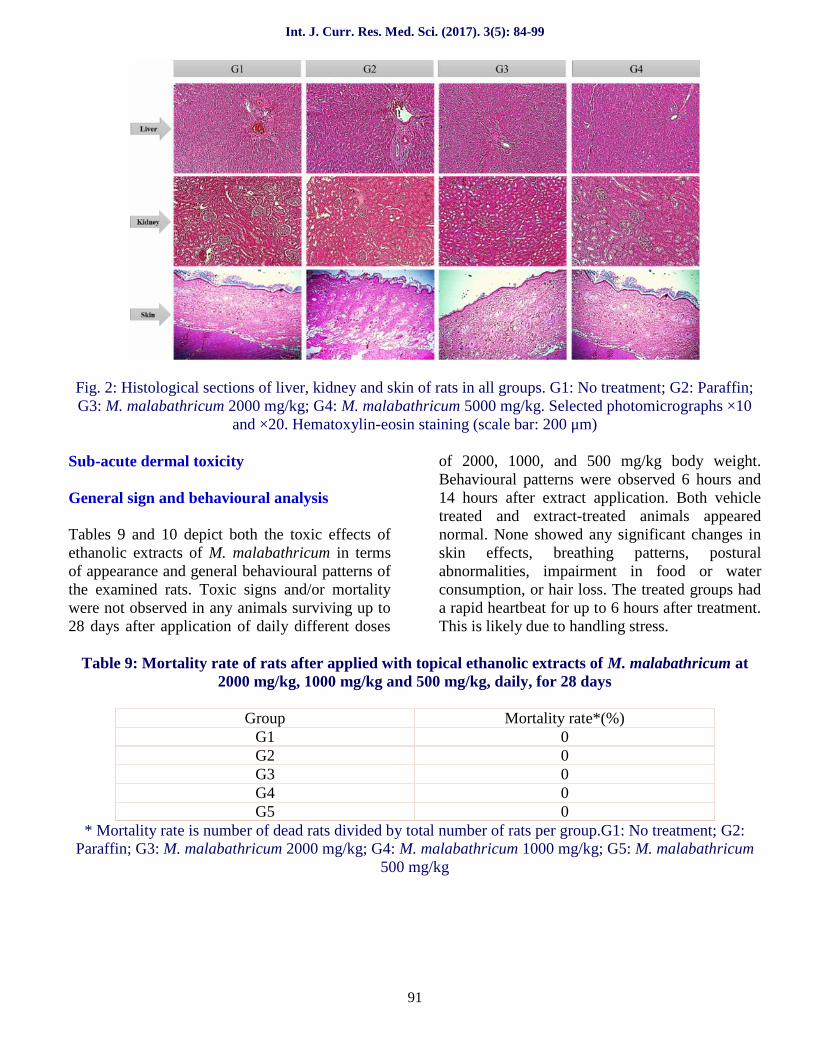
Histopathology examination
The microscopic structures of the liver, kidneysand skin are shown in Figure 2. There are nounnoticeable differences between the control andtreatment groups. Microscopic examination was
conducted showing that the organs from extract-treated rats had no any alterations in cellstructures or any other undesirable effects whenviewed under a light microscope using multiplemagnification powers.
Int. J. Curr. Res. Med. Sci. (2017). 3(5): 84-99
91
Fig. 2: Histological sections of liver, kidney and skin of rats in all groups. G1: No treatment; G2: Paraffin;G3: M. malabathricum 2000 mg/kg; G4: M. malabathricum 5000 mg/kg. Selected photomicrographs ×10
and ×20. Hematoxylin-eosin staining (scale bar: 200 μm)
Sub-acute dermal toxicity
General sign and behavioural analysis
Tables 9 and 10 depict both the toxic effects ofethanolic extracts of M. malabathricum in termsof appearance and general behavioural patterns ofthe examined rats. Toxic signs and/or mortalitywere not observed in any animals surviving up to28 days after application of daily different doses
of 2000, 1000, and 500 mg/kg body weight.Behavioural patterns were observed 6 hours and14 hours after extract application. Both vehicletreated and extract-treated animals appearednormal. None showed any significant changes inskin effects, breathing patterns, posturalabnormalities, impairment in food or waterconsumption, or hair loss. The treated groups hada rapid heartbeat for up to 6 hours after treatment.This is likely due to handling stress.
Table 9: Mortality rate of rats after applied with topical ethanolic extracts of M. malabathricum at2000 mg/kg, 1000 mg/kg and 500 mg/kg, daily, for 28 days
Group Mortality rate*(%)G1 0G2 0G3 0G4 0G5 0
* Mortality rate is number of dead rats divided by total number of rats per group.G1: No treatment; G2:Paraffin; G3: M. malabathricum 2000 mg/kg; G4: M. malabathricum 1000 mg/kg; G5: M. malabathricum
500 mg/kg

Int. J. Curr. Res. Med. Sci. (2017). 3(5): 84-99
92
Table 10: Behavioural patterns and general appearance of rats in all groups
Abnormal sign Control group:G1 (6 h)
Controlgroup:
G1 (14 h)
Treatmentgroups:
G2-G5 (6 h)
Treatmentgroups:
G2-G5 (14 h)Skin and fur Normal Normal Normal Normal
Eyes Normal Normal Normal NormalMucous membrane Normal Normal Normal Normal
Behavioural patterns Normal Normal Tachycardia NormalSalivation Normal Normal Normal NormalLethargy Normal Normal Normal Normal
Sleep Normal Normal Normal NormalDiarrhea Normal Normal Normal Normal
Coma NO NO NO NOTremors NO NO NO NO
G1: No treatment, G2: Paraffin, G3: M. malabathricum 2000 mg/kg, G4: M. malabathricum 1000 mg/kg,G5: M. malabathricum 500 mg/kg, NO: Not observed
Organ and body weight
Body weights and weights of liver and kidneys ofthe rats are shown in Figure 3 and Table 11. Nosignificant changes in body weight were noted.All rats demonstrated a normal increment in the
body weight which was not significantly (p >0.05) different between the control and treatedgroups. Similarly, there were no significant (p >0.05) differences in changes of relative organweights between groups.
Fig. 3: Mean body weight (g) of rats in all groups. Data collected were recorded and presented as (mean ±SEM). G1: No treatment group; G2: Paraffin group; G3: M. malabathricum (2000 mg/kg);
G4: M. malabathricum (1000 mg/kg); G5: M. malabathricum(500 mg/kg
Int. J. Curr. Res. Med. Sci. (2017). 3(5): 84-99
93
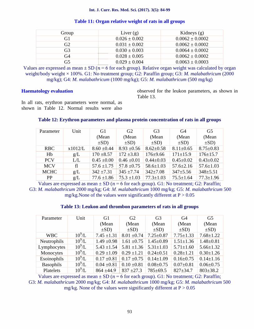
Table 11: Organ relative weight of rats in all groups
Group Liver (g) Kidneys (g)G1 0.026 ± 0.002 0.0062 ± 0.0002G2 0.031 ± 0.002 0.0062 ± 0.0002G3 0.030 ± 0.003 0.0064 ± 0.0002G4 0.028 ± 0.005 0.0062 ± 0.0002G5 0.029 ± 0.004 0.0063 ± 0.0003
Values are expressed as mean ± SD ( = 6 for each group). Relative organ weight was calculated by organweight/body weight × 100%. G1: No treatment group; G2: Paraffin group; G3: M. malabathricum (2000
mg/kg); G4: M. malabathricum (1000 mg/kg); G5: M. malabathricum (500 mg/kg)
Haematology evaluation
In all rats, erythron parameters were normal, asshown in Table 12. Normal results were also
observed for the leukon parameters, as shown inTable 13.
Table 12: Erythron parameters and plasma protein concentration of rats in all groups
Parameter Unit G1(Mean±SD)
G2(Mean±SD)
G3(Mean±SD)
G4(Mean±SD)
G5(Mean±SD)
RBC x1012/L 8.60 ±0.44 8.93 ±0.56 8.62±0.58 8.11±0.65 8.75±0.83Hb g/L 170 ±8.57 172 ±3.83 176±9.66 171±15.9 176±15.7
PCV L/L 0.45 ±0.00 0.46 ±0.01 0.44±0.03 0.45±0.02 0.43±0.02MCV fl 57.6 ±1.75 57.8 ±0.75 58.6±1.03 57.6±2.16 57.6±1.03
MCHC g/L 342 ±7.31 345 ±7.74 342±7.08 347±5.56 348±5.51PP g/L 77.6 ±1.86 75.3 ±1.03 77.3±1.03 75.5±1.64 77.3±1.96
Values are expressed as mean ± SD ( = 6 for each group). G1: No treatment; G2: Paraffin;G3: M. malabathricum 2000 mg/kg; G4: M. malabathricum 1000 mg/kg; G5: M. malabathricum 500
mg/kg.None of the values were significantly different at P > 0.05
Table 13: Leukon and thrombon parameters of rats in all groups
Parameter Unit G1(Mean±SD)
G2(Mean±SD)
G3(Mean±SD)
G4(Mean±SD)
G5(Mean±SD)
WBC 109/L 7.45 ±1.31 8.01 ±0.74 7.25±0.87 7.75±1.33 7.68±1.22Neutrophils 109/L 1.49 ±0.98 1.61 ±0.75 1.45±0.89 1.51±1.36 1.48±0.81
Lymphocytes 109/L 5.43 ±1.54 5.81 ±1.36 5.31±1.03 5.71±1.60 5.66±1.32Monocytes 109/L 0.29 ±1.09 0.29 ±1.21 0.24±0.51 0.28±1.21 0.30±1.26Esoinophils 109/L 0.17 ±0.81 0.17 ±0.75 0.14±1.09 0.16±0.75 0.14±1.16Basophils 109/L 0.04 ±0.81 0.10 ±0.81 0.08±0.75 0.07±0.81 0.06±0.75Platelets 109/L 864 ±44.9 837 ±27.3 785±69.5 827±34.7 803±38.2
Values are expressed as mean ± SD ( = 6 for each group). G1: No treatment; G2: Paraffin;G3: M. malabathricum 2000 mg/kg; G4: M. malabathricum 1000 mg/kg; G5: M. malabathricum 500
mg/kg. None of the values were significantly different at P > 0.05

Int. J. Curr. Res. Med. Sci. (2017). 3(5): 84-99
94
Blood biochemistry
Serum biochemical parameters of liver andmuscle enzymes are shown in Table 14. Table 15contains kidney parameters while protein
concentrations are shown in Table 16 for both themale rats of the control and M. malabathricumextract groups. No significant (p > 0.05) changeswere observed in any of the biochemicalparameters.
Table 14: Serum biochemical parameters of liver and muscle enzymes of rats in all groups
Parameter Unit G1(Mean±SD)
G2(Mean±SD)
G3(Mean±SD)
G4(Mean±SD)
G5(Mean±SD)
ALT (U/L) 44.1 ±8.35 42.0 ±6.41 38.5±6.34 42.5±7.06 44.1±7.35ALP (U/L) 135 ±12.3 138 ±10.1 130±12.6 131±10.7 125±13.3AST (U/L) 149 ±12.4 150 ±16.5 139±19.4 123±18.1 145±13.5CK (U/L) 201 ±22.1 221 ±26.3 202±14.2 221±19.5 213±26.2
Values are expressed as mean ± SD ( = 6 for each group). G1: No treatment group; G2: Paraffin group;G3: M. malabathricum (2000 mg/kg); G4: M. malabathricum (1000 mg/kg); G5: M. malabathricum (500
mg/kg. None of the values were significantly at P > 0.05
Table 15: Serum biochemical parameters of kidney of rats in all groups
Parameter Unit G1(Mean±SD)
G2(Mean±SD)
G3(Mean±SD)
G4(Mean±SD)
G5(Mean±SD)
Urea (mmol/L) 7.71±0.91 6.91±0.57 7.50±0.73 7.18±0.88 7.08±0.66Creatinine (μmol/L) 55.5±2.25 55.5±3.08 51.5±1.22 54.3±2.42 51.8±2.63
Values are expressed as mean ± SD ( = 6 for each group). G1: No treatment group; G2: Paraffin group;G3: M. malabathricum (2000 mg/kg); G4: M. malabathricum (1000 mg/kg); G5: M. malabathricum (500
mg/kg. None of the values were significantly different at P > 0.05
Table 16: Serum biochemical parameters of protein concentration of rats in all groups
Parameter Unit G1(Mean±SD)
G2(Mean±SD)
G3(Mean±SD)
G4(Mean±SD)
G5(Mean±SD)
TP (g/L) 72.2 ±4.09 75.5 ±4.37 72.2±2.77 72.2±5.29 70.4±2.97Albumin (g/L) 40.8 ±1.15 43.8 ±2.62 41.3±2.01 39.3±3.61 38.8±2.25Globulin (g/L) 31.4 ±3.23 31.6 ±5.26 30.9±1.84 32.8±3.06 31.6±3.02A/G ratio (g/L) 1.25 ±0.13 1.36 ±0.26 1.30±0.10 1.16±0.12 1.20±0.15
Values are expressed as mean ± SD ( = 6 for each group). G1: No treatment group; G2: Paraffin group;G3: M. malabathricum (2000 mg/kg); G4: M. malabathricum (1000 mg/kg); G5: M. malabathricum (500
mg/kg. None of the values were significantly different at P > 0.05
Histopathology examination
The liver, kidneys and skin shown in Figure 4have no noticeable differences between controland treatment groups. No organs from the extract
treated rats demonstrated any alteration in cellstructure or other negative effects when examinedunder a light microscope with variousmagnification powers.

Int. J. Curr. Res. Med. Sci. (2017). 3(5): 84-99
95
Fig. 4: Histological sections of liver, kidney and skin of rats in all groups G1: No treatment; G2: Paraffin;G3: M. malabathricum 500 mg/kg; G4: M. malabathricum 1000 mg/kg; G5: M. malabathricum 2000
mg/kg. Selected photomicrographs ×10 and ×20. Hematoxylin-eosin staining (scale bar: 200 μm)
Discussion
Phytotherapeutic products from medicinal plantshave come to be widely used in primaryhealthcare in developing countries. Some suchproducts are considered safe due to their naturalprovidence. Bioactive products from medicinalplants tend to be used more often in self-medication due to being considered naturally safe(Vaghasiya et al., 2011). However, this is adangerous assumption, as there are many suchplant compounds which are highly toxic,including the most cytotoxic anti-cancer plant-derived drugs, digitalis, the pyrrolizidinealkaloids, ephedrine, phorbol esters, and so on(Vaghasiya et al., 2011). Most herbal drugs showless harmful side effects when the drugs areappropriately used, when compared with syntheticdrugs (Kirtikar and Basu, 1975). Toxic substancesmay have compelling pharmacological effects at alower, nontoxic dose. Toxicity results as foundthrough animal testing are important indetermining the safety and usefulness ofpharmacologically active plants (Moshi and Brineshrimp, 2007). Worldwide, use of such medicinal
plants is growing; however, there remains a lackof knowledge about the relative toxicity andpotential adverse effects of such plants. Further invivo studies are called for to determine signs oftoxicity as well as the correct range of dosages ofsuch plants (Syahmi et al., 2010).
Acute and sub-acute toxicity testing in laboratoryanimal are used to evaluate natural remedies fordifferent pharmacological activities (Sasidharanet al., 2008). It is one of the necessary studiesneed to be performed for the toxicologicalanalyses of medicinal plants. The toxicity effectsof the plants are evaluated through qualitative andquantitative analyses of blood and histopathologysamples of the laboratory animal (Sasidharan etal., 2008). The present study was designed toinvestigate the toxicity of ethanolic extracts ofM. malabathricum, for topical application viaacute and sub-acute dermal toxicity analyses.However, results in this study indicated no signsof toxicity and deaths observed throughout theexperimental period in both acute and sub-acutetoxicity studies.

Int. J. Curr. Res. Med. Sci. (2017). 3(5): 84-99
96
No rats topically exposed to the ethanolic extractsof M. malabathricum demonstrated any organ orsystemic toxicity at 2000 mg/kg and 5000 mg/kgin the acute dermal toxicity study or dosages of500 mg/kg, 1000mg/kg and 2000 mg/kg in sub-acute dermal toxicity study. Rats were monitoreddaily for signs of toxicity or mortality until day 14in the acute study and until day 28 in the sub-acute study. During this monitoring period, ratsshowed no overt signs of distress. No symptomstoxicity was noted and there were no deaths.Clinical abnormality is one of the major importantobservations which indicate toxicity effects onorgans in the treated groups (Eaton and Klaassen,1996). No rats displayed significant changes inbehaviour.
In addition, the physical appearances such as skin,fur and eyes were normal and whilst the bodyweight of the rats was increased, this indicatesthat applying of the crude extracts on the skin hadnegligible level of toxicity on the growth of therats. Moreover, proper food and water intake isessential for a therapeutic product safety study.Proper nutrition has a significant effect on animalstatus and on the measured effects of the drugs tobe tested (Steven and Iversen, 2003). In thisstudy, food and water consumption remainednormal. This lack of an effect on appetite meansthere was no disturbance in carbohydrate, proteinor fat metabolism levels (Klaassen, 2001).Typically, body weight and internal organ weightgain changes are a measure of toxicity after toxicsubstance exposure (Carol, 1995). In 2002, astudy by Raza and his team found that bodyweight changes are indicators of adverse effectsof drugs and chemicals. These changes becomerelatively more significant if body weight lossexceeds 10%. Organ weight also is an importantindex of physiological and pathological status inanimals.
The relative organ weight is fundamental todiagnose whether the organ was exposed to theinjury or not (Teoet al., 2002). Liver, kidneys andspleen are the primary organs affected bymetabolic reaction caused by toxicant (Dybinget al., 2002). In this study, gross appearance of theselected organs of both control and treated groupswere normal. The relative and absolute weight ofthe organs in both control and treated groups
showed no significant differences. Body weightgain was similar in both control and treatedgroups without statistically significantdifferences. Local application of ethanolicextracts of M. malabathricum was not shown tocause adverse effects on organ weight of anyimportant organs. This suggests thatM. malabathricum extracts are virtually nontoxic.The haematopoietic system tends to be quitesensitive to toxic compounds. It acts as animportant index of physiological and pathologicalstatus in both animals and humans (Adeneye etal., 2006). After 14 and 28 days of treatment withethanolic extracts of M. malabathricum, nochanges were found in the haematologicalparameters of either the control or treatmentgroups. Also, no significant changes in levels ofall serum biochemistry parameters were found.
This evidences the nontoxic nature of ethanolicextract of M. malabathricum. This finding is inthe agreement with the safe use of the herbs bythe traditional healers. One study performed in1999 by Ajagbonna and his team found that thenormal range of haematological parameters maybe changed through the ingestion of toxic plants.Histopathology may be used for confirmation ofcell structure alteration due to toxicity.Histopathological examination remains the goldstandard method for evaluating treatment-relatedpathological changes in tissues and organs(OECD, 1995). The histopathological evaluationconducted for this study to determine acute andsub-acute dermal toxicity has indicated thatethanolic extracts of M. malabathricum did notcause any structural damage in the morphology ofliver, kidneys and skin.
Histopathological findings matched with theresults of haematological and biochemicalanalyses. The histopathology results were cross-correlated with body weight and relative organweights. In this study, the liver histology revealednormal hepatocytes; portal and central vein, bileduct as well as hepatic artery did not show anyalterations in the structure in both controls andtreated rats. In contrast to the present study, thehistological examination of a study conducted byHarizal and his team in 2010 using Mitragynaspeciosa extract revealed morphological changes

Int. J. Curr. Res. Med. Sci. (2017). 3(5): 84-99
97
in liver of mice treated with the extract at doselevels of 100 mg/kg and 500 mg/kg. Meanwhile,in 2009 another study by SalawuusingCrossopteryx febrifuga observed inflammatorychanges histologically in the liver by infiltrationof lymphocytes at portal and central veins of ratstreated at dose levels of 500 mg/kg and 1000mg/kg. Those studies showed that the extractsexerted deleterious effects on the liver.
Liver is capable of regenerating damaged tissues,hence it functions may not be impaired early onfollowing an insult from a toxicant (Salawu et al.,2009). Additionally, one toxicity study conductedon a Cassia fistula pod extract performed by(Akanmu et al., 2004) reported that thehistological examinations of liver, kidneys, andtestes showed. No potential toxicity or damage tothe cell structure of the liver, kidneys and testes ata dose of 1000 mg/kg extract were found. Nonecrosis, inflammatory reaction, fibrosis, or localfatty degeneration were observed in the liver orhepatocytes. The morphology of liver cells inboth the control and treated groups of this werealso normal. No structural alterations wereobserved under the microscope. As with the liver,kidney microphotograph histology found noadverse effects in either group. A previous studyby (Alade et al., 2009) found focal proximaltubular epithelial necrosis in the kidneys. Anotherstudy by (Akanmu et al., 2004) on the effects ofC. fistula pod extract in rats revealed weresignificant changes in the histology of kidneystreated with the extract at a dose of 1000 mg/kg.
In this present study, the microscopic examinationof the skin of rats treated with the extracts did notindicate any changes in the layers of the skin atthe epidermis, dermis and hypodermis ascompared to the control rats. Both glomeruli andBowman’s capsules were normal. Colloidal Nanosilver toxicity at doses of 100, 1000 and 10000μg/mL in guinea pigs was shown to cause skinlesions. This indicates a decreased thickness ofepidermis and dermis, increased numbers ofLangerhans and inflammatory cells, decreasedpapillary layer with regular collagens fibres,acidophilic cytoplasm in muscle fibres,degenerative fibres, and increased levels of
macrophage in the endomysium (Korani et al.,2011).
In conclusion, study results suggest that ethanolicextracts of M. malabathricum cause no obvious invivo toxicity. Deathor signs of acute toxicity werenot observed in rats treated with doses of 5000and2000 mg/kg (acute toxicity study). The sameholds for those rats treated with doses of 2000mg/kg, 1000 mg/kg,and 500 mg/kg (sub-acutetoxicity study). This indicates that the extract issafe to use. Histology examinations also found nochange in the architectures of selected organs forboth the control and treated groups. According tothe findings of this research, M. malabathricum issuitable for use as a topical medicinal agent atthose dosages. This is especially indicated in ruralcommunities, for which conventional drugs areunaffordable.
References
Adeneye, A.A., Ajagbonna, O.P., Adeleke, T.I.,Bello, S.O. 2006. Preliminarytoxicity andphytochemical studies of the stem barkaqueous extract of Musanga cecropioides inrats. J. Ethnopharmacol 105, 374-379.
Ajagbonna, O.P., Onifade, K.I., Suleiman, U.1999. Haematological and biochemicalchanges in rats given extract of C. pocera.Sokoto. J. Vet. Sci. 1, 3-12.
Akanmu, M.A., Iwalewa, E.O., Elujoba, A.A.,Adelusola, K.A. 2004. Toxicity potentials ofCassia fistula fruits as laxative with referenceto senna. Afr. J.Biomed. Res. 7, 23-26.
Alade, G.O., Akanmu, M.A., Obuotor, E.M.,Osasan, S.A., Omobuwajo, O.R. 2009. Acuteand oral sub-acute toxicity of methanolicextract of Bauhinia monandra leaf in rats. Afr.J. Pharm. Pharmacol3, 354-358.
Asante-Duah, K. 2002. Public Health RiskAssessment for Human Exposure to Chemicals(illustrated.); Kluwer Academic Publishers:Dordrecht, The Netherlands. Volume 6.
Carol, S.A. 1995. Acute, Sub-chronic and ChronicToxicology. In CRC Handbook of Toxicology;Michael, J.D., Mannfred, A.H., Eds. CRCPress Inc. Boca Raton, FL, USA 51-104.

Int. J. Curr. Res. Med. Sci. (2017). 3(5): 84-99
98
Dybing, E., Doe, J., Groten, J., Kleiner, J.,O’Brien, J. 2002. Hazard characterization ofchemicals in food and diet: dose response,mechanism and extrapolation issues. FoodChem. Toxicol 42, 237-282.
Eaton, D.L., Klaassen, C.D. 1996. Principles oftoxicology. In Casarett and Doull’sToxicology: The Basic Science of Poisons, 5thed; Klaassen, C.D. Ed.; McGraw-Hill: NewYork, NY, USA p. 13.
Grosvenor, P.W., Gothard, P.K., McWilliam,N.C., Supriono, A. and Gray, D.O. 1995.“Medicinal plants from Riau Province,Sumatra, Indonesia. Part 1: uses, ”Journal ofEthnopharmacology, vol. 45, no. 2, pp. 75-95.
Harizal, S.N., Mansor, S.M., Hasnan, J.,Tharakan, J.K.J., Abdullah, J. 2010. Acutetoxicity study of the standardized methanolicextract of Mitragyna speciosa Korthin Rodent.J. Ethnopharmacol 131, 404-409.
Iversen, P.O., Nicolaysen, G. 2003. Water for life.J. Norw. Med. Assoc 123, 3402-3405.
Kirtikar, K.R., Basu, B.D. 1975. Indian MedicinalPlants; International Book Distributors:Dehradun, India, Volume 2, p. 858.
Klaassen, C.D. 2001. Principles of Toxicology. InCasarett and Doull’s Toxicology: The BasicScience of Poisons, 5th ed.; McGraw-Hill:New York, NY, USA, p. 13.
Koay, S.S. 2008. Establishment of cell suspensionculture of Melastoma malabathricum L. for theproduction of anthocyanin, Ph.D. thesis,UniversitiSains Malaysia, Pulau Pinang,Malaysia.
Korani, M., Rezayat, S.M., Gilani, K.,Arbabi-Bidgoli, S., Adeli, S. 2011. Acute and sub-chronic dermal toxicity of nanosilver in guineapig. International Journal ofNanomedicine6:855–862.
Lipnick, R.L., Cotruvo, J.A., Hill, R.N., Bruce,R.D., Stitzel, K.A., Walker, A.P.,Chu, I.,Goddard, M., Segal, L., Springer, J.A. andMyers, R.C. 1995. Comparison of the up-anddown, conventional LD50, and fixed-doseacute toxicity procedures. Fd. Chem. Toxicol.33: 223-231.
Meyer, K. 2001. “Revision of the Southeast Asiangenus Melastoma (Melastomataceae),”Blumea: Journal of Plant Taxonomy and PlantGeography, vol. 46, no. 2, pp. 351-398.
Moshi, M.J. 2007. Brine shrimp toxicityevaluation of some Tanzanian plants usedtraditionally for the treatment of fungalinfections. Afr. J. Tradit. Complement. Altern.Med 4, 219-225.
OECD. 1995. OECD Guideline for Testing ofChemicals. Repeated Dose 28-DayOralToxicity Study in Rodents; Organisation forEconomic Co-operation and Development:Paris, France.
Rajenderan, M.T. 2010. “Ethno medicinal usesand antimicrobial properties of Melastomamalabathricum,” SEGi Review, vol. 3, pp. 34-44.
Raza, M., Al-Shabanah, O.A., El-Hadiyah, T.M.,Al-Majed, A.A. 2002. Effect of prolongedvigabatrin treatment on haematological andbiochemical parameters in plasma, liver andkidney of Swiss albino mice. Sci. Pharm 70,135-145.
Salawu, O.A., Chindo, B.A., Tijani, A.Y.,Obidike, I.C., Salawu, T.A., JamesAkingbasote, A. 2009. Acute and sub-acutetoxicological evaluation of the methanolicstem bark extract of Crossopteryx febrifuga inrats. Afr. J. Pharm. Pharmacol 3, 621-626.
Sasidharan, S., Darah, I., Jain, K. 2008. In vivoand in vitro toxicity study of Gracilariachangii. Pharm.Biol 46, 413-417.
Sunilson, J.A.J., James, J.J., Thomas, J., Jayaraj,P., Varatharajan, R. and Muthappan, M. 2008.“Antibacterial and wound healing activities ofMelastoma malabathricum Linn.,” AfricanJournal of Infectious Disease, vol. 2, pp. 68-73.
Syahmi, A.R.M., Vijayarathna, S., Sasidharan, S.,Yoga Latha, L., Kwan, Y.P., Lau, Y.L., Shin,L.N., Chen, Y. 2010. Acute oral toxicity andbrine shrimp lethality of Elaeis guineensisJacq., (Oil Palm leaf) methanol extract.Molecules 15, 8111-8121.
Teo, S.D., Stirling, S., Thomas, A., Kiorpes, A.,Vikram, K. 2002. A 90-day oral gavagetoxicity study of D-methylphenidate and D, Lmethylphenidate in Sprague-dawley rats.Toxicology 179,183-196.

Int. J. Curr. Res. Med. Sci. (2017). 3(5): 84-99
99
Vaghasiya, Y.K., Shukla, V.J., Chanda, S.V.2011. Acute oral toxicity study of Plucheaargutaboiss extract in mice. J. Pharmacol.Toxicol 6, 113-123.
Zakaria, M. and Mohd, M.A. 1994. TraditionalMalay Medicinal Plants, Fajar BaktiSdn. Bhd.,Kuala Lumpur, Malaysia.
Access this Article in Online
Website:www.ijcrims.com
Subject:Medicine
Quick Response Code
How to cite this article:Ali Khairullah Zahi, Hazilawati Hamzah, Mohd Rosly Shaari, Riyanto Teguh Widodo, Lucy Johnny,Noordin M.M, and Shanmugavelu Sithambaram. (2017). Investigation and evaluation of acute and sub-acute dermal toxicity studies of ethanolic leaves extract of Melastoma malabathricum in SpragueDawley rats. Int. J. Curr. Res. Med. Sci. 3(5): 84-99.DOI: http://dx.doi.org/10.22192/ijcrms.2017.03.05.013