
Investigating the factors determining Aspergillus ...
69
1 Investigating the factors determining Aspergillus fumigatus virulence and the variations of virulence between clinical isolates Thesis presented by, Carl Michelangelo Maliglig Ramirez BMedSc (Medical Science) Primary Supervisor, Dr C. Oliver Morton Submitted in fulfilment of requirements for the degree of, Master of Research Submitted to the School of Science and Health, Western Sydney University 03 December 2018
Transcript of Investigating the factors determining Aspergillus ...

1
Investigating the factors determining Aspergillus fumigatus virulence
and the variations of virulence between clinical isolates
Thesis presented by,
Carl Michelangelo Maliglig Ramirez
BMedSc (Medical Science)
Primary Supervisor,
Dr C. Oliver Morton
Submitted in fulfilment of requirements for the degree of,
Master of Research
Submitted to the School of Science and Health,
Western Sydney University
03 December 2018

2
Statement of Authentication
The work presented in this thesis is, to the best of my knowledge and belief, original except
as acknowledged in the text. I hereby declare that I have not submitted this material, either
in full or in part, for a degree at this or any other institution.
Carl M. M. Ramirez

3
Acknowledgements
I would like to thank Dr C. Oliver Morton for his supervision of my research project. My
research ambitions have been furthered thanks to your continuous support and guidance. I
am grateful and honoured to work with an expert in the field of medical mycology.
I would also like to thank the pathology department at Westmead Hospital for providing the
clinical isolates of Aspergillus fumigatus from cases of both colonisation and infection. Also, I
would like to thank Sam El-Kamand for assistance in conducting my experiments.
Finally, I would like to give special thanks to my family, who have supported me and
encouraged me throughout my research year. I am grateful for the support I was given to
pursue my research ambitions. Thank you to Mum, Dad and Matthew for your continuous
support of my pursuits.

4
Abstract
Aspergillus fumigatus is a leading cause of mortality in immunocompromised patients. The
most common cause of death from A. fumigatus is Invasive Aspergillosis (IA), which starts off
as a respiratory disease. IA prevalence has been rising in recent years because of the advent
of organ and bone marrow transplantation, which requires immunosuppressive drugs.
Previous studies on A. fumigatus virulence have so far failed to ascertain the factors in
determining the differences in virulence levels between isolates. It is also unknown why some
strains only cause colonising infection, while other strains cause invasive infection.
To determine the factors affecting the virulence levels of A. fumigatus, and the causes
thereof, 15 clinical isolates were obtained from Westmead Hospital, of which 10 were
colonising and 5 were invasive. A variety of phenotypic tests were carried out, such as conidial
size measurements, resistance to Reactive Oxygen Species (ROS) acute exposure, proteolysis
and urea hydrolysis. These were compared to virulence results, obtained through mortality
counts in the insect model Tenebrio molitor.
Results indicated that conidial sizes and proteolysis seemed to have little effect on virulence.
Also, the most virulent strains appeared to be the colonising isolates. While resistance to ROS
acute exposure was lower in invasive isolates, this mirrors previous findings that toxigenic A.
fumigatus strains are more affected by ROS exposure, inducing the secretion of toxins,
including those responsible for tissue and bloodstream invasion. Urea hydrolysis was also
found to be higher in invasive isolates, indicating an improved ability to survive in acidic lung
mucosa, and an ability to encourage adaptive immune responses which overlook fungal
infections. The results of this study contribute to the increasing knowledge of factors affecting
A. fumigatus virulence.

5
Table of Contents
Statement of Authentication ..................................................................................................... 2
Acknowledgements .................................................................................................................... 3
Abstract ...................................................................................................................................... 4
1. Introduction ........................................................................................................................... 7
1.1 Introduction to Aspergillus fumigatus ............................................................................. 7
1.1.1 Introduction to fungi ................................................................................................. 7
1.1.2 Pathogenic fungi ....................................................................................................... 8
1.2 Background on A. fumigatus pathogenesis ................................................................... 10
1.2.1 Disease states caused by A. fumigatus ................................................................... 10
1.2.2 Immunity against A. fumigatus ............................................................................... 11
1.3 A. fumigatus virulence ................................................................................................... 15
1.3.1 Introduction to virulence factors ............................................................................ 15
1.3.2 A. fumigatus virulence factors ................................................................................ 17
1.4 Insect models ................................................................................................................. 21
1.4.1 Usefulness of live hosts in microbial pathogenicity research and associated ethical
concerns ........................................................................................................................... 21
1.4.2 Using Tenebrio molitor as an insect model for A. fumigatus pathogenicity .......... 22
1.5 Aims of project ............................................................................................................... 25
2. Materials and methods ........................................................................................................ 26
2.1 Reagents and Chemicals ................................................................................................ 26
2.2 A. fumigatus strains ....................................................................................................... 27
2.3 Conidial size measurements .......................................................................................... 29
2.4 Acute exposure to Reactive Oxygen Species (ROS) ....................................................... 30
2.4.1 Menadione .............................................................................................................. 30
6
2.4.2 Hydrogen peroxide ................................................................................................. 30
2.5 Visual analysis of fungal enzymatic activity ................................................................... 32
2.5.1 Proteolysis on Skim Milk (SM) agar ........................................................................ 32
2.5.2 Urea hydrolysis ....................................................................................................... 33
2.6 Virulence tests in insect models .................................................................................... 34
3. Results .................................................................................................................................. 36
3.1 Conidial sizes .................................................................................................................. 36
3.2 Acute exposure to ROS .................................................................................................. 37
3.2.1 Menadione .............................................................................................................. 37
3.2.2 Hydrogen peroxide ................................................................................................. 39
3.3 Analysis of fungal enzymatic activity ............................................................................. 41
3.3.1 Proteolysis on Skim Milk (SM) agar ........................................................................ 41
3.3.2 Urea hydrolysis ....................................................................................................... 43
3.4 Virulence tests in insect models .................................................................................... 48
3.5 Results summary ............................................................................................................ 56
4. Discussion ............................................................................................................................. 58
4.1 Conidial sizes .................................................................................................................. 58
4.2 Reactive Oxygen Species (ROS) ...................................................................................... 58
4.3 Fungal enzymatic activity ............................................................................................... 59
4.3.1 Proteolysis on SM Agar ........................................................................................... 59
4.3.2 Urea Hydrolysis ....................................................................................................... 60
4.4 Virulence tests in insect models .................................................................................... 61
5. Conclusions .......................................................................................................................... 62
References ............................................................................................................................... 63

7
1. Introduction
1.1 Introduction to Aspergillus fumigatus
1.1.1 Introduction to fungi
Fungi are a kingdom within the domain of eukaryotes, which are organisms with cellular
compartmentalisation, an 80S ribosome and cells containing a true nucleus. Fungi can be
unicellular (yeasts) or multicellular (moulds). There are 50,000 to 100,000 identified species
of fungi and up to 1.5 million species yet to be identified. Fungi are distributed worldwide,
most commonly in moist terrestrial ecosystems. Most fungi grow optimally in an aerobic
environment with a neutral pH and a temperature between 25-37°C. Fungi are
heterotrophs, meaning that they depend on consumption of external energy sources. Other
distinguishing features of fungal cells are chitinous cell walls and ergosterol cell membranes
(1).
Fungi are spread through spores, which are carried around by air currents, and are generally
hydrophobic and somewhat UV-resistant to facilitate higher survival during aerial transport.
Most fungi produce non-motile spores, meaning that fungal infections are generally
epithelial, affecting skin, genitalia or respiratory tissue. More severe fungal infections also
start in epithelia prior to invasion into vasculature and other tissues. Although fungi are
heterotrophic, most are saprotrophs, meaning that they feed off dead organic matter and
thereby play an important ecological role as decomposers. Fungi utilise a process known as
osmotrophy, where they secrete degradative enzymes to transform dead organic molecules
into soluble products which they can absorb nutrients (2). A few fungi, such as Candida
albicans, are commensal, meaning that they can live on hosts without harming the host, and
forming a natural part of the hosts microbiota (3).
Fungi can have either a unicellular (yeast) or multicellular (mould) growth habit. Yeasts
almost exclusively reproduce asexually, through budding, which is when the nucleus divides
by mitosis, then the cell swells and a septum grows in the middle to produce 2 daughter
cells. Some yeasts don’t completely split into daughter cells, forming long, interconnected
filaments known as pseudohyphae. Moulds can be identified by using a light microscope to
assess their reproductive structures and body structures, which are composed of tube-like
structures known as hyphae. Hyphae can be distinguished from pseudohyphae in that

8
pseudohyphae have constrictions at the junctions between cells. Hyphae are an advantage
in terms of nutrient acquisition as they can penetrate their growth medium. As the mould
grows, the intertwined mass of hyphae can become a mat-like structure known as
mycelium. Moulds can reproduce from mycelia, asexual spores or sexual spores (1).
Aspergillus fumigatus is a one of the most widespread and common species of fungi, found
in all climates worldwide. Up to 400 A. fumigatus conidia are found per cubic metre of air. It
is found in all climates, due to its ability to grow at temperatures ranging from 12°C to 55°C.
It is typically a saprotroph (decomposer), performing vital roles in carbon and nitrogen
cycling in the environment (4-6). It is a mould of the subclass Ascomycota, which generally
reproduce by asexual means. Filamentous ascomycetes such as A. fumigatus perform
asexual reproduction through conidiospores or conidia, which are asexual spores borne on
specialised aerial hyphae known as conidiophores (7). It is distinguished by green echinulate
(spiky) conidia, 2 to 3 µm in diameter (8).
1.1.2 Pathogenic fungi
Although A. fumigatus is generally a harmless saprotroph, it is one of over 600 species of
fungi which are pathogenic, meaning that they can cause disease (4, 9). Some pathogenic
fungi, such as Tinea pedis, cause superficial, non-life-threatening symptoms such as skin
inflammation. Other pathogenic fungi cause more harmful disease states, such as
Cryptococcus neoformans, which causes cryptococcosis, leading to potentially fatal lung
infections and meningitis. Pathogenic fungi also secrete toxins, for example, A. flavus
secretes aflatoxin, a potent hepatotoxin and carcinogen (3).
Opportunistic fungal pathogens are defined as organisms that can become pathogenic in
response to a perturbation to their host (e.g., disease, wound, medication, prior infection,
immunodeficiency, and ageing). These organisms are typically commensal or saprotrophic,
such as A. fumigatus, C. albicans and C. neoformans. Virulence is evolved by otherwise
commensal or saprotrophic organisms to aid in transmission or to take advantage of a new
environment for growth within hosts (10). Due to the requirement to survive in vastly
different environments, opportunistic pathogens generally can survive on various carbon
sources (for energy) and nitrogen sources (for protein synthesis) (11).

9
In immunocompromised patients, opportunistic fungal diseases cause high mortality rates,
with invasive infections resulting in at least 30% mortality. While patients can become
immunocompromised through diseases such as leukaemia, other factors such as
chemotherapy, broad-spectrum antibiotic use, and organ and stem cell transplants are
becoming more common, leading to an increase in the prevalence of fungal diseases
associated with immunocompromised patients (3, 8, 9, 12).

10
1.2 Background on A. fumigatus pathogenesis
1.2.1 Disease states caused by A. fumigatus
Although relatively rare, there are several disease states caused by A. fumigatus, (Fig. 1.1).
Hypersensitivity to A. fumigatus can cause asthma, allergic sinusitis and allergic
bronchopulmonary aspergillosis (ABPA). A. fumigatus can also cause chronic necrotising
aspergillosis, aspergilloma, tracheobronchitis and fibrosis in patients with normal immune
function but also suffering from pre-existing lung cavities and lesions, which are usually
caused by tuberculosis (8, 13). However, the most common disease state caused by A.
fumigatus is invasive aspergillosis (IA), which poses a severe threat to neutropenic patients.
IA symptoms include fever, dyspnoea, and chest pain. Acute IA infections also cause
seizures, delirium, chills, shock and blood clots, leading to death by kidney failure, liver
failure or severe breathing difficulties (Figure 1.1) (13-15).
IA prevalence has typically been linked to chronic diseases which cause neutropenia, such as
chronic granulomatous disease and leukaemia. Neutropenia is a form of immunodeficiency
resulting from a lack of white blood cells known as neutrophils. However, recent increases in
IA prevalence are mainly driven by the advent of solid organ transplants and haematopoietic
stem cell transplants (HSCT). Tissue transplants require immunosuppressive drugs to
prevent tissue rejection, however, this induced immunosuppression has the side effect of
neutropenia and adaptive immune system dysfunction. IA was found in 3.1% of all
autopsied bodies in 1992, up from 0.4% in 1978, and was detected in 60% of autopsied
bodies in 1992, up from 17% in 197. IA is the most common fungal disease in organ
recipients, comprising 43% of fungal infections in organ recipients (13, 15, 16). IA incidence
rates are as high as 24% among acute leukaemia patients, 10% among allogeneic stem cell
transplant recipients, 26% in HSCT recipients, 26% in solid organ recipients and 5-10% in
severe burns patients (17). The mortality rate of IA ranges from 50% in neutropenic patients
to 90% in haematopoietic stem cell transplant (HSCT) recipients, with 58% in HSCT patients
and 32% in solid organ transplant recipients dying of IA within 90 days of transplantation
(13, 18). In addition, A. fumigatus clinical isolates have been detected with acquired or de
novo resistance to antifungal drugs such as itraconazole. This drug resistance reduces
treatment options for IA sufferers and potentially contributing to increased IA prevalence
(19).

11
Infections by A. fumigatus can be classified as colonization or invasive infections.
Colonization refers to A. fumigatus being identified, however, it has not penetrated tissues
or caused any symptoms unique to A. fumigatus infections. Invasive infections involve tissue
degradation and penetration, has produces identifiable signs on chest x-ray imaging, and is
confirmable using histopathological analysis of lung tissue (20). Colonization by A. fumigatus
are more common than invasive infections, being recorded in 72% of neutropenic patients,
58% of solid organ transplant recipients, and 28% of critically ill patients (21).
Figure 1.1: Acute IA is the most common disease state caused by A. fumigatus. Although IA
affects those with immune dysfunction, other fungal diseases can be caused by A.
fumigatus, most commonly in asthma sufferers (14).
1.2.2 Immunity against A. fumigatus
Innate immunity is the first line of immune defences against A. fumigatus infection. The first
features of innate immunity to counter A. fumigatus infection are physical barriers in the
lung tissue such as a low pH, defensins, surfactant and mucociliary escalators. Defensins are
nonspecific antimicrobial peptides that act mainly by disrupting the structure of cell

12
membranes. Lung surfactant contains hydrophilic surfactant proteins A and D, which
encourages agglutination, killing and phagocytosis of A. fumigatus conidia by alveolar
macrophages and neutrophils. However, the small size (2-3 microns) of A. fumigatus conidia
aids entrance into lung tissue. Additionally, A. fumigatus secretes ciliostatic compounds,
preventing the mucociliary escalator from removing A. fumigatus from lung tissue (8, 22).
Alveolar macrophages are the first immune cells recruited to counter A. fumigatus infection
Specific fungal motifs are recognised by pathogen-recognition receptors on host cells, such
as dectin-1 and toll-like receptors (TLRs). Soluble pathogen-recognition receptors (PRRs) in
the blood, such as pentraxin 3 and mannose-binding lectin can also detect fungal motifs.
Fungal-cell-wall–associated beta-glucans ligate TLR-2 and dectin-1, and Aspergillus DNA
contains unmethylated CpG sequences that ligate TLR-9, inducing the secretion of
chemokines and cytokines that activate and recruit neutrophils (16).
Neutrophils are among the first internal immune cells recruited to the point of infection.
Neutrophils contain granules, which are capsules containing proteins with antimicrobial
particles. Azurophilic granules contain proteins and peptides directed toward microbial
killing and digestion. Specific granules replenish membrane components and help to limit
free radical reactions. Neutrophils are a feature of the innate immune system, performing
functions such as degranulation and phagocytosis. Neutrophils are some of the first cells to
be recruited to fungal infection sites. During degranulation, the azurophilic granules first
release antimicrobial compounds such as defensins. After a 20 second lag phase, NADPH
oxidase, an enzyme contained in the specific granules, is activated. NADPH oxidase can also
be activated by dectin-1 and potentially primed by other PRRs such as TLR4. Activated
NADPH oxidase activates the production of antimicrobial proteases in granules and converts
oxygen carried by the blood into superoxide anions. Superoxides are a Reactive Oxygen
Species (ROS), which are released out of the neutrophil and are the precursor to the
production of other ROS with antimicrobial activity, such as hydrogen peroxide and
hypochlorous acid. Exposure to ROS comprises the antimicrobial innate immune response
known as oxidative stress. Phagocytosis, which is the engulfment of H2O2-killed pathogens,
can then take place (16, 23).
Dendritic cells also sense Aspergillus motifs through PRR activation and stimulate antigen-
dependent responses in the adaptive immune system. Activated PRRs induce maturation of

13
antigen-presenting cells that prime T-cell immunity, such as the cytokine Interferon-γ, which
primes type 1 helper T cells (Th1) and stimulates adaptive immunity. Cytokines are small
proteins which play a role in short-distance cell signalling. CD4+ T-cell differentiation during
experimental aspergillosis occurs in stages, with TLR-independent signals promoting Th1
differentiation in the lung and priming of TLR-dependent Th1 occurring in lymph nodes.
Differentiated Th1 cells provide a strong anti-fungal response, and a Th1-dominant response
results in an improved outcome for A. fumigatus-infected patients (16).
Type 2 helper T cells (Th2) produce interleukin-4, interleukin-5, and interleukin-13. In A.
fumigatus infections, they lead to sensitization and inflammation of the bronchial airway in
response to A. fumigatus antigens. This may cause the development of ABPA, especially in
cystic fibrosis sufferers. Weakening the Th2 response can help prevent the development of
ABPA. However, TH2 cell-dependent immune responses have the benefit by promoting TH1
cell responses and by altering the intracellular trafficking of fungi within macrophages (4,
16).
Interleukin-17– producing CD4+ T cells (Th17) are a subgroup of T helper cells associated
with defence against extracellular and epithelial pathogens, however, they are also
associated with autoimmune diseases and allergies. In C. albicans infection, Th17 stimulates
the Th1 response, however, C. albicans also downregulates the Th17 response. Interleukin-
17 encourages the production of specific myelopoietic growth factors and cytokines and
chemokines that promote neutrophil recruitment, however, host defence against A.
fumigatus can also be impaired by defective Th17 responses, which in turn causes a
defective Th1 response. In A. fumigatus infections, a defective Th17 response is associated
with increased Th2 responses, leading to allergic reactions to the fungus. Regulatory T (Treg)
cells have attenuate responses to fungal pathogens. In A. fumigatus infections, Treg induce
tolerance of the fungus, helping prevent allergic reactions and limiting airway remodelling
and inflammation. However, excessive Treg activity may result in immunosuppression (4,
16) (Figure 1.2).

14
Figure 1.2: Host immune responses against inhaled Aspergillus species. Fungal motifs are
detected by various cell surface receptors and soluble PRRs. These induce various innate
immune responses, such as degranulation, ROS release and phagocytosis. This induces the
Th1 adaptive immune response to eliminate the fungus, and Th17 to recruit neutrophils to
infection sites. However, the Th2 adaptive immune response may cause allergic reactions,
while Treg activity lowers immune responses to prevent allergy (16).

15
1.3 A. fumigatus virulence
1.3.1 Introduction to virulence factors
Virulence refers to an organism’s ability to cause disease. Virulence factors are typically
defined as pathogen components whose loss specifically impairs virulence but not viability
(in rich media). Virulence factors can be identified through their elimination through
mutation – functions which can be removed resulting in lower virulence but not lower
viability (ability to survive, grow and reproduce) are identified as virulence factors (3, 10).
To adapt from a saprophytic to a pathogenic form, fungi need to make adaptations to
facilitate virulence. Potentially pathogenic fungi will need to be able to enter tissue and
survive the tissue environmental features such as a 37°C temperature, acidic mucosal
surfaces, nutrient unavailability and high levels of carbon dioxide relative to oxygen (22).
Some cases of A. fumigatus in patients may be colonization, in which A. fumigatus is isolated
from mucosa, without evidence of causing tissue invasion, allergic reaction or saprotrophy
of decaying tissue. However, IA is caused by A. fumigatus invasive infection, which involves
tissue invasion and fungal entrance into the bloodstream (Figure 1.3) (13, 14).
While physical features of A. fumigatus such as conidial size may influence virulence, A.
fumigatus virulence factors include tissue destruction and cell surface disruption, toxin
secretion, induction of hypersensitivity and allergy, and resistance to immune responses
(19, 24).
16
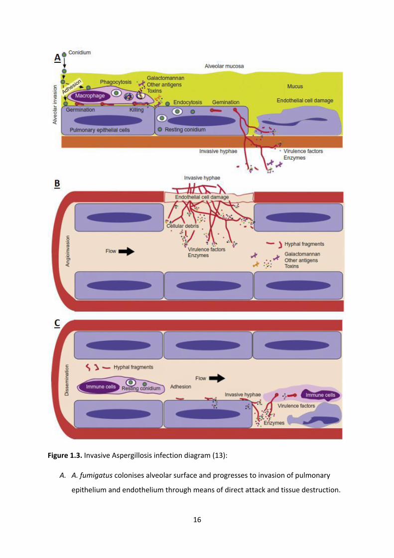
Figure 1.3. Invasive Aspergillosis infection diagram (13):
A. A. fumigatus colonises alveolar surface and progresses to invasion of pulmonary
epithelium and endothelium through means of direct attack and tissue destruction.

17
B. A. fumigatus penetrates blood capillaries in lung endothelium, leading to distribution
of hyphal fragments and virulence factors such as toxins in bloodstream.
C. A. fumigatus is spread by bloodstream and invades tissues in other parts of the body.
Immune cells may detect and react to the infection. Entering deep tissues requires
use of direct attack and tissue invasion virulence factors.
1.3.2 A. fumigatus virulence factors
The most widely studied A. fumigatus virulence factor is tissue destruction and cell surface
disruption, which is the result of proteolysis, carried out by enzymes known as proteases. In
A. fumigatus, the most widely studied proteases are ALP family alkaline serine proteases,
encoded mainly by the Alp1 gene. ALP enzymes act by degrading the extracellular matrix
and cell membranes, disrupting cytoskeletal actin filaments and destroying of focal cell
adhesion sites in lung alveolar epithelium. Serine proteases are also associated with
inflammation and countering immune reactions. Variations in the Alp1 gene results in
phenotypic differences in substrate specificity, resulting in differing tissue destruction
capabilities between A. fumigatus isolates. The progression of A. fumigatus from
colonization to invasive infection relies on hyphae invading through both alveolar and lung
endothelial tissue layers to enter vasculature, allowing A. fumigatus infection to spread to
deep organs (Figure 1.3) (24-26). In addition, deletion of the prT gene, which encodes the
transcription factor PrT, was found knock out the secretion of 6 proteases, including ALP.
Infectious A. fumigatus is more reliant on nutrients obtained through proteolysis than
saprophytic A. fumigatus. However, in vivo virulence results of prT deficient mutants in
neutropenic mice showed no difference with the wildtype (27).
A. fumigatus releases multiple toxins as virulence factors. Gliotoxin, helvolic acid and
fumagillin slow down ciliary beating by damaging airway epithelia, inhibiting the mucociliary
escalator and preventing the expulsion of A. fumigatus from lung epithelia. Gliotoxin also
induces host cell apoptosis and inhibits T-cell responses (especially Th1), phagocytosis and
oxidative stress. The mitF, asp f1 and res genes encode the toxins mitogillin, ribotoxin, and
restrictocin, respectively, inducing Type 1 hypersensitivity and cause cell death through
ribotoxicity and protein synthesis pathway inhibition. A. fumigatus, unlike less virulent

18
Aspergillus species, has sialic acid residues on its conidia, facilitating adhesion to and uptake
by lung epithelia. Asp-hemolysin, encoded by the aspHS gene, causes haemolysis and
inhibits immune reactions to A. fumigatus by preventing cytokine production (Figure 1.4)
(28, 29).
The conidial sizes of A. fumigatus are smaller than that of most fungi, facilitating easy entry
into lung tissue (8). Conidia size can affect fungal virulence, with some fungi such as Mucor
circinelloides exhibiting increased virulence with below-average conidia size due to easier
entry into lung tissue and evasion of immune detection through delayed hyphal growth (30).
In other fungi, such as Cryptococcus neoformans and Paracoccidioides brasiliensis, larger
conidia result in increased virulence due to increased resistance to phagocytosis (31).
Previous research into the effect of conidial sizes on Aspergillus virulence found that larger
conidia germinate faster in Aspergillus, however, there is an insignificant difference in
conidial sizes between strains, and that these con (32).
Reactive Oxygen Species (ROS) such as the superoxide ion (O2•−) and hydrogen peroxide
(H2O2) are by-products of aerobic metabolism with high reactivity and oxidative potential. In
addition, humans and most animals release ROS in the innate immune reaction known
oxidative stress (16, 23). ROS are harmful to all cells, including pathogens, due to their
effects on cells include DNA damage, protein inactivation, protein cross-linking and
fragmentation, and lipid peroxidation (33). Reactive Oxygen Species production may also be
stimulated by stress and by some antifungal agents such as Amphotericin B (34). Acute
exposure to high doses of ROS can help assess subtle differences in ROS resistance between
isolates. H2O2 solution is used to assess resistance to peroxide and has acts against target
cells externally. However, to assess resistance to superoxides, menadione is used, as its
metabolism releases superoxides, and this method allows assessment of how ROS affects
cells from within (35). To detoxify ROS, A. fumigatus secretes catalase and superoxide
dismutase enzymes, which decompose peroxides and superoxides, respectively. Catalases in
A. fumigatus are Cat1p, CatA/CatB and Cat2p encoded by the genes cat1, catB and cat2,
respectively. A. fumigatus has the Superoxide dismutase MnSod/Aspf6, encoded by
sod/Aspf6. Knockouts of catalase and superoxide dismutase function have been found to
reduce resistance to ROS acute exposure in various fungal pathogens, including Aspergillus.
However, A. fumigatus catalase and superoxide dismutase genes are generally poorly

19
expressed, potentially indicating influence from other genes on catalase and superoxide
dismutase secretion. Zinc needs to be acquired to produce functioning A. fumigatus
superoxide dismutases, however, iron starvation was not shown to affect A. fumigatus
superoxide dismutase activity (36-38).
Urease is a metalloenzyme which catalyses the hydrolysis of urea to ammonia and
carbamate. Urease was first suggested as a fungal virulence factor when bacteria such as
Helicobacter pylori and Proteus mirabilis were found to use urease to convert urea into
ammonia in order to neutralise acidic tissue environments. This allowed H. pylori to survive
in within the gastric mucosa, and urease-negative H. pylori strains were shown to be unable
to survive gastric mucosa (39). In the fungal pathogen Cryptococcus neoformans, strains
with urease production have demonstrated an ability to invade neuromuscular tissue and
promote Th2 adaptive immune responses instead of Th1, resulting in increased allergenicity
and inflammation, which attenuating immune responses targeted against the fungus. In
Aspergillus, urease enzymes have been isolated in A. nidulans, A. niger and A. fumigatus,
however, the role of urease in Aspergillus lung infection is yet to be determined (40-42).
Variations have been hypothesised in nutrient acquisition mechanisms to between
saprophytic and pathogenic A. fumigatus isolates, as nutrient acquisition by pathogenic
strains is challenged by competition from living cells. Pathogenic A. fumigatus requires an
ability to utilise a variety of carbon sources because supplies of glucose are usually severely
limited inside host tissues. During growth in living tissues, A. fumigatus has been shown to
increase secretion of the enzyme Isocitrate lyase (ICL), which allows utilization of Carbon
sources such as acetate and fatty acids, to the point where A. fumigatus can exclusively
obtain its carbon from fatty acids. ICL secretion in A. fumigatus has been shown to be
encouraged by exposure to macrophages (22). While nitrogen can be acquired through
proteolysis, A. fumigatus can also utilize ammonia as a nitrogen source and oxidise nitrates
into ammonia. Iron and zinc is required by A. fumigatus as essential micronutrients,
however, it cannot salvage these from digesting host cells. A. fumigatus acquires iron
through the siderophores fusarinine C and acetylfusarinine C to concentrate iron from
extracellular fluids such as blood, then uses the siderophores hyphal ferricrocin and conidial
hydroxylferroxcrocin to transfer iron into the cell. A. fumigatus obtains zinc through action

20
of zinc-binding protein Pra1, which scavenges zinc from host cells, then Zrt1, a plasma
membrane zinc transporter, moves the zinc into the cells (11).
Figure 1.4: Toxin virulence factors secreted by A. fumigatus. Gliotoxin, helvolic acid and
fumagillin, slow ciliary beating and prevent the mucociliary escalator from removing the
fungus from lung tissue. Verruculogen may aid in colonisation through tissue injury and
altering airway epithelia, and sialic acid may aid in adhesion to lung epithelial surfaces.
Serine proteases and other proteases then perform proteolysis on alveolar epithelia (29).

21
1.4 Insect models
1.4.1 Usefulness of live hosts in microbial pathogenicity research and associated
ethical concerns
Research into microbial pathogenicity in humans often uses animal models, usually
vertebrates such as mice and rats, due to similarity of metabolic pathways, anatomical
structures and immune reactions. Animal models are used to test the efficacy and side
effects of drugs and vaccines and study principles aiding in the development of new
therapies. Due to the pain, distress, and potential mortality of live hosts in microbial
pathogenicity research, ethical guidelines known as the 4Rs are used. They responsibility
(scientific correctness and integrity), reduction (finding ways to minimise the number of
animals experimented on), refinement (minimising pain and distress experienced by animal
test subjects) and replacement (replacing animal test subjects with less sentient species
whenever possible). The use of animal models fulfils the requirement for replacement of
human test subjects (Figure 1.5) (43, 44).
Testing of A. fumigatus pathogenicity has previously been performed in mouse models,
which discovered that immunocompetent mouse hosts do not produce results which mimic
clinical infections. However, neutropenic mouse hosts have demonstrated a similar pattern
of A. fumigatus infection, immunosuppression and pathogenesis as in IA clinical cases (19).
Due to the similarity between A. fumigatus infection in neutropenic mice and IA clinical
cases, this suggests the feasibility of replacement with invertebrate models, especially
insects, as they are lower order animals with only innate immune systems. Immune
responses in larval insect models show a high level of conservation with mammalian innate
immunity. For example, similar haemocyte function (e.g., phagocytosis, superoxide
production) to mammalian phagocytes, and comparable TLRs and response pathways (e.g.,
coagulation and melanisation) have been noted between insects and mammals. This means
that insect models will produce similar results to mammalian models regarding virulence of
fungal pathogens, and toxicity and in vivo efficacy of novel and conventional antifungal
drugs, and therefore, the use of insect models such as Galleria mellonella in fungal pathogen
research is increasing as they can replace more sentient model organisms such as mammals

22
(45). Aside from similarities in innate immune responses, insect models are advantageous
due to their exemption from ethical restrictions, ease of care, low maintenance
requirements, ease of breeding and short turnaround times. This means that laboratories do
not require ethical clearances, specialised equipment or specialised training to conduct
microbial pathogenicity testing in insect models. The ease of breeding and short turnaround
times allow for larger scale, more replicable experiments (43, 46, 47).
Figure 1.5: Principles of using animal models, such as the larvae of Tenebrio molitor, in
fungal pathogen research. The animal model can be infected with human fungal pathogens,
and the variations in certain virulence factors can be assessed by how they affect mortality
rates. How immunity and therapeutic treatments affect reaction to pathogens can also be
assessed. Insect models are feasible due to their comparable innate immune systems to
mammals, lack of ethical restrictions, ease of care and non-requirement of specialised
equipment (44).
1.4.2 Using Tenebrio molitor as an insect model for A. fumigatus pathogenicity
The insect model we used in this project is Tenebrio molitor, also known as mealworms.
Compared to G. mellonella, T. molitor has the advantages of ease of rearing, minimal
environmental pest potential, and a published transcriptome. Compared to other
invertebrate models, such as the nematode Caenorhabditis elegans, insect models can be
administered with a given dose of microbial inoculum through injections, instead of
inoculation through food. T. molitor can also be successfully reared at 37°C, which is human

23
body temperature and the activation temperature for most fungal virulence factors. Using
mortality counts, the virulence of each A. fumigatus isolate can be compared (48-50).
T. molitor also has a well-understood immune system and is known to have various innate
immune pathways related to those in human and mammalian innate immunity. The first line
of immune defences are the cuticle and digestive tract, which provides a hard barrier
against pathogens, and where high acidity hinders pathogen survival, respectively. Similar to
mammals, T. molitor can recognise fungal motifs using TLRs. This can direct immune cells,
such as hemocytes, to perform phagocytosis, the engulfment of foreign bodies by a single
hemocyte, in a manner comparable to vertebrate macrophages and neutrophils. Autophagy
can be used to eliminate excess hemocytes, including those which have phagocytosed
pathogens, assisting in the maintenance of stable populations of immune cells, a process
known as cellular homeostasis. In cases of pathogen burdens too high for phagocytosis,
nodulation and encapsulation processes are employed, involving the aggregation of
hemocytes around the target. Encapsulation by haemocytes is a precursor to the process of
melanization, which isolates aggregations of pathogens, killing the pathogens through
oxygen deprivation or the release of antimicrobial compounds, such as peptides with
antimicrobial activity (PAMs) and ROS, which is similar to neutrophil methods of action. In
addition, melanisation of the insect cuticle can confer a resistance to pathogen infection.
(Figure 1.6) (44, 46, 50).

24
Figure 1.6: Immune responses of Tenebrio molitor against pathogens. Cellular responses can
detect the pathogen and immune cells such as haemocytes directly kill pathogens through
phagocytosis and autophagy. In case of pathogen loads too large for phagocytosis and
autophagy, haemocytes can encapsulate pathogen aggregations, releasing compounds such
as PAMs and ROS to kill the pathogens, resulting in melanisation of the affected area (44).

25
1.5 Aims of project
To date, IA research has focused on host immunity, and there is currently a research gap
regarding which virulence factors in A. fumigatus determine whether a strain is a colonising
or invasive isolate. The aims of this project include:
Comparing the resistance to ROS acute sensitivity between A. fumigatus isolates.
Comparing the proteolytic capabilities of A. fumigatus isolates.
Comparing the urea hydrolysis capabilities of A. fumigatus isolates.
Comparing virulence levels between A. fumigatus isolates through mortality counts
in T. molitor models.
Determining which features may affect the virulence levels of A. fumigatus isolates,
and how this might determine which isolates are colonising or invasive.
The expected outcomes of this project include:
Improving diagnostic accuracy and providing more accurate prognoses regarding IA.
Improving understanding of A. fumigatus virulence resulting from this project can
help improve IA treatments, leading to socio-economic benefit by preventing organ
transplants being wasted to IA.

26
2. Materials and methods
2.1 Reagents and Chemicals
All reagents, chemicals and media are listed in Table 2.1. All concentrations of stock
solutions are specified within the table.
Table 2.1 Reagents, chemicals and media used in this project.
Reagents and
Chemicals
Abbreviation Supplier Stock Concentration
Malt extract ME Sigma-Aldrich 15g/L
Menadione - Sigma-Aldrich -
Hydrogen peroxide H2O2 Sigma-Aldrich 30% w/v
Liquid skim milk SM - -
Agar powder - Sigma-Aldrich 12 g/L (1X) or 24 g/L (2X)
Potato dextrose agar PDA Sigma-Aldrich 39 g/L
Phosphate Buffered
Saline
PBS Sigma-Aldrich 1X
Urea agar - Sigma-Aldrich 25.3 g/L
Sodium hydroxide NaOH Sigma-Aldrich 1 M

27
2.2 A. fumigatus strains
15 clinical isolates of Aspergillus fumigatus were obtained from Westmead Hospital and
labelled as AF01 to AF15. 10 of the isolates were found to have colonized patients, while 5
of the isolates were found to have caused infection (Table 2.2). These strains are considered
to be wildtypes.
These clinical isolates were then grown on PDA plates for 3 days at 37°C, after which the
conidia were suspended by swabbing with 5µL 0.05% Tween-20 solution. Conidial
suspensions are then filtered with a 0.22-micron filter, and plated on PDA plates for 1 day at
37°C. Colonies are then counted, then the conidial suspensions are diluted to 108 CFU/mL,
and plated again on PDA plates for 3 days at 37°C to confirm that the desired concentration
was reached (19).
Table 2.2: A. fumigatus clinical isolates used in this project, supplied by Westmead Hospital.
Strain Type
AF01 Colonizing
AF02 Colonizing
AF03 Colonizing
AF04 Colonizing
AF05 Colonizing
AF06 Colonizing
AF07 Colonizing
AF08 Colonizing
AF09 Colonizing
AF10 Colonizing
AF11 Infectious
AF12 Infectious
AF13 Infectious

28
AF14 Infectious
AF15 Infectious

29
2.3 Conidial size measurements
20µL of 106 CFU/mL A. fumigatus conidia suspension was added to a microscopy slide with a
graticule. The sizes of the conidia were measured using the graticule, and observed by light
microscopy, and 50 conidia from isolates AF01-10 were measured (51).

30
2.4 Acute exposure to Reactive Oxygen Species (ROS)
2.4.1 Menadione
Menadione was acquired from Sigma-Aldrich in a pure powder form and dissolved to form a
10 mM Menadione solution. 10 mM Menadione solution was further diluted in ethanol to
create 1mM (1000 µM) Menadione solution. 1000µL liquid media for acute exposure is
prepared as shown in Table 2.3 and inoculated with 100µL of 106 CFU/mL A. fumigatus
conidial suspension. The cultures are grown for 3 hours at 37°C, and plated onto PDA. These
plates are grown at 37°C for 24 hours and counted (37, 38).
Table 2.3 Preparation of liquid media for various concentrations of Menadione acute
exposure.
Concentration of
Menadione
106 CFU/mL conidial
suspension to add
1mM Menadione to
add
Malt broth
to add
0µM 100 µL 0 µL 900 µL
30µM 100 µL 30 µL 870 µL
50µM 100 µL 50 µL 850 µL
100µM 100 µL 100 µL 800 µL
2.4.2 Hydrogen peroxide
Hydrogen peroxide (H2O2) was acquired from Sigma-Aldrich as 30% w/v, or 8.821 molar,
solution. This is then diluted to 1 molar by adding 113 µL of 30% H2O2 to 887 µL of distilled
water. 1000µL liquid media for acute exposure is prepared as outlined in Table 2.4 and
inoculated with 100µL of 106 CFU/mL A. fumigatus conidial suspension. The cultures were
incubated for 3 hours at 37°C, a dilution series was prepared and 20 µL of each dilution was
plated onto PDA. These plates were incubated at 37°C for 24 hours and counted to
determine CFU/mL (52-54).
Table 2.4 Preparation of liquid media for various concentrations of Hydrogen Peroxide acute
exposure.

31
Concentration of
H2O2
106 CFU/mL conidial
suspension to add
1M H2O2 to add Malt broth to add
0mM 100 µL 0 µL 900 µL
30mM 100 µL 30 µL 870 µL
50mM 100 µL 50 µL 850 µL
100mM 100 µL 100 µL 800 µL
32
2.5 Visual analysis of fungal enzymatic activity
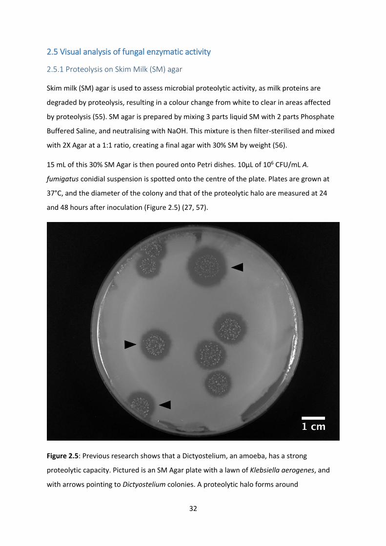
2.5.1 Proteolysis on Skim Milk (SM) agar
Skim milk (SM) agar is used to assess microbial proteolytic activity, as milk proteins are
degraded by proteolysis, resulting in a colour change from white to clear in areas affected
by proteolysis (55). SM agar is prepared by mixing 3 parts liquid SM with 2 parts Phosphate
Buffered Saline, and neutralising with NaOH. This mixture is then filter-sterilised and mixed
with 2X Agar at a 1:1 ratio, creating a final agar with 30% SM by weight (56).
15 mL of this 30% SM Agar is then poured onto Petri dishes. 10µL of 106 CFU/mL A.
fumigatus conidial suspension is spotted onto the centre of the plate. Plates are grown at
37°C, and the diameter of the colony and that of the proteolytic halo are measured at 24
and 48 hours after inoculation (Figure 2.5) (27, 57).
Figure 2.5: Previous research shows that a Dictyostelium, an amoeba, has a strong
proteolytic capacity. Pictured is an SM Agar plate with a lawn of Klebsiella aerogenes, and
with arrows pointing to Dictyostelium colonies. A proteolytic halo forms around
33
Dictyostelium colonies, indicating proteolytic activity, which is not exhibited by K. aerogenes
(55).
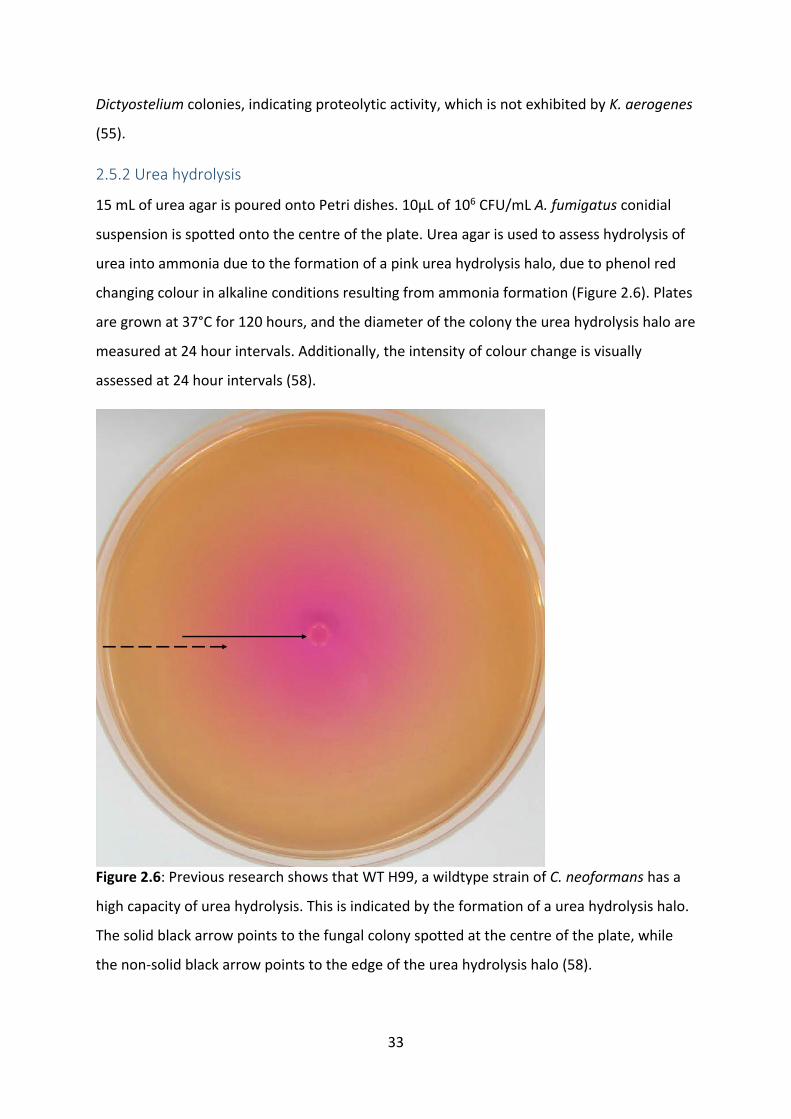
2.5.2 Urea hydrolysis
15 mL of urea agar is poured onto Petri dishes. 10µL of 106 CFU/mL A. fumigatus conidial
suspension is spotted onto the centre of the plate. Urea agar is used to assess hydrolysis of
urea into ammonia due to the formation of a pink urea hydrolysis halo, due to phenol red
changing colour in alkaline conditions resulting from ammonia formation (Figure 2.6). Plates
are grown at 37°C for 120 hours, and the diameter of the colony the urea hydrolysis halo are
measured at 24 hour intervals. Additionally, the intensity of colour change is visually
assessed at 24 hour intervals (58).
Figure 2.6: Previous research shows that WT H99, a wildtype strain of C. neoformans has a
high capacity of urea hydrolysis. This is indicated by the formation of a urea hydrolysis halo.
The solid black arrow points to the fungal colony spotted at the centre of the plate, while
the non-solid black arrow points to the edge of the urea hydrolysis halo (58).

34
2.6 Virulence tests in insect models
Mealworm (Tenebrio molitor) larvae weighing between 100 to 200 mg were selected.
Healthy individuals were used, as distinguished by a uniform yellow-brown colour and lack
of dark spots or greyish marks (Figure 2.7). Using a Hamilton Syringe (701 N, 26's gauge, 10
μL capacity), larvae are inoculated with 5 μL of 108 CFU/mL A. fumigatus conidial
suspension, the equivalent of around 5 x 10⁵ CFU per individual larva. The inoculum is
injected into the hemocoel, on the ventral side of the larva, at the second or third visible
sternite above the legs (Figure 2.8). Our preliminary testing determined that 5 x 10⁵ CFU per
individual larva was the optimal fungal load, with the larvae experiencing very little from
lesser fungal loads, and that 37°C is the ideal incubation temperature (Figure 3.12 A). After
inoculation, larvae are raised at 37 °C for 7 days, with survival assessed at 24-hour intervals.
Deaths of individual larvae are detected as a lack of movement and complete melanisation
of the body (Figure 3.12 B) (48). Mealworms are raised with a feed consisting of bran and
LSA mix (59).
Figure 2.7 Mealworm larvae used in this experiment need to weigh between 100-200 mg,
and have a uniform yellow-brown colour with no discolourations.

35
Figure 2.8: Injection of conidial suspension into the 3rd sternite of mealworm larvae, on the
ventral side.
36
3. Results
3.1 Conidial sizes
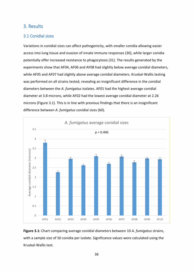
Variations in conidial sizes can affect pathogenicity, with smaller conidia allowing easier
access into lung tissue and evasion of innate immune responses (30), while larger conidia
potentially offer increased resistance to phagocytosis (31). The results generated by the
experiments show that AF04, AF06 and AF08 had slightly below average conidial diameters,
while AF05 and AF07 had slightly above average conidial diameters. Kruskal-Wallis testing
was performed on all strains tested, revealing an insignificant difference in the conidial
diameters between the A. fumigatus isolates. AF01 had the highest average conidial
diameter at 3.8 microns, while AF02 had the lowest average conidial diameter at 2.26
microns (Figure 3.1). This is in line with previous findings that there is an insignificant
difference between A. fumigatus conidial sizes (60).
Figure 3.1: Chart comparing average conidial diameters between 10 A. fumigatus strains,
with a sample size of 50 conidia per isolate. Significance values were calculated using the
Kruskal-Wallis test.
0
0.5
1
1.5
2
2.5
3
3.5
4
4.5
AF01 AF02 AF03 AF04 AF05 AF06 AF07 AF08 AF09 AF10
Ave
rage
co
nid
ial d
iam
eter
(m
icro
ns)
A. fumigatus average conidial sizes
p = 0.406

37
3.2 Acute exposure to ROS
Reactive Oxygen Species (ROS), such as the superoxide and peroxide ions, provide oxidative
stress as a non-specific immune response. Resistance to ROS acute exposure in A. fumigatus
are implicated in increased virulence and evasion of immune responses (28).
3.2.1 Menadione
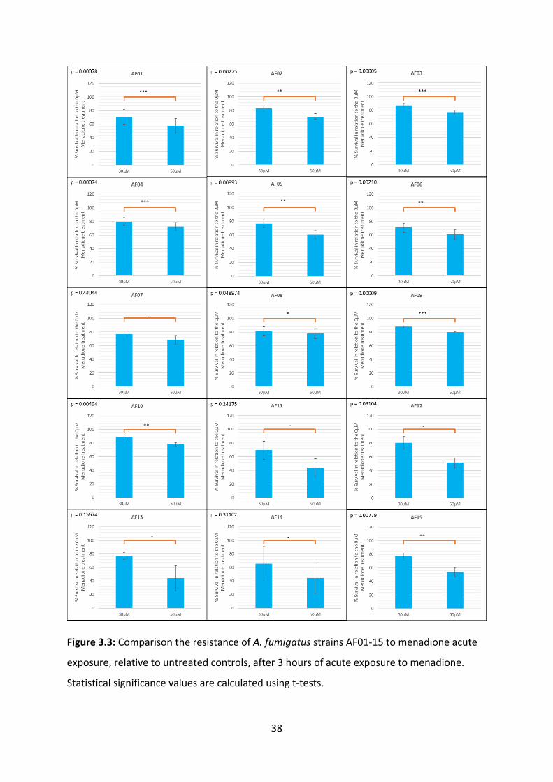
Menadione is an electron acceptor, which when metabolised, releases superoxide ions.
Differences in A. fumigatus resistance to menadione acute exposure has been linked to
secretion of superoxide dismutase enzymes (37). To investigate resistance to acute
exposure to menadione, we exposed 15 A. fumigatus clinical isolates to 30 and 50 µM of
menadione for 3 hours at 37°C then incubated the solutions on potato dextrose agar for 24
hours at 37°C. Five replicates were performed on AF01-AF10, and 3 replicates were
performed on AF11-15. The results generated demonstrate a negative effect between cell
survival and menadione concentration. Symbols denoting calculated p-values are shown in
Table 3.2. Although statistically insignificant results were found in AF11-14, this may
indicate an actual sensitivity level higher than that shown in our results. The highest
resistance to menadione acute exposure was found in AF10, AF09 and AF03, which had
survival rates at 30µM of 88.3%, 87.6% and 87.4% respectively. The lowest resistance to
menadione acute exposure was found in AF14, AF11 and AF01, which had survival rates at
30µM of 65.3%, 69.3% and 70.5% respectively (Figure 3.3). Our results are in line with our
expectations, as A. fumigatus wildtype strains exhibit high resistance to acute exposure to
superoxide ions due to the functioning of superoxide dismutase enzymes (37, 38).
Table 3.2: Symbols indicating calculated p-values.
P-value Symbol
Not significant (NS) -
<0.05 *
<0.01 **
<0.001 ***
38
Figure 3.3: Comparison the resistance of A. fumigatus strains AF01-15 to menadione acute
exposure, relative to untreated controls, after 3 hours of acute exposure to menadione.
Statistical significance values are calculated using t-tests.
39
3.2.2 Hydrogen peroxide
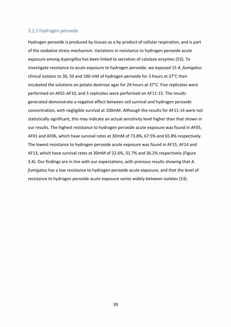
Hydrogen peroxide is produced by tissues as a by-product of cellular respiration, and is part
of the oxidative stress mechanism. Variations in resistance to hydrogen peroxide acute
exposure among Aspergillus has been linked to secretion of catalase enzymes (53). To
investigate resistance to acute exposure to hydrogen peroxide, we exposed 15 A. fumigatus
clinical isolates to 30, 50 and 100 mM of hydrogen peroxide for 3 hours at 37°C then
incubated the solutions on potato dextrose agar for 24 hours at 37°C. Five replicates were
performed on AF01-AF10, and 3 replicates were performed on AF11-15. The results
generated demonstrate a negative effect between cell survival and hydrogen peroxide
concentration, with negligible survival at 100mM. Although the results for AF11-14 were not
statistically significant, this may indicate an actual sensitivity level higher than that shown in
our results. The highest resistance to hydrogen peroxide acute exposure was found in AF05,
AF01 and AF06, which have survival rates at 30mM of 73.8%, 67.5% and 65.8% respectively.
The lowest resistance to hydrogen peroxide acute exposure was found in AF15, AF14 and
AF13, which have survival rates at 30mM of 22.6%, 31.7% and 36.2% respectively (Figure
3.4). Our findings are in line with our expectations, with previous results showing that A.
fumigatus has a low resistance to hydrogen peroxide acute exposure, and that the level of
resistance to hydrogen peroxide acute exposure varies widely between isolates (53).

40
Figure 3.4: Comparison the resistance of A. fumigatus strains AF01-15 to menadione acute
exposure, relative to untreated controls, after 3 hours of acute exposure to menadione.
Statistical significance values are calculated using t-tests.

41
3.3 Analysis of fungal enzymatic activity
3.3.1 Proteolysis on Skim Milk (SM) agar
Proteases are important virulence factors in A. fumigatus, playing a role in tissue
destruction and invasion. Loss of protease function has been implicated in decreased
virulence A. fumigatus virulence in vivo (27). To assess proteolysis, we plated 15 clinical
isolates of A. fumigatus on SM agar for 72 hours at 37°C, and monitored the growth rates of
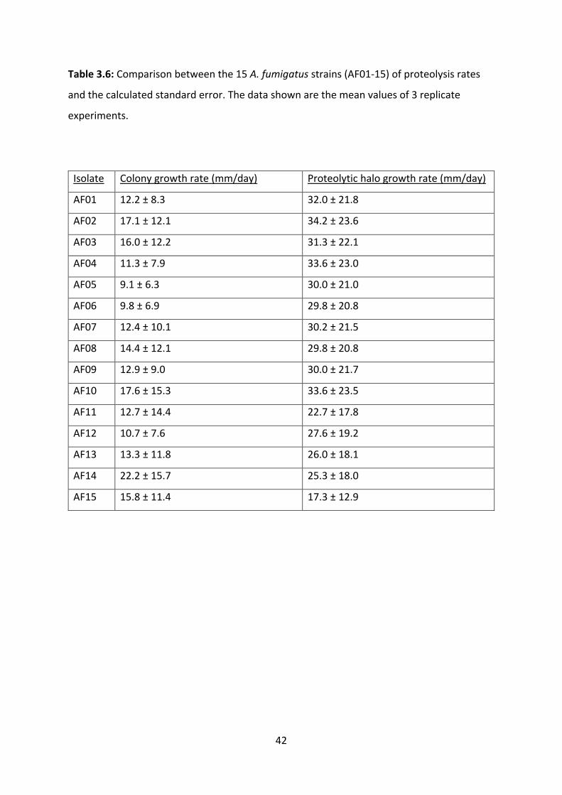
the colony and the proteolytic halo (Figure 3.5). The highest rates of proteolysis were found
in isolates AF02, AF04 and AF10, at 34.2, 33.6 and 33.6 mm per day, respectively. The lowest
rates of proteolysis were found in isolates AF15, AF11 and AF14, at 17.3, 22.7 and 25.3 mm
per day, respectively (Table 3.6). As our isolates are all wildtypes, the large differences in
proteolytic activity of was unexpected. This may indicate that proteolytic activity can be
influenced by factors other than protease function knockout, for example, environmental
conditions such as pH or nitrogen availability (27).
Figure 3.5: Image showing the SM Agar plate for isolate AF05 at 72 hours. The colony is
visible as the white area in the centre, while the proteolytic halo is visible as the clearing
around the colony.
42
Table 3.6: Comparison between the 15 A. fumigatus strains (AF01-15) of proteolysis rates
and the calculated standard error. The data shown are the mean values of 3 replicate
experiments.
Isolate Colony growth rate (mm/day) Proteolytic halo growth rate (mm/day)
AF01 12.2 ± 8.3 32.0 ± 21.8
AF02 17.1 ± 12.1 34.2 ± 23.6
AF03 16.0 ± 12.2 31.3 ± 22.1
AF04 11.3 ± 7.9 33.6 ± 23.0
AF05 9.1 ± 6.3 30.0 ± 21.0
AF06 9.8 ± 6.9 29.8 ± 20.8
AF07 12.4 ± 10.1 30.2 ± 21.5
AF08 14.4 ± 12.1 29.8 ± 20.8
AF09 12.9 ± 9.0 30.0 ± 21.7
AF10 17.6 ± 15.3 33.6 ± 23.5
AF11 12.7 ± 14.4 22.7 ± 17.8
AF12 10.7 ± 7.6 27.6 ± 19.2
AF13 13.3 ± 11.8 26.0 ± 18.1
AF14 22.2 ± 15.7 25.3 ± 18.0
AF15 15.8 ± 11.4 17.3 ± 12.9

43
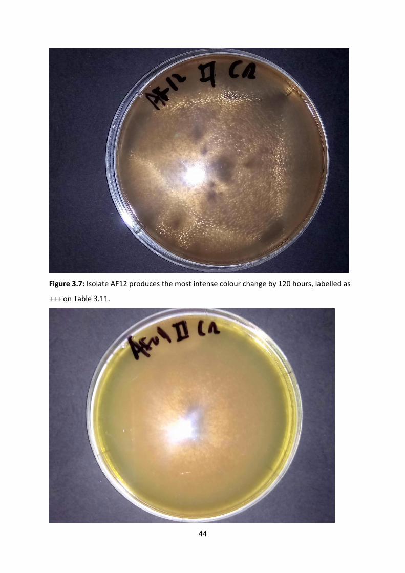
3.3.2 Urea hydrolysis
Survival of A. fumigatus in the acidic environment of lung mucosa requires a method of
neutralising the acidic environment. The enzyme urease has been found to play a role of
neutralising other acidic environments for pathogens such as C. neoformans. Urease
hydrolyses urea into ammonia and carbamate, which raises the pH. To assess urea
hydrolysis, A. fumigatus isolates were plated on urea agar for 120 hours at 37°C, and
measurements of colony growth rates, urea hydrolysis halos and the intensity of colour
change were taken (39, 42). The level of colour change was assessed visually, with AF12
producing the most intense colour change, while isolates AF03, AF11 and AF15 do not
create colour changes at all (Figures 3.7-3.10). The highest rates of urea hydrolysis were
found in isolates AF12, AF09 and AF14, at 14.1, 11.1 and 11.0 mm per day, respectively
(Table 3.11). The lowest rates of proteolysis were found in isolates AF03, AF11 and AF15, all
of which failed to produce a urea hydrolysis halo. Since proteolysis is highest under alkaline
conditions, our urea hydrolysis results resonate with our proteolysis results, with the
isolates with low urea hydrolysis capacity generally also having a low proteolysis capacity
(61).
44
Figure 3.7: Isolate AF12 produces the most intense colour change by 120 hours, labelled as
+++ on Table 3.11.

45
Figure 3.8: Isolate AF09 produces a moderate colour change by 120 hours, labelled as ++ on
Table 3.11.
Figure 3.9: Isolate AF02 produces a mild colour change by 120 hours, labelled as + on Table
3.11.

46
Figure 3.10: Isolate AF11 produces no colour change by 120 hours, labelled as - on Table
3.11.
Table 3.11: Comparison between the 15 A. fumigatus strains (AF01-15) of urea hydrolysis
rates and the calculated standard error. The data shown are the mean values of 3 replicate
experiments. The intensity of the colour change can be seen in Figures 3.14-3.17.
Isolate Colony growth rate
(mm/day)
Urea hydrolysis halo growth
rate (mm/day)
Intensity of
colour change
AF01 13.7 ± 11.0 7.0 ± 6.3 ++
AF02 11.6 ± 9.8 4.7 ± 4.4 +
AF03 11.2 ± 9.4 0 ± 0 -
AF04 13.3 ± 11.1 9.0 ± 7.7 +
AF05 13.1 ± 11.3 7.5 ± 6.6 +
AF06 13.6 ± 11.5 7.7 ± 7.2 +
AF07 14.7 ± 11.8 8.3 ± 7.5 ++

47
AF08 15.9 ± 12.8 9.6 ± 9.1 ++
AF09 15.7 ± 12.8 11.1 ± 10.3 ++
AF10 14.6 ± 12.3 10.1 ± 9.0 ++
AF11 12.7 ± 10.4 0 ± 0 -
AF12 16.5 ± 13.3 14.1 ± 12.4 +++
AF13 14.7 ± 12.1 9.7 ± 9.1 ++
AF14 15.3 ± 12.5 11.0 ± 10.3 ++
AF15 10.4 ± 8.6 0 ± 0 -

48
3.4 Virulence tests in insect models
Insects are used as animal models for fungal virulence assessments. For this experiment, the
mealworm Tenebrio molitor was used as a model organism, due to its well-understood
innate immune system, ease of rearing, and ease of replicating the experiment. Virulence
was assessed through daily counts of how many worms remained alive at 24-hour intervals
(44). We conducted preliminary experiments for protocol optimisation, finding that 37°C is
the ideal temperature for incubation and that the optimal fungal load is 5µL 105 CFU/mL of
A. fumigatus conidial suspension. This experiment involved inoculating T. molitor larvae
with A. fumigatus conidial suspensions and incubating the larvae at 37 °C, for 7 days (Figure
3.13) (48). The survival curves generated by 4 replicated experiments shows that A.
fumigatus causes a mortality “baseline” to be reached in about 3 days. The highest mortality
rates occurred in AF04, AF05, and AF01, all of which had a 6% survival rate. The lowest
mortality rates occurred in AF03, AF08 and AF09, with survival rates of 17%, 14% and 13%,
respectively. Although AF13 does exceed the mortality rate of AF04 by day 7, it does not
cause mortality as quickly. (Figure 3.14-3.18). Our results were unexpected, as the invasive
isolates generally had a lower mortality rate in T. molitor than colonising isolates, despite
the T. molitor innate immune system being comparable to that of neutropenic mice and
neutropenic human patients (19, 45).

49
Figure 3.12: Results of preliminary experiments, used for protocol optimisation:

50
A. Virulence of A. fumigatus in T. molitor activates at 37°C, which is human body
temperature.
B. Red arrows point to dead larvae, which are completely melanised and motionless.
Figure 3.13: T. molitor larval mortality count for AF01 at day 1. Individuals which have
turned black are dead. The remaining number of live worms, which are yellow-brown, are
counted as living.

51
Figure 3.14: Survival curves T. molitor larvae inoculated with isolates AF01-03. This is the
average of 4 replicated virulence tests. For comparison, the orange line indicates AF03, the
least virulent isolate, and the blue line indicates AF04, the most virulent isolate.

52
Figure 3.15: Survival curves T. molitor larvae inoculated with isolates AF04-06. This is the
average of 4 replicated virulence tests. For comparison, the orange line indicates AF03, the
least virulent isolate, and the blue line indicates AF04, the most virulent isolate.

53
Figure 3.16: Survival curves T. molitor larvae inoculated with isolates AF07-09. This is the
average of 4 replicated virulence tests. For comparison, the orange line indicates AF03, the
least virulent isolate, and the blue line indicates AF04, the most virulent isolate.

54
Figure 3.17: Survival curves T. molitor larvae inoculated with isolates AF10-12. This is the
average of 4 replicated virulence tests. For comparison, the orange line indicates AF03, the
least virulent isolate, and the blue line indicates AF04, the most virulent isolate.

55
Figure 3.18: Survival curves T. molitor larvae inoculated with isolates AF13-15. This is the
average of 4 replicated virulence tests. For comparison, the orange line indicates AF03, the
least virulent isolate, and the blue line indicates AF04, the most virulent isolate.

56
3.5 Results summary
To summarise which strain of A. fumigatus had the highest virulence level, a scoring system
was devised. The rankings of each strain regarding menadione acute exposure resistance,
hydrogen peroxide acute exposure resistance, proteolysis, urea hydrolysis and T. molitor
mortality are compiled. By summing up the ranks, this produces a score, with which a lower
score indicates a higher total virulence. The results found that AF04, AF10 and AF05 were
the most virulent strains, while AF15, AF11 and AF14 were the least virulent strains (Table
3.19). This does not align with our hypothesis that invasive isolates would exhibit a higher
total virulence score.
Table 3.19: The results of each strain on the various experiments are ranked. The ranks are
then totalled to generate a score. A lower score indicates a higher level of virulence,
allowing the determination of a total virulence level.
Isolate Resistance
to
menadione
(rank)
Resistance
to
hydrogen
peroxide
(rank)
Proteolysis
(rank)
Urea
hydrolysis
(rank)
T.
molitor
mortality
(rank)
Score Total
virulence
(rank)
AF01 13 2 4 11 =1 31 5
AF02 4 6 1 12 =11 34 =6
AF03 3 4 5 =13 15 40 =8
AF04 7 5 =2 7 =1 22 1
AF05 9 1 =7 10 =1 28 3
AF06 12 3 =9 9 10 43 12
AF07 11 9 6 8 =6 40 =8
AF08 5 8 =9 6 14 42 11
AF09 2 10 =7 2 13 34 =6
AF10 1 11 =2 4 =8 26 2
AF11 14 12 14 =13 =6 59 14
AF12 6 7 11 1 5 30 4
AF13 8 13 12 5 3 41 10

57
AF14 15 14 13 3 =8 53 13
AF15 10 15 15 =13 =11 64 15

58
4. Discussion
4.1 Conidial sizes
Other studies have found that conidial size in A. fumigatus shows no significant differences,
and germination rate has been found to be unrelated to virulence in insect models (60). In
our results, size differences between the conidia, as shown by Figure 3.1, were found to be
not significant. Smaller conidia have been shown to result in higher virulence in M.
circinelloides pathogens, due to ease of entry into respiratory tissues and avoidance of
immune detection (30), while C. neoformans and P. brasiliensis exhibit greater virulence
with larger conidia due to increased phagocytosis resistance.
The use of inoculation through injection may negate the virulence advantages of smaller
conidia, as the conidia enter directly into hemocoel of T. molitor larvae, which does not
simulate the natural process of pathogen entry through inhalation. Alternatives to injection
include forced inhalation of A. fumigatus conidia and oral administration of autoclaved
feeds inoculated with A. fumigatus. Although this method is less precise regarding the
number of fungal conidia enter each individual larva, it may allow better simulation of entry
into tissues (19, 62).
4.2 Reactive Oxygen Species (ROS)
ROS include peroxides and superoxide ions, playing a key role of the innate immune
response known as oxidative stress. Menadione is a powerful agent of oxidative stress,
forming superoxide radicals by redox cycling in cells. Detoxification of ROS are carried out by
enzymes superoxide dismutase and catalase, which detoxify superoxides and peroxides,
respectively, and the thiol-containing antioxidant glutathione (GSH). The activity of these
enzymes and the production of GSH have been shown to confer a resistance to oxidative
stress and increased virulence in insect models (36, 63, 64).
Our findings show that acute exposure to menadione reduces the survival of A. fumigatus
isolates, with survival rates between 65.3% and 88.3% in 30µM menadione, as shown in
Figures 3.2-3.6. AF10 was found to have the most resistance to menadione acute exposure,
while AF14 was found to have the least. Menadione resistance is known to vary between
various fungal pathogens, however, Aspergillus is noted to be less affected by menadione

59
acute exposure than other pathogenic fungi, with concentrations as high as 500µM required
to cause significant reductions in the viability of some Aspergillus species (65). Menadione
may not be an optimal chemical to use for comparing survival in response to ROS acute
exposure between A. fumigatus clinical isolates, due to the low effect of menadione
exposure on A fumigatus, and because menadione has previously only been used in studies
comparing wild types to mutants with a superoxide dismutase function knockout (37).
Our findings show that acute exposure to H2O2 causes a marked reduction on the survival
between various A. fumigatus isolates. The highest resistance to H2O2 acute exposure was
found in AF05, AF01 and AF06, while the lowest resistance was found in AF15, AF14 and
AF13, respectively (Figures 3.7-3.10). Our results resonate with previous findings where
resistance to H2O2 acute exposure did not influence virulence in insect models, and that the
isolates from invasive infection exhibited lower resistance to H2O2 than colonising isolates.
This may indicate a similar phenomenon to what has been observed in Aspergillus flavus,
where atoxigenic strains exhibited higher resistance to oxidative stress than toxigenic strains
(60, 66). Toxigenic strains of A. flavus were found to respond to oxidative stress by releasing
mycotoxins such as aflatoxin and are likely more virulent in human hosts. This may mean
that an assay of toxin production may be necessary to analyse the effect of acute exposure
to H2O2 on A. fumigatus virulence (53). Toxins of potential interest in A. fumigatus include
gliotoxin, fumagillin, fumigacin, fumitremorgin A and Asp-hemolysin. In particular, gliotoxin
has been shown to be released during oxidative stress, inhibiting superoxide release, and
aiding tissue penetration (24, 67).
4.3 Fungal enzymatic activity
4.3.1 Proteolysis on SM Agar
Proteolysis in A. fumigatus has been linked to various protease enzymes such as ALP1 and
Asp f13. These proteases disrupt the extracellular matrix, provokes airway hyper-
responsiveness and interfere with Ca2+ signalling, causing increased bronchoconstriction.
This leads to chronic lung inflammation due to the immune response to inhaled allergens.
Fungal allergenicity caused by the Pen c13 alkaline serine protease from Penicillium citrinum
has been shown to cause in chronic lung inflammation, leading to dysfunction of the
respiratory epithelial barrier and facilitating tissue invasion by the fungus (26).

60
Proteolysis results on SM Agar showed that while colony growth rates remained similar
between the colonising and invasive isolates, the proteolysis of the invasive isolates was
lesser than that of the colonising isolates (Table 3.12). Although proteolysis on SM Agar was
successfully used to analyse protease secretion in A. fumigatus, mutant strains with
impaired proteolysis exhibited no difference in virulence in leukopenic mouse models (27).
However, genes encoding proteases, such as prT, have been noted to be absent in the less
virulent species A. nidulans, potentially highlighting that its presence helps facilitate A.
fumigatus virulence. In addition, our SM Agar results may have been affected by the normal
atmospheric conditions, a which has higher ratio of oxygen to carbon dioxide compared to
respiratory tissue (26, 57, 68). Further research needs to be undertaken to determine
diversity in A. fumigatus genes, such as alp1 and prT which may affect protease secretions.
4.3.2 Urea Hydrolysis
Respiratory tissue is acidic, as a non-specific immune defence, however, an increased ability
to neutralise the low pH of the lung mucosa, allows fungal pathogens to survive in lung
mucosa. Urease enzymes confer an ability to neutralise low pH environments, by
hydrolysing urea into ammonia. A. fumigatus is known to be urease positive, and in C.
neoformans, urease activity has been linked to respiratory and nervous system invasion. In
addition, extracellular protease secretion in A. fumigatus requires a basic pH (42, 58, 61).
Our urea hydrolysis results on urea agar showed that the invasive isolates, especially AF12,
generally have a greater capacity for urea hydrolysis than colonising isolates (Table 3.13),
potentially indicating an improved ability to survive inside and infect human hosts compared
to other strains. Previous research with other fungal pathogens such as Cryptococcus
neoformans indicates that urease-positive strains altered the structure of microvessel walls,
facilitating microvascular sequestration and spread through the bloodstream. Urease-
positive C. neoformans strains also avoid triggering an inflammatory response, thereby
avoiding some features of the innate immune response (40). In addition, urease-positive
strains of C. neoformans and Coccidioides posadasii also discourage the anti-pathogenic Th1
adaptive immune response, instead encouraging the Th2 adaptive immune response, which
is associated with allergy and hypersensitivity. This may result in a discrepancy between the
virulence results in human patients and insect models, which lack an adaptive immune
system (42).
61
4.4 Virulence tests in insect models
Insect models are ideal tools for studying in vivo virulence of fungal pathogens, due to ease
of care and low ethical barriers to their use (69). In vivo results in insect models are
applicable to cases of infection in human hosts, as much of the immune response against A.
fumigatus is from the innate immune system. In vivo results can also differ greatly from in
vitro results, for example, A. fumigatus strains with high gliotoxin production generally
exhibit little growth in vitro but cause high mortality rates in other insect models, such as
Galleria mellonella (70, 71).
While the highest mortality in T. molitor insect models occurred in AF01, AF04 and AF05, the
next highest mortality rates were recorded in AF13 and AF12 (Table 3.20). This may be
related to the high H2O2 susceptibility of AF13 and AF12, which may indicate that they
respond to oxidative stress by releasing toxins such as gliotoxin (66). The high mortality
caused by AF01, AF12 and AF13 may also be influenced by their urease production. The high
mortality rates caused by AF04 and AF05 despite their low capacity for urea hydrolysis may
be a result of high proteolytic activity and high resistance to hydrogen peroxide. Our results
resonate with previous findings that the infectious isolates such as AF15 are less virulent in
insect models than environmental or colonising isolates such as AF04. This indicates that
adaptive immunity may also affect A. fumigatus pathogenicity in humans. Differences in
respiratory system structure may also play a role in the discrepancy between the
pathogenicity of our isolates in human hosts and the results of our virulence tests in insect
models (71, 72).

62
5. Conclusions
The results of our study suggest that A. fumigatus isolates known to cause invasive
infections, particularly AF12 and AF13, generally have an above average urea hydrolysis
capacity and a below average resistance to hydrogen peroxide acute exposure compared to
isolates which only colonising infections. This suggests that the results of other tests we
conducted may not reflect pathogenesis in live human hosts. For example, an above average
urea hydrolysis capacity results in an increased ability to raise pH, which aids in the activity
of proteases. In addition, toxigenic A. flavus strains have been shown to have a lower
resistance to H2O2 acute exposure, but release toxins in response to H2O2 exposure.
Future directions of this research include genotypic analyses, use of tissue culture and
alternative inoculation methods. Genotypic analyses can be performed on the known the.
fumigatus genes associated with the secretion of toxins, proteases, urease, catalase,
superoxide dismutase. Genotypic analyses may determine how the diversity and expression
of these genes influenced the results of our study. Use of tissue culture can be used to study
the progression of A. fumigatus infection between different types of respiratory tissues.
Additionally, structural features of human respiratory tissue differ from that of insect
models, and use of tissue culture replaces the need for testing in live models. Alternative
inoculation methods such as force-feeding and forced inhalation may also need to be
assessed, as injection into the hemocoel does not mirror natural infection mechanisms, and
injections may not accurately show how conidial size and interference with the mucociliary
escalator affect virulence.
63
References
1. Payne WC. Medical Mycology. In: Kiser KM, Payne WC, Taff TA, editors. Clinical
laboratory microbiology : a practical approach. Upper Saddle River, N.J.: Upper Saddle River,
N.J. : Prentice Hall; 2011. p. 684-733.
2. Willey JMa. Prescott's microbiology. Ninth edition. ed. Sherwood Lc-a, Woolverton
CJc-a, editors: New York, NY : McGraw-Hill, 2014; 2014.
3. Anaissie EJ, McGinnis MR, Pfaller MA. Clinical mycology. 2nd ed. ed. Edinburgh:
Edinburgh : Churchill Livingstone; 2009.
4. Romani L. Immunity to fungal infections. Nature Reviews Immunology. 2011;11:275.
5. Cunha C, Aversa F, Romani L, Carvalho A. Human genetic susceptibility to invasive
aspergillosis. PLoS Pathog. 2013;9(8):e1003434.
6. Ballard E, Melchers WJG, Zoll J, Brown AJP, Verweij PE, Warris A. In-host
microevolution of Aspergillus fumigatus: A phenotypic and genotypic analysis. Fungal Genet
Biol. 2018;113:1-13.
7. Wyatt TT, Wosten HA, Dijksterhuis J. Fungal spores for dispersion in space and time.
Adv Appl Microbiol. 2013;85:43-91.
8. Latge JP. Aspergillus fumigatus and aspergillosis. Clin Microbiol Rev. 1999;12(2):310-
50.
9. Brown GD, Denning DW, Levitz SM. Tackling human fungal infections. Science.
2012;336(6082):647.
10. Brown SP, Cornforth DM, Mideo N. Evolution of virulence in opportunistic
pathogens: generalism, plasticity, and control. Trends Microbiol. 2012;20(7):336-42.
11. Casadevall Ae, Mitchell APe, Berman Je, Kwon-Chung KJe, Perfect JRe, Heitman Je.
Human fungal pathogens: Cold Spring Harbor, New York : Cold Spring Harbor Laboratory
Press, 2014; 2014.

64
12. Leenders AC, van Belkum A, Behrendt M, Luijendijk A, Verbrugh HA. Density and
molecular epidemiology of Aspergillus in air and relationship to outbreaks of Aspergillus
infection. J Clin Microbiol. 1999;37(6):1752-7.
13. Kousha M, Tadi R, Soubani AO. Pulmonary aspergillosis: a clinical review. Eur Respir
Rev. 2011;20(121):156-74.
14. Hope WW, Walsh TJ, Denning DW. The invasive and saprophytic syndromes due to
Aspergillus spp. Medical Mycology. 2005;43(Supplement_1):S207-S38.
15. Kontoyiannis DP, Marr KA, Park BJ, Alexander BD, Anaissie EJ, Walsh TJ, et al.
Prospective surveillance for invasive fungal infections in hematopoietic stem cell transplant
recipients, 2001-2006: overview of the Transplant-Associated Infection Surveillance
Network (TRANSNET) Database. Clin Infect Dis. 2010;50(8):1091-100.
16. Segal BH. Aspergillosis. N Engl J Med. 2009;360(18):1870-84.
17. Maschmeyer G, Haas A, Cornely OA. Invasive aspergillosis: epidemiology, diagnosis
and management in immunocompromised patients. Drugs. 2007;67(11):1567-601.
18. Weber DJ, Peppercorn A, Miller MB, Sickbert-Benett E, Rutala WA. Preventing
healthcare-associated Aspergillus infections: review of recent CDC/HICPAC
recommendations. Med Mycol. 2009;47 Suppl 1:S199-209.
19. Kavanagh K. Medical mycology : cellular and molecular techniques. Chichester:
Chichester : John Wiley & Sons; 2007.
20. Barberan J, Sanz F, Hernandez JL, Merlos S, Malmierca E, Garcia-Perez FJ, et al.
Clinical features of invasive pulmonary aspergillosis vs. colonization in COPD patients
distributed by gold stage. The Journal of infection. 2012;65(5):447-52.
21. Guinea J, Torres-Narbona M, Gijon P, Munoz P, Pozo F, Pelaez T, et al. Pulmonary
aspergillosis in patients with chronic obstructive pulmonary disease: incidence, risk factors,
and outcome. Clin Microbiol Infect. 2010;16(7):870-7.
22. Cooney NM, Klein BS. Fungal adaptation to the mammalian host: it is a new world,
after all. Curr Opin Microbiol. 2008;11(6):511-6.
23. Segal AW. How neutrophils kill microbes. Annu Rev Immunol. 2005;23:197-223.

65
24. Abad A, Fernandez-Molina JV, Bikandi J, Ramirez A, Margareto J, Sendino J, et al.
What makes Aspergillus fumigatus a successful pathogen? Genes and molecules involved in
invasive aspergillosis. Rev Iberoam Micol. 2010;27(4):155-82.
25. Watson DS, Feng X, Askew DS, Jambunathan K, Kodukula K, Galande AK. Substrate
specifity profiling of the Aspergillus fumigatus proteolytic secretome reveals consensus
motifs with predominance of Ile/Leu and Phe/Tyr. PLoS One. 2011;6(6):e21001.
26. Balenga NA, Klichinsky M, Xie Z, Chan EC, Zhao M, Jude J, et al. A fungal protease
allergen provokes airway hyper-responsiveness in asthma. Nat Commun.
2015;6(6763):6763.
27. Sharon H, Hagag S, Osherov N. Transcription factor PrtT controls expression of
multiple secreted proteases in the human pathogenic mold Aspergillus fumigatus. Infect
Immun. 2009;77(9):4051-60.
28. Rementeria A, Lopez-Molina N, Ludwig A, Vivanco AB, Bikandi J, Ponton J, et al.
Genes and molecules involved in Aspergillus fumigatus virulence. Rev Iberoam Micol.
2005;22(1):1-23.
29. Dagenais TR, Keller NP. Pathogenesis of Aspergillus fumigatus in Invasive
Aspergillosis. Clin Microbiol Rev. 2009;22(3):447-65.
30. Li CH, Cervantes M, Springer DJ, Boekhout T, Ruiz-Vazquez RM, Torres-Martinez SR,
et al. Sporangiospore size dimorphism is linked to virulence of Mucor circinelloides. PLoS
Pathog. 2011;7(6):e1002086.
31. Wang L, Lin X. Morphogenesis in fungal pathogenicity: shape, size, and surface. PLoS
Pathog. 2012;8(12):e1003027.
32. Teertstra WR, Tegelaar M, Dijksterhuis J, Golovina EA, Ohm RA, Wosten HAB.
Maturation of conidia on conidiophores of Aspergillus niger. Fungal Genet Biol. 2017;98:61-
70.
33. Aguirre J, Hansberg W, Navarro R. Fungal responses to reactive oxygen species.
Medical Mycology. 2006;44(s1):S101-S7.

66
34. Devasagayam TP, Tilak JC, Boloor KK, Sane KS, Ghaskadbi SS, Lele RD. Free radicals
and antioxidants in human health: current status and future prospects. The Journal of the
Association of Physicians of India. 2004;52:794-804.
35. Kawamura F, Hirashima N, Furuno T, Nakanishi M. Effects of 2-methyl-1,4-
naphtoquinone (menadione) on cellular signaling in RBL-2H3 cells. Biological &
pharmaceutical bulletin. 2006;29(4):605-7.
36. Paris S, Wysong D, Debeaupuis JP, Shibuya K, Philippe B, Diamond RD, et al.
Catalases of Aspergillus fumigatus. Infect Immun. 2003;71(6):3551-62.
37. Lambou K, Lamarre C, Beau R, Dufour N, Latge JP. Functional analysis of the
superoxide dismutase family in Aspergillus fumigatus. Mol Microbiol. 2010;75(4):910-23.
38. Meletiadis J, Mouton JW, Meis JF, Bouman BA, Donnelly JP, Verweij PE, et al.
Colorimetric assay for antifungal susceptibility testing of Aspergillus species. J Clin Microbiol.
2001;39(9):3402-8.
39. Cox GM, Mukherjee J, Cole GT, Casadevall A, Perfect JR. Urease as a virulence factor
in experimental cryptococcosis. Infection and immunity. 2000;68(2):443-8.
40. Olszewski MA, Noverr MC, Chen GH, Toews GB, Cox GM, Perfect JR, et al. Urease
expression by Cryptococcus neoformans promotes microvascular sequestration, thereby
enhancing central nervous system invasion. The American journal of pathology.
2004;164(5):1761-71.
41. Yu J-J, Smithson SL, Thomas PW, Kirkland TN, Cole GT. Isolation and characterization
of the urease gene (URE) from the pathogenic fungus Coccidioides immitis. Gene.
1997;198(1):387-91.
42. Rutherford JC. The emerging role of urease as a general microbial virulence factor.
PLoS pathogens. 2014;10(5):e1004062-e.
43. Arora T, Mehta AK, Joshi V, Mehta KD, Rathor N, Mediratta PK, et al. Substitute of
Animals in Drug Research: An Approach Towards Fulfillment of 4R's. Indian journal of
pharmaceutical sciences. 2011;73(1):1-6.

67
44. Canteri de Souza P, Custódio Caloni C, Wilson D, Sergio Almeida R. An Invertebrate
Host to Study Fungal Infections, Mycotoxins and Antifungal Drugs: Tenebrio molitor. Journal
of Fungi. 2018;4(4).
45. Kavanagh K, Sheehan G. The Use of Galleria mellonella Larvae to Identify Novel
Antimicrobial Agents against Fungal Species of Medical Interest. Journal of Fungi. 2018;4(3).
46. J LS, Scorzoni L, H CdO, C MM, P AA, Fusco-Almeida AM, et al. Applications of
Invertebrate Animal Models to Dimorphic Fungal Infections. J Fungi (Basel). 2018;4(4).
47. Nicole Fenwick GG, Clément Gauthier. The welfare of animals used in science: How
the “Three Rs” ethic guides improvements. Can Vet J. 2009;50(5):523–30.
48. de Souza PC, Morey AT, Castanheira GM, Bocate KP, Panagio LA, Ito FA, et al.
Tenebrio molitor (Coleoptera: Tenebrionidae) as an alternative host to study fungal
infections. J Microbiol Methods. 2015;118:182-6.
49. Ribeiro NTGM. Tenebrio molitor for food or feed: Rearing conditions and the effect
of pesticides on its performance. In: Coimbra ESAd, editor. 2017. p. 1-70.
50. Moret Y, Siva-Jothy MT. Adaptive innate immunity? Responsive-mode prophylaxis in
the mealworm beetle, Tenebrio molitor. Proc Biol Sci. 2003;270(1532):2475-80.
51. Raper KB. The genus aspergillus. Fennell DI, editor. Baltimore: Baltimore : Williams &
Wilkins; 1965.
52. Dotis J, Simitsopoulou M, Chatzimoschou A, Walsh TJ, Roilides E, editors. Difference
in susceptibility to hydrogen peroxide (H2O2) between Aspergillus fumigatus and Fusarium
solani. 18th European Congress of Clinical Microbiology and Infectious Diseases; 2008.
53. Fountain JC, Scully BT, Chen ZY, Gold SE, Glenn AE, Abbas HK, et al. Effects of
Hydrogen Peroxide on Different Toxigenic and Atoxigenic Isolates of Aspergillus flavus.
Toxins (Basel). 2015;7(8):2985-99.
54. Washburn RG, Gallin JI, Bennett JE. Oxidative killing of Aspergillus fumigatus
proceeds by parallel myeloperoxidase-dependent and -independent pathways. Infect
Immun. 1987;55(9):2088-92.
55. Hirst J, R Kay R, Traynor D. Dictyostelium Cultivation, Transfection, Microscopy and
Fractionation2015.

68
56. Cruickshank R. The Practice of medical microbiology. 12th ed. ed. Cruikshank R,
J.P.Duguid, B.P.Marmion, Swain RHA, editors. Edinburgh ; New York :: Churchill Livingstone;
1975. x, 587 p. : p.
57. Shemesh E, Hanf B, Hagag S, Attias S, Shadkchan Y, Fichtman B, et al. Phenotypic and
Proteomic Analysis of the Aspergillus fumigatus DeltaPrtT, DeltaXprG and
DeltaXprG/DeltaPrtT Protease-Deficient Mutants. Front Microbiol. 2017;8:2490.
58. Li C, Lev S, Saiardi A, Desmarini D, Sorrell TC, Djordjevic JT. Identification of a major
IP5 kinase in Cryptococcus neoformans confirms that PP-IP5/IP7, not IP6, is essential for
virulence. Sci Rep. 2016;6:23927.
59. Yang YT, Lee MR, Lee SJ, Kim S, Nai YS, Kim JS. Tenebrio molitor Gram-negative-
binding protein 3 (TmGNBP3) is essential for inducing downstream antifungal Tenecin 1
gene expression against infection with Beauveria bassiana JEF-007. Insect Sci. 2017.
60. Jackson JC, Higgins LA, Lin X. Conidiation color mutants of Aspergillus fumigatus are
highly pathogenic to the heterologous insect host Galleria mellonella. PLoS One.
2009;4(1):e4224.
61. Yike I. Fungal proteases and their pathophysiological effects. Mycopathologia.
2011;171(5):299-323.
62. Panthee S, Paudel A, Hamamoto H, Sekimizu K. Advantages of the Silkworm As an
Animal Model for Developing Novel Antimicrobial Agents. Front Microbiol. 2017;8:373.
63. Ali S, Wang Z, Ren S, Huang Z. Superoxide dismutase production by Isaria
fumosorosea on metals and its role in stress tolerance and fungal virulence. Biocontrol
Science and Technology. 2011;21(12):1457-69.
64. Mutoh N, Kawabata M, Kitajima S. Effects of four oxidants, menadione, 1-chloro-2,4-
dinitrobenzene, hydrogen peroxide and cumene hydroperoxide, on fission yeast
Schizosaccharmoyces pombe. J Biochem. 2005;138(6):797-804.
65. Azevedo RF, Souza RK, Braga GU, Rangel DE. Responsiveness of entomopathogenic
fungi to menadione-induced oxidative stress. Fungal biology. 2014;118(12):990-5.
69
66. Fountain JC, Bajaj P, Pandey M, Nayak SN, Yang L, Kumar V, et al. Oxidative stress
and carbon metabolism influence Aspergillus flavus transcriptome composition and
secondary metabolite production. Sci Rep. 2016;6:38747.
67. Kamei K, Watanabe A. Aspergillus mycotoxins and their effect on the host. Medical
Mycology. 2005;43(Supplement_1):S95-S9.
68. Bergmann A, Hartmann T, Cairns T, Bignell EM, Krappmann S. A regulator of
Aspergillus fumigatus extracellular proteolytic activity is dispensable for virulence. Infect
Immun. 2009;77(9):4041-50.
69. Chamilos G, Lionakis MS, Lewis RE, Kontoyiannis DP. Role of mini-host models in the
study of medically important fungi. Lancet Infect Dis. 2007;7(1):42-55.
70. Reeves EP, Messina CG, Doyle S, Kavanagh K. Correlation between gliotoxin
production and virulence of Aspergillus fumigatus in Galleria mellonella. Mycopathologia.
2004;158(1):73-9.
71. Cheema MS. NATURALLY OCCURRING VARIATION IN VIRULENCE IN ASPERGILLUS.
SIMON FRASER UNIVERSITY: Guru Nanak Dev University; 2010.
72. Cheema MS, Christians JK. Virulence in an insect model differs between mating types
in Aspergillus fumigatus. Med Mycol. 2011;49(2):202-7.


![[Micro] aspergillus](https://static.fdocuments.in/doc/165x107/55d6fc36bb61eb0d2b8b47a8/micro-aspergillus.jpg)


