Invasive basosquamous cell carcinoma of the head treated ...
13
Archive of Clinical Cases www.clinicalcases.eu 58 Arch Clin Cases 2015; 2(2):58-70 Invasive basosquamous cell carcinoma of the head treated with wide excision and reconstruction with fascia lata graft and two muscle flaps. Case report Alina Chelmuş 1 , Codrin Nicolae Dobreanu 1 , Atena Florina Tripa 1 , Ramona Filipescu 2 , Angelica Dorina Slătineanu 3 , Daniel Mihai Rusu 4 , Dan Ferariu 5 , Gema Bugean 6 , Cristian Dumitru Lupaşcu 7 , Nicolae Gheţu* , 1, 7 1 Department of Plastic and Reconstructive Surgery, Regional Institute of Oncology Iaşi, Romania, 2 Department of Neurosurgery, ”Sf. Maria” Pediatric Emergency Hospital Iaşi , Romania, 3 Department of Radiology, Regional Institute of Oncology Iaşi, Romania, 4 Department of Anesthesia and Intensive Care, Regional Institute of Oncology Iaşi, Romania, 5 Department of Pathology, Regional Institute of Oncology Iaşi, Romania, 6 Department of Radiotherapy, Regional Institute of Oncology Iaşi, Romania, 7 ”Grigore T. Popa” University of Medicine and Pharmacy, Iaşi, Romania Abstract Background: Basosquamous carcinoma (BSC) is a variant of basal cell carcinoma subtype that is locally aggressive with high tendency for recurrence and metastasis and a poor prognosis. Up to 95% are located in head and neck area. Treatment for invasive BSC with aggressive growth pattern is wide excision and reconstruction. Complex defects require free tissue transfer to protect underlying structures and to sustain the adjuvant radiotherapy. Case report: A 57 year-old male presented with ulcerated, bleeding tumors in frontal and periauricular area, identified as BSC on biopsy. CT-scan revealed contact to dura mater. Wide excision included frontal bone and dura mater, frontal sinus, lateral orbital wall and exenteration, ear en-bloc with parotid gland. Dura mater was replaced with fascia lata graft, frontal sinus was filled with pedicled temporalis muscle and 20/25 cm soft tissue defect was covered with free Latissimus dorsi muscle anastomosed to superior thyroid artery and internal jugular vein branch, respectively. The muscle was skin grafted 14 days later. Results: Postoperative recovery was complicated: cerebrospinal fluid leak, extradural hematoma, posthemorrhagic anemia, pneumonia, and withdrawal syndrome, remitted under specific treatment. Muscle flap survived entirely with skin graft fully integrated. Positive margins on dura mater and mastoid bone required radiotherapy. Conclusions: Wide excision of head invasive BSC resulted in complex defect reconstructed with fascia lata graft, temporalis muscle and Latissimus dorsi free flap grafted secondarily. Short-term evaluation showed no recurrence and good life-quality; follow-up is needed to evaluate long-term results. Interdisciplinary approach is the key for patient’s successful treatment. Keywords: basosquamous carcinoma, head and neck, wide excision, Latissimus dorsi free flap, interdisciplinary approach Introduction Basosquamous carcinoma (BSC) was first described by Hamilton in 1928 as an aggressive basal cell carcinoma subtype [1]. It is frequently underdiagnosed, due to Received: May 2015; Accepted after review: June 2015; Published: June 2015. *Corresponding author: Nicolae Gheţu, Department of Plastic and Reconstructive Surgery, Regional Institute of Oncology Iaşi, ”Grigore T. Popa” University of Medicine and Pharmacy, 2-4, Gen. Henri Mathias Berthelot St., 700483 Iaşi, Romania Email: [email protected]
Transcript of Invasive basosquamous cell carcinoma of the head treated ...

Archive of Clinical Cases
www.clinicalcases.eu 58 Arch Clin Cases 2015; 2(2):58-70
Invasive basosquamous cell carcinoma of the head treated
with wide excision and reconstruction with fascia lata graft
and two muscle flaps. Case report
Alina Chelmuş1, Codrin Nicolae Dobreanu1, Atena Florina Tripa1, Ramona
Filipescu2, Angelica Dorina Slătineanu3, Daniel Mihai Rusu4, Dan Ferariu5, Gema
Bugean6, Cristian Dumitru Lupaşcu7, Nicolae Gheţu*, 1, 7
1Department of Plastic and Reconstructive Surgery, Regional Institute of Oncology Iaşi, Romania,
2Department of Neurosurgery, ”Sf. Maria” Pediatric Emergency Hospital Iaşi, Romania,
3 Department
of Radiology, Regional Institute of Oncology Iaşi, Romania, 4 Department of Anesthesia and Intensive
Care, Regional Institute of Oncology Iaşi, Romania, 5
Department of Pathology, Regional Institute of
Oncology Iaşi, Romania, 6 Department of Radiotherapy, Regional Institute of Oncology Iaşi, Romania,
7 ”Grigore T. Popa” University of Medicine and Pharmacy, Iaşi, Romania
Abstract
Background: Basosquamous carcinoma (BSC) is a variant of basal cell carcinoma subtype that is locally
aggressive with high tendency for recurrence and metastasis and a poor prognosis. Up to 95% are located in
head and neck area. Treatment for invasive BSC with aggressive growth pattern is wide excision and
reconstruction. Complex defects require free tissue transfer to protect underlying structures and to sustain the
adjuvant radiotherapy.
Case report: A 57 year-old male presented with ulcerated, bleeding tumors in frontal and periauricular area,
identified as BSC on biopsy. CT-scan revealed contact to dura mater. Wide excision included frontal bone and
dura mater, frontal sinus, lateral orbital wall and exenteration, ear en-bloc with parotid gland. Dura mater was
replaced with fascia lata graft, frontal sinus was filled with pedicled temporalis muscle and 20/25 cm soft tissue
defect was covered with free Latissimus dorsi muscle anastomosed to superior thyroid artery and internal jugular
vein branch, respectively. The muscle was skin grafted 14 days later.
Results: Postoperative recovery was complicated: cerebrospinal fluid leak, extradural hematoma,
posthemorrhagic anemia, pneumonia, and withdrawal syndrome, remitted under specific treatment. Muscle flap
survived entirely with skin graft fully integrated. Positive margins on dura mater and mastoid bone required
radiotherapy.
Conclusions: Wide excision of head invasive BSC resulted in complex defect reconstructed with fascia lata
graft, temporalis muscle and Latissimus dorsi free flap grafted secondarily. Short-term evaluation showed no
recurrence and good life-quality; follow-up is needed to evaluate long-term results. Interdisciplinary approach is
the key for patient’s successful treatment.
Keywords: basosquamous carcinoma, head and neck, wide excision, Latissimus dorsi free flap, interdisciplinary
approach
Introduction
Basosquamous carcinoma (BSC) was first
described by Hamilton in 1928 as an
aggressive basal cell carcinoma subtype [1]. It
is frequently underdiagnosed, due to
Received: May 2015; Accepted after review: June 2015;
Published: June 2015.
*Corresponding author: Nicolae Gheţu, Department of
Plastic and Reconstructive Surgery, Regional Institute of
Oncology Iaşi, ”Grigore T. Popa” University of Medicine and
Pharmacy, 2-4, Gen. Henri Mathias Berthelot St., 700483
Iaşi, Romania
Email: [email protected]
Archive of Clinical Cases
www.clinicalcases.eu 59 Arch Clin Cases 2015; 2(2):58-70
macroscopic resemblance with the basal cell
carcinoma [2, 3]. Up to 95% of BSC are located in the head
and neck area and have a poor prognosis as a
result of high tendency for recurrence and
metastases [2, 4].
The treatment is wide excision with
complete peripheral and deep margins
assessment and long-term follow-up, both
clinical and CT scan [5-7]. Invasive BSC with bone and meningeal
involvement yields complex defects,
challenging to reconstruct. For defects larger
than 50 cm2, free flap reconstruction is advised
[8]. Exposed brain requires dura mater plasty
and soft tissue coverage to protect the
underlying structures and to sustain the
adjuvant radiotherapy.
Due to poor long term results, primary
radiotherapy is advised for inoperable tumors
on lower eyelid, lips, nose, and ear, elders,
patients without connective tissue diseases [5-
7]. Adjuvant radiotherapy is critical for
patients with high risk of recurrence - positive
margins, perineural involvement, bone
invasion and aggressive histologic features [9].
Case presentation
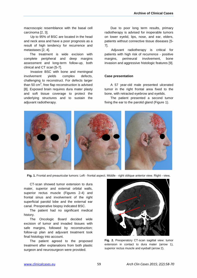
A 57 year-old male presented ulcerated
tumor in the right frontal area fixed to the
bone, with retracted eyebrow and eyelids.
The patient presented a second tumor
fixing the ear to the parotid gland (Figure 1).
Fig. 1. Frontal and preauricular tumors: Left - frontal aspect. Middle - right oblique anterior view. Right - view.
CT-scan showed tumor extension to dura
mater, superior and external orbital walls,
superior rectus muscle (Figures 2-4) and
frontal sinus and involvement of the right
superficial parotid lobe and the external ear
canal. Preoperative biopsy indicated BSC.
The patient had no significant medical
history.
The Oncologic Board decided wide
excision of tumor and invaded tissues with
safe margins, followed by reconstruction;
follow-up plan and adjuvant treatment took
final histology into account.
The patient agreed to the proposed
treatment after explanations from both plastic
surgeon and neurosurgeon were provided.
Fig. 2. Preoperatory CT-scan sagittal view: tumor
extension in contact to dura mater (arrow 1),
superior rectus muscle and eyeball (arrow 2).
1
2
Archive of Clinical Cases
www.clinicalcases.eu 60 Arch Clin Cases 2015; 2(2):58-70
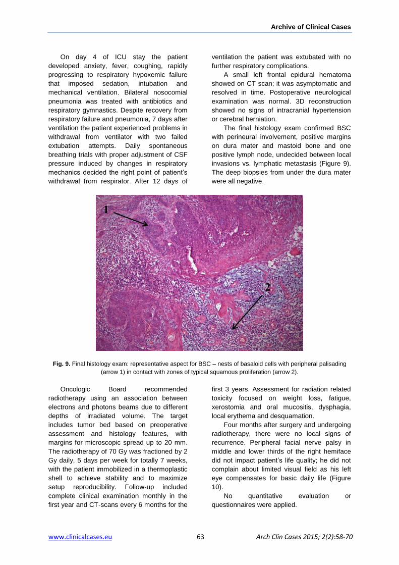
Fig. 3. Preoperatory CT-scan sagittal view: the tumor
invading the frontal bone, in vicinity of the frontal-
zygomatic suture (arrow 3)
Fig. 4. CT-scan with 3D reconstruction of the skull:
outer cortex of the right frontal bone, superior and
lateral orbital walls affected.
Anesthesiologist had their informed
consent taken. Skin was incised
circumferentially at 2 cm from tumor
macroscopic margins, down to the bone. Wide
right fronto–orbital craniectomy en-bloc with
right frontal sinus, lateral orbital wall and
zygomatic bone partially was performed, also
at 2 cm from tumor margins on CT-scan
(Figure 5).
Fig. 5. CT scan with 3D reconstruction of the skull: Left - the defect after excision of the right frontal bone (arrow
1), superior and lateral orbital walls, respectively (arrows 2); Middle - partial zygoma excision (arrow 3); Right -
right frontal sinus defect (arrow 4).
2
1
3 4

Archive of Clinical Cases
www.clinicalcases.eu 61 Arch Clin Cases 2015; 2(2):58-70
Invasion of orbital content – superior
rectus muscle and the eyeball required orbital
exenteration. Dura mater was excised in
circular fashion with wide margins. Epidural
thin layer collection resembling tumor and
puss was followed laterally until normal
macroscopic dura mater appearance. The
adherence between inner aspect of dura and
superficial cerebral vessels made the
dissection tedious. Dural margins were
suspended with silk sutures to prevent
postoperative epidural hematoma.
At 2 cm from second tumor margins the
skin incision went circumferentially around the
ear. The pavilion en-bloc with invaded parotid
gland, facial nerve, masseter muscle and
external ear canal were excised. The
subperiosteal excision of the external ear
canal went down to the tympanic membrane.
Excision of soft tissue was guided by serial
frozen sections and the bone was removed by
macroscopic appearance and clinical
judgement taking into account the local
conditions.
Fig. 6. Ear en-bloc with parotid gland, facial nerve and masseter muscle were excised with 2 cm safety
margins from tumor edges
The temporalis muscle was raised in
pedicled fashion, rotated and fixed to fill the
right orbit and frontal sinus. Dura mater defect
of 9/8 cm with exposed brain was
reconstructed with fascia lata (FL) from
ipsilateral thigh, sutured in watertight fashion
in order to prevent postoperative cerebrospinal
fluid (CFS) leak continuous locking manner to
prevent postoperative CSF leak (Figure 7).
With the patient on left lateral decubitus,
right Latissimus dorsi muscle flap was
harvested on thoracodorsal pedicle.
Donor area was closed using our preferred
method – quilting sutures, double layers for
incision on 2 suctions drains. The muscle was
transferred to cover the 20/25 cm post-
excisional defect. Under microscope
magnification, thoracodorsal pedicle was
anastomosed to superior thyroid artery and a
branch of internal jugular vein, respectively.
Muscle flap was covered with Vaseline gauze
and donor sites were closed in standard
fashion.
Flap was monitored by visual exams and
pin-prick every 2 hours for 3 days and every 6
hours thereafter.
The patient underwent monitoring for
microsurgical cases according to ICU
protocols.
Postoperative day (POD) 14 the muscle
flap was grafted using split thickness skin
harvested from anterolateral left thigh.

Archive of Clinical Cases
www.clinicalcases.eu 62 Arch Clin Cases 2015; 2(2):58-70
Fig. 7. The defect resulted after wide excision. Left - pedicled temporalis muscle was fixed in the right
orbit and frontal sinus caudal from exposed brain. Right - dura mater was reconstructed using FL graft sutured
circumferentially to the defect edges
Results
The operation lasted for 17 hours and 2
combined teams including neurosurgeon
rotated on the operating sites. Exposed brain
was covered with FL graft; the orbit and frontal
sinus were filled with pedicled temporalis
muscle. Defect of 20/25 cm was covered with
LDFF; the muscle was normally perfused
throughout the postoperative period,
completely covering the defect, with no signs
of necrosis or hemorrhage (Figure 8).
Drainage in LD donor area was removed
on POD 8; donor sites (trunk and thigh) healed
uneventfully. Split-thickness skin graft was
fully integrated at discharge POD 23.
On POD 2 after extubation, CSF started
leaking through right frontal sinus and nose
and posterior margin of the inset flap. External
lumbar drainage released the intracranial fluid
pressure and allowed for FL sealing by POD
20 when the device was removed. Daily
drainage dropped from 125 cc to 10 cc, to a
total amount of 765 cc.
Fig. 8. 3D reconstruction at POD 5 shows LDFF providing complete coverage. Left - frontal aspect. Middle - right
oblique anterior view. Right - lateral aspect.
FL
Archive of Clinical Cases
www.clinicalcases.eu 63 Arch Clin Cases 2015; 2(2):58-70
On day 4 of ICU stay the patient
developed anxiety, fever, coughing, rapidly
progressing to respiratory hypoxemic failure
that imposed sedation, intubation and
mechanical ventilation. Bilateral nosocomial
pneumonia was treated with antibiotics and
respiratory gymnastics. Despite recovery from
respiratory failure and pneumonia, 7 days after
ventilation the patient experienced problems in
withdrawal from ventilator with two failed
extubation attempts. Daily spontaneous
breathing trials with proper adjustment of CSF
pressure induced by changes in respiratory
mechanics decided the right point of patient’s
withdrawal from respirator. After 12 days of
ventilation the patient was extubated with no
further respiratory complications.
A small left frontal epidural hematoma
showed on CT scan; it was asymptomatic and
resolved in time. Postoperative neurological
examination was normal. 3D reconstruction
showed no signs of intracranial hypertension
or cerebral herniation.
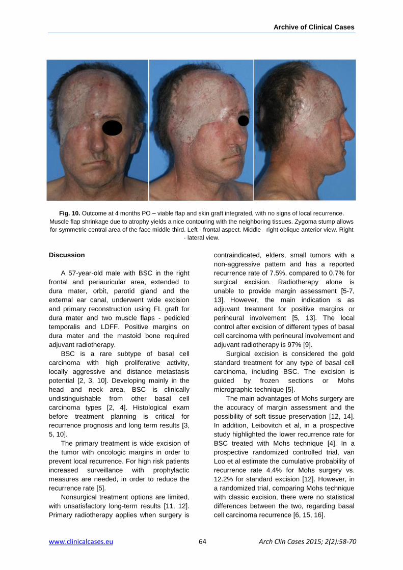
The final histology exam confirmed BSC
with perineural involvement, positive margins
on dura mater and mastoid bone and one
positive lymph node, undecided between local
invasions vs. lymphatic metastasis (Figure 9).
The deep biopsies from under the dura mater
were all negative.
Fig. 9. Final histology exam: representative aspect for BSC – nests of basaloid cells with peripheral palisading
(arrow 1) in contact with zones of typical squamous proliferation (arrow 2).
Oncologic Board recommended
radiotherapy using an association between
electrons and photons beams due to different
depths of irradiated volume. The target
includes tumor bed based on preoperative
assessment and histology features, with
margins for microscopic spread up to 20 mm.
The radiotherapy of 70 Gy was fractioned by 2
Gy daily, 5 days per week for totally 7 weeks,
with the patient immobilized in a thermoplastic
shell to achieve stability and to maximize
setup reproducibility. Follow-up included
complete clinical examination monthly in the
first year and CT-scans every 6 months for the
first 3 years. Assessment for radiation related
toxicity focused on weight loss, fatigue,
xerostomia and oral mucositis, dysphagia,
local erythema and desquamation.
Four months after surgery and undergoing
radiotherapy, there were no local signs of
recurrence. Peripheral facial nerve palsy in
middle and lower thirds of the right hemiface
did not impact patient’s life quality; he did not
complain about limited visual field as his left
eye compensates for basic daily life (Figure
10).
No quantitative evaluation or
questionnaires were applied.
Archive of Clinical Cases
www.clinicalcases.eu 64 Arch Clin Cases 2015; 2(2):58-70
Fig. 10. Outcome at 4 months PO – viable flap and skin graft integrated, with no signs of local recurrence.
Muscle flap shrinkage due to atrophy yields a nice contouring with the neighboring tissues. Zygoma stump allows
for symmetric central area of the face middle third. Left - frontal aspect. Middle - right oblique anterior view. Right
- lateral view.
Discussion
A 57-year-old male with BSC in the right
frontal and periauricular area, extended to
dura mater, orbit, parotid gland and the
external ear canal, underwent wide excision
and primary reconstruction using FL graft for
dura mater and two muscle flaps - pedicled
temporalis and LDFF. Positive margins on
dura mater and the mastoid bone required
adjuvant radiotherapy.
BSC is a rare subtype of basal cell
carcinoma with high proliferative activity,
locally aggressive and distance metastasis
potential [2, 3, 10]. Developing mainly in the
head and neck area, BSC is clinically
undistinguishable from other basal cell
carcinoma types [2, 4]. Histological exam
before treatment planning is critical for
recurrence prognosis and long term results [3,
5, 10].
The primary treatment is wide excision of
the tumor with oncologic margins in order to
prevent local recurrence. For high risk patients
increased surveillance with prophylactic
measures are needed, in order to reduce the
recurrence rate [5].
Nonsurgical treatment options are limited,
with unsatisfactory long-term results [11, 12].
Primary radiotherapy applies when surgery is
contraindicated, elders, small tumors with a
non-aggressive pattern and has a reported
recurrence rate of 7.5%, compared to 0.7% for
surgical excision. Radiotherapy alone is
unable to provide margin assessment [5-7,
13].
However, the main indication is as
adjuvant treatment for positive margins or
perineural involvement [5, 13]. The local
control after excision of different types of basal
cell carcinoma with perineural involvement and
adjuvant radiotherapy is 97% [9].
Surgical excision is considered the gold
standard treatment for any type of basal cell
carcinoma, including BSC. The excision is
guided by frozen sections or Mohs
micrographic technique [5].
The main advantages of Mohs surgery are
the accuracy of margin assessment and the
possibility of soft tissue preservation [12, 14].
In addition, Leibovitch et al, in a prospective
study highlighted the lower recurrence rate for
BSC treated with Mohs technique [4]. In a
prospective randomized controlled trial, van
Loo et al estimate the cumulative probability of
recurrence rate 4.4% for Mohs surgery vs.
12.2% for standard excision [12]. However, in
a randomized trial, comparing Mohs technique
with classic excision, there were no statistical
differences between the two, regarding basal
cell carcinoma recurrence [6, 15, 16].

Archive of Clinical Cases
www.clinicalcases.eu 65 Arch Clin Cases 2015; 2(2):58-70
Row et al recommend Mohs surgery for
recurrent basal cell carcinoma treatment [16,
17].
Exceeding the advantages, it is time-
consuming and costs are greatly higher than
classic excision [6]. Furthermore, Mohs surgery is used for
cutaneous lesions, without bone involvement
[14]. We chose the frozen section samplings
due to its straightforward and the advantage to
help timely-fashion excision. Skin tumors with
bone and deeper invasion cannot be excised
and brain let open until final pathology report is
released few days later. Clinical and
preoperative imageries assessment oriented
the gross excision margins and for deeper and
circumferential areas we do follow “sample as
you go”.
However, frozen sections extensive tissue
sampling from wide postexcisional defects
carries the risk that positive margin may be
overlooked. From a 20/25 cm excision area,
only two samples turned to be positive, one in
dura mater and second in mastoid bone,
showing frozen sections limitations.
Our patient was included in a high risk
category for recurrence based on histological
type, scalp and forehead location, size over 10
mm with poorly defined borders, aggressive
growth pattern and perineural involvement,
requiring wide excision, reconstruction with
adjuvant radiotherapy [5].
The 57 year-old patient, without
morbidities or contraindications for surgery,
was suitable to undergo excision and
reconstruction. Due to wide excision of soft
tissue, calvarial bone and dura mater,
immediate reconstruction was necessary in
order to protect the brain tissue. Dura mater
substitutes vary from autografts to semi-
/synthetic and xenografts.
Autografts are preferred, due to texture
close to human dura, flexibility, good tensile
strength, non-immunogenic and disease-free.
Fat grafting is not suitable for sizable defect of
dura mater with exposed brain, and also the
temporalis fascia or the pericranium was
insufficient to achieve coverage [18, 19].
FL was harvested by the second team
while neurosurgeon completed the excision.
Concomitant operations were decided in order
to expedite the surgery and the thigh donor
scaring site was considered acceptable [18].
Moreover, the surrounding scalp and
pericranium was left intact as a reliable lifeboat
in case of minor flap loss or secondary
revisions without the need for complex
reconstruction or another donor site.
Scalp reconstruction is challenging after
wide excision, not because of the amount of
tissue lacking but because of complex defect.
Even though highly vascularized, the scalp
has decreased elasticity, which leaves few
reconstruction techniques available for large
defects [8]. Small frontal defects can be
covered with rotated flaps; however there are
few techniques using local flaps to grant total
coverage.
Orticochea flap is not a choice for our
patient due to scarce remaining soft tissue. In
combined frontal and face defects, the
remaining scalp is largely insufficient as donor.
Rule of thumb, for a defect over 50 cm2, is
recommended coverage using local or free
flap [8, 20, 21]. For herein patient the soft
tissue defect was 20 per 25 cm. Moreover,
dura mater repair using non-vascularized
autograft needed well vascularized tissue for
coverage, also in view of subsequent
radiotherapy.
Several free flaps are available for large
defects in head and neck area, either
cutaneous, muscular or combinations. ALT is
preferred for scalp reconstruction, for medium
size defects [22-26]. The main disadvantage of
the ALT flap is unpredictable vascular
anatomy, which can increase the surgery time
[27, 28]. The perforator arteries require
meticulous dissection and are less resistant to
compression, contamination or infection [28].
When ALT is raised with a large muscle
component the risk of developing
complications is higher [28]. If is harvested
with a large skin surface, the donor site is not
able to be closed primarily, requiring a skin
graft which is also associated with a higher
rate of complications and decreased
ambulation for first 2 weeks postoperatively.
The more complex the ALT with inclusion of
adjacent tissues on the same pedicle, the
more increased the chance to develop
complications.
Multiple flaps 3D arrangements to achieve
recipient site requirements are very difficult if
the anatomy is not favorable [29, 30]. For our

Archive of Clinical Cases
www.clinicalcases.eu 66 Arch Clin Cases 2015; 2(2):58-70
patient the ALT size was insufficient, unable to
ensure complete coverage.
For subtotal or total scalp defects the only
free flaps large enough to ensure complete
coverage are the omentum flap and the LDFF
[31]. The omentum free flap with split-
thickness skin graft for scalp defects, first
described by McLean and Bunck in 1972,
needs laparotomy with increased morbidity at
the donor site. Although easy to fold on a flat
surface, on the head the omental flap
suspension is tedious. Moreover,
postoperative ambulation is limited until flap
will adhere to the recipient site [22-24].
Latissimus dorsi has a reliable anatomy
and is easy to harvest with minimal acceptable
donor site morbidity and atrophy of the muscle
provides a soft tissue thickness that closely
approximates that of the native scalp [23, 25,
31]. LD offers more tissue bulk (thickness
range 0.5-4.5 cm with an average of 1.5 cm)
and secondary surgery such as calvarial
reconstruction is possible [25, 31].
Fattah et al in case series report on giant
head and neck basal cell carcinoma with bone
and dura mater involvement, in two out of
three cases chose LD for soft tissue defect
coverage [32]. Hiernet et al favors the LDFF
over the omental flap [31]. Skin grafting was
postponed due to long operating time; it is our
preference for delayed skin grafting. There
was no donor-site morbidity in our case and
entire muscle survived on single pedicle.
The paranasal sinuses need to be covered
in order to protect the brain against nasal
contamination [33]. Whenever dura resection
and reconstruction are associated with orbital
exenteration, primary coverage of the orbit is
critical for protection against meningitis [34].
Due to complex defect, it is difficult for
single muscle flap to achieve orbital, sinus and
large defect coverage on top of it. Even though
trimming and folding of the large surface
muscle like LD could have provided all
requirements, possible failure of the free flap
would have left the whole recipient area open
again.
Our strategy is damage-control oriented:
one flap for orbit and sinus coverage and a
second flap for large defect coverage. Even if
the free flap on top of first flap fails, the barrier
from nose area can still prevent contamination
while preparing the second free flap operation.
Temporalis muscle was reported as a very
good choice for barrier to intracranial
communication [35]. For our patient, the
temporalis muscle was raised in pedicled
fashion, rotated and fixed to fill the remaining
of the right orbit and frontal sinus in a timely-
fashion manner. The technique is
straightforward, the muscle is readily available
and the contouring is satisfactorily achieved
using the muscle bulk. With smooth surfacing,
due to zygoma excision and temporalis muscle
coverage, LD mission to resurface the defect
was achieved easily with improved final
contouring.
Another option for orbital and sinus
coverage is the chimeric flap of serratus
anterior and LD [36]. The main drawback is
the functional disabilities from sacrificing two
muscles [37]. The serratus anterior muscle
enables full abduction and flexion of the arm
and sacrificing it will lead to “winging” of the
scapula. The patient will be unable to elevate
an arm above the horizontal plane [38].
Herein, the temporalis muscle was chosen due
to defect proximity, minimal donor site
comorbidities and shorter operating time.
Composite scalp and calvarial defects can
undergo primary simultaneous reconstruction
or scalp only reconstruction, knowing that
there is no significant difference in
complication rate between the two methods
[39]. However, primary calvarial and zygoma
reconstruction is contraindicated when the
paranasal sinuses are opened; moreover,
alloplastic material exposure rate after
radiotherapy is increased [40]. Due to high
recurrence risk, adjuvant radiotherapy and
close follow-up required, the patient underwent
scalp reconstruction without bone
replacement. Later reconstruction of the upper
third of the right face will be granted after 3
years follow-up with no recurrence. From our
point of view, the nice contouring of the face
middle third doesn’t require zygoma
reconstruction. Eye reconstruction with
prosthetics and lid reconstruction is also
feasible, after oncologic follow-up.
PO complications after major head and
neck surgery (involving free flaps) include
medical complications and those related to
donor and recipient site [41-43]. Most frequent
Archive of Clinical Cases
www.clinicalcases.eu 67 Arch Clin Cases 2015; 2(2):58-70
major complications are respiratory failure and
pneumonia. In a prospective cohort study on
192 patients who underwent major head and
neck surgery with free flap repair, 49
developed pneumonia [43]. Perisanidis et al
reviewed 79 patient who underwent major
head and neck surgery: 43% had pulmonary
complications, from which 9% with respiratory
failure [41]. McMahon et al report 8 cases of
delirium out of 192 patients [42]. A frequent
minor complication reported in the literature is
the need for transfusion, as Perisanidis et al
reported in 39% cases [43]. In a retrospective
study on 282 patients who underwent head
and neck surgery with free flap
reconstructions, there was no statistical
difference in flap failure rate between patient
with transfusion vs. no transfusion group.[44]
The risk factors related with PO
complications are the area of surgery, neck
dissection, operating time, comorbidities and
alcohol consumption [41, 42]. Herein, the
patient developed PO pneumonia,
posthemorrhagic anemia, and withdrawal
syndrome, remitted under specific treatment.
For CSF leak through frontal sinus the
patient underwent external lumbar drainage for
12 days and antibiotics. The CSF leak is very
difficult to be avoided after craniectomy with
dura mater plasty. In a prospective multicenter
analysis study, the frequency of CSF leak is
7.7% [45]. Most of them respond to external
lumbar drainage from 48 hours up to 10 days
and only 2% require surgery. After the CSF
leak subsides, is recommended to keep the
drainage another 3 to 5 days in order to
provide time for the fistula to heal [46].
Craniotomy, CSF leak and the effusion found
in the frontal sinus during surgery are risk
factors for meningitis [45, 47].
POD 9 CT scan reevaluation revealed an
extradural hematoma, which slowly resolved
without motor impairment under conservatory
treatment. In literature, the cases in which this
complication occurs after head surgery ranges
from 0.8% up to 1.3% [48]. Pichierri et al
recommend conservatory treatment for small
extradural hematoma, if the neurological
status of the patient permits [48].
Final histology showed positive margins
on dura mater and the mastoid bone. Sherry et
al in a retrospective study reported 3.2% rate
of positive margins after standard excision for
different histologic types of basal cell
carcinoma, including BSC [49]. There are no
clear protocols in managing incomplete
excision. In literature, three types of
approaches are described when positive
margins are involved: re-excision, adjuvant
radiotherapy and wait-and-see.
A retrospective study reveals 25% cases
of recurrence out of 62% patients with positive
margins who underwent observations [49]. Liu
et al, in a study in which 120 patients
underwent radiotherapy and 67 were under
observations after surgical treatment, reported
6% recurrence rate for adjuvant radiotherapy
and 31% for the patients under observation
[50, 51]. For our patient the decision was for
adjuvant radiotherapy, due to BSC aggressive
features and high risk of recurrence and
positive margins. Close follow-up with
complete clinical examination monthly in the
first year and CT-scans at every 6 months for
the first 2 years are advised [5].
Facial nerve palsy morbidity is major for
the eye with less morbidity for middle and
lower face [52]. The patient did not complain of
motor function absence on right hemiface.
Perisanidis et al report a median duration
of hospitalization stay after free flap surgery of
34 days (22-48 days) when major
complications are involved [41]. One of the
most important factors for hospital stay in head
and neck surgery is operation time. Lofti et al
report a hazard ratio of 1.34 for surgeries
longer than 220 minutes vs. those lasting
under 220 minutes [53]. A cost-effectiveness
analysis of 39 free tissue transfers reported an
average cost of 24.737€ per case with 16 days
of hospitalization [54]. Our patient was
discharged after major head and neck surgery
that lasted 1020 minutes and 23 days of PO
recovery, with viable flap and split-thickness
skin graft fully integrated, overall cost of
hospitalization and treatment was 5000€.
Conclusion
Patient with invasive BSC can benefit from
major surgery comprising wide excision and
soft tissue extensive reconstruction.
Interdisciplinary approach with assessment

Archive of Clinical Cases
www.clinicalcases.eu 68 Arch Clin Cases 2015; 2(2):58-70
from pathology and radiology specialists helps
designing the operating plan. Intense
reanimation, follow-up and radiotherapy for
positive margins are mandatory. This holistic
approach yields no recurrence on short-term
evaluation.
Further follow-up and life-quality
assessment is needed. The cost-benefit ratio
is favorable.
Disclosure
The authors have no conflict of interests to
declare.
Acknowledgement
This paper was published under the frame of
European Social Found, Human Resources
Development Operational Program 2007-2013,
project no. POSDRU/159/1.5/S/136893.
References
1. Hamilton M. Basal squamous cell epithelioma.
Arch Derm Syphilol 1928; 18(1):50-73.
2. De Stefano A, Dispenza F, Petrucci AG, et al.
Features of biopsy in diagnosis of metatypical
basal cell carcinoma (Basosquamous
Carcinoma) of head and neck. Otolaryngol Pol
2012; 66(6):419-423.
3. Garcia C, Poletti E, Crowson N.
Basosquamous carcinoma. J An Acad
Dermatol 2009; 60(1):137-143.
4. Leibovitch I, Huilgol SC, Selva D, et al.
Basosquamous carcinoma: treatment with
Mohs micrographic surgery. Cancer 2005;
104(1):170-175.
5. Clinical Practice in Oncology – Basal Cell Skin
Cancer. NCCN 2015.
[http://www.nccn.org/professionals/physician_gl
s/f_guidelines.asp available at 06.15.2015]
6. Smith V, Walton S. Treatment of Facial Basal
Cell Carcinoma: A Review. J Skin Cancer
2011; doi:10.1155/2011/380371.
7. Rigel DR, June K, Robinson JK, et al. Cancer
of the Skin. 2nd
Ed. China: Elsevier Saunders;
2011. Chapter 52, Radiation therapy in the
treatment of skin cancers; p. 578-581.
8. Newman MI, Hanasono MM, Disaj JJ, et al.
Scalp Reconstruction: A 15-Year Experience.
Ann Plast Surg 2004; 52(5):501-506.
9. Plichta K, Mackley HB, Radiotherapy for
cutaneous malignancies of the head and neck.
Operative Techniques in Otolaryngology 2013;
24(1):59-62. doi:10.1016/j.otot.2012.12.004.
10. Hussain SI, Hussainy AS. Baso-Squamous Cell
Carcinoma - a Case Report. Pak Med Assoc
2004; 54(1):30-32.
11. Cognetta AB, Howard BM, Heaton HP, et al.
Superficial x-ray in the treatment of basal and
squamous cell carcinomas: A viable option in
select patients. J Am Acad Dermatol 2012;
67(6):1235-1244.
12. van Loo E, Mosterd K, Krekels GAM, et al.
Surgical excision versus Mohs’ micrographic
surgery for basal cell carcinoma of the face: A
randomised clinical trial with 10 year follow-up.
Eur J Cancer 2014; 50(17):3011-3020.
13. Aguayo-Leiva IR, Ríos-Buceta L, Jaén-Olasolo
P. Surgical vs. Nonsurgical Treatment of Basal
Cell Carcinoma. Actas Dermosifiliogr 2010;
101(8):683–692.
14. Wain RAJ, Tehrani H. The plastic &
reconstructive Mohs surgery service. J Plast
Reconstr Aesthet Surg 2014; 67(3):331-335.
15. Smeets NW, Krekels GAM, Ostertag JU, et al.
Surgical excision vs. Mohs’ micrographic
surgery for basal cell carcinoma of the face:
randomised controlled trial. Lancet 2004;
364(9447):1766-1772.
16. Mosterd K, Krekels GA, Nieman FH, et al.
Surgical excision versus Mohs’ micrographic
surgery for primary and recurrent basal-cell
carcinoma of the face: a prospective
randomised controlled trial with 5-years’ follow-
up. Lancet Oncol 2008; 9(12):1149-1156.
17. Rowe DE, Carroll RJ, Day CL. Mohs surgery is
the treatment of choice for recurrent (previously
treated) basal cell carcinoma. J Dermatol Surg
Oncol 1989; 15(4):424-431.
18. Parlato C, Granata R, Moraci A, et al. Dural
Reconstruction in Meningioma Surgery. In:
Monleon D. Meningiomas - Management and
Surgery. China: InTech, 2012. Chapter 6, Dural
reconstruction in meningioma surgery; p. 103-
124.
19. Rengachary SS, Benzel EC. Calvarial and
Dural Reconstruction. U.S.A.: AANS, 1998.
Chapter 3, Cranioplasty materials; p. 35-45.
20. Baker SR. Local flaps in facial reconstruction.
3rd
Ed. China: Elsevier Saunders; 2014.
Chapter 24, Reconstruction of the scalp; p.641-
667.
21. Janis JE, Leedy JE. Lip, cheek and scalp
reconstruction. SRPS 2006; 10(13):1-39.
22. Bayles SW, Hayden RE. Gastro-omental free
flap reconstruction of the head and neck. Arch
Facial Plast Surg 2008; 10(4):255-259.

Archive of Clinical Cases
www.clinicalcases.eu 69 Arch Clin Cases 2015; 2(2):58-70
23. Kruse-Losler B, Presser D, Meyer U, et al.
Reconstruction of large defects on the scalp
and forehead as an interdisciplinary challenge:
Experience in the management of 39 cases.
Eur J Surg Oncol 2006; 32(9):1006-1014.
24. Thorne CH, Chung KC, Gosain GC, et al.
Grabb and Smith's Plastic Surgery. 7th Ed.
China: Lippincott Williams Walkins, 2014.
Chapter 31, Reconstruction of the scalp,
calvarium, and forehead; p. 342-351.
25. Wei F, Mardini S. Flaps and Reconstructive
Surgery. China: Elsevier Saunders, 2009.
Chapter 23, Latissimus dorsi flap; p.287-303.
26. Xua ZF, Suna CF, Duana WY. Clinical
anatomical study and evaluation of the use of
the free anteromedial thigh perforator flaps in
reconstructions of the head and neck. Br J Oral
Maxillofac Surg 2013; 51(8):725-730.
27. Bhujel N, Johnston C, Parmar S, et al. An
unusual anatomical variant of the vascular
anatomy in the anterolateral thigh free flap. Int
J Oral Maxillofac Surg 2010; 39(1):94-95.
28. Shawa RJ, Batstone MD, Blackburn TK, et al.
The anterolateral thigh flap in head and neck
reconstruction: “Pearls and pitfalls”. Br J Oral
Maxillofac Surg 2010; 48(1):5–10.
29. Marsh DJ, Chana JS. Reconstruction of very
large defects: a novel application of the double
skin paddle anterolateral thigh flap design
provides for primary donor-site closure. J Plast
Reconstr Aesthet Surg 2010; 63(1):120-125.
30. Agostini T, Lazzeri D, Spinelli G. Anterolateral
thigh flap: Systematic literature review of
specific donor-site complications and their
management. J Craniomaxillofac Surg 2013;
41(1):15-21.
31. Hierner R, vaan Loon J, Goffin J, et al. Free
latissimus dorsi flap transfer for subtotal scalp
and cranium defect reconstruction: report of 7
cases. Microsurgery 2007; 27(5):425-428.
32. Fattah A, Pollock J, Maheshwar A, et al. Big
Bad BCCs: Craniofacial resection and
reconstruction for atypical basal cell carcinoma.
J Plast Reconstr Aesthet Surg 2010; 63(5):433-
441.
33. Fattahi T, DiPasquale J. Utility of the pericranial
flap in frontal sinus and anterior cranial fossa
trauma. Int. J. Oral Maxillofac Surg 2009;
38(12):1263-1267.
34. Uyar Y, Kumral TL, Yıldırım G. Reconstruction
of the orbit with a temporalis muscle flap after
orbital exenteration. Clin Exp Otorhinolaryngol
2015; 8(1):52-56.
35. Bagheri SC, Bell B, Khan HA. Current Therapy
In Oral and Maxillofacial Surgery. China:
Elsevier Saunders, 2012. Chapter 64 - The
temporalis system of flaps in head and neck
reconstruction: temporoparietal fascia and
temporalis muscle flaps; p. 527-533.
36. Serra MP, Longhi P, Carminati M, et al.
Microsurgical scalp and skull reconstruction
using a combined flap composed of serratus
anterior myo-osseous flap and latissimus dorsi
myocutaneous flap. J Plast Reconstr Aesthet
Surg 2007; 60(10):1158-1161.
37. Arai H, Yanai A, Nishida M, et al.
Reconstruction of scalp and cranium defect
utilizing latissimus dorsi musculocutaneous and
serratus anterior muscle free flaps with
interpositional anastomosis of T-shaped flap
artery. Skull Base Surg 1995; 5(2):117-121.
38. Abolhoda A, Bui TD, Milliken JC, et al. Pedicled
latissimus dorsi muscle flap routine use in high-
risk thoracic surgery. Tex Heart Inst J 2009;
36(4):298-302.
39. Chao AH, Yu P, Skoracki RJ, et al.
Microsurgical reconstruction of composite scalp
and calvarial defects in patients with cancer: A
10-year experience. Head Neck 2012;
34(12):1759-1764.
40. Stula D. Cranioplasty: Indications, Techniques,
and Results. Wien: Springer-Verleg, 1984.
Chapter C, Surgical procedures and techniques
of cranial repair; p.57-61.
41. Perisanidis C, Herberger B, Papadogeorgakis
N, et al. Complications after free flap surgery:
do we need a standardized classification of
surgical complications? Br J Oral Maxillofac
Surg 2012; 50(2):113-118.
42. McMahon JD, MacIver C, Smith M, et al.
Postoperative complications after major head
and neck surgery with free flap repair –
prevalence, patterns, and determinants: a
prospective cohort study. Br J Oral Maxillofac
Surg 2013; 51(8):689-695.
43. Bianchi B, Copelli C, Ferrari S, et al. Free flaps:
outcomes and complications in head and neck
reconstructions. J Craniomaxillofac Surg 2009;
37(8): 438-442.
44. Puram SV, Yarlagadda BB, Sethi R, et al.
Transfusion in head and neck free flap patients:
practice patterns and a comparative analysis
by flap type. Otolaryngol Head Neck Surg
2015; 152(3):449-457.
45. Kehler U, Hirdes C, Weber C, et al. CSF leaks
after cranial surgery-a prospective multicenter
analysis. Innov Neurosurg 2012; 1(1):49-53.
46. Quinones-Hinojosa A, Schmidek, S. Operative
Neurosurgical Techniques: Indications,
Methods and Results. 6th
Ed. vol. 2. China:
Elsevier Saunders, 2012. Chapter 138,
Management of cerebrospinal fluid leaks;
p.1579-1595.
47. Korinek AM, Baugnon T, Golmard JL, et al.
Risk factors for adult nosocomial meningitis
Archive of Clinical Cases
www.clinicalcases.eu 70 Arch Clin Cases 2015; 2(2):58-70
after craniotomy: role of antibiotic prophylaxis.
Neurosurg 2006; 59(1):126-133.
48. Pichierri A, Ruggeri A, Donnarumma P, et al.
Postoperative Extradural Hematomas. J Neurol
Surg 2013; 74(1):25-28.
49. Sherry KR, Reid LA, Wilmshurst AD. A five
year review of basal cell carcinoma excisions, J
Plast Reconstr Aesthet Surg 2010; 63(9):1485-
1489.
50. Liu FF, Maki E, Warde P, et al. A management
approach to incompletely excised basal cell
carcinomas of skin. Int J Radiat Oncol Biol
Phys 1991; 20(3):423-428.
51. Ríos-Buceta L. Management of basal cell
carcinomas with positive margins. Actas
Dermosifiliogr 2007; 98(10):679-687.
52. Birgfeld C, Neligan P. Surgical Approaches to
Facial Nerve Deficits. Skull Base 2011;
21(3):177-184.
53. Penel N, Yann Mallet Y, Roussel-Delvallez M,
et al. Factors determining length of the
postoperative hospital stay after major head
and neck cancer surgery. Oral Oncol 2008;
44(6):555-562.
54. Miller MJ, Swartz WM, Miller RH, et al. Cost
analysis of microsurgical reconstruction in the
head and neck. J Surg Oncol 1991; 46(4):215-
277.