Invasive Carcinoma of the Breast Histopathology Reporting ...
32
Invasive Carcinoma of the Breast Histopathology Reporting Guide Sponsored by Version 1.1 Published June 2021 ISBN: 978-1-922324-12-2 Page 1 of 32 © 2021 International Collaboration on Cancer Reporting Limited (ICCR). Family/Last name Given name(s) Patient identifiers Date of request Accession/Laboratory number Elements in black text are CORE. Elements in grey text are NON-CORE. Date of birth DD – MM – YYYY SCOPE OF THIS DATASET indicates multi-select values indicates single select values CLINICAL INFORMATION (Note 1) Presentation mode Information not provided Screening Symptomatic Other clinical information, specify Information not provided Current clinical findings for which this surgery is performed (select all that apply) Information not provided Nipple discharge Other, specify Prior presurgical therapy for this diagnosis of invasive breast carcinoma Imaging modality (select all that apply) Information not provided Mammography Magnetic resonance imaging (MRI) Radiological findings (select all that apply) Information not provided Single lesion Calcifications Mass Prior history of breast cancer OPERATIVE PROCEDURE a (Note 2) Information not provided Yes, specify laterality, site(s), diagnosis, and prior treatment(s) Paget disease of the nipple Palpable mass Information not provided Yes Not specified Excision (less than total mastectomy) Diagnostic excision/excision biopsy/localisation biopsy Therapeutic wide local excision Duct excision/microdochectomy Re-excision Total mastectomy Simple mastectomy Nipple-sparing mastectomy Skin-sparing mastectomy Modified radical mastectomy Radical mastectomy Additional specimens, specify Other, specify Extent by imaging, if available mm Clip inserted Yes No Known genetic predisposition a If a lymph node staging specimen is submitted, then a separate dataset is used to record the information. Information not provided Gene predisposition, specify None Ultrasound None Multiple lesions Architectural distortion None SPECIMEN LATERALITY (Note 3) Left SPECIMEN DIMENSIONS SPECIMEN WEIGHT g Right Not specified x mm mm x mm Not known No No Other, specify DD – MM – YYYY
Transcript of Invasive Carcinoma of the Breast Histopathology Reporting ...

Invasive Carcinoma of the BreastHistopathology Reporting Guide
Sponsored by
Version 1.1 Published June 2021 ISBN: 978-1-922324-12-2 Page 1 of 32© 2021 International Collaboration on Cancer Reporting Limited (ICCR).
Family/Last name
Given name(s)
Patient identifiers Date of request Accession/Laboratory number
Elements in black text are CORE. Elements in grey text are NON-CORE.
Date of birth DD – MM – YYYY
SCOPE OF THIS DATASETindicates multi-select values indicates single select values
CLINICAL INFORMATION (Note 1)
Presentation mode
Information not providedScreeningSymptomatic Other clinical information, specify
Information not provided
Current clinical findings for which this surgery is performed (select all that apply)
Information not providedNipple dischargeOther, specify
Prior presurgical therapy for this diagnosis of invasive breast carcinoma
Imaging modality (select all that apply)
Information not providedMammographyMagnetic resonance imaging (MRI)
Radiological findings (select all that apply)
Information not providedSingle lesionCalcificationsMass
Prior history of breast cancer
OPERATIVE PROCEDUREa (Note 2)
Information not providedYes, specify laterality, site(s), diagnosis, and prior treatment(s)
Paget disease of the nipplePalpable mass
Information not providedYes
Not specifiedExcision (less than total mastectomy)
Diagnostic excision/excision biopsy/localisation biopsy Therapeutic wide local excision Duct excision/microdochectomyRe-excision
Total mastectomy Simple mastectomyNipple-sparing mastectomySkin-sparing mastectomyModified radical mastectomyRadical mastectomy
Additional specimens, specify
Other, specify
Extent by imaging, if available mm
Clip inserted Yes No
Known genetic predisposition
a If a lymph node staging specimen is submitted, then a separate dataset is used to record the information.
Information not providedGene predisposition, specify
NoneUltrasound
NoneMultiple lesionsArchitectural distortion
None
SPECIMEN LATERALITY (Note 3)
Left
SPECIMEN DIMENSIONS
SPECIMEN WEIGHT
g
Right Not specified
x mm mm x mm
Not known
No
No
Other, specify
DD – MM – YYYY

Version 1.1 Published June 2021 ISBN: 978-1-922324-12-2 Page 2 of 32© 2021 International Collaboration on Cancer Reporting Limited (ICCR).
Invasive Carcinoma of the Breast
Invasive breast carcinoma of no special type (invasiveductal carcinoma, not otherwise specified)e
Invasive lobular carcinomaTubular carcinomaCribriform carcinomaMucinous carcinoma Invasive micropapillary carcinomaCarcinoma with apocrine differentiationMetaplastic carcinoma
HISTOLOGICAL TUMOUR TYPEc (Note 7)(Value list based on the World Health OrganizationClassification of Breast Tumours (2019))
No residual invasive carcinoma
Other, specify
TUMOUR DIMENSIONS (Note 6)
Maximum dimension of largest invasivefocus >1 mm (specify exact measurementrounded to nearest mm)d
Additional dimensions of largest invasive focus
mm
c For microinvasive disease refer to the DCIS, variants of LCIS and low grade lesions dataset.
Maximum dimension of whole tumour field (invasive + DCIS)/total extent of disease mm
d Based on a combination of macroscopic and microscopic assessment.
Only microinvasion present (≤1 mm)cNo residual invasive carcinoma
Cannot be assessed, specify
TUMOUR FOCALITY (Note 5)
Cannot be assessedSingle focus of invasive carcinoma Multiple foci of invasive carcinoma
is at least
Number of foci
f Tumour exhibiting more than one tumour type should be designated mixed and the types present stated.
e Refer to Note for details of variants including medullary carcinoma.
x mm mm
Cannot be assessed
Sizes of individual focib
b Record the largest measurement of individual foci in millimetres. If there are many foci a range may be included.
Mixed, specify subtypes present
f
TUMOUR SITE (select all that apply) (Note 4)
Upper outer quadrantLower outer quadrantUpper inner quadrantLower inner quadrantCentralNippleOther, specify
Present (select all that apply)
CARCINOMA IN SITU (Note 9)
Not identified
Negative for extensive intraductal component (EIC) Positive for EIC
No residual invasive carcinomaGrade 1 (scores of 3, 4, or 5)Grade 2 (scores of 6 or 7)Grade 3 (scores of 8 or 9)
HISTOLOGICAL TUMOUR GRADE (Note 8)
Score cannot be determined, specify
Lobular carcinoma in situ (LCIS)
SPECIMEN DETAILS
Depth of tissue excised
SkinNippleSkeletal muscle
Specimen includes (select all that apply)
YesSkin to deep fascia No
Not specified
Position, specify
Distance from nipple
o’clock
mm
AND
OR
Ductal carcinoma in situ (DCIS)
Only microinvasion present (not graded)c
Mitotic count
per 10 HPF (field diameter ____ mm)
Score 1,2,3
Total score
Paget disease of the nippleEncapsulated papillary carcinomaSolid papillary carcinoma in situ
Nuclear pleomorphism 1,2,3
Tubule score 1,2,3
OR
per mm2

Version 1.1 Published June 2021 ISBN: 978-1-922324-12-2 Page 3 of 32© 2021 International Collaboration on Cancer Reporting Limited (ICCR).
CribriformMicropapillaryPapillarySolidOther (e.g., clinging/flatg), specify
Histological architectural pattern (select all that apply) (Applicable to DCIS only)
Not identifiedPresent
Necrosis
Classical LCISPleomorphic LCISFlorid LCISOther, specify
Classification of LCIS (select all that apply) (Applicable if LCIS is present in specimen)
g Applies to high nuclear grade DCIS only.
Invasive carcinoma
MARGIN STATUSi (Note 11)(For wide local excision specimens and similar non-complete mastectomy specimens)
i Core for all wide local excision specimens, similar non-complete mastectomy and some (refer to Note) complete mastectomy specimens.
Cannot be assessed, specify
Involved (select all that apply)
Anterior (superficial)
Posterior (deep)
Superior
Inferior
Medial
Lateral
Other margin, specify
Specify extent
Specify extent
Specify extent
Specify extent
Specify extent
Specify extent
Specify extent
h Where there is disease extension to involve skin, nipple or skeletal muscle, disease extent classification is a core element; in all other cases it is non-core.
Nipple tissue is not presentDCIS does not involve the nipple epidermisDCIS involves nipple epidermis (Paget disease of the nipple)
Skeletal muscle is not presentSkeletal muscle is free of carcinomaTumour involves skeletal muscleTumour involves both skeletal muscle and chest wall (classified as pT4a)
Nipple (including areola complex)
Skeletal muscle
TUMOUR EXTENSIONh (Note 10)
Skin is not presentSkin is present and uninvolvedInvasive carcinoma directly invades into the dermis or epidermis without skin ulceration Invasive carcinoma directly invades into the dermis or epidermis with skin ulceration (classified as pT4b)Satellite skin foci of invasive carcinoma are present (i.e., not contiguous with the invasive carcinoma in the breast) (classified as pT4b)
Skin
Not involved
(< or > may be used) mm
Cannot be determined, specify
Specify closest margin, if possible
Distance of invasive carcinoma to closest margin
Distance of invasive carcinoma to other margins (< or > may be used)
Anterior (superficial) mm
Posterior (deep) mm
Superior mm
Inferior mm
Medial mm
Lateral mm
Other margin, specify mm
CLASSIFICATION OF CARCINOMA IN SITU (if present) (Note 9)
Grade 1 (Low) Grade 2 (Intermediate)Grade 3 (High)
Histological nuclear grade (Applicable to DCIS, encapsulated papillary carcinoma and solid papillary carcinoma in situ)
Central (Comedo) necrosisFocal (Punctate) necrosis (<10% duct diameter)

Version 1.1 Published June 2021 ISBN: 978-1-922324-12-2 Page 4 of 32© 2021 International Collaboration on Cancer Reporting Limited (ICCR).
Invasive Carcinoma of the Breast
DCIS
j
Involved (select all that apply)
Anterior (superficial)
Posterior (deep)
Superior
Inferior
Medial
Lateral
Other margin, specify
Specify extent
Specify extent
Specify extent
Specify extent
Specify extent
Specify extent
Specify extent
MARGIN STATUS
i (Note 11)(For complete mastectomy specimens)
Invasive carcinoma
Cannot be assesessd, specify
Involved by invasive carcinoma, specify margin/sites of involvement
Involved, specify margin/sites of involvement
Not involved
(< or > may be used) mm
Cannot be determined, specify
Specify closest margin, if possible
Distance of invasive carcinoma to closest margin
Not involved
mm
Cannot be determined, specify
Specify closest margin, if possible
Distance of DCIS to closest margin
Distance of DCIS to other margins (< or > may be used)
Anterior (superficial) mm
Posterior (deep) mm
Superior mm
Inferior mm
Medial mm
Lateral mm
Other margin, specify mm
DCIS
j
Not involved
mm
Cannot be determined, specify
Specify closest margin, if possible
Distance of DCIS to closest margin
(< or > may be used)
Involved, specify margin/sites of involvement
i Core for all wide local excision specimens, similar non-complete mastectomy and some (refer to Note) complete mastectomy specimens.
Present
LYMPHOVASCULAR INVASION IN PRIMARY BREASTCARCINOMA (Note 12)
Not identified
Specify extent
Lymphovascular invasion identified elsewhere, specify
Indeterminate
j Required only if DCIS or florid LCIS or pleomorphic LCIS is also present in specimen.
Version 1.1 Published June 2021 ISBN: 978-1-922324-12-2 Page 5 of 32© 2021 International Collaboration on Cancer Reporting Limited (ICCR).
Invasive Carcinoma of the Breast
PositiveLow positive
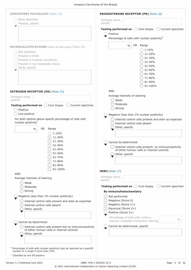
ESTROGEN RECEPTOR (ER) (Note 15)
% Range
1-10%l
11-20%21-30%31-40%41-50%51-60%61-70%71-80%81-90%91-100%
Average intensity of staining
WeakModerateStrong
Negative (less than 1% nuclear positivity)
Internal control cells present and stain as expectedInternal control cells absentOther, specify
Cannot be determined
Internal control cells present but no immunoreactivity of either tumour cells or internal controlsOther, specify
k Percentage of cells with nuclear positivity may be reported as a specific number or a range if more than 10%.l Classified as low ER positive.
For both options above specify percentage of cells with nuclear positivityk
OR
AND
MICROCALCIFICATIONS (select all that apply) (Note 14)
Other, specify
Not identified Present in DCISPresent in invasive carcinomaPresent in non-neoplastic tissue
COEXISTENT PATHOLOGY (Note 13)
None identifiedPresent, specify
Positive
PROGESTERONE RECEPTOR (PR) (Note 16)
OR Range
1-10%11-20%21-30%31-40%41-50%51-60%61-70%71-80%81-90%91-100%
Average intensity of staining
WeakModerateStrong
Negative (less than 1% nuclear positivity)
Internal control cells present and stain as expectedInternal control cells absentOther, specify
Cannot be determined
Internal control cells present; no immunoreactivity of either tumour cells or internal controlsOther, specify
%
Percentage of cells with nuclear positivityk
AND
Testing performed on Core biopsy Current specimen
Testing performed on Core biopsy Current specimen
HER2 (Note 17)
Not performedNegative (Score 0) Negative (Score 1+) Equivocal (Score 2+) Positive (Score 3+)
Cannot be determined, specify
Percentage of cells with uniform, intense, complete membrane staining %
By immunohistochemistry
Testing performed on Core biopsy Current specimen
Antibody clone, specify
Antibody clone, specify
Antibody clone, specify

Version 1.1 Published June 2021 ISBN: 978-1-922324-12-2 Page 6 of 32© 2021 International Collaboration on Cancer Reporting Limited (ICCR).
Negative (not amplified) Positive (amplified)Positive (heterogenous)Negative (heterogenous)
Cannot be determined, specify
Number of observers
Not performed
Number of invasive tumour cells counted
Dual probe assay
Average number of HER2 signals per cell
Average number of CEP17 signals per cell
HER2/CEP17 ratio
Single probe assay
Average number of HER2 signals per cell
Aneusomy
Not identified Present
Heterogeneous signals
Not identified Present
%
By in situ hybridization
Pending
Percentage of cells with amplified HER2 signals
/
PATHOLOGICAL STAGING (UICC TNM 8th edition)m (Note 19)
m - multiple foci of invasive carcinomar - recurrent
TNM Descriptors (only if applicable) (select all that apply)
TX Primary tumour cannot be assessedT0 No evidence of primary tumourT1 Tumour 2 cm or less in greatest dimension T1a More than 0.1 cm but not more than 0.5 cm in
greatest dimension T1b More than 0.5 cm but not more than 1 cm in greatest
dimension T1c More than 1 cm but not more than 2 cm in greatest
dimensionT2 Tumour more than 2 cm but not more than 5 cm in
greatest dimensionT3 Tumour more than 5 cm in greatest dimensionT4 Tumour of any size with direct extension to
chest wall and/or to skin (ulceration or skin nodules)o
T4a Extension to chest wall (does not include pectoralis muscle invasion only)
T4b Ulceration, ipsilateral satellite skin nodules, or skin oedema (including peau d’orange)
T4c Both 4a and 4b T4d Inflammatory carcinomap
m Reproduced with permission. Source: UICC TNM Classification of Malignant Tumours, 8th Edition, eds by James D. Brierley, Mary K. Gospodarowicz, Christian Wittekind. 2016, Publisher Wiley (incorporating any errata published up until 6th October 2020).
o Invasion of the dermis alone does not qualify as T4. Chest wall includes ribs, intercostal muscles, and serratus anterior muscle but not pectoral muscle.p Inflammatory carcinoma of the breast is characterised by diffuse, brawny induration of the skin with an erysipeloid edge, usually with no underlying mass. If the skin biopsy is negative and there is no localised measurable primary cancer, the T category is pTX when pathologically staging a clinical inflammatory carcinoma (T4d). Dimpling of the skin, nipple retraction, or other skin changes, except those in T4b and T4d, may occur in T1, T2, or T3 without affecting the classification.
Primary tumour (pT)n
n Note that the results of surgically removed lymph nodes are derived from a separate dataset.
ANCILLARY STUDIES (Note 18)
Not performedPerformed
Ki-67 proliferation index %
Other, specify test(s) and result(s)
Representative blocks for ancillary studies, specify those blocks best representing tumour and/or normal tissue for further study

Version 1.1 Published June 2021 ISBN: 978-1-922324-12-2 Page 7 of 32
© 2021 International Collaboration on Cancer Reporting Limited (ICCR).
Definitions CORE elements
CORE elements are those which are essential for the clinical management, staging or prognosis of the cancer. These elements will either have evidentiary support at Level III-2 or above (based on prognostic factors in the National Health and Medical Research Council (NHMRC) levels of evidence1). In rare circumstances, where level III-2 evidence is not available an element may be made a CORE element where there is unanimous agreement in the expert committee. An appropriate staging system e.g., Pathological TNM staging would normally be included as a CORE element. The summation of all CORE elements is considered to be the minimum reporting standard for a specific cancer.
NON-CORE elements
NON-CORE elements are those which are unanimously agreed should be included in the dataset but are not supported by level III-2 evidence. These elements may be clinically important and recommended as good practice but are not yet validated or regularly used in patient management. Key information other than that which is essential for clinical management, staging or prognosis of the cancer such as macroscopic observations and interpretation, which are fundamental to the histological diagnosis and conclusion e.g., macroscopic tumour details, may be included as either CORE or NON-CORE elements by consensus of the Dataset Authoring Committee.
Back
Scope This dataset has been developed for the reporting of resection specimens from patients with invasive carcinoma of the breast, with or without ductal carcinoma in situ (DCIS). DCIS without invasive carcinoma and microinvasive carcinoma (≤1 millimetres (mm)) are dealt with in a separate International Collaboration on Cancer Reporting (ICCR) dataset.2 Ipsilateral multifocal disease should be dealt with in a single report. For bilateral invasive breast tumours, a separate dataset should be completed for each side.
Surgically removed lymph nodes are dealt with in a separate ICCR dataset which may be used, as appropriate, in conjunction with this dataset.3 Invasive breast cancer for the post neoadjuvant setting is also dealt with in a separate ICCR dataset. Phyllodes tumours and needle biopsies are not covered in this dataset.
The authors of this dataset can be accessed here.
Back

Version 1.1 Published June 2021 ISBN: 978-1-922324-12-2 Page 8 of 32
© 2021 International Collaboration on Cancer Reporting Limited (ICCR).
Note 1 – Clinical information (Core) Provision of accurate clinical information and detail is considered important to provide context to the specimen, nature of the abnormality, its method of detection and patient medical history. Examples of key information include past history of breast disease or cancer, prior treatment such as neoadjuvant therapy and inherited genetic mutations such as BRCA1 or BRCA2.
Back
Note 2 – Operative procedure (Core) The nature of the operation or procedure(s) performed is important to ensure appropriate pathological examination protocols are followed, and accurate clinical correlation and post-operative management discussion. The nature, extent, focality of the abnormality and patient choice can influence the type of operation. Multiple procedures may be performed and sent as separate specimens which require cross correlation. The forms of surgical procedure used to manage breast disease are considerable and more specific detail of the specimen can be provided. Partial mastectomy, lumpectomy and quadrantectomy/segmental excision are considered synonymous with wide local excision.
Back
Note 3 – Specimen laterality (Core) Specification of the side and site in the breast is important for clinical correlation and accuracy of the patient medical record. For bilateral invasive breast tumours, a separate dataset should be completed for each side.
Back
Note 4 – Tumour site (Core) A measure of distance from the nipple is required. Clock face delineation of location is a more commonly used determination of site than quadrant alone, but either is acceptable. Specification of the side and site in the breast is important for clinical correlation, post-operative management discussion and accuracy of the patient medical record, especially when there are multiple lesions for correlation with radiology/prior biopsies.
Back

Version 1.1 Published June 2021 ISBN: 978-1-922324-12-2 Page 9 of 32
© 2021 International Collaboration on Cancer Reporting Limited (ICCR).
Note 5 – Tumour focality (Core and Non-core) Presence of a single tumour focus is the most common clinical situation, but breast cancer can present with multiple tumour foci as a consequence of a number of scenarios, including:
• Extensive DCIS with multiple associated foci of invasive carcinoma. • A large dominant primary tumour focus with surrounding smaller satellite foci. • In-breast metastatic deposits due to lymphovascular invasion (LVI). • Multiple synchronous independent primary tumours which may be of different type, grade
and receptor status (historically this form of multifocality has been classified as multicentricity).
Identification of the presence of multiple tumour foci requires further clarification through measurement of the main foci, the overall extent of disease (DCIS and invasive foci) and their type, grade and receptor status to determine which of the above forms of multifocality is present. Ipsilateral multifocal disease, even if of different types, should be dealt with in a single report. It can be difficult, if not impossible, on rare occasions to determine whether two adjacent foci represent satellite foci or one lesion mimicking this process due to the plane of sectioning. A practical approach is required; the presence of intervening normal tissue and increasing distance between foci are features that indicate that these are more likely to be multiple foci than a localised process. A distance of 5 mm or greater is used to define a separate focus.
Back
Note 6 – Tumour dimensions (Core and Non-core) The size of the tumour or of the largest/dominant invasive tumour focus is a key variable required for breast cancer staging and requires accurate assessment to the nearest mm. Histological tumour size is deemed the gold standard but should be correlated with the gross macroscopic size measurement and where possible with the imaging size. On rare occasions, the tumour size is obtained from a previous core needle biopsy specimen, as the tumour in the core may be larger than the tumour in the excision specimen or the entire invasive tumour has been removed by the needle sampling procedure. In the context of extensive surrounding DCIS (and/or florid or pleomorphic lobular carcinoma in situ (LCIS), the total extent of the entire disease process including all invasive tumour foci and associated DCIS should be provided as the whole tumour size (Figure 1). This information is useful for clinical and radiological correlation and to assist in the determination of completeness of disease excision. In the context of multiple invasive tumours without associated extensive DCIS, the total extent of disease can be used to indicate the total size of area involved by invasive carcinoma (Figure 2). However for more complex tumours, such as synchronous primary carcinomas in separate quadrants, a pragmatic description of each tumour and its accompanying features will be necessary. Where microscopic size measurement is not possible or deemed inaccurate, for example prior needle biopsy partial removal or piecemeal resection of the tumour at single or multiple operations (Figure 3), the gross macroscopic, magnetic resonance imaging (MRI), ultrasound, mammographic and clinical tumour size, listed here in priority sequential order, should be used.

Version 1.1 Published June 2021 ISBN: 978-1-922324-12-2 Page 10 of 32
© 2021 International Collaboration on Cancer Reporting Limited (ICCR).
It is recognised that the distinction between a separate satellite invasive tumour focus based on a distance of 5 mm or greater is arbitrary, but this distance has been accepted as a pragmatic approach.
Figure 1: Invasive carcinoma with DCIS. © 2021 International Collaboration on Cancer Reporting Limited (ICCR).

Version 1.1 Published June 2021 ISBN: 978-1-922324-12-2 Page 11 of 32
© 2021 International Collaboration on Cancer Reporting Limited (ICCR).
Figure 2: Invasive carcinoma without DCIS. © 2021 International Collaboration on Cancer Reporting Limited (ICCR).
Figure 3: Piecemeal tumour resection by: I) prior partial removal by diagnostic needle biopsy sampling; II) same invasive tumour in two or more portions of tissue; III) tumour resected at multiple operations. © 2021 International Collaboration on Cancer Reporting Limited (ICCR).

Version 1.1 Published June 2021 ISBN: 978-1-922324-12-2 Page 12 of 32
© 2021 International Collaboration on Cancer Reporting Limited (ICCR).
Recommendation: Do not add together the maximum tumour dimensions from each separate sample, which is likely to lead to an overestimate of true invasive tumour size. Default to imaging size, or if not available, then clinical size.
Back
Note 7 – Histological tumour type (Core) To ensure consensus and consistency of reporting, it is recommended to use the most recent edition of the World Health Organization (WHO) Classification of Breast Tumours, 5th edition, 2019, nomenclature and definitions for diagnosis and classification of invasive tumour type (Table 1).4 The ICCR dataset includes 5th edition Corrigenda, September 2020.5 Determination of histologic type is based on routine histologic examination; special stains such as e-cadherin are not required for determining histologic type. Special type carcinomas should consist of at least 90% pure pattern. Therefore, classification as a pure special type cannot be determined with certainty on a limited core biopsy sample and will usually require findings in the resection. Some invasive breast carcinomas and invasive breast carcinoma of no special type (IBC-NST) can contain a mixture of both no special type and a special subtype. If the special subtype makes up 10-90% of the cancer, the term “mixed IBC-NST and special subtype carcinoma” may be used. For this type of mixed IBC-NST and special subtype, it is recommended to report both elements present, as well as the overall percentage of the special subtype. For example, “mixed IBC-NST and invasive lobular carcinoma (30% lobular)”. Cancers with <10% special subtype should be classified as IBC-NST, while cancers with >90% specialized subtype should be classified as the special subtype. Note that the 2019 WHO classification now considers carcinoma with medullary pattern, invasive carcinoma with neuroendocrine differentiation, carcinomas with pleomorphic and choriocarcinomatous patterns, tumours with melanocytic features, oncocytic, lipid-rich, glycogen-rich, clear cell, and sebaceous carcinomas as special morphological patterns of IBC-NST.4 These tumours are considered morphological patterns of IBC-NST regardless of the extent of differentiation/pattern, and the 90% rule for special subtype is not applied to tumours showing any of these patterns. Where no residual invasive carcinoma is present, for example if the invasive tumour has been removed entirely by a previous operation or biopsy sampling, the tumour characteristics identifiable in the prior diagnostic specimen should be used to fill out the synoptic report, with an explanatory note.
Version 1.1 Published June 2021 ISBN: 978-1-922324-12-2 Page 13 of 32
© 2021 International Collaboration on Cancer Reporting Limited (ICCR).
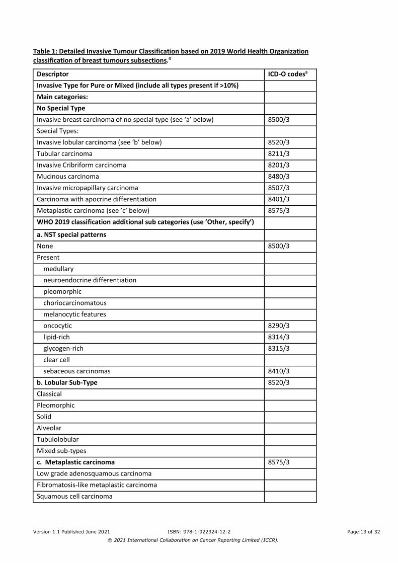
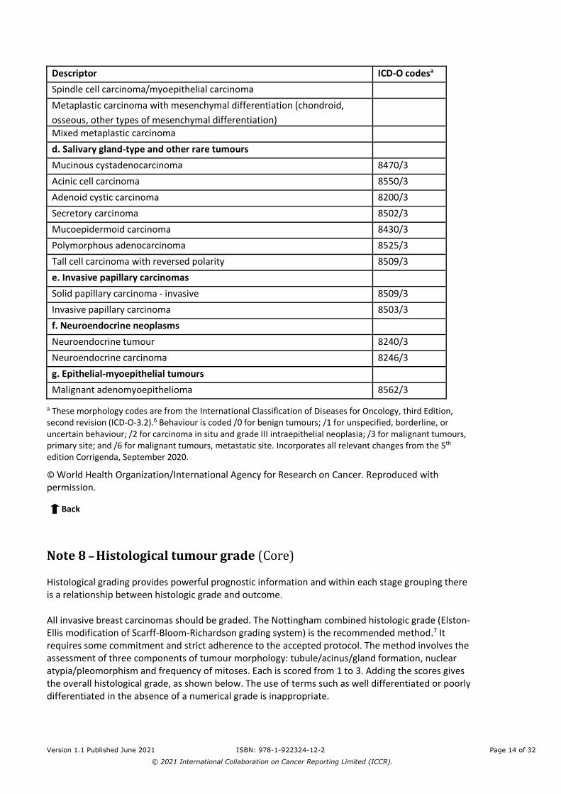
Table 1: Detailed Invasive Tumour Classification based on 2019 World Health Organization classification of breast tumours subsections.4
Descriptor ICD-O codesa Invasive Type for Pure or Mixed (include all types present if >10%) Main categories: No Special Type Invasive breast carcinoma of no special type (see ‘a’ below) 8500/3 Special Types: Invasive lobular carcinoma (see ‘b’ below) 8520/3 Tubular carcinoma 8211/3 Invasive Cribriform carcinoma 8201/3 Mucinous carcinoma 8480/3 Invasive micropapillary carcinoma 8507/3 Carcinoma with apocrine differentiation 8401/3 Metaplastic carcinoma (see ’c’ below) 8575/3 WHO 2019 classification additional sub categories (use ’Other, specify’)
)
a. NST special patterns None 8500/3 Present
medullary
neuroendocrine differentiation pleomorphic choriocarcinomatous melanocytic features oncocytic 8290/3 lipid-rich 8314/3 glycogen-rich 8315/3 clear cell sebaceous carcinomas 8410/3
b. Lobular Sub-Type 8520/3 Classical Pleomorphic Solid Alveolar Tubulolobular Mixed sub-types c. Metaplastic carcinoma 8575/3 Low grade adenosquamous carcinoma Fibromatosis-like metaplastic carcinoma Squamous cell carcinoma
Version 1.1 Published June 2021 ISBN: 978-1-922324-12-2 Page 14 of 32
© 2021 International Collaboration on Cancer Reporting Limited (ICCR).
Descriptor ICD-O codesa Spindle cell carcinoma/myoepithelial carcinoma
Metaplastic carcinoma with mesenchymal differentiation (chondroid, osseous, other types of mesenchymal differentiation)
Mixed metaplastic carcinoma d. Salivary gland-type and other rare tumours Mucinous cystadenocarcinoma 8470/3 Acinic cell carcinoma 8550/3 Adenoid cystic carcinoma 8200/3 Secretory carcinoma 8502/3 Mucoepidermoid carcinoma 8430/3 Polymorphous adenocarcinoma 8525/3 Tall cell carcinoma with reversed polarity 8509/3 e. Invasive papillary carcinomas Solid papillary carcinoma - invasive 8509/3 Invasive papillary carcinoma 8503/3 f. Neuroendocrine neoplasms Neuroendocrine tumour 8240/3 Neuroendocrine carcinoma 8246/3 g. Epithelial-myoepithelial tumours
Malignant adenomyoepithelioma 8562/3
a These morphology codes are from the International Classification of Diseases for Oncology, third Edition, second revision (ICD-O-3.2).6 Behaviour is coded /0 for benign tumours; /1 for unspecified, borderline, or uncertain behaviour; /2 for carcinoma in situ and grade III intraepithelial neoplasia; /3 for malignant tumours, primary site; and /6 for malignant tumours, metastatic site. Incorporates all relevant changes from the 5th edition Corrigenda, September 2020.
© World Health Organization/International Agency for Research on Cancer. Reproduced with permission.
Back
Note 8 – Histological tumour grade (Core) Histological grading provides powerful prognostic information and within each stage grouping there is a relationship between histologic grade and outcome. All invasive breast carcinomas should be graded. The Nottingham combined histologic grade (Elston-Ellis modification of Scarff-Bloom-Richardson grading system) is the recommended method.7 It requires some commitment and strict adherence to the accepted protocol. The method involves the assessment of three components of tumour morphology: tubule/acinus/gland formation, nuclear atypia/pleomorphism and frequency of mitoses. Each is scored from 1 to 3. Adding the scores gives the overall histological grade, as shown below. The use of terms such as well differentiated or poorly differentiated in the absence of a numerical grade is inappropriate.
Version 1.1 Published June 2021 ISBN: 978-1-922324-12-2 Page 15 of 32
© 2021 International Collaboration on Cancer Reporting Limited (ICCR).
Overall grade
• Grade 1 = Scores of 3–5 • Grade 2 = Scores of 6 or 7 • Grade 3 = Scores of 8 or 9.
Published ratios for grades 1, 2 and 3 are approximately 2:3:5 in symptomatic breast cancer, with about half of all symptomatic cancers assigned as grade 3. Screen detected cancer series are likely to include a smaller proportion of high-grade cases. Poor fixation impairs accurate assessment of mitotic frequency reducing their visibility which can result in a change in grade ratios typically with a larger proportion of grade 2 cases and a lower proportion of grade 3 cases. If audit of grade distribution in symptomatic cancers shows substantially fewer grade 3 cases, or a majority of grade 2 cases, fixation and grading protocols should be reviewed. Some degree of variation in appearance from one part of a tumour to another undoubtedly occurs; this is particularly true of tumours of mixed type. Assessment of tubular differentiation is made on the overall appearances of the tumour and so account is taken of any variation. Nuclear appearances are evaluated at the periphery and/or least differentiated area of the tumour to obviate differences between the growing edge and the less active centre. The mitotic score is determined by the number of mitotic figures found in representative 10 consecutive high power fields (HPF) in the most mitotically active part of the tumour. Representative field selection is based on fields having appropriate tumour cellularity based on assessment of the overall cellularity of the tumour identified at low magnification scanning. Fields with low or no tumour cells should not be counted. A random meander approach counting only representative fields is recommended. Only clearly identifiable mitotic figures should be counted; hyperchromatic, karyorrhectic, or apoptotic nuclei are excluded. Because of variations in field size, the HPF size must be determined for each microscope and the appropriate point score determined accordingly, which can also be designated as mitoses/mm2 (see separate section below). Assessment of grade on needle core biopsies
Histological grade can be assessed on core biopsies using the approach described above. This is of particular value if the patient has pre-operative systemic treatment or if grade in the surgical specimen is not assessable. There is about 70% agreement on grade between core biopsy and subsequent surgical specimen. Usually the histological grade in the surgical specimen is used in preference to the core grade. However, if assessment of grade in the surgical specimen is compromised, for example by poor fixation or pre-operative systemic treatment it is reasonable to use the mitotic count score in the core biopsy. Another alternative is to use the mitotic count score in nodal metastases if interpretation of grade is difficult in the primary carcinoma. Assignment of Glandular (Acinar)/Tubular differentiation score
All parts of the tumour are scanned, and the proportion occupied by tumour islands showing clear acinar or gland formation or defined tubular structures with a luminal space is assessed semi-quantitatively. This assessment is generally carried out during the initial low power scan of the tumour sections. A tumour in which 75% or more of its area is composed of such structures would score 1 point for gland/tubule formation. A tumour with between 75% and 10% of glandular/tumour area would score 2 points. A tumour with less than 10% gland/tubule formation would score 3 points. These rules apply to tumours with simple gland/tubule formation such as invasive tubular carcinoma, and those exhibiting complex gland formations such as invasive cribriform carcinoma.
Version 1.1 Published June 2021 ISBN: 978-1-922324-12-2 Page 16 of 32
© 2021 International Collaboration on Cancer Reporting Limited (ICCR).
In the assessment of gland/tubule formation, only structures in which there are clearly defined central lumens, surrounded by polarised tumour cells, should be counted. This does, however, include larger islands of tumour with central gland formation, as may be seen in mucinous carcinoma or invasive micropapillary tumours. Thus mucinous, micropapillary and pure papillary tumours without, or with <10%, secondary luminal spaces are classified as having no tubular or glandular formation and assigned a score of 3. Papillary structures are also not regarded as glandular/tubular structures. Artefactual ‘false’ spaces can occur as a consequence of sub optimal fixation and tissue freezing. Such spaces should be excluded from assessment. Intracytoplasmic lumen formation (intracytoplasmic vacuoles with true luminal microvillar surface, PAS+) does not count as gland formation whatever the size of the intracytoplasmic vacuoles. Assignment of Nuclear Pleomorphism score
Individual pathologists differ markedly in their approach to nuclear grading, and breast specialists appear to allocate higher grades than non-specialists. Few cancers possess the very bland nuclei warranting an atypia/pleomorphism score of 1, and obvious atypia/pleomorphism should attract a score of 3. The minimum proportion of tumour nuclei which should show marked nuclear atypia/ pleomorphism before a score of 3 is allocated has not been defined, but the finding of an occasional enlarged or bizarre nucleus should not be used to give a score of 3 rather than a score of 2. Assignment of Mitotic Frequency score
Accurate mitosis counting requires high quality fixation, obtained when fresh specimens are sliced into promptly after surgery and fixed immediately in neutral buffered formalin. This can be achieved without compromising the evaluation of resection margins. Poor quality fixation can result in underscoring of mitotic frequency; optimal fixation is therefore essential. A minimum of 10 HPFs should be counted at the periphery of the tumour, where it has been demonstrated that mitotic activity is greatest on lower power search. If there is variation in the number of mitoses in different areas of the tumour, the least differentiated area (i.e., with the highest mitotic count) should be assessed. If the mitotic frequency score falls very close to a score cut point, one or more further groups of 10 HPFs should be assessed to establish the correct (highest) score. It is recommended that identification of the most mitotically active or least differentiated part of the tumour forms part of the low magnification preliminary assessment of the histological section. If there is no evidence of heterogeneity, mitotic scoring can be carried out at a part of the tumour periphery chosen at random. Fields chosen for scoring are selected during a random meander along the peripheral margin of the selected tumour area. Only fields with a representative tumour burden should be used. The low power scan of the tumour can be used to provide an assessment of the typical tumour to stromal ratio. Only definite mitotic figures (in any phase of the growth cycle) should be counted. Hyperchromatic nuclei and/or apoptotic nuclei should not be scored. The mitosis score depends on the number of mitoses per 10 HPFs. The size of HPFs of modern microscopes is very variable, so it is necessary to standardise the mitotic count using Table 2 below. Field diameter is a function of the objective lens and the eyepiece, so if either of these is changed this exercise should be repeated. The field diameter of the microscope should be measured using the stage graticule, a Vernier scale or one of the simplified methods detailed below. The scoring category should be assigned from the corresponding line of Table 2. Mitotic counts can also be expressed per mm2 which may be amenable to digital microscopy assessment.4
Version 1.1 Published June 2021 ISBN: 978-1-922324-12-2 Page 17 of 32
© 2021 International Collaboration on Cancer Reporting Limited (ICCR).
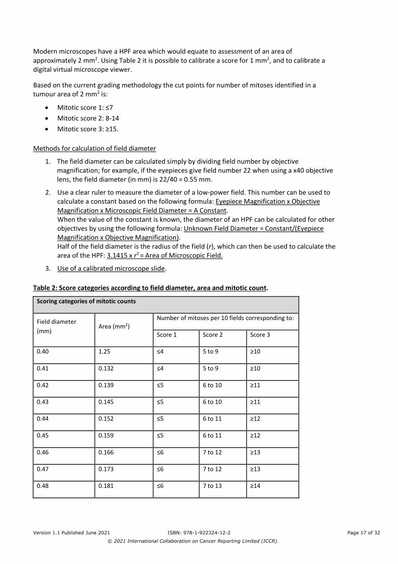
Modern microscopes have a HPF area which would equate to assessment of an area of approximately 2 mm2. Using Table 2 it is possible to calibrate a score for 1 mm2, and to calibrate a digital virtual microscope viewer.
Based on the current grading methodology the cut points for number of mitoses identified in a tumour area of 2 mm2 is:
• Mitotic score 1: ≤7 • Mitotic score 2: 8-14 • Mitotic score 3: ≥15.
Methods for calculation of field diameter
1. The field diameter can be calculated simply by dividing field number by objective magnification; for example, if the eyepieces give field number 22 when using a x40 objective lens, the field diameter (in mm) is 22/40 = 0.55 mm.
2. Use a clear ruler to measure the diameter of a low-power field. This number can be used to calculate a constant based on the following formula: Eyepiece Magnification x Objective Magnification x Microscopic Field Diameter = A Constant. When the value of the constant is known, the diameter of an HPF can be calculated for other objectives by using the following formula: Unknown Field Diameter = Constant/(Eyepiece Magnification x Objective Magnification). Half of the field diameter is the radius of the field (r), which can then be used to calculate the area of the HPF: 3.1415 x r2 = Area of Microscopic Field.
3. Use of a calibrated microscope slide.
Table 2: Score categories according to field diameter, area and mitotic count.
Scoring categories of mitotic counts
Field diameter (mm)
Area (mm2) Number of mitoses per 10 fields corresponding to:
Score 1 Score 2 Score 3
0.40 1.25 ≤4 5 to 9 ≥10
0.41 0.132 ≤4 5 to 9 ≥10
0.42 0.139 ≤5 6 to 10 ≥11
0.43 0.145 ≤5 6 to 10 ≥11
0.44 0.152 ≤5 6 to 11 ≥12
0.45 0.159 ≤5 6 to 11 ≥12
0.46 0.166 ≤6 7 to 12 ≥13
0.47 0.173 ≤6 7 to 12 ≥13
0.48 0.181 ≤6 7 to 13 ≥14

Version 1.1 Published June 2021 ISBN: 978-1-922324-12-2 Page 18 of 32
© 2021 International Collaboration on Cancer Reporting Limited (ICCR).
0.49 0.189 ≤6 7 to 13 ≥14
0.50 0.196 ≤7 8 to 14 ≥15
0.51 0.204 ≤7 8 to 14 ≥15
0.52 0.212 ≤7 8 to 15 ≥16
0.53 0.221 ≤8 9 to 16 ≥17
0.54 0.229 ≤8 9 to 16 ≥17
0.55 0.238 ≤8 9 to 17 ≥18
0.56 0.246 ≤8 9 to 17 ≥18
0.57 0.255 ≤9 10 to 18 ≥19
0.58 0.264 ≤9 10 to 19 ≥20
0.59 0.273 ≤9 10 to 19 ≥20
0.60 0.283 ≤10 11 to 20 ≥21
0.61 0.292 ≤10 11 to 21 ≥22
0.62 0.302 ≤11 12 to 22 ≥23
0.63 0.312 ≤11 12 to 22 ≥23
0.64 0.322 ≤11 12 to 23 ≥24
0.65 0.332 ≤12 13 to 24 ≥25
0.66 0.342 ≤12 13 to 24 ≥25
0.67 0.353 ≤12 13 to 25 ≥26
0.68 0.363 ≤13 14 to 26 ≥27
0.69 0.374 ≤13 14 to 27 ≥28
Reproduced with permission from The Royal College of Pathologists (2016). Pathology reporting of breast disease in surgical excision specimens incorporating the dataset for histological reporting of breast cancer. The Royal College of Pathologists and National Coordinating Committee for Breast Pathology.8
Back

Version 1.1 Published June 2021 ISBN: 978-1-922324-12-2 Page 19 of 32
© 2021 International Collaboration on Cancer Reporting Limited (ICCR).
Note 9 – Carcinoma in situ (Core and Non-core) and Classification of carcinoma in situ (Core and Non-core)
The presence of coexisting DCIS (and/or florid or pleomorphic LCIS) is commonplace with invasive carcinomas of the breast and forms part of the overall disease process which requires complete surgical excision to reduce the risk of local recurrence. It is recognised that the term “Extensive Intraductal Component” (EIC) has different definitions in different countries and centres. Most refer to either substantial volume of DCIS within the invasive carcinoma and/or substantial DCIS quantity beyond the limits of the invasive cancer. No preferred definition is provided as there is a limited evidence base for each of these proffered definitions, with no international consensus. For this reason, sub-categorisation as EIC is deemed non-core and its use is optional. Classification of DCIS and accompanying in situ lesions with respect to histological nuclear grade (core), presence or absence of necrosis (core), and architectural pattern (non-core) is dealt with in the ICCR DCIS, variants of LCIS and low grade lesions dataset.2 Nuclear grade of DCIS is largely determined by size and pleomorphism, although other morphologic features (see Table 3) are also of help. Table 3: Nuclear grade of ductal carcinoma in situ.
Feature Grade I (Low) Grade II (Intermediate) Grade III (High)
Pleomorphism Monotonous (monomorphic)
Intermediate Markedly pleomorphic
Size 1.5 to 2 x the size of a normal RBC or a normal duct epithelial cell nucleus
Intermediate >2.5 x the size of a normal red blood cell or a normal duct epithelial cell nucleus
Chromatin Usually diffuse, finely dispersed chromatin
Intermediate Usually vesicular with irregular chromatin distribution
Nucleoli Only occasional Intermediate Prominent, often multiple
Mitoses Only occasional Intermediate May be frequent
Orientation Polarized toward luminal spaces
Intermediate Usually not polarized toward the luminal space
Definition: RBC, red blood cell.
Reproduced with permission from College of American Pathologists (CAP). Protocol for the Examination of Resection Specimens From Patients With Ductal Carcinoma In Situ (DCIS) of the Breast. Breast DCIS Resection 4.3.0.2. College of American Pathologists, February 2020.9

Version 1.1 Published June 2021 ISBN: 978-1-922324-12-2 Page 20 of 32
© 2021 International Collaboration on Cancer Reporting Limited (ICCR).
Pleomorphic and florid LCIS have overlapping features with DCIS and may be treated similarly, but at present there is insufficient evidence to establish definitive recommendations for treatment. The current understanding of the natural history of pleomorphic LCIS and florid LCIS is limited, and the optimal treatment is unknown with regard to pursuing negative margins and consideration of additional adjuvant therapies. Nevertheless, although pleomorphic and florid LCIS are not currently included in the but not classic LCIS which is considered a ‘benign’ lesion in the American Joint Committee on Cancer (AJCC) pTis classification,10 they remain as a category in the Union for International Cancer Control (UICC) TNM 8th edition,11 and there is emerging evidence suggesting that these forms of LCIS might be better treated as DCIS,4,12 in particular the practice of excision to negative margins.
Back Note 10 – Tumour extension (Core) Tumour extension to involve overlying skin or underlying skeletal muscle is a variable which influences TNM staging and should be recorded when present. It is recognised that in the context of primary operable breast cancer these phenomena are rare. The majority of cancer resection cases will be confined to the breast with no skin, nipple or underlying skeletal muscle involvement and in this context disease extent classification is deemed non-core. The finding of invasive carcinoma that directly invades into the dermis or epidermis without skin ulceration does not change the pT stage. Satellite skin nodules must be separate from the primary tumour and macroscopically identified to assign a category as pT4b. Skin nodules identified only on microscopic examination and in the absence of epidermal ulceration or skin oedema (clinical peau d’orange) do not qualify as pT4b. Such tumours should be categorised based on tumour size. The finding of tumour extension into the nipple does not change the pT classification of invasive carcinomas. Invasion into pectoralis muscle is not considered chest wall invasion, and cancers are not classified as pT4a unless there is invasion deeper than this muscle.
Back
Note 11 – Margin status (Core and Non-core) There is an assumption that all breast tissue will be resected in patients undergoing a complete mastectomy and that pathological examination of margins is of limited value. However, there is evidence that margin involvement can increase the risk of local recurrence after mastectomy and modification of the comprehensive margin analysis and reporting recommendations for wide local excision and other similar specimens are adopted for reporting of mastectomy specimens to include a statement of the distance to the closest margin(s) or site(s) of margin involvement.

Version 1.1 Published June 2021 ISBN: 978-1-922324-12-2 Page 21 of 32
© 2021 International Collaboration on Cancer Reporting Limited (ICCR).
Assessment of adequacy of excision requires close correlation between the surgical excision procedure and pathological examination. In particular it is essential that the pathologist is made aware of the depth of tissue excised and whether the surgeon has excised all the tissue from the subcutis to the pectoral fascia. Similarly it has been recognised that involvement of a margin, particularly the posterior margin in a mastectomy specimen, should also be described as this could result in a recommendation for further surgery or radiotherapy. There remains some controversy regarding the minimum width of uninvolved tissue that defines ‘complete’ excision, although narrower margins are now more widely accepted as adequate than previously. For this reason, it is recommended that the pathologist reports the measurement to the inked margins of DCIS and invasive carcinoma rather than quoting ‘complete’ excision or ‘not at ink’ in histology reports. Some centres find it helpful to report the approximate extent of margin involvement. The following system is recommended - this is considered a non-core feature:
• Unifocal: one focal area of carcinoma at the margin, <5 mm • Multifocal: two or more foci of carcinoma at the margin • Extensive: carcinoma present at the margin over a broad front (≥5 mm).
Back
Note 12 – Lymphovascular invasion in primary breast carcinoma (Core and Non-core) The presence of LVI is an adverse feature providing independent prognostic information about both local recurrence and survival. It is therefore important to record whether or not it is present. Reporting the LVI status for stage IIA and IIB patients who have an axillary lymph node dissection may influence the use of adjuvant radiotherapy. As it is difficult to distinguish between lymphatic and venous channels, findings should be categorised as LVI rather than define a specific channel. This is supported by evidence identifying that most tumour emboli are present in lymphatic channels.13 The presence of unequivocal tumour in lymphovascular spaces should be recorded. ‘Indeterminate’ may be used where it is equivocal or uncertain. If there is doubt about the presence of tumour in lymphovascular spaces, but it is considered to be very likely, it should be recorded as ‘indeterminate’. Useful criteria for recognition of LVI include:
• Groups of tumour cells in spaces around the main tumour mass; ensure that any spaces are lined by a rim of endothelial cells and are not fat spaces.
• The presence of adjacent channels that may be of varying sizes. • The presence within the space of lymphocytes, erythrocytes and/or thrombus. Note that true
blood vascular involvement in the breast is rare. • Shrinkage artefact results in nests of cells having the shape of the space in which they lie; and
endothelial cells will not be seen.
Version 1.1 Published June 2021 ISBN: 978-1-922324-12-2 Page 22 of 32
© 2021 International Collaboration on Cancer Reporting Limited (ICCR).
The best method for assessing LVI is the use of good quality, optimally fixed and processed haematoxylin-eosin (H&E) stained sections. Immunostaining for endothelial and/or lymphoendothelial markers does not generally contribute further but could be considered for difficult critical cases. Shrinkage artefact may also involve DCIS, where the myoepithelial layer may mimic endothelial cells, and it should be recognised that both lymphatic endothelial cells and myoepithelial cells stain positively with the lymphendothelial marker podoplanin/D2-40 antibody. One of the major problems in trying to determine whether or not tumour cells are in a vessel is shrinkage artefact, so care should be taken, wherever possible, to ensure that there is optimal tissue fixation and processing. Only LVI identified in breast tissue associated with the primary breast carcinoma should be recorded. LVI identified elsewhere, for example in axillary tissue, may be described but not recorded formally as LVI positive. Perineural invasion should not be recorded as LVI. Documenting the presence of dermal LVI is valuable because of its strong association with the clinical findings of inflammatory breast carcinoma. There is no agreed definition of extensive LVI and no substantive evidence base. Sub categorisation of LVI as extensive or non-extensive is therefore subjective and considered optional/non-core.
Back
Note 13 – Coexistent pathology (Non-core)
In some situations, inclusion of coexisting conditions can be considered beneficial if this supports clinicopathological correlation or patient management. Examples include microcalcification detected mammographically and extension into or involvement of a benign lesion such as a sclerosing lesion, papillary lesion or fibroepithelial lesion. An exhaustive description of all coexisting conditions is not required.
Back
Note 14 – Microcalcifications (Non-core) Ductal carcinoma in situ (DCIS) found in biopsies performed for microcalcifications will almost always be at the site of the microcalcifications or in close proximity.14,15 Some of these lesions may also include an invasive component. The pathologist must be satisfied that the specimen has been sampled in such a way that the lesion responsible for the microcalcifications has been examined microscopically. The presence of the targeted microcalcifications in the specimen can be confirmed by specimen radiography. The relationship of the radiologic microcalcifications to the DCIS should be indicated.
Back

Version 1.1 Published June 2021 ISBN: 978-1-922324-12-2 Page 23 of 32
© 2021 International Collaboration on Cancer Reporting Limited (ICCR).
Note 15 – Estrogen receptor (ER) (Core and Non-core)
Use of hormone receptor scoring systems such as Allred, Quick score and H score are optional (see methodology details below). Hormone receptor status is determined primarily to identify patients who may benefit from endocrine therapy. About 75 to 80% of invasive breast cancers are positive for estrogen receptor (ER) and progesterone receptor (PR), including almost all well-differentiated (grade 1) cancers and most moderately differentiated (grade 2) cancers, and studies have shown a substantial survival benefit from endocrine therapy among patients with ER positive tumours. Receptor status is only a weak prognostic factor. Currently ER status is used to select patients suitable for endocrine therapy. PR status has been shown to provide information on degree of response to endocrine therapy in patients for ER positive tumours. Hormone receptor status
True ER negative, PR positive carcinomas are extremely rare, but patients with such tumours are also considered eligible for endocrine therapy. The finding of an ER negative PR positive tumour can indicate a false negative ER assessment or a false positive PR assessment and audit or repeat staining is recommended. Hormone receptor status is most often determined in formalin-fixed, paraffin-embedded tissue sections by immunohistochemistry (IHC). Only nuclear staining is considered positive. Single-gene expression assays are not recommended for routine use. The American Society of Clinical Oncology (ASCO) and College of American Pathologists (CAP), The Royal College of Pathologists UK (RCPath), and The Royal College of Pathologists of Australasia (RCPA) have issued recommendations for reporting the results of IHC assays for ER and PR.16-18
Studies using both IHC and the ligand binding assay suggest that patients with higher hormone receptor levels have a higher probability of response to endocrine therapy, but expression as low as 1% positive staining has been associated with a clinical response. As a result, the guidelines recommend classifying all cases with at least 1% positive cells as receptor positive. For patients with low ER expression (1 to 10% positive cells), the decision on endocrine therapy should be based on an analysis of its risks and potential benefits.19 Definition of a negative result
All current guidelines recommend that carcinomas with <1% positive cells be considered negative for ER and PR.8,20,21 In the Allred system (see Table 4), the survival of patients whose carcinomas had a score of 2 (corresponding to <1% weakly positive cells) was similar to that of patients whose carcinomas were completely negative for ER.22 Therefore, a score of 2 was considered to be a negative result. Using the Allred or Quickscore system23 carcinomas with <1% positive cells and intensity scores of 2 or 3 would have a total score of 3 or 4 and historically were considered positive. These are rare carcinomas, and their response to endocrine therapy has not been specifically studied. Thus use of the Allred/Quickscore assessment methods can, in a small proportion of cases, conflict with the 1% cut point for positivity/negativity recommended above. It is recommended that all cases showing ≥1% of tumour cells positive should be classified as receptor positive regardless of their Allred/Quickscore. Reports should include the overall percentage of positive cells and the average intensity regardless of whether additional scoring systems, such as Allred or H score, are also reported. All cases showing <1% of tumour cells positive should be classified as receptor negative regardless of their Allred score.

Version 1.1 Published June 2021 ISBN: 978-1-922324-12-2 Page 24 of 32
© 2021 International Collaboration on Cancer Reporting Limited (ICCR).
It has become increasingly recognised that there are limited data on response to endocrine therapy in carcinomas with low level ER expression, defined as 1-10% positive cells, although the available information currently supports possible benefit. Furthermore, recent studies of ER gene expression have shown profiles more similar to ER negative cancers. It is recommended that these tumours remain classified as positive and considered eligible for endocrine treatment, but be designated Low ER Positive.19 The following reporting comment is recommended in ER Low Positive cases, to aid in communicating the challenges and more limited data on cancers with this result: “The cancer in this sample has a low level (1-10%) of ER expression by IHC. There are limited data on the overall benefit of endocrine therapies for patients with low level (1-10%) ER expression but they currently suggest possible benefit, so patients are considered eligible for endocrine treatment. There are data that suggest invasive cancers with these results are heterogeneous in both behaviour and biology and often have gene expression profiles more similar to ER negative cancers.” When a tumour is negative but no internal control cells are present, the pathologist must exercise judgment as to whether the assay can be interpreted as a true negative. If there is doubt then a recommendation to repeat on another block or specimen that contains internal controls should be made. ‘Cannot be determined’ is used when any issue prevents reliable interpretation of the result. This can include suboptimal specimen handling, presence of artefacts (crush or edge artefacts) making interpretation difficult, or if the analytical testing procedure failed. Quantification of ER and PR
There is a wide range of receptor levels in cancers as shown by the biochemical ligand binding assay and as observed with IHC. Patients whose carcinomas have higher levels have improved survival when treated with endocrine therapy. Quantification systems may use only the proportion of positive cells or may include the intensity of immunoreactivity:
• Number of positive cells: The number of positive cells can be reported as a percentage or within discrete categories (Figure 4).
• Intensity: Refers to the degree of nuclear positivity (i.e., pale to dark). The intensity can be affected by the amount of protein present, as well as the antibody used, the antigen retrieval system and the detection system. In most cancers, there is heterogeneous immunoreactivity with pale to darkly positive cells present.
Figure 4: Quantification of immunohistochemical findings.
Reproduced with permission from College of American Pathologists (2020). Template for Reporting Results of Biomarker Testing of Specimens From Patients With Carcinoma of the Breast. College of American Pathologists.24

Version 1.1 Published June 2021 ISBN: 978-1-922324-12-2 Page 25 of 32
© 2021 International Collaboration on Cancer Reporting Limited (ICCR).
Two methods of quantifying ER by using both intensity and percentage of positive cells are the Allred score (Table 4) and the H score (Table 5). The two systems classify carcinomas into similar, but not identical, groups. If high-affinity antibodies are used with sensitive detection systems, most carcinomas will fall into clearly positive (score 7 or 8) or clearly negative (score 0) categories by Allred score. A small group of carcinomas (<1% of total) show intermediate levels of immunoreactivity. Table 4: Allred score* for estrogen and progesterone receptor evaluation.
Proportion Score Positive Cells, % Intensity Intensity Score
0 0 None 0
1 <1 Weak 1
2 1 to 10 Intermediate 2
3 11 to 33 Strong 3
4 34 to 66
5 ≥67
* The Allred score combines the percentage of positive cells and the intensity of the reaction product in most of the carcinoma.22 The two scores are added together for a final score with eight positive values. Scores of 0 and 2 are considered negative. Scores of 3 to 8 are considered positive.
Reproduced with permission from College of American Pathologists (2020). Template for Reporting Results of Biomarker Testing of Specimens From Patients With Carcinoma of the Breast. College of American Pathologists.24 Table 5: H score* for estrogen and progesterone receptor evaluation.
Calculation of H Score
Cell Signal Percentage of Cells Value Multiplied
Cells with no signal % x 0 = 0
Cells with weak signal % x 1 =
Cells with moderate signal % x 2 =
Cells with strong signal % x 3 =
Total score =
* The H score is determined by multiplying the percentage of cells demonstrating each intensity (scored from 0 to 3) and adding the results.25 There are 300 possible values. In this system, <1% positive cells is considered to be a negative result.
Reproduced with permission from College of American Pathologists (2020). Template for Reporting Results of Biomarker Testing of Specimens From Patients With Carcinoma of the Breast. College of American Pathologists.24

Version 1.1 Published June 2021 ISBN: 978-1-922324-12-2 Page 26 of 32
© 2021 International Collaboration on Cancer Reporting Limited (ICCR).
Quality assurance
There are many preanalytic, analytic and postanalytic variables that can affect test results, and the assays must be validated to ensure their accuracy. External quality assurance proficiency testing surveys for ER and PR are invaluable tools to help ensure that assays perform as expected, and they are available from established immunocytochemistry external quality assurance (EQA) scheme providers (CAP, United Kingdom NEQAS, NordiQC, CPQA, CBQA etc).
Back
Note 16 – Progesterone receptor (PR) (Core and Non-core) The value of PR in the selection of endocrine therapy in both the adjuvant and metastatic settings has not been demonstrated and at present ER status is used to predict the benefit of endocrine therapy. Within the group of cancers that are ER positive, PR expression levels (the percentage of stained cells) are considered a prognostic marker: cases with lower PR expression levels are associated with worse outcomes, but patients still receive benefit from endocrine therapy. When a tumour is negative but no internal control cells are present, the pathologist must exercise judgment as to whether the assay can be interpreted as a true negative. If there is doubt then a recommendation to repeat on another block or specimen that contains internal controls should be made. ‘Cannot be determined’ is used when any issue prevents reliable interpretation of the result. This can include suboptimal specimen handling, presence of artefacts (crush or edge artefacts) making interpretation difficult, or if the analytical testing procedure failed.
Back
Note 17 – HER2 (Core and Non-core)
A subset of breast carcinomas (approximately 15 to 20%) overexpress human epidermal growth factor receptor 2 (HER2; HUGO nomenclature ERBB2). Protein overexpression is usually due to gene amplification. Assays for gene copy number, mRNA quantity, and protein generally give similar results; gene amplification correlates with protein overexpression in about 95% of cases. In a small subset of carcinomas (probably <5%), protein overexpression may occur by different mechanisms. Overexpression is both a prognostic and predictive factor. HER2 status is primarily evaluated to determine patient eligibility for anti-HER2 therapy. It may also identify patients who have a greater benefit from anthracycline-based adjuvant therapy. HER2 status can be determined in formalin-fixed paraffin-embedded tissue by assessing protein overexpression on the membrane of tumour cells using IHC or by assessing the number of HER2 gene copies using in situ hybridization (ISH). When both IHC and ISH are performed on the same tumour, the results should be correlated. The most likely reason for a discrepancy is a false result of one of the assays, but in a small number of cases there may be protein overexpression without amplification, amplification without protein overexpression (especially in low-level amplification), or marked intratumoural heterogeneity.
Version 1.1 Published June 2021 ISBN: 978-1-922324-12-2 Page 27 of 32
© 2021 International Collaboration on Cancer Reporting Limited (ICCR).
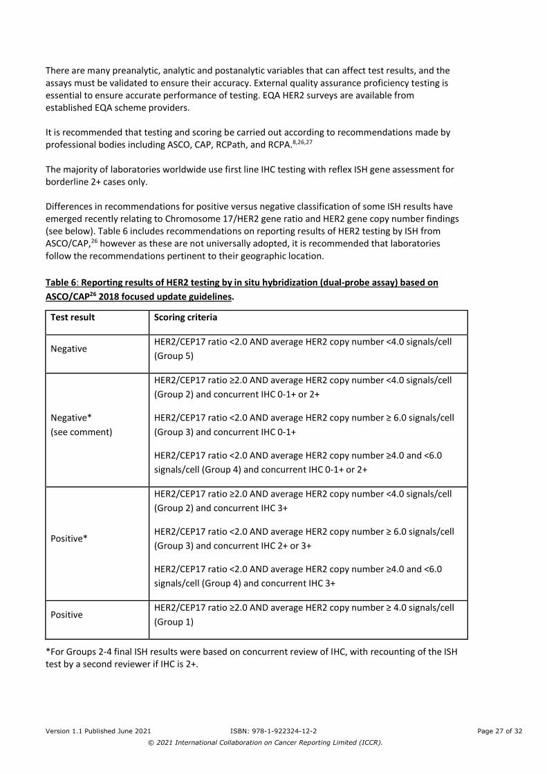
There are many preanalytic, analytic and postanalytic variables that can affect test results, and the assays must be validated to ensure their accuracy. External quality assurance proficiency testing is essential to ensure accurate performance of testing. EQA HER2 surveys are available from established EQA scheme providers. It is recommended that testing and scoring be carried out according to recommendations made by professional bodies including ASCO, CAP, RCPath, and RCPA.8,26,27 The majority of laboratories worldwide use first line IHC testing with reflex ISH gene assessment for borderline 2+ cases only. Differences in recommendations for positive versus negative classification of some ISH results have emerged recently relating to Chromosome 17/HER2 gene ratio and HER2 gene copy number findings (see below). Table 6 includes recommendations on reporting results of HER2 testing by ISH from ASCO/CAP,26 however as these are not universally adopted, it is recommended that laboratories follow the recommendations pertinent to their geographic location. Table 6: Reporting results of HER2 testing by in situ hybridization (dual-probe assay) based on ASCO/CAP26 2018 focused update guidelines.
Test result Scoring criteria
Negative HER2/CEP17 ratio <2.0 AND average HER2 copy number <4.0 signals/cell (Group 5)
Negative* (see comment)
HER2/CEP17 ratio ≥2.0 AND average HER2 copy number <4.0 signals/cell (Group 2) and concurrent IHC 0-1+ or 2+
HER2/CEP17 ratio <2.0 AND average HER2 copy number ≥ 6.0 signals/cell (Group 3) and concurrent IHC 0-1+
HER2/CEP17 ratio <2.0 AND average HER2 copy number ≥4.0 and <6.0 signals/cell (Group 4) and concurrent IHC 0-1+ or 2+
Positive*
HER2/CEP17 ratio ≥2.0 AND average HER2 copy number <4.0 signals/cell (Group 2) and concurrent IHC 3+
HER2/CEP17 ratio <2.0 AND average HER2 copy number ≥ 6.0 signals/cell (Group 3) and concurrent IHC 2+ or 3+
HER2/CEP17 ratio <2.0 AND average HER2 copy number ≥4.0 and <6.0 signals/cell (Group 4) and concurrent IHC 3+
Positive HER2/CEP17 ratio ≥2.0 AND average HER2 copy number ≥ 4.0 signals/cell (Group 1)
*For Groups 2-4 final ISH results were based on concurrent review of IHC, with recounting of the ISH test by a second reviewer if IHC is 2+.

Version 1.1 Published June 2021 ISBN: 978-1-922324-12-2 Page 28 of 32
© 2021 International Collaboration on Cancer Reporting Limited (ICCR).
• Comment for Group 2 Negative result: Evidence is limited on the efficacy of HER2-targeted therapy in the small subset of cases with HER2/CEP17 ratio ≥2.0 and an average HER2 copy number <4.0/cell. In the first generation of adjuvant trastuzumab trials, patients in this subgroup who were randomized to the trastuzumab arm did not appear to derive an improvement in disease free or overall survival, but there were too few such cases to draw definitive conclusions. IHC expression for HER2 should be used to complement ISH and define HER2 status. If IHC result is not 3+ positive, it is recommended that the specimen be considered HER2 negative because of the low HER2 copy number by ISH and lack of protein overexpression.
• Comment for Group 3 Negative result: There are insufficient data on the efficacy of HER2-targeted therapy in cases with HER2/CEP17 ratio <2.0 in the absence of protein overexpression because such patients were not eligible for the first generation of adjuvant trastuzumab clinical trials. When concurrent IHC results are negative (0-1+), it is recommended that the specimen be considered HER2 negative.
• Comment for Group 4 Negative result: It is uncertain whether patients with ≥4.0 and <6.0 average HER2 signals/cell and HER2/CEP17 ratio <2.0 benefit from HER2 targeted therapy in the absence of protein overexpression (IHC 3+). If the specimen test result is close to the ISH ratio threshold for positive, there is a high likelihood that repeat testing will result in different results by chance alone. Therefore, when IHC results are not 3+ positive, it is recommended that the sample be considered HER2 negative without additional testing on the same specimen.
Back
Note 18 – Ancillary studies (Non-core)
The results of any additional ancillary studies, such as multigene test results, when performed are recommended to be included or added subsequently to the pathology report, to ensure a record of all assays performed on the case in a single comprehensive report. Ki-67 is a nuclear protein found in all phases of the cell cycle and is a marker of cell proliferation. The percentage of Ki-67 positive tumour cells determined by IHC has been used to stratify patients into good and poor prognostic groups, but there is a lack of consensus on scoring, definition of low versus high expression, an appropriate cut point for positivity, or which part of the tumour should be scored (e.g., leading edge, hot spots, overall average). There is also a paucity of data on the effects of pre-analytic variables (e.g., ischaemic time, length of fixation, antigen retrieval) on Ki-67 staining. For these reasons, routine testing of breast cancers for Ki-67 expression is not currently recommended or deemed required by organisations such as ASCO, National Comprehensive Cancer Network (NCCN), and RCPath. However, it is recognised that Ki-67 testing is routine in some countries. International collaborative efforts aim to develop standardised validated staining and scoring methodology which may lead to more widespread adoption.28-30 Other tests may become relevant in classification of some forms of breast cancer and the results of these assays, when performed, should be included in the report. For example, tumour-infiltrating lymphocyte (TIL) assessment is gaining importance as a prognostic marker. High numbers of TILs are associated with better outcome and better response to neoadjuvant therapy in triple-negative (ER, PR and HER2 negative) and HER2-positive breast carcinomas. It is recommended to follow the international consensus scoring recommendations for quantifying TILs.31
Back

Version 1.1 Published June 2021 ISBN: 978-1-922324-12-2 Page 29 of 32
© 2021 International Collaboration on Cancer Reporting Limited (ICCR).
Note 19 – Pathological staging (Core) The Tumour Node Metastasis (TNM) system of the UICC is recommended.11 Pathologic Classification
Additional descriptors can be used:
The suffix ‘m’ indicates the presence of multiple primary tumours in a single site and is recorded in parentheses, e.g., pT(m) NM. The ‘r’ prefix indicates a recurrent tumour when staging is carried out after a documented disease-free interval. Pathological T (pT): Histological assessment of the primary tumour (pT) generally is based on the largest invasive tumour focus. See Note 6 TUMOUR DIMENSIONS for methodology details.
Back
References 1 Merlin T, Weston A and Tooher R (2009). Extending an evidence hierarchy to include topics
other than treatment: revising the Australian 'levels of evidence'. BMC Med Res Methodol 9:34.
2 International Collaboration on Cancer Reporting (2021). Ductal Carcinoma In Situ, Variants of
Lobular Carcinoma In Situ and Low Grade Lesions Histopathology Reporting Guide. Available from: http://www.iccr-cancer.org/datasets/published-datasets/breast (Accessed 20th June 2021).
3 International Collaboration on Cancer Reporting (2021). Surgically Removed Lymph Nodes for
Breast Tumours Histopathology Reporting Guide. Available from: http://www.iccr-cancer.org/datasets/published-datasets/breast (Accessed 20th June 2021).
4 WHO Classification of Tumours Editorial Board (ed) (2019). WHO Classification of Tumours,
Breast Tumours, 5th Edition. IARC Publications, Lyon. 5 WHO Classification of Tumours Editorial Board (2020). Breast Tumours, WHO Classification of
Tumours, 5th Edition, Volume 2 - Corrigenda September 2020. Available from: https://publications.iarc.fr/Book-And-Report-Series/Who-Classification-Of-Tumours/Breast-Tumours-2019 (Accessed 16th June 2021).
6 Fritz A, Percy C, Jack A, Shanmugaratnam K, Sobin LH, Parkin DM, Whelan SL and World
Health Organization (2000). International classification of diseases for oncology, World Health Organization, Geneva.
7 Elston CW and Ellis IO (1991). Pathological prognostic factors in breast cancer. I. The value of
histological grade in breast cancer: experience from a large study with long-term follow-up. Histopathology 19(5):403-410.

Version 1.1 Published June 2021 ISBN: 978-1-922324-12-2 Page 30 of 32
© 2021 International Collaboration on Cancer Reporting Limited (ICCR).
8 Royal College of Pathologists and National Coordinating Committee for Breast Pathology (2016). Pathology reporting of breast disease in surgical excision specimens incorporating the dataset for histological reporting of breast cancer. Available from: https://www.rcpath.org/profession/guidelines/cancer-datasets-and-tissue-pathways.html (Accessed 26th March 2020).
9 College of American Pathologists (2020). Protocol for the Examination of Resection Specimens
From Patients With Ductal Carcinoma In Situ (DCIS) of the Breast. Available from: https://documents.cap.org/protocols/cp-breast-dcis-resection-19-4301.pdf (Accessed 20th September 2020).
10 Amin MB, Edge S, Greene FL, Byrd DR, Brookland RK, Washington MK, Gershenwald JE,
Compton CC, Hess KR, Sullivan DC, Jessup JM, Brierley JD, Gaspar LE, Schilsky RL, Balch CM, Winchester DP, Asare EA, Madera M, Gress DM and Meyer LR (eds) (2017). AJCC Cancer Staging Manual. 8th ed. Springer, New York.
11 Brierley JD, Gospodarowicz MK and Wittekind C (eds) (2016). Union for International Cancer
Control. TNM Classification of Malignant Tumours, 8th Edition, Wiley, USA. 12 Foschini MP, Miglio R, Fiore R, Baldovini C, Castellano I, Callagy G, Bianchi S, Kaya H,
Amendoeira I, Querzoli P, Poli F, Scatena C, Cordoba A, Pietribiasi F, Kovács A, Faistova H, Cserni G and Quinn C (2019). Pre-operative management of Pleomorphic and florid lobular carcinoma in situ of the breast: Report of a large multi-institutional series and review of the literature. Eur J Surg Oncol 45(12):2279-2286.
13 Mohammed RA, Martin SG, Gill MS, Green AR, Paish EC and Ellis IO (2007). Improved
methods of detection of lymphovascular invasion demonstrate that it is the predominant method of vascular invasion in breast cancer and has important clinical consequences. Am J Surg Pathol 31(12):1825-1833.
14 Owings DV, Hann L and Schnitt SJ (1990). How thoroughly should needle localization breast
biopsies be sampled for microscopic examination? A prospective mammographic/pathologic correlative study. Am J Surg Pathol 14(6):578-583.
15 Silverstein MJ, Lagios MD, Recht A, Allred DC, Harms SE, Holland R, Holmes DR, Hughes LL,
Jackman RJ, Julian TB, Kuerer HM, Mabry HC, McCready DR, McMasters KM, Page DL, Parker SH, Pass HA, Pegram M, Rubin E, Stavros AT, Tripathy D, Vicini F and Whitworth PW (2005). Image-detected breast cancer: state of the art diagnosis and treatment. J Am Coll Surg 201(4):586-597.
16 Hammond ME, Hayes DF, Dowsett M, Allred DC, Hagerty KL, Badve S, Fitzgibbons PL, Francis
G, Goldstein NS, Hayes M, Hicks DG, Lester S, Love R, Mangu PB, McShane L, Miller K, Osborne CK, Paik S, Perlmutter J, Rhodes A, Sasano H, Schwartz JN, Sweep FC, Taube S, Torlakovic EE, Valenstein P, Viale G, Visscher D, Wheeler T, Williams RB, Wittliff JL and Wolff AC (2010). American Society of Clinical Oncology/College Of American Pathologists guideline recommendations for immunohistochemical testing of estrogen and progesterone receptors in breast cancer. J Clin Oncol 28(16):2784-2795.
17 Royal College of Pathologists. Cancer datasets and tissue pathways. Available from:
https://www.rcpath.org/profession/guidelines/cancer-datasets-and-tissue-pathways.html (Accessed 25th March 2020).

Version 1.1 Published June 2021 ISBN: 978-1-922324-12-2 Page 31 of 32
© 2021 International Collaboration on Cancer Reporting Limited (ICCR).
18 Royal College of Pathologists of Australasia. Cancer Protocols. Available from: https://www.rcpa.edu.au/Library/Practising-Pathology/Structured-Pathology-Reporting-of-Cancer/Cancer-Protocols (Accessed 26th March 2020).
19 Allison KH, Hammond MEH, Dowsett M, McKernin SE, Carey LA, Fitzgibbons PL, Hayes DF,
Lakhani SR, Chavez-MacGregor M, Perlmutter J, Perou CM, Regan MM, Rimm DL, Symmans WF, Torlakovic EE, Varella L, Viale G, Weisberg TF, McShane LM and Wolff AC (2020). Estrogen and Progesterone Receptor Testing in Breast Cancer: ASCO/CAP Guideline Update. J Clin Oncol:Jco1902309.
20 Hammond ME, Hayes DF, Dowsett M, Allred DC, Hagerty KL, Badve S, Fitzgibbons PL, Francis
G, Goldstein NS, Hayes M, Hicks DG, Lester S, Love R, Mangu PB, McShane L, Miller K, Osborne CK, Paik S, Perlmutter J, Rhodes A, Sasano H, Schwartz JN, Sweep FC, Taube S, Torlakovic EE, Valenstein P, Viale G, Visscher D, Wheeler T, Williams RB, Wittliff JL and Wolff AC (2010). American Society of Clinical Oncology/College of American Pathologists guideline recommendations for immunohistochemical testing of estrogen and progesterone receptors in breast cancer (unabridged version). Arch Pathol Lab Med 134(7):e48-72.
21 Royal College of Pathologists of Australasia (2012). Invasive breast cancer structured
reporting protocol (2nd edition). Available from: https://www.rcpa.edu.au/getattachment/9c857cb6-6878-4004-bf8a-37761873cf13/Protocol-invasive-breast-cancer.aspx (Accessed 26th March 2020).
22 Harvey JM, Clark GM, Osborne CK and Allred DC (1999). Estrogen receptor status by
immunohistochemistry is superior to the ligand-binding assay for predicting response to adjuvant endocrine therapy in breast cancer. J Clin Oncol 17(5):1474-1481.
23 Detre S, Saclani Jotti G and Dowsett M (1995). A "quickscore" method for
immunohistochemical semiquantitation: validation for oestrogen receptor in breast carcinomas. J Clin Pathol 48(9):876-878.
24 College of American Pathologists (2020). Template for Reporting Results of Biomarker Testing
of Specimens From Patients With Carcinoma of the Breast. Available from: https://documents.cap.org/protocols/cp-breast-biomarker-20-1400.pdf (Accessed 22nd September 2020).
25 McCarty KS, Jr., Miller LS, Cox EB, Konrath J and McCarty KS, Sr. (1985). Estrogen receptor
analyses. Correlation of biochemical and immunohistochemical methods using monoclonal antireceptor antibodies. Arch Pathol Lab Med 109(8):716-721.
26 Wolff AC, Hammond MEH, Allison KH, Harvey BE, McShane LM and Dowsett M (2018). HER2
Testing in Breast Cancer: American Society of Clinical Oncology/College of American Pathologists Clinical Practice Guideline Focused Update Summary. J Oncol Pract 14(7):437-441.
27 Royal College of Pathologists of Australasia (2018). ASCO CAP 2018 HER2 Testing for Breast
Cancer Guidelines - Recommendations for Practice in Australasia. Available from: https://www.rcpa.edu.au/getattachment/fecd094c-aaf4-416b-9ed5-4a61f5ac1a93/ASCO-CAP-2018-HER2-Testing-for-Breast-Cancer-Guide.aspx (Accessed 26th March 2020).

Version 1.1 Published June 2021 ISBN: 978-1-922324-12-2 Page 32 of 32
© 2021 International Collaboration on Cancer Reporting Limited (ICCR).
28 Leung SCY, Nielsen TO, Zabaglo LA, Arun I, Badve SS, Bane AL, Bartlett JMS, Borgquist S, Chang MC, Dodson A, Ehinger A, Fineberg S, Focke CM, Gao D, Gown AM, Gutierrez C, Hugh JC, Kos Z, Laenkholm AV, Mastropasqua MG, Moriya T, Nofech-Mozes S, Osborne CK, Penault-Llorca FM, Piper T, Sakatani T, Salgado R, Starczynski J, Sugie T, van der Vegt B, Viale G, Hayes DF, McShane LM and Dowsett M (2019). Analytical validation of a standardised scoring protocol for Ki67 immunohistochemistry on breast cancer excision whole sections: an international multicentre collaboration. Histopathology 75(2):225-235.
29 Leung SCY, Nielsen TO, Zabaglo L, Arun I, Badve SS, Bane AL, Bartlett JMS, Borgquist S, Chang
MC, Dodson A, Enos RA, Fineberg S, Focke CM, Gao D, Gown AM, Grabau D, Gutierrez C, Hugh JC, Kos Z, Laenkholm AV, Lin MG, Mastropasqua MG, Moriya T, Nofech-Mozes S, Osborne CK, Penault-Llorca FM, Piper T, Sakatani T, Salgado R, Starczynski J, Viale G, Hayes DF, McShane LM and Dowsett M (2016). Analytical validation of a standardized scoring protocol for Ki67: phase 3 of an international multicenter collaboration. NPJ Breast Cancer 2:16014.
30 Polley MY, Leung SC, Gao D, Mastropasqua MG, Zabaglo LA, Bartlett JM, McShane LM, Enos
RA, Badve SS, Bane AL, Borgquist S, Fineberg S, Lin MG, Gown AM, Grabau D, Gutierrez C, Hugh JC, Moriya T, Ohi Y, Osborne CK, Penault-Llorca FM, Piper T, Porter PL, Sakatani T, Salgado R, Starczynski J, Laenkholm AV, Viale G, Dowsett M, Hayes DF and Nielsen TO (2015). An international study to increase concordance in Ki67 scoring. Mod Pathol 28(6):778-786.
31 International Immuno-Oncology Biomarker Working Group on Breast Cancer. International
Guidelines on TIL-assessment in Breast Cancer. Available from: https://www.tilsinbreastcancer.org/#train1 (Accessed 23rd March).