Introduction to the Endocrine System - Professor Welday's...
18
© 2013 Pearson Education, Inc. Introduction to the Endocrine System Chapter 7a 1 About This Chapter • Hormones • The classification of hormones • Control of hormone release • Hormone interactions • Endocrine pathologies • Hormone evolution © 2013 Pearson Education, Inc. 2 Hormones: Function • Control – Rates of enzymatic reactions – Transport of ions or molecules across cell membranes – Gene expression and protein synthesis © 2013 Pearson Education, Inc. 3 Figure 7.1 An endocrine disorder in ancient art 4
Transcript of Introduction to the Endocrine System - Professor Welday's...

© 2013 Pearson Education, Inc.
Introduction to the Endocrine System
Chapter 7a
1
About This Chapter
• Hormones
• The classification of hormones
• Control of hormone release
• Hormone interactions
• Endocrine pathologies
• Hormone evolution
© 2013 Pearson Education, Inc.
2
Hormones: Function
• Control – Rates of enzymatic reactions
– Transport of ions or molecules across cell membranes
– Gene expression and protein synthesis
© 2013 Pearson Education, Inc.
3 Figure 7.1 An endocrine disorder in ancient art 4
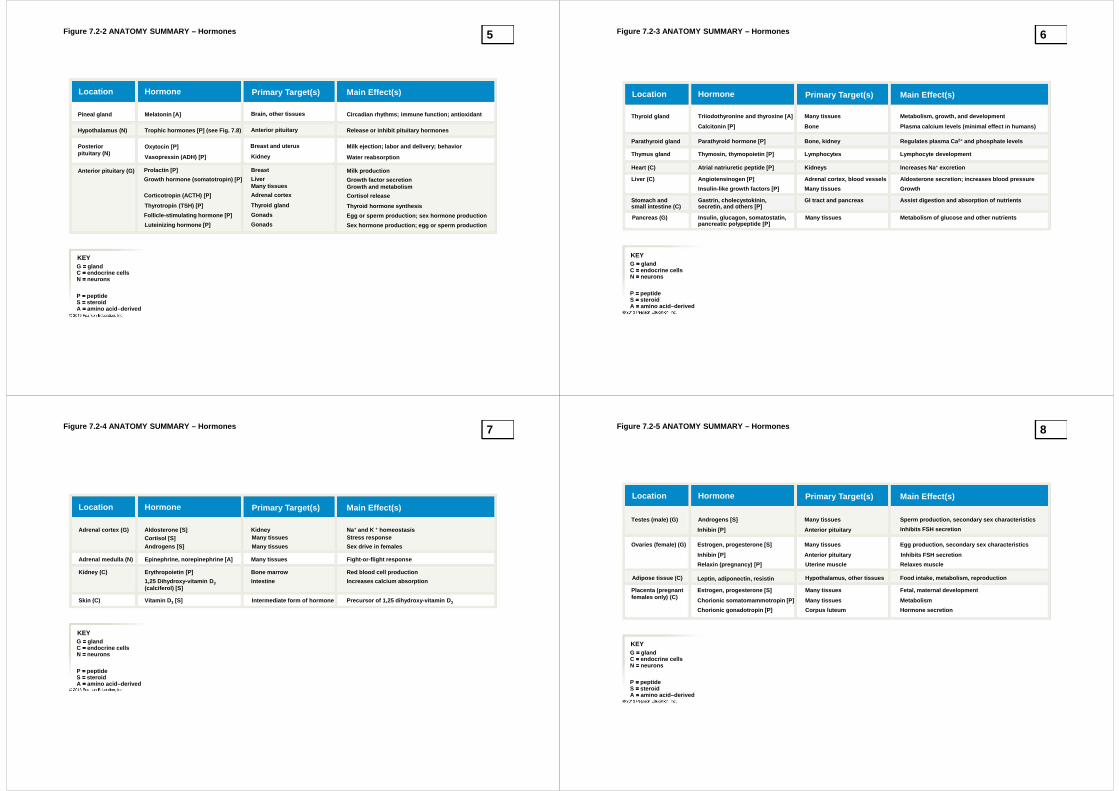
Figure 7.2-2 ANATOMY SUMMARY – Hormones
KEYG ==== glandC ==== endocrine cellsN ==== neurons
P ==== peptideS ==== steroidA ==== amino acid–derived
Pineal gland
Hypothalamus (N)
Posteriorpituitary (N)
Anterior pituitary (G)
Melatonin [A]
Trophic hormones [P] (see Fig. 7.8)
Oxytocin [P]
Vasopressin (ADH) [P]
Prolactin [P]
Luteinizing hormone [P]
Growth hormone (somatotropin) [P]
Follicle-stimulating hormone [P]
Corticotropin (ACTH) [P]
Thyrotropin (TSH) [P]
Brain, other tissues
Anterior pituitary
Breast and uterus
Kidney
Breast
LiverMany tissues
Adrenal cortex
Thyroid gland
Gonads
Gonads Sex hormone production; egg or sperm production
Egg or sperm production; sex hormone production
Thyroid hormone synthesis
Cortisol release
Growth and metabolismGrowth factor secretion
Milk production
Water reabsorption
Milk ejection; labor and delivery; behavior
Release or inhibit pituitary hormones
Circadian rhythms; immune function; antioxidant
Main Effect(s)Primary Target(s)HormoneLocation
5 Figure 7.2-3 ANATOMY SUMMARY – Hormones
KEYG ==== glandC ==== endocrine cellsN ==== neurons
P ==== peptideS ==== steroidA ==== amino acid–derived
Main Effect(s)Primary Target(s)HormoneLocation
Thyroid gland
Parathyroid gland
Thymus gland
Heart (C)
Liver (C)
Stomach andsmall intestine (C)
Pancreas (G)
Triiodothyronine and thyroxine [A]
Calcitonin [P]
Parathyroid hormone [P]
Thymosin, thymopoietin [P]
Atrial natriuretic peptide [P]
Angiotensinogen [P]
Insulin-like growth factors [P]
Gastrin, cholecystokinin,secretin, and others [P]
Insulin, glucagon, somatostatin,pancreatic polypeptide [P]
Many tissues
Many tissues
Many tissues
Bone
Bone, kidney
Lymphocytes
Kidneys
Adrenal cortex, blood vessels
GI tract and pancreas
Metabolism, growth, and development
Plasma calcium levels (minimal effect in humans)
Regulates plasma Ca 2+ and phosphate levels
Lymphocyte development
Increases Na + excretion
Aldosterone secretion; increases blood pressure
Growth
Assist digestion and absorption of nutrients
Metabolism of glucose and other nutrients
6
Figure 7.2-4 ANATOMY SUMMARY – Hormones
KEYG ==== glandC ==== endocrine cellsN ==== neurons
P ==== peptideS ==== steroidA ==== amino acid–derived
Main Effect(s)Primary Target(s)HormoneLocation
Adrenal cortex (G)
Adrenal medulla (N)
Kidney (C)
Skin (C)
Aldosterone [S]Cortisol [S]Androgens [S]
Epinephrine, norepinephrine [A]
Erythropoietin [P]
1,25 Dihydroxy-vitamin D 3(calciferol) [S]
Vitamin D 3 [S]
KidneyMany tissues
Many tissues
Many tissues
Bone marrow
Intestine
Intermediate form of hormone
Na+ and K + homeostasisStress response
Sex drive in females
Fight-or-flight response
Red blood cell production
Increases calcium absorption
Precursor of 1,25 dihydroxy-vitamin D 3
7 Figure 7.2-5 ANATOMY SUMMARY – Hormones
KEYG ==== glandC ==== endocrine cellsN ==== neurons
P ==== peptideS ==== steroidA ==== amino acid–derived
Main Effect(s)Primary Target(s)HormoneLocation
Testes (male) (G)
Ovaries (female) (G)
Adipose tissue (C)
Placenta (pregnantfemales only) (C)
Androgens [S]
Inhibin [P]
Estrogen, progesterone [S]
Inhibin [P]
Relaxin (pregnancy) [P]
Leptin, adiponectin, resistin
Estrogen, progesterone [S]
Chorionic somatomammotropin [P]
Chorionic gonadotropin [P]
Many tissues
Many tissues
Many tissues
Many tissues
Anterior pituitary
Anterior pituitary
Uterine muscle
Hypothalamus, other tissues
Corpus luteum
Sperm production, secondary sex characteristics
Inhibits FSH secretion
Egg production, secondary sex characteristics
Inhibits FSH secretion
Relaxes muscle
Food intake, metabolism, reproduction
Fetal, maternal development
Metabolism
Hormone secretion
8
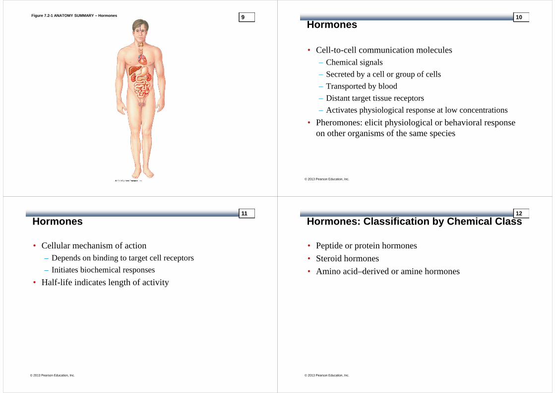
Figure 7.2-1 ANATOMY SUMMARY – Hormones 9
Hormones
• Cell-to-cell communication molecules– Chemical signals
– Secreted by a cell or group of cells
– Transported by blood
– Distant target tissue receptors
– Activates physiological response at low concentrations
• Pheromones: elicit physiological or behavioral response on other organisms of the same species
© 2013 Pearson Education, Inc.
10
Hormones
• Cellular mechanism of action– Depends on binding to target cell receptors
– Initiates biochemical responses
• Half-life indicates length of activity
© 2013 Pearson Education, Inc.
11
Hormones: Classification by Chemical Class
• Peptide or protein hormones
• Steroid hormones
• Amino acid–derived or amine hormones
© 2013 Pearson Education, Inc.
12

Table 7.1 Comparison of Peptide, Steroid, and Amino Acid-Derived Hormones 13
Hormones: Peptides or Proteins
• Preprohormone– Large, inactive precursor
• Prohormone– Smaller, inactive
– Proteolytic, post-translational modification
• Peptide/protein hormones– Bind surface membrane receptors
– Cellular response through signal transduction system
© 2013 Pearson Education, Inc.
14
Figure 7.3 ESSENTIALS – Peptide Hormone Synthesis an d Processing 15 Figure 7.3 ESSENTIALS – Peptide Hormone Synthesis an d Processing
mRNA
Preprohormone Ribosome
Endoplasmicreticulum (ER)
Cytoplasm
ECF
Plasma Capillaryendothelium
Messenger RNA onthe ribosomes bindsamino acids into apeptide chain called apreprohormone. Thechain is directed intothe ER lumen by asignal sequence ofamino acids.
Slide 1
© 2013 Pearson Education, Inc.
16

Figure 7.3 ESSENTIALS – Peptide Hormone Synthesis an d Processing
mRNA
Preprohormone
Signalsequence
Prohormone
Ribosome
Endoplasmicreticulum (ER)
Cytoplasm
ECF
Plasma Capillaryendothelium
Enzymes in the ERchop off the signalsequence, creating aninactive prohormone.
Messenger RNA onthe ribosomes bindsamino acids into apeptide chain called apreprohormone. Thechain is directed intothe ER lumen by asignal sequence ofamino acids.
Slide 2
© 2013 Pearson Education, Inc.
17 Figure 7.3 ESSENTIALS – Peptide Hormone Synthesis an d Processing
mRNA
Preprohormone
Signalsequence
Prohormone
Ribosome
Endoplasmicreticulum (ER)
Transportvesicle
Golgicomplex
Cytoplasm
ECF
Plasma Capillaryendothelium
Messenger RNA onthe ribosomes bindsamino acids into apeptide chain called apreprohormone. Thechain is directed intothe ER lumen by asignal sequence ofamino acids.
Enzymes in the ERchop off the signalsequence, creating aninactive prohormone.
The prohormonepasses from the ERthrough the Golgicomplex.
Slide 3
© 2013 Pearson Education, Inc.
18
Figure 7.3 ESSENTIALS – Peptide Hormone Synthesis an d Processing
mRNA
Preprohormone
Signalsequence
Prohormone
Ribosome
Endoplasmicreticulum (ER)
Transportvesicle
Golgicomplex
Cytoplasm
ECF
Plasma
Secretoryvesicle
Capillaryendothelium
Peptide fragment
Active hormone
Messenger RNA onthe ribosomes bindsamino acids into apeptide chain called apreprohormone. Thechain is directed intothe ER lumen by asignal sequence ofamino acids.
Enzymes in the ERchop off the signalsequence, creating aninactive prohormone.
The prohormonepasses from the ERthrough the Golgicomplex.
Secretory vesiclescontaining enzymesand prohormone budoff the Golgi. Theenzymes chop theprohormone into oneor more activepeptides plusadditional peptidefragments.
Slide 4
© 2013 Pearson Education, Inc.
19 Figure 7.3 ESSENTIALS – Peptide Hormone Synthesis an d Processing
mRNA
Preprohormone
Signalsequence
Prohormone
Ribosome
Endoplasmicreticulum (ER)
Transportvesicle
Golgicomplex
Cytoplasm
ECF
Plasma
Secretoryvesicle
Releasesignal
Capillaryendothelium
Peptide fragment
Active hormone
Messenger RNA onthe ribosomes bindsamino acids into apeptide chain called apreprohormone. Thechain is directed intothe ER lumen by asignal sequence ofamino acids.
Enzymes in the ERchop off the signalsequence, creating aninactive prohormone.
The prohormonepasses from the ERthrough the Golgicomplex.
Secretory vesiclescontaining enzymesand prohormone budoff the Golgi. Theenzymes chop theprohormone into oneor more activepeptides plusadditional peptidefragments.
The secretory vesiclereleases its contentsby exocytosis into theextracellular space.
Slide 5
© 2013 Pearson Education, Inc.
20

Figure 7.3 ESSENTIALS – Peptide Hormone Synthesis an d Processing
mRNA
Preprohormone
Signalsequence
Prohormone
Ribosome
Endoplasmicreticulum (ER)
Transportvesicle
Golgicomplex
Cytoplasm
ECF
Plasma
Secretoryvesicle
Releasesignal
Capillaryendothelium To target
Peptide fragment
Active hormone
Messenger RNA onthe ribosomes bindsamino acids into apeptide chain called apreprohormone. Thechain is directed intothe ER lumen by asignal sequence ofamino acids.
Enzymes in the ERchop off the signalsequence, creating aninactive prohormone.
The prohormonepasses from the ERthrough the Golgicomplex.
Secretory vesiclescontaining enzymesand prohormone budoff the Golgi. Theenzymes chop theprohormone into oneor more activepeptides plusadditional peptidefragments.
The secretory vesiclereleases its contentsby exocytosis into theextracellular space.
The hormone movesinto the circulation fortransport to its target.
Slide 6
© 2013 Pearson Education, Inc.
21 Figure 7.3a ESSENTIALS – Peptide Hormone Synthesis a nd Processing
Preprohormones
Preprohormone
PreproTRH (242 amino acids)
6 TRH(3 amino acids each)
Other peptide fragments
Signal sequence
processes to
PreproTRH (thyrotropin-releasinghormone) has six copies of the3-amino acid hormone TRH.
22
Figure 7.3b ESSENTIALS – Peptide Hormone Synthesis a nd Processing
Prohormones
Pro-opiomelanocortin
Prohormones, such as pro-opiomelanocortin, the prohormone forACTH, may contain several peptidesequences with biological activity.
ACTH
Peptide fragment
γγγγ lipotropin ββββ endorphin
processes to
23 Figure 7.3c ESSENTIALS – Peptide Hormone Synthesis a nd Processing
Prohormones Process to Active Hormone Plus Peptide Fragments
Proinsulin
Insulin C-peptideprocesses to
The peptide chain ofinsulin’s prohormonefolds back on itself withthe help of disulfide(S—S) bonds. Theprohormone cleaves toinsulin and C-peptide.
24

Endocrine System Review
© 2013 Pearson Education, Inc.
Interactive Physiology® Animation: Endocrine System: Endocrine System Review
25 Figure 7.4 Membrane receptors and signal transducti on for peptide hormones
Peptide hormones (H) cannot enter their target cell s andmust combine with membrane receptors (R) that initi atesignal transduction processes.
KEY
TK ==== Tyrosine kinaseAE ==== Amplifier enzymeG ==== G protein
Opens ionchannel
Secondmessenger
systems
Proteins
Cellularresponse
H H
R R
G AE TK
phosphorylate
26
Hormones: Steroid
• Cholesterol-derived– Lipophilic and easily cross membranes
• Bind carrier proteins in blood– Longer half-life
• Cytoplasmic or nuclear receptors– Genomic effect to activate or repress genes for protein
synthesis– Slower acting
• Cell membrane receptors– Nongenomic responses
© 2013 Pearson Education, Inc.
27 Figure 7.5a ESSENTIALS – Steroid Hormones
Cholesterol is the parent compound for all steroid hormones.
DHEA ==== dehydroepiandrosterone
==== intermediate compoundswhose names have beenomitted for simplicity.
Cholesterol
DHEA
Progesterone
Andro-stenedione
Testosterone
Dihydro-testosterone
(DHT)
Estradiol
Estrone
Ovary
Cortisol
Corticosterone Aldosterone
Adrenalcortex
Each step is catalyzed by an enzyme, but only two e nzymes are shown in this figure.
aromatase
aromatase
21-hydroxylase
21-hydroxylase
28

Figure 7.5b ESSENTIALS – Steroid Hormones
Steroid hormones act primarily on intracellular receptors.
Cell surface receptor
Rapid responses
Nucleus
DNA
Nuclearreceptor
Transcriptionproduces mRNA
TranslationNew
proteins
Endoplasmicreticulum
Cytoplasmicreceptor
Cellmembrane
Proteincarrier
Bloodvessel
Steroidhormone
Interstitialfluid
Most hydrophobic steroids are bound toplasma protein carriers. Only unboundhormones can diffuse into the target cell.
Steroid hormone receptors are in thecytoplasm or nucleus.
Some steroid hormones also bind tomembrane receptors that use secondmessenger systems to create rapidcellular responses.
The receptor-hormone complex binds toDNA and activates or represses one ormore genes.
Activated genes create new mRNA thatmoves back to the cytoplasm.
Translation produces new proteinsfor cell processes.
29 Figure 7.5b ESSENTIALS – Steroid Hormones
Bloodvessel
Most hydrophobic steroids are bound toplasma protein carriers. Only unboundhormones can diffuse into the target cell.
Proteincarrier
Interstitialfluid
Nucleus
Cellmembrane
Slide 1
© 2013 Pearson Education, Inc.
30
Figure 7.5b ESSENTIALS – Steroid Hormones
Bloodvessel
Most hydrophobic steroids are bound toplasma protein carriers. Only unboundhormones can diffuse into the target cell.
Steroidhormone
Proteincarrier
Cytoplasmicreceptor
Interstitialfluid
Cellmembrane
Nucleus
Nuclearreceptor
Steroid hormone receptors are in thecytoplasm or nucleus.
Slide 2
© 2013 Pearson Education, Inc.
31 Figure 7.5b ESSENTIALS – Steroid Hormones
Bloodvessel
Most hydrophobic steroids are bound toplasma protein carriers. Only unboundhormones can diffuse into the target cell.
Steroidhormone
Proteincarrier
Cell surface receptor
Rapid responses
Cytoplasmicreceptor
Interstitialfluid
Cellmembrane
Nucleus
Nuclearreceptor
Steroid hormone receptors are in thecytoplasm or nucleus.
Some steroid hormones also bindto membrane receptors that usesecond messenger systems tocreate rapid cellular responses.
Slide 3
© 2013 Pearson Education, Inc.
32

Figure 7.5b ESSENTIALS – Steroid Hormones
Bloodvessel
Most hydrophobic steroids are bound toplasma protein carriers. Only unboundhormones can diffuse into the target cell.
Steroidhormone
Proteincarrier
Cell surface receptor
Rapid responses
Cytoplasmicreceptor
Interstitialfluid
Cellmembrane
Nucleus
Nuclearreceptor
DNA
Steroid hormone receptors are in thecytoplasm or nucleus.
The receptor-hormone complex binds toDNA and activates or represses one ormore genes.
Some steroid hormones also bindto membrane receptors that usesecond messenger systems tocreate rapid cellular responses.
Slide 4
© 2013 Pearson Education, Inc.
33 Figure 7.5b ESSENTIALS – Steroid Hormones
Bloodvessel
Most hydrophobic steroids are bound toplasma protein carriers. Only unboundhormones can diffuse into the target cell.
Steroidhormone
Proteincarrier
Cell surface receptor
Rapid responses
Cytoplasmicreceptor
Interstitialfluid
Cellmembrane
Translation
Transcriptionproduces mRNA
Nucleus
Nuclearreceptor
DNA
Steroid hormone receptors are in thecytoplasm or nucleus.
The receptor-hormone complex binds toDNA and activates or represses one ormore genes.
Activated genes create new mRNA thatmoves back to the cytoplasm.
Some steroid hormones also bindto membrane receptors that usesecond messenger systems tocreate rapid cellular responses.
Slide 5
© 2013 Pearson Education, Inc.
34
Figure 7.5b ESSENTIALS – Steroid Hormones
Bloodvessel
Most hydrophobic steroids are bound toplasma protein carriers. Only unboundhormones can diffuse into the target cell.
Steroidhormone
Proteincarrier
Cell surface receptor
Rapid responses
Cytoplasmicreceptor
Endoplasmicreticulum
Interstitialfluid
Cellmembrane
Newproteins Translation
Transcriptionproduces mRNA
Nucleus
Nuclearreceptor
DNA
Steroid hormone receptors are in thecytoplasm or nucleus.
The receptor-hormone complex binds toDNA and activates or represses one ormore genes.
Activated genes create new mRNA thatmoves back to the cytoplasm.
Translation produces new proteinsfor cell processes.
Some steroid hormones also bindto membrane receptors that usesecond messenger systems tocreate rapid cellular responses.
Slide 6
© 2013 Pearson Education, Inc.
35
Hormones: Amino Acid–Derived, or Amine
• Derived from one of two amino acids– Tryptophan
– Tyrosine
• Ring structure
© 2013 Pearson Education, Inc.
36
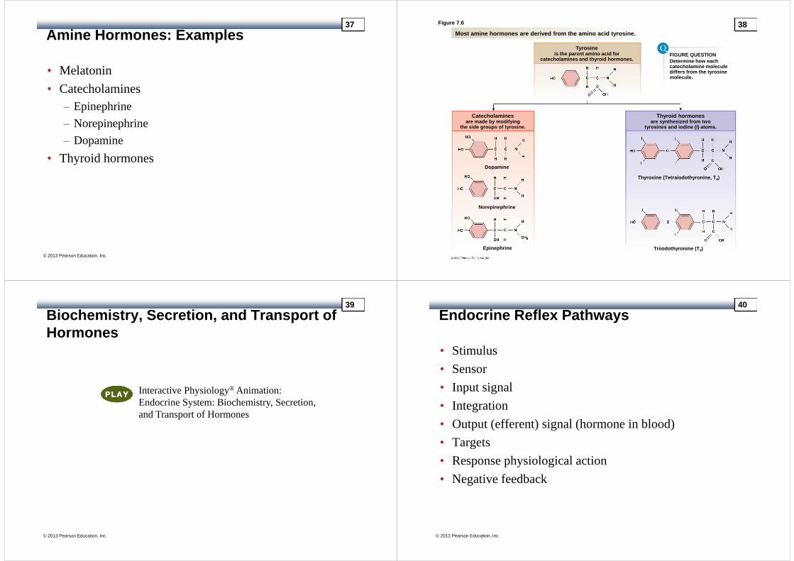
Amine Hormones: Examples
• Melatonin
• Catecholamines – Epinephrine
– Norepinephrine
– Dopamine
• Thyroid hormones
© 2013 Pearson Education, Inc.
37 Figure 7.6
Most amine hormones are derived from the amino acid tyrosine.
FIGURE QUESTIONDetermine how eachcatecholamine moleculediffers from the tyrosinemolecule.
Tyrosineis the parent amino acid for
catecholamines and thyroid hormones.
Catecholaminesare made by modifying
the side groups of tyrosine.
Thyroid hormonesare synthesized from two
tyrosines and iodine (I) atoms.
Thyroxine (Tetraiodothyronine, T 4)
Triiodothyronine (T 3)Epinephrine
Norepinephrine
Dopamine
38
Biochemistry, Secretion, and Transport of Hormones
© 2013 Pearson Education, Inc.
Interactive Physiology® Animation: Endocrine System: Biochemistry, Secretion, and Transport of Hormones
39
Endocrine Reflex Pathways
• Stimulus
• Sensor
• Input signal
• Integration
• Output (efferent) signal (hormone in blood)
• Targets
• Response physiological action
• Negative feedback
© 2013 Pearson Education, Inc.
40
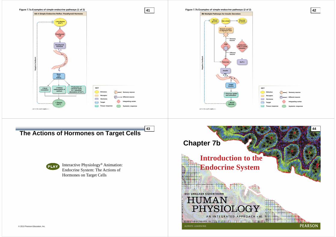
Figure 7.7a Examples of simple endocrine pathways ( 1 of 2)
Stimulus
Receptor
Hormone
Target
Tissue response
Sensory neuron
Efferent neuron
Integrating center
Systemic response
KEY
A Simple Endocrine Reflex: Parathyroid Hormone
Low plasma[Ca2+]
Parathyroidcell
Parathyroidhormone
Boneand
kidney
↑↑↑↑ Boneresorption
↑↑↑↑ Kidneyreabsorption of
calcium
Production ofcalcitriol leadsto ↑↑↑↑ intestinal
absorption of Ca 2+]
↑↑↑↑ Plasma[Ca2+]
Neg
ativ
e fe
edba
ck
41 Figure 7.7b Examples of simple endocrine pathways ( 2 of 2)
Stimulus
Receptor
Hormone
Target
Tissue response
Sensory neuron
Efferent neuron
Integrating center
Systemic response
KEY
Neg
ativ
e fe
edba
ck
↓↓↓↓ Bloodglucose
↑↑↑↑ Glucose uptakeand utilization
Targettissues
Insulin
Pancreas GLP-1
Efferent neuron
Sensoryneuron
CNS Endocrinecells in small
intestine
Glucosein lumen
Stretch receptorin digestive tract
↑↑↑↑ Bloodglucose Eat a meal
Multiple Pathways for Insulin Secretion
42
The Actions of Hormones on Target Cells
© 2013 Pearson Education, Inc.
Interactive Physiology® Animation: Endocrine System: The Actions of Hormones on Target Cells
43
© 2013 Pearson Education, Inc.
Introduction to the Endocrine System
Chapter 7b
44

Neurohormones: Major Groups
• Adrenal medulla – Catecholamines
• Hypothalamus– Posterior pituitary is neural tissue
– Anterior pituitary is endocrine tissue
© 2013 Pearson Education, Inc.
45 Figure 7.8a ESSENTIALS – The Pituitary Gland
ANTERIOR POSTERIOR
HYPOTHALAMUS
The pituitary gland sitsin a protected pocketof bone, connected tothe brain by a thin stalk. Infundibulum is the
stalk that connectsthe pituitary to the brain.
Posterior pituitary isan extension of theneural tissue.
Anterior pituitary is atrue endocrine gland ofepithelial origin.
Sphenoid bone
46
Figure 7.8c ESSENTIALS – The Pituitary Gland
HYPOTHALAMUS
The posterior pituitary is anextension of the brain thatsecretes neurohormonesmade in the hypothalamus.
Neurohormone is madeand packaged in cellbody of neuron.
Vesicles are trans-ported down the cell.
Vesicles containingneurohormone arestored in posteriorpituitary.
Neurohormones arereleased into blood.
POSTERIOR PITUITARY
Vein
Oxytocin Vasopressin
KidneysMammary glands and uterus
Ile Gln
Tyr Asp
Cys Cys
ProLeu
GlyCys Cys
Tyr Asp
Phe Gln
ProArg
Gly
47 Figure 7.8c ESSENTIALS – The Pituitary Gland
HYPOTHALAMUS
Neurohormone is madeand packaged in cellbody of neuron.
POSTERIOR PITUITARY
Slide 1
© 2013 Pearson Education, Inc.
48

Figure 7.8c ESSENTIALS – The Pituitary Gland
HYPOTHALAMUS
Neurohormone is madeand packaged in cellbody of neuron.
Vesicles are trans-ported down the cell.
POSTERIOR PITUITARY
Slide 2
© 2013 Pearson Education, Inc.
49 Figure 7.8c ESSENTIALS – The Pituitary Gland
HYPOTHALAMUS
Neurohormone is madeand packaged in cellbody of neuron.
Vesicles are trans-ported down the cell.
Vesicles containingneurohormone arestored in posteriorpituitary.
POSTERIOR PITUITARY
Slide 3
© 2013 Pearson Education, Inc.
50
Figure 7.8c ESSENTIALS – The Pituitary Gland
HYPOTHALAMUS
Neurohormone is madeand packaged in cellbody of neuron.
Vesicles are trans-ported down the cell.
Vesicles containingneurohormone arestored in posteriorpituitary.
POSTERIOR PITUITARY
Vein
Neurohormones arereleased into blood.
Slide 4
© 2013 Pearson Education, Inc.
51 Figure 7.8b ESSENTIALS – The Pituitary Gland
HYPOTHALAMUS
TO TARGET ORGANS
ProlactinGH TSH ACTH
Gonadotropins (LH & FSH)
Mammary glands Musculoskeletal system Thyroid gland Ad renal cortex
Ovary Testis
Gonads
Veins
ANTERIOR PITUITARY
Capillary bed
POSTERIOR PITUITARY
Artery
Capillary bed
Neurons synthesizingtrophic neurohormonesrelease them intocapillaries of the portalsystem.
Portal vessels carry thetrophic neurohormonesdirectly to the anteriorpituitary, where they acton the endocrine cells.
Endocrine cells releasetheir peptide hormonesinto the second set ofcapillaries for distributionto the rest of the body.
The anterior pituitary is a true endocrine gland that secretes six classichormones. Neurohormones from the hypothalamus contr ol release of theanterior pituitary hormones. The hypothalamic hormo nes reach the anteriorpituitary through a specialized region of the circu lation called a portal system.
52

Figure 7.9 ESSENTIALS – Hormones of the Hypothalamic –Anterior Pituitary Pathway 53
Endocrine Control
• A trophic hormone controls the secretion of another hormone
• Hypothalamic-hypophyseal portal system
• Three integrating centers– Hypothalamic stimulation—from CNS
– Anterior pituitary stimulation—from hypothalamic trophic hormones
– Endocrine gland stimulation—from anterior pituitary trophic hormones (except prolactin)
© 2013 Pearson Education, Inc.
54
Figure 7.10
The Growth Hormone Pathway
HYPOTHALAMUS
Hypothalamic growth hormone–releasing hormone (GHRH )stimulates growth hormone (GH) secretion. Growth ho rmone actsdirectly on many body tissues but also influences l iver productionof insulin-like growth factors (IGFs or somatomedin s), anothergroup of hormones that regulate growth.
Hypothalamus
GHRH
ANTERIORPITUITARY GH cells
in anteriorpituitary
GH
Liver
IGFs
Growth
T TBone andsoft tissue
55 Figure 7.11a (1 of 2)
In complex endocrine pathways, the hormones of thepathway serve as negative feedback signals.
Stimulus
Hypothalamus(IC1)
Anteriorpituitary
(IC2)
Trophic hormone (H 1)
Trophic hormone (H 2)
Sho
rt-lo
op n
egat
ive
feed
back
Long-loop negative feedback
Endocrinegland(IC3)
Hormone (H 3)
Target tissue
Response
56

Figure 7.11b (2 of 2)
Targettissue
Response
Long-loop negative feedback
Cortisol
To targettissue
Adrenalcortex
Anteriorpituitary
ACTH
CRH
Hypothalamus
Control Pathway for Cortisol SecretionCortisol is a steroid hormone secreted by the adren al cortex.ACTH ==== corticotropin or adrenocorticotropic hormone;CRH ==== corticotropin-releasing hormone.
Draw in the short-loopnegative feedbackfor this pathway.
FIGURE QUESTION
57
Hormone Interactions
• Synergism– Combined effect is greater than the sum of individual
effects
• Permissiveness– Need second hormone to get full effect
• Antagonism– One substance opposes the action of another
– Competitive inhibitors vs. functional antagonism
– Glucagons oppose insulin
© 2013 Pearson Education, Inc.
58
Figure 7.12 Synergism
Glucagon ++++ Epinephrine ++++ Cortisol
Glucagon ++++ Epinephrine
Epinephrine
GlucagonCortisol
Time (hours)
Blo
od g
luco
se (
mg/
dL)
250
200
150
100
0 1 2 3 4 5
59
Endocrine Pathologies
• Hypersecretion: excess hormone– Caused by tumors or exogenous iatrogenic treatment
– Negative feedback
• Hyposecretion: deficient hormone– Caused by decreased synthesis materials or atrophy
– Absence of negative feedback
© 2013 Pearson Education, Inc.
60

Figure 7.13
↓↓↓↓CRH
↓↓↓↓ACTH
↓↓↓↓Cortisol
Response
Targettissue
Exogenouscortisol
(Hypothalamus)
(Anterior pituitary)
(Adrenal cortex)
Exogenous hormone has the same negative feedbackeffect as endogenous hormone.
61
Pathologies: Abnormal Receptors
• Down-regulation – Decreased number of receptors
– Hyperinsulinemia
• Receptor and signal transduction abnormalities– Testicular feminization syndrome
– Pseudohypothyroidism
© 2013 Pearson Education, Inc.
62
Figure 7.14 Primary and secondary hypersecretion of cortisol
Secondary HypersecretionDue to Pituitary Problem
Secondary Hypersecretion Dueto Hypothalamic Problem
Primary Hypersecretion Due toProblem with Adrenal Cortex
Hypothalamus ↓↓↓↓CRH ↓↓↓↓CRH ↑↑↑↑CRH
↑↑↑↑ACTH
↑↑↑↑Cortisol↑↑↑↑Cortisol↑↑↑↑Cortisol
↑↑↑↑ACTH↓↓↓↓ACTH
Hypothalamus
Anteriorpituitary
Anteriorpituitary
Adrenalcortex
Adrenalcortex Negative
feedbackfails
PATHOLOGYIN ADRENAL
CORTEX
PATHOLOGYIN ANTERIOR
PITUITARY
HYPERSECRETINGTUMOR IN
HYPOTHALAMUS
Symptomsof excess
Symptomsof excess
Symptomsof excess
• CRH levels – low• ACTH levels – low• Cortisol levels – high
• CRH levels – low• ACTH levels – high• Cortisol levels – high
• CRH levels – high• ACTH levels – high• Cortisol levels – high
63 Figure 7.15 Patterns of hormone secretion in hypoco rtisolism
Hyposecretion from Atrophyof the Adrenal Cortex
Hyposecretion fromDamage to the Pituitary
Hypothalamus CRH Hypothalamus CRH
ACTHAnterior pituitaryACTHAnterior pituitary
CortisolAdrenal cortexCortisolAdrenal cortex
Symptomsof
deficiency
Symptomsof
deficiency
For each condition, use arrows to indicate whetherlevels of the three hormones in the pathway will beincreased, decreased, or unchanged. Draw innegative feedback loops where functional.
FIGURE QUESTION
64
The Hypothalamic-Pituitary Axis
© 2013 Pearson Education, Inc.
Interactive Physiology® Animation: Endocrine System: The Hypothalamic-Pituitary Axis
65
Hormone Evolution
• Evolutionary conservation of hormone function
• Proteomics– Calcitonin gene-related peptide example
• Vestigial– Melanocyte-stimulating hormone example
• Comparative endocrinology– Pineal gland and melatonin example
© 2013 Pearson Education, Inc.
66
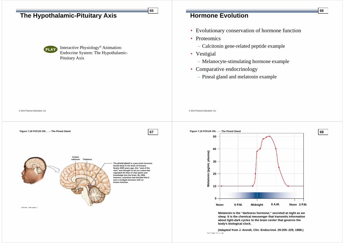
Figure 7.16 FOCUS ON. . . – The Pineal Gland
ThalamusCorpus
callosum
The pineal gland is a pea-sized structureburied deep in the brain of humans.Nearly 2000 years ago, this “seat of thesoul” was thought to act as a valve thatregulated the flow of vital spirits andknowledge into the brain. By 1950,however, scientists had decided that itwas a vestigial structure with noknown function.
67 Figure 7.16 FOCUS ON. . . – The Pineal Gland
Melatonin is the “darkness hormone,” secreted at ni ght as wesleep. It is the chemical messenger that transmits informationabout light-dark cycles to the brain center that go verns thebody’s biological clock.
(Adapted from J. Arendt, Clin. Endocrinol. 29:205–229, 1988.)
Noon 6 P.M. Midnight 6 A.M. Noon 3 P.M.
50
40
30
20
10
0
Mel
aton
in (
pg/m
L pl
asm
a)
68

Figure 7.16 FOCUS ON. . . – The Pineal Gland
Melatonin is an amino acid–derivedhormone made from tryptophan.
69
Summary
• Hormones
• The classification of hormones
• Control of hormone release
• Hormone interactions
• Endocrine pathologies
• Hormone evolution
© 2013 Pearson Education, Inc.
70