Introduction to reproductive health and environment rev
74
<<NOTE TO USER: Please add details of the date, time, place and sponsorship of the meeting for which you are using this presentation in the space indicated.>> <<NOTE TO USER: This is a large set of slides from which the presenter should select the most relevant ones to use in a specific presentation. These slides cover many facets of the issue. Present only those slides that apply most directly to the local situation in the region or country.>> <<NOTE TO USER: This module presents several examples of risk factors that affect reproductive health. You can find more detailed information in other modules of the training package that deal with specific risk factors, such as lead, mercury, pesticides, persistent organic pollutants, endocrine disruptors, occupational exposures; or disease outcomes, such as developmental origins of disease, reproductive effects, neurodevelopmental effects, immune effects, respiratory effects, and others.>> <<NOTE TO USER: For more information on reproductive health, please visit the website of the Department of Reproductive Health and Research at WHO: www.who.int/reproductivehealth/en/>> 1 TRAINING FOR THE HEALTH SECTOR [Date ... Place ... Event ... Sponsor ... Organizer} INTRODUCTION TO REPRODUCTIVE HEALTH AND THE ENVIRONMENT November 2011 Training Module 1 Children's Environmental Health Public Health and the Environment World Health Organization www.who.int/ceh WHO/HSE/PHE/EPE/11.01.10
Transcript of Introduction to reproductive health and environment rev

<<NOTE TO USER: Please add details of the date, time, place and sponsorship of the meeting for which you are using this presentation in the space indicated.>>
<<NOTE TO USER: This is a large set of slides from which the presenter should select the most relevant ones to use in a specific presentation. These slides cover many facets of the issue. Present only those slides that apply most directly to the local situation in the region or country.>>
<<NOTE TO USER: This module presents several examples of risk factors that affect reproductive health. You can find more detailed information in other modules of the training package that deal with specific risk factors, such as lead, mercury, pesticides, persistent organic pollutants, endocrine disruptors, occupational exposures; or disease outcomes, such as developmental origins of disease, reproductive effects, neurodevelopmental effects, immune effects, respiratory effects, and others.>>
<<NOTE TO USER: For more information on reproductive health, please visit the website of the Department of Reproductive Health and Research at WHO: www.who.int/reproductivehealth/en/>>
1
TRAINING FOR THE HEALTH SECTOR [Date ... Place ... Event ... Sponsor ... Organizer}
INTRODUCTION TO REPRODUCTIVE HEALTH AND THE
ENVIRONMENT
November 2011
Training Module 1
Children's Environmental Health Public Health and the Environment
World Health Organization
www.who.int/ceh
WHO/HSE/PHE/EPE/11.01.10

<<READ SLIDE.>>
According to the formal definition by the World Health Organization (WHO), health is more than absence of illness. It is a state of complete physical, mental and social well-being. Similarly, reproductive health also represents a state of complete physical, mental and social well-being, and not merely the absence of reproductive diseases or alterations.
This presentation will introduce you to the basics of reproductive health and the important role that the environment plays in influencing the health of individuals.
Refs:•WHO. Department of Reproductive Health and Research, Partner Brief. Geneva, Switzerland, World Health Organization, 2009. WHO/RHR/09.02. Available at whqlibdoc.who.int/hq/2009/WHO_RHR_09.02_eng.pdf – accessed 15 June 2011•WHO. Preamble to the Constitution of the World Health Organization as adopted by the International Health Conference. New York, United States of America, World Health Organization, 1946.
2
Reproductive Health and the Environment
LEARNING OBJECTIVES
After this presentation individuals should be able to understand, recognize, and know:
❖ Basic components of reproductive health
❖ Basic hormone and endocrine functions
❖ Reproductive physiology
❖ Importance of environmental exposures on reproductive health endpoints
2

<<READ SLIDE.>>
<<NOTE TO USER: You may decide to delete certain parts of the presentation depending on time. Please correct the outline accordingly.>>
<<NOTE TO USER: If your audience is already familiar with the reproductive system, you may skip the introductory basic slides (slides 14 to 39), and go directly to the section on the role of environmental contaminants on reproductive health (slide 40 and onwards).>>
3
Reproductive Health and the Environment
OUTLINE
❖ The concept of reproductive health
❖ The role of hormones and the endocrine system
❖ Review of the female reproductive system
❖ Review of the male reproductive system
❖ Role of environmental contaminants on reproductive health
❖ Introduction to endocrine disruptors
3

<<READ SLIDE.>>
<<NOTE TO USER: Due to the amount of information presented in this introductory module, it will be divided into three sections. Each section is important for a thorough understanding of the fundamentals of reproductive health and the environment. However, you may decide to delete certain parts of the sections depending on time and relevance to the region or country.>>
Images: WHO
4
Reproductive Health and the Environment
SECTIONS OF MODULE 1
❖ Section 1: Introduction to reproductive health
❖ Section 2: Biology and physiology of the reproductive systems
❖ Section 3: Environmental exposures and reproductive health
WHO

<<READ SLIDE.>>
Section 1 will introduce the foundations of reproductive health according to the definitions of the WHO.
Image: WHO
5
Reproductive Health and the Environment
SECTION 1: Introduction to
reproductive health
WHO 5

The WHO defines reproductive health as a state of complete physical, mental and social well-being, and not merely the absence of reproductive disease or infirmity. Reproductive health involves all of the reproductive processes, functions and systems at all stages of human life. This definition implies that people are able to have a satisfying and safe sex life and that they have the capability to reproduce and the freedom to decide if, when and how often to do so. Men and women have the right to be informed and to have access to safe, effective, affordable and acceptable methods of family planning of their choice that are not against the law. Furthermore, men and women should have access to appropriate health care services that will enable women to go safely through pregnancy and childbirth, as well as to provide couples with the best chance of having a healthy infant. Reproductive health is a universal concern, but is of special importance for women particularly during the reproductive years. However, men also demand specific reproductive health needs and have particular responsibilities in terms of women's reproductive health because of their decision-making powers in some reproductive health matters. Reproductive health is a fundamental component of an individual’s overall health status and a central determinant of quality of life.
Refs:•UNDP/UNFPA/WHO/World Bank. Social science methods for research on reproductive health topics. Geneva, Switzerland, UNDP/UNFPA/WHO/World Bank Special Programme on Research, Development, and Training in Human Reproduction, 2006. Available at whqlibdoc.who.int/hq/1999/WHO_RHR_HRP_SOC_99.1.pdf -accessed 22 June 2010. •United Nations Population Information Network (POPIN). Guidelines on reproductive health. Geneva, Switzerland, United Nations Population Information Network (POPIN), 2002. Available at www.un.org/popin/unfpa/taskforce/guide/iatfreph.gdl.html - accessed 22 June 2010.
Images :•UNDP/UNFPA/WHO/World Bank. Providing the foundation for sexual and reproductive health: A record of achievement. Geneva, Switzerland, UNDP/UNFPA/WHO/World Bank Special Programme on Research, Development, and Research Training in Human Reproduction, 2008. Available at www.who.int/reproductivehealth/publications/general/hrp_brochure.pdf - accessed 23 June 2010. •WHO. Department of Reproductive Health partner brief, Geneva, Switzerland, World Health Organization, 2009. Available at whqlibdoc.who.int/hq/2009/WHO_RHR_09.02_eng.pdf - Accessed 23 June 2010.
6
Reproductive Health and the Environment
1. REPRODUCTIVE HEAL TH
❖ Reproductive processes, functions , and systems at all stages of life
❖ Freedom to make decis ions rega rding a healthy sex life
❖ Access to approp riate reproduct ive health serv ices
www.who.inllreproducliveh6althlpub/icationslg,,nerallh,p_bfochure .pdf
... for both men and womenl
wl>qlibdoc.who.inl/hq/2009IWHO_RHR_09 .02_eng.pdl

The WHO’s definition of reproductive health specifically highlights the importance of an individual’s right to maintain their own sexual health status. Sexual health is the integration of emotional, intellectual, and social aspects of sexual being in order to positively enrich personality, communication, relationships and love. The three fundamental principles of sexual health are: 1) capacity to enjoy and control sexual and reproductive behavior; 2) freedom from shame, guilt, fear, and other psychological factors that may impair sexual relationships; and 3) freedom from organic disorder or disease that interferes with sexual and reproductive function.Reproductive health further implies the right to satisfying and safe sex life. This includes the ability to reproduce, but also the personal freedom to decide if, when and how often to do so. Both men and women have the right to be informed and to have access to safe, effective, affordable and acceptable methods of family planning that are not against the law. Reproductive health should also be understood in the context of healthy relationships in which there is an understanding of the balance between fulfillment and risk. Reproductive health contributes enormously to physical and psychosocial comfort and closeness between individuals. Poor reproductive health is frequently associated with disease, abuse, exploitation, unwanted pregnancy, and death.
Refs:•UNDP/UNFPA/WHO/World Bank. Social science methods for research on reproductive health topics. Geneva, Switzerland, UNDP/UNFPA/WHO/World Bank Special Programme on Research, Development, and Training in Human Reproduction, 2006. Available at whqlibdoc.who.int/hq/1999/WHO_RHR_HRP_SOC_99.1.pdf -accessed 22 June 2010. •United Nations Population Information Network (POPIN). Guidelines on reproductive health. Geneva, Switzerland, United Nations Population Information Network (POPIN), 2002. Available at www.un.org/popin/unfpa/taskforce/guide/iatfreph.gdl.html - accessed 22 June 2010. •WHO. The Reproductive Health Library (RHL), Geneva, Switzerland, World Health Organization, 2008. Available at apps.who.int/rhl/en/index.html - accessed 22 June 2010.
Image: UNDP/UNFPA/WHO/World Bank. Providing the foundation for sexual and reproductive health: A record of achievement. Geneva, Switzerland, UNDP/UNFPA/WHO/World Bank Special Programme on Research, Development, and Research Training in Human Reproduction, 2008. Available at www.who.int/reproductivehealth/publications/general/hrp_brochure.pdf - accessed 23 June 2010.
7
Reproductive Health and the Environment
REPRODUCTIVE HEAL TH
www.whoJnVreproducllvehe~blicationslgeneratnJrp_brochura .pdf
Right to a satisfying and safe sex life with the freedom to decide to reproduce and how often to do so ❖ Safe, effective, affordable access to family planning methods
❖ Access to appropriate reproductive health services 7

Reproductive health is a crucial feature of healthy human development and of general health. It may be a reflection of a healthy childhood, is crucial during adolescence, and sets the stage for health in adulthood and beyond the reproductive years for both men and women. Reproductive life span does not begin with sexual development at puberty and end at menopause for a woman or when a man is no longer likely to have children. Rather, it follows throughout an individual’s life cycle and remains important in many different phases of development and maturation. At each stage of life, individual reproductive health needs may differ. However, there is a cumulative effect across the life course, and each phase has important implications for future well-being. An inability to deal with reproductive health problems at any stage in life may set the scene for later health problems. This is known as the life cycle perspective for reproductive health.
Refs:•UNDP/UNFPA/WHO/World Bank. Social science methods for research on reproductive health topics. Geneva, Switzerland, UNDP/UNFPA/WHO/World Bank Special Programme on Research, Development, and Training in Human Reproduction, 2006. Available at whqlibdoc.who.int/hq/1999/WHO_RHR_HRP_SOC_99.1.pdf -accessed 22 June 2010. •United Nations Population Information Network (POPIN). Guidelines on reproductive health. Geneva, Switzerland, United Nations Population Information Network (POPIN), 2002. Available at www.un.org/popin/unfpa/taskforce/guide/iatfreph.gdl.html - accessed 22 June 2010.
Image: WHO. Mental health aspects of women's reproductive health: A global review of the literature. Geneva, Switzerland, World Health Organization, 2009. Available at whqlibdoc.who.int/publications/2009/9789241563567_eng.pdf - accessed 23 June 2010.
8
Reproductive Health and the Environment
THE LIFE CYCLE PERSPECTIVE
❖ Individual reproductive health needs differ at each stage of life
❖ Reproductive health status may reflect cumulative effects and experiences that occurred in earlier life phases
wflql;bdoc .who.Inl/p(Jblications/200f/19789241563567_ong .pd(
An inability to address reproductive health concerns may result in future health complications
Reproductive health is important for healthy social, economic, and human development/
8

Healthy reproductive systems, processes, and function are imperative components of adequate overall health. However, many internal as well as external factors may challenge an individual's ability to maintain reproductive health. It is important to keep in mind that reproductive health status may be determined by occurrences and exposures from in utero development until the final stages of life. Numerous factors directly effect how well an individual maintains his or her reproductive health status. While some factors may be pre-determined, such as genetic susceptibility to a particular disorder or disease, other factors that relate to the maintenance of reproductive health may be behavioural and involve an individual's participation in risky practices. Furthermore, the environment in which an individual lives, both natural and physical, may present important risk that may directly influence reproductive health. For instance, some occupational exposures (e.g works with hazardous pesticides) can have adverse effects in reproductive life.
Ref:•UNDP/UNFPA/WHO/World Bank. Providing the foundation for sexual and reproductive health: A record of achievement. Geneva, Switzerland, UNDP/UNFPA/WHO/World Bank Special Programme on Research, Development, and Research Training in Human Reproduction, 2008. Available at www.who.int/reproductivehealth/publications/general/hrp_brochure.pdf - accessed 23 June 2010.
Images:•WHO•UNDP/UNFPA/WHO/World Bank. Providing the foundation for sexual and reproductive health: A record of achievement. Geneva, Switzerland, UNDP/UNFPA/WHO/World Bank Special Programme on Research, Development, and Research Training in Human Reproduction, 2008. Available at www.who.int/reproductivehealth/publications/general/hrp_brochure.pdf - accessed 23 June 2010.
9
Reproductive Health and the Environment
MAINTAINING REPRODUCTIVE HEAL TH
❖ Engaging in healthy behaviors
❖ Appropriate access to health care
❖ Condition of immediate environment ❖ Natural , physical, socio-economic , political, others
WHO
9 www.who.inl/reproducJivehealtNpubl ications/general/htp _ brochure .pd

The World Health Organization defines the term primary infertility as the inability to bear any children, whether this is the result of the inability to conceive a child, or the inability to carry a child to full term after 12 months of unprotected sexual intercourse. Primary infertility is sometimes known as primary sterility. However, in many medical studies, the term primary infertility is only used to describe a situation where a couple is not able to conceive. Secondary infertility is defined as the inability to have a second child after a first birth. Secondary infertility has shown to have a high geographical correlation with primary infertility. Fecundity describes the ability to conceive after several years of exposure to risk of pregnancy. Fecundity is often evaluated as the time necessary for a couple to achieve pregnancy. The World Health Organization recommends defining fecundity as the ability for a couple to conceive after two years of attempting to become pregnant. The terms infertility and infecundity are often confused. Fertility describes the actual production of live offspring, while fecundity describes the ability to produce live offspring. Fecundity cannot be directly measured, though it may be assessed clinically. Typically, fecundity may be assessed by the time span between a couple’s decision to attempt to conceive and a successful pregnancy.
Ref:• Rutsein S, Iqbal S. Infecundity, infertility, and childlessness in the developing world. Geneva, Switzerland, World Health Organization and ORC Macro, 2004. DHS Comparative Report, No. 9.
Image: WHO
10
Reproductive Health and the Environment
INFERTILITY AND FECUNDITY
❖ Primary infertility - failure to bear any children after 12 months of unprotected sexual intercourse
❖ Secondary infertility - failure to have a second child after a first birth
❖ Fecundity - the ability of a
couple to conce ive after a
certain time of attempting to
become pregnant
WHO
10

There are specific reproductive health problems that directly describe the health of an early pregnancy or the development of the fetus in utero. The World Health Organization describes the term congenital abnormalities as all structural, functional, and genetic abnormalities diagnosed in aborted fetuses, at birth or in the neonatal period. Congenital abnormalities are sometimes known as birth defects. An ectopic pregnancy describes a complication in the early stages of pregnancy when a fertilized egg is implanted in an area outside of the uterine cavity. A majority of ectopic pregnancies occur in the fallopian tube, but may also occur in the cervix, ovary, or abdomen. If not treated properly, an ectopic pregnancy may be life threatening for the woman. Fetal death (commonly known as a stillbirth) occurs when an infant does not survive complete expulsion from the mother or after twenty completed weeks of gestational age. Death is evidenced by a lack of vital signs following separation from the womb, for example, lack of fetal breath, heart beat, umbilical cord pulsation, or definite movement of voluntary muscles.
<< NOTE TO USER: For further information, please refer to module 2, “Female Environmental Reproductive Health" or to the module on "Developmental and Environmental Origins of Disease”>>
Refs:•Rutsein S, Iqbal S. Infecundity, infertility, and childlessness in the developing world. Geneva, Switzerland, World Health Organization and ORC Macro, 2004. DHS Comparative Report, No. 9. •United Nations Population Information Network (POPIN). Guidelines on reproductive health. Geneva, Switzerland, United Nations Population Information Network (POPIN), 2002. Available at www.un.org/popin/unfpa/taskforce/guide/iatfreph.gdl.html - accessed 22 June 2010.
11
Reproductive Health and the Environment
REPRODUCTIVE HEAL TH ISSUES DURING AND AFTER PREGNANCY
❖ Congenital anomalies - all structural, functional, and genetic abnormalities diagnosed at birth or in the neonatal period
❖ Ectopic pregnancy - when implantation occurs outside the uterus
❖ Fetal death (stillbirth) - death prior to the complete birth after 20 weeks of gestation. Evidenced by lack of vital life signs
❖ Spontaneous abortion (miscarriage) - spontaneous loss of a pregnancy that occurs before 20 weeks of gestational age
11

As previously mentioned, reproductive health describes multiple components of health status and is essential in the development of social, economic, spiritual, and mental well-being. For this reason, reproductive health research is a diverse field that encompasses numerous disciplines. This diagram from the WHO demonstrates the different components of reproductive health research while emphasizing that each component remains part of one single unit of investigation. This diagram also portrays the imperative role of society and culture on the outcome of reproductive health status.
Ref:•United Nations Population Information Network (POPIN). Guidelines on reproductive health.Geneva, Switzerland, United Nations Population Information Network (POPIN), 2002. Available at www.un.org/popin/unfpa/taskforce/guide/iatfreph.gdl.html - accessed 22 June 2010..
Image: UNDP/UNFPA/WHO/World Bank. Social science methods for research on reproductive health topics. Geneva, Switzerland, UNDP/UNFPA/WHO/World Bank Special Programme on Research, Development, and Training in Human Reproduction, 2006. Available at whqlibdoc.who.int/hq/1999/WHO_RHR_HRP_SOC_99.1.pdf - accessed 22 June 2010.
12
Reproductive Health and the Environment
RESEARCH IN REPRODUCTIVE HEAL TH An expansive field that incorporates biological
evidence, clinical investigations, and social sciences
. • Valuu • Bellell • Soc:ioOCXlnomlt
cond~loll• , SeMcet • Undar•SMllld
QIOIIPS
whq/ibdoc.who.inllhq/ t999/WHO_RHR_HR P_SOC_99 . 1.pdf
STDs: Sexually Transmitted Diseases RT/s: Reproductive Tract Infections HIV: Human immunode ficienc virus
12

While many reproductive health questions are still left unanswered, a solid foundation of biological reproductive health knowledge exists that should be understood before attempting to investigate more complex reproductive health topics. Examples include knowledge of the hormonal pathways that control the reproductive system, the central components and functions of the female reproductive system, and the central components and functions of the male reproductive system. These topics will be described in the upcoming slides.
Ref:•United Nations Population Information Network (POPIN). Guidelines on reproductive health. Geneva, Switzerland, United Nations Population Information Network (POPIN), 2002. Available at www.un.org/popin/unfpa/taskforce/guide/iatfreph.gdl.html - accessed 22 June 2010..
Image: WHO. Mental health aspects of women's reproductive health: A global review of the literature. Geneva, Switzerland, World Health Organization, 2009. Available at whqlibdoc.who.int/publications/2009/9789241563567_eng.pdf - accessed 23 June 2010.
13
Reproductive Health and the Environment
THE BIOLOGICAL FOUNDATIONS
wnq1;bc1oc. wno.inl/p<Jblications/2009/9789241563Sii7 _ eng.pdf
Many advances have expanded our knowledge on
reproductive health biology
13

<<READ SLIDE.>>
Section 2 will introduce the biology and physiology of the reproductive health systems. The section will be divided into 5 parts. It will start with an overview of genetics, and then describe the endocrine system, next describe the female and the male reproductive systems, and finally close with an explanation of fetal development.
Image: WHO
14
Reproductive Health and the Environment
SECTION 2: Biology and physiology of reproductive
systems
A. Genetics
B. Endocrine system
C. Female reproductive system
D. Male reproduct ive system
E. Fetal development WHO 14

Genetics is the study of heredity in living organisms. Heredity describes the passing of parental traits to offspring. For example, if a mother has black hair and her biological son has black hair, we can assume that heredity was involved in the passing of hair colour from mother to son. The study of genetics can thus explain why children look more like one parent or another, and why biological siblings have some similarities and differences. The term genetics contains the root word, gene. Genes are specific human traits that are passed from parents to their children. Essentially, genes are the basic units of heredity and heredity is the passing of traits to offspring. Humans have thousands of genes each. These genes are made of DNA or deoxyribonucleic acid. DNA is a chain of molecules that uniquely defines the individual traits that you possess. All humans have a different DNA pattern and this explains why no two human beings are exactly the same. The specific molecules of DNA that carry the hereditary information of humans are known as chromosomes. It is important to remember that each of our cells throughout our body thus contains these genes with our unique DNA pattern. However, all humans have two copies of every gene. One gene was inherited from the mother, and the other gene was inherited from the father. The image shows different types of human chromosomes pairs.
Refs:•Hartl D, Jones E. Genetics: analysis of genes and genomes. Sudbary, MA, USA: Jones and Bartlett Publishers, 2005. •Klug, M. Concepts of Genetics, 9th Edition. New York: Benjamin Cummings Publishing, 2008.
Image: Phelps J. Headliners: Neurodevelopment: genome-wide screen reveals candidate genes for neural tube defects. Environmental Health Perspectives, 2006, 114:A351-A351.
Reproductive Health and the Environment
2.A. INTRODUCTION TO GENETICS
❖ Many human traits are passed from parents to offspring
❖ Genes are the basic units of heredity
❖ Genes are situated on chromosomes, which are comprised of DNA
♦:♦ Humans have two copies of every gene : one from their mother and one from their father
ehs6hplp03 .niohs.nih .govl af1ic/olfelchAl1ic/e.acUoo?arlic/6URl=inlo %3Adoi%2FIO . I 289%2Fehp . I l+a351

There are a specific type of chromosomes that are known as the sex chromosomes. In humans, the mother's egg cell contains an X chromosome. The father’s sperm cell contains either an X or a Y chromosome. An infant will inherit one pair of sex chromosomes, either one X from the mother and one X from the father OR one X from the mother and one Y from the father. This random inheritance will determine whether the child is a boy or a girl. It is important to note that the sex chromosome that a child inherits from its father will determine its sex. This is because the child will automatically inherit one X from its mother. For example if a baby inherits the X chromosome from its father, the child will be a girl, represented by a double X (XX). If the baby inherits the Y chromosome from its father the child will be a boy, represented by an X and a Y (XY).
Refs:•Hartl D, Jones E. Genetics: analysis of genes and genomes. Sudbary, MA, USA: Jones and Bartlett Publishers, 2005. •Klug M. Concepts of genetics, 9th Edition. New York: Benjamin Cummings Publishing, 2008. •NIH. Genetics home reference. National Institutes of Health. Available at ghr.nlm.nih.gov/ -accessed 20 March 2010.
Image: WHO
Reproductive Health and the Environment
SEX DETERMINATION
❖ Sex chromosomes determine whether a baby will be male or female
❖ The mother has two of the same kind of chromosomestwo X's (XX)
,:. The father has two different chromosomes - X and Y (XY)
❖ The child will randomly inherit one chromosome from the father to be either XX (female)
or XY (male)

Epigenetics is the study of inherited changes in phenotype (factors that account for appearance) that are not directly related to, nor explained by changes in our DNA pattern. For this reason, this field of study is known as "epi," the greek root for "above," indicating that a change has occurred that is not directly related to the genetic code, but above it somehow. In epigenetics, non-genetic causes are responsible for different expressions of phenotypes. Or, termed in a different way, epigenetics describes changes in the expression of our genes that are not caused by alterations in the DNA sequence. Essentially, a different factor accounts for the change in the gene expression. Exogenous, or environmental components may affect gene regulation and thus, potentially, subsequent expression in the phenotype. Changes to gene expression that are induced by environmental contaminants can be permanent or transient. Research has shown that epigenetic changes may in fact be reversed.
Refs:•Anway MD, Skinner MK. Epigenetic transgenerational actions of endocrine disruptors. Endocrinology, 2006, 147:S43-9.•Diamanti-Kandarakis E et al. Endocrine-disrupting chemicals: an Endocrine Society scientific statement. The Endocrine Society. 2009.•Hartl D, Jones E. Genetics: analysis of genes and genomes. Sudbary, MA, USA: Jones and Bartlett Publishers, 2005. •Klug M. Concepts of genetics. 9th Edition. New York: Benjamin Cummings Publishing, 2008.
Image: WHO
17
Reproductive Health and the Environment
EPIGENETICS
❖ Changes in expression of genes not caused by alterations of the DNA sequence
❖ May be caused by factors in the
environment ❖ Potentially alter gene
expression
❖ Potentially suppress
or activate specific genes
❖ Epigenetic changes may be reversible
or permanent and passed down
to subsequent generations (transgenerational) WHO
17
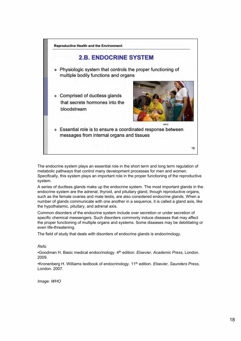
The endocrine system plays an essential role in the short term and long term regulation of metabolic pathways that control many development processes for men and women. Specifically, this system plays an important role in the proper functioning of the reproductive system. A series of ductless glands make up the endocrine system. The most important glands in the endocrine system are the adrenal, thyroid, and pituitary gland, though reproductive organs, such as the female ovaries and male testis, are also considered endocrine glands. When a number of glands communicate with one another in a sequence, it is called a gland axis, like the hypothalamic, pituitary, and adrenal axis. Common disorders of the endocrine system include over secretion or under secretion of specific chemical messengers. Such disorders commonly induce diseases that may affect the proper functioning of multiple organs and systems. Some diseases may be debilitating or even life-threatening. The field of study that deals with disorders of endocrine glands is endocrinology.
Refs:•Goodman H. Basic medical endocrinology. 4th edition. Elsevier, Academic Press, London. 2009.•Kronenberg H. Williams textbook of endocrinology. 11th edition. Elsevier, Saunders Press,London. 2007.
Image: WHO
18
Reproductive Health and the Environment
2.B. ENDOCRINE SYSTEM
❖ Physiologic system that controls the proper function ing of multiple bodily functions and organs
❖ Compr ised of ductless glands
that secrete hormones into the
bloodstream
WHO
❖ Essential role is to ensure a coordinated response between messages from internal organs and tissues
18

19
Hormones are chemical signals that transmit messages from one part of the body to another. They travel through the bloodstream to target specific tissues or organs. Hormones regulate many different processes, such as growth and development, metabolism, sexual function, reproduction, and mood. Hormones also work to control natural chemical balances to ensure the body is in a stable state. They may induce immediate bodily effects, or work slowly, over time, to affect entire bodily processes. Hormones act as very powerful signals. It takes a very small amount of hormonal imbalance to cause significant changes in the human body.
Refs:•Goodman H. Basic medical endocrinology. 4th edition. Elsevier, Academic Press, London. 2009.•Kronenberg H. Williams textbook of endocrinology. 11th edition. Elsevier, Saunders Press,London. 2007.
Reproductive Health and the Environment
HORMONES
♦:♦ Diverse effects in the body - Growth and development
- Mood
- Metabolism regulation
- Reproduct ive cycle control
Essential to maintaining health
of the bodyl
♦:♦ Fundamental components of the endocrine system
19

20
There are different classes of hormones within the human body. Each type of hormone has a different function in the body. They are divided into three classes. The first class of hormones are peptide-derived hormones, and are made of single amino acids that link to form amino acid chains. Some examples of peptide-derived hormones include insulin and two female reproductive hormones: Luteinizing Hormone (LH) and Follicle Stimulating Hormone (FSH). Insulin is the hormone that is responsible for maintaining appropriate blood sugar levels. LH and FSH will be discussed in upcoming slides. A second class of hormones are the steroid-derived hormones. Examples of steroid hormones are estrogen and testosterone. These reproductive hormones will be described in the upcoming sections. The third class of hormones are the amine-derived hormones. Similar to peptide-derived hormones, as they are also made of amino acids that link to form amino acid chains. Amine-derived hormones are specifically comprised of the specific amino acid that are known as tyrosine and tryptophan. Tryptophan is the precursor of serotonin and melatonin synthesis.Note: Peptides consist of chains of amino-acids (oligo- or poly-peptides, proteins). Amines are derived from single amino-acids.
Refs:•Goodman H. Basic medical endocrinology. 4th edition. Elsevier, Academic Press, London. 2009.•Kronenberg H. Williams textbook of endocrinology. 11th edition. Elsevier, Saunders Press,London. 2007.
Reproductive Health and the Environment
TYPES OF HORMONES
Three basic classes of hormones
1. Peptide-derived Amino acid chains
• Insulin, Luteinizing Hormone (LH), Follicle Stimulating Hormone (FSH)
2. Steroid-derived • Derived from cholesterol, cortisone, aldosterone
, Estrogen, testosterone
3. Amine-derived , Amino acids chains
• Catecholamine 20

A central function of the endocrine system is the maintenance of homeostasis in the body. Homeostasis is defined as a stable, constant condition of a living organism, free from sudden fluctuation. Several regulatory mechanisms in the endocrine system allow homeostasis to occur. The endocrine system, and its complex processes, are responsible for a proper internal balance of the human body. Homeostatic regulation allows humans to function effectively even when they are exposed to different environmental conditions, such as temperature. For example, when human beings are in hot temperatures, sweat glands in the skin will produce sweat, thus bringing liquid to the surface for evaporation, and acting to internally reduce our body heat. This is an example of a homeostatic process. When the endocrine system is not able to properly maintain homeostasis, serious life-threatening disorders and diseases may occur.The endocrine system regulates proper balance of hormones in the body through a process known as the “see-saw principle.” This principle describes the communication between cells and glands that secrete hormones to ensure proper hormonal balance. When there is an improper level of hormones in the bloodstream, cells will communicate with other cells to increase or decrease (as needed) the production of a certain hormone. The slide demonstrates how the endocrine system regulates itself to promote homeostasis. Cell A (pink and red colored) will secrete hormone A which will influence cell B, and thus the secretion of cell B (gray colored). If cell B produces too much hormone B, cell A will sense this imbalance and decrease secretion of hormone A, which will in turn decrease secretion of hormone B by cell B. This regulation is known as a negative feedback loop because the resulting return to homeostasis occurs due to a decrease in hormone secretion.
Refs:•Goodman H. Basic medical endocrinology. 4th edition. Elsevier, Academic Press, London. 2009.•Kronenberg H. Williams textbook of endocrinology. 11th edition. Elsevier, Saunders Press, London. 2007. •WHO. Global assessment of the state of the science of endocrine disruptors. Geneva, Switzerland, WHO/PCS/EDC, 2002. Available at www.who.int/ipcs/publications/new_issues/endocrine_disruptors/en/ -accessed 23 June 2010.
Image: WHO. Global assessment of the state of the science of endocrine disruptors. Geneva, Switzerland, WHO/PCS/EDC, 2002.
21
Reproductive Health and the Environment
THE IMPORTANCE OF HOMEOSTASIS
❖ A state of internal balance
❖ Avoiding dramatic shifts in hormone levels that may negatively affect health
❖ See-saw principle: cells send feedback signals to regulate proper levels of each hormone ❖ Usually negative feedback-signals tell
endocrine system to reduce production of certain hormones
Regulation by hlgta' centres
<~- ~ ) Inhibition
www.who.int/,pcslpublicationslnew_issues/Mdocrine_disroptorslena 21

A good example of how the endocrine system maintains homeostasis via the see-saw principle is the maintenance of blood sugar levels by the hormone insulin. Insulin is a hormone that causes certain cells to take up sugar, in the form of glucose, from the blood. The level of insulin in the bloodstream is a very important mechanism of central metabolic control. If blood sugar falls too low in the body, the person may experience unconsciousness due to a lack of glucose. However, insulin also acts to ensure that blood sugar does not rise too high. When endocrine control of insulin fails, diabetes mellitus may occur. However, if the body is in proper homeostatic balance via the see-saw principle, levels of blood sugar will be stable.
Refs:•Goodman H. Basic medical endocrinology. 4th edition. Elsevier, Academic Press, London. 2009.•Kronenberg H. Williams textbook of endocrinology. 11th edition. Elsevier, Saunders Press,London. 2007. •WHO. Global assessment of the state of the science of endocrine disruptors. Geneva, Switzerland, WHO/PCS/EDC, 2002. Available at www.who.int/ipcs/publications/new_issues/endocrine_disruptors/en/ - accessed 23 June 2010.
Image: WHO. Global assessment of the state of the science of endocrine disruptors. Geneva, Switzerland, WHO/PCS/EDC, 2002.
22
Reproductive Health and the Environment
EXAMPLE OF SEE-SAW PRINCIPLE
❖ Insulin maintains proper levels of blood sugar ❖ If blood sugar falls too low =
unconsciousness
❖ If blood sugar is too high= wasteful excretion into urine
❖ Diabetes mellitus: occurs when insulin does not properly respond to changes in blood sugar levels
Homeorasis A B
A
...i. (nc:tnts.ed s~rion of A
B
i Oectii'itsad .sear..lon c;f A
I Oec,>oa::ed s.;cr,r.ion of B +'!IT<ct.ll roli.x• • .'0 ,1.-... ,.-'C'O~b!>'d
B
www.who.int/ipcslpublicatio/new_iss.lNJS!endocrine_disruplorslen
22

Different types of hormones are responsible for different processes in the human body. For example, there is a group of hormones responsible for the reproductive processes of the body. These are known as reproductive hormones and are responsible for many different processes related to sexual development and reproduction. Estrogen is a type of reproductive hormone and is the primary female reproductive hormone. Estrogen promotes the development of breasts and regulates the process of the menstrual cycle. Details of the menstrual cycle will be described in further slides. Progesterone is another type of female reproductive hormone. It is responsible for many processes during pregnancy, including the development of the fetus in the mother’s womb. Luteinizing hormone, known as LH, is yet another female reproductive hormone. LH is essential for female reproduction. LH triggers ovulation and is responsible for releasing the female egg. Therefore, it plays an important role in the menstrual cycle. Finally, Follicle Stimulating Hormone (FSH), is a female reproductive hormone that regulates the development, growth, pubertal maturation, and reproductive processes of the female body. It also initiates follicular growth and prepares the body for the start of the next ovulation cycle. LH and FSH are reproductive hormones that work together and help control the menstrual cycle. Men do not have the same reproductive hormones as women. Testosterone is the male reproductive hormone and is the principal male reproductive hormone. Testosterone is important in the development of male reproductive tissues such as the testes and the prostate. Testosterone also promotes hair growth and muscle development during adolescence, a stage known as puberty.Sexual development and reproductive health for both women and men are dependent on the action of these reproductive hormones.
Refs:•Goodman H. Basic medical endocrinology. 4th edition. Elsevier, Academic Press, London. 2009.•Kronenberg H. Williams textbook of endocrinology. 11th edition. Elsevier, Saunders Press, London. 2007. •Nelson R. An introduction to behavioral endocrinology. Sunderland, Mass: Sinauer Associates, 2005.
Reproductive Health and the Environment
REPRODUCTIVE HORMONES
♦:♦ Responsible for sexual development and reproductive health in women and men
♦:♦ Female primary reproductive hormones: - Estrogen - Progesterone - Luteinizing hormone - Follicle stimulating hormone
♦:♦ Male primary reproductive hormones: - Testosterone - Luteinizing hormone - Follicle stimulating hormone 23

Reproductive hormones are responsible for deciding whether a developing embryo will become a phenotypic male or a female. Whether an embryo will develop into a male or female depends on the formation of reproductive duct systems and the differentiation of external genitalia. When a fetus is approximately 8 weeks old, it will begin to develop either male or female reproductive systems. The mechanism that decides the sex of the fetus is the secretion, or release, of testosterone or a lack of testosterone secretion. For example, if testosterone is released at approximately 8 weeks of fetal life, the fetus will develop a male duct system and external male genitalia. However, if secretion of testosterone does not occur, there will be no induction of male duct system differentiation, thus leading to development of the female duct system. A lack of testosterone release will lead to the development of female characteristics.
Refs:•Goodman H. Basic medical endocrinology. 4th edition. Elsevier, Academic Press, London. 2009.•Kronenberg H. Williams textbook of endocrinology. 11th edition. Elsevier, Saunders Press, London. 2007.
Image: UNDP/UNFPA/WHO/World Bank. Providing the foundation for sexual and reproductive health: A record of achievement. Geneva, Switzerland, UNDP/UNFPA/WHO/World Bank Special Programme on Research, Development, and Research Training in Human Reproduction, 2008. Available at www.who.int/reproductivehealth/publications/general/hrp_brochure.pdf - accessed 23 June 2010.
Reproductive Health and the Environment
ROLE OF HORMONES IN SEX DIFFERENTIATION
❖ Testosterone secretion determines whether an embryo will develop into a phenotypic male or a female
❖ Secretion of testosterone will induce formation of male reproductive system
❖ Lack of secretion and/or response to testosterone can lead to failure to form the male reproductive system completely, leading to female characteristics
UNDP/UNFPAIWHOIWORL.D BANK
24

The female reproductive pathway is comprised of the vagina, uterus, fallopian tubes, and ovaries. The vagina is where the male sperm first enter the internal pathway of the female reproductive tract and is also where a baby will leave the female’s body once ready for birth. The next compartment in the female reproductive system is the uterus. This is the space where the fetus will develop. Next are the fallopian tubes. The female reproductive tract has two fallopian tubes, one on each side of the top of the uterus where mature eggs or ova move through to reach the uterus. Finally, the ovaries are the two round organs that produce the female egg cells. The ovaries rest outside of the openings of the fallopian tubes.
<<NOTE TO USER: Please refer to the diagram on the slide for greater detail into the exact location of these different anatomical features. >>
Refs:•Jones R. Human reproductive biology, 3rd Edition. Burlington, MA, USA: Elsevier Academic Press, 2006. • Pinon R. Biology of human reproduction. Sausalito, CA, USA: University College Books, 2002.
Image: Centers for Disease Control and Prevention. United States Department of Health and Human Services. Available at www.cdc.gov/cancer/nbccedp/cc_basic.htm - accessed 20 June 2010.
25
Reproductive Health and the Environment
2.C. FEMALE REPRODUCTIVE SYSTEM
♦:♦ Ovaries - where egg is produced
♦:♦ Oviduct (Fallopian tube) -where fertilization takes place
♦:♦ Uterus - where the embryo grows and develops
♦:♦ Vagina - birth canal
F~llopl~n tubes O ~rlu
y CDC
25

26
Oogenesis is the term used to describe the creation of the egg, or ovum. The egg is the female cell that will be fertilized by a sperm to create an embryo that can develop into a fetus. The process of oogenesis begins when a female is still just an embryo. The creation of the female eggs occurs before or slightly after the birth of a female. At birth, a female will have 1,000,000 primary eggs, but only 200,000 are left by puberty. No additional primary eggs are created. Over a woman’s reproductive lifetime, only 450 eggs complete oogenesis. The latency period render the eggs particularly vulnerable to environmental exposures.You will notice in the diagram that for every mature ovum that is created, three polar bodies will also be created. However, the mature ovum is what is known as the egg. It is this egg that must be fertilized by a sperm in order for an embryo to grow into a fetus. Oocyte formation in utero provides a susceptibility to epigenetics effects from maternal exposures and to third-generation effects.
In any one human generation, the egg’s development starts before the female that carries it is even born; 8 to 20 weeks after the fetus has started to grow, cells that are to become mature ova have been multiplying, and by the time that the female is born, all of the egg cells that the ovaries will release during the active reproductive years of the female are already present in the ovaries. These cells, known as the primary ova,…, remain dormant until just prior to ovulation, when an egg is released from the ovary. Some egg cells may not mature for 40 years; others degenerate and never mature.
Refs: •Jones R. Human reproductive biology, 3rd Edition. Burlington, MA, USA: Elsevier Academic Press, 2006. •NIH. Women’s Health. National Institutes of Health. Available at health.nih.gov/category/WomensHealth - accessed March 20, 2010.
Image: Wikimedia Commons. http://en.wikipedia.org/wiki/File:Gray5.svg - accessed 10 July 2010. This image is public domain.
Reproductive Health and the Environment
OOGENESIS
♦:♦ Oogenesis is the production of the egg in the ovaries
♦:♦ Begins when the female is an embryo ❖ all primary oocytes develop before
birth and remain "dormant" in this stage until ready for maturation
en.wikipedia_cwglwik11Flte:Gray5.svg>
26

The female reproductive system is regulated by a the signaling pathways of the female reproductive hormones described in previous slides. There are three key places in the female body that serve as hormonal messaging centers. They are the hypothalamus in the brain, the anterior pituitary gland, and the ovaries. The hypothalamus secretes a hormone that is called the gonadotropin-releasing hormone (GnRH). This hormone (GnRH) regulates the release of the luteinizing hormone (LH) and follicle-stimulating hormone (FSH) from specialized cells in the anterior pituitary gland. These hormones are released in short bursts. LH and FSH promote ovulation and stimulate secretion of the sex hormone estradiol, (an estrogen) and progesterone from the ovaries. These hormones circulate in the bloodstream and stimulate the target organs of the reproductive system, including the breasts, uterus, vagina. Proper functioning of the female reproductive system is dependent on the chemical messaging of the described hormonal pathways.
Refs:•Jones R. Human reproductive biology, 3rd Edition. Burlington, MA, USA: Elsevier Academic Press, 2006. •National Institutes of Health. Female reproductive system. Medline Plus. National Institutes of Health. Available at www.nlm.nih.gov/medlineplus/femalereproductivesystem.html -accessed 20 March 2010.
Image: WHO
27
Reproductive Health and the Environment
FEMALE REPRODUCTIVE HORMONE PATHWAYS
❖ The hypothalamus and the pituitary gland are both in the brain.
❖ Hormonal messaging between the hypothalamus , anter ior pituitary gland, and ovar ies regulates female reproductive system .
❖ Feedback loop controls proper
hormonal balance.
❖ Hormones signal target female
reproductive organs : breasts ,
uterus , and vagina. WHO
27

The female menstrual cycle is necessary for reproduction. This process is under control of the endocrine system. The menstrual cycle occurs as a result of a female body’s preparation for potential fertilization of an egg by a sperm cell to create an embryo. During the ovarian cycle, a primary oocyte matures into a egg ready for release. The egg is released into the fallopian tube and is ready for potential fertilization by a sperm cell. At this moment, the uterus begins forming a layer of nutrient rich cells on its inner walls. This lining will serve as an implantation bed for a potentially fertilized egg. However, if the egg is not fertilized by the time the egg reaches the uterus, the uterus will shed the lining that was created. This is because there is no longer a need for an implantation bed because fertilization has not occurred. Menstruation occurs in monthly cycles throughout a woman's reproductive life. However, menstruation does not occur while a woman is pregnant, in the majority of women. Menstruation starts during puberty and ends permanently at menopause.
Refs:•Jones R. Human reproductive biology, 3rd Edition. Burlington, MA, USA: Elsevier Academic Press, 2006. •Menstrual cycle. MERCK Medical Library. Available at www.merck.com/mmhe/sec22/ch241/ch241e.html - accessed 20 March 2010.
28
Reproductive Health and the Environment
FEMALE MENSTRUAL CYCLE
❖ Each month, a woman's body prepares for the possible fertilization of an egg
❖ Ovarian Cycle An egg matures in the ovary and is released about 14 days from the last menstruation
❖ Uterine Cycle The lining of the uterus builds up to prepare for a fertilized egg. If no fertilization occurs, the lining breaks down and menstruation occurs
28

The female menstrual cycle is regulated by specific hormones. Luteinizing hormone and follicle-stimulating hormone promote ovulation and stimulate the ovaries to produce estrogen and progesterone. These two hormones stimulate the uterus to prepare for potential fertilization. The cycle has three phases: follicular (before release of the egg), ovulatory (egg release), and luteal (after egg release). The menstrual cycle begins with the first day of bleeding. This is counted as day 1. The cycle ends just before the next menstrual period. Menstrual cycles typically range from about 25 to 36 days.
Refs:•Jones R. Human reproductive biology, 3rd Edition. Burlington, MA, USA: Elsevier Academic Press, 2006. •Menstrual cycle. MERCK Medical Library. Available at www.merck.com/mmhe/sec22/ch241/ch241e.html - accessed 20 March 2010.
29
Reproductive Health and the Environment
FEMALE MENSTRUAL CYCLE
❖ Follicle is the egg and the cells surrounding it.
❖ One follicle fully matures during each ovar ian cycle.
❖ Ovulation is the release of egg from ovary triggered by LH (Luteinizing Hormone).
29

Unlike the female reproductive system, most of the male reproductive system is located outside of the body. These external structures include the penis, scrotum, and testicles. Sperm are the male reproductive cells. The sperm cell consists of a head, a midpiece and a tail. The head contains the nucleus. The head is surrounded by an acrosome that contains enzymes for penetrating the female egg. The body has many mitochondria to provide energy for the journey through the female cervix. The tail or “flagellum” allows the sperm to move. Sperm is expelled (ejaculated) through the end of the penis. The penis is the male organ used in sexual intercourse. It has three parts: the root, which attaches to the wall of the abdomen; the body, or shaft; and the glans, which is the cone-shaped part at the end of the penis.Most men have two testes. The testes are responsible for making testosterone, the primary male sex hormone, and for generating sperm.The scrotum is a sac of skin that hangs behind and below the penis. It contains the testes. The scrotum acts as a "climate control system" for the testes. For normal sperm development, the testes must be at a temperature slightly cooler than body temperature.
Refs:•Jones R. Human reproductive biology, 3rd Edition. Burlington, MA, USA: Elsevier Academic Press, 2006. •National Institutes of Health. Male reproductive system. National Institutes of Health. Available at www.nlm.nih.gov/medlineplus/malereproductivesystem.html - accessed 20 March 2010.
30
Reproductive Health and the Environment
2.D. MALE REPRODUCTIVE SYSTEM
❖ Sperm are the male reproductive cells
❖ The penis is the male organ used in sexual intercourse
❖ The testes are where sperm are produced
❖ The scrotum contains the testes and controls temperature for sperm development
30

<<READ SLIDE.>>
Testosterone is an imperative sex hormone in male reproductive function. Specifically, it regulates spermatogenesis, the production of the sperm. A proper balance of testosterone in the male body is essential to maintain not only reproductive health, but overall health status. The figure is the representation of the testosterone molecule. Testosterone is a steroid hormone. Genetics also has a role in the development of the male reproductive system. For instance, they act as regulators of testicular descent.
Refs:•Jones R. Human reproductive biology, 3rd Edition. Burlington, MA, USA: Elsevier Academic Press, 2006. •Kaleva M, Toppari J. Genetics and hormones in testicular descent. Hormones (Athens), 2003, 2(4):211-6.•NIH. Male reproductive system. National Institutes of Health. Available at www.nlm.nih.gov/medlineplus/malereproductivesystem.html - accessed 20 March 2010.
Image: Testosterone. Wikimedia Commons. Available at en.wikipedia.org/wiki/File:Testosterone.svg - accessed 10 July 2010. This image is public domain.
31
Reproductive Health and the Environment
THE ROLE OF TESTOSTERONE
♦:♦ Determines male sexual development
♦:♦ Involved in healthy sperm and semen production
♦:♦ Controls and regulates sexual behavior
0
31

32
Spermatogenesis describes the process of sperm cell development in the male. It is initiated in the male testis with the beginning of puberty. It entails numerous steps that one by one, lead to the development of a mature sperm cell. First, the process begins with rounded immature sperm cells. These cells first undergo mitotic division in which the genetic material in the immature sperm cell is replicated. Mitotic division indicates that the genetic material was separated into two identical copies of each other. Next, is a phase of meiotic division. Meiotic division refers to a cellular process that occurs in sex cells. This process divides the chromosome in half and produces two cells with half the genetic material in each cell. Finally, the sperm cells undergo a final metamorphic change to produce mature sperm. Spermatogenesis is heavily dependent on appropriate levels of testosterone and may be affected by hormonal fluctuations in the body.
Refs:•Johnson et. al. Efficiency of spermatogenesis: a comparative approach. Animal Reproduction Science. 2000, 60-61: 471-480.•Jones R. Human reproductive biology, 3rd Edition. Burlington, MA, USA: Elsevier Academic Press, 2006. •National Institutes of Health. Male reproductive system. National Institutes of Health. Available at www.nlm.nih.gov/medlineplus/malereproductivesystem.html - accessed 20 March 2010.
Image: Histology of selected organs of the reproductive system and embryo. Available at science.tjc.edu/images/reproduction/spermatogenesis.jpg - accessed 20 June 2010. Copyright permission courtesy of the author, Pamela Gregory, Professor of Biology, Tyler Junior College, Tyler, Texas, US:
Reproductive Health and the Environment
SPERMATOGENESIS
❖ Occurs in the testes in the seminiferous tubules
Mature in the epid idymis and gain motility
❖ Produced constantly after puberty
UI
UI D Ill l:
R MA T O GE N e; IS
_inelo9oniu ..
..
bio 1152.nicerweb. com/Locked/media/ch46146_ 12Spermatogenesis.jpg Pamela Gregory, Professor of Biology, Tyler Junior College, Tyler, Texas, US 32

Spermatogenesis is a complex physiological process. The production of relatively high concentrations of sperm is essential for healthy reproductive function of males. On average, the process of sperm production takes between 70 to 80 days. A very large volume of individual sperm are produced every day, though it varies widely between 20 million and 375 million individual sperm cells.Semen is the liquidly medium that provides sperm with nutrients as they travel through the female reproductive tract to fertilize the egg. In every millimeter of semen, there are approximately 100,000,000 sperm, however, this number can also vary depending on a numerous internal and external factors. Epidemiologic research has ascertained that within a millimeter of male sperm, approximately 20,000,000 will be unviable to fertilize a female egg due to physiologic or chemical dysfunction.
Refs:•Cooper TG et al. WHO reference values for human semen characteristics. Human Reproduction Update. 2009, 0:1-15.•Jones R. Human reproductive biology, 3rd Edition. Burlington, MA, USA: Elsevier Academic Press, 2006. •National Institutes of Health. Male reproductive system. National Institutes of Health. Available at www.nlm.nih.gov/medlineplus/malereproductivesystem.html - accessed 20 March 2010.
33
Reproductive Health and the Environment
PRODUCTION FACTORS IN SPERMATOGENESIS
❖ Sperm production takes 70 to 80 days
❖ Between 20 and 375 million sperm are produced per day
WHO lower reference values for human semen
❖ Total sperm number 39 million per ejaculate
❖ Sperm concentration 15 million per ml
❖ Vitality 28% live
❖ Progress ive motility 32%
❖ Total (progressive+ non-progressive motility) 40%
❖ Morphologically normal forms 4%
This data represents reference distributions of semen characteristics of fertile men in a number of countries. They provide an appropriate tool in conjunction with clinical data to evaluate a patient's semen quality and prospects for fertility. 33

34
Conception describes the process of the joining of sperm and egg. This process can only can occur during a short window of the menstrual cycle, either 2 days before or 15 hours following ovulation. Developmental processes related to human fertility and conception occur during limited periods of time. If sperm meets and penetrates a mature egg after ovulation, it will fertilize it. When the sperm penetrates the egg, changes occur in the protein coating around it to prevent other sperm from entering. However, both the female egg and male sperm must demonstrate proper structure and function in order for conception to occur.
Refs:•Jones R. Human reproductive biology, 3rd Edition. Burlington, MA, USA: Elsevier Academic Press, 2006. •National Institutes of Health. Reproductive health. National Institutes of Health. National Institute of Child Health and Human Development. Available at www.nichd.nih.gov/health/topics/ - accessed 22 March 2010.
Image: Conception. National Institutes of Health (NIH). Available at www.ncbi.nlm.nih.gov/bookshelf/br.fcgi?book=dbio&part=A2609 - accessed 15 November 2009. Reproduced with permission from NIH.
Reproductive Health and the Environment
2.E. CONCEPTION AND FETAL LIFE
❖ Ovulation occurs approximately 14 days after start of menses
❖ Sperm must be introduced in female system at any time from 2 days before to 15 hours after ovulation
❖ One sperm joins with an ovum forming an embryo
NatkmaJ Institutes of Health
Reproductive health extends through conception and the development of
the embryo and fetus!
34

35
Human embryogenesis is a process of very specific cell divisions and differentiations following fertilization of a male sperm with a female egg. Embryogenesis begins at the moment that a sperm meets with the egg and lasts until the eighth week of development. The original cell at fertilization is known as a zygote. The zygote will undergo specific cell division, known as cleavages. The different types of cleavages are outlined in the diagram. At approximately day five following fertilization, the zygote has divided multiple times and is now formally referred to as a blastocyst. In the first week, the blastocyst will implant into the uterine wall. Following this period, the mass of cells will differentiate and the first physiologic structures will be formed, including the beginnings of the umbilical cord.
Refs:•Jones R. Human reproductive biology, 3rd Edition. Burlington, MA, USA: Elsevier Academic Press, 2006. •National Institutes of Health. Reproductive health. National Institutes of Health. National Institute of Child Health and Human Development. Available at www.nichd.nih.gov/health/topics/ - accessed 22 March 2010.
Image: Human embryogenesis. Available at en.wikipedia.org/wiki/File:Human_embryogenesis.png - accessed 29 June 2010. This image is public domain.
Reproductive Health and the Environment
EMBRYOGENESIS
fertilise d egg 8 -cell zygote cell adhesion 16-cell morula blastocyst ... 6 7 .. , 8 ~ ~- 9@ .. ,,. _
· .. : ; . . --- -. : .._._ ,,,-
zona hatch ing invades uterine Willl cell mass different iates bllam lnar disc forms
mesode rm forms mesoderm spreads amn iotic sac grows
en. wfkipedia .orglwfkVFilfl :Human_ embryogenesis .png 35

Organogenesis describes the process by which the cells differentiate into specific tissues that will later become internal organs. There are three specific tissues that develop during this period. They are the ectoderm, mesoderm, and endoderm. The ectoderm will form the external layer of organs. For example, the skin cells of the body will be formed from the ectoderm developed during organogenesis. The next type of tissue is the mesoderm, which will represent the middle layer of the organs. This type of tissue includes the red blood cells and skeletal muscle cells. Finally, endodermic tissue will be formed. The endoderm will comprise the internal layer of organs. An example of endoderm formation is the development of thyroid cells. Together, the ectoderm, mesoderm, and endoderm will create the essential organs for the developing fetus. Internal organs begin to develop in utero between the 3rd and 8th week. During this critical window of development, the process of cell differentiation may be adversely affected by various environmental exposures. However, due to the complexity of organogenesis, as well as the poorly understood mechanisms of action for many exogenous elements on organogenesis, the exact health effects are difficult to presume. On this diagram, you will notice the transition from the zygote to the blastocyst described in the previous slide.
Refs:•Evers C, Starr L. Organogenesis, biology: concepts and aplications. 6th ed. United States: Thomson, 2006.•Jones R. Human reproductive biology, 3rd Edition. Burlington, MA, USA: Elsevier Academic Press, 2006. •Van De Graaf K. Human anatomy, 5th ed. New York: McGraw-Hill, 2000.
Image: Organogenesis. image.absoluteastronomy.com/images/encyclopediaimages/c/ce/cell_differentiation.gif -accessed 14 May 2010. This image is public domain
36
Reproductive Health and the Environment
Zygote
C#dooc M ......
ORGANOGENESIS
....,,. Cel Of Cels MUSCie lhe Kidney (In Gut )
Lu ng Cell Thyr06CI (Alv eolo< Cell Cell)
Panc reabC c .. ,
Nlp:M,,.....abKAu!MMrcnom,o__oom'i'nag~p,uilh\agestc>cMOII _~~
36

37
Following the end of embryogenesis in the 8th week, the developing organism is officially known as a fetus. From this point forward, the developmental period is known as the fetal period. During the fetal period, the beginnings of all of the major organs are created through intricate cell divisions and differentiations. In addition to individual organs, physiologic systems are also developed. The fetal period is defined by two major stages: rapid growth and cell differentiation. The first stage is rapid growth. Fetal growth is the most intense at the first week of fetal period and lasts through week sixteen. In this rapid growth phase, the fetus experiences a 25-fold increase in body weight. The second phase of growth is defined by dramatic tissue and organ development. During the rapid growth period previously described, the fetus has not developed any mechanisms for proper cell differentiation. Therefore, during this second phase, complex processes of cell differentiation and cleavaging develop specific organs, organ systems, and tissues. In addition, the fetus takes the shape representative of an infant. Throughout the fifth month, muscle mass begins to develop and the mother may experience the first fetal movements due to this occurrence. By the six month, the lungs have fully differentiated and the fetus is able to independently breathe. The seventh month is marked by the development of the nervous system and the fetus is able to respond to basic reflexes, including constriction of the pupils in response to light. The fetal period lasts until birth, at approximately the 38th week of development.
<< NOTE TO USER: For further information, please refer to module 2, “Female Environmental Reproductive Health" or to the module on "Developmental and Environmental Origins of Disease”>>
Refs:•Jones R. Human reproductive biology, 3rd Edition. Burlington, MA, USA: Elsevier Academic Press, 2006. •National Institutes of Health. Reproductive health. National Institutes of Health. National Institute of Child Health and Human Development. Available at www.nichd.nih.gov/health/topics/ - accessed 22 March 2010.•Van De Graaf K. Human anatomy, 5th ed. New York: McGraw-Hill, 2000.
Image: Environmental Health Perspectives. Available at ehp.niehs.nih.gov/docs/2005/113-8/fetus.jpg -accessed 29 June 2010. Reproduced with permission from EHP.
Reproductive Health and the Environment
THE FETAL PERIOD
Begins at the termination of embryogenesis, or after the 8th week and lasts until the end of pregnancy
ehp.niells .nih.gov/docs/2005/113-8/fe tus.jpg>
Characterized by two specific mechanisms:
1. Rapid growth 2. Cellular differentiation
and organ development
37

A critical window of susceptibility is a period where there are numerous changing capabilities in the developing organism. Exposures to environmental contaminants during this window may result in permanent damage to a fetus and may have lifelong effects on health. Given that development continues after birth, critical and sensitive windows occur before, during, and shortly after the fertilization of the egg. Critical windows of development are also present during pregnancy, infancy, childhood, and puberty. The diagram provided demonstrates the particular windows of susceptibility for the developing fetus, in this case, of the nervous system. The maternal environment at these specific temporal windows has important implications for the healthy development of a fetus.
<< NOTE TO USER: For further information, please refer to the module on "Developmental and Environmental Origins of Disease”>>
Refs:•Calabrese E. Sex differences in susceptibility to toxic industrial chemicals. British Journal of Industrial Medicine, 1986, 43: 577–579.•Rice D et al. Critical periods of vulnerability for the developing nervous system: evidence from humans and animal models. Environmental Health Perspectives, 2000, 108 (Suppl 3): 511-33Vulnerable periods during the development of the nervous system are sensitive to environmental insults because they are dependent on the temporal and regional emergence of critical developmental processes (i.e. proliferation, migration, differentiation, synaptogenesis, myelination, and apoptosis). Evidence from numerous sources demonstrates that neural development extends from the embryonic period through adolescence. In general, the sequence of events is comparable among species, although the time scales are considerably different. Developmental exposure of animals or humans to numerous agents (e.g. X-ray irradiation, methylazoxymethanol, ethanol, lead, methyl mercury, or chlorpyrifos) demonstrates that interference with one or more of these developmental processes can lead to developmental neurotoxicity. Different behavioural domains (e.g. sensory, motor, and various cognitive functions) are subserved by different brain areas. Although there are important differences between the rodent and human brain, analogous structures can be identified. Moreover, the ontogeny of specific behaviours can be used to draw inferences regarding the maturation of specific brain structures or neural circuits in rodents and primates, including humans. Furthermore, various clinical disorders in humans (e.g. schizophrenia, dyslexia, epilepsy, and autism) may also be the result of interference with normal ontogeny of developmental processes in the nervous system. Of critical concern is the possibility that developmental exposure to neurotoxicants may result in an acceleration of age-related decline in function. This concern is compounded by the fact that developmental neurotoxicity that results in small effects can have a profound societal impact when amortized across the entire population and across the lifespan of humans.Image: Rice D et al. Critical periods of vulnerability for the developing nervous system: evidence from humans and animal models. Environmental Health Perspectives, 2000, 108 (Suppl 3): 511-33. Reproduced with permission from Environmental Health Perspectives
38
Reproductive Health and the Environment
CRITICAL WINDOWS OF SUSCEPTIBILITY
Sensitive time interval during development when exposu res to endocrine disruptors can interfere with physiology of cell, tissue, or organ
Miouboft Cell of Subpl.1• SVNP••
oW..-abon ,_•roas neu,ront ,O,matl,on
,_,,.2. c.o.n.,.,iMln rJ timc!h.e,. b de,,e..,_. aipaatSS1H 1ahun- lhepm~ snJOd ts~ • ~ onda'litpo1tl!Mef~l•aic.ed .. ,,_..,~~~.9Wit.r~"l~..cl"PW'lltedV1.-.'lliflO',.... .- ofHad.r.flltSV~G,ntji
Exposure at specific windows may result in adverse and irreversible effects
Rice D et al. Critic.a, peritxh of vulnerability for the developing n9fVOUs system : a'Wdence from humans and anima l mo</6/s. Environmen tal Health Perspecove&, 2000, 108 (Suppl 3): 511-33.
38

The majority of the endocrine system is developed during the fetal or neonatal period in humans. Most endocrine glands, like the thyroid, pancreas, adrenals, and gonads (reproductive organs), will form early in the second month and then differentiate into their respective physiologic forms in the third month. This development entails the initial programming of homeostasis, and will establish the definition of an appropriate hormonal balance for an individual throughout the rest of life. This can be thought of as an initial programming of the hormones needed to maintain the "see-saw" principle defined in a previous slide. For this reason, this specific period of endocrine system programming is imperative for future healthy homeostatic balance. Research has demonstrated that abnormal environmental factors during fetal or neonatal development may potentially result in permanent "mis-programming" of the endocrine system. "Mis-programming" may lead to the development of homeostatic imbalance and potentially subsequent disease.
<< NOTE TO USER: For further information, please refer to module 2, “Female Environmental Reproductive Health" or to the module on "Developmental and Environmental Origins of Disease”>>
Refs:•Goodman H. Basic Medical Endocrinology, 4th Edition. Elsevier, Academic Press: London, 2009.•Kronenberg H. Williams Textbook of Endocrinology, 11th Edition. Elsevier, Saunders Press: London, 2007.
Image: UNDP/UNFPA/WHO/World Bank. Providing the foundation for sexual and reproductive health: A record of achievement. Geneva, Switzerland, UNDP/UNFPA/WHO/World Bank Special Programme on Research, Development, and Research Training in Human Reproduction, 2008. Available at www.who.int/reproductivehealth/publications/general/hrp_brochure.pdf, accessed 23 June 2010.
39
Reproductive Health and the Environment
DEVELOPMENT OF THE FETAL ENDOCRINE SYSTEM
❖ The endocrine system develops during an especially vulnerable fetal period
❖ Involves the programming of
homeostasis
❖ An unhealthy environment
may result in permanent
"mis-programming" of the endocrine system UNDP/UNFPAIWHO/Wond Bank
39

<<READ SLIDE.>>
Section 3 will describe environmental exposures and the relationship to reproductive health. This section will introduce the concept of endocrine disruptors and overview pertinent factors related to this topic. The section will also provide some evidence of endocrine disruptors and their role on reproductive health from wildlife and human studies.
Image: WHO
40
Reproductive Health and the Environment
SECTION 3: Environmental exposures and reproductive
health
WHO
A. Introduction to environmental reproductive health
B. Endocrine disruptors
C. Examples from wildlife and humans
40

Reproductive health and the environment focuses on exposures to environmental contaminants during critical periods of human development. These periods are directly related to reproductive health throughout the life course, including the period before conception, at conception, fertility, pregnancy, child and adolescent development, and adult health. Exposures to different environmental contaminants may influence reproductive health status through the process of epigenetics. Environmental toxicants may potentially induce effects in human reproductive processes. However, the extent of this hypothesis must be supported through greater levels of research.
Refs:•WHO. Global assessment of the state of the science of endocrine disruptors. Geneva, Switzerland, WHO/PCS/EDC, 2002. Available at www.who.int/ipcs/publications/new_issues/endocrine_disruptors/en/ - accessed 23 June 2010. •Woodruff T. Proceedings of the Summit on Environmental Challenges to Reproductive Health and Fertility: executive summary. Fertility and Sterility, 2003, 89 (2),1-20.
41
Reproductive Health and the Environment
3. A. REPRODUCTIVE HEAL TH AND THE ENVIRONMENT
❖ Focuses on exposure to contaminants found in the environment, specifically during critical periods of development.
❖ All the physical, chemical, biological and social factors that may affect the origin, growth , development and survival of a person in a given setting.
Examples include :
- Specific synthetic chemicals - Some metals
- Air pollutants
Still an emerging issue!
41

The slide shows that the dose and the time of the exposure, as well as the "danger" posed by the properties and toxicity of the environmental factor determine the risk for health.
Slide kindly provided by Dr Amalia Laborde, Professor of Department of Toxicology, Faculty of Medicine, Uruguay.
Reproductive Health and the Environment

The slide shows that the dose and the time of the exposure (in this case we take the example of pesticide ingestion) as well as the "danger" posed by the properties and toxicity of the environmental factor determine the risk for health.
Slide kindly provided by Dr Amalia Laborde, Professor of Department of Toxicology, Faculty of Medicine, Uruguay.
Reproductive Health and the Environment
ACUTE TOXICITY
43

In the case of repeated low doses in the occupational setting, of pesticides for instance, the toxicity is chronic and might affect the regulations/metabolism of genes and the immune, endocrine and other systems. The effects are chronic and might only be visible after a latency period.
Slide kindly provided by Dr Amalia Laborde, Professor of Department of Toxicology, Faculty of Medicine, Uruguay.
Reproductive Health and the Environment
Repeated low doses Occupational setting
and environment
exposure
CHRONIC EFFECTS Respiratory Skin Neurological Cancer Reproductive
dang
CHRONIC TOXICITY
genotoxicity ndocrine disruptio
inmunotoxicity 44

Several chemicals, compounds (both synthetic and organic), metals, and other environmental toxicants have been associated with adverse human health effects. Significant scientific concerns over the potential impact of these environmental hazards on reproductive health have increased research and public debate on this issue. For instance, evidence is arising on relationships between spontaneous abortion as well as reduction of anogenital distance and exposure to dichlorodiphenyltrichloroethane (DDT) during pregnancy.Refs:•CHE. Chemical contaminants in the environment. Collaborative on Health & the Environment (CHE). www.healthandenvironment.org - accessed 20 March 2010. •Swan SH et al. Decrease in anogenital distance among male infants with prenatal phthalate exposure. Environ. Health Perspect.2005, 113 (8):1056–61. Prenatal phthalate exposure impairs testicular function and shortens anogenital distance (AGD) in male rodents. We present data from the first study to examine AGD and other genital measurements in relation to prenatal phthalate exposure in humans. A standardized measure of AGD was obtained in 134 boys 2–36 months of age. AGD was significantly correlated with penile volume (R = 0.27, p = 0.001) and the proportion of boys with incomplete testicular descent (R = 0.20, p = 0.02). We defined the anogenital index (AGI) as AGD divided by weight at examination [AGI = AGD/weight (mm/kg)] and calculated the age-adjusted AGI by regression analysis. We examined nine phthalate monoester metabolites, measured in prenatal urine samples, as predictors of age-adjusted AGI in regression and categorical analyses that included all participants with prenatal urine samples (n = 85). Urinary concentrations of four phthalate metabolites [monoethyl phthalate (MEP), mono-n-butyl phthalate (MBP), monobenzyl phthalate (MBzP), and monoisobutyl phthalate (MiBP)] were inversely related to AGI. After adjusting for age at examination, p-values for regression coefficients ranged from 0.007 to 0.097. Comparing boys with prenatal MBP concentration in the highest quartile with those in the lowest quartile, the odds ratio for a shorter than expected AGI was 10.2 (95% confidence interval, 2.5 to 42.2). The corresponding odds ratios for MEP, MBzP, and MiBP were 4.7, 3.8, and 9.1, respectively (all p-values < 0.05). We defined a summary phthalate score to quantify joint exposure to these four phthalate metabolites. The age-adjusted AGI decreased significantly with increasing phthalate score (p-value for slope = 0.009). The associations between male genital development andphthalate exposure seen here are consistent with the phthalate-related syndrome of incomplete virilization that has been reported in prenatally exposed rodents. The median concentrations of phthalate metabolites that are associated with short AGI and incompletetesticular descent are below those found in one-quarter of the female population of the United States, based on a nationwide sample. These data support the hypothesis that prenatal phthalate exposure at environmental levels can adversely affect male reproductive development in humans.•Torres-Sanchez L et al. Dichlorodiphenyldichloroethylene exposure during the first trimester of pregnancy alters the anal position in male infants. Annals of the New York Academy of Sciences, 2008, 1140:155–162. •WHO. Chemicals assessment. Geneva, Switzerland, WHO/IPCS, 2007. www.who.int/ipcs/assessment/en/index.html - accessed 20 June 2010. •WHO. Fact sheet: Dioxins and their effects on human health. Geneva, Switzerland, World Health Organization, 2008. www.who.int/mediacentre/factsheets/fs225/en/index.html - accessed 20 June 2010. •Woodruff T. Proceedings of the Summit on Environmental Challenges to Reproductive Health and Fertility: executive summary.
45
Reproductive Health and the Environment
CHEMICALS POT NTIALLY ASSOCIATED WITH REPRODUCTIVE HEAL TH EFFECTS
Commonly used pesticides
Flame retardants
Dioxin-like substances
Phthalates
Addit ives to consumer products (plasticizers)
DDT ( dichlorodi phenyltrichloroethane ) Organophosphates
PBDEs (polybrominated dipheny1ethers)
PCBs (polychlorinated biphenyls)
PVC (polyvinyl chloride) Di ethyl hexyl phthalate
BPA (bisphenol A)
Multiple case studies from wildlife exposures ; some human evidence
Animal exposure models/data
-Animal exposure models/data -Wild life exposure studies -Weak human exposure data
-Animal exposure models/data - Emerging human studies (surveys , biomarker associations)
- Evidence from animal exposu re models/data
45

Fertility and Sterility, 2003, 89 (2),1-20.
45

An endocrine disruptor is an exogenous substance or mixture that alters the function of the endocrine system and consequently causes adverse health effects in an intact organism, or its progeny, or subpopulations.A potential endocrine disruptor is an exogenous substance or mixture that possesses properties that might be expected to lead to endocrine disruption in an intact organism, or its progeny, or subpopulations.Key issues in understanding the role and action of endocrine disruptors and potential endocrine disruptors are the mechanisms of action and consequences of exposure to endocrine disrupting chemicals, including mixture of various chemical compounds, dose response relationships, latent effects, and age of exposure. It is important to acknowledge that the state of science regarding endocrine disruptor research is only beginning and much more is yet to be learned about the specific qualities of these environmental compounds. Endocrine disruption has sometimes been demonstrated for animals (wildlife/in vivo and in vitro studies). For instance, tributyl tin (TBT) originating from antifouling paints used to treat boat hulls induces a form of pseudohermaphroditism (termed imposex) in female gastropods by an endocrine-disrupting mechanism.
Refs: •Calafat AM, Needham LL. Human exposures and body burdens of endocrine-disrupting chemicals. In: Gore AC, ed. Endocrine-disrupting chemicals: from basic research to clinical practice. Totowa, NJ: Humana Press, 2007, 253–268. •Dickerson SM, Gore AC. Estrogenic environmental endocrine-disrupting chemical effects on reproductive neuroendocrine function and dysfunction across the life cycle. Rev Endocr Metab Disord, 2007, 8:143–159 15. •Gore AC, Crews D. Environmental endocrine disruption of brain and behavior. In: Pfaff DW et al, eds. Hormones, Brain and Behavior. 2009, 1789 –1816. •WHO. Global assessment of the state of the science of endocrine disruptors. Geneva, Switzerland, WHO/PCS/EDC, 2002. Available at www.who.int/ipcs/publications/new_issues/endocrine_disruptors/en/ - accessed 23 June 2010.
46
Reproductive Health and the Environment
3.8. ENDOCRINE DISRUPTORS: DEFINITIONS
❖ Endocrine Disruptor is an exogenous substance or mixture that alters the function of the endocrine system and consequent ly results in adverse health effects
❖ Potential endocrine disruptor is an exogenous substance or mixture that possesses properties that might lead to endocr ine disruption
Research in this field is just beginning! 46

The endocrine system is a complex network of hormones that regulates various bodily functions such as growth and development. The endocrine glands include the pituitary, thyroid, adrenal, thymus, pancreas, ovaries, and testes. These glands or organs release carefully-measured levels of hormones into the bloodstream that act as natural chemical messengers to control important processes of the body. Specific environmental toxicants directly affect the endocrine system. Endocrine disruptors are exogenous agents that interfere with the synthesis, secretion, transport, binding, action, or elimination of natural hormones in the body that are responsible for the maintenance of homeostasis, reproduction, development, and/or behavior. Endocrine disruptors can change normal hormone levels, stimulate or halt the production of certain hormones, or change the way hormones move through the body. However, greater research is still needed to substantiate this hypothesis.
DDT: dichlorodiphenyltrichloroethaneDDE: dichlorodiphenyldichloroethylene
Refs: •Calafat AM, Needham LL. Human exposures and body burdens of endocrine-disrupting chemicals. In: Gore AC, ed. Endocrine-disrupting chemicals: from basic research to clinical practice. Totowa, NJ: Humana Press, 2007, 253–268. •Gray LE et al. Adverse effects of environmental antiandrogens and androgens on reproductive development in mammals. Int Journal of Androgens, 2007, 29:96–104.•WHO. Global assessment of the state of the science of endocrine disruptors. Geneva, Switzerland, WHO/PCS/EDC, 2002. Available at www.who.int/ipcs/publications/new_issues/endocrine_disruptors/en/ - accessed 23 June 2010. •WHO. Persistent organic pollutants: impact on child health. WHO, 2010. Available at www.who.int/ceh/publications/persistent_organic_pollutant/en/index.html - accessed 31 October 2011.
Image: WHO
47
Reproductive Health and the Environment
ENDOCRINE DISRUPTORS
♦:♦ Endocrine disruptors interfere with the production, metabolism, and action of natural hormones in the body - Disrupt hormones needed for homeostasis and
developmental processes
- Alter estrogen , androgen, thyroid , neuroendocrine and metabolic signaling
Endocrine disruptors include: • Some pesticides (DDT, DOE)
• Some herbicides (atrazine)
• Some persistent organic
pollutants (dioxin)
• Potential : phthalates 47

<<READ SLIDE>>Refs:•Amaral Mendes JJ. The endocrine disrupters: a major medical challenge. Food and Chemical Toxicology. 2002, 40(6): 781-788.•Diamanti-Kandarakis, et al. Endocrine-disrupting chemicals: an Endocrine Society scientific statement. The Endocrine Society. 2009•Miller MD et al. Thyroid-Disrupting Chemicals: Interpreting Upstream Biomarkers of Adverse Outcomes. Environ Health Perspect. 2009, 117(7): 1033-1041. There is increasing evidence in humans and in experimental animals for a relationship between exposure to specific environmental chemicals and perturbations in levels of critically important thyroid hormones (THs). Identification and proper interpretation of these relationships are required for accurate assessment of risk to public health.Objectives: We review the role of TH in nervous system development and specific outcomes in adults, the impact of xenobiotics on thyroid signaling, the relationship between adverse outcomes of thyroid disruption and upstream causal biomarkers, and the societal implications of perturbations in thyroid signaling by xenobiotic chemicals.Data sources: We drew on an extensive body of epidemiologic, toxicologic, and mechanistic studies.Data synthesis: THs are critical for normal nervous system development, and decreased maternal TH levels are associated with adverse neuropsychological development in children. In adult humans, increased thyroid-stimulating hormone is associated with increased blood pressure and poorer blood lipid profiles, both risk factors for cardiovascular disease and death. These effects of thyroid
Reproductive Health and the Environment
POSSIBLE MECHANISMS OF ENDOCRINE DISRUPTION
• Mimic effects of endogenous hormones • Antagonize effects of endogenous hormones • Disrupt synthesis and metabolism of endogenous
hormones • Disrupt synthesis of hormone receptors • Alter target cell sensitivity • Organs vulnerable to endocrine disruption, e.g
breast, uterus, cervix, vagina, testis, brain • Co-existing mechanisms such as thyroid disruption
or reduced energy intake • Limitations of in vivo animal models
48

suppression are observed even within the “normal” range for the population. Environmental chemicals may affect thyroid homeostasis by a number of mechanisms, and multiple chemicals have been identified that interfere with thyroid function by each of the identified mechanisms.Conclusions: Individuals are potentially vulnerable to adverse effects as a consequence of exposure to thyroid-disrupting chemicals. Any degree of thyroid disruption that affects TH levels on a population basis should be considered a biomarker of adverse outcomes, which may have important societal outcomes.
48

Endocrine disrupting compounds act by mimicking or antagonizing naturally occurring hormones in the body. It is believed that endocrine disruptors act by interfering with synthesis, secretion, transport, metabolism, binding action, or elimination of natural hormones that are present in the body and are responsible for homeostasis, reproduction, and developmental process.
Refs: •Calafat AM, Needham LL. Human exposures and body burdens of endocrine-disrupting chemicals. In: Gore AC, ed. Endocrine-disrupting chemicals: from basic research to clinical practice. Totowa, NJ: Humana Press, 2007, 253–268. •Dickerson SM, Gore AC. Estrogenic environmental endocrine-disrupting chemical effects on reproductive neuroendocrine function and dysfunction across the life cycle. Rev Endocr Metab Disord, 2007, 8:143–159 15. •WHO. Global assessment of the state of the science of endocrine disruptors. Geneva, Switzerland, WHO/PCS/EDC, 2002. Available at www.who.int/ipcs/publications/new_issues/endocrine_disruptors/en/ - accessed 23 June 2010.
Image: Endocrine disruptor. National Institutes of Environmental Health Sciences. Available at www.niehs.nih.gov/news/newsletter/2009/july/images/endocrine-disruptor-graphic.jpg -accessed 20 March 2010.
49
Reproductive Health and the Environment
ACTION OF ENDOCRINE DISRUPTORS
\ Endocrine ~ Disruptor
NIEHS
Hormone
Response
Endocrine disruptors may
potentially disrupt the physiological
function of naturally occurring hormones
49

Endocrine disruptors act by mimicking or antagonizing naturally occurring hormones in the body. However, there are different types of hormones in the body as explained in previous slides. For this reason, endocrine disruptors must exert their effects by mimicking or antagonizing specific types of hormones in the body. In fact, endocrine disruptors may be categorized according to their mimicking or antagonizing behavior. The largest body of evidence exists for endocrine disruptors that mimic estrogen. These are called estrogenic or estrogen agonists because they behave similarly to naturally occurring estrogens in the body. Endocrine disruptors that are estrogenic (green molecule) will mimic the naturally occurring estrogen (orange molecule) and bind with the receptor (purple molecule) that was meant for the naturally occurring estrogen. Endocrine disruptors may also antagonize estrogen activity, and are thus called estrogen antagonists. This means that they may bind the same site on the receptor of the agonist does but are incapable of doing the task that the agonist can do. Similarly, this same concept may apply to other types of hormones, including androgens. Endocrine disruptors, through the same principle as described above, can be either androgen agonists or androgen antagonists. The largest body of evidence exists for compounds that are estrogenic in nature.Endocrine disruptors can also affect hormonal mechanisms (e.g thyroid).
Refs: •Calafat AM, Needham LL. Human exposures and body burdens of endocrine-disrupting chemicals. In: Gore AC, ed. Endocrine-disrupting chemicals: from basic research to clinical practice. Totowa, NJ: Humana Press, 2007, 253–268. •Dickerson SM, Gore AC. Estrogenic environmental endocrine-disrupting chemical effects on reproductive neuroendocrine function and dysfunction across the life cycle. Rev Endocr Metab Disord, 2007, 8:143–159 15. •WHO. Global assessment of the state of the science of endocrine disruptors. Geneva, Switzerland, WHO/PCS/EDC, 2002. Available at www.who.int/ipcs/publications/new_issues/endocrine_disruptors/en/ - accessed 23 June 2010.
Image: Endocrine disruptor. National Institutes of Environmental Health Sciences. Available at www.niehs.nih.gov/news/newsletter/2009/july/images/endocrine-disruptor-graphic.jpg - accessed 20 March 2010.
50
Reproductive Health and the Environment
HORMONAL MECHANISMS OF ENDOCRINE DISRUPTORS
Endocrine disruptors can be classified based on their antagonizing or mimicking behaviors
❖ Estrogen agonists : mimic estrogens
❖ Estrogen antagonists: block estrogens
❖ Androgen agonists: mimic androgens
❖ Androgen antagonists: block androgens
Hc<mon•
\ Endoaft \ OIIIUPIO<
NIEHS
50

Some environmental toxicants, such as pesticides, are intentionally released into the environment. Others, however, are released unintentionally during manufacturing, use, and disposal. A few chemicals are created unintentionally as by-products of industrial processes. This is true for a group of chemicals known as "dioxin-like" pollutants. Environmental toxicants that may exert adverse effects on reproductive health are present in media such as the water, air, soil, dust, food, and consumer products. Humans are exposed to these contaminants in the home, community, school, or workplace. To potentially cause harm, a toxicant must come into contact with a individual and enter the body, a step referred to as biologic uptake. Biologic uptake is the moment at which exposure occurs. Toxicants enter the body in one or more of three ways: inhalation, ingestion, or dermal absorption through the skin. Toxicants are then distributed to tissues an organs and subject to metabolism. Toxicants, or their metabolites, travel to target organs, such as the thyroid, ovaries, or testes, where they exert biological effects. Certain toxicants are stored in the body for long periods of time in muscle, bones or adipose tissue.
<< NOTE TO USER: For further information, please refer to the module on Endocrine Disruptors>>
Refs:•CDC. Public health assessment guidance manual. Chapter 8: Health effects evaluation: in-depth analysis. Atlanta, Georgia, Centers for Disease Control and Prevention, 2006. Available at: www.atsdr.cdc.gov/HAC/phamanual/ch8.html - accessed 19 March 2010. •WHO. Global assessment of the state of the science of endocrine disruptors. Geneva, Switzerland, WHO/PCS/EDC, 2002. Available at www.who.int/ipcs/publications/new_issues/endocrine_disruptors/en/ - accessed 23 June 2010.
Image: WHO
Reproductive Health and the Environment
THE EXPOSURE PATHWAY TO ENDOCRINE DISRUPTORS
❖ Sources ❖ Water, air, soil, dust, food, and consumer products
❖ Biological uptake is the
moment of exposure
when endocrine disruptors
enter the body
❖ Potential uptake routes: ❖ Inhalation of gas or part icles
❖ Ingestion of food , water, non-food elements
❖ Dermal absorpt ion through the skin
WHO
51

Normal endocrine signaling involves very small changes in hormone levels, yet these changes can have significant biological effects. Studies of endocrine disruptors in animal models have sometimes demonstrated that no threshold dose could be detected, that is, health effects were apparent at the lowest doses tested. Secondly, environmental contaminants may demonstrate a wide range of effects. Endocrine signals govern virtually every organ and process in the body. That means that when outside chemicals interfere with those systems, the effects can be seen in many different diseases and conditions –some of which we are just learning to recognize as the result of endocrine disruption.On a daily basis, humans can be exposed a mixture of environmental contaminants that are found in the air, water, and food. Certain chemicals can have a greater adverse effect when other chemicals are present in the body.
Refs: •Calafat AM, Needham LL. Human exposures and body burdens of endocrine-disrupting chemicals. In: Gore AC, ed. Endocrine-disrupting chemicals: from basic research to clinical practice. Totowa, NJ: Humana Press, 2007, 253–268. •CDC. Fourth national report on human exposure to environmental chemicals. Atlanta, Georgia, Centers for Disease Control and Prevention, 2009. •Dickerson SM, Gore AC. Estrogenic environmental endocrine-disrupting chemical effects on reproductive neuroendocrine function and dysfunction across the life cycle. Rev Endocr Metab Disord, 2007, 8:143–159 15. •Woodruff TJ, Zota AR, Schwartz JM. Environmental chemicals in pregnant women in the United States: NHANES 2003–2004. Environ Health Perspect. 2011, 119(6)
Some endocrine disrupting chemicals whose effects can be seen cause effects in low doses but not at high doses, in opposition to the usual dose response curve familiar to toxicologists, which shows continually increasing responses with increases in dose. In vivo effects of bisphenol A (BPA), induce effects at low dose exposure during development on brain structure, function and behavior in rats and mice. (L. Birnbaum, Endocrine Disrupting Chemicals in Drinking Water: Risks to Human Health and the Environment, www.hhs.gov/asl/testify/2010/02/t20100225a.html)
52
Reproductive Health and the Environment
SPECIFIC ASPECTS OF EXPOSURE
1. Effect of low doses Subtle disruptions of endocrine signaling can have significant effects on the body
2. Wide range of effects Endocrine signals govern virtually all processes in the body Effects can be seen in many different diseases and conditions
Changes may be seen at population level (e.g. population shifts in fertility) but difficult to demonstrate at the individual level
3. Multiple exposures Humans are exposed to a mixture of environmental contaminants and endocrine disruptors
Important in determining risk and the potential for synergistic effects
52

Endocrine disruptors may impact physiological processes in multiple ways. However, there are important points that must be stressed. First of all, if exposure to endocrine disruptors occurs in adulthood, the effects of exposure may be compensated for by homeostatic mechanisms and thus may not result in evident health disparities. Additionally, the period in which the endocrine system is being programmed is incredibly vulnerable to damage. Thus, if an individual is exposed to an endocrine disruptor during this stage, permanent damage to the function of hormonal signalling pathways may occur. This can be specifically evident for stimulatory and inhibitory signalling pathways in the endocrine system. It is also important to note that the effects of endocrine disruptors are not ubiquitous throughout the different stages of life. That is to say that exposures to even the same levels of endocrine disruptors in different stages of development may induce different effects. Finally, due to the complexity involved in endocrine signalling, many different organ systems may be affected by a change in the function of one branch of the endocrine system. For this reason, when endocrine disruptors induce effects in the body, the extent of their action is largely unpredictable for other organ systems or hormonal signalling pathways.
Refs:•CDC. Public health assessment guidance manual. Chapter 8: Health effects evaluation: in-depth analysis. Atlanta, Georgia, Centers for Disease Control and Prevention, 2006. Available at: www.atsdr.cdc.gov/HAC/phamanual/ch8.html - accessed 19 March 2010. •WHO. Global assessment of the state of the science of endocrine disruptors. Geneva, Switzerland, WHO/PCS/EDC, 2002. Available at www.who.int/ipcs/publications/new_issues/endocrine_disruptors/en/ -accessed 23 June 2010.
53
Reproductive Health and the Environment
IMPORTANT POINTS RELATED TO ENDOCRINE DISRUPTORS' HEAL TH EFFECTS
❖ Adults may compensate for endocrine disruptors' effects
❖ Exposure when endocrine system is developing may result in permanent damage
❖ Exposure to same endocrine disruptors in different stages of life results in different effects
❖ Endocrine disruptors do not always affect the system predicted
53
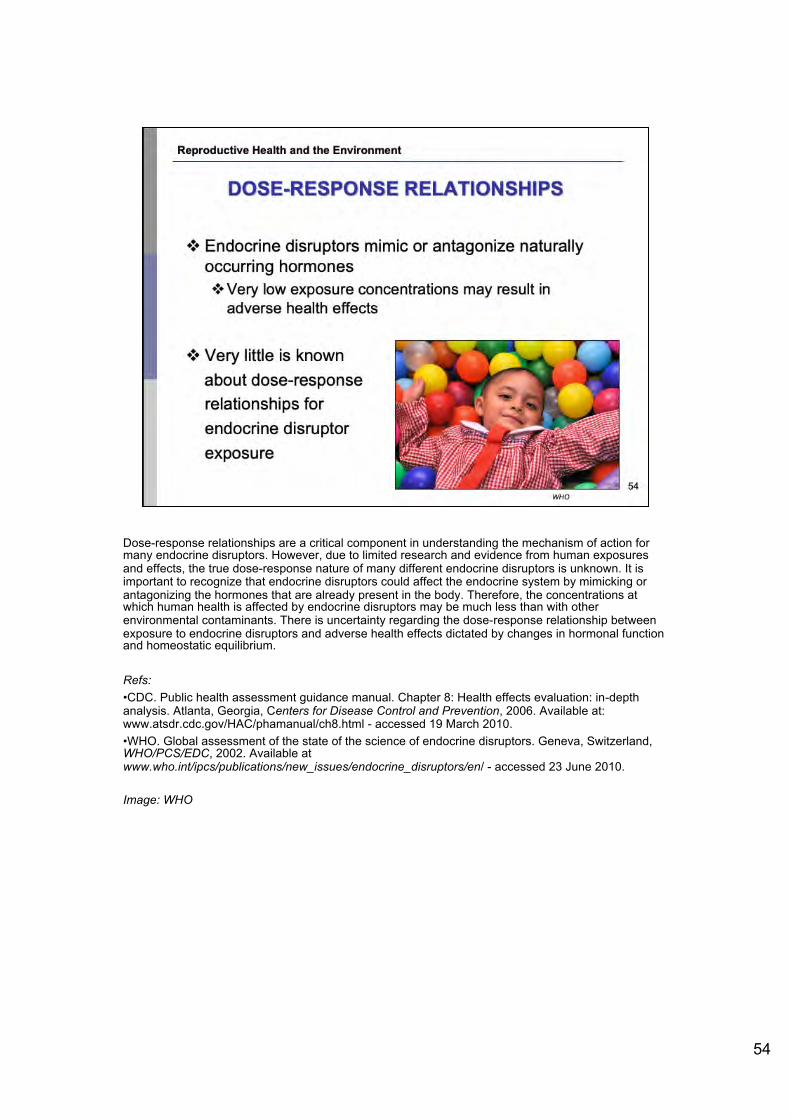
Dose-response relationships are a critical component in understanding the mechanism of action for many endocrine disruptors. However, due to limited research and evidence from human exposures and effects, the true dose-response nature of many different endocrine disruptors is unknown. It is important to recognize that endocrine disruptors could affect the endocrine system by mimicking or antagonizing the hormones that are already present in the body. Therefore, the concentrations at which human health is affected by endocrine disruptors may be much less than with other environmental contaminants. There is uncertainty regarding the dose-response relationship between exposure to endocrine disruptors and adverse health effects dictated by changes in hormonal function and homeostatic equilibrium.
Refs:•CDC. Public health assessment guidance manual. Chapter 8: Health effects evaluation: in-depth analysis. Atlanta, Georgia, Centers for Disease Control and Prevention, 2006. Available at: www.atsdr.cdc.gov/HAC/phamanual/ch8.html - accessed 19 March 2010. •WHO. Global assessment of the state of the science of endocrine disruptors. Geneva, Switzerland, WHO/PCS/EDC, 2002. Available at www.who.int/ipcs/publications/new_issues/endocrine_disruptors/en/ - accessed 23 June 2010.
Image: WHO
54
Reproductive Health and the Environment
DOSE-RESPONSE RELATIONSHIPS
❖ Endocrine disruptors mimic or antagonize naturally occurring hormones ❖ Very low exposure concentrations may result in
adverse health effects
❖ Very little is known
about dose-response
relationships for
endocrine disruptor
exposure
WHO

More than 850 chemicals directly impact the nervous system and may cause adverse health effects. This includes some metals, organic solvents, agrochemicals, poly-halogenated aromatic hydrocarbons, and pharmaceuticals. Some of these environmental contaminants may be endocrine disrupting compounds because the reproductive endocrine system is primarily regulated by the neuroendocrine system. There are two different hypothesized mechanisms of actions of endocrine disruptors on the nervous system. Endocrine disruptors may activate specific properties in adults and produce transient changes in the nervous system, or, exposure to endocrine disruptors during neural development may induce changes in neurobehavioral function, specifically sex-related behaviours. Also, there is direct toxic action on neuronal cells with effects seen via the immune system, hormonal effects (thyroid) and systemic diseases.Specific chemicals may alter neurotransmitter concentrations, thus influencing neuroendocrine function and eventually reproduction. Some studies have indicated that poly-chlorinated biphenyls (PCBs) may act through this mechanism, but greater research in necessary.
<< NOTE TO USER: For further information, please refer to the module on Endocrine Disruption and the module on Neurodevelopment>>
Refs:•Khan IA, Thomas P. Disruption of neuroendocrine control of luteinizing hormone secretion by Aroclor 1254 involves inhibition of hypothalamic tryptophan hydroxylase activity. Biology of Reproduction, 2001, 64:955-964. •Mensah-Nyagan AG et al. Neurosteroids: Expression of steroidogenic enzymes and regulation of steroid biosynthesis in the central nervous system. Pharmacology Review, 1999, 51:63-81.
Reproductive Health and the Environment
ENDOCRINE DISRUPTORS AND NEUROTOXICITY
❖ Hundreds of chemicals and environmental toxicants can affect the nervous system
❖ Mechanism of action ❖ transient changes in adult nervous system
❖ permanent changes induced in neural development
❖ direct tox ic action on neurona l cells, effects via immune system, hormonal effects (thyroid), system ic diseases
❖ Some pesticides and poly-chlorinated biphenyls (PCBs) are neurotoxic
55

•Polybrominated diphenyl ethers (PBDEs) are widely used flame retardant compounds that are persistent and bioaccumulative and therefore have become ubiquitous environment contaminants. A number of toxicologic studies have demonstrated that exposure to PBDEs may have endocrine-disrupting effects. Most of these studies have focused on thyroid hormone disruption and a smaller number on disruption of the estrogen/androgen hormone system. Animal studies suggest that prenatal PBDE exposure may result in adverse neurodevelopmental effects. A study of 100 children prenatally exposed to PBDE demonstrated that the chemical altered the neurodevelopment of children up to 72 months of age.
•At least 10 different phthalates are used commercially as plasticizers and solvents. Human exposure to phthalates can occur through inhalation, ingestion, and dermal contact. Once absorbed, they are rapidly metabolized to monoesters, and the high-molecular-weight monoesters can undergo further oxidation to form oxidative metabolites. Antagonistic effects of phthalates on the thyroid gland in vivo and thyroid tissue in vitro have been reported. Recently, human studies have demonstrated that phthalate exposure in childhood was associated with attention deficit hyperactivity disorder (ADHD). Engel et al. determined that behavioral domains adversely associated with prenatal exposure to phthalates are commonly affected in children clinically diagnosed with conduct or attention deficit hyperactivity disorders. Refs:•Engel SM et al. Prenatal phthalate exposure is associated with childhood behavior and executive functioning.Environ Health Perspect. 2010, 118:565-571. Experimental and observational studies have reported biological consequences of phthalate exposure relevant to neurodevelopment. Engel et al. (p. 565) examined the association of prenatal phthalate exposure with offspring behavior in a multiethnic prenatal population enrolled in the Mount Sinai Children’s Environmental Health Study in New York City between 1998 and 2002. Third-trimester maternal urine samples were collected and analyzed for phthalate metabolites, and the cognitive and behavioral development of the children was assessed between the ages of 4 and 9 years. In multivariate adjusted models, increased loge concentrations of low-molecular-weight (LMW) phthalate metabolites were associated with poorer scores on the Aggression, Conduct Problems, Attention Problems, and Depression, and Externalizing Problems and Behavioral Symptom Index composite scales. Increased loge concentrations of LMW phthalates were also associated with poorer scores on the Global Executive Composite index and the Emotional Control scale. The authors note that behavioral domains adversely associated with prenatal exposure to LMW phthalates in this study are commonly affected in children clinically diagnosed with conduct or attention deficit hyperactivity disorders. •Herbstman JB et al. Prenatal exposure to PBDEs and neurodevelopment. Environ Health Perspect. 2010, 118:712-719. Polybrominated diphenyl ethers (PBDEs) are widely used flame retardant compounds that are persistent and bioaccumulative. Animal studies suggest that prenatal PBDE exposure may result in adverse neurodevelopmental effects. Herbstman et al. (p. 712) initiated a longitudinal cohort following the World Trade Center attack on 11 September 2001 to evaluate associations between concentrations of individual PBDE congeners and neurodevelopmental indices. Outcomes were evaluated in approximately 100 children with PBDE concentrations measured in cord blood samples. After adjustment for potential confounders, higher concentrations of BDEs 47, 99, or 100 were associated with lower scores on tests of mental and physical development at 12–48 and 72
Reproductive Health and the Environment
ENDOCRINE DISRUPTORS AND NEUROTOXICITY: PBDEs AND PHTHALATES
❖ Prenatal exposure to polybrominated diphenyl ethers (PBDEs) may result in neurodevelopmental effects in infants ❖ Thyroid disruption
❖ Estrogen/androgen disruption
❖ Prenatal exposure to phthalates associated with Attention-deficit hyperactivity disorder (ADHD) ❖ Oxidative metabolites formed
❖ Antagonistic effects on the thyroid gland
56

months. The authors conclude that developmental exposure to flame retardants following the World Trade Center disaster was associated with altered neurodevelopment of children up to 72 months of age.
56

The effects of environmental contaminants and potential endocrine disruptors have been studied extensively in wildlife. Specific mechanisms of the endocrine system, as well as hormonal events that regulate reproduction, are quite similar between some wildlife species and humans. Evidence from case studies has shown that wildlife may even be more sensitive to the action of certain environmental contaminants and endocrine disruptors. A specific case study involves a decline in reproductive function of marine seals exposed to increased levels of two organochlorines: PCBs and DDE. Organochlorines are man-made organic compounds commonly used as pesticides. They are extremely persistent in the environment and are known to bioaccumulate in living organisms. It has been demonstrated that the population of Baltic ringed seals has declined significantly in the last century due to reproductive disorders. Specific abnormalities have included partial or complete sterility, interruptions during early pregnancy, and stenosis (abnormal narrowing) or necessary reproductive organs. A time trend study conducted by a team of scientists demonstrated that the seal populations affected by the reproductive disorder contained high levels of PCBs and DDE levels. The results specifically highlight that high levels of PCBs were the leading cause of reproductive malfunction. However, greater research is needed to elucidate the mechanism of action in this case study. DDT: dichlorodiphenyltrichloroethaneDDE: dichlorodiphenyldichloroethylene PCBs: polychlorinated biphenyls
Refs:•Hayes, TB. There is no denying this: defusing the confusion about Atrazine. Bioscience, 2004, 54 (112): 1138–1149•Roos A et al. Time trend studies on DDT and PCB in juvenile grey seals, fish and guillemot eggs from the Baltic Sea. Organochlorine Compounds, 1998, 39:109-112.•Van Der Kraak G et al. Comparative endocrinology and mechanisms of endocrine modulation in fish and wildlife. In: Kendall RJ, Dickerson RL, Giesy JP & Suk WA, eds. Principles and Processes for Evaluating Endocrine Disruption in Wildlife. SETAC Technical Publication. Pensacola, FL: SETAC Press, 1998: 97-119. Image: National Oceanic and Atmospheric Administration.
Reproductive Health and the Environment
3.C. REPRODUCTIVE HEALTH EFFECTS OF ENDOCRINE DISRUPTORS: EXAMPLES FROM
WILDLIFE
❖ Laboratory and field studies have
demonstrated adverse reproductive
health effects in wildlife exposed to
environmental contam inants
National Oceanic and Almospherlc Aomnlstlaliof'I
❖ Some adverse effects may be directly linked to endocrine disrupt ion, but the causa l pathway is largely inconclus ive
❖ Case studies to date have only involved high contam ination exposures
57

A rather famous case study for environmental exposures and effects in wildlife has been the loss of top level predatory birds due to egg shell thinning. Due to their specific feeding behaviour, carnivorous birds are exposed to a higher level of bio-accumulative organic compounds. Numerous studies have observed abnormal reproductive morphologies, deformities, and alterations in behaviour for bird colonies exposed to organic compound contaminants, specifically DDE, the breakdown product of the pesticide, DDT. DDE directly effects the shell gland and results in egg shell breaking or cracking, and thus severe decreases in avian populations. The widespread use of DDT as a insecticide in North America nearly resulted in the extinction of several bird species. However, it is widely understood that birds may be more susceptible to environmental contaminants due to their modes of reproduction, specifically, the vulnerability of egg shells to specific toxicants. However, research still remains inconclusive and greater research is needed in this area to understand the mechanisms of action of these environmental contaminants.
DDT: dichlorodiphenyltrichloroethaneDDE: dichlorodiphenyldichloroethylene
Refs: •Elliott JE, Norstrom RJ, Keith JA. Organochlorines and eggshell thinning in northern gannets (Sula bassanus) from eastern Canada. Environmental Pollution. 1998, 52:81-102.•Giesy JP, Ludwig JP & Tillitt DE. Dioxins, dibenzofurans, PCBs and colonial, fish-eating water birds. In: Schecter A, ed. Dioxin and Health. New York: Plenum Press, 1994: 254-307.
Image: WHO
58
Reproductive Health and the Environment
CASE STUDY: EGG SHELL THINNING
❖ Predatory birds exposed to organochlorine compounds experience abnorma l reproductive morphologies
❖ DDT and breakdown product, ODE
❖ Direct impact on egg shell strength
❖ Near extinction of many North
American bird species WHO
DDT: dichlorodiphenyltrichloroethane 58 ODE: dlchlorodlphenyldlchloroethylene

In 1980, a chemical spill in the state of Florida in the United States contaminated a stream with very high concentrations of dicofol, including its metabolites DDE and chloro-DDT. Dicofol is an organochlorine pesticide, similar in chemical nature and environmental effects to DDT. After the spill, the alligator population in the region decreased by 90% and adult alligators in the region were found to have high levels of dicofol and its by-products in their bodies. Alligators in the affected region demonstrated numerous reproductive dysfunctions, such as abnormal gonadal morphology and gonadal deformities. Specifically, male alligators in the contaminated area had significantly decreased phallic size and alterations in sperm cells. A proposed hypothesis is that the by-product of dicofol, DDE, may interact with the estrogen receptor and thus affect proper reproductive development in the juvenile alligator. However, it must be stated that this specific case study demands greater research to document a dose response relationship between dicofol exposure and the reproductive health effects witnessed. Large carnivorous reptiles may bioaccumulate greater levels of environmental contaminants due to their feeding habits as well as their long life expectancies in the wild. Studies have shown that in fact, certain reptiles can bioaccumulate and biomagnify environmental contaminants to an equal or even greater scale than birds.The diagram demonstrates male alligator phallic size in relation to the contaminated sites. The Woodruff lake region (far left bar) represents the control lake, while Apopka-GNS (far right bar) shows average phallic index in alligators at the contaminated site, and Apopka-NW (middle bar) shows the average phallic index in alligators in a contaminated area further away from the contaminated site.
DDT: dichlorodiphenyltrichloroethaneDDE: dichlorodiphenyldichloroethylene
Refs:•Cobb GP, Wood PD. PCB concentrations in eggs and chorioallantoic membranes of loggerhead sea turtles (Caretta caretta) from the Cape Romain National Wildlife Refuge. Chemosphere, 1997, 34(3):539-549.•Crain DA et al. Sex-steroid and thyroid hormone concentrations in juvenile alligators (Alligator mississippiensis) from contaminated and reference lakes in Florida, USA. Environmental Toxicology and Chemistry, 1998, 17:446-452.
Image: WHO. Global assessment of the state of the science of endocrine disruptors. Geneva, Switzerland, WHO/PCS/EDC, 2002. Available at www.who.int/ipcs/publications/new_issues/endocrine_disruptors/en/ -accessed 23 June 2010.
59
Reproductive Health and the Environment
CASESTUDY:DEVELOPMENTAL ABNORMALITIES IN ALLIGATORS
❖ Dicofol, organochlorine (OC) pesticide released into stream
❖ 90% decrease in alligator species
❖ High levels of OC compounds in alligators associated with significant decrease in male penis size
❖ Dicofol may interact with
estrogen receptor during
juvenile development
1
0.8 ~ ] 0.6
~ 0.4 !.
0.2
I,
-
0 _ _.___,..__-,--.....,__.__-,-- ......... _---,
Woodruff Apopka -NW Apopka-GNS
WHO'IPCSIEDC, 2002

As increasingly more women enter the workforce, they may be exposed to a variety of occupational chemicals and hazards that may lead to adverse health and reproductive effects. In addition, smoking, alcohol consumption, and other lifestyle factors play an increasingly important role in determining the health status of women. There is now abundant evidence that environmental factors may contribute to many of the disease processes discussed above. Some examples of likely environmental impact on women's health include the following: Among the most widespread and persistent environmental toxicants are chlorinated hydrocarbons (such as dichlorodiphenyltrichloroethane (DDT) and polychlorinated biphenyls), which are known to possess estrogenic potential, i.e., the ability to mimic the biological effects of estrogens. Imbalanced or unopposed estrogen exposure is a leading risk factor for many gynecologic malignancies, as well as benign proliferative disorders such as endometriosis and leiomyoma. The potential impact of these compounds on hormone-dependent physiological processes such as conception and fetal development, as well as on disease processes such as osteoporosis and cardiovascular disease, demands further exploration.
DDT: dichlorodiphenyl trichloroethane
Refs: •Anger DL, Foster WG. The link between environmental toxicant exposure and endometriosis. Frontiers in Bioscience, 2008, 13:1578-1593. •Best Start. Workplace reproductive health: research and strategies. Best Start. Ontario's Maternal Newborn and Early Child Development Resource Centre, 2001.•Diamanti-Kandarakis, et al. Endocrine-disrupting chemicals: an Endocrine Society scientific statement. The Endocrine Society. 2009
Reproductive Health and the Environment
3.D REPRODUCTIVE HEAL TH EFFECTS OF ENDOCRINE DISRUPTORS: EXAMPLES OF EFFECTS ON HUMAN
HEALTH
Widespread and persistent, chlorinated hydrocarbons -may mimic the biological effects of estrogens.
Excessive estrogen exposure is a key risk factor for gynaecologic malignancies and benign proliferative disorders - e.g. endometriosis and leiomyoma.
Also on hormone-dependent physiological processes
- e.g. conception; fetal development
- Possibly on osteoporosis and cardiovascular disease
60
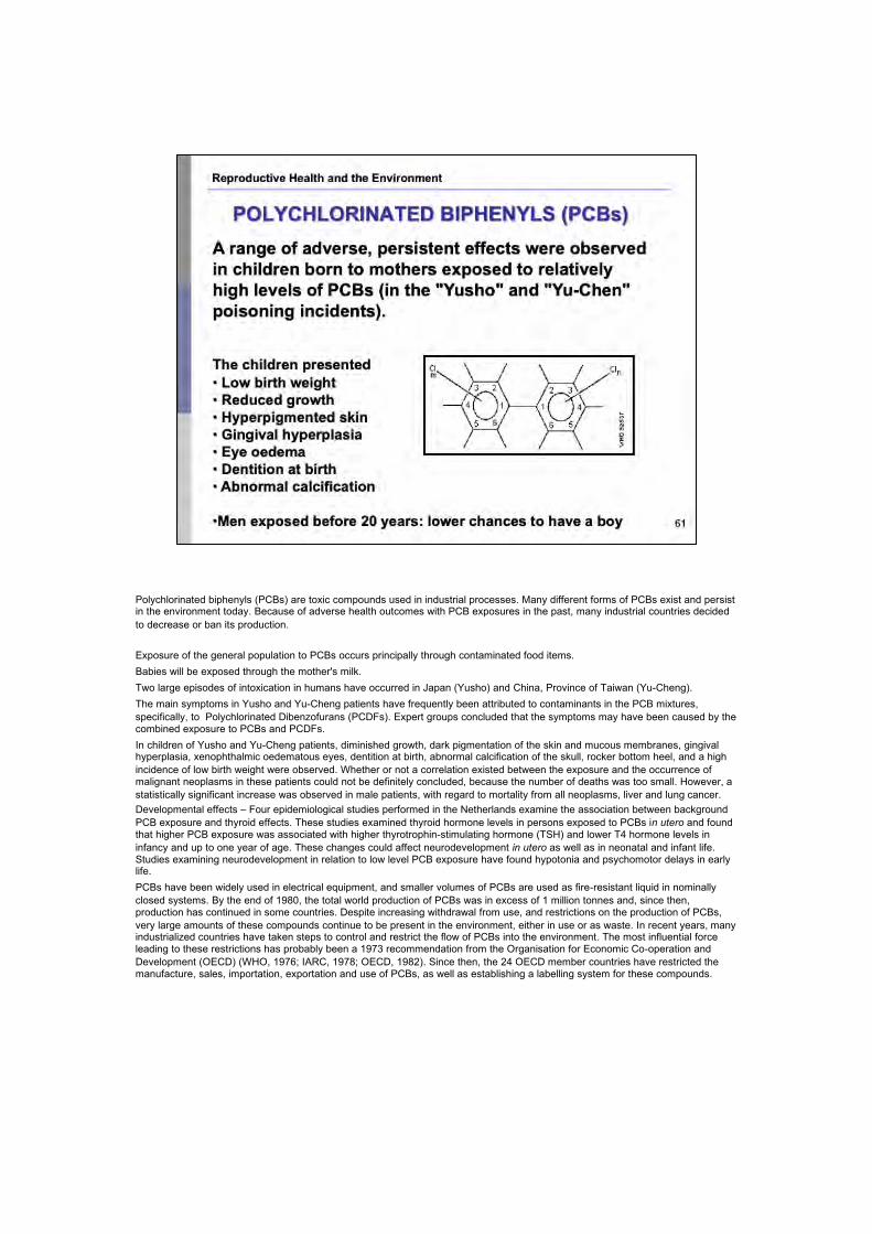
Polychlorinated biphenyls (PCBs) are toxic compounds used in industrial processes. Many different forms of PCBs exist and persist in the environment today. Because of adverse health outcomes with PCB exposures in the past, many industrial countries decided to decrease or ban its production.
Exposure of the general population to PCBs occurs principally through contaminated food items. Babies will be exposed through the mother's milk.Two large episodes of intoxication in humans have occurred in Japan (Yusho) and China, Province of Taiwan (Yu-Cheng).The main symptoms in Yusho and Yu-Cheng patients have frequently been attributed to contaminants in the PCB mixtures, specifically, to Polychlorinated Dibenzofurans (PCDFs). Expert groups concluded that the symptoms may have been caused by the combined exposure to PCBs and PCDFs. In children of Yusho and Yu-Cheng patients, diminished growth, dark pigmentation of the skin and mucous membranes, gingival hyperplasia, xenophthalmic oedematous eyes, dentition at birth, abnormal calcification of the skull, rocker bottom heel, and a high incidence of low birth weight were observed. Whether or not a correlation existed between the exposure and the occurrence of malignant neoplasms in these patients could not be definitely concluded, because the number of deaths was too small. However, a statistically significant increase was observed in male patients, with regard to mortality from all neoplasms, liver and lung cancer. Developmental effects – Four epidemiological studies performed in the Netherlands examine the association between background PCB exposure and thyroid effects. These studies examined thyroid hormone levels in persons exposed to PCBs in utero and found that higher PCB exposure was associated with higher thyrotrophin-stimulating hormone (TSH) and lower T4 hormone levels in infancy and up to one year of age. These changes could affect neurodevelopment in utero as well as in neonatal and infant life. Studies examining neurodevelopment in relation to low level PCB exposure have found hypotonia and psychomotor delays in earlylife. PCBs have been widely used in electrical equipment, and smaller volumes of PCBs are used as fire-resistant liquid in nominally closed systems. By the end of 1980, the total world production of PCBs was in excess of 1 million tonnes and, since then, production has continued in some countries. Despite increasing withdrawal from use, and restrictions on the production of PCBs, very large amounts of these compounds continue to be present in the environment, either in use or as waste. In recent years, many industrialized countries have taken steps to control and restrict the flow of PCBs into the environment. The most influential force leading to these restrictions has probably been a 1973 recommendation from the Organisation for Economic Co-operation and Development (OECD) (WHO, 1976; IARC, 1978; OECD, 1982). Since then, the 24 OECD member countries have restricted the manufacture, sales, importation, exportation and use of PCBs, as well as establishing a labelling system for these compounds.
Reproductive Health and the Environment
POL YCHLORINATED BIPHENYLS (PCBs)
A range of adverse, persistent effects were observed in children born to mothers exposed to relatively high levels of PCBs (in the "Yusho" and "Yu-Chen" poisoning incidents).
The children presented • Low birth weight • Reduced growth • Hyperpigmented skin • Gingival hyperplasia • Eye oedema • Dentition at birth • Abnormal calcification
•Men exposed before 20 years: lower chances to have a boy s1

Current sources of PCB release include volatilization from landfills containing transformer, capacitor, and other PCB-containing wastes, sewage sludge, spills, and dredge spoils, and improper (or illegal) disposal in open areas. Pollution may occur during the incineration of industrial and municipal waste. Most municipal incinerators are not effective in destroying PCBs. Explosions or overheating of transformers and capacitors may release significant amounts of PCBs into the local environment. Refs:•Brouwer A et al. Characterization of potential endocrine-related health effects at low-dose levels of exposure to PCBs. Environmental Health Perspectives, 1999, 107:639-49.•Landrigan P, Garg A, Droller D. Assessing the effects of endocrine disruptors in the National Chidren's Study. Environmental Health Perspectives. Environmental Health Perspectives, 2003, 111(13): 1678.•WHO. Persistent organic pollutants: impact on child health. WHO, 2010. Available at www.who.int/ceh/publications/persistent_organic_pollutant/en/index.html - accessed March 2011.
<<NOTE TO USER: See module on persistent organic pollutants for more information.>>
61

In Seveso Italy, 2,3,7,8 tetrachlorodibenzo-p- dioxin (TCDD) was released when a chemical factor exploded. In the years that followed, males were born at half the rate of females in families where the father was exposed to the chemical. This study has not been reproduced in animals or humans.
<<READ SLIDE>>
Refs:•Eskenazi B et al. Seveso Women's Health Study: a study of the effects of 2,3,7,8-tetrachlorodibenzo-p-dioxin on reproductive health. Chemosphere, 2000, 40(9-11):1247-53.•Landrigan P, Garg A, Droller D. Assessing the effects of endocrine disruptors in the National Chidren's Study. Environmental Health Perspectives, 2003, 111(13): 1678.•WHO. Persistent organic pollutants: impact on child health. WHO, 2010. Available at www.who.int/ceh/publications/persistent_organic_pollutant/en/index.html - accessed March 2011.
<<NOTE TO USER: See module on persistent organic pollutants for more information.>>
Reproductive Health and the Environment
Seveso, Italy -10 July 1976, chemical explosion that releases the highest levels of 2,3,7,8-tetrachlorodibenzo-p-dioxin (TCDD) exposure experienced by a human population.
Seveso Women's Health Study:1996 retrospective cohort study of females
- Age of menopause : increase risk of early menopause - Breast cancer : associated with individual high TCDD levels - Menstrual cycles: more regular - Endometriosis: doubled , non-significant risk - Leiomyoma : ... . seems to have an anti-estrogenic effect on
myometrium - Ovarian function : no evidence - Menarche: no evidence
• Confusing picture: in some tissues TCDD may be estrogen like (breast) and in others it may be blocking estrogen (uterus).
• Timing of exposure seems to be important • Excess of female births in a region after toxic chemical spill of TCDQ

DES taught us three important lessons that can guide our investigations on endocrine disrupting chemicals in the future: 1. Exposure to endocrine disruptors during early (fetal) development can induce disorders of the endocrine system in the fetus, whilst the mother may appear healthy2. The risk of health impacts from exposure to hormone disruptors is especially high during early development.3. An endocrine disease or disorder induced during early development might only be apparent decades later, and exposure to this one chemical could lead to multiple health risks. Please note pharmaceuticals are prescribed and taken on higher doses and often regular basis, so are usually high level exposures.<<READ SLIDE>>Ref:•Diamanti-Kandarakis E. et al. Endocrine-disrupting chemicals: an Endocrine Society scientific statement. 2009. Endocrine reviews. 30(4): 293-342.•Hoover RN et al. Adverse Health Outcomes in Women Exposed In Utero to Diethylstilbestrol. N Engl J Med. 2011; 365:1304-1314.•NIEHS. Diethylstilbestrol (DES) as a model for environmental estrogens. Environews, NIEHS news. 1993. Available at www.ncbi.nlm.nih.gov/pmc/articles/PMC1519726/pdf/envhper00372-0021b-color.pdf – accessed 15 June 2011.DES was administered to pregnant women during the 1940s through 1960s, originally for high-risk pregnancies but later to promote "healthier babies" as well. Subsequently, the drug was linked to the development of an otherwise extremely rare malignancy, clear-cell carcinoma of the vagina, in young female offspring exposed in utero. In addition, a number of more common non-neoplastic changes in the reproductive tract of DES-exposed daughters were identified, including vaginal adenosis, cervical ectropion, and numerous other structural abnormalities. Although the public health hazards associated with further exposure to DES have been largely eliminated, there are a number of compelling reasons for the continued study of DES-exposed women, as well as for basic research on the biological effects of DES and other environmental estrogenic compounds. First, it is unclear whether the human cancer incidence resulting from DES exposure has peaked. Although the majority of DES daughters have passed the age range for vaginal carcinoma development, few have reached the age range (postmeno-pausal) in which endometrial carcinoma typically occurs in the DES unexposed population, and endometrial carcinoma occurs with a much higher prevalence than vaginal carcinoma in DES-treated mice. Similarly, the threat of breast cancer is still a concern in this population. The identification of molecular genetic markers for DES carcinogenicity is therefore a continuing priority; such markers would also be of value in predicting risk for third-generation DES offspring, for whom little is known about potential health risks. Second, DES may be viewed as a model compound for other environmental agents with estrogenic potential. The bioaccumulation of these environmental estrogens is recognized as a problem of increasing magnitude, and certain human populations in the United States have been shown to carry amounts of these fat-soluble compounds which, in fish and other wildlife, cause significant endocrine dysfunction and developmental anomalies of the reproductive tract. Insights into the biological effects of DES should therefore provide a foundation upon which future environmental health problems may be effectively addressed. NIEHS has a long history of accomplishments in conducting and supporting research on estrogen action, hormonal carcinogenesis, and other types of estrogen-related pathology, particularly for DES and similar compounds. More recent achievements have provided insights into basic mechanisms of estrogen receptor action at the molecular level. A transgenic mouse that overexpresses the estrogen receptor is being developed to study tissue susceptibility and mechanisms for hormonal carcinogenesis. New endeavors include the analysis of human and animal tumors resulting from DES exposure in utero for molecular genetic alterations. Rapid advances in the fields of molecular and developmental biology have provided numerous insights into relevant genes and molecular pathways involved in reproductive tract development. Epidemiologic studies are fo-cused on a broad range of health effects among DES-exposed men and women. Expanded research efforts are necessary to use this knowledge in exploring the epigenetic effects of DES in relation to reproductive tract malformations at the molecular level. In addition to the estrogen receptor, research on the role of "orphan receptors" in environmental disease is promising. Identification and characterization of orphan receptors and their endogenous ligands will provide a link to understanding the molecular mechanisms through which exogenous chemicals may exert toxic effects and through which natural substances influence physiologic processes. For example, a recently discovered member of the nuclear receptor family apparently recognizes a class of foreign chemicals called peroxisome proliferators, which includes industrial plasticizers, herbicides, and hypolipidemic agents. Similarly, a receptor from another gene family exists for the ubiquitous xenobiotic dioxin, or TCDD. A related example is the retinoids, which regulate differentiation and growth of a variety of epithelial tissues including mammary gland, cervical, vaginal, and uterine epithelium. Ongoing research at NIEHS is directed toward understanding the process of squamous differentiation in gynecologic epithelial tissues by retinoids and estrogens, and interactions between the retinoic acid receptor and estrogen receptor signaling pathways. Further research is necessary to define these pathways at the molecular level and to elucidate possible therapeutic applications of retin-oids in breast and other cancers. An additional complexity is that age and the timing of exposures to environmental agents can have a profound effect on individual susceptibility. For example, the well-known adverse effects of the antimiscarriage drug diethylstilbestrol (DES) and subsequent development of vaginal cancer in the daughters who were exposed during in utero development and the perinatal DES-exposed experimental animal model point to critical stages of susceptibility. In fact, the hypothesis that exposure to environmental agents early in life is of greater health significance than adult exposure is currently being investigated by NIEHS researchers studying the effects of endocrine-disrupting chemicals during development and the importance of timing of exposure in cancer risk. Yet another aspect of timing that complicates environmental health research is that the appearance of disease often occurs much later than the causative exposure. This was the case with DES, and this delay makes identifying the contributing factor(s) difficult but challenging, requiring both laboratory- and human population-based studies.
Reproductive Health and the Environment
DIETHYLSTILBESTROL
Another type of cancer: Diethylstilbestrol (DES) as a model for environmental estrogens
• DES administered to pregnant women 1940-1960 for high-risk pregnancies but later to promote "healthier babies" as well.
• Female offspring developed clear-cell carcinoma of the vagina, vaginal adenosis, cervical ectropion, and other abnormalities.
• Males: reproductive tract abnormalities.
Has human cancer incidence resulting from DES exposure peaked? DES daughters are reaching post-menopause, the age of endometrial carcinoma ...
• DES may be a model compound for other environmental agents with estrogenic potential. 63

Studies performed in North Carolina (US) and Northern Mexico showed a statistically significant association between DDE levels in breast milk, maternal serum, and cord blood and duration of lactation. It was thought that DDE acts through an estrogen receptor to oppose prolactin activity (for normal milk production, estrogen falls and prolactin rises) and interfere with milk synthesis.In a US study, it was found that the higher the DDE exposure prenatally, the taller and heavier boys were at 14 years of age.There was no effect on when pubertal milestones were reached.CB-153 and DDE in semen of 149 Swedish fishermen from the eastern Baltic coast had a high proportion of Y-chromosome bearing semen. Also high levels of persistent organic pollutants in blood.Higher prevalence of chryptorchidism in Lithuania. Environmental factors may be changing the ratio of sperm carrying the X or Y (sex determining) chromosomes and may be contributing to male reproductive disordersExposure to p,p´-DDT early in life may increase breast cancer risk. Many U.S. women heavily exposed to DDT in childhood have not yet reached 50 years of age. The public health significance of DDT exposure in early life may be large. 1973: Accidental contamination of Michigan food chain by a fire retardant containing PBBs. 4000 people ingested contaminated meat and milk. Maternal PBB exposure and/or exposure through breastfeeding seemed to cause earlier onset of puberty in their daughters Farm families. Gestation and breast milk.
DDT: dichlorodiphenyl trichloroethane DDE: dichlorodiphenyldichloroethylene PBBs: polybrominated biphenyls
Refs:•Cohn BA. DDT and breast cancer in young women: New data on the significance of age at exposure. Environmental Health Perspectives, 2007, 115(10):1406-14.•Landrigan P, Garg A, Droller D. Assessing the effects of endocrine disruptors in the National Chidren's Study. Environmental Health Perspectives, 2003, 111(13): 1678.•Rogan WJ and Ragan NB. Evidence of effects of environmental chemicals on the endocrine system in children. Pediatrics, 2003, 112; 247-252.•WHO. Persistent organic pollutants: impact on child health. WHO, 2010. Available at www.who.int/ceh/publications/persistent_organic_pollutant/en/index.html - accessed March 2011.
Reproductive Health and the Environment
DDE, DDT & PBBs
• Dichlorodiphenyl dichloroethylene (DDE) exposure through breast milk may alter duration of lactation by acting through the estrogen receptor
• DDE exposures prenatally may have potential effects on adolescent growth and sexual maturation
• Dichlorodiphenyl trichloroethane (DDT) might be connected to breast cancer
• Polybrominated biphenyls (PBBs) and accelerated onset of puberty
64

Certain environmental contaminants, specifically endocrine disruptors, may lead to adverse reproductive health effects in humans. A majority of the reproductive health effects of endocrine disruptors in humans have been witnessed through high exposure scenarios (for instance, the Seveso incident). For this reason, the research concerning low dose exposures is much less conclusive. However, low level exposures may potentially pose the greatest health threat for chronically exposed populations. Even small shifts in reproductive patterns may result in tremendous impacts for the population at large. Data from human epidemiologic studies and case studies remain predominantly from North America and Canada. Exposures from the developing world are much less documented, though could potentially be more significant and impact vulnerable populations. Specific examples of reproductive health endpoints resulting from exposures to endocrine disruptors will be explained in modules 2, 3, 4, and 5.
Refs: •Calafat AM, Needham LL. Human exposures and body burdens of endocrine-disrupting chemicals. In: Gore AC, ed. Endocrine-disrupting chemicals: from basic research to clinical practice. Totowa, NJ: Humana Press, 2007, 253–268. •Dickerson SM, Gore AC. Estrogenic environmental endocrine-disrupting chemical effects on reproductive neuroendocrine function and dysfunction across the life cycle. Rev Endocr Metab Disord, 2007, 8:143–159 15. •WHO. Global assessment of the state of the science of endocrine disruptors. Geneva, Switzerland, WHO/PCS/EDC, 2002. Available at www.who.int/ipcs/publications/new_issues/endocrine_disruptors/en/ - accessed 23 June 2010.
Image: WHO
65
Reproductive Health and the Environment
REPRODUCTIVE HEAL TH EFFECTS OF ENDOCRINE DISRUPTORS: HUMANS
❖ High exposure case studies provide greatest evidence for adverse human effects (e.g. polychlorinated biphenyls , diethylstilbestrol)
❖ Inconclusive data related to low dose ,
chronic exposures ❖ Though may be most relevant!
❖ Lack of data from developing nations
WHO
65

The current state of the knowledge regarding endocrine disruptors and their mechanism of action demands greater research. While a few high level exposure studies have found an association between exposures to potentially endocrine disrupting chemicals and adverse reproductive health effects, the exact mechanism of action for many of these compounds of poorly understood
Ref:•WHO. Global assessment of the state of the science of endocrine disruptors. Geneva, Switzerland, WHO/PCS/EDC, 2002. Available at www.who.int/ipcs/publications/new_issues/endocrine_disruptors/en/ - accessed 23 June 2010.
Image: WHO
66
Reproductive Health and the Environment
LIMITATIONS OF KNOWLEDGE ON ENDOCRINE DISRUPTORS
❖ Studies examining reproductive effects of low levels of endocrine disruptors in humans are inconclusive
However , high level exposures
provide evidence of human
susceptibility to endocrine disruptor
effects
❖ Research needs : ❖ Effects of low level exposures
❖ Effects of early-life exposures
to adult reproduct ive health
❖ Dose-response relationships
❖ Exposure during life-cycle and for future generations WHO
66

<<READ SLIDE>>
Images:•WHO•Fatherhood and Health Outcomes in Europe. Geneva, Switzerland, World Health Organization, 2002. Available at www.euro.who.int/document/e91129.pdf - accessed 10 June 2010
Reproductive Health and the Environment
TOWARDS THE FUTURE The importance of research on reproductive health and
environment
• Consideration and study of environmental factors in reproductive diseases key for:
- developing new prevention strategies
- decreasing disease prevalence and severity
• Increased understanding of the mechanisms through which environmental agents disrupt organs/systems will help improve prevention and treatment strategies.
• Benefits from research will translate into better health for men and women , and yield findings that will benefit everybody
WHO

<<NOTE TO USER: Add points for discussion according to the needs of your audience.>>
68
Reproductive Health and the Environment
POINTS FOR DISCUSSION
68

69
Reproductive Health and the Environment
ACKNOWLEDGEMENTS
WHO and its partners are grateful to the US EPA Office of Children's Health Protection and the UK Department of Health for the financial support that made this
project possible.
First draft prepared by a working group at Johns Hopkins Bloomberg School of Public Health (Halshka Graczyk, MPH, with the lead of Dr. Lynn Goldman) and WHO (Marie-Noel Brune, MSc)
Expert reviewers: Dr. Hell Bathija (WHO); Prof. MS. Riana Bornman (South Africa); Dr. Bremen De Mucio (CLAP SMR); Dr. Pablo Duran (CLAP/SMR); Dr. Ricardo Fesclna (CLAP/SMR); Prof. Dr. Jean Golding ~UK); Prof. Dr. Eun-Hee Ha (Rep. ofl<orea); Dr. Woong Ju (Rep. of Korea ; Prof. Dr. Young Ju Kim (Rep. of Korea); Prof. Dr. Merci Kusel (Aus ralia); Prof. Dr. Amalia Laborde (Uruguay); Dr. Lizbeth L6pez Carrillo (Mexico); Dr. Hanns Moshammer (ISDE); Dr. Joanne Perron (US); Dr. Suzanne Serruya (CLAP/SMR); Prof. Dr. Peter Sit (Australia); Prof. Dr. Shanna Swan (US); Prof. Dr. Oriol Vall Combelles (Spain) ; Dr. Sheryl Vanderpoel (WHO).
Additional collaborators: Dr. Brenda Eskenazi (US), Dr. Jenny Pronczuk (WHO), Anne Sweeney (US), Anna Pollack (US)
WHO Training Project Coordination:
Last update: November 2011
Dr. Ruth Etzel (WHO) Marie-Noel Brune, MSc (WHO)
69

70
Reproductive Health and the Environment
Disclaimer
Disclaimer C World Health Organization 2011. Some rights reserved . This wort is available under the Creative Commons Attribution -NonCommerc ial-ShareAlike 3.0 IGO licence (CC BY-NC-SA 3.0 IGO; htt ps :l(creativ ecommo ns.orgQicenses(b y--nc-sa/3 .0Jigo t Und er the term s of theCC BY-NC-SA 3.0 IGOlicence, you may ropy , redistr ibute and adapt t he work for non.commer cial purposes, provided the wor k is appropriately cited, as indicated below. In any use of this work, there should be no suggestion that WHO endorses any specific organization. products or services. The use of the WHO logo is not permitted . If you ada pt the work, the n you must license your work under the same or equivalent (re.alive Commons licence. ff you ere.ate a translatio n of t his work , you should add the following disclaimer along with the suggested citation: "This t ranslation was not create d by the World Health Organizat ion (WHO). WHO is not responsible for the content or accuracy of this translation. The original English edition shall be the binding and authentic edition". Any mediat ion re lating to disputes arising under the licence shall be conducted in accordance with the med iation rules of the World Intellectual Property Organization. Third-party materials. If you wish to reuse mater ial from this work that is attributed to a third party, such as tables, fq ures or images, it is your respons ibility to determine whethe r permission is needed for that reuse and to obta in permission fTom the copyri&ht holder . The risk of claims resulting from infrin&ement of any third-party-owned component in the wort: rests solety with the user . This t raining module was developed by the Wortd Health Organization (WHO) and is intended to be used for training purposes only. The opinions, conclusions, and statistics expressed in the training module do not necessarily represent the official position of the Worid Health Organization. The contents of this tra ining module are based upon references available in the published literature as of the last stated up date date. Users are encouraged to search sta ndard medical databases for update s in the science for issues of particular interest or sensitivity in the ir region sand areas of specific concem . All reasonab le precautions have been taken by WHO to verify the information contained in this tra ining module. However, the training module is being distributed without warranty of any kind, either expressed or implied. The responsibility for the interpretation and use of the e-leaming tra ining lies with the reader . In no event shall WHO be liable for damages arising from its use. The des ignations employed and the presentatio n of the material in this tra ining module do not imply the expression of any opinion whatsoever on the part of the World Health Organization concerning the Jegal status of any count ry, territory , city or area or of its author ities, or concerning the delimitat ion of its frontiers or boundaries. Dotted lines on maps represent approximate borde r lines for which the re may not yet be full agreement . The mention of specific companies or of c-ertain manufacturers ' products does not Imply that they are endorsed or recommended by the World Health Organization in prefe rence to others of a similar nature that are not mentioned. Errors and omissions excepted, the names of proprietary products are distinguished by initial capital letters . The authors alone are responsible for the views expressed in this tra ining package and they do not necessa rily represe nt the decisions, policy or views of the World Health Organization.
70
WHO/HSE/PHE/EPE/11.01.10 - © World Health Organization 2011. All rights reserved.
This e-learning training was developed by the World Health Organization (WHO}. It is Intended to be used as a self-learning course on Children's Health and
the Environment. All reasonable precautions have been taken by WHO to verify the information contained in this e-4earning training. However, the
e-learning training is being distributed without warranty of any kind, either expressed or implied. The responsibility for the interpretation and use of the
e-learning training lies with the reader. In no event shall WHO be liable for damages arising from its use. The mention of specific companies or of certain
manufacturers' products does not impty that they are endorsed or recommended by the Workl Health Organization in preference to others of a similar
nature that are not mentioned. Errors and omissions excepted, the names of proprietary products are distinguished by initial capital letters.
Disclaimer
Reproductive Health and the Environment


![for “Male reproductive health and the environment” Special Issue final.pdf · 2016. 8. 26. · [1] Invited Review for “Male reproductive health and the environment” Special](https://static.fdocuments.in/doc/165x107/5fd7079cad639c2e876f609c/for-aoemale-reproductive-health-and-the-environmenta-special-finalpdf-2016.jpg)