Introducing the Endocrine System Copyright © 2005 Pearson Education, Inc. publishing as Benjamin...
61
Introducing the Endocrine System
-
Upload
conrad-haynes -
Category
Documents
-
view
215 -
download
0
Transcript of Introducing the Endocrine System Copyright © 2005 Pearson Education, Inc. publishing as Benjamin...
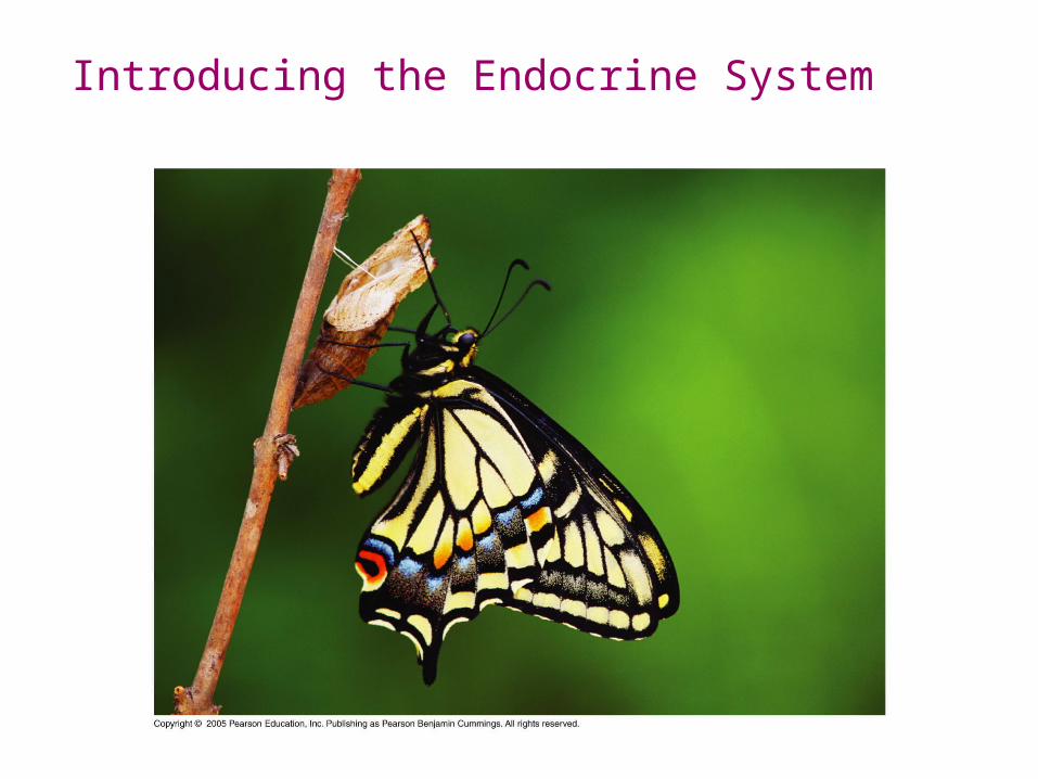
Introducing the Endocrine System
Copyright © 2005 Pearson Education, Inc. publishing as Benjamin Cummings
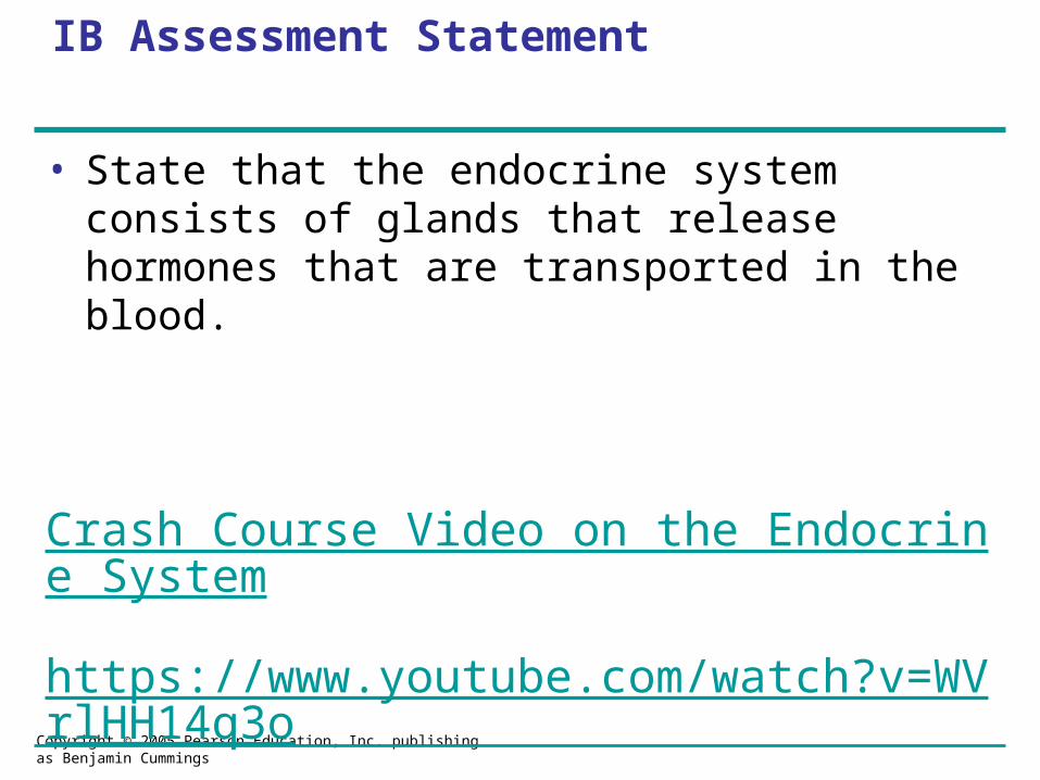
IB Assessment Statement
• State that the endocrine system consists of glands that release hormones that are transported in the blood.
Crash Course Video on the Endocrine System
https://www.youtube.com/watch?v=WVrlHH14q3o
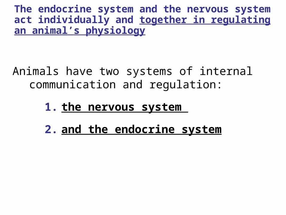
The endocrine system and the nervous system act individually and together in regulating an animal’s physiology
Animals have two systems of internal communication and regulation:
1. the nervous system
2. and the endocrine system
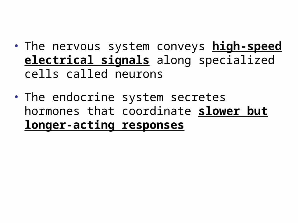
• The nervous system conveys high-speed electrical signals along specialized cells called neurons
• The endocrine system secretes hormones that coordinate slower but longer-acting responses
Overlap Between Endocrine and Nervous Regulation
• The endocrine and nervous systems function together in maintaining homeostasis, development, and reproduction
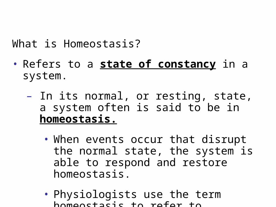
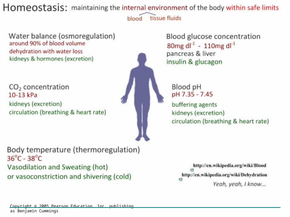
What is Homeostasis?
• Refers to a state of constancy in a system.
– In its normal, or resting, state, a system often is said to be in homeostasis.
• When events occur that disrupt the normal state, the system is able to respond and restore homeostasis.
• Physiologists use the term homeostasis to refer to maintenance of a constant internal environment despite fluctuations in external conditions.
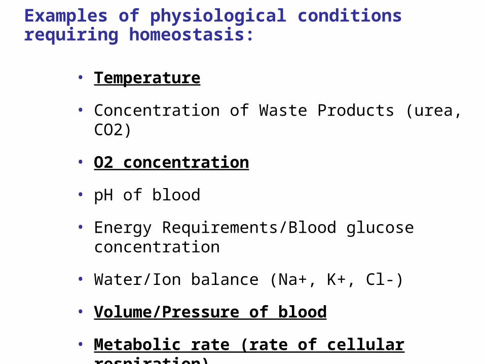
Examples of physiological conditions requiring homeostasis:
• Temperature
• Concentration of Waste Products (urea, CO2)
• O2 concentration
• pH of blood
• Energy Requirements/Blood glucose concentration
• Water/Ion balance (Na+, K+, Cl-)
• Volume/Pressure of blood
• Metabolic rate (rate of cellular respiration)

Homeostasis – is important for our internal environment
• Internal environment consists of:
– Blood
– Tissue fluid
Copyright © 2005 Pearson Education, Inc. publishing as Benjamin Cummings

Copyright © 2005 Pearson Education, Inc. publishing as Benjamin Cummings
IB Learning Objective
• Define Homeostasis and explain how it is regulative by a negative feedback system.
Copyright © 2005 Pearson Education, Inc. publishing as Benjamin Cummings
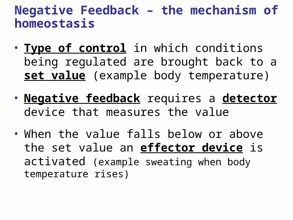
Negative Feedback – the mechanism of homeostasis
• Type of control in which conditions being regulated are brought back to a set value (example body temperature)
• Negative feedback requires a detector device that measures the value
• When the value falls below or above the set value an effector device is activated (example sweating when body temperature rises)

Homeostatic Mechanism
Negative-Feedback Regulation
– An external stimuli or activity alters a condition of the internal environment, which triggers a response by the body. This response reverses the altered condition.
70 72 74 76 78 80 82 84 86 88 90 92
Effectors – in Negative Feedback
– Effectors are of 2 types: Muscles & Glands
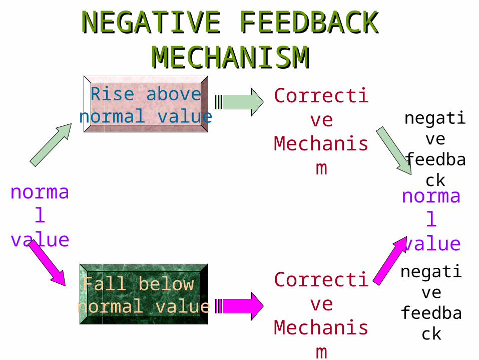
Rise abovenormal value
Fall below normal value
normal value
Corrective Mechanis
m
Corrective Mechanis
m
NEGATIVE FEEDBACK NEGATIVE FEEDBACK MECHANISMMECHANISM
normal
value
negative
feedback
negative
feedback
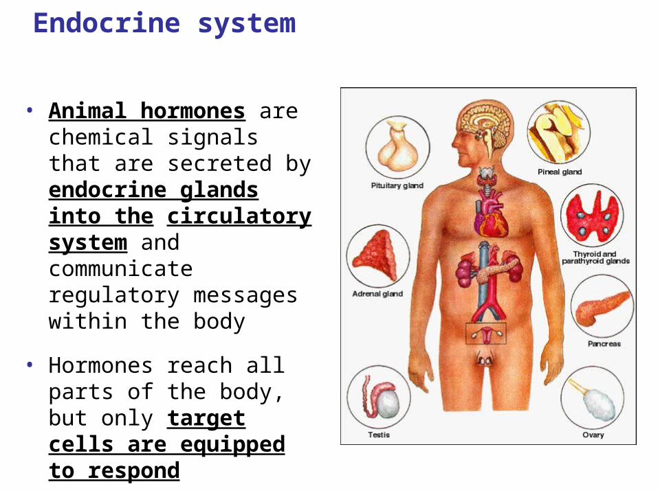
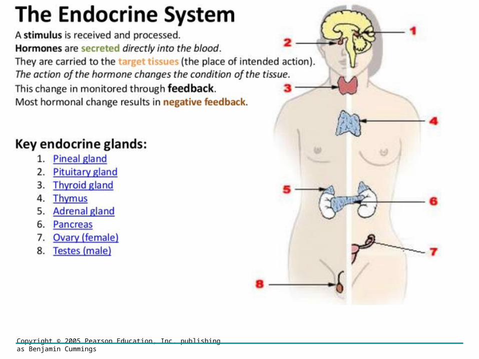
Endocrine system
• Animal hormones are chemical signals that are secreted by endocrine glands into the circulatory system and communicate regulatory messages within the body
• Hormones reach all parts of the body, but only target cells are equipped to respond
Copyright © 2005 Pearson Education, Inc. publishing as Benjamin Cummings
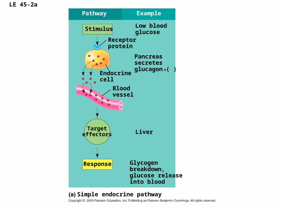
LE 45-2a
Targeteffectors
Response
Simple endocrine pathway
Glycogenbreakdown,glucose releaseinto blood
Liver
Bloodvessel
Pancreassecretesglucagon ( )
Endocrinecell
Low bloodglucose
Receptorprotein
Stimulus
Pathway Example

LE 45-2b
Targeteffectors
Response
Simple neurohormone pathway
Stimulus
Pathway Example
Suckling
Milk release
Smooth musclein breast
Neurosecretorycell
Bloodvessel
Posterior pituitarysecretes oxytocin( )
Hypothalamus/posterior pituitary
Sensoryneuron
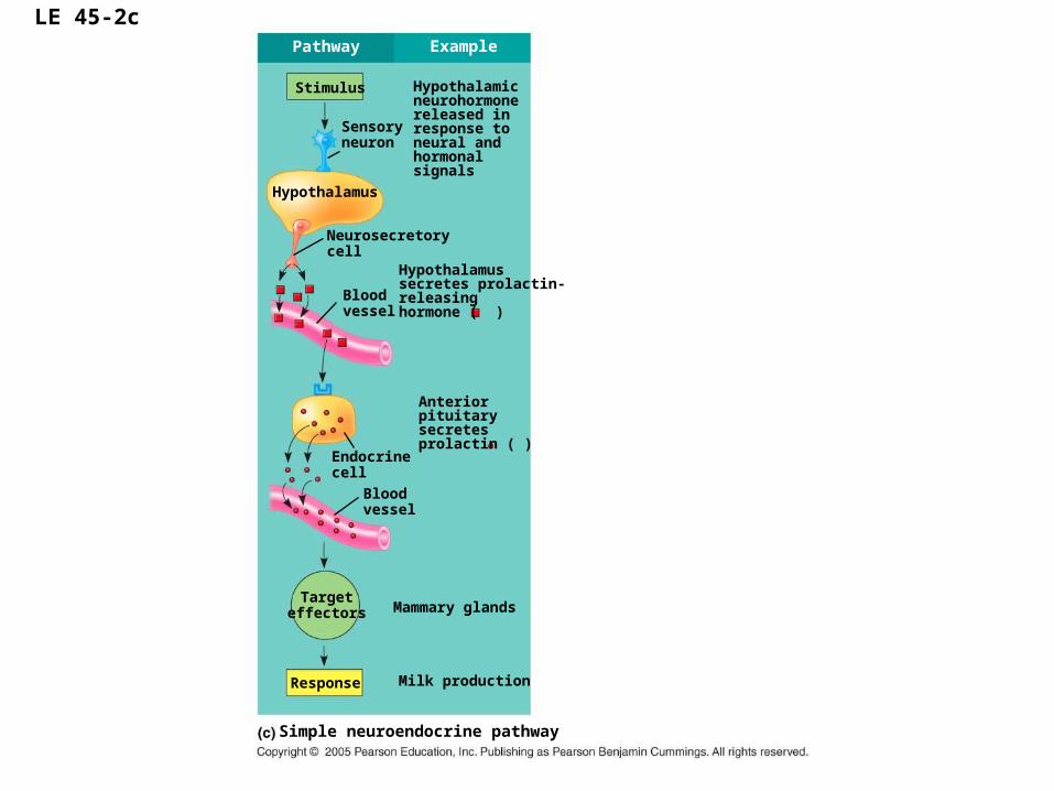
LE 45-2c
Targeteffectors
Response
Simple neuroendocrine pathway
Stimulus
Pathway Example
Milk production
Bloodvessel
Hypothalamus
Sensoryneuron
Mammary glands
Endocrinecell
Bloodvessel
Anteriorpituitarysecretesprolactin ( )
Hypothalamussecretes prolactin-releasinghormone ( )
Neurosecretorycell
Hypothalamicneurohormonereleased inresponse to neural andhormonalsignals
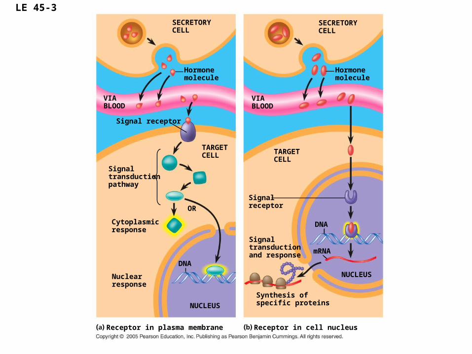
LE 45-3
SECRETORYCELL
Hormonemolecule
Signal receptor
VIABLOOD
VIABLOOD
TARGETCELL TARGET
CELLSignaltransductionpathway
OR
Cytoplasmicresponse
DNA
NUCLEUS
Nuclearresponse
Receptor in plasma membrane Receptor in cell nucleus
DNA
NUCLEUS
mRNA
Synthesis ofspecific proteins
Signaltransductionand response
Signalreceptor
Hormonemolecule
SECRETORYCELL
Copyright © 2005 Pearson Education, Inc. publishing as Benjamin Cummings
IB Assessment Statement
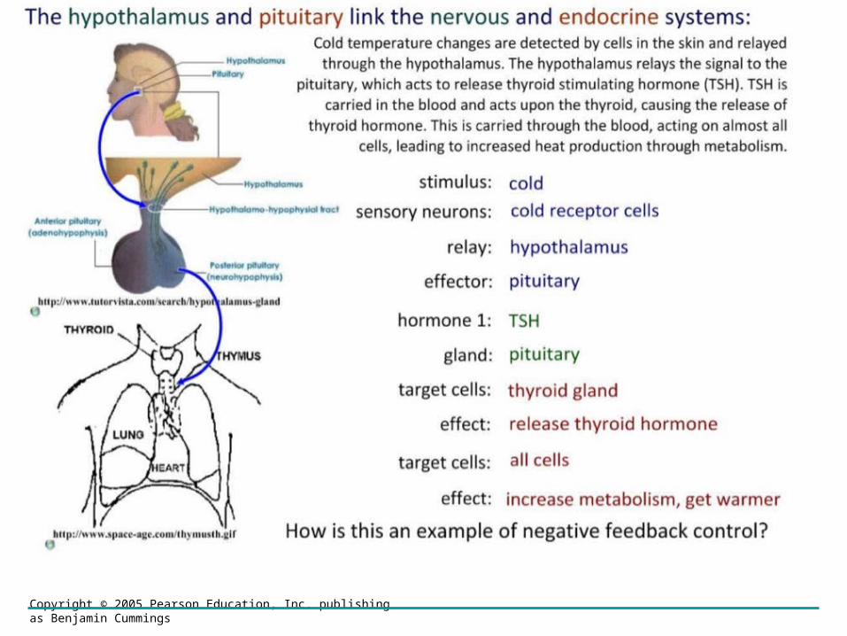
• Explain the control of body temperature, including the transfer of heat in blood, and the roles of the hypothalamus, sweat glands, skin arterioles and shivering.
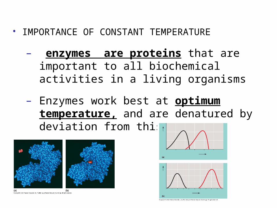
• IMPORTANCE OF CONSTANT TEMPERATURE
– enzymes are proteins that are important to all biochemical activities in a living organisms
– Enzymes work best at optimum temperature, and are denatured by deviation from this.
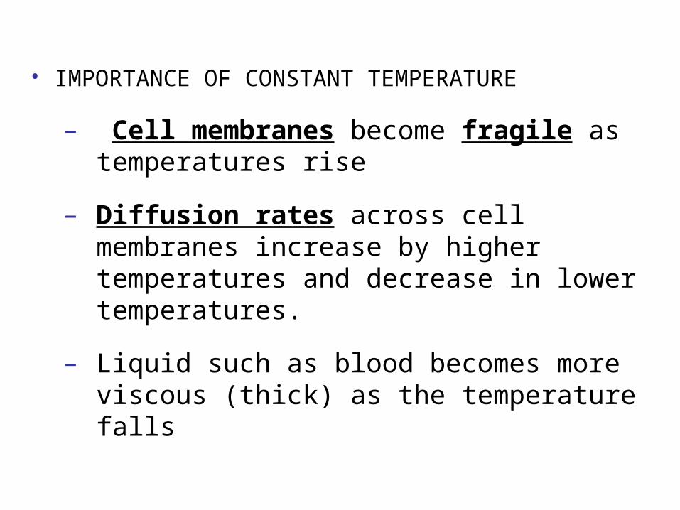
• IMPORTANCE OF CONSTANT TEMPERATURE
– Cell membranes become fragile as temperatures rise
– Diffusion rates across cell membranes increase by higher temperatures and decrease in lower temperatures.
– Liquid such as blood becomes more viscous (thick) as the temperature falls
Mechanism to Maintain Body temperature
• Maintaining constant temperature,
• Generation of Heat:
– Biochemical reactions, such as respiration generate heat.
– Movement generates heat by friction within muscles

Copyright © 2005 Pearson Education, Inc. publishing as Benjamin Cummings
Mechanism to Maintain Body temperature
• Maintaining constant temperature,
• Losing heat:
– Evaporation of water, from the lungs and sweat from the skin.
– Excretion: Urine and Faeces heat is lost as theses substances are removed from the body.
Copyright © 2005 Pearson Education, Inc. publishing as Benjamin Cummings
Mechanism to Maintain Body temperature
• Insulation:
– Fat stores under the skin prevent heat loss.
Copyright © 2005 Pearson Education, Inc. publishing as Benjamin Cummings
Mechanism to Maintain Body temperature
• The hypothalamus as the co-ordinator of temperature regulation:
• Vasoconstriction: is a cold adaptation narrowing of arterioles that reduces blood flow to the surface of the skin is coupled with a dilation of the horizontal shunt vessels. This prevents heat loss from blood near the skin surface and retains heat in the body core for essential organs.
• Vasodilation: is an adaptation to warm conditions in which arterioles dilate sending more blood closer to the skin surface from where heat can be radiated to the surrounding environment. The horizontal shunt vessels are constricted sending most blood closer to the skin surface. Additionally sweat (mainly water) is released onto the surface of the skin where it enters the vapour phase when warmed by the heat carried by blood. Therefore the vapour of sweat carried away heat energy from blood.
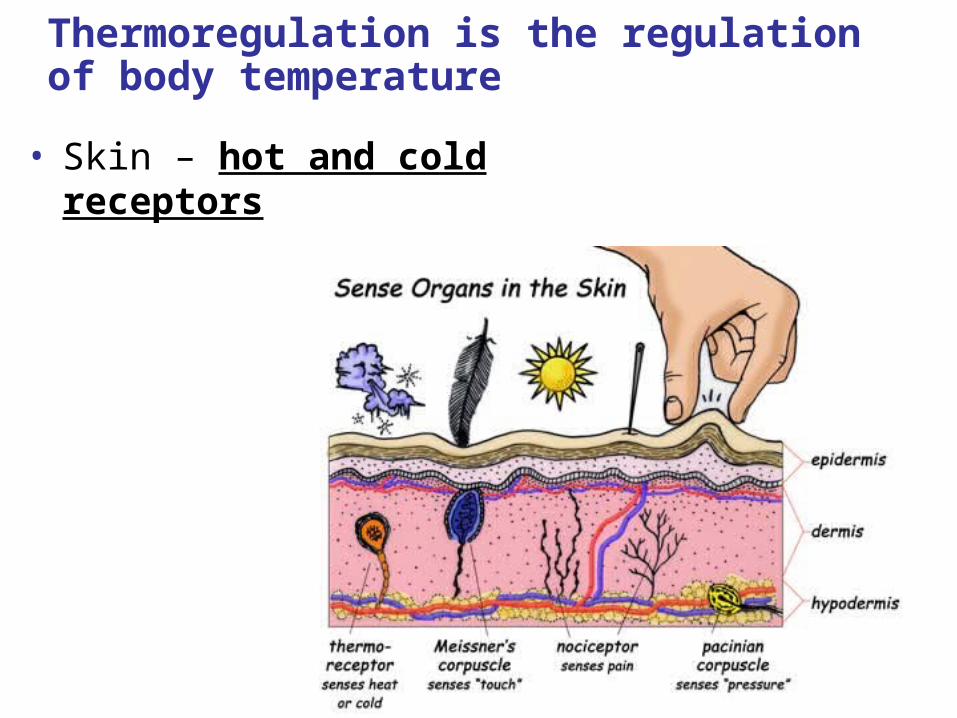
Thermoregulation is the regulation of body temperature
• Skin – hot and cold receptors
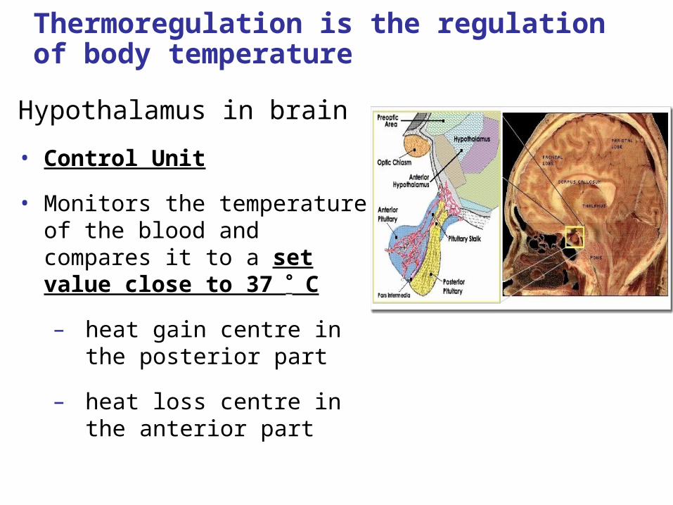
Thermoregulation is the regulation of body temperature
Hypothalamus in brain
• Control Unit
• Monitors the temperature of the blood and compares it to a set value close to 37 C
– heat gain centre in the posterior part
– heat loss centre in the anterior part
Thermoregulation – Response to overheating
Skin
– Heat exchange between body and surrounding occur through our skin
– Sweat glands produce sweat. Water evaporates off the skin and this has a cooling effect.
– Capillaries will dilate (vasodilation). More blood will flow through the skin. The blood transfers heat from the core to the skin. This heat will be lost to environment
– Hair erector muscles which relax. Allows more air flow on skin, which promotes cooling.
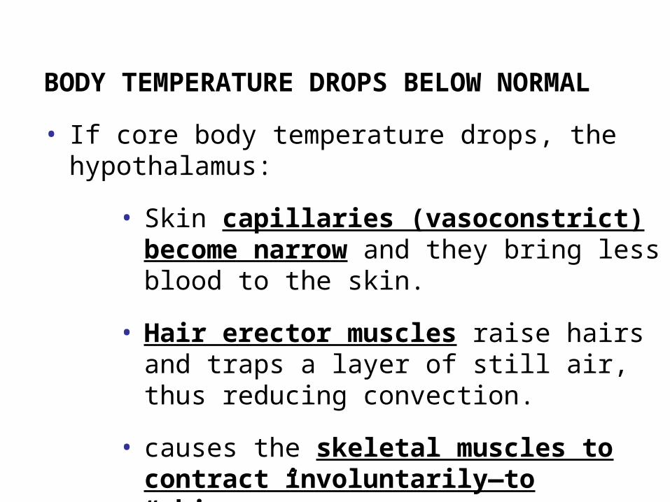
BODY TEMPERATURE DROPS BELOW NORMAL
• If core body temperature drops, the hypothalamus:
• Skin capillaries (vasoconstrict) become narrow and they bring less blood to the skin.
• Hair erector muscles raise hairs and traps a layer of still air, thus reducing convection.
• causes the skeletal muscles to contract involuntarily—to “shiver.”
– This causes the body temperature to increase.
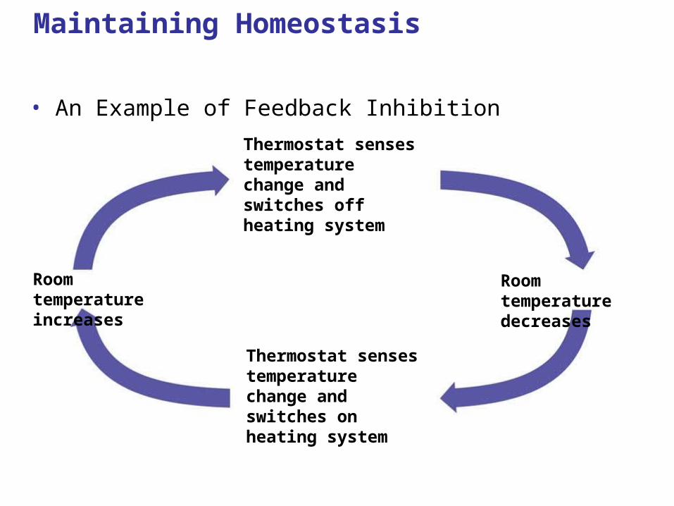
Maintaining Homeostasis
• An Example of Feedback Inhibition
Thermostat senses temperature change and switches off heating system
Thermostat senses temperature change and switches on heating system
Room temperature increases
Room temperature decreases
Copyright © 2005 Pearson Education, Inc. publishing as Benjamin Cummings
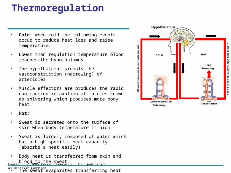
Thermoregulation
• Cold: when cold the following events occur to reduce heat loss and raise temperature.
• Lower than regulation temperature blood reaches the hypothalamus.
• The hypothalamus signals the vasoconstriction (narrowing) of arterioles
• Muscle effectors are produces the rapid contraction relaxation of muscles known as shivering which produces more body heat.
• Hot:
• Sweat is secreted onto the surface of skin when body temperature is high
• Sweat is largely composed of water which has a high specific heat capacity (absorbs a heat easily)
• Body heat is transferred from skin and blood to the sweat
• The sweat evaporates transferring heat away and in doing so cools the body

Copyright © 2005 Pearson Education, Inc. publishing as Benjamin Cummings

Copyright © 2005 Pearson Education, Inc. publishing as Benjamin Cummings
Copyright © 2005 Pearson Education, Inc. publishing as Benjamin Cummings
Copyright © 2005 Pearson Education, Inc. publishing as Benjamin Cummings
Videos/ Tutorials on Thermoregulation of Body Temperature
• http://bcs.whfreeman.com/thelifewire/content/chp41/4101s.swf
• http://www.think-bank.com/iwb/flash/homeostasis.swf
• http://www.phys.unsw.edu.au/biosnippets/

Copyright © 2005 Pearson Education, Inc. publishing as Benjamin Cummings
IB Assessment Statement
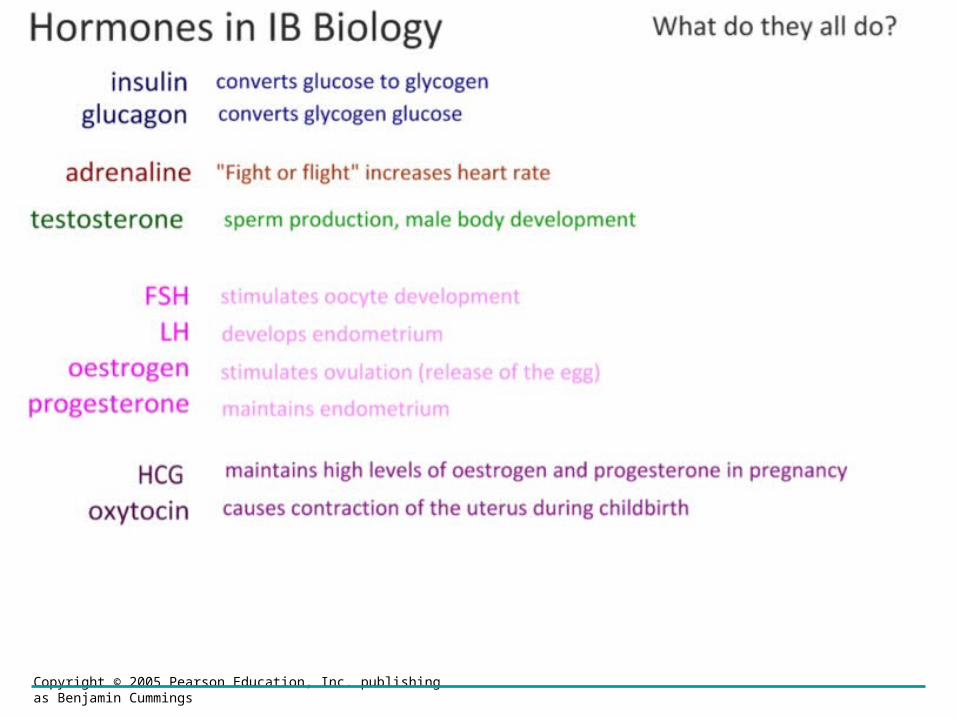
• Explain the control of blood glucose concentration, including the roles of glucagon, insulin and α and β cells in the pancreatic islets.
Copyright © 2005 Pearson Education, Inc. publishing as Benjamin Cummings
Why blood sugar is regulated
Blood sugar concentration is regulated for a number of reason amongst which:
•Osmosis. content of a tissue is determined by the concentration of the surrounding tissues.
•Respiration: Some tissues are entirely dependent on blood sugar as a respiratory substrate being unable to either store glucose of metabolise fat.
Insulin and Glucagon: Control of Blood Glucose
• The pancreas secretes insulin and glucagon, antagonistic hormones that help maintain glucose homeostasis
• Glucagon is produced by alpha cells
• Insulin is produced by beta cells

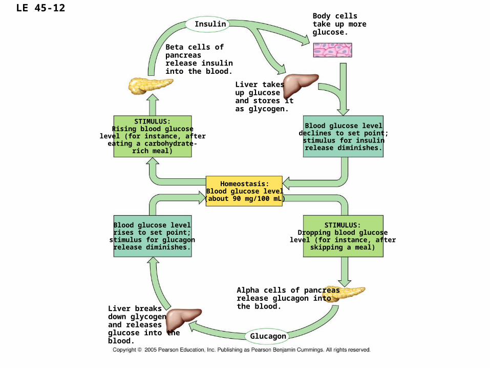
LE 45-12
Beta cells ofpancreasrelease insulininto the blood.
Insulin
Liver takesup glucoseand stores itas glycogen.
STIMULUS:Rising blood glucose
level (for instance, aftereating a carbohydrate-
rich meal)
Blood glucose leveldeclines to set point;stimulus for insulinrelease diminishes.
Homeostasis:Blood glucose level
(about 90 mg/100 mL)
STIMULUS:Dropping blood glucoselevel (for instance, after
skipping a meal)
Blood glucose levelrises to set point;
stimulus for glucagonrelease diminishes.
Liver breaksdown glycogenand releasesglucose into theblood.
Body cellstake up moreglucose.
Alpha cells of pancreasrelease glucagon into the blood.
Glucagon
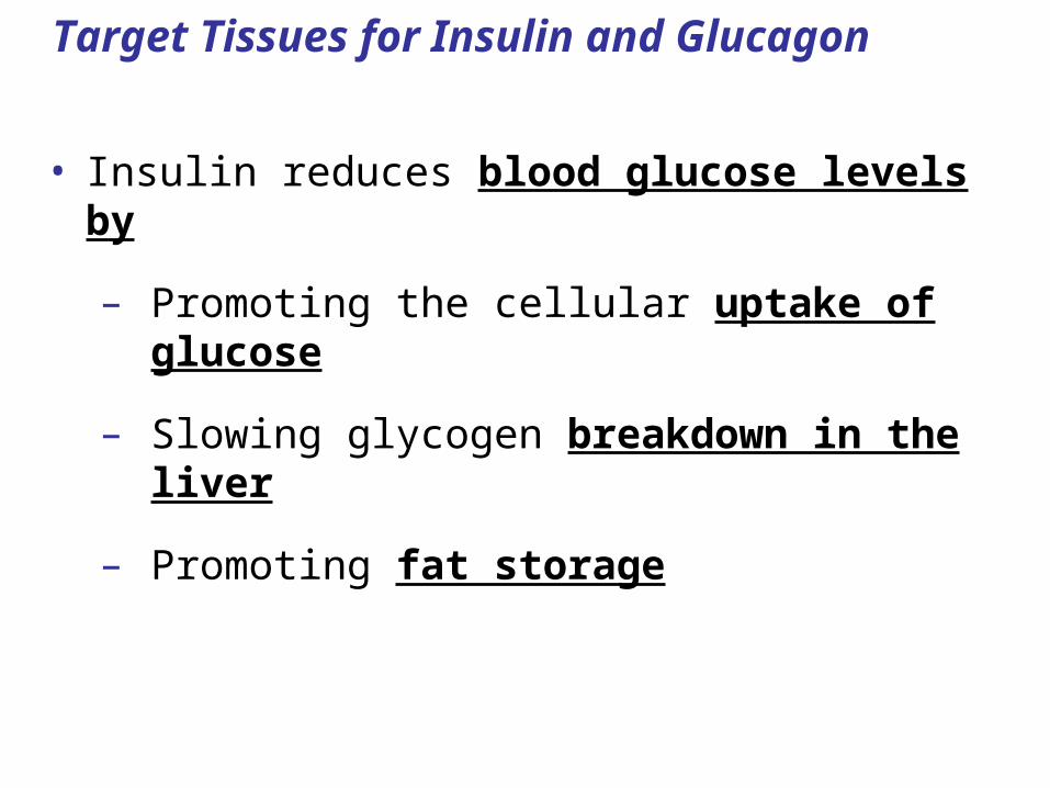
Target Tissues for Insulin and Glucagon
• Insulin reduces blood glucose levels by
– Promoting the cellular uptake of glucose
– Slowing glycogen breakdown in the liver
– Promoting fat storage

• Glucagon increases blood glucose levels by
– Stimulating conversion of glycogen to glucose in the liver
– Stimulating breakdown of fat and protein into glucose

Copyright © 2005 Pearson Education, Inc. publishing as Benjamin Cummings
Copyright © 2005 Pearson Education, Inc. publishing as Benjamin Cummings
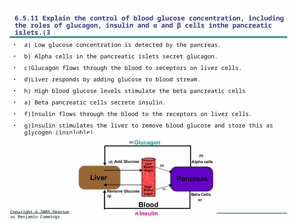
6.5.11 Explain the control of blood glucose concentration, including the roles of glucagon, insulin and α and β cells inthe pancreatic islets.(3
• a) Low glucose concentration is detected by the pancreas.
• b) Alpha cells in the pancreatic islets secret glucagon.
• c)Glucagon flows through the blood to receptors on liver cells.
• d)Liver responds by adding glucose to blood stream.
• h) High blood glucose levels stimulate the beta pancreatic cells
• a) Beta pancreatic cells secrete insulin.
• f)Insulin flows through the blood to the receptors on liver cells.
• g)Insulin stimulates the liver to remove blood glucose and store this as glycogen (insoluble)
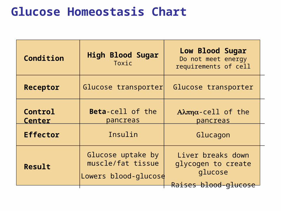
Glucose Homeostasis Chart
Liver breaks down glycogen to create
glucose
Raises blood-glucose
Glucose uptake by muscle/fat tissue
Lowers blood-glucoseResult
GlucagonInsulinEffector
-cell of the pancreas
Beta-cell of the pancreasControl Center
Glucose transporterGlucose transporterReceptor
Low Blood SugarDo not meet energy requirements of cell
High Blood SugarToxic
Condition

Liver converts glycogen
to glucose
normal normal blood blood
glucose glucose levellevel
Blood glucose Blood glucose level fallslevel falls
SooSoon n
after after a a
mealmeal
Long Long after after
a a mealmeal
Blood Blood glucose glucose
level riseslevel rises
normal normal blood blood
glucose glucose levellevel
Too Too HigHighh
Too Too LowLow
Pancreas Pancreas secretes secretes insulininsulin
Pancreas Pancreas secretes secretes
less insulinless insulin
Liver coverts glucose
to glycogen

Copyright © 2005 Pearson Education, Inc. publishing as Benjamin Cummings

Copyright © 2005 Pearson Education, Inc. publishing as Benjamin Cummings
Copyright © 2005 Pearson Education, Inc. publishing as Benjamin Cummings
IB Assessment Statement
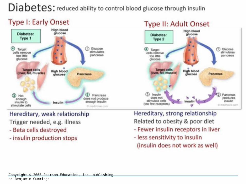
• Distinguish between type I and type II diabetes.
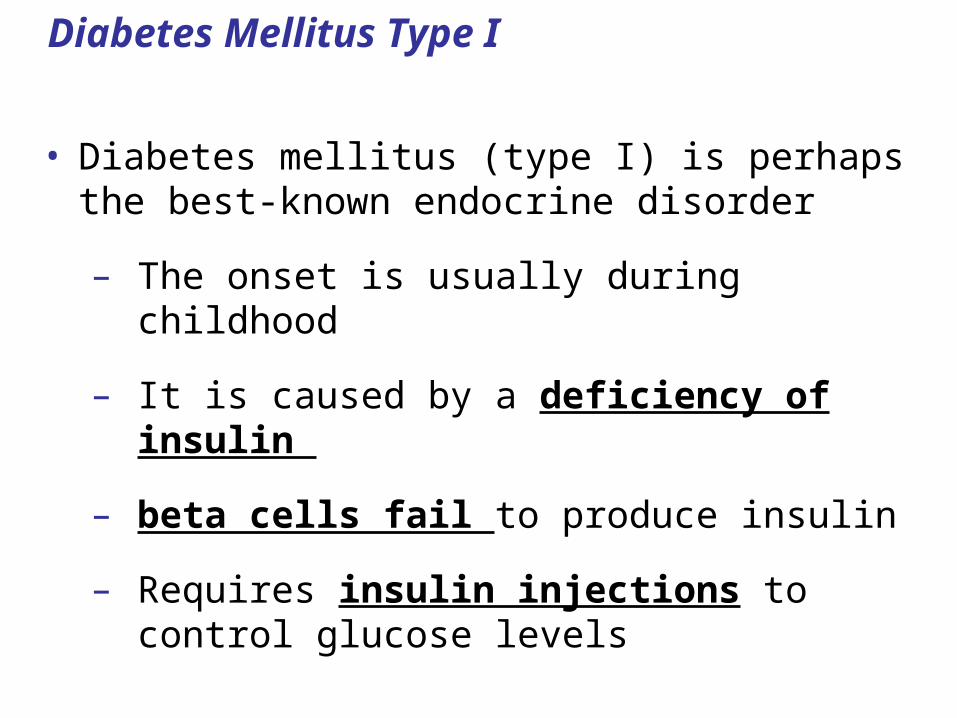
Diabetes Mellitus Type I
• Diabetes mellitus (type I) is perhaps the best-known endocrine disorder
– The onset is usually during childhood
– It is caused by a deficiency of insulin
– beta cells fail to produce insulin
– Requires insulin injections to control glucose levels
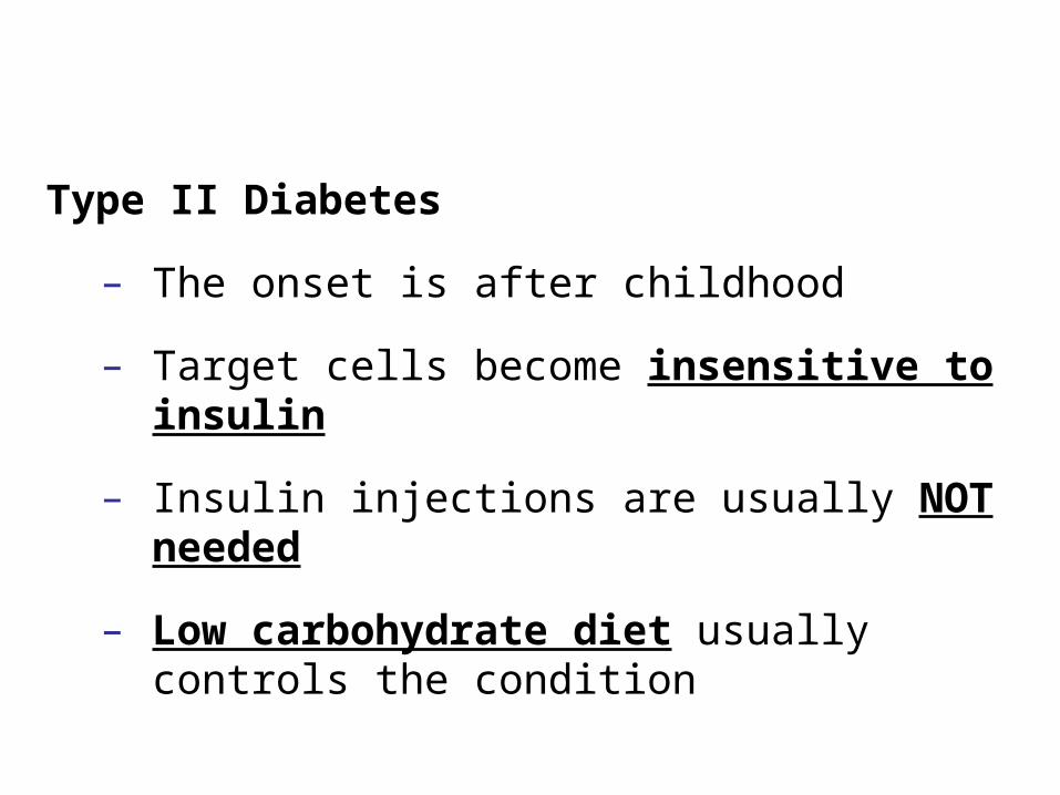
Type II Diabetes
– The onset is after childhood
– Target cells become insensitive to insulin
– Insulin injections are usually NOT needed
– Low carbohydrate diet usually controls the condition
Copyright © 2005 Pearson Education, Inc. publishing as Benjamin Cummings
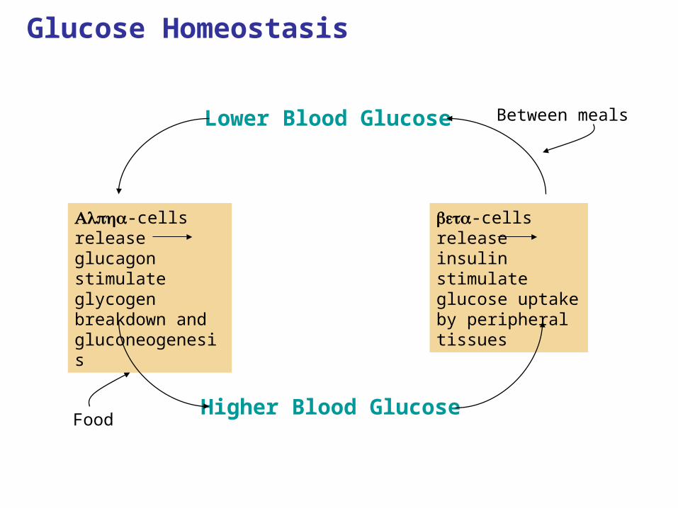
-cells release glucagon stimulate glycogen breakdown and gluconeogenesis
-cells release insulin stimulate glucose uptake by peripheral tissues
Glucose Homeostasis
Lower Blood Glucose
Higher Blood GlucoseFood
Between meals
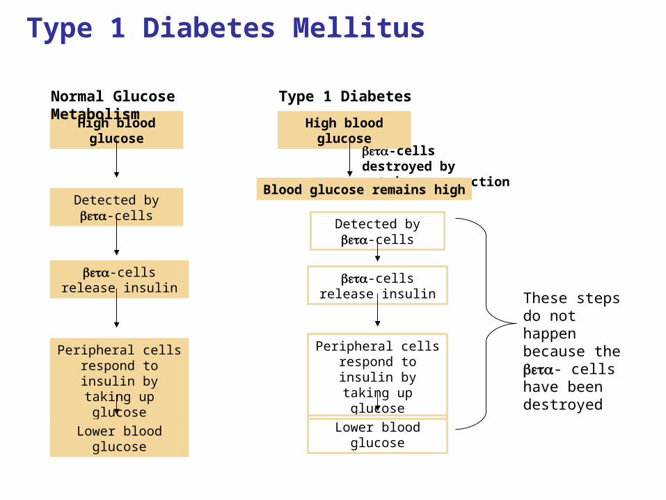
Type 1 Diabetes Mellitus
High blood glucose
Detected by -cells
-cells release insulin
Peripheral cells respond to insulin
by taking up glucose
Lower blood glucose
Normal Glucose Metabolism Type 1 Diabetes
High blood glucose
Detected by -cells
-cells release insulin
Peripheral cells respond to insulin
by taking up glucose
Lower blood glucose
-cells destroyed by autoimmune reaction
These steps do not happen because the - cells have been destroyed
Blood glucose remains high
Copyright © 2005 Pearson Education, Inc. publishing as Benjamin Cummings
Blood sugar regulation Animations/ Tutorials
• http://highered.mcgraw-hill.com/sites/0072495855/student_view0/chapter20/animation__blood_sugar_regulation_in_diabetics.html
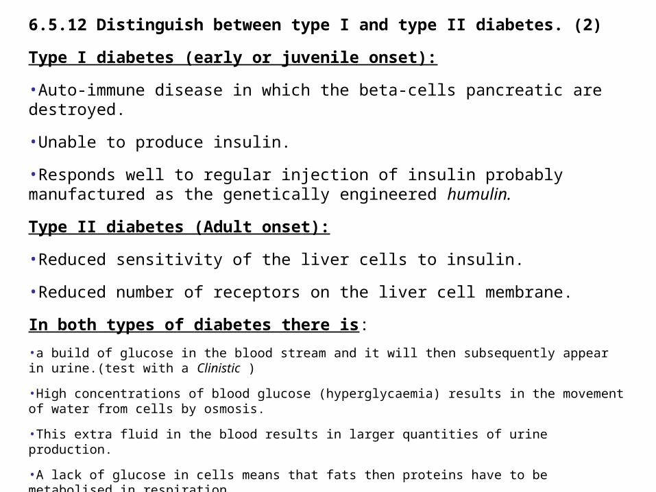
6.5.12 Distinguish between type I and type II diabetes. (2)
Type I diabetes (early or juvenile onset):
•Auto-immune disease in which the beta-cells pancreatic are destroyed.
•Unable to produce insulin.
•Responds well to regular injection of insulin probably manufactured as the genetically engineered humulin.
Type II diabetes (Adult onset):
•Reduced sensitivity of the liver cells to insulin.
•Reduced number of receptors on the liver cell membrane.
In both types of diabetes there is:
•a build of glucose in the blood stream and it will then subsequently appear in urine.(test with a Clinistic )
•High concentrations of blood glucose (hyperglycaemia) results in the movement of water from cells by osmosis.
•This extra fluid in the blood results in larger quantities of urine production.
•A lack of glucose in cells means that fats then proteins have to be metabolised in respiration.
•Particularly the breakdown of protein for energy creates organ damage.
Copyright © 2005 Pearson Education, Inc. publishing as Benjamin Cummings
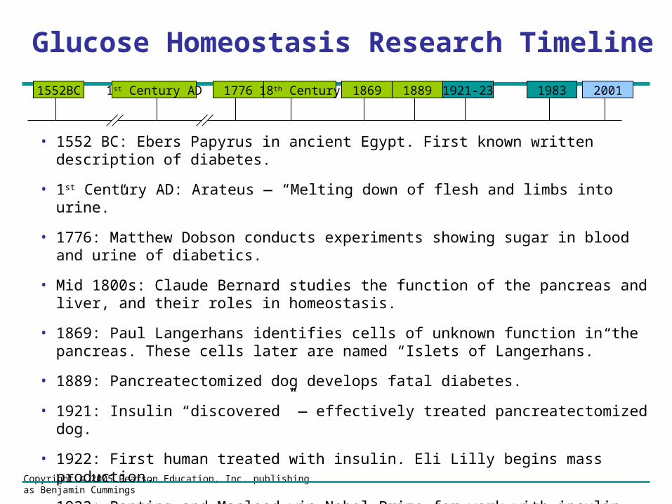
Glucose Homeostasis Research Timeline
• 1552 BC: Ebers Papyrus in ancient Egypt. First known written description of diabetes.
• 1st Century AD: Arateus — “Melting down of flesh and limbs into urine.”
• 1776: Matthew Dobson conducts experiments showing sugar in blood and urine of diabetics.
• Mid 1800s: Claude Bernard studies the function of the pancreas and liver, and their roles in homeostasis.
• 1869: Paul Langerhans identifies cells of unknown function in the pancreas. These cells later are named “Islets of Langerhans.”
• 1889: Pancreatectomized dog develops fatal diabetes.
• 1921: Insulin “discovered” — effectively treated pancreatectomized dog.
• 1922: First human treated with insulin. Eli Lilly begins mass production.
• 1923: Banting and Macleod win Nobel Prize for work with insulin.
• 1983: Biosynthetic insulin produced.
• 2001: Human genome sequence completed.
1552BC 1st Century AD 1776 1869 188918th Century 1921-23 1983 2001

Copyright © 2005 Pearson Education, Inc. publishing as Benjamin Cummings
Current/Future Research in Diabetes• Current
– Clinical Trials: physiological traits of patients/response to therapeutics
– Genetic Approaches: candidate genes; family-based studies; genome-wide scans
– Animal Models: developing gene-knockout models; large-scale mutagenesis studies to produce diabetic phenotypes
– Microarray Analysis: analysis of gene expression in tissues
• Future
– Continued utilization of human genome sequence data
• Sequencing multiple ethnicities
– Beta-cell transplantation
– Use of stem cells to halt or reverse symptoms