Intrauterine Development of Female Genital Organs … (12) PJZ-2229-15 30-11-15 final... ·...
9
Pakistan J. Zool., vol. 46(2), pp. 389-397, 2016. Intrauterine Development of Female Genital Organs in Cavia porcellus (Rodentia: Caviidae) Amilton Cesar dos Santos, 1* Phelipe Oliveira Favaron, 1 Diego Carvalho Viana, 1 Amanda Olivotti Ferreira, 1 Fernanda Menezes Oliveira e Silva, 1 Dayane Alcântara, 1 Bruno Gomes Vasconcelos, 2 Rose Eli Grassi Rici, 1 Antônio Chaves de Assis-Neto 1 and Maria Angélica Miglino 1 1 Department of Surgery, Sector of Anatomy of Domestic and Wild Animals, School of Veterinary Medicine and Animal Science, University of Sao Paulo, Sao Paulo SP, Brazil 2 Institute of Health Science, Federal University of Mato Grosso, Sinop MT, Brazil A B S T R A C T To contribute to the knowledge of the embryology of an important experimental model for studies related to the reproduction, a study was conducted to describe the morphological events of sexual differentiation of female genital organs during the intrauterine life of Cavia porcellus from 20 th to 68 th gestational day. Eighteen embryos and fetuses at days 20, 25, 30, 45, and 60 of pregnancy and near term (n = 3 animals for each gestational period) were utilized. Embryos and fetuses were dissected, and the genital organs processed for light microscopy. Sexual differentiation on females of Cavia porcellus was observed only after the 25th day of gestation with the development of the ovary. However external genitalia acquired female characteristics only at 45 days of gestation. At the end of gestation, the external genitalia had labia, vaginal external ostium occluded by the vaginal closure membrane and penile clitoris with the external urethral ostium opening at the top of the organ. Microscopically, the tubular genital organs (uterine tubes and horns, uterus and vagina) were lined with internal epithelium and loose connective tissue at 30 days of pregnancy. At the end of the pregnancy tissue layers characteristics of tubular genital organs are slightly differentiated. INTRODUCTION The guinea pig (Cavia porcellus, Caviidae, Linnaeus, 1758) is a hystricomorph rodent, widely used as animal model for research development, especially that related to placentation (Miglino et al., 2004; Mess, 2007) and development of embryonic membranes (Oliveira et al., 2012; Vasconcelos et al., 2013), which have been studied by members of our research group. Other studies as female reproductive cycle (Stockard and Papanicolaou, 1917, 1919; Selle, 1922), immunological studies against vaginal diseases, which in this species closely resemble those found in women (Lilley et al., 1997) and toxicological tests (Leite et al., 2002; Savransky et al., 2013) also were conducted. In addition, the classic study performed by Phoenix et al. (1959) using Cavia porcellus as experimental model, and embryological and hormonal descriptions of Jost (1965) gave rise the model for sexual differentiation, widely used in books of embryology and developmental biology (Wolpert, 1998; Sinowatz, 2010). In adults Cavia porcellus, Cooper and Schiller (1975) describe in general terms the anatomy of this __________________________________ * Corresponding author: [email protected] 0030-9923/2016/0002-0389 $ 8.00/0 Copyright 2016 Zoological Society of Pakistan species, although it has been noticed the absence of more detailed data concerning the urogenital tract. Regarding the embryology of this species Evans and Sack (1973) describes some events of embryonic development. In relation to the genital organs however, only the presence of the external genitalia in a given period is described, but development of internal and external genitalia events were not described by these authors. Mammals initiate embryonic development without phenotypic sexual differentiation, and an ovary is not required for the female phenotype, but a testicle and the production of androgenic hormones are essential for the development of a male phenotype, since both, Wolff and the Müllerian ducts are found in sexually undifferentiated embryos. In females, Müllerian ducts develop into a duct- gonadal system and Wolff ducts atrophy. In the male the opposite occurs, stimulated by anti-Müllerian hormone and androgens produced by the testicles (Wolpert, 1998; Arnold, 2009; Nakamura, 2010; Sinowatz, 2010). In order to contribute to the knowledge of the embryology of this important experimental model, the aims of this study was to describe the morphological events related to the sexual differentiation of the female genital organs in Cavia porcellus with ages ranging from the 20 th to the 68 th day of pregnancy. Article Information Received 25 March 2015 Revised 22 April 2015 Accepted 13 June 2015 Available online 1 March 2016 Authors’ Contributions MAM and ACAN designed and coordinated the study. DCV and AOF performed macroscopic analysis, FMOS and DA performed dissections. ACDS and POF performed light microscopy of samples. BGV and AOF performed vaginal cytology analysis. ACDS wrote the article. POF and REGR helped in writing the article. Key words Cavia porcellus, guinea pig, penile clitoris, sexual differentiation, reproductive tract.
-
Upload
truonghanh -
Category
Documents
-
view
214 -
download
0
Transcript of Intrauterine Development of Female Genital Organs … (12) PJZ-2229-15 30-11-15 final... ·...

Pakistan J. Zool., vol. 46(2), pp. 389-397, 2016.
Intrauterine Development of Female Genital Organs in Cavia porcellus (Rodentia: Caviidae) Amilton Cesar dos Santos,1* Phelipe Oliveira Favaron,1 Diego Carvalho Viana,1 Amanda Olivotti Ferreira, 1 Fernanda Menezes Oliveira e Silva,1 Dayane Alcântara,1 Bruno Gomes Vasconcelos,2 Rose Eli Grassi Rici,1 Antônio Chaves de Assis-Neto1 and Maria Angélica Miglino1 1Department of Surgery, Sector of Anatomy of Domestic and Wild Animals, School of Veterinary Medicine and Animal Science, University of Sao Paulo, Sao Paulo SP, Brazil 2Institute of Health Science, Federal University of Mato Grosso, Sinop MT, Brazil A B S T R A C T To contribute to the knowledge of the embryology of an important experimental model for studies related to the reproduction, a study was conducted to describe the morphological events of sexual differentiation of female genital organs during the intrauterine life of Cavia porcellus from 20th to 68th gestational day. Eighteen embryos and fetuses at days 20, 25, 30, 45, and 60 of pregnancy and near term (n = 3 animals for each gestational period) were utilized. Embryos and fetuses were dissected, and the genital organs processed for light microscopy. Sexual differentiation on females of Cavia porcellus was observed only after the 25th day of gestation with the development of the ovary. However external genitalia acquired female characteristics only at 45 days of gestation. At the end of gestation, the external genitalia had labia, vaginal external ostium occluded by the vaginal closure membrane and penile clitoris with the external urethral ostium opening at the top of the organ. Microscopically, the tubular genital organs (uterine tubes and horns, uterus and vagina) were lined with internal epithelium and loose connective tissue at 30 days of pregnancy. At the end of the pregnancy tissue layers characteristics of tubular genital organs are slightly differentiated.
INTRODUCTION
The guinea pig (Cavia porcellus, Caviidae, Linnaeus, 1758) is a hystricomorph rodent, widely used as animal model for research development, especially that related to placentation (Miglino et al., 2004; Mess, 2007) and development of embryonic membranes (Oliveira et al., 2012; Vasconcelos et al., 2013), which have been studied by members of our research group. Other studies as female reproductive cycle (Stockard and Papanicolaou, 1917, 1919; Selle, 1922), immunological studies against vaginal diseases, which in this species closely resemble those found in women (Lilley et al., 1997) and toxicological tests (Leite et al., 2002; Savransky et al., 2013) also were conducted. In addition, the classic study performed by Phoenix et al. (1959) using Cavia porcellus as experimental model, and embryological and hormonal descriptions of Jost (1965) gave rise the model for sexual differentiation, widely used in books of embryology and developmental biology (Wolpert, 1998; Sinowatz, 2010). In adults Cavia porcellus, Cooper and Schiller (1975) describe in general terms the anatomy of this __________________________________ * Corresponding author: [email protected] 0030-9923/2016/0002-0389 $ 8.00/0 Copyright 2016 Zoological Society of Pakistan
species, although it has been noticed the absence of more detailed data concerning the urogenital tract. Regarding the embryology of this species Evans and Sack (1973) describes some events of embryonic development. In relation to the genital organs however, only the presence of the external genitalia in a given period is described, but development of internal and external genitalia events were not described by these authors. Mammals initiate embryonic development without phenotypic sexual differentiation, and an ovary is not required for the female phenotype, but a testicle and the production of androgenic hormones are essential for the development of a male phenotype, since both, Wolff and the Müllerian ducts are found in sexually undifferentiated embryos. In females, Müllerian ducts develop into a duct-gonadal system and Wolff ducts atrophy. In the male the opposite occurs, stimulated by anti-Müllerian hormone and androgens produced by the testicles (Wolpert, 1998; Arnold, 2009; Nakamura, 2010; Sinowatz, 2010). In order to contribute to the knowledge of the embryology of this important experimental model, the aims of this study was to describe the morphological events related to the sexual differentiation of the female genital organs in Cavia porcellus with ages ranging from the 20th to the 68th day of pregnancy.
Article Information Received 25 March 2015 Revised 22 April 2015 Accepted 13 June 2015 Available online 1 March 2016 Authors’ Contributions MAM and ACAN designed and coordinated the study. DCV and AOF performed macroscopic analysis, FMOS and DA performed dissections. ACDS and POF performed light microscopy of samples. BGV and AOF performed vaginal cytology analysis. ACDS wrote the article. POF and REGR helped in writing the article. Key words Cavia porcellus, guinea pig, penile clitoris, sexual differentiation, reproductive tract.

A.C. SANTOS ET AL. 390
MATERIALS AND METHODS Animals Eighteen Cavia porcellus embryos and fetuses at 20, 25, 30, 45 and 60 days of gestation and full term (68 days), as estimated by Deanesly (1975) (n = 3 animals for each gestational age) were utilized in this study. Embryos and fetuses were derived from pregnancies of nine adult females, which were paired with three adult males (n = 3 females and 1 male per box). The methodology employed have been authorized by the Bioethics Committee of the School of Veterinary Medicine and Animal Science, at the University of São Paulo (Process number: 2521/2012). Establishment of gestational age and collection of embryos and fetuses Daily monitoring of copulation by the method of vaginal exfoliative cytology and confirmation of mating through the presence of spermatozoa in vaginal smears by light microscopy in stained slides, by Panoptic-fast method were conducted. The time of detection of spermatozoa in the vaginal smear was determined as day 0 of gestation. In order to collect embryos and fetuses, pregnant females were pre-anesthetized with xylazine 2% (40mg/kg/IM) and ketamine hydrochloride 1% (60mg/kg/IM) and euthanized by administration of thiopental sodium 2.5% (60mg/Kg/IC).
Dissection and macroscopic analysis After euthanasia, pregnant females were dissected and embryos and fetuses removed from the uterus. All specimens were weighted in a digital balance (g) and the crown-rump length (mm) was determined according to methodology established by Evans and Sack (1973) using a digital scale and precision calipers Mitotoyo ®. After the measurement, specimens were dissected and the organs of the urogenital system were exposed. Macroscopic photodocumentation was performed using an Olympus digital camera SP-81OUZ.
Light microscopy Samples were fixed in 10% formalin solution, and dehydrated in a series of ethanol in increasing concentrations (from 50% to 100%) and diaphanized in xylene for later inclusion in paraffin. Sections of 5 µm of thickness were obtained in microtome Leica RM 2155 and stained by H/E (Hematoxylin/Eosin). The microscopic photodocumentation was performed in a photomicroscope Olympus BX61VS. Embryos in early development (20 and 25 days) were entire included and sliced with the following method: a cut with 5 µm was
used and four cuts with 5 µm were discarded. The method was serially performed until the gonads of embryos were found in the sample.
RESULTS Embryos and fetuses measurements Biometric results of weight and length were collected and listed according to gestational age (Table I). An increase in length (crown-rump) was related to weight gain of embryos and fetuses (Fig.1).
Fig. 1. Measurements of embryos and fetuses at 20, 25, 30, 45, 60 days of gestation and full term (T, 68 days).
Table I.- Measurements of weight and crown-rump length related to gestational age in Cavia porcellus.
Gestational age (days) Weight (g) Crown-rump (mm) 20 2.74 ± 0.21 16.60 ± 2.10 25 6.75 ± 0.69 23.01 ± 1.98 30 14.88 ± 1.05 49.90 ± 3.07 45 16.33 ± 1.12 52.10 ± 2.89 60 55.51 ± 1.90 77.44 ± 2.92 Full term 82.90 ± 3.21 97.66 ± 3.79 Morphology of external genitalia The first observed anatomical structures on the external genitalia of sexually undifferentiated embryos at 20 days of gestation were the lateral genital elevations (left and right), located caudal to the genital tubercle. During this period no cloacal opening was observed (Fig. 2A). At 25 days however, was possible to observe cloacal folds and a cloacal membrane (Fig. 2B). After 30 days it was possible to distinguish females from males by the formation of urogenital folds and sulcus, positioned caudally to the clitoris. At 45 days, the urogenital folds already had characteristics of future labia, not yet fused

DEVELOPMENT OF FEMALE GENITAL ORGANS IN CAVIA PORCELLUS 391
Fig. 2. External genitalia of embryos and fetuses of Cavia porcellus. (A) 20 days. Undifferentiated embryo. Genital elevations (arrows) and genital tubercle (t). (B) 25 days. Female embryo. Cloacal folds (arrows), cloacal membrane (*) and genital tubercle (t). (C) 45 days. Female embryo. Urogenital folds (arrows), urogenital membrane (*) and clitoris (c). (D) full term (68 days). Female embryo. Labia (empty arrowhead), vagina (full arrowhead), perineum (*) and clitoris (c) with external uretral ostium (arrow). Bar: A, B, C, D, 5 mm.
urogenital sulcus, covered by the urogenital membrane was evident and the perineum was not formed (Fig.2C). From the 60 days until the end of gestation it was observed the formation of the labia, fusion of urogenital folds with perineum dividing the anal orifice from the external vaginal orifice, and, the formation of the vaginal closure membrane. The urogenital sulcus was incorporated into the genital tubercle, developing in a penile structure (Fig. 2D).
Morphology of internal genitalia Regarding the internal genital organs, gonads at 25 days were already differentiated into ovaries. At 30 days, uterine tubes and uterine horns were present. In this period, the uterus was short and did not have a division with too short and flacid vagina (Fig. 3A). At 45 days, a septate uterus (partially double) and a single cervix with a fornix dividing the uterus from a little elongated but still flaccid vagina were observed. From 60 days until the end of pregnancy, the tubular organs (uterine tubes, uterine horns, uterus and vagina) are lengthened and the double uterus with a single cervix, divided from the vagina by the fornix was observed. The vagina remained flaccid, but was elongated, allowing a good view of the organ. The pelvic urethra was present from the 25th day until the end of pregnancy. This organ extends ventrally to the vagina, from urinary bladder until the clitoris, with the external urethral ostium opening at the top of the clitoris after 60 days. The vaginal vestibule was not observed (Fig. 3B).
Fig. 3. Urogenital organs of embryos and fetuses of Cavia porcellus. (A) 30 days. Note ovary (o), uterine horns (uh), uterus (u), vagina (v), urinary bladder (b) and urethra (ur). (B) full term (68 days). Note kidney (k), adrenal (a), ureter (ur), urinary bladder (b), urethra (urt), ovary (o), uterine tubes (ut), uterine horns (uh), uterus (u), vagina (v), clitoris (c) and labia (l). Bar: A, B, 25 mm.
Histological structure of female genital organs A pair of ovaries lined by cuboidal epithelium was observed, with still undifferentiated tissue. Oogonia and stromal cells inside were observed on the 25th day of gestation (Fig. 4A-B). At 30 days many blood vessels arise in the ovarian parenchyma (Fig. 4C). At 45 days of

A.C. SANTOS ET AL. 392
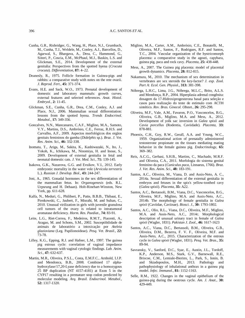
Fig. 4. Ovaries of embryos and fetuses of Cavia porcellus. (A) 25 days: ovary (o) mesonephros (m) and Wolff duct (arrow). (B) 25 days: oogonia (arrowhead, circle) and the ovarian stromal cells (arrow). (C) 30 days: ovary (o) and blood vessels (arrows). (D) 45 days: cortex (c) medulla (m) and blood vessels (arrows). (E) 45 days: oocyte (arrow) and primordial follicle (circle). (F) 60 days. oocyte (arrow) and primordial follicle (circle). (G) 68 days. follicles (F) and the blood vessels (V). (H) 68 days: primordial follicles (arrowhead), large primordial follicles (*) and oocytes (arrow). Bar: A, D, 200 µm; B, 20 µm; C, E, 100 µm; G, H, F, 50 µm. Staining: hematoxylin/eosin.
gestation, the ovary had a differentiated medulla and cortex, in which primordial follicles in the cortical region were present (Fig. 4D-E). At 60 days, several clusters of oocytes and large primordial follicles in the cortex (Fig.4F) were observed. At 68 days, numerous primordial follicles were noticed in the cortex. During this period, large primordial follicles could be found in the cortex and medulla of the organ (Fig. 4G).
Fig. 5. Uterine tube of embryos and fetuses of Cavia porcellus. (A) 30 days. Infundibulum with cuboidal epithelium (arrow) and loose connective tissue (ct). (B) 68 days. Infundibulum with serous (s) and muscular (m) layers, and folded mucosa (arrow). (C) 30 days. Ampoule with epithelium (arrow) and loose connective tissue (ct). (D) 68 days. Ampoule with serous (s) and muscular (m) layers, and folded mucosa (arrow). (E) 30 days. Isthmus with epithelium (arrow) and loose connective tissue (ct). (F) 68 days. Isthmus with serous (s) and muscular (m) layers, and folded mucosa (arrow). A, 20 µm; B, 200 µm; C, E, 50 µm; D, F, 200 µm. Staining: hematoxylin/eosin.
At 30 days of gestation, the uterine tube (infundibulum, ampulla and isthmus) had cubic epithelium and loose connective tissue lining externally the organ. From 30 days until 60 days of gestation, tissue layers were not evident. After 68 days the outermost

DEVELOPMENT OF FEMALE GENITAL ORGANS IN CAVIA PORCELLUS 393
serous layer, the poorly developed muscular layer and a pleated mucosa were present (Fig. 5). At 30 days of gestation the uterine horns had columnar epithelium and loose connective tissue lining externally the organ (Fig. 6A). At 68 days, mucosa with columnar epithelium and not modeled dense connective tissue, an inner and outer muscular layer and serous layer lining externally the organ were observed (Fig. 6B). The body of the uterus (partially double) at 30 days had cubical epithelium and loose connective tissue lining externally the organ (Fig. 6C). Tissue layers were not evident from 30 days until 60 days of gestation. However, after 68 days of gestation, mucosa with cubical epithelium, and muscular and serous layer were present (Fig. 6D).
Fig. 6. Uterine horn and corpus of embryos and fetuses of Cavia porcellus. (A) 30 days. Uterine horn with epithelium (arrow) and loose connective tissue (ct). (B) 68 days. Uterine horn with epithelium (arrow), connective tissue (ct), internal muscular layer (m), longitudinal muscular layer and serosa (arrowhead). (C) 30 days. Corpus of the uterus with epithelium (arrows) and loose connective tissue (ct). (D) 68 days. Corpus of the uterus with epithelium (arrow) and connective tissue (ct), internal muscular layer (m), longitudinal muscular layer and serosa (arrowhead). Bar: A, B, C, 100 µm; D, 200 µm. Staining: hematoxylin/eosin.
The cervix was developed from 45 to 60 days of gestation, but was at the end of pregnancy that this organ showed development of cervical folds and layers
differentiation. The mucosa comprised cubic epithelium and not modeled dense connective tissue, and a muscular layer, and a serous layer lining externally the organ were present (Fig. 7A-B). At 30 days, the vagina had stratified epithelium and loose connective tissue lining externally the organ (Fig. 7C). Tissue layers were not evident from 30 until 60 days of gestation, but at 68 days of gestation, mucosa with stratified epithelium and not modeled dense connective tissue, muscular and serous layer lining the organ were present (Fig. 7D).
Fig. 7. Uterine cervix, vaginal closure membrane and vagina of embryos and fetuses of Cavia porcellus. (A) 68 days. Uterine cervix with pleated epithelium (arrow), connective tissue (ct), inner (m) and outer (arrowhead) muscular layer. (B) 68 days. Vaginal closure membrane (vcm) with epithelium (arrow) and vaginal connective tissue (ct). (C) 30 days. Vaginal epithelium (arrow) and loose connective tissue (ct). (D) 68 days. Vaginal epithelium (arrow), connective tissue (ct) and muscular layer (m). Bar: A, C, 100 µm; B, 20 µm; D, 200 µm. Staining: hematoxylin/eosin.
DISCUSSION Chronological documentation of events during embryogenesis in different species is of great importance to estimate possible teratogenic effects and other developmental anomalies in those stages when the fetus is more susceptible to failures during its formation and differentiation (Evans and Sack, 1973; Isakova et al., 2012). Results of embryonic and fetal development of

A.C. SANTOS ET AL. 394
Cavia porcellus found in this study are within the expected by Banks et al. (2010) for weight of the animal at birth, which may vary between 45-115 grams. The largest body mass gain was observed in the final third, comprised from 45 to 68 days of gestation when the average weight was from 16.33 to 82.90 g. In the adult stage, females of these animals can reach up to 900 g (Banks et al., 2010). Length measurements for crown-rump accompanied the gain in body mass at the end of pregnancy. Evans and Sack (1973) and Sterba (1995) observed that the crown-rump length can reach up to 100 mm at the end of pregnancy, but the authors did not mention the sex of fetuses. In our study, the crown-rump length was 97, 66 mm at the end of gestation. The formation of the external genitalia in our study could be noted from 20 days with the lateral genital elevations and the genital tubercle presence, although at this stage it is not possible to differentiate the external genitalia of the embryo. Evans and Sack (1973) described that external genitalia is visible at 24 days of gestation in Cavia porcellus and a differentiated external genitalia is visible at 28 days in pigs, at 35 days in dogs, at 43 days in sheep and 45 days in horses. In cows, the genital tubercle is visible at 38 days of gestation. In cats with 3.2-3.3 cm crown-rump length, the external genitalia were first observed (Inomata et al., 2009). In the present study, the urogenital folds fuse in the dorsal and ventral extremities, forming the perineum. The lateral elevations form the labia, occurring absence of a vaginal vestibule. The urogenital sulcus is incorporated into the genital tubercle, developing in a penile structure, similar to males, differently than that described for females of domestic mammals (Sinowatz, 2010) as cats (Inomata et al., 2009). In our previous studies (Santos et al., 2014a), the genital tubercle is present at 15th day of gestation in Galea spixii, but the sexual differentiation occurs only after 22th day of gestation and those results is very similar with these found in Cavia porcellus. The formation of the ovaries, with differentiation of oocytes and primordial follicles follows that described by Sinowatz (2010), except for the final period of gestation where we can observe large primordial follicles that were not described in other species of domestic mammals. In the present study, these primordial follicles first appear in the cortex and then can be found in the hilum and medulla region, results that agree with those of Deanesly (1975) in the same species. The author also noted that in this species, meiosis begins after 35 days, however, primordial follicles are observed at 48 days. These observations are similar to our results, in which primordial follicles arise at 45 days of gestation. Deanesly (1975) points out that approximately 87% of oocytes degenerate during and after meiotic prophase in
fetal ovaries of Cavia porcellus. In early development, the oogonia and oocytes are found among the stromal cells in the specimens of our study, similar to that cited by Deanesly (1975) in Cavia porcellus, humans, mice, rabbits and ferrets. This above description is very important because Yang et al. (2010) propose that normal external genital development in mice requires androgen and estrogen action produced by both ovaries and testes and the masculinized clitoris of female require estrogen for development, which could be produced by primordial follicles. Some studies describe that the uterus can be double or partially double in some species of hystricomorphs rodents (Weir, 1971; Reis et al., 2011). The partially double uterus is common in rodents and the Cavia porcellus used in our study showed these characteristics in the embryonic period. They present a single cervix that opens into a single vagina as that found in the capybara (Hydrochoerus hydrochaeris) (Costa et al., 2002), paca (Cuniculus paca) (Reis et al., 2011) and Galea spixii (Santos et al., 2014b). Female viscacha (Lagostamus maximus) (Weir, 1971) have double uterus and double cervix, as found in marsupials (Gonçalves et al., 2009). However, these authors did not study this organ during the embryonic period. The flaccid vagina found in our study may be due to the lack of muscular layers of the vagina, throughout almost the entire development. Sinowatz (2010) describes that the vagina has two origins: one anterior, derived from the paramesonephric ducts and one posterior derived from the urogenital sinus. However, the microstructure of the vagina is not described. At 68 days of gestation, the external vaginal ostium occluded by the presence of “vaginal closure membrane”, as described in C. porcellus adults by Stockard and Papanicolaou (1917) was observed. Stockard and Papanicolaou (1917) called the vaginal closure membrane and compared this structure with the hymen, which is formed by epithelium of the urogenital sinus and a thin lining of vaginal cells, in domestic animals (Sinowatz, 2010). The vaginal closure membrane is an embryonic structure as the hymen but develops throughout the reproductive life of the female, from the fetal period until puberty when it breaks. After puberty, this membrane develops and breaks down during each estrous cycle, indicating the period of estrus and is present throughout pregnancy, breaking at delivery (Stockard and Papanicolaou, 1919). Other species of rodents as Galea musteloides, Cavia aperea (Touma et al., 2001) and Galea spixii (Santos et al., 2014b; 2015) also have this feature; however the studies do not describe the embryonic development of species.

DEVELOPMENT OF FEMALE GENITAL ORGANS IN CAVIA PORCELLUS 395
A clitoris trespassed by the urethra, as found in our study, has been described in studies of hyenas (Crocruta crocruta), which have a hypertrophied clitoris trespassed by the urethra. The formation of a urogenital sinus in these animals occurs due absence of an external vaginal ostium (Yalcinkaya et al., 1993; Browne et al., 2006; Glickman et al., 2006). In adult Galea spixii, Santos et al. (2014c) describe a clitoris trespassed by the urethra with differences in morphology from apex without urethral glands to basis with presence of urethral glands. Yang et al. (2010) show that a clitoris trespassed by the urethra is associated with the balance in the production of androgens and estrogens, with emphasis in estrogen participation during sexual differentiation in mice. Studies about sexual hormones balance and their receptors during sexual differentiation have been useful for that studies, which have aimed to understand the development of external genitalia in mice (Weiss et al., 2012) and hyenas (Crocruta crocruta) (Cunha et al., 2014). Then, these species have been used as experimental model for studies on hypospadias, which are caused by abnormalities in the penis morphology during human development (Blaschko et al., 2012). The clitoral urethra found in C. porcellus require further studies related to the hormones and hormonal receptors responsible for the development of female external genitalia and those hormones responsible for initial sexual differentiation. Then, this species as mice and hyena may also be useful as experimental model for studies about hypospadias. Moreover, female of C. porcellus when used as experimental models may be useful in those studies on sexual infantilism in women, polycystic ovary syndrome, ambiguous genitalia at birth, pseudohermaphroditism, female genital virilization (Nóbrega et al., 2004; Martin et al., 2008; Costenaro et al., 2010; Kalfa et al., 2010) and other studies on genetic syndromes associated with female pseudohermaphroditism, with alterations in genitalia in the female fetus, as clitoromegaly and hypoplasia of labia minora (Arnold et al., 1999) and McKusick-Kaufman syndrome, in which occurs association with hydrometrocolpos and agenesis of the vagina with the presence of the female urogenital sinus (Slavotinek and Biesecker, 2000). Other studies on sexual dimorphism in guinea pig in order to elucidate the importance of masculinized genitalia are required, because this fact is still not well understood in the literature, but may be associated with sexual selection in these species that present these characteristics. These studies are relevant because Adrian and Sachser (2011) found different mating system in different species of cavies (Caviidae).
In conclusion, females of Cavia porcellus were sexually differentiated only after the 25th day of gestation with the formation of the ovary, but the external genitalia acquired female characteristic only at 45 days of gestation. At the end of gestation, the external genitalia had labia, vaginal external ostium occluded by the vaginal closure membrane and penile clitoris with the external urethral ostium opening at the top of organ. Microscopically, tubular genital organs (uterine tubes and horns, uterus and vagina) arise with internal epithelium and loose connective tissue externally lining the organs at 30 days. At the end of pregnancy tissue layer characteristics of tubular genital organs were slightly differentiated.
ACKNOWLEDGEMENT Thanks to FAPESP (Fundação de Amparo a Pesquisa do Estado de São Paulo) for financial support.
REFERENCES Adrian, O. and Sachser, N., 2011. Diversity of social and
mating systems in cavies: a review. J. Mammal., 92: 39-53.
Arnold, P.A., 2009. The organizational-activational hypothesis as the foundation for a unified theory of sexual differentiation of all mammalian tissues. Horm. Behavior., 55: 570-578.
Arnold, S.R., Spicer, D., Kouseff, B., Lacson, A. and Gilbert-Barness, E., 1999. Seckel-like syndrome in three siblings. Ped. Develop. Pathol., 2: 180-187.
Banks, R.E., Sharp, J.M., Doss, S.D. and Vanderford, D.A., 2010. Exotic Small Mammal Care and Husbandry. Wiley-Blackwell, Iowa.
Blaschko, S.D., Cunha, G.R. and Baskin, L.S., 2012. Molecular mechanisms of external genitalia development. Differentiation, 843: 261-268.
Browne, P., Place, N.J., Vidal, J.D., Moore, G.I., Cunha, T.R., Glickman, S.E. and Conley, A.J., 2006. Endocrine differentiation of fetal ovaries and testes of the spotted hyena (Crocuta crocuta): timing of androgen-independent versus androgen-driven genital development. Reproduction, 132: 649-659.
Cooper, G. and Schiller, A.L., 1975. Anatomy of the guinea pig. Harvard University Press, Cambridge.
Costa, D.S., Paula, T.A.R., Fonseca, C.C. and Neves, M.T.D., 2002. Reprodução de capivaras. Arq. Ciências Vet. Zool. UNIPAR, 5: 111-118.
Costenaro, F., Rodrigues, T.C., Kater, C.E., Auchus, R.J., Zareei, P.M. and Czepielewski, M.A., 2010. Combined 17α-hydroxylase/17, 20 -lyase deficiency due to p.R96W mutation in the CYP17 gene in a Brazilian patient. Arq. Brasil. Endocrinol. Metabol., 54: 744-748.
A.C. SANTOS ET AL. 396
Cunha, G.R., Risbridger, G., Wang, H., Place, N.J., Grumbach, M., Cunha, T.J., Weldele, M., Conley, A.J., Barcellos, D., Agarwal, S., Bhargava, A., Drea, C., Hammond, G., Siiteri, P., Coscia, E.M., McPhaul, M.J., Baskin, L.S. and Glickman, S.E., 2014. Development of the external genitalia: Perspectives from the spotted hyena (Crocuta crocuta). Differentiation, 87: 4–22.
Deanesly, R., 1975. Follicle formation in Guinea-pigs and rabbits: a comparative study with notes on the rete ovarii. J. Reprod. Fert., 45: 371-374.
Evans, H.E. and Sack, W.O., 1973. Prenatal development of domestic and laboratory mammals: growth curves, external features and selected references. Anat. Histol. Embryol., 2: 11-45.
Glickman, S.E., Cunha, G.R., Drea, C.M., Conley, A.J. and Place, N.J., 2006. Mammalian sexual differentiation: lessons from the spotted hyena. Trends Endocrinol. Metabol., 17: 349-356.
Goncalves, N.N., Mancanares, C.A.F., Miglino, M.A., Samoto, V.Y., Martins, D.S., Ambrósio, C.E., Ferraz, R.H.S. and Carvalho, A.F., 2009. Aspectos morfológicos dos orgãos genitais femininos do gamba (Didelphis sp.). Braz. J. Vet. Res. Anim. Sci., 46: 332-338.
Inomata, T., Ariga, M., Sakita, K., Kashiwazaki, N., Ito, J., Yokoh, K., Ichikawa, M., Ninomiya, H. and Inoue, S., 2009. Development of external genitalia in fetal and neonatal domestic cats. J. Vet. Med. Sci., 71: 139-145.
Isakova, G.K., Nazarova, G.G. and Evsikov, V.I., 2012. Early embryonic mortality in the water vole (Arvicola terrestris L.). Russian J. Develop. Biol., 43: 244-247.
Jost, A., 1965. Gonadal hormones in the sex differentiation of the mammalian fetus. In: Organogenesis (eds. R.L. Urpsrung and H. Dehaan). Holt-Rinehart-Winston, New York, pp. 611-628.
Kalfa, N., Meduri, G., Philibert, P., Patte, B.B.B., Thibaut, E., Pienkowski, C., Jaubert, F., Misrahi, M. and Sultan, C., 2010. Unusual virilization in girls with juvenile granulosa cell tumors of the ovary is related to intratumoral aromatase deficiency. Horm. Res. Paediat., 74: 83-91.
Leite, L.G., Riet-Correa, F., Medeiros, R.M.T., Piacenti, A., Aragao, M. and Schons, S.M., 2002. Susceptibilidade de animais de laboratório a intoxicação por Ateleia glazioviana (Leg. Papilionoideae). Pesq. Vet. Brasil., 22: 73-78.
Lilley, K.G., Epping, R.J. and Hafner, L.M., 1997. The guinea pig estrous cycle: correlation of vaginal impedance measurements with vaginal cytologic findings. Lab. Anim. Sci., 47: 632-637.
Martin, M.R., Oliveira, P.S.L., Costa, E.M.F.C., Arnhold, I.J.P. and Mendonca, B.B., 2008. Combined 17 alpha-hydroxylase/17,20-Lyase deficiency due to a homozygous 25 BP duplication (NT 4157-4181) at Exon 5 in the CYP17 resulting in a premature stop codon predicted by molecular modeling. Arq. Brasil. Endocrinol. Metabol., 52: 1317-1320.
Miglino, M.A., Carter, A.M., Ambrósio, C.E., Bonatelli, M., Oliveira, M.F., Santos, F., Rodrigues, R.F. and Santos, T.C., 2004. Vascular organization of the hystricomorph placenta: a comparative study in the agouti, capybara, guinea pig, paca and rock cavy. Placenta, 25: 438-448.
Mess, A., 2007. The Guinea pig placenta: model of placental growth dynamics. Placenta, 28: 812-815.
Nakamura, M., 2010. The mechanism of sex determination in vertebrates are sex steroids the key-factor? J. exp. Zool. Part A: Ecol. Gen. Physiol., 313: 381-398.
Nóbrega, L.H.C., Lima, J.G., Nóbrega, M.L.C., Brito, A.L.S. and Mendonça, R.P., 2004. Hiperplasia adrenal congênita: dosagem da 17-Hidroxiprogesterona basal para seleção e casos para realização do teste de estimulo com ACTH sintético. Rev. Bras. Ginecol. Obstet., 26: 295-298.
Oliveira, M.F., Vale, A.M., Favaron, P.O., Vasconcelos, B.G., Oliveira, G.B., Miglino, M.A. and Mess, A., 2012. Development of yolk sac inversion in Galea spixii and Cavia porcellus (Rodentia, Caviidade). Placenta, 33: 878-881.
Phoenix, C.H., Goy, R.W., Gerall, A.A. and Young, W.C., 1959. Organizational action of prenatally administered testosterone propionate on the tissues mediating mating behavior in the female guinea pig. Endocrinology, 65: 369–382.
Reis, A.C.G., Gerbasi, S.H.B., Martins, C., Machado, M.R.F. and Oliveira, C.A., 2011. Morfologia do sistema genital feminino da paca (Cuniculus paca, Linnaeus, 1766). Braz. J. Vet. Res. Anim. Sci., 48: 183-191.
Santos, A.C., Oliveira, M., Viana, D. and Assis-Neto, A. C., 2014a. Sexual differentiation of the external genitalia in embryos and fetuses in the spix's yellow-toothed cavy (Galea spixii). Placenta, 35: A22.
Santos, A.C., Bertassoli, B.M., Viana, D.C., Vasconcelos, B.G., Oliveira, M.F., Miglino, M.A. and Assis-Neto, A.C., 2014b. The morphology of female genitalia in Galea spixii (Caviidae, Caviinae). Biosci. J., 30: 1793-1802.
Santos, A.C., Olio, R.L., Viana, D.C., Oliveira, M.F., Miglino, M.A. and Assis-Neto, A.C., 2014c. Morphological description of unusual urinary tract in female of Galea spixii (Wagler, 1831). Pakistan J. Zool., 46: 1617-1623
Santos, A.C., Viana, D.C., Bertassoli, B.M., Oliveira, G.B., Oliveira, D.M., Bezerra, F. V. F., Oliveira, M.F. and Assis-Neto, A.C., 2015. Characterization of the estrous cycle in Galea spixii (Wagler, 1831). Pesq. Vet. Bras., 35: 89-94.
Savransky, V., Sanford, D.C., Syar, E., Austin, J.L., Tordoff, K.P., Anderson, M.S., Stark, G.V., Barnewall, R.E., Briscoe, C.M., Lemiale-Bierinx, L., Park, S., Ionin, B. and Skiadopoulos, M.H., 2013. Pathology and pathophysiology of inhalational anthrax in a guinea pig model. Infec. Immunol., 81: 1152-1163.
Selle, R.M., 1922. Changes in the vaginal epithelium of the guinea-pig during the oestrous cycle. Am. J. Anat., 30: 429-449.

DEVELOPMENT OF FEMALE GENITAL ORGANS IN CAVIA PORCELLUS 397
Sinowatz, F., 2010. Development of the urogenital system. In: Essential of domestic animal embryology. (eds. P. Hyttel, F. Sinowatz and M. Vejlsted). Saunders-Elsevier, Edinburgh-London-New York-Oxford-Philadelphia-St Louis-Sydney-Toronto, pp. 252-283.
Slavotinek, A.M. and Biesecker, L.G., 2000. Phenotypic overlap of McKusick-Kaufman syndrome with Bardet-Biedl syndrome: a literature review. Am. J. med. Gen., 95: 208-215.
Sterba, O., 1995. Staging and ageing of mammalian embryos and fetuses. Acta Vet. Brno, 64: 83-89.
Stockard, C.R. and Papanicolaou, G.N., 1917. The existence of a typical oestrous cycle in the guinea-pig – with a study of its histological and physiological changes. Am. J. Anat., 22: 225-283.
Stockard, C.R. and Papanicolaou, G.N., 1919. The vaginal closure membrane, copulation and the vaginal plug in the guinea pig, with further considerations of the oestrous rhytm. Biol. Bull., 37: 222-244.
Touma, C., Palme, R. and Sachser, N., 2001. Different types of oestrous cycle in two closely related South American rodents (Cavia aperea and Galea musteloides) with different social and mating systems. Reproduction, 121:
791-801. Vasconcelos, B.G., Favaron, P.O., Miglino, M.A. and Mess,
A.M., 2013. Development and morphology of the inverted yolk sac in the guinea pig (Cavia porcellus). Theriogenology, 80: 636-641.
Weir, J. B., 1971. The reproductive organs of the female plains viscacha, Lagostomus maximus, J. Reprod. Fert., 25: 365-373.
Weiss, D.A., Rodriguez Jr, E., Cunha, T., Menshenina, J., Barcellos, D., Chan, L.Y., Risbridger, G., Baskin, L. and Cunha, G., 2012. Morphology of the external genitalia of the adult male and female mice as an endpoint of sex differentiation. Mol. Cell. Endocrinol., 354: 94–102.
Wolpert, L., 1998. Principles of development. Oxford University Press, London.
Yalcinkaya, T.M., Siiteri, P.K., Licht, P. and Frank, L.G., 1993, A mechanism for virilization of female spotted hyenas in utero. Science, 260: 1929-1931.
Yang, J.H., Menshenina, J., Cunha, G.R., Place, N. and Baskin, L.S., 2010. Morphology of mouse external genitalia: implications for a role of estrogen in sexual dimorphism of the mouse genital tubercle. J. Urol., 184: 1604-1609.