Intracellular Cytokine Staining on PBMCs Using CyTOF … · Vol 5, Iss 1, Jan 05, 2015 ....
7
http://www.bio-protocol.org/e1370 Vol 5, Iss 1, Jan 05, 2015 Intracellular Cytokine Staining on PBMCs Using CyTOF TM Mass Cytometry Dongxia Lin 1* , Sheena Gupta 2 and Holden T. Maecker 1, 2* 1 Department of Microbiology and Immunology; 2 Institute for Immunity, Transplantation, and Infection, Stanford University, Stanford, USA * For correspondence: [email protected]; [email protected] [Abstract] In this protocol, we use a CyTOF TM mass cytometry to collect single-cell data on a large number of cytokines/chemokines as well as cell-surface proteins that characterize T cells and other immune cells. The current selected mass window in AW 103-203 includes the lanthanides used for most antibody labeling, along with iridium and rhodium for DNA intercalators. The output data are in the format as .txt and .fcs files, which is compatible with many analysis programs. This protocol could be adapted to include tetramers into the staining panel, but we have not optimized for that purpose. The principal steps of intracellular cytokine staining are as follows: First, cells are activated for a few hours using either a specific peptide or a non-specific activation cocktail. An inhibitor of protein transport (e.g. Brefeldin A) is added to retain the cytokines within the cell. Next, EDTA is added to remove adherent cells from the activation vessel. After washing, antibodies to cell surface markers are added to the cells. The cells are then fixed in paraformaldehyde and permeabilized. We use a gentle detergent, saponin, as the permealization buffer because it is less destructive to surface and intracellular epitopes compared to harsh detergents or methanol. After permeabilization, the metal-conjugated anti-cytokine antibodies are added into the cell suspension. The stained cells are then sequentially introduced into the mass cytometry for signal intensity analysis. Materials and Reagents 1. PBMC (fresh or thawed frozen) 2. RPMI-1640 (Hyclone, catalog number: SH30027.01) 3. FBS (Atlanta Biologicals, catalog number: S11150) 4. Pen-strep-Glutamin 100X (Hyclone, catalog number: SV30082.01) 5. Benzonase (2.5 x 10 5 U/ml) (Pierce, catalog number: 88701) 6. Brefeldin A (Sigma-Aldrich, catalog number: B7651) 7. Monensin (Sigma-Aldrich, catalog number: M5273) 8. 0.5 M EDTA (Hoefer, catalog number: GR-123-100) 9. Sodium azide (10% w/v solution) (Teknova, catalog number: S0209) 10. 16% para-formaldehyde (PFA) (Alfa Aesar, catalog number: 43368)) Copyright © 2015 The Authors; exclusive licensee Bio-protocol LLC. 1
-
Upload
duonghuong -
Category
Documents
-
view
218 -
download
0
Transcript of Intracellular Cytokine Staining on PBMCs Using CyTOF … · Vol 5, Iss 1, Jan 05, 2015 ....
http://www.bio-protocol.org/e1370 Vol 5, Iss 1, Jan 05, 2015
Intracellular Cytokine Staining on PBMCs Using CyTOFTM Mass Cytometry
Dongxia Lin1*, Sheena Gupta2 and Holden T. Maecker1, 2*
1Department of Microbiology and Immunology; 2Institute for Immunity, Transplantation, and
Infection, Stanford University, Stanford, USA *For correspondence: [email protected]; [email protected]
[Abstract] In this protocol, we use a CyTOFTM mass cytometry to collect single-cell data on a
large number of cytokines/chemokines as well as cell-surface proteins that characterize T cells
and other immune cells. The current selected mass window in AW 103-203 includes the
lanthanides used for most antibody labeling, along with iridium and rhodium for DNA intercalators.
The output data are in the format as .txt and .fcs files, which is compatible with many analysis
programs. This protocol could be adapted to include tetramers into the staining panel, but we have
not optimized for that purpose. The principal steps of intracellular cytokine staining are as follows: First, cells are activated for a
few hours using either a specific peptide or a non-specific activation cocktail. An inhibitor of protein
transport (e.g. Brefeldin A) is added to retain the cytokines within the cell. Next, EDTA is added to
remove adherent cells from the activation vessel. After washing, antibodies to cell surface markers
are added to the cells. The cells are then fixed in paraformaldehyde and permeabilized. We use a
gentle detergent, saponin, as the permealization buffer because it is less destructive to surface
and intracellular epitopes compared to harsh detergents or methanol. After permeabilization, the
metal-conjugated anti-cytokine antibodies are added into the cell suspension. The stained cells
are then sequentially introduced into the mass cytometry for signal intensity analysis.
Materials and Reagents
1. PBMC (fresh or thawed frozen)
2. RPMI-1640 (Hyclone, catalog number: SH30027.01)
3. FBS (Atlanta Biologicals, catalog number: S11150)
4. Pen-strep-Glutamin 100X (Hyclone, catalog number: SV30082.01)
5. Benzonase (2.5 x 10⁵ U/ml) (Pierce, catalog number: 88701)
6. Brefeldin A (Sigma-Aldrich, catalog number: B7651)
7. Monensin (Sigma-Aldrich, catalog number: M5273)
8. 0.5 M EDTA (Hoefer, catalog number: GR-123-100)
9. Sodium azide (10% w/v solution) (Teknova, catalog number: S0209)
10. 16% para-formaldehyde (PFA) (Alfa Aesar, catalog number: 43368))
Copyright © 2015 The Authors; exclusive licensee Bio-protocol LLC. 1
http://www.bio-protocol.org/e1370 Vol 5, Iss 1, Jan 05, 2015
11. 10x PBS (ROCKLAND, catalog number: MB-008)
12. BSA (Sigma-Aldrich, catalog number: A7284)
13. Maleimide-DOTA (Macrocyclics, catalog number: B-272)
14. Lanthanum (III) chloride heptahydrate (Sigma-Aldrich, catalog number: 203521)
15. Indium (III) chloride (Sigma-Aldrich, catalog number: 203440)
16. MilliQ water
Note: Beakers or bottles used here are not washed with soap due to barium content of
most commercial soaps.
17. Phenotyping antibodies (filtered with 0.1 µm spin filters) (Millipore, catalog number:
UFC30VV00)
18. Ir-intercalator stock solution (Fluidigm, catalog number: 201192)
Note: Rh103-intercalator can be used.
19. 10x saponin-based permeabilization buffer (eBioscience, catalog number: 00 8333-56)
20. Phorbol 12-myristate 13-acetate (PMA) (Sigma-Aldrich, catalog number: P8139)
21. Ionomycine (Sigma-Aldrich, catalog number: I0634)
22. Phytohemagglutinin (PHA) (Sigma-Aldrich, catalog number: 61764)
23. SEB (Sigma-Aldrich, catalog number: S0812)
24. Anti-CD3/CD28 (various vendors)
25. Peptide mixes (JPT)
26. Complete RPMI (see Recipes)
27. CyPBS (see Recipes)
28. CyFACS buffer (see Recipes)
29. Live-dead stain (see Recipes) Equipment
1. 96- well round-bottom plates
2. 37 °C water bath
3. Biosafety cabinet
4. Centrifuge
5. CO2 incubator at 37 °C
6. Calibrated pipettes
Procedure A. Thaw PBMC
Copyright © 2015 The Authors; exclusive licensee Bio-protocol LLC. 2
http://www.bio-protocol.org/e1370 Vol 5, Iss 1, Jan 05, 2015
1. Warm complete RPMI media to 37 °C in water bath. Each sample will require 22 ml of
media with benzonase. Calculate the amount needed to thaw all samples, and prepare a
separate aliquot of warm media with 1:10,000 benzonase (final concentration 25 U/ml).
Benzonase is added into the media to prevent dead cell aggregation. Thaw no more than
3 samples at a time. Run one control PBMC with each batch of samples.
2. Remove samples from liquid nitrogen and transport to lab on dry ice.
3. Place 10 ml of warmed benzonase media into a 15 ml tube, making a separate tube for
each sample.
4. Thaw frozen vials in 37 °C water bath.
5. When cells are nearly completely thawed, carry to hood.
6. Add 1 ml of warm benzonase media from appropriately labeled centrifuge tube slowly to
the cells, then transfer the cells to the centrifuge tube. Rinse vial with more media from
centrifuge tube to retrieve all cells.
7. Continue with the rest of the samples as quickly as possible.
8. Centrifuge cells at 1,550 rpm (RCF = 473) for 8 min at room temperature.
9. Remove supernatant from the cells and resuspend the pellet by tapping the tube.
10. Gently resuspend the pellet in 1 ml warmed benzonase media. Filter cells through a 70
micron cell strainer if needed. Add 9 ml more warmed benzonase media to the tube.
11. Centrifuge cells at 1,550 rpm (RCF = 473) for 8 min at room temperature. Remove
supernatant from the cells and resuspend the pellet by tapping the tube.
12. Resuspend cells in 1 ml warm media.
13. Count cells with Vicell (or hemocytometer if necessary). To count, take 20 μl cells and
dilute with 480 μl PBS in Vicell counting chamber. Load onto Vicell as PBMC with a 1:25
dilution factor.
14. Adjust the cell concentration to 5-10 x 106 cells/ml with warm media (no more benzonase
at this point).
15. Using a multichannel pipette, add 200 µl cells (for at least 1 x 10⁶ cells) into each well of a
96-well deep well plate. Split each sample equally into two or more wells keeping one as
an unstimulated control and the others for different types of stimulation.
16. Rest overnight (6-18 h) at 37 °C in CO2 incubator.
B. Stimulate cells
1. After overnight rest at 37 °C, add the activation reagents and secretion inhibitor (brefeldin
A/monensin) to the well for stimulation. Add only the secretion inhibitor to the unstimulated
control well. If doing CD107a staining, add CD107a antibody during the stimulation.
Copyright © 2015 The Authors; exclusive licensee Bio-protocol LLC. 3
http://www.bio-protocol.org/e1370 Vol 5, Iss 1, Jan 05, 2015
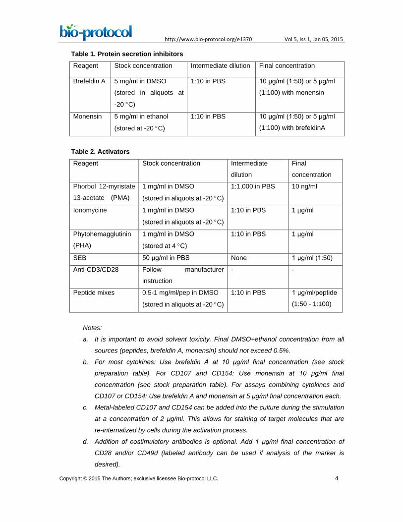
Table 1. Protein secretion inhibitors Reagent Stock concentration Intermediate dilution Final concentration
Brefeldin A 5 mg/ml in DMSO
(stored in aliquots at
-20 °C)
1:10 in PBS 10 μg/ml (1:50) or 5 μg/ml
(1:100) with monensin
Monensin 5 mg/ml in ethanol
(stored at -20 °C)
1:10 in PBS 10 μg/ml (1:50) or 5 μg/ml
(1:100) with brefeldinA
Table 2. Activators Reagent Stock concentration Intermediate
dilution
Final
concentration
Phorbol 12-myristate
13-acetate (PMA)
1 mg/ml in DMSO
(stored in aliquots at -20 °C)
1:1,000 in PBS 10 ng/ml
Ionomycine 1 mg/ml in DMSO
(stored in aliquots at -20 °C)
1:10 in PBS 1 µg/ml
Phytohemagglutinin
(PHA)
1 mg/ml in DMSO
(stored at 4 °C)
1:10 in PBS 1 µg/ml
SEB 50 μg/ml in PBS None 1 μg/ml (1:50)
Anti-CD3/CD28 Follow manufacturer
instruction
- -
Peptide mixes 0.5-1 mg/ml/pep in DMSO
(stored in aliquots at -20 °C)
1:10 in PBS 1 μg/ml/peptide
(1:50 - 1:100)
Notes:
a. It is important to avoid solvent toxicity. Final DMSO+ethanol concentration from all
sources (peptides, brefeldin A, monensin) should not exceed 0.5%.
b. For most cytokines: Use brefeldin A at 10 μg/ml final concentration (see stock
preparation table). For CD107 and CD154: Use monensin at 10 μg/ml final
concentration (see stock preparation table). For assays combining cytokines and
CD107 or CD154: Use brefeldin A and monensin at 5 μg/ml final concentration each.
c. Metal-labeled CD107 and CD154 can be added into the culture during the stimulation
at a concentration of 2 μg/ml. This allows for staining of target molecules that are
re-internalized by cells during the activation process.
d. Addition of costimulatory antibodies is optional. Add 1 μg/ml final concentration of
CD28 and/or CD49d (labeled antibody can be used if analysis of the marker is
desired).
Copyright © 2015 The Authors; exclusive licensee Bio-protocol LLC. 4
http://www.bio-protocol.org/e1370 Vol 5, Iss 1, Jan 05, 2015
2. Incubate the cells for 4 h (PMA + ionomycin stimulation, PHA + ionomycin stimulation) or
6-8 h (SEB, anti-CD3/CD28 stimulation, peptide stimulation) at 37 °C, in a CO2 incubator.
Note: For most cytokines 6-12 h incubation at 37 °C is sufficient; For IL-10 optimal
incubatation time is 12-24 h, but it is possible to detect in 6 h.
3. At the end of stimulation, add EDTA to a final concentration of 2 mM and incubate for 15
min at room temperature.
C. Staining
1. Wash 2x with 250 µl CyFACS buffer per well 1,550 rpm (RCF = 473), 10 min at room
temperature. The same volume and centrifuge conditions are used in additional wash
steps before fixation with PFA (step C6).
2. Make surface Ab cocktail in CyFACS buffer (filter with 0.1 µm spin filter) 100 µl per
reaction. Incubate on ice for 45 min. Use vendor’s recommended concentration (or
optimal titer as determined for self-made conjugates) for each antibody.
3. Wash 2x in CyFACS buffer.
4. Resuspend cells in 100 µl of 1:3,000 diluted 5 mg/ml 115-DOTA maleimide (titrated if new
stock) in CyPBS, Incubate 30 min on ice.
5. Wash 3x in CyFACS buffer.
6. Resuspend in 100 µl of 2% PFA in CyPBS, Incubate 4 °C overnight.
7. Wash 2x in 1x eBioscience perm buffer (1x in milliQ water), 2,000 rpm (RCF = 787), 10
min at 4 °C. The same volume and centrifuge conditions are used in the following wash
steps.
8. Make intracellular staining cocktail in 1x perm buffer and filter with 0.1 µm spin filter, 100 µl
per reaction. Incubate on ice for 45 min.
9. Wash 3x in CyFACS buffer.
10. Resuspend in 100 µl 1:2,000 Ir-Interchelator diluted in 2% PFA (in CyPBS).
11. Incubate 20 min at room temp.
12. Wash 2x in CyFACS buffer.
13. Wash 3x in MilliQ water.
14. Resuspend in MilliQ water (1 to 1.5 ml) for running on CyTOF.
Copyright © 2015 The Authors; exclusive licensee Bio-protocol LLC. 5
http://www.bio-protocol.org/e1370 Vol 5, Iss 1, Jan 05, 2015
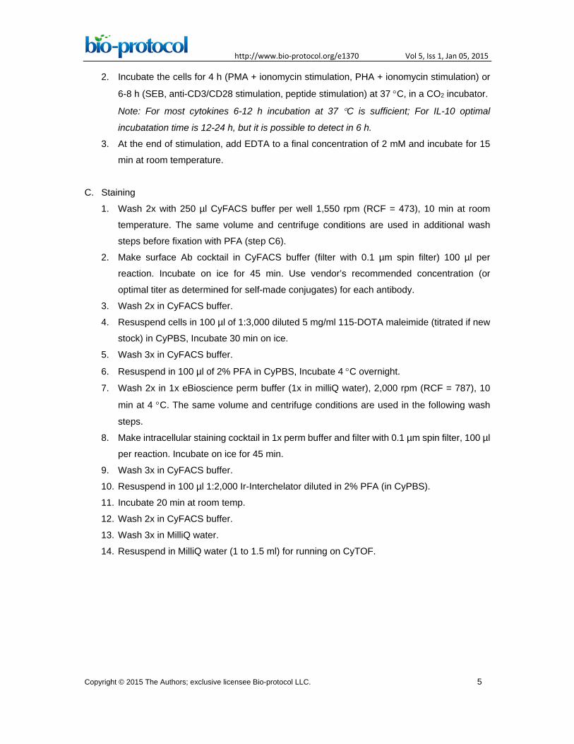
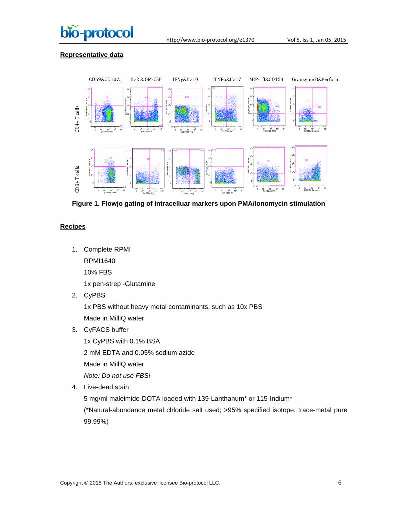
Representative data
Figure 1. Flowjo gating of intracelluar markers upon PMA/Ionomycin stimulation
Recipes
1. Complete RPMI
RPMI1640
10% FBS
1x pen-strep -Glutamine
2. CyPBS
1x PBS without heavy metal contaminants, such as 10x PBS
Made in MilliQ water
3. CyFACS buffer
1x CyPBS with 0.1% BSA
2 mM EDTA and 0.05% sodium azide
Made in MilliQ water
Note: Do not use FBS!
4. Live-dead stain
5 mg/ml maleimide-DOTA loaded with 139-Lanthanum* or 115-Indium*
(*Natural-abundance metal chloride salt used; >95% specified isotope; trace-metal pure
99.99%)
Copyright © 2015 The Authors; exclusive licensee Bio-protocol LLC. 6
http://www.bio-protocol.org/e1370 Vol 5, Iss 1, Jan 05, 2015
Acknowledgments
This work was supported by grants # S10RR027582 and 5U19AI057229 from the National
Institutes of Health. We thank Evan Newell and Mark Davis for help in the initial protocol
development.
References
1. Bendall, S. C., Simonds, E. F., Qiu, P., Amir el, A. D., Krutzik, P. O., Finck, R., Bruggner,
R. V., Melamed, R., Trejo, A., Ornatsky, O. I., Balderas, R. S., Plevritis, S. K., Sachs, K.,
Pe'er, D., Tanner, S. D. and Nolan, G. P. (2011). Single-cell mass cytometry of differential
immune and drug responses across a human hematopoietic continuum. Science
332(6030): 687-696.
2. Maecker, H. T. (2009). Multiparameter flow cytometry monitoring of T cell responses.
Methods Mol Biol 485: 375-391.
3. Newell, E. W., Sigal, N., Bendall, S. C., Nolan, G. P. and Davis, M. M. (2012). Cytometry
by time-of-flight shows combinatorial cytokine expression and virus-specific cell niches
within a continuum of CD8+ T cell phenotypes. Immunity 36(1): 142-152.
Copyright © 2015 The Authors; exclusive licensee Bio-protocol LLC. 7