
Interleukin 16 in Atherosclerosis and Cardiovascular ... · PDF fileInterleukin 16 in...
87
Interleukin 16 in Atherosclerosis and Cardiovascular Disease Grönberg, Caitriona Published: 2016-01-01 Document Version Publisher's PDF, also known as Version of record Link to publication Citation for published version (APA): Grönberg, C. (2016). Interleukin 16 in Atherosclerosis and Cardiovascular Disease Lund: Lund University, Faculty of Medicine General rights Copyright and moral rights for the publications made accessible in the public portal are retained by the authors and/or other copyright owners and it is a condition of accessing publications that users recognise and abide by the legal requirements associated with these rights. • Users may download and print one copy of any publication from the public portal for the purpose of private study or research. • You may not further distribute the material or use it for any profit-making activity or commercial gain • You may freely distribute the URL identifying the publication in the public portal Take down policy If you believe that this document breaches copyright please contact us providing details, and we will remove access to the work immediately and investigate your claim.
Transcript of Interleukin 16 in Atherosclerosis and Cardiovascular ... · PDF fileInterleukin 16 in...

LUND UNIVERSITY
PO Box 117221 00 Lund+46 46-222 00 00
Interleukin 16 in Atherosclerosis and Cardiovascular Disease
Grönberg, Caitriona
Published: 2016-01-01
Document VersionPublisher's PDF, also known as Version of record
Link to publication
Citation for published version (APA):Grönberg, C. (2016). Interleukin 16 in Atherosclerosis and Cardiovascular Disease Lund: Lund University,Faculty of Medicine
General rightsCopyright and moral rights for the publications made accessible in the public portal are retained by the authorsand/or other copyright owners and it is a condition of accessing publications that users recognise and abide by thelegal requirements associated with these rights.
• Users may download and print one copy of any publication from the public portal for the purpose of privatestudy or research. • You may not further distribute the material or use it for any profit-making activity or commercial gain • You may freely distribute the URL identifying the publication in the public portalTake down policyIf you believe that this document breaches copyright please contact us providing details, and we will removeaccess to the work immediately and investigate your claim.

Interleukin 16 in Atherosclerosis and Cardiovascular DiseaseCAITRÍONA GRÖNBERG | FACULTY OF MEDICINE | LUND UNIVERSITY
Lund University, Faculty of MedicineDepartment of Clinical Sciences, Malmö
Doctoral Dissertation Series 2016:75ISBN 978-91-7619-301-3
ISSN 1652-8220
My sister recently asked if I could write a short description of her for her thesis. The challenge was not the description per se, but rather to keep it short, for my sister has many facets to her character. She studied biotechnology at Swedish University of Agricultural Sciences and shortly thereafter went to an interview for the PhD position she later acquired. Rumours say that the interview was something special. You see, Caitríona herself would never claim success quite as brazenly, which is why I am doing it now. She’ll be cringing when she reads this and debating with herself if she should omit some of the description. Don’t. She is an older sister, a mother, a scientist, a wife, a daughter, a fantastic cheese cake maker, a gardener, an animal lover. But what always surprises me is not her long list of skills, but her will of steel and stamina. Every morning, early mind you, she sets out to reach goals and do a full day’s worth of work, after which she comes home to look after her family. Every morning, relentless. She’s a steam engine which simply cannot be stopped, an oak tree which cannot be uprooted, a powerful working horse ploughing the fi eld for seeds to be planted in. Her thesis is one of those seeds.
Úna Eriksson
Printed by Media-Tryck, Lund U
niversity 2016 N
ordic Ecolabel 341903
9789176
193013
CA
ITRÍO
NA
GR
ÖN
BER
G
Interleukin 16 in Atherosclerosis and C
ardiovascular Disease
75

1
Interleukin 16 in Atherosclerosis and
Cardiovascular Disease
Caitríona Grönberg
DOCTORAL DISSERTATION
by due permission of the Faculty of Medicine, Lund University, Sweden.
To be defended in the lecture hall at Kvinnokliniken, Skåne University Hospital,
Malmö on June 9th 2016 at 9.00.
Faculty opponent
Professor Bente Halvorsen

2
Organization
LUND UNIVERSITY
Document name
Doctoral Dissertation
Department of Clinical Sciences, Malmö Date of issue: June 9th 2016
Author: Caitríona Grönberg Sponsoring organization
Title and subtitle: Interleukin 16 in atherosclerosis and cardiovascular disease.
Abstract
Background and aim - The development of clinical manifictations due to an eroded or ruptured atherosclerotic plaque, or occluded vessel, are one of the major causes of death world wide. It has been known for some time that atherosclerosis develops due to retention and modification of LDL particles in the vessel wall and the subsequent triggering of the immune system. The research presented within this thesis has focused on IL-16, a signaling molecule, in the immune system. IL-16 has shown pleiotropic functions in inflammatory diseases. IL-16 has been described to have the capacity to induce T cell unresponsivness and increase the regulatory T cell population. Regulatory T cells are known to be protective in atherosclerosis by dampening the immune responses. There has been no extensive research on the role of IL-16 in atherosclerosis disease and the clinical manifestations thereof. The aim of the collected work in this thesis was to investigate if IL-16 can induce anti-inflammatory and atherosclerosis dampening effects, if IL-16 holds potential as a biomarker for prediciting future cardiovascular events, and if IL-16 is altered in an retrospective cardiovascular case-control study.
Results – (I) Administration of IL-16, in an experimental model of atherosclerosis consisting of hypercholesterolemic female mice, increased anti-inflammatory factors and decreased the atherosclerotic plaque burden. Male mice, which were defective of IL-16, displayed an increased atherosclerotic burden compared to control mice. (II) Elevated levels of IL-16, in carotid plaques, from individuals with severe carotid atherosclerosis displayed associations to plaque stabilizing components (collagen, elastin and FoxP3). High levels of carotid plaque IL-16 were associated to a decreased risk of suffering from a cardiovascular event. (III) Individuals suffering from severe carotid atherosclerosis had a decreased risk of suffering from a post-operative cardiovascular event, or a cardiovascular event leading to death, if they had high levels of circulating IL-16 compared to individuals with low levels of IL-16. (IV) In a population-based prospective study four single nucleotide polymophisms (SNPs) were found to be associated with IL-16 plasma levels. High plasma levels of IL-16 were associated with an increased risk of myocaridal infarction in women, compared to women with low levels of circulating IL-16. None of the SNPs were associated with an increased risk of cardiovascular events. One of the identified SNPs was associated with a decreased risk of all-cause mortality during the 20-year follow-up period. (V) In a retrospective case-control study, including individuals suffering from diabetes, IL-16 was 50% elevated in individuals whom also suffered from cardiovascular complications compared to the individuals only suffering from diabetes. Plasma levels of IL-16 were associated with surrogate markers of atherosclerosis which was further supported by SNP analysis.
Conclusion - IL-16 in plasma did not display associtions to a decreased risk of cardiovascular events in a prospective population-based study, rather the opposite. We have presented supporting evidence of a protective role of IL-16 in severe and experimental atherosclerosis, reinforced by the associations between high IL-16 levles and increasing amounts of stabilizing factors in the atherosclerotic plaque. We also present supporting evidence, in an experimental setting, for an anti-inflammatory role and plaque burden limiting role of IL-16. The data presented within this thesis warrents futher investigation of the plaque stabilizing properties of IL-16 in severe atherosclerosis and the role of IL-16 in promoting anti-inflammatory mediators.
Key words: Athersoclerosis, cardiovascular disease, interleukin- (IL)16, immunity,T cells, myocardial infarction, stroke.
Classification system and/or index terms (if any)
Supplementary bibliographical information Language: English
ISSN and key title: 1652-8220 ISBN: 978-91-7619-301-3
Recipient’s notes Number of pages 83 Price
Security classification
I, the undersigned, being the copyright owner of the abstract of the above-mentioned dissertation, hereby grant to all reference sources permission to publish and disseminate the abstract of the above-mentioned dissertation.
Signature Date

3
Interleukin 16 in Atherosclerosis and
Cardiovascular Disease
A fire fighter counteracting an inferno in the vessel wall.
Caitríona Grönberg

4
Coverphoto by Caitríona Grönberg produced in Sony Sketch, as has Fig 1-10, and
16-17.
Copyright (Caitríona Grönberg and Stroke: 1 September 2015 – Volume 47 –
Issue 10, p 2748-2754. Wolters Kluwer Health Lippincott Williams & Wilkins ©)
Faculty of Medicine | Department of Clinical Sciences, Malmö | Lund University
ISBN 978-91-7619-301-3
ISSN 1652-8220
Faculty of Medicine Doctoral Dissertation series 2016:75
Printed in Sweden by Media-Tryck, Lund University
Lund 2016

5
To my family
Hampus, Áine & Éirinn
Mamma, Pappa, Úna & Thomas

6
Table of contents
Preface 9
Original Papers 11
Published or submitted papers not included in this thesis 12
Abbreviations 13
Background 15
Lipoprotein metabolism 15
Atherosclerosis 16 The healthy artery 16 Atherosclerotic plaque development 17 Stable vs. vulnerable plaque 19
CVD 20 CV events 20 Risk factors 20 Treatment 20
Immune system 21 Innate immunity 21 Adaptive immunity 23
Apoptosis and anergy 26 Apoptosis 26 Anergy 27
IL-16 28 Lymphocyte Chemoattractant Factor 28 Pre-form and secreted IL-16 28 IL-16 effects on T cells and disease 29 IL-16 in CVD 30
Methods 31
Experimental atherosclerosis 31
Cohorts 32 CPIP 32

7
MDC 33 SUMMIT 33
Statistics 34 Statistical workflow 34
Techniques 37
Aims and key findings 41
Paper I 41
Paper II 41
Paper III 42
Paper IV 43
Paper V 43
Results and discussion 45
IL-16 in experimental atherosclerosis 45 Apoptosis 46
IL-16 and T cells 47
IL-16 in severe carotid atherosclerosis 49
IL-16 gene polymorphism 51
IL-16 and cardiovascular risk factors 52
IL-16 as a biomarker in primary prevention 53 IL-16 and obesity 54
IL-16 is associated with vascular complications of CVD 55
Pro and cons of IL-16 in the atherosclerotic disease 57
Conclusions and Future Perspectives 59
Populärvetenskaplig sammanfattning 61
Interleukin 16 - brandmannen som släcker farliga
eldsvådor i kärlväggen. 61
Science for everyone 65
Interleukin 16 – a fire fighter counteracting an
inferno in the vessel wall. 65
Acknowledgments 71
References 75

8

9
Preface
Atherosclerosis affects us all sooner or later, the question is whether the
atherosclerotic plaque will cause clinical symptoms, such as stroke and myocardial
infarction. It has been known for some time that atherosclerosis develops due to
deficient lipid clearance and modification, triggering the immune system, and due
to vessel wall defectiveness. Atherosclerosis is an intriguing and multifaceted
disease, demonstrated by the fact that one individual can have several different
atherosclerotic plaques in several different locations although only a few of them
might be prone to rupture and cause an acute clinical event.
The main focus for many researchers around the world is to find techniques to
identify the vulnerable plaques and to promote plaque stabilizing properties
allowing prevention of clinical events. Research in the field has generated great
insight and help to millions of patients every year. The new treatments have
however modified the disease and researchers are now struggling to “catch up”
with new therapies to be able to coevolve with the disease.
The research presented in this thesis is focused on a signaling molecule,
interleukin 16 (IL-16), in the immune system and whether this cytokine can hold
potential to both predict cardiovascular events but also if it may be used as a
therapy to limit atherosclerosis progression.
In the following pages you will find my perception of atherosclerosis, the immune
system and interleukin 16. This thesis does not present all the answers to the role
of interleukin 16 in this fascinating and deadly disease, although it raises some
possibilities and a lot of intriguing questions.

10
11
Original Papers
I. Caitríona Grönberg, Lukas Tomas, Sara Rattik, Úna Eriksson, Ingrid
Yao Mattisson, Anahita Abdali, Emelie Larsson, Linda Andersson,
Ragnar Alm, Lena Sundius, Ingrid Söderberg, Hardy Kornfeld, Gunilla
Nordin Fredrikson, Jan Nilsson, and Harry Björkbacka. IL-16 Reduces
Atherosclerotic Development in Hypercholesterolemic Apolipoprotein E
Deficient Mice. Manuscript.
II. Caitríona Grönberg, Eva Bengtsson, Gunilla Nordin Fredrikson,
Mihaela Nitulescu, Giuseppe Asciutto, Ana Persson, Linda Andersson,
Jan Nilsson, Isabel Gonçalves, and Harry Björkbacka. Human Carotid
Plaques with High Levels of Interleukin 16 are Associated with Reduced
Risk for Cardiovascular Events. Stroke 2015 Oct;46(10):2748-54.
III. Caitríona Grönberg; Giuseppe Asciutto, Ana Persson, Gunilla Nordin
Fredrikson, Jan Nilsson, Isabel Gonçalves, and Harry Björkbacka.
Endarterectomy Patients with Elevated Levels of Circulating IL-16 Have
Fewer Post-operative Cardiovascular Events. Submitted.
IV. Caitríona Grönberg, Gunilla Nordin Fredrikson, Marju Orho-Melander,
Gunnar Engström, Olle Melander, Jan Nilsson and Harry Björkbacka.
Interleukin 16 Levels in Plasma and Polymorphisms in Relation to
Incident Cardiovascular Events: A Prospective Population-Based Cohort
Study. Manuscript.
V. Caitríona Grönberg, Emma Ahlqvist, Gunilla Nordin Fredrikson, Isabel
Gonçalves, Andreas Edsfeldt, Helen M Colhoun, Angela C. Shore, Carlo
Palombo, Andrea Natali, Maria Wigren, Eva Bengtsson, Gerd Östling,
Kunihiko Aizawa, Francesco Casanova, Margaretha Persson, Kim
Gooding, Phil Gates, Faisel Khan, Helen C Looker, Fiona Adams, Jill
Belch, Silvia Pinnola, Elena Venturi, Michaela Kozakova, Li-Ming Gan,
Volker Schnecke, Jan Nilsson, Harry Björkbackaon behalf of the
SUMMIT consortium. Elevated Levels of Circulating IL-16 are
Associated with Vascular Complications of Diabetes. Manuscript.

12
Published or submitted papers not
included in this thesis
1. Caitríona Grönberg and Harry Björkbacka. Atherosclerosis: cell biology
and lipoproteins. Curr Opin Lipidol. 2012 Oct;23(5):505-8.
2. Andreas Edsfeldt, Mihaela Nitulsecu, Helena Grufman, Caitríona
Grönberg, Ana Persson, Marie Nilsson, Margareta Persson, Harry
Björkbacka and Isabel Gonçalves. Soluble urokinase plasminogen
activator receptor is associated with inflammation in the vulnerable
human atherosclerotic plaque. Stroke. 2014 Dec;43(12):3305-12.
3. Sara Rattik, Caitríona Grönberg, Maria Gomez, Harry Björkbacka,
Gunilla Nordin-Fredrikson, Jan Nilsson and Maria Wigren.
Apolipoprotein B-100 peptide p210 inhibits proliferation of naive T
effector cells and promotes induction of tolerogenic antigen presenting
cells and regulatory T cells in vitro. J Clin Cell Immunol 2015;6:3.
4. Giuseppe Asciutto, Maria Wigren, Gunilla Nordin-Fredrikson, Ingrid Yao
Mattison, Caitríona Grönberg, Ragnar Alm, Harry Björkbacka, Nuno
Dias, Andreas Edsfeldt, Isabel Gonçalves and Jan Nilsson. Apolipoprotein
B-100 anitbody interaction with atherosclerotic plaque inflammation and
repair processes. Stroke 2016;Apr;47(4):1140-3.
5. Maria Wigren, Sara Rattik, Caitríona Grönberg, Lukas Tomas, Ingrid
Yao Mattisson, Ingrid Söderberg, Ragnar Alm, Lena Sundius, Irena
Ljungcrantz, Harry Björkbacka, Gunilla Nordin Fredrikson, and Jan
Nilsson. Lack of ability to present antigens on MHC class II molecules
aggravates atherosclerosis in ApoE-/- mice. Submitted.

13
Abbreviations
AF amourosis fugaux
APC anitgen presenting cell
ApoA apolipoprotein A
ApoB apolipoprotein B
ApoC apolipoprotein C
ApoE apolipoprotein E
Au arbitrary unit
B cell B-lymphocytes
C57Bl/6 mice mice on the genetic background of C57 black 6
CD cluster of differentiation
CD25 IL-2 receptor alpha chain
CD4 expressed on Th cells, IL-16 ligand
CD8 expressed on Tc cells
CPIP carotid plaque imaging project
CRS cardiorenal syndrome
CTLA-4 cytotoxic T lymphocyte antigen 4
CV cardiovascular
CVD cardiovascular disease
DAMP danger associated molecular pattern
DC dendritic cell
de novo starting from the beginning
DNA deoxyribonucleic acid
DR death receptor
EAE experimental autoimmune encephalomyelitis
EC endothelial cell
ECM extracellular matrix
eGFR estimated glomerular filtration rate
FASL Fas ligand, CD95
FoxP3 forkhead box protein 3, Treg transcription factor
GATA3 GATA binding protein 3, Th2 transcription factor
HFD high fat diet
HLA-DR human leukocyte antigen - antigen D related, MHC class II
IFN-γ interferon gamma
IL interleukin
IL-10 interleukin 10, cytokine synthesis inhibitory factor
IL-12 interleukin 12

14
IL-16 interleukin 16, lymphocyte chemoattractant factor
IL-2 interleukin 2
ILC innate lymphoid cell
in vitro outside a living organism
in vivo inside a living organism
LDL low-density lipoprotein
LDLR low-density lipoprotein receptor
MDC Malmö diet and cancer study
MHC major histocompatibility complex
MI myocardial infarction
MMP metalloproteinase
NK cell natural killer cell
NOD mice non-obese diabetic mice
oxLDL oxidized low-density lipoprotein
PAMP pathogen-associated moleculer pattern
PBMC peripheral blood mononulear cell
PD-1 programmed cell death receptor 1, CD279
PDZ domain structural domain in signaling proteins
PRR pattern recognition receptor
RA rheumatoid arthritis
RNA ribonucleic acid
SMC smooth muscle cell
SNP single nucleotide polymorphism
SR scavenger receptor
SUMMIT surrogate markers for micro- and macrovascular hard end points for innovative diabetes tools
T cell T-lymphocyte
T1D type 1 diabetes
T2D type 2 diabetes
T-bet T box 21, transcription factor for Th1 cells
Tc cytotoxic T cells, CD8+
TCR T cell receptor
TG triglyceride
TGF-β transforming growth factor beta 1
Th T helper cell, CD4+
Th1 T helper cell type 1
Th2 T helper cell type 2
TIA transient ischemic attack
TLR toll-like receptor
Treg regulatory T cell
WBC white blood cell
VLDL very low-density lipoprotein
VSMC vascular smooth muscle cell
CD9 tetraspannin, alternative IL-16 ligand

15
Background
The word atherosclerosis comes from the Greek language and in direct translation
would be “disgusting/sticky hard tissue, or mass, of the arteries”. Factors that
compose life and factors that we expose ourselves to (genes, nutrient intake,
immune system, stress and toxins) determine the risk an individual has of
developing cardiovascular disease. Cardiovascular disease (CVD) represents the
clinical manifestations of atherosclerosis. How can this disgusting hard tissue be a
result of the same factors essential for life? All of these factors will be discussed in
the chapters below except for one, which is the ability to reproduce. The fact that
atherosclerosis causes clinical symptoms, which generally appear in individuals
after reproductive age, implies that atherosclerosis will never resolve due to
natural selection. With a constant increase in life expectancy and more effective
treatment of infectious diseases, there will also be an increasing population with
CVD. Today the clinical complications of atherosclerosis are the main cause of
death worldwide, therefore more research is needed to understand, find and treat
this disease.
Lipoprotein metabolism
Lipids such as cholesterol and triglycerides, which are essential for the cells, are
transported in the circulation enclosed by phospholipids and lipoproteins. The
lipids are enclosed by these proteins to facilitate the dissolvement in the blood, due
to the fact that the lipids are hydrophobic. As the body digests nutrients in the gut,
TGs are packed together with lipoprotein (apolipoprotein B48) to form
chylomicrons which are then passed on to the circulation.[1] Chylomicrons and
very low-density lipoproteins (VLDL) are the main carriers of TGs. The remnants
of chylomicrons are taken up by the liver. The master regulator of cholesterol in
the body is the liver, where VLDL is formed by fusing triglycerides (TGs),
cholesterol and apolipoprotein B100.
As the chylomicrons and VLDL deliver some of its lipids to cells, they become
denser and hydrolyzed and turn into low-density-lipoprotein (LDL). The major
source of cholesterol, from the liver, is delivered to the periphery by LDL. The

16
apolipoprotein B-100, on the surface of LDL, binds to receptors on cells and the
LDL is taken up by endocytosis. The left-overs from this transportation of lipids
are called high-density lipoprotein (HDL). HDL has the capacity to clear excess
cholesterol, and chylomicron remnants, from the circulation by binding scavenger
receptor class B type 1 in the liver (Figure 1).[1] The excess cholesterol is then
depleted from the system together with bile salts.
Most of us have heard about bad cholesterol (LDL) and good cholesterol (HDL),
this is however misleading as they are both essential for the body although due to
excessive intake of cholesterol and fat (or by familial hypercholesterolemia) the
balance is shifted and the LDL can become toxic.[2, 3]
FIgure 1. The chylomicron (largest particle) transports triglycerides and cholesterol encapsuled in phospholipids and apolipoproteins cells. VLDL contains mainly triglycerides, wheras LDL and HDL have increasing fraction of cholesterol compared to the triglyceride level.
Atherosclerosis
The healthy artery
The healthy artery is built up of three layers; the adventitia, tunica media and
tunica intima (closest to the lumen). The outermost layer, adventitia, consists of
extracellular matrix (ECM) proteins, immune cells, fibroblasts, and vasa vasora
which supplies nutrients to the vessel.[4] The intermediate layer, tunica media,
consists of smooth muscle cells (SMCs) surrounded by a basement membrane and

17
ECM. The ECM consists of several different types of collagens, elastin,
fibronectin and proteases.[5, 6] The tunica intima is the inner most layer closest to
the blood flow and is made up of endothelial cells (ECs)(Figure 2). The three
layers work together to supply the elasticity and compression needed to maintain
blood flow and pressure.[7]
Figure 2. A normal vessel wall (left) with the tunica intima (I) innermost to the lumen surrounded by SMCs in the tunica media (M) and the adventita (A). Formation of an atherosclerotic plaque (right) with a disturbed blood flow, attachement of white blood cells to the endothelial layer and accumulation of foam cells and immune cells in the subendothelial layer.
Atherosclerotic plaque development
The concept of atherosclerosis was observed and described more than a hundred
years ago, even in experimental models, as well as the connection between
atherosclerosis and the accumulation of lipids in the vessel wall.[8, 9] The
atherosclerotic lesion is initiated as fatty streaks in the vessel wall, in the
subendothelial cell layer, the occurrence of which can start in adolescence.[10]
The fatty streaks can be found in large to medium sized arteries and consists of
lipids and phagocytic white blood cells, called macrophages. There are some extra
vulnerable places in the arteries where the initiation of atherosclerosis normally
takes place (Figure 3).[11] Such places are characterized by regions with disturbed
or altered blood flow, which can trigger endothelial permeabilization and invasion
of white blood cells.

18
Figure 3. Atherosclerosis prone sites leading to CVD, in the arterial tree are manily the arteries in the; carotids, coronary, aorta, iliaca and femoral.
There are different theories to why atherosclerosis develops, Tabas and Williams
presented the response-to-retention model in 1995.[12] They propose that the key
factor for the development of atherosclerosis is the retention of apolipoprotein
B100 in the subendothelial space. The trapped lipoproteins bind to ECM where
they are retained and undergo modifications and oxidation and are engulfed by
macrophages.[13] Ross and Glomset have presented a model based on the
response-to-injury concept, which focuses on a disturbance of the endothelial layer
(injury or by activation) leading to an increased influx of cholesterol and immune
cells.[14]
The LDL that enters the vessel wall may be subjected to oxidation and
modification. The oxidized LDL (oxLDL) is thought to be the main immunogenic
factor in atherosclerosis, leading to increased responsiveness of the macrophages.
The oxLDL is recognized as foreign, non-self material, due to the modifications
on the surface making leading to binding, and activation of pattern-recognition
receptors (PPRs), scavenger receptors (SRs), and toll-like receptors (TLRs).[15-
17] The uptake of the oxLDL, which resembles pathogens for the macrophages,
will induce a release of pro-inflammatory factors from the macrophages and will
further attract immune cells by activation of the ECs.
The SMCs respond to the inflammation by migrating to the intima and by
producing ECM proteins to repair the wound. As the macrophages in the fatty
streaks take up more lipids, they will swell into lipid-laden foam cells. The foam
cells, which are stuck in the vessel wall, are prone to die a messy death, called
necrosis.[18] The necrosis results in a release of a dangerous mixture of
intracellular proteins and oxLDL into the subendothelial layer, which will further
contribute to inflammation and the formation of a toxic necrotic core.[19]

19
Stable vs. vulnerable plaque
We can live with atherosclerotic plaques for decades without noticing that they are
present in our arteries. Some people may even have large atherosclerotic plaques
without having any symptoms. These plaques are referred to as stable plaques, and
consist of higher levels of SMCs, which produce ECM and a thicker fibrous cap
(enclosing the plaque from the lumen) (Figure 4). The only time the individual
will notice a stable plaque is due to a massively enlarged plaque and that it thereby
restricts the blood flow.
Figure 4. A stable plaque (not prone to rupture) is charecterized by SMCs, ECM proteins, and a thick fibrous cap (left). A vulnerable plaque is characterized by immune cell infiltration, large lipid and necrotic core and a thin fibrous cap (right).
A vulnerable plaque is characterized by a large necrotic/lipid core, more
inflammation and a thin fibrous cap. Macrophages have the possibility to produce
matrix metalloproteinases (MMPs) which can degrade ECM and the fibrous
cap.[20, 21] If the cap ruptures, the plaque content enter the lumen and during
contact with the blood flow, cause a thrombosis (blood clot) formation. The
thrombus can occlude the lumen and cut off blood flow at the site of the rupture or
it can be flushed with the blood flow and occlude smaller vessels further on in the
arterial tree. The occlusion causes ischemia, and symptoms from such thrombus
formations are known as stroke and myocardial infarction (MI). There is immense
research focused on identifying vulnerable plaques and how to promote these
plaques to switch into the stable phenotype, where silencing the immune system is
thought to be one key factor.[22, 23]

20
CVD
CV events
As atherosclerosis progresses it is possible that the atherosclerotic plaque erodes,
leaks or blocks the arteries and clinical symptoms may arise. Cardiovascular
disease (CVD) includes symptoms in the brain such as ischemic stroke, transient
ischemic attack (TIA) and amourosis fugax (AF), which all can be caused by
atherosclerotic plaques situated in the carotid arteries. MI and unstable angina are
caused by arteries being obstructed in the vessels surrounding and supplying the
heart with blood. Peripheral artery disease is an atherosclerotic disease affecting
the extremities of the body and can lead to intermittent claudication. Interestingly,
not all of the atherosclerotic plaques lead to a cardiovascular (CV) symptom.
Risk factors
As mentioned before, the atherosclerotic plaques may be relatively large without
affecting the individual. The Framingham Heart Study was initiated in1948 to
identify risk factors associated with increased risk of suffering from CVD.[24, 25]
The study concluded that the following risk factors are of interest; age, male sex,
smoking, total cholesterol, HDL cholesterol, and systolic blood pressure with the
addition of diabetes mellitus and body-mass-index (when cholesterol levels are not
available).[3, 26] The risk factors established in the Framingham Heart Study
seems to be reliable when calculating risk for CVD in a previously healthy
individual, however, when the individual has established CVD or is on medication
for one or several of these risk factors, they do not seem to predict risk as well.
Treatment
Today the current treatment is to suggest lifestyle changes, remove or stent the
atherosclerotic plaques or/and to medicate with aspirin, beta-blocker, ACE
inhibitors and statins. Aspirin decreases the platelets ability to form aggregates and
thereby decreases the risk for thrombus formation. Beta-blockers block the
response to stress hormones in the heart, arteries and kidneys by interfering with
the receptors for adrenalin and thereby not allowing the sympathetic system to
“over heat”. ACE inhibitors decrease the contraction of blood vessels and reduce
blood pressure by inhibiting the conversion of angiotensin I to angiotensin II.
Statins have been proven to lower lipid levels through inhibition of cholesterol
production in the liver, by reducing LDL through an up regulation of LDL

21
receptors in the liver.[27] Statin treatment has also been associated with increased
levels of the athero-protecitve regulatory T cells.[28, 29]
Recently an already approved compound, cyclodextrin, has been proposed as a
possible new treatment for cholesterol in atherosclerotic plaques. The authors
present evidence, in an experimental setting, for an increase in cholesterol efflux
from macrophages and reduction in atherosclerotic progression after treatment
with cyclodextrin.[30] As hypercholesterolemia, hypertension and diabetes is
dramatically increasing, the usage of medication escalates. The treatment will
most probably change the phenotype of the atherosclerotic plaques and the need
for new biomarkers and therapies are essential to be able to coevolve with the
disease and to resolve CVD.
Immune system
The immune system is an essential part of the body and it is responsible for
protecting the body from invading hostile particles. It is a myriad of cells and
molecules that patrol and “talk” with each other to ensure that the body is not
being infected. The immune system has traditionally been divided into two parts,
the innate, which is the first line of defense, and the adaptive which is responsible
for remembering previously encountered “wrong doers”. Most of the cells that are
a part of the immune system are produced in the bone marrow, by hematopoietic
cells, and are then relocated to lymphoid organs (thymus, spleen, lymph nodes)
and into other tissues.
Innate immunity
First line of defense
The innate immune system is our first line of defense, and therefore needs to be
located where the body is most likely to come across pathogens (skin, gut, liver
and lungs). Examples of such cells are mast cells, macrophages and neutrophils.
Mast cells in the skin and lungs are responsible for the rapid response to allergens
involved in symptoms such as hay fever, contact allergy and bee stings. People
suffering from these illnesses are of course not so pleased with the immune system
but without these cells harmful pathogens would have a free passage and could
destroy the body. We also have tissue resident phagocytes, such as macrophages
and dendritic cells, which engulf foreign particles to degrade and present them to
effective killer cells which can seek out and kill pathogens. The phagocytic cells
can recognize these foreign particles by cell surface receptors called DAMPs
22
(danger-associated molecular pattern molecules), PAMPs (pathogen-associated
molecular pattern molecules) and TLRs (toll like receptors).[16]
Common for all types of cells in the immune system is that they communicate
with each other by releasing molecules, called chemokines and cytokines, which
attracts other immune cells and fine-tunes them to respond in an appropriate way.
The phagocytic cells also have the possibility to present small parts of the engulfed
particles on their cell surface to enable the adaptive immune cells to respond.
Previously it was thought that the innate immune system did not have any cells
which had the ability of creating immunological memory, however during the
recent years more evidence has been presented for an emerging field of cells
representing innate phenotypes but also displaying capacity to confer memory or
to produce the same effector molecules as the adaptive immune system.[31]
Among these cells are B1 cells, innate lymphoid cells (ILCs) and natural killer
cells (NKs).[32-34] These cells are outside the scope of this thesis, even if they are
extremely interesting.
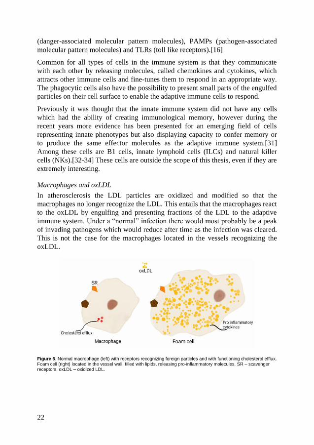
Macrophages and oxLDL
In atherosclerosis the LDL particles are oxidized and modified so that the
macrophages no longer recognize the LDL. This entails that the macrophages react
to the oxLDL by engulfing and presenting fractions of the LDL to the adaptive
immune system. Under a “normal” infection there would most probably be a peak
of invading pathogens which would reduce after time as the infection was cleared.
This is not the case for the macrophages located in the vessels recognizing the
oxLDL.
Figure 5. Normal macrophage (left) with receptors recognizing foreign particles and with functioning cholesterol efflux. Foam cell (right) located in the vessel wall, filled with lipids, releasing pro-inflammatory molecules. SR – scavenger receptors, oxLDL – oxidized LDL.

23
Increased cholesterol intake will most likely increase the levels of oxLDL
available for the macrophages in the vessel wall. As a consequence of this the
macrophages in the vessel wall will engulf more and more oxLDL and finally
swell into lipid laden foam cells.[35] The foam cells will eventually burst and a
highly toxic mass of cell debris and lipids will leak out and further activate the
immune system in the vessel wall, and a destructive cycle has been initiated
(Figure 5).[36, 37]
Adaptive immunity
The adaptive immunity has the capacity of producing memory cells which are
derived from cells that have encountered their specific antigen. There are two main
populations of cells belonging to the adaptive immunity, B and T cells. These cells
have a total repertoire of over ten billion antigen receptors. Several of the different
subtypes of B and T cells have been described to affect atherosclerosis.[38]
B cells
B cells are responsible for producing antibodies that bind to specific sequences on
cells or particles. The antibody-particle complex is then either cleared away by
phagocytic cells or forms an immune complex. The first time a B cell encounters
its pathogen it takes approximately a week to produce the specific antibody
required to bind to the pathogen of interest.
The second time the body is infected with the same pathogen there are memory B
cells, which were produced from the primary infection, that are specialized to
recognize this specific pathogen and a rapid antibody production is assembled. It is
also possible to introduce a part of the pathogen, which is not as toxic as the entire
pathogen, to produce memory B cells. This memory response is the key for how
vaccination functions in such an effective way.
There are several known subtypes of B cells where B1 cells have been associated
with athero-protection and B2 and regulatory B cells (Breg) have displayed both
pro- and anti-atherosclerotic properties.[39-43]
T cells
Stem cells, which produce T cells, are located in the bone marrow and are later
transported to the thymus. In the thymus the T cells mature and undergo positive
and negative selection for binding to antigen, here apoptosis (programmed cell
death) plays a crucial role in clearing defective T cells.
24
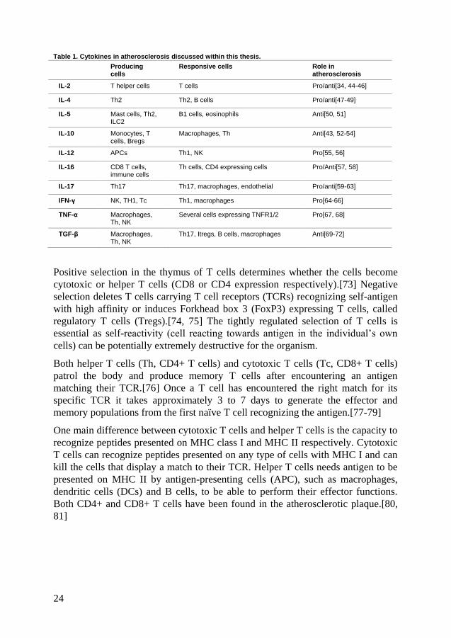
Table 1. Cytokines in atherosclerosis discussed within this thesis.
Producing cells
Responsive cells Role in atherosclerosis
IL-2 T helper cells T cells Pro/anti[34, 44-46]
IL-4 Th2 Th2, B cells Pro/anti[47-49]
IL-5 Mast cells, Th2, ILC2
B1 cells, eosinophils Anti[50, 51]
IL-10 Monocytes, T cells, Bregs
Macrophages, Th Anti[43, 52-54]
IL-12 APCs Th1, NK Pro[55, 56]
IL-16 CD8 T cells, immune cells
Th cells, CD4 expressing cells Pro/Anti[57, 58]
IL-17 Th17 Th17, macrophages, endothelial Pro/anti[59-63]
IFN-γ NK, TH1, Tc Th1, macrophages Pro[64-66]
TNF-α Macrophages, Th, NK
Several cells expressing TNFR1/2 Pro[67, 68]
TGF-β Macrophages, Th, NK
Th17, Itregs, B cells, macrophages Anti[69-72]
Positive selection in the thymus of T cells determines whether the cells become
cytotoxic or helper T cells (CD8 or CD4 expression respectively).[73] Negative
selection deletes T cells carrying T cell receptors (TCRs) recognizing self-antigen
with high affinity or induces Forkhead box 3 (FoxP3) expressing T cells, called
regulatory T cells (Tregs).[74, 75] The tightly regulated selection of T cells is
essential as self-reactivity (cell reacting towards antigen in the individual’s own
cells) can be potentially extremely destructive for the organism.
Both helper T cells (Th, CD4+ T cells) and cytotoxic T cells (Tc, CD8+ T cells)
patrol the body and produce memory T cells after encountering an antigen
matching their TCR.[76] Once a T cell has encountered the right match for its
specific TCR it takes approximately 3 to 7 days to generate the effector and
memory populations from the first naïve T cell recognizing the antigen.[77-79]
One main difference between cytotoxic T cells and helper T cells is the capacity to
recognize peptides presented on MHC class I and MHC II respectively. Cytotoxic
T cells can recognize peptides presented on any type of cells with MHC I and can
kill the cells that display a match to their TCR. Helper T cells needs antigen to be
presented on MHC II by antigen-presenting cells (APC), such as macrophages,
dendritic cells (DCs) and B cells, to be able to perform their effector functions.
Both CD4+ and CD8+ T cells have been found in the atherosclerotic plaque.[80,
81]

25
Th1
Depending on which factors are available at the encounter with the antigen, the T
helper cells can be divided into different subtypes; Th1, Th2, Th17 and regulatory
T cells. Th1 cells are produced to fight intracellular pathogens, such as viruses.
Phagocytic cells produce among others interleukin (IL)-12 and TNF-α when
presenting the antigen to T cells and prime the T cells towards a Th1 response.[82]
The Th1 cells are characterized by producing large amounts of interferon gamma
(IFN-γ) and have the T-box 21 (T-bet) transcription factor readily transcribed.
Macrophages react to the IFN-γ by producing pro-inflammatory cytokines, nitric
oxide and matrix metalloproteinases MMPs.[18, 83] Th1 cells have been implied
to be harmful in the atherosclerotic disease, which was demonstrated in studies by
generating Tbet or Ifnγ deficient, atherosclerotic prone, mice (Table 1).[64, 84]
Th2
Th2 cells are traditionally evoked as a response to extracellular pathogens and are
important in the humoral response of B cells. Th2 cells are characterized by their
production of IL-4, IL-5 and IL-13 and Th2 cells have the GATA binding protein
3 (GATA3) as their hallmark transcription factor. The role of Th2 cells in
atherosclerosis have yielded both pro and anti-atherosclerotic results. [47-49, 55,
85] IL-5 deficiency in atherosclerosis prone mice did however increase
atherosclerosis and there was also a reduction in natural antibodies.[50] Studies on
IL-5 and IL-13 are not easily interpreted as we now know that ILC type 2 have the
same cytokine profile and have been demonstrated to be athero-protective (Table
1).[33, 34]
Tregs
The first time I heard about regulatory T cells was at a course in advanced
immunology in the year of 2007. In this course the teachers presented regulatory T
cells as a really hot field in immunology. Now, nearly ten years later, the
regulatory T cells (Tregs) are still a hot topic especially in the field of
atherosclerosis.[86] The reason for this is that the Tregs have the ability to reduce
responsiveness of the T effector cells and APCs, which is of course beneficial in
the non-resolving inflammatory setting of atherosclerosis.
Tregs are generally described as expressing the transcription factor FoxP3 and the
cell surface markers CD4 and CD25. The Tregs can either be produced in the
thymus, natural Tregs, or in the periphery, which are called induced Tregs. There
is however an ever emerging field of markers and subtypes of these
immunosuppressive Tregs being described.[86, 87]
There are several potential mechanisms where the Tregs may be beneficial in
reducing atherosclerosis. The most commonly described is their ability to produce

26
the anti-inflammatory cytokines IL-10 and transforming growth factor beta 1
(TGF-β), which suppress effector cells. Tregs are known to consume large
amounts of IL-2, reducing the availability for other proliferating T cells and
thereby decreasing the cells proliferative capacity.[88] Cell to cell contact can also
induce immunosuppression.in which Tregs express CTLA-4 and by binding CD80
and CD86 on APCs they can reduce the pro-inflammatory response in the
APCs.[86]
There is substantial evidence for a protective role of Tregs in atherosclerosis, both
in humans and in experimental models.[86, 88-93] In mice there has even been
evidence that Tregs play a role in the clearance of VLDL and chylomicrons from
the circulation.[94] In humans, statin treatment has not only been seen to reduce
the cholesterol levels but also in increasing the circulating Tregs after
treatment.[29, 95]
Apoptosis and anergy
Apoptosis
Apoptosis is essential for a functioning development of the fetus and for clearance
of unneeded or exhausted cells in the body. Apoptosis is a highly controlled
clearing mechanism, where particles of the cells are packaged and released for
other cells to clear. The opposite of apoptosis is necrosis, where the cell dies in an
uncontrolled fashion, releasing cell debris and toxins into the surrounding tissue.
Apoptosis can be induced by the extrinsic pathway, or by the intrinsic pathway.
The extrinsic pathway is initiated by ligands binding to death receptors (DRs) on
the surface of cells, followed by an intracellular activation of caspases (Figure
6).[96] The intrinsic pathway is promoted by mitochondrial dysfunction and the
release of cytochrome c, which will in turn activate caspases (Figure 6).[97] Both
pathways are known to traditionally result in caspase-3 activation, which will in
turn result in certain effector functions of apoptosis. Recent studies, however,
suggest the possibility of caspase-3 to have an important role in silencing the
immune system by decreasing interferon production.[98, 99]

27
Figure 6. Apoptosis can either be initiated by extrinsic factors binding to death receptors (DRs) or by cellular stress, which induces cytochrome c release from the mitochondria (intrinisic). Both the extrinsic and the intrinsic pathway activate caspase-3 and 7 and activates the ”find me”, ”eat me”, ”tolerate me” signals produced by the apoptotic cell.
Anergy
Anergy is a term used for cells, in this case T cells, that have been silenced and are
not able to undertake their effector functions. Anergy is normally initiated after an
incomplete activation of a T cell. T cells also undergo deletion if they recognize
self-antigen, although not all of the self-antigens are present in the thymus and
therefore some of the T cells will encounter self-antigen in the periphery. To
prevent harmful auto-reactivity, anergy is essential.
In the spleen and lymph nodes anergic CD4+ T cells are characterized by increased
expression of CD44, FR4, CD73, Nrp1, Ki67, CD69, PD-1, CTLA-4, and Nur77
compared to naïve T cells.[100] Caspase-3 activation has also been reported to
induce anergic T cells.[101] Recent studies have showed a role of anergic T cells
in promoting Tregs.[100] To date I do not know of any evidence of anergic T cells
presented as athero-protective. The data supporting the protective role of Tregs in
atherosclerosis, however, makes the anergic T cells a really promising target to
study in atherosclerosis.

28
IL-16
Lymphocyte Chemoattractant Factor
Interleukin (IL-)16 was originally described as Lymphocyte Chemoattractant
Factor (LCF) in 1982 by David Center and William Cruikshank, although was
later given the name IL-16 in 1995.[102, 103] The newly discovered cytokine was
described as having chemoattractant properties in naive T4+ lymphocytes (CD4+
T cells).[104] Early studies in human T cells also showed an increase of IL-2
receptor (CD25) and HLA-DR (MHC class II) on T cells treated with IL-16, as
well as chemoattractant properties for monocytes expressing CD4.[104]
Further studies demonstrated a specific binding of IL-16 to the CD4 molecule
which could be blocked by anti-CD4 antibody, the study also showed that the
binding interacted with the D4 domain.[105] There are a number of different cells
expressing CD4, which include T helper cells, monocytes, macrophages, dendritic
cells, eosinophils, epithelial cells, NK cells, mast cells. The tetraspanin CD9 has
also been discussed as a possible receptor for IL-16 on mast cells, as has the
involvement of CCR5 on T cells.[106-108]
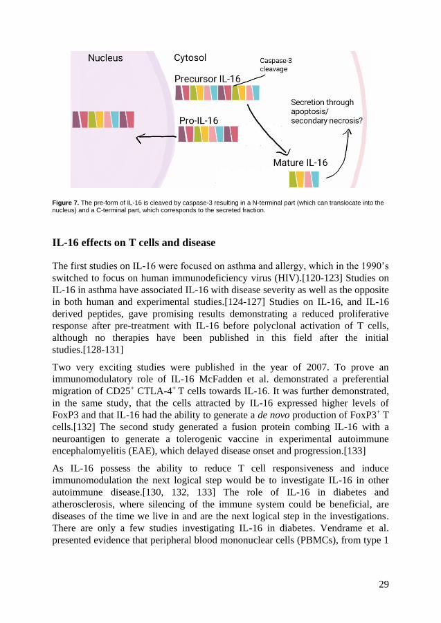
Pre-form and secreted IL-16
IL-16 is a unique cytokine with no significant sequence homology to other known
cytokines. The IL-16 gene is located on chromosome 15 and is produced in a pre-
form (631 amino acids). The pre-form is cleaved by caspase-3 to produce the
secreted C-terminal (121 amino acids) part and the N-terminal part which has the
ability to translocate into the nucleus and to induce cell-cycle arrest (Figure
7).[109-112] The secreted form can be found pre-made in the cytosol of CD8+ T
cells and in fibroblasts, while other cell types cleave the pre-form to produce the
bioactive IL-16 shortly before secretion.[113, 114]
There is high homology within IL-16 between different species and human IL-16,
and the blocking antibody, has been proven to be effective in rodent
experiments.[115-117] IL-16 is rare in the cytokine world due to the PDZ-like
motif and is the first extracellular protein found to have a PDZ motif.[110, 118]
PDZ motifs are important in anchoring receptor proteins in the membrane to
cytoskeletal factors and regulate biological processes such as ion channel signaling
and transduction. The mechanism by how IL-16 is secreted is not fully understood,
however recent work on neutrophils show extracellular IL-16 only after secondary
necrosis.[119]
29
Figure 7. The pre-form of IL-16 is cleaved by caspase-3 resulting in a N-terminal part (which can translocate into the nucleus) and a C-terminal part, which corresponds to the secreted fraction.
IL-16 effects on T cells and disease
The first studies on IL-16 were focused on asthma and allergy, which in the 1990’s
switched to focus on human immunodeficiency virus (HIV).[120-123] Studies on
IL-16 in asthma have associated IL-16 with disease severity as well as the opposite
in both human and experimental studies.[124-127] Studies on IL-16, and IL-16
derived peptides, gave promising results demonstrating a reduced proliferative
response after pre-treatment with IL-16 before polyclonal activation of T cells,
although no therapies have been published in this field after the initial
studies.[128-131]
Two very exciting studies were published in the year of 2007. To prove an
immunomodulatory role of IL-16 McFadden et al. demonstrated a preferential
migration of CD25+ CTLA-4+ T cells towards IL-16. It was further demonstrated,
in the same study, that the cells attracted by IL-16 expressed higher levels of
FoxP3 and that IL-16 had the ability to generate a de novo production of FoxP3+ T
cells.[132] The second study generated a fusion protein combing IL-16 with a
neuroantigen to generate a tolerogenic vaccine in experimental autoimmune
encephalomyelitis (EAE), which delayed disease onset and progression.[133]
As IL-16 possess the ability to reduce T cell responsiveness and induce
immunomodulation the next logical step would be to investigate IL-16 in other
autoimmune disease.[130, 132, 133] The role of IL-16 in diabetes and
atherosclerosis, where silencing of the immune system could be beneficial, are
diseases of the time we live in and are the next logical step in the investigations.
There are only a few studies investigating IL-16 in diabetes. Vendrame et al.
presented evidence that peripheral blood mononuclear cells (PBMCs), from type 1

30
diabetic patients, produced lower levels of active caspase-3, which correlated to
the decreased levels of IL-16.[134] Non-obese diabetic mice (NOD), treated with a
blocking IL-16 antibody, displayed less type 1 diabetes.[115] There is also
evidence for increasing IL-16 plasma levels in obese individuals and decreased
levels of IL-16 in CD8+ T cells from individuals that smoke with a subsequent
increase in bronchoalveolar lavage.[135, 136]
IL-16 in CVD
IL-16 seems to be involved in several of the key risk factors for CVD, however the
involvement of IL-16 in atherosclerosis, and metabolic disease, has just recently
started to be investigated. At present there is one study investigating the potential
role of IL-16 as a prospective biomarker for CVD. In this particular study, IL-16 is
presented in an algorithm-based approach combining IL-16 with several other
markers to be able to detect individuals with intermediate risk of CVD.[57]
Findings of Cross et al. associate increased IL-16 plasma levels with an increased
risk of coronary heart disease during a five-year follow-up reporting a twofold
increased risk after adjustment for Framingham risk factors.[57]
There have been several studies associating IL-16 gene polymorphism (rs8034928,
rs3848180, rs1131445, rs11556218, rs4778889 and rs4072111) with coronary
artery disease (CAD) and ischemic stroke.[137-140] None of the genetic
polymorphism studies have investigated the SNPs in a prospective cohort.
Recently there was a study investigating the role of IL-16 on vascular smooth
muscle cells (VSMCs) which demonstrated an increased migration and invasion of
VSMCs after IL-16 treatment, with a specific induction of metalloproteinase
(MMP)-9 and activation of transcription factors nuclear factor (NF)-κβ, activator
protein (AP)-1, as well as the cell-cycle-inhibitor p21WAF1 in VSMCs.[141]
Tamaki et al. showed that IL-16 can possibly impact cardiac fibrosis and
myocardial stiffening in patients with preserved ejection fraction, which was
further confirmed in experimental animal models.[116]
There is one very interesting unpublished study on Ldlr deficient mice
investigating the role of IL-16 in atherosclerosis development by abolishing the
circulating IL-16 through DNA vaccination. The results seemed to astound the
authors where they described an increased lesion formation in the carotid artery
and in the aorta, with increased levels of circulating T cells after abrogating IL-16
levels (Wanrooij. Thesis. 2007. http://hdl.handle.net/1887/12357). These results do
however fit very well with the data presented in paper I within this thesis.

31
Methods
I would like to start by emphasizing that in scientific research the most important
factor is the study design. With a correct and thoroughly thought through study
design you can be sure that the read out will answer your hypothesis. If the study
design is not optimal, there can be difficulties in both achieving an answer to the
hypothesis or even to prove that the finding is true, due to lack of statistical power.
I think that the PhD years have given me this insight and that the study design is
no simple matter, but essential.
Experimental atherosclerosis
In this thesis I have used both experimental models, in this case laboratory mice,
and human samples. The reason we use mice, to study atherosclerosis, is that they
are mammals, and are relatively close to humans evolutionary. The mice are easy
to house and can generate offspring rapidly. There are a lot of tools and methods
established for working and investigating the role of the immune system and
atherosclerosis in mice. These mice have a life expectancy of approximately 2
years, which is advantageous when studying a disease that develops over time.
In paper I, we have used a mouse model that spontaneously develops
atherosclerotic plaques, due to a deficiency in the Apoe gene. The deficiency
results in a lack of clearance of cholesterol particles in the circulation, causing
excess cholesterol to enter the vessel wall and initiate atherosclerotic lesions. The
mice are further pushed into an atherosclerotic phenotype by giving them a high
fat diet (HFD, 21% fat and 0.15% cholesterol) resembling a Western diet.
The mice are on C57Bl/6 background which has been proven to be the most
susceptible for atherosclerosis, this is important as wild mice are resistant to
atherosclerosis, most probably due to their high levels of HDL.[142-145]
Furthermore, female mice develop larger lesions than male mice, which is evident
in paper I.[146] The mice that are referred to as wild-type, in paper I, are
C57Bl/6 mice.
The mice display plaque formation in the aortic root, aortic arch and finally in the
descending aorta. The morphology of the plaques resembles the human

32
atherosclerotic plaques although with one difference; they do not tend to rupture
and produce thrombus formations. This implies that the mice are a tool for
investigating plaque initiation, progression and phenotype although not for
studying the events associated with the human cardiovascular disease.
Cohorts
At the University of Lund and the Skåne University Hospitals (SUS), in
collaboration with other universities, there have been great efforts to produce high
quality databases of human material. I have had the opportunity to work with three
of these databases (Figure 8). All individuals included, in any of the cohorts, gave
written informed consent.
Figure 8. Overview of the clinical cohorts used in this thesis, the number of individuals analyzed in each cohort and the associations to IL-16.
CPIP
In paper II and III I have worked with the cohort called Carotid Plaque Imaging
Project (CPIP). This biobank was, and is currently, collected from patients
undergoing carotid endarterectomy (the carotid plaque was surgically removed) at
the Vascular Department at SUS, Malmö. It is designed to prospectively
investigate secondary prevention in patients with severe carotid atherosclerosis.
Inclusion criteria were carotid stenosis degree above 80% (asymptomatic) or
carotid stenosis above 70% with stroke, TIA or AF occurring within one month

33
before surgery (symptomatic). The patients had blood taken the day before surgery
and the carotid plaque was snap frozen in liquid nitrogen until further processing.
The most highly stenotic part was saved for sectioning and histological analysis
whereas the rest was homogenized and analyzed for other components such as
lipids, RNA and protein levels. This database is extremely valuable because it
contains both general patient data on risk factors, circulating factors, and an
extensive characterization of the plaque phenotype by ultrasound, histology,
protein, lipid and RNA profiling. The patients in this cohort are generally old,
have considerable medication treatment and have advanced atherosclerosis.
Plasma samples (n=473), and carotid homogenates from 206 patients were
assessed on the O-link platform in 2014, where a total of 92 analytes were
measured, including IL-16. The individuals in the CPIP cohort, were generally a
few years older and more extensively medicated (statins, anti-hypertensive and
beta-blockers) than the individuals included in the SUMMIT study (paper V).
Another difference in the CPIP cohort (paper II-III) is that not all of the
individuals have had CV events, or diabetes, compared to the individuals in the
SUMMIT study (paper V).
MDC
The Malmö Diet and Cancer Study (MDC) was initiated in the 1990’s to facilitate
research on how environmental factors predict diseases like cancer, diabetes and
CVD in a population-based prospective study on primary prevention of the Malmö
inhabitants. The entire study consists of approximately 30 000 individuals and
6103 of these were randomly selected to be a part of the cardiovascular arm of
MDC. Blood samples were taken and plasma and cells were stored until further
investigated. In 2014 plasma samples from 4742 individuals were analyzed on the
O-link platform to determine the levels of 151 analytes in the circulation. Data was
collected in 2013 from the National registries to follow-up on morbidity, mortality
causes and events.
SUMMIT
The SUMMIT study (surrogate markers for micro- and macrovascular hard end
points for innovative diabetes tools) is the latest and in some ways the trickiest
cohort that I have worked with. The cohort is a multicenter study investigating
cardiovascular complications of diabetes in 1500 individuals in Dundee, Exeter,
Lund and Pisa. The design of the study is a retrospective case-control study
including four different groups; relatively healthy individuals, individuals with

34
CVD, individuals with type 2 diabetes (T2D), and individuals with a combination
of CVD and T2D. There was no retrospective limit in time for the occurrence of
the CV event or the onset of T2D, which results in a relatively heterogeneous
diseased population.[147]
Plasma samples were taken from all of the individuals and circulating cells were
analyzed on 211 individuals included in Lund. Plasma samples were sent to O-link
to analyze 92 circulating factors where IL-16 was included. The strength of the
cohort is that it is a multicenter study, although the multicenter aspect also makes
the analysis complicated. It became evident that the different centers had slightly
different populations when analyzing risk factors and medication.
In paper V we associated the prevalence of CVD and T2D with IL-16 plasma
levels. It would be very interesting to reassess the findings, within the same cohort
after a few years, on follow-up data describing the incidence of CVD and the
progression of surrogate markers of vascular complications.
Statistics
Statistical workflow
To be able to answer the hypothesis that IL-16 could be a biomarker in CVD I
needed to acquire some basics in statistics. Before I even could start to test my
hypothesis, in the different cohorts, I had to contemplate on a number of different
matters.
Confounders
The first matter was which potential confounders should I adjust for? A
confounder is something that could impact the factors, directly or indirectly, that
you are measuring although they do not necessarily have to impact them (Figure
9). Without adjusting for confounders these factors can cause associations to arise
even though there might not be an association. Therefore, it is essential to try to
identify possible confounders so not to draw invalid conclusions from the tested
hypothesis.

35
Figure 9. A confounding factor can impact both the measured variable (exposure) and the measured outcome.
There are several different strategies to use to determine which confounders
should be used. The simplest is to do a literature search and accept the generally
established factors (and/or proven) involved in a disease. The classical example
would be the Framingham risk factor score, which was developed from a study
investigating a relatively healthy population in the 1950’s and factors associated to
CVD 5-30 years after the individuals were given a baseline examination. The
Framingham risk factors should therefore be suitable to use in the population-
based MDC cohort.
There is also the more tedious way of comparing all the risk factors measured with
both the associations to disease and with the factor of interest (in this case IL-16).
At a biostatistics course I was given the advice to include all associations with a P-
value under 0.2. In such an immense material as MDC, with hundreds of variables
in the database, this would take some time and most probably generate too many
confounders. The general rule of thumb is that it is possible to adjust for 1
confounder or factor per 100 individuals in the analysis. In the MDC this would
allow me to adjust for 46 confounders when analyzing all 4742 individuals
together.
A middle of the road approach is to analyze the general risk factors that have been
described and the factors known to impact the variable of interest (in my case IL-
16). This approach will most probably generate less confounders than the
previously described method. This is the approach I have used to analyze the CPIP
cohort, as this cohort is a severely diseased and medicated population, I do not
believe that the Framingham risk factors are sufficient enough. I have also chosen
to set the cut off for the confounders at a p-value of 0.05 as is generally expected
as the level of statistical significance.

36
Baseline clinical characterization
The second matter is if the variables are normally distributed or not. If they are
not, one can then either: choose to logarithmically transform them, or to use them
in statistical tests that are non-parametric (the statistical test takes into account that
the variable is not normally distributed). A normal workflow for me would be to
describe the cohort by investigating the risk factor distribution and other factors
such as medication and to present the findings in a baseline clinical
characterization table. I then continue to check if there is any significant difference
in the levels of the tested factor (IL-16) in the groups of interest (i.e. CV events vs.
controls). Next I associate the levels of the factor of interest with other risk factors
or variables to see if there are any associations. Here I also establish which
confounders are essential to include.
Regression analysis
Depending on the cohort design, I can either perform a binary logistic regression,
where I answer the question if my tested factor (IL-16) is independently associated
with prevalent CVD after adjusting for potential confounders. If the cohort is of a
prospective character, I use Kaplan Meier curve to test if IL-16 is associated with
incidence of CV event over time. If the Kaplan Meier curve is significant and none
of the sub fractions (e.g. tertiles) of the IL-16 crosses each other I can conclude
that the sub fractions are statistically separated from each other and that the
survival distribution is different in the population, I can then progress with
analyzing my data in a Cox regression.
The Cox regression resembles the binary logistic regression with the addition of
time from measurement until event or censoring. In both the binary logistic
regression, and in the Cox regression, I test the continuous factor and the factor
divided into groups based on the level of IL-16 (dichotomized, tertiles or quartiles
depending on the cohort size), as well as the trend across the n-tiles. If the p-value
is significant (the 95% confidence interval does not intercept 1) then the factor is
significantly associated with events independently of the confounders that I have
determined previously. A value greater than 1 describes an increasing event rate
with increasing IL-16 levels, whereas a value below 1 describes a decreasing event
rate with increasing IL-16 levels.
Associations
The last matter is that the data generated by producing statistical analysis in paper
II-V are based on association, which have been based on one single measurement
at one time-point. These associations do not have to describe any causality, and
always needs to be confirmed in other cohorts, different populations, and in
experimental studies before any strong conclusions can be drawn. For the role of
IL-16 in atherosclerosis and CVD, I have tried my best to test whether IL-16 has
37
potential as both a biomarker and as a possible therapy in experimental
atherosclerosis, by investigating plaque burden in atherosclerosis prone mice
treated with IL-16.
Techniques
To test the hypothesis that IL-16 is associated with a decreased risk of developing
CVD and atherosclerosis, we have used several different techniques.
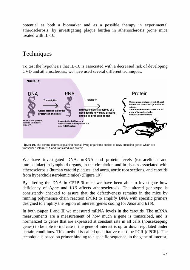
Figure 10. The central dogma explaining how all living organisms cosists of DNA encoding genes which are transcribed into mRNA and translated into protein.
We have investigated DNA, mRNA and protein levels (extracellular and
intracellular) in lymphoid organs, in the circulation and in tissues associated with
atherosclerosis (human carotid plaques, and aorta, aortic root sections, and carotids
from hypercholesterolemic mice) (Figure 10).
By altering the DNA in C57Bl/6 mice we have been able to investigate how
deficiency of Apoe and Il16 affects atherosclerosis. The altered genotype is
consistently checked to assure that the defectiveness remains in the mice by
running polymerase chain reaction (PCR) to amplify DNA with specific primers
designed to amplify the region of interest (genes coding for Apoe and Il16).
In both paper I and II we measured mRNA levels in the carotids. The mRNA
measurements are a measurement of how much a gene is transcribed, and is
normalized to genes that are expressed at constant rate in all cells (housekeeping
genes) to be able to indicate if the gene of interest is up or down regulated under
certain conditions. This method is called quantitative real time PCR (qPCR). The
technique is based on primer binding to a specific sequence, in the gene of interest,

38
and of a probe binding in between the primer pair. As the mRNA is amplified the
probe will detach and emit a fluorescent signal which is detected for each
amplification cycle. The amount of fluorescence is relative to the copy number of
the PCR product and then further normalized to the amount of housekeeping gene
copies. Measuring mRNA does not resemble the absolute amount of the translated
protein, and therefore measuring protein content is also important to be able to
explain the biological implications.
We have measured protein levels in several different ways. One of the “easiest”
techniques is by measuring proteins by ELISA (enzyme-linked immunosorbent
assay), which utilizes specific antibodies binding the protein of interest. These
antibodies are then detected by a secondary antibody which has an enzyme
conjugated to it allowing a substrate to be degraded and produce a shift in colour
which is detectable. The colour intensity is related to a known serial dilution of the
protein of interest and thereby the amount in the test samples can be calculated.
This technique has also been adapted to facilitate the measurement of several
different proteins in the same sample, here we have used both MesoScale and
Luminex technology.
The MesoScale technology utilizes electrochemiluminescense, by tagging the
protein of interest with Sulfo-Tags, which emit light when they are stimulated with
electrochemical stimuli. Each well consists of specific spots, corresponding to the
site where the specific protein of interest is detected. The light intensity in each of
these spots is then compared to the standard curve and an estimated concentration
can be calculated. The draw-back of this technique is that it is not possibly to rerun
the plate if any errors occurred during measurement.
The Luminex technology utilizes fluorescent beads, which carry antibodies
specific for the protein of interest. The assay requires lasers and sensors to detect
the emission wavelengths of the beads carrying the captured protein. There are two
really advantageous sides of Luminex. Firstly, the numbers of beads available are
up to a hundred, which allows for 100 different proteins to be assessed in the same
sample. The other benefit of using Luminex is that the beads can be reanalyzed if
any errors occurred during the first measurement.
Measuring proteins in aortic root section or in sections from human carotids by
immunohistochemistry utilizes the same technique of a specific antibody designed
to measure the protein of interest and detecting the cleavage of the added
substrate.
To identify immune cells, the most commonly favorable method is flow cytometry
(in daily use called FACS). This method also utilizes the specific binding of
antibodies to protein on the surface or inside the cell. The advantage of flow
cytometry is that several antibodies for different proteins can be combined with

39
size and density measurements of cells, and thereby each cell can be thoroughly
characterized, which is needed for immune cells, as many of them carry shared
proteins on their cell surface.
The most recently invented technique, to be used in this thesis, is the technique
developed by O-link (http://olink.com). This method utilizes antibodies for
specific binding to the protein of interest. The antibodies have specific DNA
probes attached to them and when two antibodies have bound the same protein
these probes can hybridize, extend and amplify the specific sequence of the protein
of interest allowing a read out similar to the qPCR. The main advantage of this
system is that it has the capacity to measure 92 analytes in 1 µl of sample due to
multiplexing and high sensitivity which is of great interest while analyzing human
material.

40
41
Aims and key findings
The aim of the collected work in this thesis was to investigate the potential of IL-
16 as a biomarker in the different stages of the atherosclerotic disease in humans,
and to validate these findings in an experimental setting. Given the limited
knowledge of the relationship between IL-16 and development of CVD we wanted
to investigate the role of IL-16 in primary prevention at population level, in
established CVD and in cardiovascular complications of diabetes.
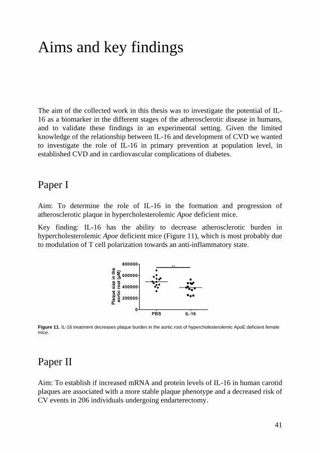
Paper I
Aim: To determine the role of IL-16 in the formation and progression of
atherosclerotic plaque in hypercholesterolemic Apoe deficient mice.
Key finding: IL-16 has the ability to decrease atherosclerotic burden in
hypercholesterolemic Apoe deficient mice (Figure 11), which is most probably due
to modulation of T cell polarization towards an anti-inflammatory state.
Figure 11. IL-16 treatment decreases plaque burden in the aortic root of hypercholesterolemic ApoE deficient female mice.
Paper II
Aim: To establish if increased mRNA and protein levels of IL-16 in human carotid
plaques are associated with a more stable plaque phenotype and a decreased risk of
CV events in 206 individuals undergoing endarterectomy.

42
Key finding: IL-16 was associated with plaque stabilizing factors collagen, elastin
and FoxP3 expression. High IL-16 protein levels in human carotid plaques were
independently associated with a decreased risk of suffering a CV event during a
two-year follow-up after surgery (Figure 12).
Figure 12. IL-16 protein in homogenates from human carotid plaques (n=206) is independetly associated with decreased incidence of cardiovascular events. IL-16 mRNA in homogenates from human carotid plaques (n=206) is independtly associated with asymptomatic patients. Black lines display the hazard ratios (with 95% CI) for incidence of cariovascular events and grey lines depicts the odds ratios (with 95% CI) for associating with symptomatic carotid plaques.
Paper III
Aim: To investigate if plasma levels of IL-16 can predict CV outcome in 473
patients who have undergone surgery to remove the carotid plaque.
Key Finding: High levels of IL-16 in plasma were independently associated with a
decreased risk of suffering from CV events and death due to CV events, during a
follow-up period of three years (Figure 13).
Figure 13. Kaplan-Meier curve depicting cardiovascular event free survival during follow-up of IL-16 plasma tertiles in individuals who underwent endarerectomy (n=473).
43
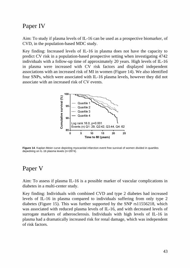
Paper IV
Aim: To study if plasma levels of IL-16 can be used as a prospective biomarker, of
CVD, in the population-based MDC study.
Key finding: Increased levels of IL-16 in plasma does not have the capacity to
predict CV risk in a population-based prospective setting when investigating 4742
individuals with a follow-up time of approximately 20 years. High levels of IL-16
in plasma were increased with CV risk factors and displayed independent
associations with an increased risk of MI in women (Figure 14). We also identified
four SNPs, which were associated with IL-16 plasma levels, however they did not
associate with an increased risk of CV events.
Figure 14. Kaplan-Meier curve depicting myocaridal infarction event free survival of women divided in quartiles depedning on IL-16 plasma levels (n=2874).
Paper V
Aim: To assess if plasma IL-16 is a possible marker of vascular complications in
diabetes in a multi-center study.
Key finding: Individuals with combined CVD and type 2 diabetes had increased
levels of IL-16 in plasma compared to individuals suffering from only type 2
diabetes (Figure 15). This was further supported by the SNP rs11556218, which
was associated with reduced plasma levels of IL-16, and with decreased levels of
surrogate markers of atherosclerosis. Individuals with high levels of IL-16 in
plasma had a dramatically increased risk for renal damage, which was independent
of risk factors.
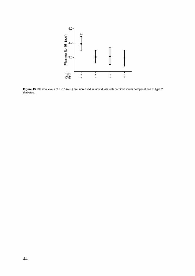
44
Figure 15. Plasma levels of IL-16 (a.u.) are increased in individuals with cardiovascular complications of type 2 diabetes.
45
Results and discussion
In the following sections I will present, and discuss, the data describing the role of
IL-16 in atherosclerosis and cardiovascular disease. From studies in an
experimental model of atherosclerosis, I will present our findings of the role of IL-
16 in dampening plaque progression, and the connection to apoptosis and T cells. I
will then continue to present the associations found with IL-16 in secondary
prevention in individuals with severe carotid atherosclerosis. I will present the
inherited predisposition of IL-16 in the form of genotyped single nucleotide
polymorphisms (SNPs) and their associations to IL-16 plasma levels. Finally, I
will present whether or not IL-16 plasma levels can be used in primary prevention
of cardiovascular risk and in cardiovascular complications in diabetic subjects.
IL-16 in experimental atherosclerosis
In paper I we tested whether IL-16, in a controlled environment, impacted
atherosclerosis formation and plaque phenotype in an experimental model of
atherosclerosis. The benefit of studying atherosclerotic development in mice is that
there is limited “background noise” due to the identical genetic and environmental
background. The drawback is that the mice never generate atherosclerotic plaques
leading to a CV event. Administration of IL-16 to hypercholesterolemic Apoe
deficient female mice reduced the atherosclerosis burden compared to control
treated mice. This was further supported by the fact that Il16 and Apoe deficient
hypercholesterolemic male mice had increased atherosclerotic burden compared to
control mice.
We could not identify any differences in the compositions of the plaques, situated
in the aortic root, when investigating inflammation (MOMA and CD4 staining) or
collagen (Van Gieson staining). In the brachiocephalic artery (which we use as the
equivalent to the carotid in humans) there was increased expression levels of genes
associated with apoptosis (caspase-3 and BAX) in mice treated with IL-16
compared to control. There was also a significant decrease in caspase-3 gene
activity in mice deficient in both Il16 and Apoe compared to Apoe deficient mice.
The results indicate a connection between apoptosis and IL-16 in experimental

46
atherosclerosis (paper I) and the connection is also present in human carotid
plaques (paper II).
Apoptosis
Is apoptosis beneficial in atherosclerosis? Promoting controlled apoptosis in the
atherosclerotic plaque could be advantageous as this will clear cell components in
an anti-inflammatory fashion. The opposite to apoptosis, necrosis, is thought to be
harmful in the atherosclerotic disease and believed to promote vulnerable plaques.
Immune cells are readily produced from stem cells in the bone marrow, and will
be replenished in the circulation, this is however not the case of all of the other cell
types in the body. For example, apoptosis in cardiomyocytes is not beneficial due
to that less than 50% of the cardiomyocytes are renewed after birth.[148] Death of
the cardiomyocytes will therefore, most probably, result in a weakened heart
increasing the risk of morbidity and mortality. This is of course not a desirable
consequence of IL-16 treatment.
Table 2. Spearman correlations between IL-16 and death receptors.
MDC
SUMMIT
CPIP plasma
CPIP plaque
n 4742
1435
558
202
r P-value r P-value r P-value r P-value
TRAIL 0.537 0.0E0 0.232 6.3E-19 0.159 0.00017 0.505 1.9E-14
TRAIL-R2 0.533 0.0E0 0.397 1.9E-54 0.268 1.1E-10 0.454 1.1E-11
TNF-R1 0.625 0.0E0 0.576 2.2E-127 0.328 1.8E-15 -0.008 ns
TNF-R2 0.622 0.0E0 0.572 3.3E-125 0.363 8.4E-19 0.272 0.00014
FAS 0.586 0.0E0 0.490 2.0E-87 0.394 3.7E-22 0.399 4.2E-9
Evidence put forward in this thesis supports the connection between IL-16 and
increased apoptosis, both in mice (paper I) and in humans (paper II), as well as
in Table 2 and Figure 16. There is, however, a risk of undesirable side effects. IL-
16 seems to be a consequence of activated caspase-3, as has been described
previously. We have seen that IL-16 has a role in inducing apoptosis by detecting
an enhanced gene activity in genes involved in the apoptotic pathway from studies
on IL-16 treated mice (paper I).
Further characterization of the atherosclerotic plaques from IL-16 treated, or
deficient mice, is needed to conclude that IL-16 induces apoptosis. This can for
example be done by TUNEL or active caspase-3 measurements in the
atherosclerotic plaques. To be able to consider IL-16 as a potential therapy further
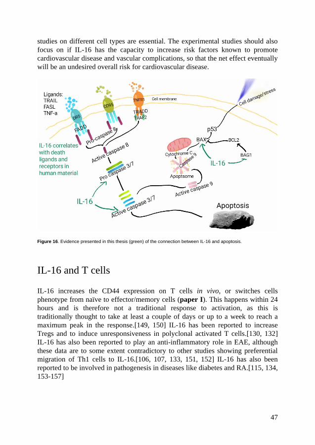
47
studies on different cell types are essential. The experimental studies should also
focus on if IL-16 has the capacity to increase risk factors known to promote
cardiovascular disease and vascular complications, so that the net effect eventually
will be an undesired overall risk for cardiovascular disease.
Figure 16. Evidence presented in this thesis (green) of the connection between IL-16 and apoptosis.
IL-16 and T cells
IL-16 increases the CD44 expression on T cells in vivo, or switches cells
phenotype from naïve to effector/memory cells (paper I). This happens within 24
hours and is therefore not a traditional response to activation, as this is
traditionally thought to take at least a couple of days or up to a week to reach a
maximum peak in the response.[149, 150] IL-16 has been reported to increase
Tregs and to induce unresponsiveness in polyclonal activated T cells.[130, 132]
IL-16 has also been reported to play an anti-inflammatory role in EAE, although
these data are to some extent contradictory to other studies showing preferential
migration of Th1 cells to IL-16.[106, 107, 133, 151, 152] IL-16 has also been
reported to be involved in pathogenesis in diseases like diabetes and RA.[115, 134,
153-157]

48
An increase in the effector/memory population of the T cells has been proposed to
promote atherosclerosis, and the associations between IL-16 and increased levels
of CD44 expressing T cells and atherosclerotic burden in paper I is puzzling.[158]
A similar association between IL-16 and effector/memory T cells was seen in a
sub-cohort of the SUMMIT study (Table 3).
Table 3. Spearman correlations between IL-16 plasma levels and number of circulating T cell populations in 211 individulas of the SUMMIT cohort.
Correlation P-value
CD4+CD45RA+ -0.164 0.017
CD4+CD45RO+ 0.142 0.039
CD4+CD62L+CD45RO- -0.160 0.020
CD4+CD62L+CD45RO+ 0.220 0.001
CD4+CD197+CD45RO- -0.126 0.067
CD4+CD197+CD45RO+ 0.130 0.060
CD8+CD62L-CD45RO+ -0.138 0.046
CD8+CD197+CD45RO- -0.133 0.054
The mice which were treated with IL-16, in paper I, displayed increased
percentages of CD44+ T helper cells. To further analyze these cells, we
investigated makers known to be associated with cells identified of anti-
inflammatory capacity. The CD44+ T cells, from IL-16 treated mice, had a higher
fraction of regulatory T cells and T cells expressing the immune regulator CTLA-4
compared to control treated mice. The increase of CD44+ Tregs and CTLA-4+ T
cells, in paper I, could explain the decreased levels of atherosclerosis in IL-16
treated mice. The anti-inflammatory properties of IL-16 were further supported by
the decrease in circulating TNF-α and the decreased release of IFN-γ from
stimulated splenocytes (paper I).
In a sub-cohort of the MDC study (n=540), PBMCs were analyzed by flow
cytometry and correlated to IL-16 levels in plasma. There are clear sex-specific
differences where women with increased IL-16 levels in plasma have more
cytotoxic T cells as well as Tregs (Table 4). CD8+ T cells have been described to
have pre-made bioactive IL-16. There are also evident differences in sex-specific
correlations between HDL and T cells, increasing HDL levels associates with
decreasing T cell levels in women but no associations were found in men (Table
3).

49
Table 4. Sex-specific Spearman correlations for IL-16 with T cell populations as well as sex-specific Spearman correlations with HDL. A total of 222 men and 318 women were studied for T cell numbers and IL-16 levels in the circulation.
IL-16
HDL
Men Women Men Women
CD3+ T cells 0.028 0.138* -0.079 -0.247***
CD3+CD4+ T cells -0.026 0.045 -0.048 -0.153**
CD3+CD8+ T cells 0.044 0.161*** -0.071 -0.257***
CD3+CD4+IFN-γ+ T cells -0.115 -0.016 -0.092 -0.105*
CD3+CD4+IL4+ T cells -0.141* 0.007 -0.060 -0.077
CD3+CD4+CD25+ T cells -0.072 0.005 -0.018 -0.033
CD3+CD4+CD25+FoxP3+ T cells -0.010 0.118* 0.025 -0.076
In a linear regression model IL-16 was positively associated (B=0.055, P=0.026)
and HDL was inversely associated (B=-0.156, P=0.000002) with the number of
cytotoxic T cells when adjusting for Framingham risk factors in women. No
significant associations were found in men except for smoking (B=0.125,
P=0.0004). Regulatory T cells were independently associated with IL-16
(B=0.003, P=0.033) and age (B=0.001, P=0.027) in women, whereas no
associations were found between IL-16 and Tregs in men.
The evidence for a connection between IL-16 and caspase-3, and the knowledge
that IL-16 is involved in increasing the Treg population, as well as inducing
unresponsiveness in T cells, is very interesting.[130, 132] There is a proven role of
caspase-3 in anergy and evidence of anergic T cells generating regulatory T
cells.[100, 101] Further investigation is needed to elucidate the potential role of
IL-16 in inducing anergic/regulatory T cells. It is also interesting to investigate the
potential anti-atherosclerotic properties of anergic T cells.
It is fascinating that the sub-types of T cells correlate differently with IL-16 once
analyzing men and women separately. Further studies investigating the association
between lipid metabolism, sex-hormones, immunology and IL-16 could
potentially hold valuable information.
IL-16 in severe carotid atherosclerosis
To be able to study how IL-16 impacts plaque phenotype and CV risk we had the
opportunity to measure IL-16 in carotid plaques surgically removed from 206
patients, presented in paper II, in the CPIP cohort. The carotid plaques have been

50
extensively studied and characterized allowing us to associate IL-16 levels to
markers known to stabilize, or to promote, plaque rupture.
In paper II we measured both IL-16 mRNA and protein levels in the carotid
plaques. Increasing IL-16 mRNA levels were associated with asymptomatic
patients, and increased expression of CD4 and FoxP3. Increased protein levels of
IL-16, in the carotid plaques, were associated with elevated levels of collagen,
elastin and caspase-3 activity. Collagen and elastin are ECM proteins generally
accepted to promote stabilization of the atherosclerotic plaque, which should lead
to less rupture prone plaques and less clinical manifestations of CVD.
The aim was to investigate if the increased IL-16 protein levels also associated
with less CV events by recording CV events for approximately 2 years after
removing the carotid plaque (paper II). During follow-up the individuals with
levels of IL-16 above median, in the carotid plaque, displayed association with a
decreased risk of suffering a CV event after adjusting for potential confounders
(HR 0.47, 95% CI 0.22-0.99, P=0.047). No significant associations were detected
between IL-16 mRNA expression levels in the carotid plaque, or with plasma IL-
16 levels, and CV events during follow-up.
In paper III we had the possibility to analyze plasma samples from 473
individuals undergoing carotid endarterectomy and to follow these individuals for
approximately 3 years. During this time CV events and CV events leading to death
were recorded. The associations between high levels of IL-16 and a decreased risk
of CV events was significant in a Cox regression when comparing to the lowest
tertile and adjusting for potential confounders (HR 0.535, 95% CI 0.27-0.81,
P=0.007). An even stronger reduction in risk was found between the highest tertile
of plasma IL-16 and risk of dying due to CV complications (HR 0.252, 95% CI
0.09-0.70, P=0.008) compared to individuals within the lowest tertile, after
adjusting for potential confounders.
The individuals included in these studies (paper II-III) are all affected by severe
atherosclerosis. The CPIP study is a large and well characterized database,
although the design of the study has not included any healthy individuals as
controls, allowing us only to draw conclusions based on this highly selected, and
diseased, population. Plasma levels of IL-16 from healthy individuals that are age
and sex matched would have been valuable, however carotid vessels from healthy
individuals do not have plaques, which make a comparison difficult and complex.
It is also puzzling that IL-16 plasma levels do not display any association to the
primary CV event, demonstrated by the lack of association with the asymptomatic
group. There is a possibility that IL-16 is elevated as a repair mechanism in both
the symptomatic and asymptomatic group. Further experimental and mechanistic

51
studies are needed to evaluate the potential of IL-16 in severe atherosclerosis and
the clinical complications thereof.
In conclusion, IL-16 displays associations with increased stabilizing factors and a
decreased risk of post-operative CV events. Our data support the hypothesis that
high levels of IL-16 confers a protective role in severe carotid atherosclerosis, in
an elderly population.
IL-16 gene polymorphism
The aim was to identify SNPs associated with IL-16 plasma levels to further
strengthen the conclusions drawn from the associations regarding IL-16 plasma
levels. The SNPs also hold potential as a screening method if they are significantly
impacting the risk of suffering from CVD. In paper IV and V, we measured IL-16
in plasma and SNPs located in, or close to, the IL-16 gene in the MDC and
SUMMIT studies. SNP data can indicate if certain alleles are associated with
increased, or decreased, levels of IL-16 in plasma and can therefore support a
predisposition from birth to have altered risk of CVD due to IL-16 plasma levels.
By researching literature, we identified four SNPs associated with IL-16 plasma
levels (rs4072111, rs8034928, rs3848180, and rs1131445) in the prospective
population-based MDC study (paper IV). All but one were associated with
increased levels of IL-16 in plasma, whereas rs3848180 was associated with a
reduction in plasma levels after adjusting for Framingham risk factors. None of the
SNPs were associated with an increased risk of MI, stroke or all-cause mortality
after adjusting for Framingham risk factors. Rs1131445 in men, which associated
with increased levels of IL-16, was significantly associated with a 40% reduction
in risk of suffering a MI. The same SNP (rs1131445) was associated with a 23%
risk reduction for all-cause mortality in the entire population, and a 35% risk
reduction in women.
In the SUMMIT study (paper V), rs11556218 (which was not available on the
chip used to identify SNPs in the MDC study) was associated with reduced levels
of IL-16 in plasma and was inversely associated with markers of atherosclerosis,
supporting a harmful role of IL-16 in vascular complications of type 2 diabetes.
Interestingly, rs11556218 has previously been described to be associated with
increased risk of ischemic stroke, which makes it a potential candidate for further
analysis in the MDC and CPIP cohorts.[140]
In paper IV and V, we identified in total five SNPs that were associated with IL-
16 plasma levels. The data presented are based on associations and do not prove
causality. There are several different steps between the DNA sequence, where we

52
have measured the SNPs, and the functional IL-16 protein which can impact the
levels of the circulating IL-16 (Figure 8). There is also a possibility that it is not
the identified SNP that controls the associations with the plasma levels of IL-16,
but instead a sequence or SNP that is in linkage disequilibrium with the measured
SNP. Finally, one needs to be aware of the fact that we are testing 4 or 5 SNPs
with several different outcomes (variables) which increases the number of
statistical tests that have been performed. As the p-value is set as being significant
if under 0.05 there is a chance of 1 in 20 of getting a false positive significance.
There have been several reports on IL-16 associated SNPs in case-controls studies
of CVD but only one of them have investigated the associations to plasma levels
of IL-16.[137-140] To my knowledge the two studies presented in paper IV and
V are the two largest cohorts that have been analyzed for SNPs associated with IL-
16 plasma levels and further associated with CVD. Paper IV is also the first to
perform an extensive prospective study of the role of IL-16 in CVD, and paper V
is the first to investigate the role of IL-16 in vascular complications of T2D.
The presented results imply that the inherited genetic material determines the IL-
16 plasma levels later in life, however these SNPs do not seem to be strong
predictors of CV risk in a large population-based prospective study (paper IV). In
a case-control study (paper V) the SNP rs11556218 was associated with reduced
levels of plasma IL-16 and less atherosclerotic manifestations, supporting a
harmful role of IL-16 in vascular complications of type 2 diabetes.
IL-16 and cardiovascular risk factors
There are several known risk factors which are used to determine the future risk of
suffering from a CV event, of which the most commonly used is the Framingham
risk score which includes; age, sex, smoking, cholesterol, HDL and systolic blood
pressure. To understand the role of IL-16 in CVD we therefore first explored the
associations between IL-16 plasma levels and risk factors in MDC, which is a
prospective population-based cohort of relatively healthy individuals.
Interestingly, in paper IV, IL-16 levels were significantly lower in females, and as
a consequence the analysis was split on sex so not to mask any sex-specific
differences.
All of the Framingham risk factors were associated with increasing IL-16 levels,
except for smoking, in the MDC study. After expanding the analysis to include
diabetes mellitus, BMI, LDL, diastolic blood pressure, white blood cell count and
lymphocyte count, all of the risk factors associated positively with increasing IL-
16 levels in plasma in both men and women. In women the risk factors most

53
closely associated with IL-16 levels were BMI, cholesterol, LDL, HDL (inversely)
and triglycerides. Men displayed a stronger association between IL-16 with
systolic and diastolic blood pressure, which was then followed by BMI, HDL
(inversely) and triglycerides.
In paper V, where we investigated the vascular complications of T2D, it became
evident that renal dysfunction was highly associated with increasing IL-16 levels
in individuals with T2D, due to the increasing levels of creatinine and decrease in
eGFR with increasing IL-16 plasma levels. In the SUMMIT study (paper V) IL-
16 displayed associations to HDL (inversely), LDL, cholesterol and systolic blood
pressure although the majority of individuals were medicated (i.e. with statins and
metformin).
In a prospective population-based study (paper IV), consisting of relatively
healthy individuals, IL-16 was associated with most of the measured risk factors
known to impact CVD. In an elderly population already suffering from disease
which was more medicated, creatinine was associated with IL-16 plasma levels in
individuals suffering from T2D (paper V) or from severe atherosclerosis (paper
III). It is left unclear if the high levels of IL-16 are a compensatory response to the
increased levels of risk factors, or if it is the actual increase in risk factor levels
that drives the augmented IL-16 levels.
IL-16 as a biomarker in primary prevention
To determine if IL-16 could be a potential biomarker in primary prevention we
performed Cox regressions adjusted for potential confounders by Framingham risk
factors and diabetes mellitus. There were no significant associations between
increased risk of suffering from a CV event with high IL-16 levels in men. There
was a 64% increased risk for suffering from a MI for women within the highest
quartile of IL-16, compared to the women in the lowest quartile, of the individuals
included in the MDC study (paper IV).
The increased risk for MI remained significant after adjusting for all of the risk
factors that were associated with IL-16. There were no associations to stroke, CV
death or all-cause mortality with high IL-16 levels. So far we could conclude that
IL-16, measured in plasma, was not a universal biomarker for CVD in primary
prevention, but perhaps a sex-specific marker for MI in women.

54
IL-16 and obesity
We have described that IL-16 levels in plasma correlates to hypercholesterolemia,
and it has previously been shown that IL-16 is increased in individuals that are
overweight.[135] Due to the increasing evidence supporting a connection between
IL-16 and lipid metabolism, I wondered if IL-16 could predict CV risk in obese
subjects in the MDC cohort and therefore I selected individuals with body-mass-
index (BMI) over 30 (n=597). Obese individuals displayed a tendency of increased
risk of MI with increasing IL-16 levels in tertiles (Kaplan Meier Log rank test
5.132, P=0.077). Next I performed a sex-specific analysis, which displayed no
association with increasing IL-16 levels with risk for MI for men (n=215),
whereas there was a striking association in obese women (n=380) with increasing
IL-16 levels for risk of MI (Log rank test 9.916, P=0.007) (data not included in
paper IV).
In a Cox regression, women within the highest tertile of IL-16, had a 3-fold
increased risk for MI compared to the lowest (HR 2.972, 95% CI 1.239-7.126,
P=0.015) after adjusting for potential confounders. Men displayed no significant
association between IL-16 levels and MI (HR 0.974, 95% CI 0.466-2.038,
P=0.945). In paper IV, we reported an association between IL-16 and MI in all
obese and non-obese women collectively (HR 1.640, 95% CI 1.04-2.57, P=0.032),
however after only selecting women with a BMI below 30 there was no significant
association between increasing IL-16 tertiles and increased risk of MI (HR 1.277,
95% CI 0.842-1.917, P=0.255), suggesting that there is a link between obesity,
increased IL-16 levels and risk of MI (data not included in paper IV). Though, it is
possible that the exclusion of the high risk obese groups impairs the statistical
power by reducing the event rate.
The data presented in paper IV supports the perception that CV risk factors may
impact IL-16 plasma levels, or vice versa, in the MDC population-based
prospective study.
As has been mentioned before, the results presented in paper II-V are based on
associations and do not allow us to prove causality. With this said the number of
individuals included in the MDC study (paper IV) is, to my knowledge, the
largest of its kind to investigate the potential of IL-16 as a biomarker for primary
prevention. There is one study investigating the potential of IL-16 in a relatively
healthy population to identify individuals with intermediate risk of suffering from
CV event within a 5-year follow-up period. IL-16 was, in this study, combined
with several other biomarkers in an algorithm based approach and was associated
with increased risk of suffering from CVD.[57]
There is a general consensus that the risk prediction of CVD in women is
understudied. It is also known that women during fertile age, prior to menopause,

55
have a decreased risk of CVD. This is most probably due to increased HDL levels
as a result of estrogen levels and this protection decreases after menopause. More
research is needed to gain insight into the development and risk prediction of CVD
in general, and also more specifically into the characteristics of the disease in
women.
In conclusion, the value of measuring IL-16 in plasma for determining primary
prevention of CVD is however most likely limited, although IL-16 might be
valuable to measure in obese women to predict future risk of MI.
IL-16 is associated with vascular complications of CVD
Since there were evident connections between high IL-16 levels with several of
the risk factors known to impact CVD, we went on to study the role of IL-16 in
established atherosclerosis. In paper V, we investigated if IL-16 could be a
marker of vascular complications in subjects suffering from type 2 diabetes in the
SUMMIT study, including 1500 individuals.
In paper V we report a significant increase of circulating IL-16 in subjects with
CVD and diabetes, compared to subjects with only CVD, diabetes or controls. In a
binary logistic regression, the subjects with diabetes within the highest quartile of
IL-16 levels had a 50% increased risk for CVD compared to the lowest quartile,
after adjusting for Framingham risk factors. We could also confirm that plasma
levels of IL-16 are associated with increased amount of risk factors in subjects
with diabetes. Creatinine, eGFR and systolic blood pressure were the risk factors
displaying the strongest associations with IL-16. Furthermore, we report
associations between increasing IL-16 levels with surrogate markers of
atherosclerosis, such as ABPI (inversely) and with increased plaque area of the
common carotid artery.
In the CPIP study (paper III), plasma levels of IL-16 in 473 individuals displayed
significant associations with increasing serum creatinine, supporting the strong
associations found in the SUMMIT study (paper V). The increased, respectively
decreased, levels of creatinine and eGFR with increasing IL-16 levels purported us
to reflect upon whether kidney damage was associated with IL-16.
Since diabetes is known to affect vasculature in the whole body we had the
privilege of having data on renal dysfunction, in the SUMMIT study (paper V),
reported as microalbuminuria, proteinuria or renal failure. To our surprise the
quartile including subjects with the highest levels of IL-16 in plasma had more
than a 6-fold increased risk of suffering from renal dysfunction compared to the
individuals with the lowest quartile, after adjusting for risk factors.

56
It is known that heart and kidney function is closely linked. The term cardio-renal
syndrome (CRS) is used to describe the condition of the two-way relationship
between CVD and the worsening of kidney function (Figure 17). Several different
factors are thought to play a role in CRS including the immune system, cytokines
and apoptosis.[159, 160]
It is clear that individuals with both diabetes and CVD have an increased risk of
suffering from renal dysfunction. The association between IL-16 and increased
risk of CVD could indicate a stronger association with renal dysfunction. In a
linear regression model IL-16 was as significant a marker for renal dysfunction as
was CVD. IL-16 was only exceeded by creatinine in predicting risk of renal
dysfunction, whereas other risk factors did not predict risk of renal damage to the
same level.
Figure 17. Cardiorenal syndrome - the interplay between decreased heart function and decreased renal function.
Whether the increased IL-16 levels are a result of the increased risk factor levels
or a consequence thereof we cannot answer. Furthermore, there is a possibility that
confounding factors, which might have not been taken into consideration, are
responsible for the presented associations between IL-16 and risk factors. There
are a few other studies describing an association between IL-16 and obesity, as
well as with diabetes.[115, 134, 135]
It was evident that IL-16 was strongly associated with renal damage. If IL-16 is
responsible for driving renal damage, CVD, or both, we cannot answer in this
study. There is even a possibility that the increased IL-16 levels are a
compensatory factor trying to reduce the vascular damage. Zhao et al. have
proposed a compensatory role of IL-16 in the outgrowth of endothelial progenitor
cells in individuals with kidney disease.[161] Further experimental studies are
called for to elucidate the role of IL-16 in vascular complications of diabetes, as
well as in the connection to CV risk factors.

57
Our data support the hypothesis that IL-16 is associated with an increased rate of
prevalent CVD in individuals with T2D in a multicenter study. The presented data
are based on statistical tests, calculating the probability of co-variance and do not
predict if IL-16 has a driving or inhibiting effect on the disease. There has,
however, been evidence presented for a pathological role of IL-16 in an
experimental study in non-obese diabetic (NOD) mice where the authors used the
human IL-16 blocking antibody 14.1.[115] The same antibody, 14.1, was used in
paper I included within this thesis, where we failed to measure any reduction of
circulating levels of IL-16 after 14.1 treatment. To my knowledge there have been
no larger studies investigating the associations between IL-16 and cardiovascular
complications in diabetic subjects.
The multicenter aspect of the SUMMIT study (paper V) was one of its strengths
although also one of the complicating matters of this study. It became evident that
the four different study centers (Dundee, Exeter, Lund and Pisa) differed slightly
in the enrolled populations when accounting for age, gender, medication and so
forth. The difference in the study center populations made the confounding factors
difficult to determine. Further research on the role of IL-16 in vascular
complications of IL-16 is needed, preferably both by re-evaluating our findings in
other cohorts as well as validation in experimental studies.
In conclusion, the levels of IL-16 in plasma were higher in individuals with
cardiovascular complications of T2D, as well as associated with surrogate markers
of atherosclerosis. To understand how IL-16 impacts vascular complications from
diabetes, more research is called for to identify the underlying mechanisms, and
the potency of IL-16 as a biomarker.
Pro and cons of IL-16 in the atherosclerotic disease
The collected work of this thesis has aimed to establish a role of IL-16 in
atherosclerosis and CVD. The hypothesis from the start was that IL-16 carry anti-
inflammatory functions, by inhibition of T cell responses. In paper I, the net
effect of IL-16 in experimental atherosclerosis was anti-inflammatory and perhaps
thereby resulted in a decrease in atherosclerotic burden. The results from paper I
were also reflected in the association between IL-16 and stabilizing plaque
components presented in paper II. High levels of IL-16 in carotid plaques were
associated with a decreased risk of suffering a post-operative CV event during a
two-year follow-up period (paper II).
As carotid plaque homogenates require surgical removal, and thereafter thorough
processing, plasma samples are preferable in a clinical setting, when discussing
58
biomarkers. Therefore, we tested whether plasma IL-16 levels, from 473
individuals with severe carotid atherosclerosis, could predict future CV events
(paper III). High IL-16 levels in plasma associated with a decreased risk of
suffering from CV events or CV events so severe leading to death as final outcome
(paper III). Paper I-III supports a protective role of IL-16 in atherosclerosis and
CVD.
The results presented in paper I-III, are contradicted by the results from IL-16 in
subjects with established T2D and with cardiovascular complications (paper V).
The data presented in paper V, indicates that IL-16 is elevated in individuals with
T2D who also suffer from CVD, compared to individuals only suffering from
T2D. Furthermore, IL-16 was connected to renal dysfunction and associated with
surrogate markers of atherosclerosis. One SNP, rs11556218 which associated with
decreased levels of IL-16 in plasma, was inversely associated with surrogate
markers of atherosclerosis, further supporting a disease driving role of IL-16 in
T2D (paper V).
Paper IV is the largest study, to my knowledge, performed on circulating IL-16 as
a biomarker for primary prevention. There was no association between SNPs,
associated with IL-16 plasma levels, with an increased risk for future CVD. There
was even one SNP (rs1131445) which associated with decreased risk for all-cause
mortality during the 20-year follow-up period. Furthermore, it was evident that
women had decreased levels of IL-16 compared to men, and that IL-16 correlated
to several of the known cardiovascular risk factors (paper IV). Women with high
levels of IL-16 had a 64% increased risk of suffering from a MI, compared to the
individuals with low levels. Obese women had an even further increased risk of
suffering from CVD if they had elevated levels of IL-16 (paper IV). The
conclusion from paper IV is that IL-16 may mainly predict risk in obese women
but IL-16 is not useful in a “normal”, heterogeneous population to predict future
CV risk.
I believe that IL-16 possesses anti-atherogenic properties, evident in paper I-III.
The reason why IL-16 is elevated in subjects with T2D with cardiovascular
complications, and in obese women, is because IL-16 is a firefighter trying to put
out an already raging fire.
59
Conclusions and Future Perspectives
In this thesis, I have shown that;
I. IL-16 decreases the atherosclerotic burden in hypercholesterolemic
mice.
II. IL-16 levels depend on which gene polymorphisms (SNPs) are
inherited from parents to offspring and are closely associated with risk
factors driving CVD.
III. IL-16 is associated with stabilizing factors in human carotid plaques
and is associated with less post-operative CV events and CV event
leading to death.
IV. IL-16 is not a potential prognostic biomarker in a relatively healthy
population, but perhaps it should be further evaluated as a biomarker
for CVD in obese women.
V. In subjects with type 2 diabetes, IL-16 is associated with several
vascular complications and should be further validated as a biomarker
for risk prediction in subjects with diabetes.
Furthermore, there are strong associations between increasing IL-16 levels and
many of the risk markers for atherosclerosis, especially cholesterol, HDL
(inversely) and LDL levels. In the future I believe that the connection between
lipid metabolism, risk factors and the association to increasing IL-16 levels will
remain extremely interesting and should be further investigated. As previously
mentioned, the study design of paper II–V present associations and do not prove
causality and need to be further tested.
The relatively rapid effect of IL-16 on T cells is, to my mind amazing, and should
be further evaluated as a future therapy. The manuscripts presented in this thesis
only highlight the effect of IL-16 on T cells. There are still, however, many
potential effects of IL-16 on other cells affecting atherosclerosis development,
which have not been included here as they are outside the scope of this thesis.
Finally, there is a protective role of IL-16 in experimental and advanced
atherosclerosis, which is most likely connected to T cells and apoptosis. A few
more years and I will have solved the remaining question marks .

60

61
Populärvetenskaplig sammanfattning
Interleukin 16 - brandmannen som släcker farliga
eldsvådor i kärlväggen.
Immunförsvaret påverkar risken för hjärt-kärlsjukdom. En signalsubstans i
immunförsvaret, IL-16, har visat sig öka antalet anti-inflammatoriska
immunceller. Vi har bevis för att IL-16 minskar åderförfettningen i
kärlväggen samt minskar risken för att drabbas av hjärt-kärlsjukdom.
Vår kropp består av många olika typer av celler, där vissa av cellerna har som
uppgift att skydda oss mot farliga inkräktare som bakterier och virus. Dessa celler
ingår i vårt immunförsvar. Under vissa förhållande så blir cellerna farliga för
kroppen då de tolkar kroppsegna byggstenar (i detta fall det onda kolesterolet) som
inkräktare. När detta sker i kärlväggen kan en lokal inflammation uppstå och kan
resultera i startskottet för åderförfettning (arteroskleros), vilket senare kan ge
upphov till hjärtinfarkt och stroke. En av orsakerna som gör sjukdomen
fascinerande är att celler har förmåga att kommunicera med varandra genom att
utsöndra protein som programmerar omgivningens gensvar. Kan dessa
kommunikationsfaktorer vara vägen till att hitta, förstå och lindra
sjukdomsförloppet?
Över tid så kan inflammationen i kärlväggen leda till att ett plack uppstår.
Immunförsvaret kommunicerar med proteiner och ett sådant protein är interleukin-
16 (IL-16). Om placket brister kan det innebära allvarliga konsekvenser för
individen i form av hjärtinfarkt eller stroke. Placket kan ge upphov till
komplikationer på olika sätt, antingen kan det täppa till kärlet, eller kan en
blodpropp bildas som en följd av placket, som sedan transporteras med blodet och
fastnar i ett trängre kärl. När blodet inte kan transporteras till hjärnan eller hjärtat
uppstår syre och näringsbrist vilket leder till att vävnaden dör.
Komplikationer av åderförfettning är den största dödsorsaken i världen och
mycket forskning fokuserar på att hitta, karakterisera och behandla åderförfettning.
Denna avhandling har haft som fokus att utvärdera om IL-16 kan vara en lämplig
markör för att identifiera risken för att drabbas av åderförfettning samt om IL-16
kan påverka placket till att bli mindre bristningsbenäget.

62
Det finns flera faktorer som bidrar till om en individ har en högre risk att drabbas
av åderförfettning och dessa inkluderar manligt kön, rökning, hög ålder, höga
nivåer av det ”onda” kolesterolet (LDL), högt blodtryck, lider av diabetes, fetma
eller har defekta nedärvda gener som påverkar kolesterolnivåerna i blodet. En av
orsakerna till varför hjärt-kärlsjukdom är så vanligt är att sjukdomen ger sig oftast
till uttryck när personen har passerat fertil ålder. Detta innebär att sjukdomen inte
kommer försvinna ur populationen på grund av naturlig selektion. I den tid vi
lever, med mycket fysisk inaktivitet och tillgången till mat i överflöd, bidrar detta
till en växande grupp individer med stor risk för att drabbas av hjärt-kärlsjukdom.
För att förstå sjukdomsförloppet så behöver man bakgrundskunskapen att i
kroppen finns immunceller som ansvarar för bort städningen. Dessa städarceller
kallas makrofager och de har som uppgift att äta upp allt de träffar på som kan
vara farligt för kroppen, så som bakterier eller delar av andra döda celler. När
kroppen transporter fett till celler så packas det i speciella ”fartyg” (låg-densitets
lipoprotein, LDL). Dessa fartyg kan ändra utseende p.g.a. att det är väldigt
stormigt (oxideras) eller för att de stöter på hinder (modifieras). Fartygen kommer
då att ändra skepnad så att makrofagerna inte känner igen dem som ett inhemskt
(kroppseget) material och kommer att hindra det genom att förtära och bryta ner
hotet, och därmed minska risken för allvarliga konsekvenser.
Om detta sker i kärlväggen så uppstår det en fettansamling inuti makrofagerna och
de blir skumceller. Skumcellerna är klumpiga, inte mobila, och kommer tillslut att
spricka av alla fartyg som de har inuti sig. Ut från skumcellerna läcker då en
blandning av nedbrutna fartyg och cellrester som uppfattas som inkräktare av
andra immunceller. Nu har en negativ spiral startat i kärlväggen och ytterligare
makrofager kommer anlända till platsen för att städa upp, men dessa kommer gå
samma öde till mötes. Det bildas helt enkelt en kyrkogård av vrakrester som
kommer kalla på ytterligare hjälp från immunförsvaret.
Hjälpsignalerna som immuncellerna skickar ut kallas för kemokiner och cytokiner.
Kemokiner hjälper cellerna att hitta till platsen genom att gå mot en ökad gradient
av kemokinerna. Cytokinerna berättar vilken typ av inkräktare immuncellerna har
stött på och hur det inkommande försvaret bör förbereda sig. En sådan
signalsubstans, eller cytokin, är IL-16. De celler som leder angreppet kallas för T
celler. Det finns flera olika typer av T celler och beroende på vilken T cell som
attraheras till platsen så kommer immunförsvaret styras till att svara på hotet på
olika sätt. Vissa T celler är för aktiv krigsföring och driver på det aterosklerotiska
sjukdomsförloppet (dessa benämns som T hjälpar celler typ 1) och andra har
förmåga att dämpa den inflammatoriska responsen mot hotet (regulatoriska T
celler).
Det finns bevis för att IL-16 binder till T celler som har en specifik faktor på ytan
som kallas CD4. CD4 finns på alla T hjälpar cellerna samt en del andra celler som

63
ingår i immunsystemet. IL-16 har förmågan att hämma T hjälpar cellernas
förökning (delning) samt att prioritera ett regulatoriskt T cells svar. Det finns
forskning som visar att ett av de proteinerna som är involverat vid celldöd
(caspase-3) också är involverat vid aktiveringen av IL-16. Det finns fortfarande
frågetecken på hur IL-16 tar sig ut från cellerna där det har producerats och hur
exakt IL-16 påverkar andra celler. Eftersom man vet att IL-16 kan öka antalet
regulatoriska T celler, och att dessa celler kan verka skyddande mot
sjukdomsförloppet, har arbetena som är presenterade i denna avhandling fokuserat
på IL-16s roll i ateroskleros och komplikationerna där av.
I papper IV var frågeställningen huruvida höga IL-16 nivåer i blodet kunde
förutsäga risken för att drabbas av hjärtkärlsjukdom upp till tjugo år efter att
proverna togs. Till vår förvåning så visade sig höga nivåer av IL-16 vara kopplat
till ökad risk för kvinnor att drabbas av hjärtinfarkt, men inga samband fanns för
män. Vidare så kunde vi se starka kopplingar mellan ökade IL-16 nivåer med flera
riskfaktorer kopplade till hjärt-kärlsjukdom. Designen på studien gör det svårt för
oss att säga om de höga nivåerna var en orsak av hjärtkärlsjukdomen, eller om det
var en konsekvens av den, men slutsatsen är att IL-16 kan inte användas som en
universal markör för att identifiera personer med risk för hjärt-kärlsjukdom. Vi
hade även tillgång till analyser av arvsmassan, från dessa individer, där vi kunde
identifiera mutationer i arvsmassan som var kopplade till mängden IL-16 i blodet.
En av dessa mutationer, som var kopplad till en ökad mängd IL-16, visade sig vara
korrelerad till en minskad risk för dödlig utgång för både män och kvinnor.
Vidare ställde vi oss frågan om prover tagna från personer, som redan har drabbats
av hjärt-kärlsjukdom kunde förutsäga risken för att få ett nytt kliniskt symptom
orsakat av åderförfettningen i kärlväggen. Här hade vi förmånen av att ha tillgång
till plack i halspulsådern. Placken var tagna från patienter med kliniska symptom
av placken, eller från patienter med en plackstorlek som täppte till minst 80 % av
kärlet. Det visade sig att höga nivåer av IL-16 i placket, samt i blodet, minskade
risken för att få en komplikation av åderförfettningen samt att det minskade risken
av att dö av komplikationer av åderförfettningen.
Eftersom placket börjar bildas redan vid en ung ålder, långt innan man utvecklar
symptom, så pågår mycket forskning på vad som bidrar till att placket är stabilt
och inte riskerar att brista. Vi kunde koppla samman de höga nivåerna av IL-16, i
placket, med ökade mängder av bindvävsprotein (plack stabiliserande) samt med
regulatoriska T celler. Slutsatsen av papper II och III är att höga nivåer av IL-16, i
en population som lider av kraftig åderförfettning, är kopplat till minskad risk för
nya kliniska symptom av åderförfettningen.
Vi vet att individer som lider av diabetes har ökad risk att få en defekt kärlvägg. I
papper V, undersökte vi om det fanns ett samband mellan hjärt-kärlsjukdom och
IL-16 hos personer med diabetes. IL-16 var även i denna population starkt kopplat

64
till riskfaktorer som påverkar hjärt-kärlsjukdom. Det visade sig att individer med
höga nivåer av IL-16 hade en 50 % ökad risk för att lida av både diabetes och
hjärt-kärlsjukdom jämfört med individer som enbart hade diabetes. Det som
verkligen var överraskande var att de personer som hade höga IL-16 nivåer i
blodet hade 6 gånger högre risk för att lida av nedsatt njurfunktion. Slutsatsen från
denna studie är att det finns en koppling mellan höga IL-16 nivåer i blodet med
nedsatt kärlfunktion som antagligen påverkar både njurfunktion och hjärt-
kärlsjukdom.
Papper II-V i avhandlingen är baserade på associationer som beräknas med
statistiska metoder och indikerar sannolikheter för ett samband. Studier som är
baserade på associationer, i mänskliga populationer, är svåra att dra några
definitiva slutsatser ifrån. Vi vet med andra ord inte med säkerhet att IL-16 är
skyddande mot hjärt-kärlsjukdom i individer som redan lider av allvarlig
åderförfettning.
För att svara på frågan om IL-16 kan hämma åderförfettningsprocessen behövde vi
utföra ett mer kontrollerat försök. I papper I har vi använt oss av möss som är
åderförfettningsbenägna och är genetiskt identiska. Vi har injicerat IL-16 i början
av sjukdomsprocessen och sedan kvantifierat mängden fett och plack i kärlväggen
hos dessa möss. Slutsatsen var att behandling med IL-16 minskade mängden, och
storleken, på placken i kärlväggen. För att bekräfta dessa fynd genererade vi en
mus som saknade funktionellt IL-16. Det visade sig att hanar som saknade IL-16
hade mer plack än de som hade IL-16, medan honorna inte visade någon skillnad i
plackstorlek.
Vad drar jag för slutsats av de studier jag har varit involverade i angående IL-16
påverkan på åderförfettning? Jag har inte alla svaren och jag anser att mer
forskning behövs för att förstå IL-16 roll i hjärt-kärlsjukdom. Åderförfettning är en
komplex och fascinerade sjukdom som drabbar många människor årligen. Det
verkar inte som att IL-16 kan användas som en indikator för att beräkna risken för
hjärt-kärlsjukdom, hos relativt friska individer, men mer forskning behövs för att
titta närmre på varför IL-16 är kopplat till en ökad risk för hjärtinfarkt hos kvinnor.
Jag förordar också att mer forskning bör bedrivas på kopplingen mellan
njursjukdom och IL-16, samt varför IL-16 är starkt kopplat till kolesterolnivåer.
Vi har presenterat bevis på att IL-16 är kopplat till en minskad risk för nya kliniska
symptom hos personer som lider av allvarlig åderförfettning och att detta styrks av
de data vi har genererat under experimentella förhållanden. I denna avhandling har
jag sökt svaret på om IL-16 är en skyddande faktor i hjärt-kärlsjukdom. Jag har
inte svaret på den frågan men jag tror att IL-16 kan vara en stabiliserande faktor i
placken och mer forskning behövs för att förstå IL-16 i sig självt och hur IL-16
påverkar faktorer som är involverade i ateroskleros.
65
Science for everyone
Interleukin 16 – a fire fighter counteracting an inferno in
the vessel wall.
The immune system impacts the risk of suffering from cardiovascular
disease. A signal substance in the immune system, IL-16, has been reported to
increase anti-inflammatory immune cells. We have proof of a protective role
of IL-16 in cardiovascular disease, both by stabilizing atherosclerotic plaques,
and by associating with a decreased risk of suffering from cardiovascular
event.
The body consists of numerous cell types, of which certain types of cells are
designed to protect us from dangerous intruders such as bacteria and viruses.
These cells are a part of our immune system. Under certain conditions the same
cells that are designed to protect us, turn against one’s own body and interpret
essential building-stones as foreign. If this event occurs within the vessel wall,
there is a possibility that an inflammation arises and initiates an atherosclerotic
plaque, which later can result in myocardial infarction or stroke. The cells in the
immune system have an amazing capacity to communicate with each other
through releasing proteins which can program the surroundings’ response. Is it
possible that one of these proteins, interleukin-16 (IL-16), has the propensity to
identify, alter and limit the disease severity?
Inflammation in the vessel wall is thought to initiate atherosclerosis and over time
an atherosclerotic plaque can develop. If the plaque ruptures the individual is at
risk of suffering from a myocardial infarction or stroke, which can massively
impact the quality of life or even cause death. The atherosclerotic plaque can
either occlude the vessel where it is situated, or it can rupture, causing a blood
clot. The blood clot will travel with the blood flow until it blocks a smaller vessel.
If this obstruction occurs in the brain the individual can suffer a stroke, or if it
happens in the vessels supplying the heart with blood, it can lead to a myocardial
infarction. The obstruction causes a lack of oxygen and cells in the tissue die.
Complications of atherosclerosis, cardiovascular disease (CVD), are the main
cause of death in the world and great efforts are made to identify and to treat this
disease. This thesis has focused on evaluating if IL-16 can alter the atherosclerotic

66
plaque to be less prone to rupture and thereby reduce the risk of suffering from
CVD. Furthermore, we have assessed to possibility of utilizing IL-16 as a
biomarker for predicting CVD.
There are several different factors that contribute to the risk of developing CVD
such as being of the male gender, smoking, high age, high levels of the “bad”
cholesterol (LDL), high blood pressure, diabetes and obesity and mutations in
genes which control cholesterol levels. One of the reasons behind the large number
of individuals suffering of CVD is that the disease normally affects people that
have passed child bearing years. This implies that the disease will not disappear
due to natural selection. The increasing sedentary lifestyle and problems with
obesity will undoubtedly influence the number of individuals suffering from CVD.
Every grain of new insight into the disease is therefore extremely valuable.
To understand the disease one has to understand the role of a certain type of
immune cells called macrophages. The macrophages eat, or engulf, particles that
are seen as a threat to the body. To facilitate the transportation of cholesterol to the
cells that need it, the lipids are packed into “cargo ships” called low-density
lipoprotein (LDL). The cargo ships can change their appearance due to stormy
weather (oxidation), or by crashing into obstacles (modification). The cargo ships
will then change their appearance and the macrophages will recognize the cargo
ships as foreign (non-self) and will evaporate the threat by engulfing them. If this
occurs repeatedly in the vessel wall, the macrophages will eventually swell into
lipid laden foam cells. The foam cells are immobilized and finally burst, or die,
from all the engulfed cargo ships packed with lipids. The foam cells will release a
toxic mess of cargo ships recognized as foreign and cell debris which will further
attract immune cells to clear the threat. A negative cycle has now been initiated
and more macrophages will enter the site, get stuck and die. This graveyard of ship
scraps, lipids and cell debris can eventually form an atherosclerotic plaque.
Macrophages that encounter foreign particles will release signaling molecules
which are called chemokines and cytokines. Chemokines help incoming immune
cells to locate the site of inflammation. Cytokines are released to prepare the
incoming immune cells for the type of pathogen the macrophages have
encountered and enabling them to respond in an appropriate way. One of these
signaling molecule, or cytokine, is IL-16.
The cells that are responsible for coordinating the attack on the pathogens are
called T cells. There are several different types of T cells. Depending on which
type of T cell that is attracted to the site, the immune system will respond to the
invasion with different tactics. Certain T cells are for active warfare and have been
found to be driving the atherosclerotic disease (these cells are called T helper cells
type 1, Th1) and other have the ability of dampening the immune response
(regulatory T cells).
67
There is evidence that IL-16 can bind to T cells that express a specific factor on
their cells surface which is called CD4. CD4 is expressed on all types of helper T
cells and also some other immune cells. IL-16 has the capacity to reduce the T
helper cells response to activation and to induce a regulatory T cell response.
Research has shown that one of the proteins, caspase-3, involved in controlled cell
death (apoptosis) is responsible for producing the active form of IL-16. It is
neither fully understood how IL-16 is released from the cells, nor the exact
mechanism behind IL-16 functions. Since IL-16 has the capacity to increase the
number of regulatory T cells, and these regulatory T cells have been proven to be
involved in decreasing the risk of CVD, the aim of this thesis was to investigate
the role of IL-16 in the atherosclerotic plaque progression and the complications
thereof.
In paper IV the aim was to evaluate if high levels of IL-16 in blood could predict
the risk of suffering from CVD up to 20 years after the blood sample was taken.
To our surprise, high levels of IL-16 were related to an increased risk of suffering
of a myocardial infarction for women although not for men. Furthermore, we
could identify strong associations between increased IL-16 levels with several risk
factors known to be involved in CVD. Analysis of the mutations associated with
the levels of IL-16 resulted in identification of one mutation which was associated
with a decreased risk of all-cause mortality in both women and men. The design of
the study does not allow us to draw a conclusion if the high levels of IL-16 are
causing the CVD or if it is a consequence of the disease, however IL-16 is not a
unisex marker for predicting CVD.
Next we asked the question if IL-16 levels in individuals, who have already
suffered from CVD, could predict the risk of suffering an additional
cardiovascular (CV) event. To answer this question, we had the benefit of
measuring IL-16 in carotid plaques surgically removed from patients, who had
either suffered a CV event to the brain (stroke, TIA or amourosis fugax) or had at
least 80% blockage of the carotid vessel by the atherosclerotic plaque. The results
indicated that high levels of IL-16 in the carotid plaques, as well as in the blood,
decreased the risk of suffering an additional CV event or death by a CV event.
Since the atherosclerotic plaque is initiated at a young age, decades before the
clinical symptom arise, a lot of research is focused on identifying factors involved
in stabilizing the already existing atherosclerotic plaque, making it less rupture
prone.
To understand why high levels of IL-16 were connected to a decreased risk of
suffering from a new CV event, we correlated the levels of IL-16 with factors
known to stabilize the plaques such as collagen, elastin and regulatory T cells. The
results showed that increased levels of IL-16 went hand in hand with an increase
in stabilizing factors. The conclusion of the data presented in paper II-III was that
68
high levels of IL-16, in an elderly population with severe atherosclerosis was
associated to a decreased risk of suffering a new CV event.
To further evaluate the possibility of utilizing IL-16 as a risk predictor for CVD
we assessed the levels of IL-16 in blood from individuals with diabetes, which is
presented in paper V. It is known that individuals suffering from diabetes have an
increased risk of developing a defective vessel wall and therefore to suffer from
CVD. We found that individuals who had been unfortunate enough to suffer from
both a CV event and diabetes had a 50% increase in IL-16 in their blood compared
to diabetic individuals without CVD. An interesting finding in this study was that
individuals with diabetes and high levels of IL-16 had a six-fold increased risk of
suffering from renal dysfunction. The conclusion from the presented results in
paper V was that there is an association between IL-16 and a decreased vascular
function in diabetic individuals, which probably affects both CVD and kidney
function.
The papers II-V presented within this thesis are based on associations, which are
calculated with statistic methods and indicate the probability for a connection.
Studies which are based on associations in human cohorts are hard to draw any
definitive conclusions from. We cannot say for sure if IL-16 is protective against
CVD. To be able to answer the question if IL-16 holds potential to reduce the
atherosclerotic progression we had to perform an experimental study.
In paper I we used mice which were predispose to atherosclerosis and were
genetically identical, which limits the background noise. We injected IL-16 at the
initiation of the disease and quantified the amount of lipid and plaque content in
the vessel wall. The results displayed evidence for reduced amount, and size, of
atherosclerotic plaques in the IL-16 treated mice. We could also see an increase in
the amount of regulatory T cells after injecting IL-16. To verify our results, we
generated a mouse model deficient in IL-16. Male mice lacking IL-16 had larger
atherosclerotic plaques compared to mice with functional IL-16, whereas the
female mice did not differ.
What conclusions can I draw from the studies conducted to evaluate the role of IL-
16 in atherosclerosis and the clinical events thereof? IL-16 is not a universal
marker for predicting CV risk in a relatively healthy population; however, more
research is needed to understand why IL-16 is connected to an increased risk for
women, and more specifically obese women, to suffer from myocardial infarction.
I strongly feel that more research is needed to elucidate the connection between
IL-16, cholesterol levels and the risk of developing kidney disease.
In this thesis we have presented evidence that IL-16 is associated with less CV
events in a population suffering from severe atherosclerosis and that the protective
properties of IL-16 in atherosclerosis. We have also strengthened the results of the

69
protective properties of IL-16 with experimental data. In this thesis I have tried to
answer the question if IL-16 has the ability to reduce progression of
atherosclerosis and the clinical complications thereof. I cannot say that this is
proven beyond doubt, however I strongly believe that IL-16 can act as a stabilizing
factor in the atherosclerotic plaques. Trying to elucidate the role of IL-16 in the
immune system, atherosclerosis and CVD is not over, and I hope that this thesis
was merely a start to understanding this complex cytokine in this deadly and
fascinating disease.

70

71
Acknowledgments
This is the part I always read first when I get my hands on a thesis and I will try
my best to express my gratitude to everyone who supported, worked and
participated in any way towards this thesis. This thesis would not have come about
without the support from the Experimental Cardiovascular Unit, colleagues,
family and friends. I would like to thank:
I would like to start off by expressing my gratitude towards my main supervisor,
Harry. You have never said “I don’t have time right now “(when you perhaps
should have done now and then). You have always been encouraging, helpful, a
good listener and optimistic. I treasure the knowledge, the drive you have for new
exciting experiments, the input and support you have given me. You have taught
me more than I thought was possible and you are my scientific role model. I would
also like to thank Anna and Olle for supporting Harry and giving him a break
from the scientific world.
Gunilla and Jan, my two co-supervisors. Gunilla, I am amazed by the way you
can remember things, your organizational skills and accuracy when undertaking
anything. I am trying every day to reach up to a fraction of this. I would also like
to thank you for always being interested and discussing important things both at
meetings but also at fika (such as horses and dogs).
Jan, you have created a wonderful place to work, with incredible opportunities to
conduct research but perhaps more importantly by building around you a group of
people who feel free to express what they are thinking, to interact and to learn
from everyone. I am grateful for all of your input to my work and for the
collaborations you have undertaken.
To my former fellow PhD colleagues, Sara, Xenia, Daniel, Andreas. You have
given me so much help by discussing my research, preparing me for my halftime
and defense, teaching me new techniques, and helping out whenever it was
needed. Sara, it has been so comforting to have had you around, always willing to
talk or help without any restrictions. I have really missed you this last year and I
have wondered a few times if I could make it without you. Xenia, I cherish your
integrity and determination, the way you stand up for your gut feeling and for
always being there for a chat or just being able to be quiet together. Daniel, for
teaching me techniques and discussing research without being patronizing. I would
72
also like to thank you for being messier, in your office and lab space, than I am so
that I sometimes look a bit tidier than I really am ☺. Andreas for always being
friendly and nice and to make my work load seem like a bit more achievable than
yours.
Previous or current post-docs and PhD students; Goran, Na-Young, Adrian,
Karin, Maria, Katti, Daniel and Pontus, never in a bad mood and always willing
to help.
To the backbone of our group, the very best and only Gertrud and the technicians
Ingrid, Irena, Mihaela, Wiaam, Fong, Christina, Ann-Magret, Lena and
Linda. The experimental data would not have been possible (or at least not as
extensive and high-quality) without Ingrid, Lena and Irena. A special thanks to
Linda for the support (experimental and when discussing children), and for all the
lovely chats.
Current PhD students Ingrid, Jenifer, Christoffer and Lukas. Ingrid for the
discussions about death receptors and horses and to Jenifer for being a superb co-
office person and person in general. I would like to express my sincere gratitude to
Lukas for developing new methods and for your interest in new knowledge and
for all the time you have spent on IL-16.
To senior researchers; Isabel, Eva and Alex. For your valuable knowledge of the
atherosclerosis disease and the mechanisms behind it.
To Maria, Lisa, Anna, Olga, Anna-Maria, Eliana and Fabiana, for fun times
and for participating and reflecting on data with “fresh” eyes.
To the Bachelor and Master students, Ida, Fatme, Úna, Jabed, Oscar, Emelie
and Viktoria, for hard work, generating data, asking questions that challenged me
and for giving me valuable insight in to myself and my research.
This thesis is built on two different mammalian models. One part is on
experimental data generated from laboratory mice of which I am really grateful for
their part of the thesis. I am also proud to see how we in Sweden respect life and
have improved the welfare of the animals. I would also like to take the opportunity
to thank one special mouse, Åke, for improving my handling skills. When
mentioning the mice I would also like to thank everyone in the Animal facility
(Gerry, Henrik, Anna, Anki, and Lennart) for looking after the animals in such
a good way, for answering numerous questions from me or just for having a chat.
The majority of the manuscripts in this thesis are based on human cohorts, all in
all including 6700 people, of whom I do not have the names. The fact that people
are willing to give material and time to be a part of research is something that I as
a PhD student, and as a potential patient in the future, am so extremely thankful
for. So to everyone that have been a part of the Malmö Diet and Cancer Study,

73
the SUMMIT cohort, and the CPIP Cohort, I thank you! I also would like to
thank the masterminds behind the cohorts who have spent a lot of time planning,
interviewing, sampling, and collecting the data.
To my friends who have supported me throughout; Lotta, Nathalie, Holly,
Magnus, Katie, H-C and Sara.
To my new family; Marianne, Sven, Malin, Mathias, Villiam, Engla, Vilda,
Hannes, Therese, Lars, Ann-Sofi, Dorit and Valter, for support, fun-times and
scientific discussions (about dental plaque and AS-factor).
To Kerstin and Sture for introducing me to the world of plants and animals. To
Lars-Göran, Annalena, Victor, Lovisa, Carola, Jan Åke, Paula, Adam, Patrik
and Magnus for interesting discussions and support.
To Carmel, Maura, Tim, Martin, Mel, Fergal, Ruairí and Noel for all your
support and love.
To Mamma, Pappa, Úna and Thomas for always believing in me and saying that
nothing is impossible, for endless love and support. I doubt I will be able to pay it
back, but I will do my uttermost to do so. I owe you everything!
To Hampus for being my biggest fan and supporter, never doubting a second that
there would be a thesis with my name on it. Thank you for all your love and for
giving me Áine and Éirinn (who serve as excellent distractions from the scientific
world).
To Áine and Éirinn, for being strong minded and simply the best.

74

75
References
1. Hegele, R.A., Plasma lipoproteins: genetic influences and clinical implications.
Nat Rev Genet, 2009. 10(2): p. 109-21.
2. Vergeer, M., et al., The HDL hypothesis: does high-density lipoprotein protect
from atherosclerosis? J Lipid Res, 2010. 51(8): p. 2058-73.
3. Gordon, T., et al., High density lipoprotein as a protective factor against
coronary heart disease. The Framingham Study. Am J Med, 1977. 62(5): p. 707-
14.
4. Hu, Y. and Q. Xu, Adventitial biology: differentiation and function. Arterioscler
Thromb Vasc Biol, 2011. 31(7): p. 1523-9.
5. Ponticos, M. and B.D. Smith, Extracellular matrix synthesis in vascular disease:
hypertension, and atherosclerosis. J Biomed Res, 2014. 28(1): p. 25-39.
6. Wagenseil, J.E. and R.P. Mecham, Vascular extracellular matrix and arterial
mechanics. Physiol Rev, 2009. 89(3): p. 957-89.
7. Raines, E.W., The extracellular matrix can regulate vascular cell migration,
proliferation, and survival: relationships to vascular disease. Int J Exp Pathol,
2000. 81(3): p. 173-82.
8. Adler, I., Studies in Experimental Atherosclerosis : A Preliminary Report. J Exp
Med, 1914. 20(2): p. 93-107.
9. Gofman, J.W., et al., Blood lipids and human atherosclerosis. Circulation, 1950.
2(2): p. 161-78.
10. Holman, R.L., et al., The natural history of atherosclerosis: the early aortic
lesions as seen in New Orleans in the middle of the of the 20th century. Am J
Pathol, 1958. 34(2): p. 209-35.
11. Simon, A., P. Giral, and J. Levenson, Extracoronary atherosclerotic plaque at
multiple sites and total coronary calcification deposit in asymptomatic men.
Association with coronary risk profile. Circulation, 1995. 92(6): p. 1414-21.
12. Williams, K.J. and I. Tabas, The response-to-retention hypothesis of early
atherogenesis. Arterioscler Thromb Vasc Biol, 1995. 15(5): p. 551-61.
13. Skalen, K., et al., Subendothelial retention of atherogenic lipoproteins in early
atherosclerosis. Nature, 2002. 417(6890): p. 750-4.
14. Ross, R., J. Glomset, and L. Harker, Response to injury and atherogenesis. Am J
Pathol, 1977. 86(3): p. 675-84.
15. Hansson, G.K. and A. Hermansson, The immune system in atherosclerosis. Nat
Immunol, 2011. 12(3): p. 204-12.
16. Miller, Y.I., et al., Oxidation-specific epitopes are danger-associated molecular
patterns recognized by pattern recognition receptors of innate immunity. Circ
Res, 2011. 108(2): p. 235-48.

76
17. Greaves, D.R. and S. Gordon, The macrophage scavenger receptor at 30 years of
age: current knowledge and future challenges. J Lipid Res, 2009. 50 Suppl: p.
S282-6.
18. Tabas, I., Macrophage death and defective inflammation resolution in
atherosclerosis. Nat Rev Immunol, 2010. 10(1): p. 36-46.
19. Ball, R.Y., et al., Evidence that the death of macrophage foam cells contributes to
the lipid core of atheroma. Atherosclerosis, 1995. 114(1): p. 45-54.
20. Davies, M.J. and A.C. Thomas, Plaque fissuring--the cause of acute myocardial
infarction, sudden ischaemic death, and crescendo angina. Br Heart J, 1985.
53(4): p. 363-73.
21. Falk, E., P.K. Shah, and V. Fuster, Coronary plaque disruption. Circulation,
1995. 92(3): p. 657-71.
22. Hansson, G.K., P. Libby, and I. Tabas, Inflammation and plaque vulnerability. J
Intern Med, 2015. 278(5): p. 483-93.
23. Nilsson, J. and G.K. Hansson, The changing face of atherosclerotic plaque
inflammation. J Intern Med, 2015. 278(5): p. 430-2.
24. Dawber, T.R., G.F. Meadors, and F.E. Moore, Jr., Epidemiological approaches to
heart disease: the Framingham Study. Am J Public Health Nations Health, 1951.
41(3): p. 279-81.
25. Kannel, W.B. and D.L. McGee, Diabetes and cardiovascular risk factors: the
Framingham study. Circulation, 1979. 59(1): p. 8-13.
26. Preis, S.R., et al., Trends in cardiovascular disease risk factors in individuals with
and without diabetes mellitus in the Framingham Heart Study. Circulation, 2009.
120(3): p. 212-20.
27. Wang, C.Y., P.Y. Liu, and J.K. Liao, Pleiotropic effects of statin therapy:
molecular mechanisms and clinical results. Trends Mol Med, 2008. 14(1): p. 37-
44.
28. Forero-Pena, D.A. and F.R. Gutierrez, Statins as modulators of regulatory T-cell
biology. Mediators Inflamm, 2013. 2013: p. 167086.
29. Meng, X., et al., Statins induce the accumulation of regulatory T cells in
atherosclerotic plaque. Mol Med, 2012. 18: p. 598-605.
30. Zimmer, S., et al., Cyclodextrin promotes atherosclerosis regression via
macrophage reprogramming. Sci Transl Med, 2016. 8(333): p. 333ra50.
31. Iwasaki, A. and R. Medzhitov, Control of adaptive immunity by the innate
immune system. Nat Immunol, 2015. 16(4): p. 343-53.
32. Libby, P., A.H. Lichtman, and G.K. Hansson, Immune effector mechanisms
implicated in atherosclerosis: from mice to humans. Immunity, 2013. 38(6): p.
1092-104.
33. Mantani, P.T., et al., IL-25 inhibits atherosclerosis development in apolipoprotein
E deficient mice. PLoS One, 2015. 10(1): p. e0117255.
34. Engelbertsen, D., et al., Expansion of CD25+ Innate Lymphoid Cells Reduces
Atherosclerosis. Arterioscler Thromb Vasc Biol, 2015. 35(12): p. 2526-35.
35. Brown, M.S. and J.L. Goldstein, Lipoprotein metabolism in the macrophage:
implications for cholesterol deposition in atherosclerosis. Annu Rev Biochem,
1983. 52: p. 223-61.

77
36. Bjorkbacka, H., et al., Reduced atherosclerosis in MyD88-null mice links elevated
serum cholesterol levels to activation of innate immunity signaling pathways. Nat
Med, 2004. 10(4): p. 416-21.
37. Cookson, F.B., The origin of foam cells in atherosclerosis. Br J Exp Pathol, 1971.
52(1): p. 62-9.
38. Ketelhuth, D.F. and G.K. Hansson, Adaptive Response of T and B Cells in
Atherosclerosis. Circ Res, 2016. 118(4): p. 668-78.
39. Sage, A.P., et al., Regulatory B cell-specific interleukin-10 is dispensable for
atherosclerosis development in mice. Arterioscler Thromb Vasc Biol, 2015.
35(8): p. 1770-3.
40. Lewis, M.J., et al., Immunoglobulin M is required for protection against
atherosclerosis in low-density lipoprotein receptor-deficient mice. Circulation,
2009. 120(5): p. 417-26.
41. Mantani, P.T., et al., Circulating CD40+ and CD86+ B cell subsets demonstrate
opposing associations with risk of stroke. Arterioscler Thromb Vasc Biol, 2014.
34(1): p. 211-8.
42. Gjurich, B.N., et al., L-selectin deficiency decreases aortic B1a and Breg subsets
and promotes atherosclerosis. Thromb Haemost, 2014. 112(4): p. 803-11.
43. Strom, A.C., et al., B regulatory cells are increased in hypercholesterolaemic
mice and protect from lesion development via IL-10. Thromb Haemost, 2015.
114(4): p. 835-47.
44. Durda, P., et al., Plasma Levels of Soluble Interleukin-2 Receptor alpha:
Associations With Clinical Cardiovascular Events and Genome-Wide Association
Scan. Arterioscler Thromb Vasc Biol, 2015. 35(10): p. 2246-53.
45. Dinh, T.N., et al., Cytokine therapy with interleukin-2/anti-interleukin-2
monoclonal antibody complexes expands CD4+CD25+Foxp3+ regulatory T cells
and attenuates development and progression of atherosclerosis. Circulation,
2012. 126(10): p. 1256-66.
46. Dietrich, T., et al., Local delivery of IL-2 reduces atherosclerosis via expansion of
regulatory T cells. Atherosclerosis, 2012. 220(2): p. 329-36.
47. Engelbertsen, D., et al., T-helper 2 immunity is associated with reduced risk of
myocardial infarction and stroke. Arterioscler Thromb Vasc Biol, 2012. 33(3): p.
637-44.
48. King, V.L., L.A. Cassis, and A. Daugherty, Interleukin-4 does not influence
development of hypercholesterolemia or angiotensin II-induced atherosclerotic
lesions in mice. Am J Pathol, 2007. 171(6): p. 2040-7.
49. King, V.L., S.J. Szilvassy, and A. Daugherty, Interleukin-4 deficiency decreases
atherosclerotic lesion formation in a site-specific manner in female LDL
receptor-/- mice. Arterioscler Thromb Vasc Biol, 2002. 22(3): p. 456-61.
50. Binder, C.J., et al., IL-5 links adaptive and natural immunity specific for epitopes
of oxidized LDL and protects from atherosclerosis. J Clin Invest, 2004. 114(3): p.
427-37.
51. Silveira, A., et al., Plasma IL-5 concentration and subclinical carotid
atherosclerosis. Atherosclerosis, 2015. 239(1): p. 125-30.
52. Mallat, Z., et al., Protective role of interleukin-10 in atherosclerosis. Circ Res,
1999. 85(8): p. e17-24.

78
53. Caligiuri, G., et al., Interleukin-10 deficiency increases atherosclerosis,
thrombosis, and low-density lipoproteins in apolipoprotein E knockout mice. Mol
Med, 2003. 9(1-2): p. 10-7.
54. Potteaux, S., et al., Leukocyte-derived interleukin 10 is required for protection
against atherosclerosis in low-density lipoprotein receptor knockout mice.
Arterioscler Thromb Vasc Biol, 2004. 24(8): p. 1474-8.
55. Davenport, P. and P.G. Tipping, The role of interleukin-4 and interleukin-12 in
the progression of atherosclerosis in apolipoprotein E-deficient mice. Am J
Pathol, 2003. 163(3): p. 1117-25.
56. Lee, T.S., et al., The role of interleukin 12 in the development of atherosclerosis
in ApoE-deficient mice. Arterioscler Thromb Vasc Biol, 1999. 19(3): p. 734-42.
57. Cross, D.S., et al., Coronary risk assessment among intermediate risk patients
using a clinical and biomarker based algorithm developed and validated in two
population cohorts. Curr Med Res Opin, 2012. 28(11): p. 1819-30.
58. Gronberg, C., et al., Human Carotid Plaques With High Levels of Interleukin-16
Are Associated With Reduced Risk for Cardiovascular Events. Stroke, 2015.
46(10): p. 2748-54.
59. Taleb, S., A. Tedgui, and Z. Mallat, IL-17 and Th17 cells in atherosclerosis:
subtle and contextual roles. Arterioscler Thromb Vasc Biol, 2015. 35(2): p. 258-
64.
60. Lim, H., et al., Proatherogenic conditions promote autoimmune T helper 17 cell
responses in vivo. Immunity, 2014. 40(1): p. 153-65.
61. Madhur, M.S., et al., Role of interleukin 17 in inflammation, atherosclerosis, and
vascular function in apolipoprotein e-deficient mice. Arterioscler Thromb Vasc
Biol, 2011. 31(7): p. 1565-72.
62. Smith, E., et al., Blockade of interleukin-17A results in reduced atherosclerosis in
apolipoprotein E-deficient mice. Circulation, 2010. 121(15): p. 1746-55.
63. Danzaki, K., et al., Interleukin-17A deficiency accelerates unstable
atherosclerotic plaque formation in apolipoprotein E-deficient mice. Arterioscler
Thromb Vasc Biol, 2012. 32(2): p. 273-80.
64. Buono, C., et al., Influence of interferon-gamma on the extent and phenotype of
diet-induced atherosclerosis in the LDLR-deficient mouse. Arterioscler Thromb
Vasc Biol, 2003. 23(3): p. 454-60.
65. Gupta, S., et al., IFN-gamma potentiates atherosclerosis in ApoE knock-out mice.
J Clin Invest, 1997. 99(11): p. 2752-61.
66. Whitman, S.C., et al., Exogenous interferon-gamma enhances atherosclerosis in
apolipoprotein E-/- mice. Am J Pathol, 2000. 157(6): p. 1819-24.
67. Branen, L., et al., Inhibition of tumor necrosis factor-alpha reduces
atherosclerosis in apolipoprotein E knockout mice. Arterioscler Thromb Vasc
Biol, 2004. 24(11): p. 2137-42.
68. Ridker, P.M., et al., Elevation of tumor necrosis factor-alpha and increased risk
of recurrent coronary events after myocardial infarction. Circulation, 2000.
101(18): p. 2149-53.
69. Frutkin, A.D., et al., TGF-[beta]1 limits plaque growth, stabilizes plaque
structure, and prevents aortic dilation in apolipoprotein E-null mice. Arterioscler
Thromb Vasc Biol, 2009. 29(9): p. 1251-7.

79
70. Mallat, Z., et al., Inhibition of transforming growth factor-beta signaling
accelerates atherosclerosis and induces an unstable plaque phenotype in mice.
Circ Res, 2001. 89(10): p. 930-4.
71. Lutgens, E., et al., Transforming growth factor-beta mediates balance between
inflammation and fibrosis during plaque progression. Arterioscler Thromb Vasc
Biol, 2002. 22(6): p. 975-82.
72. Robertson, A.K., et al., Disruption of TGF-beta signaling in T cells accelerates
atherosclerosis. J Clin Invest, 2003. 112(9): p. 1342-50.
73. Rothenberg, E.V., J.E. Moore, and M.A. Yui, Launching the T-cell-lineage
developmental programme. Nat Rev Immunol, 2008. 8(1): p. 9-21.
74. Hsieh, C.S., H.M. Lee, and C.W. Lio, Selection of regulatory T cells in the
thymus. Nat Rev Immunol, 2012. 12(3): p. 157-67.
75. Sprent, J. and C.D. Surh, Normal T cell homeostasis: the conversion of naive cells
into memory-phenotype cells. Nat Immunol, 2011. 12(6): p. 478-84.
76. Jameson, S.C., Maintaining the norm: T-cell homeostasis. Nat Rev Immunol,
2002. 2(8): p. 547-56.
77. Gerritsen, B. and A. Pandit, The memory of a killer T cell: models of CD8(+) T
cell differentiation. Immunol Cell Biol, 2016. 94(3): p. 236-41.
78. Jelley-Gibbs, D.M., et al., Two distinct stages in the transition from naive CD4 T
cells to effectors, early antigen-dependent and late cytokine-driven expansion and
differentiation. J Immunol, 2000. 165(9): p. 5017-26.
79. Swain, S.L., Generation and in vivo persistence of polarized Th1 and Th2
memory cells. Immunity, 1994. 1(7): p. 543-52.
80. Jonasson, L., et al., Regional accumulations of T cells, macrophages, and smooth
muscle cells in the human atherosclerotic plaque. Arteriosclerosis, 1986. 6(2): p.
131-8.
81. Hansson, G.K., J. Holm, and L. Jonasson, Detection of activated T lymphocytes in
the human atherosclerotic plaque. Am J Pathol, 1989. 135(1): p. 169-75.
82. Watford, W.T., et al., The biology of IL-12: coordinating innate and adaptive
immune responses. Cytokine Growth Factor Rev, 2003. 14(5): p. 361-8.
83. Tabas, I. and K.E. Bornfeldt, Macrophage Phenotype and Function in Different
Stages of Atherosclerosis. Circ Res, 2016. 118(4): p. 653-67.
84. Buono, C., et al., T-bet deficiency reduces atherosclerosis and alters plaque
antigen-specific immune responses. Proc Natl Acad Sci U S A, 2005. 102(5): p.
1596-601.
85. Huber, S.A., et al., T helper-cell phenotype regulates atherosclerosis in mice
under conditions of mild hypercholesterolemia. Circulation, 2001. 103(21): p.
2610-6.
86. Spitz, C., et al., Regulatory T cells in atherosclerosis: critical immune regulatory
function and therapeutic potential. Cell Mol Life Sci, 2016. 73(5): p. 901-22.
87. Vignali, D.A., L.W. Collison, and C.J. Workman, How regulatory T cells work.
Nat Rev Immunol, 2008. 8(7): p. 523-32.
88. Mor, A., et al., Role of naturally occurring CD4+ CD25+ regulatory T cells in
experimental atherosclerosis. Arterioscler Thromb Vasc Biol, 2007. 27(4): p.
893-900.
89. Ait-Oufella, H., et al., Natural regulatory T cells control the development of
atherosclerosis in mice. Nat Med, 2006. 12(2): p. 178-80.

80
90. Hasib, L., et al., Functional and homeostatic defects of regulatory T cells in
patients with coronary artery disease. J Intern Med, 2016. 279(1): p. 63-77.
91. Dietel, B., et al., Decreased numbers of regulatory T cells are associated with
human atherosclerotic lesion vulnerability and inversely correlate with infiltrated
mature dendritic cells. Atherosclerosis, 2013. 230(1): p. 92-9.
92. Wigren, M., et al., Low levels of circulating CD4+FoxP3+ T cells are associated
with an increased risk for development of myocardial infarction but not for
stroke. Arterioscler Thromb Vasc Biol, 2012. 32(8): p. 2000-4.
93. Gotsman, I., et al., Impaired regulatory T-cell response and enhanced
atherosclerosis in the absence of inducible costimulatory molecule. Circulation,
2006. 114(19): p. 2047-55.
94. Klingenberg, R., et al., Depletion of FOXP3+ regulatory T cells promotes
hypercholesterolemia and atherosclerosis. J Clin Invest, 2013. 123(3): p. 1323-
34.
95. Rodriguez-Perea, A.L., et al., Statins increase the frequency of circulating CD4+
FOXP3+ regulatory T cells in healthy individuals. J Immunol Res, 2015. 2015: p.
762506.
96. Kiraz, Y., et al., Major apoptotic mechanisms and genes involved in apoptosis.
Tumour Biol, 2016.
97. Tait, S.W. and D.R. Green, Mitochondria and cell death: outer membrane
permeabilization and beyond. Nat Rev Mol Cell Biol, 2010. 11(9): p. 621-32.
98. Rongvaux, A., et al., Apoptotic caspases prevent the induction of type I
interferons by mitochondrial DNA. Cell, 2014. 159(7): p. 1563-77.
99. White, M.J., et al., Apoptotic Caspases Suppress mtDNA-Induced STING-
Mediated Type I IFN Production. Cell, 2014. 159(7): p. 1549-62.
100. Kalekar, L.A., et al., CD4(+) T cell anergy prevents autoimmunity and generates
regulatory T cell precursors. Nat Immunol, 2016. 17(3): p. 304-14.
101. Puga, I., A. Rao, and F. Macian, Targeted cleavage of signaling proteins by
caspase 3 inhibits T cell receptor signaling in anergic T cells. Immunity, 2008.
29(2): p. 193-204.
102. Cruikshank, W. and D.M. Center, Modulation of lymphocyte migration by human
lymphokines. II. Purification of a lymphotactic factor (LCF). J Immunol, 1982.
128(6): p. 2569-74.
103. Center, D.M. and W. Cruikshank, Modulation of lymphocyte migration by human
lymphokines. I. Identification and characterization of chemoattractant activity for
lymphocytes from mitogen-stimulated mononuclear cells. J Immunol, 1982.
128(6): p. 2563-8.
104. Cruikshank, W.W., et al., Lymphokine activation of T4+ T lymphocytes and
monocytes. J Immunol, 1987. 138(11): p. 3817-23.
105. Liu, Y., et al., Identification of a CD4 domain required for interleukin-16 binding
and lymphocyte activation. J Biol Chem, 1999. 274(33): p. 23387-95.
106. Lynch, E.A., et al., Cutting edge: IL-16/CD4 preferentially induces Th1 cell
migration: requirement of CCR5. J Immunol, 2003. 171(10): p. 4965-8.
107. Rahangdale, S., et al., Chemokine receptor CXCR3 desensitization by IL-16/CD4
signaling is dependent on CCR5 and intact membrane cholesterol. J Immunol,
2006. 176(4): p. 2337-45.

81
108. Qi, J.C., et al., Human and mouse mast cells use the tetraspanin CD9 as an
alternate interleukin-16 receptor. Blood, 2006. 107(1): p. 135-42.
109. Zhang, Y., et al., Processing and activation of pro-interleukin-16 by caspase-3. J
Biol Chem, 1998. 273(2): p. 1144-9.
110. Zhang, Y., et al., Nuclear translocation of the N-terminal prodomain of
interleukin-16. J Biol Chem, 2001. 276(2): p. 1299-303.
111. Wilson, K.C., et al., Prointerleukin-16 contains a functional CcN motif that
regulates nuclear localization. Biochemistry, 2002. 41(48): p. 14306-12.
112. Baier, M., et al., Molecular cloning, sequence, expression, and processing of the
interleukin 16 precursor. Proc Natl Acad Sci U S A, 1997. 94(10): p. 5273-7.
113. Wu, D.M., et al., Processing and release of IL-16 from CD4+ but not CD8+ T
cells is activation dependent. J Immunol, 1999. 162(3): p. 1287-93.
114. Sciaky, D., et al., Cultured human fibroblasts express constitutive IL-16 mRNA:
cytokine induction of active IL-16 protein synthesis through a caspase-3-
dependent mechanism. J Immunol, 2000. 164(7): p. 3806-14.
115. Meagher, C., et al., Neutralization of interleukin-16 protects nonobese diabetic
mice from autoimmune type 1 diabetes by a CCL4-dependent mechanism.
Diabetes, 2010. 59(11): p. 2862-71.
116. Tamaki, S., et al., Interleukin-16 promotes cardiac fibrosis and myocardial
stiffening in heart failure with preserved ejection fraction. PLoS One, 2013. 8(7):
p. e68893.
117. Yoshimoto, T., et al., Role of IL-16 in delayed-type hypersensitivity reaction.
Blood, 2000. 95(9): p. 2869-74.
118. Muhlhahn, P., et al., Structure of interleukin 16 resembles a PDZ domain with an
occluded peptide binding site. Nat Struct Biol, 1998. 5(8): p. 682-6.
119. Roth, S., W. Solbach, and T. Laskay, IL-16 and MIF: messengers beyond
neutrophil cell death. Cell Death Dis, 2016. 7: p. e2049.
120. Baier, M. and R. Kurth, Interleukin-16 for the gene therapy of HIV infection.
Expert Opin Investig Drugs, 1997. 6(12): p. 1879-81.
121. Center, D.M., et al., Interleukin 16: implications for CD4 functions and HIV-1
progression. Immunol Today, 2000. 21(6): p. 273-80.
122. Maciaszek, J.W., et al., IL-16 represses HIV-1 promoter activity. J Immunol,
1997. 158(1): p. 5-8.
123. Zhou, P., et al., Human CD4+ cells transfected with IL-16 cDNA are resistant to
HIV-1 infection: inhibition of mRNA expression. Nat Med, 1997. 3(6): p. 659-64.
124. Laberge, S., et al., Histamine-induced secretion of lymphocyte chemoattractant
factor from CD8+ T cells is independent of transcription and translation.
Evidence for constitutive protein synthesis and storage. J Immunol, 1995. 155(6):
p. 2902-10.
125. De Bie, J.J., et al., Exogenous interleukin-16 inhibits antigen-induced airway
hyper-reactivity, eosinophilia and Th2-type cytokine production in mice. Clin Exp
Allergy, 2002. 32(11): p. 1651-8.
126. Baumann, R., et al., Nasal levels of soluble IL-33R ST2 and IL-16 in allergic
rhinitis: inverse correlation trends with disease severity. Clin Exp Allergy, 2013.
43(10): p. 1134-43.
127. Cruikshank, W.W., et al., Early identification of interleukin-16 (lymphocyte
chemoattractant factor) and macrophage inflammatory protein 1 alpha (MIP1

82
alpha) in bronchoalveolar lavage fluid of antigen-challenged asthmatics. Am J
Respir Cell Mol Biol, 1995. 13(6): p. 738-47.
128. Nicoll, J., et al., Identification of domains in IL-16 critical for biological activity.
J Immunol, 1999. 163(4): p. 1827-32.
129. Theodore, A.C., et al., CD4 ligand IL-16 inhibits the mixed lymphocyte reaction. J
Immunol, 1996. 157(5): p. 1958-64.
130. Cruikshank, W.W., et al., IL-16 inhibition of CD3-dependent lymphocyte
activation and proliferation. J Immunol, 1996. 157(12): p. 5240-8.
131. de Bie, J.J., et al., Effect of interleukin-16-blocking peptide on parameters of
allergic asthma in a murine model. Eur J Pharmacol, 1999. 383(2): p. 189-96.
132. McFadden, C., et al., Preferential migration of T regulatory cells induced by IL-
16. J Immunol, 2007. 179(10): p. 6439-45.
133. Mannie, M.D. and D.J. Abbott, A fusion protein consisting of IL-16 and the
encephalitogenic peptide of myelin basic protein constitutes an antigen-specific
tolerogenic vaccine that inhibits experimental autoimmune encephalomyelitis. J
Immunol, 2007. 179(3): p. 1458-65.
134. Vendrame, F., et al., In type 1 diabetes immunocompetent cells are defective in
IL-16 secretion. Scand J Immunol, 2012. 75(1): p. 127-8.
135. Lichtenauer, M., et al., Elevated plasma levels of interleukin-12p40 and
interleukin-16 in overweight adolescents. Biomed Res Int, 2015. 2015: p. 940910.
136. Andersson, A., et al., Effects of tobacco smoke on IL-16 in CD8+ cells from
human airways and blood: a key role for oxygen free radicals? Am J Physiol
Lung Cell Mol Physiol, 2011. 300(1): p. L43-55.
137. Hai-Feng, T., et al., Association between Polymorphisms in IL-16 Genes and
Coronary Heart Disease risk. Pak J Med Sci, 2013. 29(4): p. 1033-7.
138. Huang, H., et al., The association of interleukin-16 gene polymorphisms with
susceptibility of coronary artery disease. Clin Biochem, 2013. 46(3): p. 241-4.
139. Tong, Z., et al., Association between interleukin 6 and interleukin 16 gene
polymorphisms and coronary heart disease risk in a Chinese population. J Int
Med Res, 2013. 41(4): p. 1049-56.
140. Liu, X.L., et al., Interleukin-16 polymorphism is associated with an increased risk
of ischemic stroke. Mediators Inflamm, 2013. 2013: p. 564750.
141. Park, S.L., et al., p21WAF1 Is Required for Interleukin-16-Induced Migration and
Invasion of Vascular Smooth Muscle Cells via the p38MAPK/Sp-1/MMP-9
Pathway. PLoS One, 2015. 10(11): p. e0142153.
142. Getz, G.S. and C.A. Reardon, Diet and murine atherosclerosis. Arterioscler
Thromb Vasc Biol, 2006. 26(2): p. 242-9.
143. Daugherty, A., Mouse models of atherosclerosis. Am J Med Sci, 2002. 323(1): p.
3-10.
144. Stewart-Phillips, J.L. and J. Lough, Pathology of atherosclerosis in cholesterol-
fed, susceptible mice. Atherosclerosis, 1991. 90(2-3): p. 211-8.
145. Dansky, H.M., et al., Genetic background determines the extent of atherosclerosis
in ApoE-deficient mice. Arterioscler Thromb Vasc Biol, 1999. 19(8): p. 1960-8.
146. Paigen, B., et al., Comparison of atherosclerotic lesions and HDL-lipid levels in
male, female, and testosterone-treated female mice from strains C57BL/6,
BALB/c, and C3H. Atherosclerosis, 1987. 64(2-3): p. 215-21.
83
147. Shore, A.C., et al., Measures of atherosclerotic burden are associated with
clinically manifest cardiovascular disease in type 2 diabetes: a European cross-
sectional study. J Intern Med, 2015. 278(3): p. 291-302.
148. Bergmann, O., et al., Evidence for cardiomyocyte renewal in humans. Science,
2009. 324(5923): p. 98-102.
149. De Boer, R.J., D. Homann, and A.S. Perelson, Different dynamics of CD4+ and
CD8+ T cell responses during and after acute lymphocytic choriomeningitis virus
infection. J Immunol, 2003. 171(8): p. 3928-35.
150. De Boer, R.J., et al., Recruitment times, proliferation, and apoptosis rates during
the CD8(+) T-cell response to lymphocytic choriomeningitis virus. J Virol, 2001.
75(22): p. 10663-9.
151. Skundric, D.S., et al., Anti-IL-16 therapy reduces CD4+ T-cell infiltration and
improves paralysis and histopathology of relapsing EAE. J Neurosci Res, 2005.
79(5): p. 680-93.
152. Skundric, D.S., et al., Increased levels of bioactive IL-16 correlate with disease
activity during relapsing experimental autoimmune encephalomyelitis (EAE). J
Autoimmun, 2005. 25(3): p. 206-14.
153. Blaschke, S., et al., Interleukin 16 expression in relation to disease activity in
rheumatoid arthritis. J Rheumatol, 2001. 28(1): p. 12-21.
154. Cho, M.L., et al., IL-17 induces the production of IL-16 in rheumatoid arthritis.
Exp Mol Med, 2008. 40(2): p. 237-45.
155. Franz, J.K., et al., Interleukin-16, produced by synovial fibroblasts, mediates
chemoattraction for CD4+ T lymphocytes in rheumatoid arthritis. Eur J Immunol,
1998. 28(9): p. 2661-71.
156. Pritchard, J., et al., Synovial fibroblasts from patients with rheumatoid arthritis,
like fibroblasts from Graves' disease, express high levels of IL-16 when treated
with Igs against insulin-like growth factor-1 receptor. J Immunol, 2004. 173(5):
p. 3564-9.
157. Weis-Klemm, M., et al., Synovial fibroblasts from rheumatoid arthritis patients
differ in their regulation of IL-16 gene activity in comparison to osteoarthritis
fibroblasts. Cell Physiol Biochem, 2004. 14(4-6): p. 293-300.
158. Li, J., et al., CCR5+T-bet+FoxP3+ Effector CD4 T Cells Drive Atherosclerosis.
Circ Res, 2016.
159. Granata, A., et al., Cardiorenal syndrome type 4: From chronic kidney disease to
cardiovascular impairment. Eur J Intern Med, 2016.
160. Virzi, G.M., A. Clementi, and C. Ronco, Cellular apoptosis in the cardiorenal
axis. Heart Fail Rev, 2016. 21(2): p. 177-89.
161. Zhao, J., et al., Functional characterization of late outgrowth endothelial
progenitor cells in patients with end-stage renal failure. Transpl Int, 2014. 27(5):
p. 437-51.

Interleukin 16 in Atherosclerosis and Cardiovascular DiseaseCAITRÍONA GRÖNBERG | FACULTY OF MEDICINE | LUND UNIVERSITY
Lund University, Faculty of MedicineDepartment of Clinical Sciences, Malmö
Doctoral Dissertation Series 2016:75ISBN 978-91-7619-301-3
ISSN 1652-8220
My sister recently asked if I could write a short description of her for her thesis. The challenge was not the description per se, but rather to keep it short, for my sister has many facets to her character. She studied biotechnology at Swedish University of Agricultural Sciences and shortly thereafter went to an interview for the PhD position she later acquired. Rumours say that the interview was something special. You see, Caitríona herself would never claim success quite as brazenly, which is why I am doing it now. She’ll be cringing when she reads this and debating with herself if she should omit some of the description. Don’t. She is an older sister, a mother, a scientist, a wife, a daughter, a fantastic cheese cake maker, a gardener, an animal lover. But what always surprises me is not her long list of skills, but her will of steel and stamina. Every morning, early mind you, she sets out to reach goals and do a full day’s worth of work, after which she comes home to look after her family. Every morning, relentless. She’s a steam engine which simply cannot be stopped, an oak tree which cannot be uprooted, a powerful working horse ploughing the fi eld for seeds to be planted in. Her thesis is one of those seeds.
Úna Eriksson
Printed by Media-Tryck, Lund U
niversity 2016 N
ordic Ecolabel 341903
9789176
193013
CA
ITRÍO
NA
GR
ÖN
BER
G
Interleukin 16 in Atherosclerosis and C
ardiovascular Disease
75


![Cardiovascular Disease [70-80%] atherosclerosis [the disease of the arteries] endothelial dysfunction Laszlo G. Meszaros Dept. Animal Physiology Univ.](https://static.fdocuments.in/doc/165x107/56649eab5503460f94bb0424/cardiovascular-disease-70-80-atherosclerosis-the-disease-of-the-arteries.jpg)


