Interleukin-1 Synergy with Phosphoinositide ... · Vol. 10, No. 12 Interleukin-1 Synergy with...
10
Vol. 10, No. 12 Interleukin-1 Synergy with Phosphoinositide Pathway Agonists for Induction of Interleukin-2 Gene Expression: Molecular Basis of Costimulation THOMAS J. NOVAK,t DAN CHEN, AND ELLEN V. ROTHENBERG* Division of Biology, California Institute of Technology, Pasadena, California 91125 Received 4 June 1990/Accepted 24 August 1990 The macrophage-derived cytokine interleukin-1 (IL-1) can provide a second signal with antigen to elicit production of interleukin-2 (IL-2) by helper T cells. The pathway(s) involved remains controversial, with protein kinase C and cyclic AMP (cAMP) invoked as possible second messengers. In the murine thymoma EL4.E1, IL-1 could synergize with the phosphoinositide pathway, because the cells made higher levels of IL-2 in the presence of IL-1 than could be induced by phorbol ester plus calcium ionophore alone. IL-1 is unlikely to act through a sustained increase in cAMP in these cells because it did not raise cAMP levels detectably and because IL-1 and forskolin had opposite effects on IL-2 gene expression. Inducible expression of a transfected reporter gene linked to a cloned fragment of the murine IL-2 gene promoter was initially increased by IL-1 costimulation, implying that IL-1 can increase the rate of transcription of IL-2. The minimal promoter elements required for IL-1 responsiveness were located within 321 bp of the IL-2 RNA cap site, and further upstream sequences to -2800 did not modify this response. IL-1 costimulation resulted in enhanced activity of both an inducible NF-KB-like factor and one of two distinct AP-1-like factors that bind to IL-2 regulatory sequences. Neither was induced, however, by IL-1 alone. Another AP-1-like factor and NFAT-1, while inducible in other cel types, were expressed constitutively in the EL4.E1 cells and were unaffected by IL-1. These results are discussed in terms of the combinatorial logic of IL-2 gene expression. Interleukin-1 (IL-1) is a potent mediator of cellular func- tion that is produced primarily by activated macrophages (19), and a role for IL-1 in T-cell activation has been demonstrated in a number of experimental systems (7, 8, 25). In vitro, IL-1 synergizes with mitogenic lectins or anti-T-cell receptor (TcR) antibodies to induce expression of interleukin 2 (IL-2) and IL-2 receptor a-chain genes (13, 18, 44). In these roles, IL-1 can be replaced by tumor-promoting phorbol esters (e.g., 12-0-tetradecanoylphorbol 13-acetate [TPA]) (11). As IL-1 was known to provide a necessary, antigen- nonspecific signal for T-cell activation (9), this apparent equivalence with TPA initially suggested that its action might result from activation of protein kinase C (28). How- ever, IL-1 does not promote phosphoinositide hydrolysis or cause protein kinase C translocation to an active membrane- bound form (1). It therefore seems unlikely that its role is to augment the low levels of diacylglycerol produced by TcR- mediated phosphoinositide breakdown. More recently, it has been demonstrated that IL-1 treatment of the human T-cell leukemia line Jurkat causes increased production of two protein kinase C cofactors, phosphatidylserine and diacyl- glycerol, the latter from hydrolysis of membrane phosphati- dylcholine (23, 34). However, because Jurkat lacks conven- tional IL-1 receptors (IL-lRs) (34), the significance of these results for other T cells is unknown. The studies that have reported a costimulatory role for IL-1 have usually demonstrated this effect on cells subopti- mally stimulated with mitogenic lectins (13, 15, 18, 44). Several laboratories have obtained results that call into doubt the notion that IL-1 is necessary at all for the * Corresponding author. t Present address: Section of Immunobiology, Howard Hughes Medical Institute, Yale University School of Medicine, New Haven, CT 06510. high-level production of IL-2 when the TcR signal is opti- mized. We have shown that when fresh splenic T cells produce IL-2 in response to TPA and the calcium ionophore A23187, their response is both independent of and insensi- tive to added IL-1 (35, 36). Lichtman et al. (21) have obtained similar results with a panel of cloned antigen- dependent THi cell lines stimulated with an anti-CD3 mono- clonal antibody. Two other groups have shown that long- term TH1 (IL-2-producing) lines may lack IL-iRs altogether (14, 20). Thus, these systems do not provide access to any unique pathway through which IL-1 may act. Recent data suggest, however, that a requirement for IL-1 may reflect the developmental status of a T cell. These differences are most apparent in induction of IL-2 expression, a response subject to complex regulation (16, 27, 32). Mature TcR+ thymocytes resemble peripheral T cells in their insen- sitivity to IL-1 when they are stimulated to produce IL-2 in response to TPA plus A23187. These chemical proxies for mediators of the phosphatidylinositol bisphosphate hydroly- sis pathway may be able to induce IL-2 expression in all mature T cells, for they can also elicit high-level responses in most fresh peripheral CD8+ cells (24), cells which generally do not express IL-2 mRNA in response to either antigen or mitogenic lectins. In contrast, however, immature TcR- double-negative thymocytes do not make IL-2 in response to TPA and A23187 at all unless IL-1 is present (35, 36). Thus, these immature thymocytes differ from their more mature descendents in two important aspects of their IL-2 inductive signal requirements. First, in these cells, phosphoinositide pathway agonists are insufficient to activate IL-1 expression. Second, they possess a pathway allowing IL-1 effects to synergize with phosphoinositide pathway mediators. Here we report that the cloned murine thymoma EL4.E1 possesses characteristics of both immature and mature IL-2 producers. Like mature T cells, it can respond to phorbol 6325 MOLECULAR AND CELLULAR BIOLOGY, Dec. 1990, p. 6325-6334 0270-7306/90/126325-10$02.00/0 Copyright C) 1990, American Society for Microbiology
Transcript of Interleukin-1 Synergy with Phosphoinositide ... · Vol. 10, No. 12 Interleukin-1 Synergy with...

Vol. 10, No. 12
Interleukin-1 Synergy with Phosphoinositide Pathway Agonists forInduction of Interleukin-2 Gene Expression: Molecular
Basis of CostimulationTHOMAS J. NOVAK,t DAN CHEN, AND ELLEN V. ROTHENBERG*
Division ofBiology, California Institute of Technology, Pasadena, California 91125
Received 4 June 1990/Accepted 24 August 1990
The macrophage-derived cytokine interleukin-1 (IL-1) can provide a second signal with antigen to elicitproduction of interleukin-2 (IL-2) by helper T cells. The pathway(s) involved remains controversial, withprotein kinase C and cyclic AMP (cAMP) invoked as possible second messengers. In the murine thymomaEL4.E1, IL-1 could synergize with the phosphoinositide pathway, because the cells made higher levels of IL-2in the presence of IL-1 than could be induced by phorbol ester plus calcium ionophore alone. IL-1 is unlikelyto act through a sustained increase in cAMP in these cells because it did not raise cAMP levels detectably andbecause IL-1 and forskolin had opposite effects on IL-2 gene expression. Inducible expression of a transfectedreporter gene linked to a cloned fragment of the murine IL-2 gene promoter was initially increased by IL-1costimulation, implying that IL-1 can increase the rate of transcription of IL-2. The minimal promoterelements required for IL-1 responsiveness were located within 321 bp of the IL-2 RNA cap site, and furtherupstream sequences to -2800 did not modify this response. IL-1 costimulation resulted in enhanced activity ofboth an inducible NF-KB-like factor and one of two distinct AP-1-like factors that bind to IL-2 regulatorysequences. Neither was induced, however, by IL-1 alone. Another AP-1-like factor and NFAT-1, whileinducible in other cel types, were expressed constitutively in the EL4.E1 cells and were unaffected by IL-1.These results are discussed in terms of the combinatorial logic of IL-2 gene expression.
Interleukin-1 (IL-1) is a potent mediator of cellular func-tion that is produced primarily by activated macrophages(19), and a role for IL-1 in T-cell activation has beendemonstrated in a number of experimental systems (7, 8, 25).In vitro, IL-1 synergizes with mitogenic lectins or anti-T-cellreceptor (TcR) antibodies to induce expression of interleukin2 (IL-2) and IL-2 receptor a-chain genes (13, 18, 44). In theseroles, IL-1 can be replaced by tumor-promoting phorbolesters (e.g., 12-0-tetradecanoylphorbol 13-acetate [TPA])(11). As IL-1 was known to provide a necessary, antigen-nonspecific signal for T-cell activation (9), this apparentequivalence with TPA initially suggested that its actionmight result from activation of protein kinase C (28). How-ever, IL-1 does not promote phosphoinositide hydrolysis orcause protein kinase C translocation to an active membrane-bound form (1). It therefore seems unlikely that its role is toaugment the low levels of diacylglycerol produced by TcR-mediated phosphoinositide breakdown. More recently, it hasbeen demonstrated that IL-1 treatment of the human T-cellleukemia line Jurkat causes increased production of twoprotein kinase C cofactors, phosphatidylserine and diacyl-glycerol, the latter from hydrolysis of membrane phosphati-dylcholine (23, 34). However, because Jurkat lacks conven-tional IL-1 receptors (IL-lRs) (34), the significance of theseresults for other T cells is unknown.The studies that have reported a costimulatory role for
IL-1 have usually demonstrated this effect on cells subopti-mally stimulated with mitogenic lectins (13, 15, 18, 44).Several laboratories have obtained results that call intodoubt the notion that IL-1 is necessary at all for the
* Corresponding author.t Present address: Section of Immunobiology, Howard Hughes
Medical Institute, Yale University School of Medicine, New Haven,CT 06510.
high-level production of IL-2 when the TcR signal is opti-mized. We have shown that when fresh splenic T cellsproduce IL-2 in response to TPA and the calcium ionophoreA23187, their response is both independent of and insensi-tive to added IL-1 (35, 36). Lichtman et al. (21) haveobtained similar results with a panel of cloned antigen-dependent THi cell lines stimulated with an anti-CD3 mono-clonal antibody. Two other groups have shown that long-term TH1 (IL-2-producing) lines may lack IL-iRs altogether(14, 20). Thus, these systems do not provide access to anyunique pathway through which IL-1 may act.Recent data suggest, however, that a requirement for IL-1
may reflect the developmental status of a T cell. Thesedifferences are most apparent in induction of IL-2 expression,a response subject to complex regulation (16, 27, 32). MatureTcR+ thymocytes resemble peripheral T cells in their insen-sitivity to IL-1 when they are stimulated to produce IL-2 inresponse to TPA plus A23187. These chemical proxies formediators of the phosphatidylinositol bisphosphate hydroly-sis pathway may be able to induce IL-2 expression in allmature T cells, for they can also elicit high-level responses inmost fresh peripheral CD8+ cells (24), cells which generallydo not express IL-2 mRNA in response to either antigen ormitogenic lectins. In contrast, however, immature TcR-double-negative thymocytes do not make IL-2 in response toTPA and A23187 at all unless IL-1 is present (35, 36). Thus,these immature thymocytes differ from their more maturedescendents in two important aspects of their IL-2 inductivesignal requirements. First, in these cells, phosphoinositidepathway agonists are insufficient to activate IL-1 expression.Second, they possess a pathway allowing IL-1 effects tosynergize with phosphoinositide pathway mediators.Here we report that the cloned murine thymoma EL4.E1
possesses characteristics of both immature and mature IL-2producers. Like mature T cells, it can respond to phorbol
6325
MOLECULAR AND CELLULAR BIOLOGY, Dec. 1990, p. 6325-63340270-7306/90/126325-10$02.00/0Copyright C) 1990, American Society for Microbiology

6326 NOVAK ET AL.
ester and ionophore alone. In addition, it possesses a traitnormally associated with immature thymocytes, namely,responsiveness to IL-1 as a costimulus with TPA plusA23187. We have used these cells to explore the mechanismby which the synergy between IL-1 and TPA plus A23187occurs.
MATERIALS AND METHODS
Reagents. TPA and A23187 were from Sigma or Calbio-chem. They were dissolved in dimethyl sulfoxide to finalconcentrations of 10 ,ug/ml and 0.37 mg/ml, respectively, andstored in small samples at -20°C. Recombinant human andmouse IL-la (IL-1) was purchased from Genzyme. Thespecific activity, as reported by the supplier, was 108 U/mg.
Cells. EL4.E1, a mouse IL-2-producing thymoma cell line(originally donated by V. Paetkau, University of Alberta),and the human T-cell leukemic line Jurkat (generouslyprovided by G. Crabtree, Stanford University) were grownin RPMI 1640 supplemented with 10% fetal bovine serum, 2mM L-glutamine, 50 p.M 2-mercaptoethanol, and antibiotics.
Plasmids. The series of pIL2-CAT plasmids containingvarying lengths of the mouse IL-2 gene 5' DNA linked to thebacterial gene for chloramphenicol acetyltransferase (CAT)has been described in detail (30). Briefly, cloned fragmentsof the IL-2 gene 5'-flanking region, all terminating 3' at +45(in the 5' untranslated region), were ligated to the CAT genefrom pTK-CAT in the vector pSP65 (Promega). They aredesignated pIL2(-X), where X is the 5'-terminal nucleotide,relative to the IL-2 transcriptional start site, that is present inthe construct. All plasmids were purified in CsC1-ethidiumbromide density gradients before use.
Transfections and CAT assays. Transfection of logarithmi-cally growing cells was by DEAE-dextran facilitation asdescribed elsewhere (30). In short, cells were washed inserum-free Dulbecco modified Eagle medium with 10 mM N-2-hydroxylethylpiperazine-N'-2-ethanesulfonic acid (HEPES;pH 7.0) (DME/H) and then suspended at 107/ml in a trans-fection cocktail containing 250 ,ug of DEAE-dextran (molec-ular weight, 2 x 10⁶) per ml, 0.1 mM chloroquine, and 10 ,ugof supercoiled plasmid DNA per ml, all in DME/H. Afterincubation for 30 to 60 min at 37°C in 7% C02, cells werepelleted, washed, and plated into 4 to 12 identical cultures.Approximately 20 h after plating, cells were stimulated withTPA and A23187 at final concentrations of 10 and 37 ng/ml,respectively. These dosages were previously determined tobe optimal for both IL-2 mRNA expression and secretionover 18 to 20 h in the population overall. Some ofthe cells alsoreceived IL-1 to the indicated concentrations. Human recom-binant IL-la was found to be as effective as the mouseversion on ELA.E1 cells and was used in most experiments.
Cells were harvested after various times of stimulation,and extracts from equivalent numbers of cells were assayedfor CAT activity in a 5-h assay as described previously (29).RNA analysis. Cytoplasmic RNA was extracted by the
method of Favaloro et al. (12). The RNA from equivalentcell numbers was electrophoresed in denaturing 1% agarose-formaldehyde gels, stained with acridine orange to visualizethe rRNA, and then blotted onto nylon membranes (Nytran;Schleicher & Schuell). The RNA was fixed to the membraneby baking at 80°C for 60 min. Hybridization probes weregenerated by random priming of cDNAs for mouse IL-2 (46)and mouse skeletal a-actin (S. Sharp and N. Davidson,unpublished data) as described elsewhere (30). Hybridiza-
tions were carried out for 20 h at 42°C in 5 x SSPE-50%formamide-0.2% sodium dodecyl sulfate (SDS)-5 x Den-hardt solution-10% dextran sulfate. Filters were washed threetimes at room temperature for 1 min each in 2x SSC(SSC is0.15 M NaCl prus 0.015 M sodium citrate-0.2% SDS-0.05%sodium pyrophosphate, followed by two 30-min washes at680C in 0.2x SSC-0.1% SDS-0.05% sodium pyrophosphate.Filters were exposed to film at -700C with an intensifyingscreen, and autoradiograms in the linear response range weredensitometrically scanned. The amount of IL-2 mRNA wasnormalized with respect to the actin signal.cAMP determination. EL4.E1 cells were resuspended in
medium at a concentration of 1.2 x 10⁶/ml and were stimulatedat 37°C as indicated in Table 2. Cell extracts were assayed forcyclic AMP (cAMP) by using a competition binding assay(RPA.509; Amersham) as described elsewhere (T. J. Novakand E. V. Rothenberg, Proc. Natl. Acad. Sci. USA, in press).Extracts were acetylated before assay to increase sensitivity.
Nuclear extracts. Nuclear proteins were extracted accord-ing to published procedures (6, 8; G. Crabtree, personalcommunication) from cells that had been stimulated for 3 to4 h under the conditions described for Fig. 4 and Table 3.Proteins extracted by either method gave similar results ingel mobility shift assays, and the data presented in Table 4were obtained by using both types of extraction protocols.Extracts were stored at -80°C until use.
Gel mobility shift assay. Mobility shift assays were carriedout essentially as described elsewhere (3), with minor mod-ifications. Synthetic double-stranded oligonucleotides wereend labeled by filling in with either 35S- or 32P-labeled dTTPand dATP. For binding assays, 2.5 to 5.0 ,ug of nuclearprotein was incubated for 15 min at 250C with -0.1 ng ofprobe in the presence of a nonspecific competitor, usually0.5 jig of poly(dI - dC) for NF-KB, 0.25 to 0.5 p.g of poly(dIdC) for AP-1D and AP-1p, or 2.5 to 10 ,ug of poly(dA * dT)
for NFAT-1.Complexes were separated from free probe by electropho-
resis through 8% acrylamide gels which were cast and run inlow-ionic-strength buffer. Gels were dried and exposed tofilm at -70°C with intensifying screens. For experimentswith 35S-labeled probes, gels were fixed in Amplify (Amer-sham) and complexes were detected by fluorography.For competition binding studies, 2.5 to 5.0 p.g of nuclear
extract was preincubated for 15 min at 25°C with theindicated molar excess of competitor DNA (either a syn-thetic oligonucleotide or a cloned genomic fragment) beforeaddition of labeled probe.The synthetic oligonucleotides contained the binding sites
for NFAT-1, NF-KB, AP-1P and AP-1D as found upstream ofthe mouse IL-2 gene (30) and were of the following se-quences (coding strands on top):
AP-1 consensus(Stratagene)
AP-lp
AP-iD
NF-KB
NFAT-1
5'-CTAGTGATGAGTCAGCCGGATC-3'5'-GATCCGGCTGACTCATCAGTAG-3'
5'-AATTCCAGAGAGTCATCAG-3'5'-CTGATGACTCTCTGG-3'
5'-AAATCCATTCAGTCAGTG-3'5'-CACTGTCTGAATGG-3'
5'-AAGAGGGATTTCACCT-3'5'-ATTTAGGTGAAATCCCTCTT-3'
5'-AAGAGGAAAATTTGTTTCATACAGAAGGCG-3'5'-AATTCGCCTTCTGTATGAAACAAATTTTCCTCTT-3'
MOL. CELL. BIOL.

IL-1 COINDUCTION OF TRANSCRIPTIONAL ACTIVATORS 6327
TABLE 1. Effects of different stimuli on IL-2 production byEL4.EI cells
IL-2 titer (UIml)bStimulationa
Expt 1 Expt 2
None <4 <4TPA 256 386IL-1 <4 <4TPA + A23187 307 448TPA + A23187 + IL-1 1,280 614TPA + A23187 + forskolin 106 204
a Cells were incubated for 20 h in the presence or absence of TPA (35 nM),A23187 (140 nM), IL-1 (20 U/ml), and forskolin (10 FLM). Confirmation of theinhibitory effects of forskolin at the RNA level is presented by Novak andRothenberg (in press).
b Titer in supernatants, determined by colorimetric assay on CTLL-2 cellsas previously described (36). The presence of IL-1 in the assay supematantshas no effect on the indicator cells (36), and the addition of forskolin did notinterfere with the quantitative detection of IL-2 in a control experiment (afteradjustment to 10 pLM forskolin, a sample with 450 U of IL-2 per ml gave a titerof 530 U of IL-2 per ml in this assay; E. V. Rothenberg, unpublished data).
RESULTSIL-1 is a costimulus for IL-2 expression by phorbol ester-
and ionophore-treated EL4.E1 cells. The EL4.E1 thymoma isa convenient murine model for studies of IL-2 gene expres-sion, but it differs from normal THi cells in several ways.First, it is responsive to TPA alone. A Ca2+ costimulus
elicits higher levels of IL-2 but is not required. Second,unlike normal mature T cells (35), it shows synergy betweenIL-1 and TPA + A23187. These features are illustrated inTable 1, which presents typical yields of IL-2 from EL4.E1cells after 20 h of stimulation under different conditions. Theaddition of calcium ionophore generally increased the levelsof IL-2 relative to those elicited by TPA alone, to a maxi-mum level that could not be surpassed by increasing thedoses of TPA + A23187 (data not shown). The furtheraddition of IL-1, however, resulted in even higher levels ofIL-2 secretion. Nevertheless, IL-1 alone did not stimulateany detectable production of IL-2. Thus, the effect of IL-1 inthese cells was to potentiate the effects of the TPA andA23187 stimuli on IL-2 synthesis. Such a potentiation couldoperate at pretranscriptional, posttranscriptional, or post-translational levels. To dissect the basis for these differencesin cumulative IL-2 synthesis over 20 h, we examined thetime course of IL-2 RNA accumulation.The accumulation of IL-2 transcripts in EL4.E1 cells
stimulated with TPA + A23187 alone was rapid and bimodal(Fig. 1A, lanes 2, 6, and 10). IL-2 mRNA levels peakedaround 5 h and declined slowly during the next 13 h.Costimulation of EL4.E1 cells with 2.5 or 20 U of IL-1 perml resulted in a two- or sixfold increase, respectively, in IL-2mRNA at 5 h (Fig. 1A, lanes 1 to 4). This effect was primarilyobserved at early times of stimulation. At 12 h the enhance-ment due to IL-1 was only 1.5-fold (Fig. 1A, lanes 6 to 9),
A.TPA + A23187 -IL-I (UNITS/ML) 0
5h 12h
+ + + - +
0 2.5 20 0 0+ + - +
2.5 20 0 0.-28s
_18sX- ACTIN
IL2
1 2 3 4 5 6 7 8 9 10 11 12
Bew0
ICA] [9
®--*------DE) e Ie e e e o .ee
1 2 3 4 5 6 7 8 9 10 11 12 13 14
FIG. 1. IL-1 costimulation of IL-2 induction: effects on the IL-2 promoter. (A) Gel blot of cytoplasmic RNA extracted from unstimulatedor stimulated EL4.E1 cells. Cells were stimulated as indicated for 5 to 18 h, and RNA from 106 cells was extracted, electrophoresed on a
denaturing 1% agarose-formaldehyde gel, transferred to a nylon membrane (Nytran), and hybridized to cDNA probes for IL-2 and skeletala-actin. (B) CAT assay of extracts from EL4.E1 cells transfected with pIL2(-2800). Cells were stimulated as for panel A. Each lanerepresents the extract from transfected cells stimulated under conditions identical to those of the sample used for the corresponding lane ofthe RNA gel blot. Extracts were assayed at 37°C for 5 h. Lanes 13 and 14 represent negative and positive assay controls, respectively. Positivecontrol contains commercial CAT enzyme. [C], Unacetylated chloramphenicol; [CA], monoacetylated chloramphenicol.
18h
+ +
2.5 20
__ANIOL'UMMU&
VOL. 10, 1990
6328 NOVAK ET AL.
A. EL4.EI
* TPA+ A23187& TPA + A23187 + 20u/nVILI
pIL2 (-321) pIL2 (-1890)
STIMULATION TIME (hr)
B. JURKAT0,o TPA+A23187A a TPA + A23187 + 20 u/nmILl
PIL2 (-2800)
STIMULATION TIME (hr)
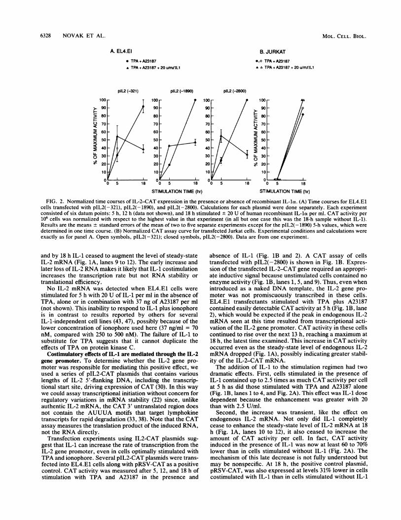
FIG. 2. Normalized time courses of IL-2-CAT expression in the presence or absence of recombinant IL-la. (A) Time courses for EL4.E1cells transfected with pIL2(-321), pIL2(-1890), and pIL2(-2800). Calculations for each plasmid were done separately. Each experimentconsisted of six datum points: 5 h, 12 h (data not shown), and 18 h stimulated ± 20 U of human recombinant IL-la per ml. CAT activity per
106 cells was normalized with respect to the highest value in that experiment (in all but one case this was the 18-h sample without IL-1).Results are the means ± standard errors of the mean of two to five separate experiments except for the pIL2(-1890) 5-h values, which were
determined in one time course. (B) Normalized CAT assay curve for transfected Jurkat cells. Experimental conditions and calculations were
exactly as for panel A. Open symbols, pIL2(-321); closed symbols, pIL2(-2800). Data are from one experiment.
and by 18 h IL-1 ceased to augment the level of steady-stateIL-2 mRNA (Fig. 1A, lanes 9 to 12). The early increase andlater loss of IL-2 RNA makes it likely that IL-1 costimulationincreases the transcription rate but not RNA stability ortranslational efficiency.No IL-2 mRNA was detected when EL4.E1 cells were
stimulated for 5 h with 20 U of IL-1 per ml in the absence ofTPA, alone or in combination with 37 ng of A23187 per ml(not shown). This inability to respond to IL-1 plus ionophoreis in contrast to results reported by others for severalIL-1-independent cell lines (43, 47), possibly because of thelower concentration of ionophore used here (37 ng/ml = 70nM, compared with 250 to 500 nM). The failure of IL-1 tosubstitute for TPA suggests that it cannot duplicate theeffects of TPA on protein kinase C.
Costimulatory effects of IL-1 are mediated through the IL-2gene promoter. To determine whether the IL-2 gene pro-moter was responsible for mediating this positive effect, weused a series of pIL2-CAT plasmids that contains variouslengths of IL-2 5'-flanking DNA, including the transcrip-tional start site, driving expression of CAT (30). In this waywe could assay transcriptional initiation without concern forregulatory variations in mRNA stability (22) since, unlikeauthentic IL-2 mRNA, the CAT 3' untranslated region doesnot contain the AUUUA motifs that target lymphokinetranscripts for rapid degradation (33, 38). Note that the CATassay measures the translation product of the induced RNA,not the RNA directly.
Transfection experiments using IL2-CAT plasmids sug-gest that IL-1 can increase the rate of transcription from theIL-2 gene promoter, even in cells optimally stimulated withTPA and ionophore. Several pIL2-CAT plasmids were trans-fected into EL4.E1 cells along with pRSV-CAT as a positivecontrol. CAT activity was measured after 5, 12, and 18 h ofstimulation with TPA and A23187 in the presence and
absence of IL-1 (Fig. 1B and 2). A CAT assay of cellstransfected with pIL2(-2800) is shown in Fig. 1B. Expres-sion of the transfected IL-2-CAT gene required an appropri-ate inductive signal because unstimulated cells contained noenzyme activity (Fig. 1B, lanes 1, 5, and 9). Thus, even whenintroduced as a naked DNA template, the IL-2 gene pro-moter was not promiscuously transcribed in these cells.EL4.E1 transfectants stimulated with TPA plus A23187contained easily detectable CAT activity at 5 h (Fig. 1B, lane2), which would be expected if the peak in endogenous IL-2mRNA seen at this time resulted from transcriptional acti-vation of the IL-2 gene promoter. CAT activity in these cellscontinued to rise over the next 13 h, reaching a maximum at18 h, the latest time examined. This increase in CAT activityoccurred even as the steady-state level of endogenous IL-2mRNA dropped (Fig. 1A), possibly indicating greater stabil-ity of the IL-2-CAT mRNA.The addition of IL-1 to the stimulation regimen had two
dramatic effects. First, cells stimulated in the presence ofIL-1 contained up to 2.5 times as much CAT activity per cellat 5 h as did those stimulated with TPA and A23187 alone(Fig. 1B, lanes 1 to 4, and Fig. 2A). This effect was IL-1 dosedependent because the enhancement was greater with 20than with 2.5 U/ml.
Second, the increase was transient, like the effect on
endogenous IL-2 mRNA. Not only did IL-1 completelycease to enhance the steady-state level of IL-2 mRNA at 18h (Fig. 1A, lanes 10 to 12), it also ceased to increase theamount of CAT activity per cell. In fact, CAT activityinduced in the presence of IL-1 was now at least 60 to 70%lower than in cells stimulated without IL-1 (Fig. 2A). Themechanism of this late decrease is not fully understood butmay be nonspecific. At 18 h, the positive control plasmid,pRSV-CAT, was also expressed at levels 31% lower in cellscostimulated with IL-1 than in cells stimulated without IL-1
L.0
MOL. CELL. BIOL.

IL-1 COINDUCTION OF TRANSCRIPTIONAL ACTIVATORS 6329
TABLE 2. Failure of IL-1 to increase cAMP in stimulatedEL4.E1 cells
cAMP concnStimulation Time (fmol/106 cells)bconditionsa (min)
Expt 1 Expt 2
No stimulation 350 320TPA + A23187 15 300 320
30 250 31060 271 380
TPA + A23187 + 20 U of IL-1/ml 15 246 30030 308 34060 258 460
TPA + A23187 + 10-7 M forskolin 15 ND 46030 ND 47060 ND 750
TPA + A23187 + 10-5 M forskolin 15 ND >4,00030 ND >4,00060 ND >4,000
a EL4.E1 cells were stimulated with TPA (10 ng/ml) + A23187 (37 ng/ml) ata density of 1.2 x 10⁶/ml.
b Measured by using a commercial assay kit as described in Materials andMethods. All values are from assays with acetylated standards and samples.The high values for samples with 10'5 M forskolin were corroborated byreassay with nonacetylated standards and samples. ND, Not done.
(see below and Table 3). By contrast, the early enhancementof CAT expression from the IL-2 gene promoter was pro-moter specific. Expression of pRSV-CAT in stimulatedEL4.E1 cells was not affected by the presence of IL-1 whenexamined after 3 or 5 h of stimulation (unpublished data).This result suggests that at later times, IL-1 caused a generaldecrease in CAT expression, possibly via reduced transla-tional efficiency, which was distinct from its promoter-specific transcriptional enhancing effects seen at 5 h.
Possible mediators contributing to the effects of IL-1 onEL4.E1 cells. (i) IL-1R. The effects of IL-1 on the IL-2 genepromoter require the expression of IL-1R, as demonstratedby the results from identical transfection experiments usingIL-1R- Jurkat cells instead of EL4.E1 cells. A time courseof IL-2-CAT expression in Jurkat cells is shown in Fig. 2B.These cells showed much slower kinetics of expression ofpIL2-CAT plasmids than did EL4.E1 cells. No activity wasdetected at 5 h even in the presence of 20 U of IL-1 per ml.Cells had begun to express by 12 h, and CAT activity wasstill rising by 18 h. However, no significant effect, early orlate, could be attributed to IL-1. These results provideevidence that surface expression of a canonical IL-1R is aprerequisite for the IL-1 effects that we have described. It isthus unlikely that the effects seen in EL4.E1 cells are due tothe type of phosphatidylcholine hydrolysis reported to occurin Jurkat cells (34).
(ii) cAMP. It has been shown that IL-1 can utilize cAMP asan intracellular second messenger in a number of non-T-celllines (42). Our results argue against a simple cAMP elevationmechanism as the pathway for IL-1 costimulation in EL4.E1cells. First, IL-1 costimulation had minimal effects on cAMPlevels in these cells (Table 2). Second, the effects of 20 U ofIL-1 per ml and 10 ,uM forskolin were clearly different (Table3). Expression of pRSV-CAT was enhanced at 18 h withforskolin but inhibited with the concentration of IL-1 usedhere. Whereas expression of pIL2(-321) was depressed at18 h by both IL-1 and forskolin, the 5-h results clearlydemonstrated the dissimilarity of these two stimuli. Forsko-
TABLE 3. Distinct effects of IL-1 and forskolin on expression ofpRSV-CAT and plL2(-321) in EL4.E1 cells
Stimulation CAT activity (% of control)aConstruct time +IL-1 +10 FM
(h) (20 U/ml) forskolinb
pRSV-CAT 18 68 ± 0.5 427 ± 99pIL2(-321) 5 169 ± 22 28 ± 4
18 43 6 51 17a Cells were stimulated for the indicated lengths of time with TPA (10 ng/ml)
+ A23187 (37 ng/ml) in the presence or absence of IL-1 or forskolin. CATactivity per 106 cell equivalents was determined, and the value for thedrug-treated samples was normalized with respect to that of the drug-freecontrol. Results are presented as means + standard errors of the mean or asmeans + ranges (in the cases with two experiments) of two to four experi-ments each.
b Results are presented in more detail elsewhere (Novak and Rothenberg, inpress).
lin was even more suppressive at 5 h than at 18 h, whereasIL-1 was stimulatory at this time point (Table 3; see also Fig.1B, lanes 1 to 4, and Fig. 2A). We have also shown thatelevation of cAMP by a variety of other agents can depressIL-2 gene expression in a dose-dependent fashion (Novakand Rothenberg, in press), supporting the conclusion thatelevation of cAMP levels per se has a net negative effect onIL-2 gene promoter function. While these results do not ruleout cAMP-responsive signaling molecules as participants inthe IL-1 enhancement pathway, they do show that thestimulatory effect of IL-1 is unlikely to be a result of anysustained increase in cAMP concentration.
IL-2 gene promoter sequences upstream of -321 are notrequired for IL-1 costimulation. Our previous work on theupstream region of the mouse IL-2 gene indicated thepresence of several positive and negative regulatory ele-ments between -321 and -2800, any of which might beinvolved in IL-1-mediated effects. However, the data pre-sented in Fig. 2A make it unlikely that far-upstream se-quences are required.When normalized for maximal levels of CAT activity, the
kinetics of IL-1 costimulation for pIL2(-321), pIL2(-1890),and pIL2(-2800) were virtually superimposable (Fig. 2A).Thus, the degrees of both the 5-h enhancement and the 18-hsuppression were unaffected by IL-2 5'-flanking sequencesextending upstream of -321. It should be noted, however,that the absolute amount of CAT activity per cell at eachtime point was higher with pIL2(-1890) and pIL2(-2800)than with pIL2(-321), in accord with our previously re-ported results (30).
Further truncation of the IL-2 gene promoter region led tosevere loss of inducibility, such that any IL-1-dependentenhancement was difficult to detect. We evaluated the re-sponses of two plasmids with deletions in the region from-321 to -261. One, pIL2(-232), failed to give significantexpression with or without IL-1 at any time point. Expres-sion of the other, pIL2(-1890; ANFAT-1), which bears adeletion from -261 to -321 (30), was also undetectable at 5h of stimulation, when IL-1 would be expected to exert apositive effect (P. M. White, T. J. Novak, and E. V.Rothenberg, unpublished results; 30). Thus, retention ofsequences between -261 and -321 appeared to be essentialfor expression and IL-1 costimulation.
Effect of IL-1 costimulation on sequence-specific DNA-binding factors. The sequences from -50 to -300 in thehuman IL-2 gene include binding sites for several DNA-binding proteins of different specificities which have beenimplicated in regulation (4, 10). One factor, binding se-
VOL. 10, 1990

6330 NOVAK ET AL.
A NFAT-1 Probe B NFkB Probe CAP-1 p Probe AP-1 D Probe
COMPET NFAT-1 NFKB (P) (D)E0 tIOO 0 0 ZOO O O
EXCESS 0 so 100 200 SO 100 zoo ?00 200
NF#4B AP-1 D (-148/217)0 --,,,8 ..
0 80 160 3ZO 80 160 320 20 40 80
AP-1p AP-1D (A) (N) - - AP-lp AP-1D (A) (N)1 ZO 10 O t5 ZO 0 0 10--02 10f -O\100 zoo tO0 Z00 so 200 0 0 tO0 200 tO0 zoo 50 zoo
I I 4 P
1 2 3 4 5 6 7 8 91 2 3 4 5 6 7 8 9 10
1 2 3 4 5 6 7 8 9 10 111213 14
FIG. 3. Specificities of complexes with binding activity for IL-2 regulatory sequences. (A) Homologous and heterologous competition forthe complex binding the NFAT-1 oligonucleotide. Competition gel shift assays were carried out as described in Materials and Methods witha labeled NFAT-1 oligonucleotide. Unlabeled competitor oligonucleotides were added as indicated, to 50- to 200-fold molar excesses. Thearrow indicates the specific NFAT-1 complex. (P) and (D) represent the AP-1P and AP-1D oligonucleotides, respectively. Lane 1, No specificcompetitor. Each lane contained 2.5 ,ug of nuclear extract from cells stimulated with TPA + A23187 + IL-1 with 2.3 ,ug of poly(dA dT) asnonspecific competitor. (B) As panel A, using labeled NF-KB oligonucleotide with the indicated competitors at the molar excesses shown.Lane 1, No specific competitor. (- 148/-217) is a restriction fragment from the natural IL-2 gene promoter that contains the NF-KB site. Itsgreater potency as a competitor in part reflects a general effect of length (F. J. Calzone and E. H. Davidson, personal communication). Eachlane contained 5 jig of extract from TPA + A23187-stimulated cells and 0.5 jig of poly(dI dC). (C) Cross-competition of complexes bindingthe AP-1p and AP-1D oligonucleotides. Lanes: 1 to 7, AP-1p probe with the indicated competitors (lane 7, no specific competitor); 8 to 14,AP-1D probe with the indicated competitors (lane 8, no specific competitor). (A) denotes a commercial consensus AP-1 22-mer (Stratagene),used at 50-fold molar excess; (N) denotes the NF-KB oligonucleotide used at 200-fold molar excess. Arrows indicate the mobilities of the mainAP-1p complex (left) and the AP-1D complex (right). Each lane contained 5 jig of extract as in panel A, with 0.125 jig of poly(dI dC). Bothextracts used in this figure were made by the method of Dignam et al. (6).
quences from -66 to -90, is the constitutive Oct-1 protein,which also appears to bind sequences between -240 and-250 (10). Factors whose binding activity is known to bedependent on cellular activation bind at -264 to -284(NFAT-1) and -196 to -205 (NF-KB or a close relative) (5,17, 39). In addition, there are two potential sites for thebinding of the canonical TPA-response factor AP-1 (2),although neither contains a perfect consensus sequence.One, from -186 to -195 (AP-1D), has been proposed as acritical site for IL-1 effects in another cell line (26). Theother, from -145 to -153 (AP-1p), has been shown to binda purified AP-1 factor (37).To dissect the mechanism of IL-1 costimulation of EL4.E1
cells, we focused on the four sites for potentially induciblefactors: NFAT-1, NF-KB, AP-1D, and AP-1p. Syntheticdouble-stranded oligonucleotides containing these sites asfound in the murine IL-2 gene were used in gel retardationassays to detect DNA-binding proteins in nuclear extractsfrom EL4.E1 cells after different types of stimulation. Typ-ical results, representative of four to seven independentanalyses, are presented in Fig. 3 and 4. A summary isprovided in Table 4.Four distinct binding specificities. Confirmation of the
distinct specificities of these factors is presented in Fig. 3.
The binding of NFAT-1 was highly sensitive to competitionwith its homologous target sequence and impervious tocompetition with the other oligonucleotides used (Fig. 3A).Figure 3B similarly confirms the specificity of the NF-KBcomplexes and their ability to be competed for by a longerIL-2 DNA fragment (-148 to -217) that contains this site.We routinely observed a doublet of complexes binding theNF-KB site, both of which appear specific by competitionanalysis. Only the upper band was strongly induction depen-dent, however, and in the text that follows we will use"NF-KB" to refer only to this slower-migrating species.The factors that bind to the two AP-1 sites are not
identical, although their target sites are of related sequence.This conclusion was confirmed by the cross-competitionanalysis shown in Fig. 3C. First, the two complexes haddifferent mobilities (compare lanes 7 and 8). Second, it isclear that they had different binding preferences. Both werelegitimately capable of binding a commercial consensusAP-1 oligonucleotide (Fig. 3C, lanes 5 and 13), and neitherwas detectably competed for by the NF-KB site oligonucle-otide (lanes 6 and 14). However, while the AP-1P oligonu-cleotide competed for the complex binding its homologoussequence (lanes 1 and 2), it did not compete for the AP-1Dcomplex (lanes 9 and 10). Correspondingly, the AP-1D
MOL. CELL. BIOL.

IL-1 COINDUCTION OF TRANSCRIPTIONAL ACTIVATORS 6331
A NFAT-1
(dA-dT) 5 pg9 10,yJurkat EL4
Extract: U S I U T I T/A+I +F
B NF#B
T/A T/AU T T/A + + F
C AP-1D
T/A T/AU T I T/A +l +F__¢.> - *X
D AP-1 p
T/A T/AU T T/A+I+F Extract
wt <,_
mefta e fte i4-
mm1 2 3 4 5 6 7 8 9 1 2 3 4 5 6 1 2 3 4 5 6 1 2 3 4 5 6
FIG. 4. Differential activation of IL-2 DNA-binding factors under different conditions of stimulation. In each panel, the indicated probeswere used for gel shift analysis, each lane containing 5 jig of extract from EL4.E1 cells that were stimulated for 3.5 h under the indicatedconditions: U, unstimulated; T, TPA alone; I, IL-1 alone; T/A, TPA plus A23187; T/A+I, TPA plus A23187 plus IL-1; T/A+F, TPA plusA23187 plus 10 FjM forskolin. In panel A only, NFAT-1 complexes from EL4.E1 cells (lanes 3 to 9) are compared with complexes formedby nuclear extracts from Jurkat cells, either unstimulated (U) or stimulated with TPA plus A23187 (S). (A) NFAT-1 factor. The arrow
indicates the NFAT-1 complex, which is inducible in Jurkat cells (lanes 1 and 2) but constitutive in EL4.E1 cells (lanes 4 to 9). Lane 3 containsa duplicate of the extract used in lane 6 with a lower amount of nonspecific competitor, to demonstrate that the lower band seen in the Jurkatsample (lane 1) is nonspecific. (B) NF-KB factors. The arrow indicates the upper complex, which is more strictly dependent on induction thanthe lower complex and which is the subject of discussion in the text. Each lane contained 0.5 p.g of poly(dI-dC). (C) AP-1D factor. Theincreased band intensity seen in lane 4 was highly irreproducible (see Table 4), in contrast to the differences seen in panel B. In panels C andD, 0.25 ,ug of poly(dI dC) was used as nonspecific competitor. Arrows indicate the specific complexes. (D) AP-1p factor, analysis as for panelC.
oligonucleotide showed homologous competition (lanes 11and 12) but did not compete for the AP-lp complex (lanes 3and 4). The quantitative reciprocity of these effects, as
measured at 100- and 200-fold molar excess, rules out thetrivial explanation that the two factors had identical speci-ficities but were present at different concentrations in thenuclear extract. These results show that the two AP-1
factors are distinct both in size and in specificity. Asdiscussed below, they are also distinct in regulation.
Constitutive factors. Two of the factors detected by theseassays appeared to be expressed constitutively in theEL4.E1 thymoma cells. One, unexpectedly, was NFAT-1. Acomplex of the expected mobility (indicated by an arrow inFig. 4A) was highly inducible in Jurkat cells (Fig. 4A, lanes
TABLE 4. Summary of effects of different stimulation conditions on specific IL-2 DNA-binding proteins
DNA-binding Stimulation conditionsa No. offactor U T T/A T/A+I T/A+F exptsb
NFAT-1 +/-- + + + + + 2(D) + 3(C)NF-KBC - ++ +±+ ++I+ +++ ++/+ 2(D) + 2(C)AP lDc + + + + + + 2(D) + 2(C)AP1lpC ±1- + - ++ +++ +++ 2(D) + 2(C)a Terminology as in the legend to Fig. 4.b Number of independent experiments (separate stimulation and extract preparation), counting separately cases in which extracts were generated by the method
of Dignam et al. (6) (D) or Crabtree (4) (C). Similar relative results were obtained with both kinds of extracts although the absolute concentrations of differentfactors were somewhat different for the two protocols.
c One set of data was obtained from an experiment with extracts prepared by the method of Dignam et al. (6) which compared only the U, T, T/A, and T/A+ 1conditions of culture.
VOL. 10, 1990
---* m,d, :O- 06 i. -
--, I.,

6332 NOVAK ET AL.
1 and 2). However, the corresponding complex was presentat indistinguishable levels in nuclear extracts from oursubline of EL4.E1 cells, whether or not they were stimulatedand regardless of the stimulation conditions used (Fig. 4A,lanes 4 to 9). This complex appeared unlikely to represent aspurious DNA-binding activity, since it was specific by thecompetition assays in Fig. 3 but quantitatively unaffected byincreasing the concentration of nonspecific competitor (Fig.4A, lanes 3 and 6). Thus, although the NFAT-1 site appearedessential for expression, on the basis of our deletion studies,the presence of its binding factor was not sufficient tosupport IL-2 expression.The other factor present at unchanged levels was the
complex of well-defined mobility binding the AP-1D site (Fig.4C). This factor exhibited slight fluctuations in concentrationin different samples, but no reproducible increase was ob-served upon stimulation under any conditions (Table 4). Infact, in some experiments there was an indication that itsconcentration diminished in stimulated cells. The possibilitythat it might exert a negative regulatory effect is underfurther study.
Inducible factors. Only two of the candidate induciblefactors showed increased DNA binding in response to induc-tion. These were NF-KB (Fig. 4B) and the factor binding theAP-lp site (Fig. 4D). In agreement with the ability of EL4.E1cells to make IL-2 in response to TPA alone (Table 1), bothwere activated in cells stimulated with TPA alone (Fig. 4Band D, lanes 2). The further addition of A23187 up regulatedboth binding activities in most experiments, in accord withits effect on IL-2 production (Table 1), although the extentsof up regulation did not match precisely (Fig. 4B and D;compare lanes 2 and 4). The binding activities of bothfactors, however, were reproducibly enhanced further byaddition of IL-1, which consistently yielded the highestlevels observed of both complexes (Fig. 4B and D, lanes 5).This result was not due to additive activation of these factorsby IL-1 and by TPA + A23187, for in most experiments IL-1alone induced little or no binding activity of either type(compare lanes 3 in Fig. 4B and D). Thus, while IL-1 wasonly marginally and irreproducibly capable of activatingeither binding factor alone, it was a potent enhancer of thestimulating capacity of A23187 and TPA.Summary. The results of multiple analyses of these four
factors are collated in Table 4. Overall, these data agreeplausibly with the IL-2 titers reported in Table 1, if it isassumed that all four factors are required for optimal pro-moter activity. The surprising, constitutive expression ofNFAT-1 in this subline of cells, and possibly that of theAP-1D binding factor a well, may simply provide thebiochemical explanation For the unique ability of EL4.E1cells to express IL-2 in response to TPA alone. Neverthe-less, this set of factors may not be sufficient to account for allregulatory modulation of IL-2 gene expression in EL4.E1cells. As shown previously (Novak and Rothenberg, inpress; Tables 1 and 3), 10' M forskolin inhibits IL-2induction by two- to threefold under these conditions. It istherefore surprising that forskolin, when added to TPA andA23187, induced effects on these DNA-binding proteins thatwere very similar to those of IL-1 (Fig. 4A, lane 9; Fig. 4B toD, lanes 6). While in several experiments the addition offorskolin appeared to decrease NF-KB expression selec-tively (data not shown), this effect was often modest (e.g.,Fig. 4B). It is thus possible that the ratio of AP-lp to NF-KBaffects expression. However, it seems more likely that theactivity of the four putative positive regulators can beinfluenced by other regulatory proteins not examined here.
Preliminary results suggest that the net inhibitory effects ofelevated cAMP depend on the presence of additional dis-crete sequences in the IL-2 regulatory region (P. M. Whiteand E. V. Rothenberg, unpublished results).
DISCUSSION
IL-1 has a long history as a costimulator of T-cell re-sponses (9, 13, 25, 44). In recent years, it has become clearthat this cytokine carries out different essential functions,depending on the cell type being stimulated. Most effects ofIL-1 on mature T cells, however, can be substituted for bythe addition of phorbol esters, presumably via stimulation ofprotein kinase C. Biochemical evidence does not support theconclusion that IL-1 acts directly via protein kinase C (1),but in most cases activation of this kinase can obscure theeffects of IL-1 itself on T cells. In this study we tookadvantage of the unusual properties of EL4.E1 cells, whichrespond to IL-1 as a costimulus even with optimal levels ofTPA, to investigate the molecular mediators by which IL-1can cooperate with the phosphoinositide pathway. Our re-sults show that IL-1 costimulates IL-2 gene expression byenhancing the activity of the IL-2 gene promoter, correlatedwith augmented levels of an NF-KB-like factor and one oftwo distinct AP-1-like factors that bind to IL-2 regulatorysequences.Recent work by Shirakawa and colleagues has shown that
IL-1 can utilize cAMP as an intracellular second messengerfor the induction of K immunoglobulin light-chain synthesisin the mouse pre-B-cell line 70Z/3 (42). In these cells,IL-1-mediated induction coincides with the activation of aprotein that can bind to the NF-KB consensus site in the5'-flanking region of the K-chain genes (40). NF-KB is not theonly transcription factor implicated in IL-1 effects, however.IL-1 can also induce transcription of the c-jun proto-onco-gene, which encodes a component of AP-1, in the mouseIL-1-dependent T-cell line LBRM-331A5 (26). In that cellline, expression of a linked reporter gene from the humanIL-2 promoter required an intact AP-1 site at -185 relativeto the transcriptional start site. However, the deletionstested which removed this site also removed the NF-KB sitecentered at -200. Therefore, the resulting loss of promoterfunction (26) cannot be attributed unequivocally to loss ofthis distal AP-1 site. In fact, deletion analysis of the humanIL-2 gene promoter has not demonstrated an important rolefor this AP-1 site in vivo (8). Footprint analysis of the mouseIL-2 promoter has led to the same conclusion (37).
In several respects, our results are in agreement with theseprevious reports. Thus, our data support the interpretationthat one mediator of the IL-1 effect may be NF-KB or arelated factor (31, 40). The addition of IL-1 as a costimulusinvariably augmented NF-KB binding activity over thatinduced by the cocktail of TPA and A23187, and in someexperiments (e.g., Fig. 4) we could even detect enhancedNF-KB binding after stimulation with IL-1 alone. As ex-pected if IL-1 activated NF-KB, it also enhanced the induc-tion of IL-2 receptor a-chain transcripts in the EL4.E1 cells(T. J. Novak and E. V. Rothenberg, unpublished results).Even the failure of IL-1 to affect IL-2 gene promoter activityin Jurkat cells is also consistent with its working throughNF-KB, since in these cells IL-1 does not induce appearanceof this DNA-binding protein (31). The resulting implicationthat NF-KB may be rate limiting for IL-2 induction is inaccord with published evidence that mutation of the NF-KBsite depresses IL-2 gene promoter function, relative to wildtype, in optimally stimulated cells (17).
MOL. CELL. BIOL.

IL-1 COINDUCTION OF TRANSCRIPTIONAL ACTIVATORS 6333
In contrast to other cell types (41, 42), however, EL4.E1cells appear to be able to respond to IL-1 as a costimuluswithout using cAMP as the principal mediator. As we reporthere and in more detail elsewhere (Novak and Rothenberg,in press), IL-2 gene promoter activity is significantly inhib-ited by treatments that cause a sustained elevation of intra-cellular cAMP. This inhibition is especially sharp at the earlytimes when IL-1 is most potent in its stimulatory activity.Thus, the cooperative effects of IL-1 with phosphoinositidepathway stimulation and those of cAMP appear quite dis-tinct.
It seems paradoxical, then, that both IL-1 and cAMP mayenhance activation of NF-KB. Indeed, both also hyperacti-vate the binding factor of the proximal AP-1 site. However,while the induced DNA-binding activities that we havestudied probably participate in the mechanism regulatingIL-2 gene promoter activity, they may not be the onlycomponents, or the only decisive components, of a combi-natorial mechanism. The resolution of this paradox is mostlikely to come, therefore, from more extensive monitoring ofthe ensemble of regulatory proteins that control IL-2 geneexpression, rather than a focus on any single one. Ourresults with only four such factors provide compelling evi-dence for the combinatorial nature of IL-2 gene regulation.The NFAT-1 factor, the regulation of which may be suffi-cient to account for the response to TcR ligands in Jurkatcells (4, 10) is not rate limiting in EL4.E1 cells. TheNFAT-1-binding activity is present at maximal levels even incells which cannot secrete IL-2 and cannot express IL-2promoter constructs. Yet the cooperation of this factorappears to be required for efficient IL-2 induction, as shownby the minimal activity, with or without IL-1, of constructsfrom which its binding site has been deleted. In this context,the shared abilities of forskolin and IL-1 to stimulate theAP-lp factor and NF-KB imply less that these agents mediatethe same signal than that the set of factors we monitor maybe incomplete.How can the costimulatory effects of IL-1 on AP-lp and
NF-KB activation be explained? There are two generalpossibilities. First, IL-1/IL-1R triggering may directly induceactivation of these two factors, and possibly others uniquelyresponsive to IL-1, mobilizing pools that are not accessibleto the calcium ionophore/phorbol ester stimulation pathway.The effects of IL-1 would then be additive with those of thephosphoinositide pathway agonists, at the level of the DNA-protein contacts in chromatin. Another possibility is that theability of A23187/TPA to activate these DNA-binding pro-teins is itself modulated by an intracellular gating functionthat is subject to IL-1 regulation. In this case, IL-1 need notinduce any DNA-binding protein activities by itself in orderto be a potent regulator of the efficiency with which themajor activating pathway mobilizes gene expression. Whiletoo few factors have yet been examined to confirm or ruleout the first mechanism, several of our findings lend plausi-bility to the second. First, IL-1 alone is strikingly poor ateffecting any increase or decrease in the binding activity ofany of the factors studied here. In EL4.E1 cells, it is farmore potent as a costimulus than as a stimulus (see, e.g.,Table 4). Second, the factors that IL-1 coactivates thus farappear to be only those that are activated in any case byA23187 and TPA. Third, our survey of IL-1 effects on theexpression of IL-2 promoter constructs with widely differingnumbers of positive and negative modulatory sites (30)indicates that all expressible constructs are affected in par-allel. Thus, IL-1 may act in these cells by enhancing thepotency of A23187 and TPA as inducing agents, its signals
converging with the phosphoinositide pathway before theactivation of the specific DNA-binding proteins.The mechanism of IL-1 costimulation may help to illumi-
nate key stages in the development of T-cell functionalcompetence. We have previously reported that immaturethymocytes can activate the IL-2 gene, but only subject to anabsolute requirement for IL-1 costimulation. The locus ofthe IL-1 effect may thus identify a stage-specific block to theexercise of function in immature cells which is later over-come by the complex physiology of positiv,e~selection in thethymus. Work is now under way to explo#e this possibility.
ACKNOWLEDGMENTS
We thank Jerry Crabtree for his generous provision of cells,protocols, and advice, Frank Calzone and Eric Davidson for theircritical guidance and encouragement, Cherrie Leighton for excellentgraphics, and Cathy Blagg and Renee Thorf for careful preparationof the manuscript.
Support for this work was provided by the Lucille P. MarkeyCharitable Trust and by Public Health Service grant CA 39605 fromthe National Cancer Institute to E.V.R. T.J.N. was supported inpart by a predoctoral training grant from the Public Health Service,and D.C. gratefully acknowledges support from a Gordon RossMedical Fellowship.
LITERATURE CITED1. Abraham, R. T., S. N. Ho, T. J. Barna, and D. J. McKean. 1987.
Transmembrane signaling during interleukin 1-dependent T-cellactivation. J. Biol. Chem. 262:2719-2728.
2. Angel, P., M. Imagawa, R. Chiu, B. Stein, R. J. Inbra, M. J.Rahmsdorf, C. Jonat, P. Herrlich, and M. Karin. 1987. Phorbolester-inducible genes contain a common cis element recognizedby a TPA-modulated trans-acting factor. Cell 49:729-739.
3. Calzone, F. J., N. Th6zt, P. Thiebaud, R.L. Hill, R. J. Britten,and E. H. Davidpon. 1988. Developmental appearance-oftactorsthat bind specifically to cis-reguto'y sequences of a geneexpressed in the sea-Trcliin embryo. Genes Dev. 2:1074-1088.
4. Crabtree, G. R. 1989. Contingent genetic regulatory events in Tlymphocyte activation. Science 243:355-361.
5. Cross, S. L., N. F. Halden, M. J. Lenardo, and W. J. Leonard.1989. Functionally distinct NF-KB binding sites in the immuno-globulin K and IL-2 receptor ax chain genes. Science 244:466-469.
6. Dignam, J. D., R. M. Lebovitz, and R. G. Roeder. 1983.Accurate transcription initiation by RNA polymerase II in asoluble extract from isolated mammalian nuclei. Nucleic AcidsRes. 11:1475-1489.
7. Dinarello, C. A. 1989. Interleukin-1 and its biologically relatedcytokines. Adv. Immunol. 44:156-205.
8. Durand, D. B., J.-P. Shaw, M. R. Bush, R. E. Reployle, R.Belagaje, and G. R. Crabtree. 1988. Characterization of antigenreceptor response elements within the interleukin-2 enhancer.Mol. Cell. Biol. 8:1715-1724.
9. Durum, S. K., J. A. Schmidt, and J. J. Oppenheim. 1985.Interleukin 1: an immunological perspective. Annu. Rev. Im-munol. 3:263-287.
10. Emmel, E. A., C. L. Verwei, D. B. Durand, K. M. Higgins, E.Lacy, and G. R. Crabtree. 1989. Cyclosporin A specificallyinhibits function of nuclear proteins involved in T-cell activa-tion. Science 246:1617-1670.
11. Farrar, J. J., S. B. Mizel, J. Fufler-Farrar, W. L. Farrar, andM. L. Hilfiker. 1980. Macrophage-independent activation ofhelper T cells. I. Production of interleukin 2. J. Immunol.125:793-798.
12. Favaloro, J., R. Treisman, and R. Kamen. 1980. Transcriptionmaps of polyoma virus-specific RNA: analysis by two-dimen-sional nuclease S1 gel mapping. Methods Enzymol. 65:718-749.
13. Gillis, S., and S. B. Mizel. 1981. T-cell lymphoma model for theanalysis of interleukin 1-mediated T-cell activation. Proc. Natl.Acad. Sci. USA 78:1133-1137.
14. Greenbaum, L. A., J. B. Horowitz, A. Woods, T. Pasqualini,
VOL. 10, 1990

6334 NOVAK ET AL.
E. P. Reich, and K. Bottomly. 1988. Autocrine growth of CD4+T cells. Differential effects of IL-1 on helper and inflammatory Tcells. J. Immunol. 140:1555-1560.
15. Hackett, R. J., L. S. Davis, and P. E. Lipsky. 1988. Comparativeeffects of tumor necrosis factor-a and IL-1, on mitogen-inducedT cell activation. J. Immunol. 140:2639-2644.
16. Heckford, S. E., E. P. Gelmann, C. L. Agnor, S. Jacobson, S.Zinn, and L. A. Matis. 1986. Distinct signals are required forproliferation and lymphokine gene expression in murine T cellclones. J. Immunol. 137:3652-3663.
17. Hoyos, B., D. W. Ballard, E. Bohnlein, M. Siekevitz, and W. C.Greene. 1989. Kappa B-specific DNA binding proteins: role inthe regulation of human interleukin-2 gene expression. Science244:457-460.
18. Kaye, J., S. Gillis, S. B. Mizel, E. M. Shevach, T. R. Malek,C. A. Dinarello, L. B. Lachman, and C. A. Janeway, Jr. 1984.Growth of a cloned helper T cell line induced by a monoclonalantibody specific for the antigen receptor: interleukin 1 isrequired for the expression of receptors for interleukin 2. J.Immunol. 133:1339-1345.
19. Koide, S., and R. M. Steinman. 1987. Induction of murineinterleukin 1: stimuli and responsive primary cells. Proc. Natl.Acad. Sci. USA 84:3802-3806.
20. Kurt-Jones, E. A., S. Hamberg, J. Ohara, W. E. Paul, and A. K.Abbas. 1987. Heterogeneity of helper/inducer T lymphocytes. I.Lymphokine production and lymphokine responsiveness. J.Exp. Med. 166:1774-1787.
21. Lichtman, A. M., J. Chin, J. A. Schmidt, and A. K. Abbas. 1988.Role of interleukin 1 in the activation of T lymphocytes. Proc.Natl. Acad. Sci. USA 85:9699-9703.
22. Lindsten, T., C. H. June, J. A. Ledbetter, G. Stella, and C. B.Thompson. 1989. Regulation of lymphokine messenger RNAstability by a surface-mediated T-cell activation pathway. Sci-ence 244:339-343.
23. Mary, D., C. Aussel, C. Pelassy, and M. Fehlmann. 1988. IL-1signaling for IL-2 production in T cells involves a rise inphosphatidylserine synthesis. J. Immunol. 141:3078-3080.
24. McGuire, K. L., J. A. Yang, and E. V. Rothenberg. 1988.Influence of activating stimulus on functional phenotype: inter-leukin-2 mRNA accumulation differentially induced by iono-phore and receptor ligands in subsets of murine T cells. Proc.Natl. Acad. Sci. USA 85:6503-6507.
25. Mizel, S. B. 1982. Interleukin 1 and T cell activation. Immunol.Rev. 63:51-72.
26. Muegge, K., T. M. Williams, J. Kant, M. Karin, R. Chiu, A.Schmidt, U. Siebenlist, H. A. Young, and S. K. Durum. 1989.Interleukin-1 costimulatory activity on the interleukin-2 pro-moter via AP-1. Science 246:249-251.
27. Nau, G. J., D-K. Kim, and F. W. Fitch. 1988. Agents that mimicantigen receptor signaling inhibit proliferation of cloned murineT lymphocytes induced by IL-2. J. Immunol. 141:3557-3563.
28. Nishizuka, Y. 1984. The role of protein kinase C in cell surfacesignal transduction and tumour promotion. Nature (London)308:693-698.
29. Novak, T. J., and E. V. Rothenberg. 1986. Differential transientand long-term expression of DNA sequences introduced into Tlymphocyte lines. DNA 5:439-451.
30. Novak, T. J., P. M. White, and E. V. Rothenberg. 1990.Regulatory anatomy of the murine interleukin-2 gene. Nucl.Acids Res. 18:4523-4533.
31. Osborn, L., S. Kunkel, and G. J. Nabel. 1989. Tumor necrosisfactor a and interleukin 1 stimulate the human immunodefi-ciency virus enhancer by activation of the nuclear factor NF-
KB. Proc. Natl. Acad. Sci. USA 86:2336-2340.32. Otten, G., K. C. Herold, and F. W. Fitch. 1987. Interleukin 2
inhibits antigen-stimulated lymphokine synthesis in helper Tcells by inhibiting calcium-dependent signaling. J. Immunol.139:1348-1353.
33. Reeves, R., T. S. Elton, M. S. Nissen, D. Lehn, and K. R.Johnson. 1987. Posttranscriptional gene regulation and specificbinding of the nonhistone protein HMG-I by the 3' untranslatedregion of bovine interleukin 2 cDNA. Proc. Natl. Acad. Sci.USA 84:6531-6535.
34. Rosoff, P. M., N. Savage, and C. A. Dinarello. 1988. Interleu-kin-1 stimulates diacylglycerol production in T lymphocytes bya novel mechanism. Cell 54:73-81.
35. Rothenberg, E. V., R. A. Diamond, T. J. Novak, K. A. Pepper,and J. A. Yang. 1990. Mechanisms of effector lineage commit-ment in T-lymphocyte development. UCLA Symp. Mol. Cell.Biol. 125:225-249.
36. Rothenberg, E. V., R. A. Diamond, K. A. Pepper, and J. A.Yang. 1990. Interleukin-2 gene inducibility in T cells prior toT-cell receptor expression: changes in signalling pathways andgene expression requirements during intrathymic maturation. J.Immunol. 144:1614-1624.
37. Serfling, E., R. Barthelmas, I. Pfeuffer, B. Schenk, S. Zarius, R.Swoboda, F. Mercurio, and M. Karin. 1988. Ubiquitous andlymphocyte-specific factors are involved in the induction of themouse interleukin 2 gene in T lymphocytes. EMBO J. 8:465-473.
38. Shaw, G., and R. Kamen. 1986. A conserved AU sequence fromthe 3' untranslated region of GM-CSF mRNA mediates selec-tive mRNA degradation. Cell 46:659-667.
39. Shaw, J.-P., P. J. Utz, D. B. Duncan, J. J. Toole, E. A. Emmel,and G. R. Crabtree. 1988. Identification of a putative regulatorof early T cell activation genes. Science 241:202-205.
40. Shirakawa, F., M. Chedid, J. Suttles, B. A. Polk, and S. B.Mizel. 1989. Interleukin 1 and cyclic AMP induce K immuno-globulin light-chain expression via activation of an NF-KB-likeDNA-binding protein. Mol. Cell. Biol. 9:959-964.
41. Shirakawa, F., and S. B. Mizel. 1989. In vitro activation andnuclear translocation of NF-KB catalyzed by cyclic AMP-dependent protein kinase and protein kinase C. Mol. Cell. Biol.9:2424-2430.
42. Shirakawa, F., U. Yamashita, M. Chedid, and S. B. Mizel. 1988.Cyclic AMP-an intracellular second messenger for interleukin1. Proc. Natl. Acad. Sci. USA 85:8201-8205.
43. Simon, P. L. 1984. Calcium mediates one of the signals requiredfor interleukin 1 and 2 production by murine cell lines. Cell.Immunol. 87:720-726.
44. Smith, K. A., K. J. Gilbride, and M. F. Favata. 1980. Lympho-cyte activating factor promotes T cell growth factor productionby cloned murine lymphoma cells. Nature (London) 287:853-855.
45. Williams, T. M., L. Eisenberg, J. E. Burlein, C. A. Norris, S.Pancer, D. Yao, S. Burger, M. Kamoun, and J. A. Kant. 1988.Two regions within the human IL-2 gene promoter are impor-tant for inducible IL-2 expression. J. Immunol. 141:662-666.
46. Yokota, T., N. Arai, F. Lee, D. Rennick, T. Mosmann, and K.-I.Arai. 1985. Use of a cDNA expression vector for isolation ofmouse interleukin 2 cDNA clones: expression of T-cell growth-factor activity after transfection of monkey cells. Proc. Natl.Acad. Sci. USA 82:68-72.
47. Zlotnik, A., and B. Daine. 1986. Activation of ILl-dependentand ILl-independent T cell lines by calcium ionophore andphorbol ester. J. Immunol. 136:1033-1037.
MOL. CELL. BIOL.