Intensive Care (ESPNIC) Critically Ill Children – Expert ...
29
Page 1/29 Recommendations for hemodynamic Monitoring for Critically Ill Children – Expert Consensus Statement issued by the Cardiovascular Dynamics Section of the European Society of Paediatric and Neonatal Intensive Care (ESPNIC) Yogen Singh ( [email protected] ) Cambridge University Hospitals and University of Cambridge School of Clinical Medicine https://orcid.org/0000-0002-5207-9019 Javier Urbano Villaescusa Hospital General Universitario Gregorio Maranon Eduardo M. da Cruz Children's Hospital Colorado Shane M Tibby Guy's and Saint Thomas' NHS Foundation Trust Gabriella Bottari Ospedale Pediatrico Bambino Gesu Rohit Saxena Great Ormond Street Hospital For Children NHS Foundation Trust Marga Guillén Newcastle Upon Tyne Hospitals NHS Foundation Trust Jesus Lopez Herce Hospital General Universitario Gregorio Maranon Matteo Di Nardo Ospedale Pediatrico Bambino Gesu Corrado Cecchetti Ospedale Pediatrico Bambino Gesu Joe Brierley Great Ormond Street Hospital For Children NHS Foundation Trust Willem de Boode Radboud Universiteit Joris Lemson Radboud Universiteit
Transcript of Intensive Care (ESPNIC) Critically Ill Children – Expert ...
Page 1/29
Recommendations for hemodynamic Monitoring forCritically Ill Children – Expert Consensus Statementissued by the Cardiovascular Dynamics Section ofthe European Society of Paediatric and NeonatalIntensive Care (ESPNIC)Yogen Singh ( [email protected] )
Cambridge University Hospitals and University of Cambridge School of Clinical Medicinehttps://orcid.org/0000-0002-5207-9019
Javier Urbano Villaescusa Hospital General Universitario Gregorio Maranon
Eduardo M. da Cruz Children's Hospital Colorado
Shane M Tibby Guy's and Saint Thomas' NHS Foundation Trust
Gabriella Bottari Ospedale Pediatrico Bambino Gesu
Rohit Saxena Great Ormond Street Hospital For Children NHS Foundation Trust
Marga Guillén Newcastle Upon Tyne Hospitals NHS Foundation Trust
Jesus Lopez Herce Hospital General Universitario Gregorio Maranon
Matteo Di Nardo Ospedale Pediatrico Bambino Gesu
Corrado Cecchetti Ospedale Pediatrico Bambino Gesu
Joe Brierley Great Ormond Street Hospital For Children NHS Foundation Trust
Willem de Boode Radboud Universiteit
Joris Lemson Radboud Universiteit
Page 2/29
Research
Keywords: Hemodynamic monitoring (HD), Paediatric intensive care unit (PICU), Children, Cardiovascularinstability, Recommendations
Posted Date: September 22nd, 2020
DOI: https://doi.org/10.21203/rs.3.rs-53557/v2
License: This work is licensed under a Creative Commons Attribution 4.0 International License. Read Full License
Version of Record: A version of this preprint was published on October 22nd, 2020. See the publishedversion at https://doi.org/10.1186/s13054-020-03326-2.

Page 3/29
AbstractBackground: Cardiovascular instability is common in critically-ill children. There is scarcity of publishedhigh-quality studies to develop meaningful evidence-based hemodynamic monitoring guidelines andhence, with the exception of management of shock, currently there are no published guidelines forhemodynamic monitoring in children. The European Society of Paediatric and Neonatal Intensive Care(ESPNIC) Cardiovascular Dynamics section aimed to provide expert consensus recommendations onhemodynamic monitoring in critically ill children.
Methods: Creation of a panel of experts in cardiovascular hemodynamic assessment and hemodynamicmonitoring and review of relevant literature - a literature search was performed, and recommendationswere developed through discussions managed following a Quaker-based consensus technique andevaluating appropriateness using a modi�ed blind RAND/UCLA voting method. The AGREE statementwas followed to prepare this document.
Results: Of 100 suggested recommendations across 12 subgroups concerning hemodynamic monitoringin critically ill children, 72 reached “strong agreement”, 20 “weak agreement” and 2 had “no agreement”.Six statements were considered as redundant after rephrasing of statements following �rst round ofvoting. The agreed 72 recommendations were then coalesced into 36 detailing four key areas ofhemodynamic monitoring in the main manuscript. Due to lack of published evidence to develop evidence-based guidelines, most of the recommendations are based upon expert consensus.
Conclusions: These expert consensus-based recommendation may be used to guide clinical practice forhemodynamic monitoring in critically-ill children and they may serve as a basis for highlighting gaps inthe knowledge base to guide further research in hemodynamic monitoring.
IntroductionCirculatory shock is de�ned as a “life threatening generalized maldistribution of blood �ow resulting infailure to deliver and / or utilize adequate amount of oxygen, leading to tissue dysoxia” [1].Cardiovascular instability with or without shock is common in children admitted to pediatric intensivecare units. Over half of the children with hemodynamic instability in intensive care units have multiple-organ dysfunction and sepsis remains the leading cause [2]. Similarly, a multi-center international study(Sepsis Prevalence, Outcomes, and Therapies; SPROUT) reported that over two-thirds of children withsepsis had multiorgan dysfunction, which was associated with a very high mortality. These data aresimilar to what has been described in the adult population [3]. However, there remains paucity of dataregarding the epidemiology of circulatory derangement in children and how best hemodynamic statuscan be evaluated or monitored in the pediatric intensive care units.
Multiple studies have established that early recognition and treatment of pediatric circulatoryinsu�ciency or shock is crucial to improve survival. However, the optimal way to resuscitate children withcirculatory failure is controversial. The exact order and quantity of �uids or vasoactive drug
Page 4/29
administration in the critically-ill child with shock is supported by little evidence, although there areseveral consensus statements [4, 5]
Overzealous �uid resuscitation is detrimental to some children [6, 7]. There is some consensus regardingthe �rst-line treatment in patients with shock or signi�cant hemodynamic instability, but debate remainsconcerning how much �uid should be administered, which type of vasoactive drug should be used, howto assess the hemodynamic changes and ultimately what hemodynamic clinical goal should be targetedto guide optimal treatment.
Ideal hemodynamic monitoring should accurately determine the severity of circulatory derangement andillustrate the underlying pathophysiologic mechanism to enable the clinician to choose the mostappropriate treatment and to guide the therapy [7, 8]. Furthermore, hemodynamic monitoring can provideuseful information about the circulatory condition in almost all types of life-threatening shock. This canbe hemodynamic instability as a result of major surgery, cardiac failure, trauma, sepsis or other causes ofshock in children. The current guidelines mostly focus on early recognition and treatment but do notspecify the type of monitoring technology that can or should be used under various circumstances. Thereare still no published evidence-based or even consensus guidelines speci�cally for hemodynamicmonitoring in neonates and children. However, there is a wide range of devices and techniques availableto evaluate the hemodynamic status and an increasing number of these methods have become availablefor use in children. In the annual meeting of the European Society of Paediatric and Neonatal IntensiveCare (ESPNIC) in October 2016, members of the Cardiovascular Dynamics Section were tasked to developevidence-based guidelines if at all possible, or an expert consensus statement on the hemodynamicmonitoring speci�cally for use in children.
MethodsA steering committee (SC) of three lead authors, one pediatric intensivist / anesthesiologist (JL), oneneonatologist / pediatric cardiologist (YS) and one pediatric intensivist (JU), identi�ed nine expert panelmembers who signi�cantly contributed with publications in hemodynamic monitoring or cardiovascularstatus assessment in children in the last ten years, similarly to what had been done with previous ESPNICguidelines [9-11]. Further, the three selection criteria for inclusion as panel member were: 1) must beclinicians working in a pediatric or neonatal intensive care; 2) needed to have experience with some formof HD monitoring; and 3) should have published in peer reviewed journals concerning the topic. Panelists’selection was performed prior to the literature search and for logistic reasons the number of participantswas limited to a maximum of 12. All invited experts agreed to participate.
The working group had a face-to-face meetings during the ESPNIC conference in Lisbon 2017 and theEuropean Academy of Paediatric Societies (EAPS) conference in Paris in 2018 and unanimously agreedto provide recommendations from full term infants over 37 weeks of gestation and over four weeks ofpostnatal age (lower limit) to 18 years old (upper limit), in order not to overlap with preterm-neonatal andadult guidelines. The panel subdivided hemodynamic monitoring into 12 subgroups: arterial blood
Page 5/29
pressure, central venous pressure, pulmonary artery catheter, cardiac output, transpulmonarythermodilution, central venous oxygen saturation measurement, lactate levels, clinical signs, near-infraredspectroscopy, �uid responsiveness, microcirculation and role of ultrasonography.
Panel members were assigned in pairs to one of the subgroups, and each subgroup was coordinated byone of the steering committee members. The tasks of each subgroups consisted of: performing athorough review of the literature, writing a short description of the parameter/method, the technicalbackground and physiological basis, writing a short overview of the reliability of the method if applicable,establishing, when possible, normal or target values, estimating the clinical value of the method orparameter in relation to the patient categories mentioned. Because of the hemodynamic knowledgeablebackground of the panel members, these documents served as an overview to provide the entire panelwith recent collective knowledge. The documents were not intended as a structured systemic review ofthe speci�c technology or method. Also, given that there is only low-quality data available in manyaspects of hemodynamic monitoring in children, and that the focus of this work was to reach consensuswithin the panel of experts, the working group decided not to use the GRADE system to evaluate theliterature [12]. De�nition of hemodynamic instability was not ill-de�ned but intended as a clinicaldescription re�ecting the child in need for �uid resuscitation or vasoactive drugs.
Finally, 3 types of recommendations were formulated: 1) recommendations considering the reliability ofmethods, but only if applicable; 2) recommendations considering normal or target values, but only ifapplicable; 3) recommendations considering clinical use in relation to speci�c patient groups.
The setup and some proposed recommendations were discussed in a face-to-face meeting during theEuropean Academy of Pediatric Societies (EAPS) in Paris, France (October 2018). In May 2019 ananonymous electronic voting system (Survey Monkey®, San Mateo, USA) was used to vote on allrecommendations by each panel member including the SC. Each panel member was given access to allthe work from other subgroups with text, results and full text publications in order to vote with allavailable evidence. Panel members scored all the recommendations individually from 1 (completedisagreement) to 9 (complete agreement). Median score was calculated after eliminating one lowest andone highest value. Recommendations were labelled “strong agreement” (median 7–9 and with noindividual score <7), “equipoise” (median 4–6) or “disagreement” (median 1–3).
All recommendations that reached “strong agreement” in the �rst voting round according to the abovescoring system acquired the strong agreement label. All recommendations that reached “equipoise” or“disagreement” in the �rst voting round were discussed and rephrased in the panel meeting during theESPNIC conference in Salzburg, Austria (June 2019). The revised recommendations underwent the samevoting process as in the �rst round. The revised recommendations retaining “strong agreement” after thesecond electronic voting were labelled as “weak agreement”. All other revised recommendations wereclassi�ed as “no agreement”. Guidelines have been prepared according to the international Appraisal ofGuidelines, Research and Evaluation (AGREE) [13].

Page 6/29
ResultsOne hundred recommendations were drafted and voted by the panel for level of agreement. Seventy-tworecommendations reached “strong agreement” after the �rst round. The remaining 28 recommendations,where no “strong agreement” was reached, were discussed in a face-to-face panel meeting during theESPNIC conference in Salzburg, Austria (June 2019): 21 were rephrased for 2nd round of voting, one wasdesignated as “no agreement”, and panel proposed deleting 6 recommendations as they were thought tobe redundant after discussing all other recommendations. (“Additional �le 1 - Figure 1”)
Finally, of the total 94 recommendations, 72 reached “strong agreement”, 20 “weak agreement”, and on 2proposed recommendations “no agreement” was reached as summarized in “supplementary �le - table1”.
Discussion And Evidence For The RecommendationsThe details of technique, methods, reliability, search for published evidence and references are provided inthe online supplement (ESM supplement; website link…please insert when ready). The commonly usedparameters for hemodynamic monitoring in critically-ill children are measurements of: blood pressure,central venous pressure (CVP), central venous oxygen saturation, cardiac output, serum lactate,pulmonary artery catheter, transpulmonary dilution, clinical signs, near-infrared spectroscopy, �uidresponsiveness, microcirculation and role of ultrasonography A brief summary of the evidence related toeach sub-section has been summarized below.
1. Clinical signs
Pediatric resuscitation courses (such as ETAT WHO, APLS) teach initial assessment of the shocked childwell. Most caregivers will be familiar with the clinical signs and symptoms that help assess thehemodynamic status in children, including heart rate (HR); blood pressure; respiratory rate (RR); state ofconsciousness; diuresis; core and peripheral temperature; capillary re�ll time and peripheral perfusion.Some of these parameters are age-dependent and some can be altered by ambient temperature, pain,anxiety and many other factors. The mission of the primary resuscitation team is to identify the shockedchild in need for urgent intervention and treatment, usually with �uids and then inotropes or vasopressorsin some combination.
All recommendations reached a high level of agreement, both in identifying children in need for treatmentand in the limited value of clinical signs to guide hemodynamic treatment. There is a signi�cantvariability in clinicians’ abilities to assess hemodynamic clinical parameters at the bedside. Early signs ofhemodynamic decompensation may be subtle and can be easily missed by the clinicians [14]. For thesereasons, the frequent and trend evaluation of clinical signs are more important than a single speci�cdetermination. A combination of vital signs can be more useful to evaluate hemodynamic state thanindividual parameters [15].

Page 7/29
Disappointingly, there is no good correlation between clinical assessment and invasive hemodynamicparameters, which only indicates that clinical parameters and invasive parameters do not measure thesame compartment [16]. Hence, in hemodynamically unstable patients apart from frequent meticulousassessment, monitoring trends of several measurable clinical, biochemical and monitoring parametersshould be used to guide the therapy timely and accurately.
Table 1: Recommendations on use of clinical examination and blood pressure measurement
in hemodynamic monitoring in critically ill children.

Page 8/29
SrNo
Recommendation Level ofagreement
Clinical signs
1. There is no single clinical parameter that allows to evaluate the globalhemodynamic status in children and, therefore, we recommend toanalyze several parameters and make frequent assessments.
Strongagreement
2. We recommend to perform a clinical assessment as the initial evaluationin all patients for the detection of hemodynamic alterations and toevaluate clinical signs periodically together with hemodynamicmonitoring parameters in unstable patients.
Strongagreement
3. We do not recommend to titrate hemodynamic therapy or fluid loadingsolely based upon clinical signs or a reduced urine output alone inunstable patients with the exception of the initial resuscitation phase.
Strongagreement
Arterial blood Pressure
4. We recommend the use of intra-arterial blood pressure (IBP) overoscillometric blood pressure (OBP) measurement when a reliable bloodpressure (BP) measurement is of importance or when fast changes inblood pressure need to be detected.
Strongagreement
5. In children over 12 years of age we recommend a target blood pressureof 65 mmHg MAP (according to adults surviving sepsis guidelines)unless in children known to have prior hypertension.
Strongagreement
6. We recommend not to use BP as the only therapeutic target in unstablechildren. The hemodynamic state should be evaluated integratingseveral clinical and hemodynamic parameters.
Strongagreement
7. We recommend IBP monitoring in children in shock not responsive toinitial fluid therapy or requiring vasopressor treatment, andhypertensive emergencies to control the effect of continuous invasivehypotensive drugs.
Strongagreement
2. Arterial blood pressure
Blood pressure (BP) measurement is one of the most commonly used hemodynamic parameters fordiagnostic and therapeutic decisions in critically-ill children, not least due to ease of utilization and, ifinvasive, the additional bene�t of arterial blood sampling, as well as continuous data sampling. Both alow and a high BP on admission are related to an increased mortality [17]. Accurate measurement of BP

Page 9/29
is considered essential for the diagnosis and treatment of hypertension as well as of hypotension,including various categories of hemodynamic shock [18, 19]. BP can be measured invasively but also byusing several less reliable non-invasive methods [20].
The committee strongly agreed on the use of intra-arterial blood pressure (IBP) over oscillometric bloodpressure (OBP) measurement when there is a need for reliable BP measurement in children with shocknot responding to initial �uid therapy or requiring inotropes or vasoactive medication; in patients withintracranial hypertension and intracranial pressure monitoring to measure cerebral perfusion pressure;during major surgery and in children with malignant hypertension or other hypertensive emergencies andto monitor the effect of continuous intravenous vasoactive medications or inotropes. However, theclinical value of BP in guiding hemodynamic therapy was not appreciated equally among the panelmembers. Nevertheless, there was strong agreement that BP should not be used as the only therapeutictarget in unstable children, so the hemodynamic state should be evaluated integrating BP with severalclinical and additional hemodynamic parameters [21].
Optimal values for BP in healthy and critically-ill children, including therapeutic thresholds, should berelated to the clinical condition, age, sex and body size [20-25]. There was only weak agreementconcerning BP values in children under 12 years of age. In children over 12 years of age generally westrongly recommend a target mean arterial pressure (MAP) of ³ 65 mmHg, although in speci�c situationsthe targeted BP may be higher such as when managing raised intracranial pressure.
3. Central venous pressure (CVP)
The committee shared a strong common opinion regarding CVP. CVP should be measured as accuratelyas possible and be evaluated only as part of multi-modal hemodynamic monitoring to assessintravascular volume and cardiac function [26, 27]. Isolated CVP measurement is of limited value buttrends of CVP, both the value and the wave morphology, or change in CVP in response to �uid orvasoactive therapy may provide useful information about overall hemodynamic status andcardiovascular physiology in critically-ill children. Speci�cally, a rise or high levels of CVP should beavoided [28]. The committee agreed that CVP is not of great value in the initial treatment of critically illchildren but it can deliver important additional information in children with shock refractory to initialhemodynamic treatment, however, use of CVP requires a good understanding of its limitations andpathophysiology of underlying disease process. For example, CVP should not be used as a soleparameter to guide �uid therapy [29-32].
4. Central venous oxygen saturation measurement
Central venous oxygen saturation (ScvO2) approximates but does not equal mixed oxygen saturation(SmvO2). The normal ranges for ScvO2 and SmvO2 are 70-80% and 60-70%, respectively, in the setting ofa normal aortic saturation [33, 34]. Trends between ScvO2 and SmvO2 are often interchangeable,although SmvO2 values are generally around 7-10% lower than ScvO2. A low ScvO2 typically indicates amismatch between oxygen supply and utilization. Conversely, a normal, or high ScvO2 value, does not

Page 10/29
necessarily signify supply-demand adequacy, as tissue dysoxia (which may occur in sepsis) may causean arti�cial elevation (or normalization) of ScvO2. Lastly, ScvO2 in isolation cannot be considered asurrogate of cardiac index / cardiac output [35]. However, there is some evidence that resuscitation insepsis might be more bene�cial when ScvO2 is incorporated in the treatment strategy [36].
The committee agreed that ScvO2 is an important parameter in unstable patients not responding to �rsttreatment and that its trend is helpful in hemodynamic management. However, we recommend thathemodynamic therapy should not be targeted solely based upon ScvO2 levels.
5. Volume resuscitation and �uid responsiveness
Volume resuscitation is one of the most commonly used therapeutic options. Nevertheless, excessive�uid administration may impair tissue perfusion even further by promoting edema and third-space �uidaccumulation [6, 7, 37]. A rise in cardiac output (or stroke volume) as a result of volume resuscitation iscalled �uid responsiveness. To prevent unnecessary �uid administration, it could be bene�cial to predict�uid responsiveness before the �uids are delivered. Unfortunately, there is no clear, simple and provenmethod to predict �uid responsiveness in children. Static measures, mostly CVP, are not appropriate totest �uid responsiveness [29, 31]. The published evidence suggests that respiratory variation in aorticblood �ow peak velocity {(ΔVPeak / velocity time integral (VTI)} is the most reliable indicator of �uidresponsiveness, but only in ventilated children that ful�l various criteria [38]. Other dynamic methods, likepassive leg raising test and liver pressure, have not been adequately assessed in children of all ages [39].
Due to the lack of simple bedside available methods to determine �uid responsiveness and the risk of�uid overload with aggressive approach, the committee recommended the following: recurrent smaller�uid boluses (maximal 5-10 ml/kg) in a short time interval in patients with hemodynamic instability whiletracking changes in cardiac output, blood pressure and CVP to con�rm or assess �uid responsiveness.Furthermore, we strongly agreed to recommend withholding �uid therapy in patients with an increasingCVP and no signi�cant increase in blood pressure or cardiac output as a result of previous �uid therapy.No speci�c recommendations regarding estimating �uid responsiveness can be made in patients withraised intracranial pressure or extracorporeal life support (ECLS).
Table 2: Recommendations on use of measurement of CVP, SCVO2, and prediction of fluid
responsiveness in hemodynamic monitoring in critically ill children

Page 11/29
SrNo
Recommendation Level ofagreement
Central venous pressure
1. We recommend to place the tip of a central venous catheter at thejunction of the superior caval vein (SCV) and the right atrium to obtainan optimal central venous pressure (CVP) measurement or ScvO2
sample.
Strongagreement
2. We recommend to measure CVP in all unstable patients refractory toinitial hemodynamic treatment.
Strongagreement
3. We recommend against the use of CVP to predict fluid responsiveness;Fluid loading should not be started solely based upon a low CVP.
Strongagreement
4. An isolated CVP measurement is of limited value in clinical practice.However, trends in CVP may provide important information regardingchanges in cardiovascular pathophysiology such as evolving right heartfailure and an abrupt elevation in CVP upon fluid administration shouldraise suspicion of significant cardiac dysfunction.
Strongagreement
Central venous oxygen saturation measurement
5. We recommend to measure central venous oxygen saturation (ScvO2) inunstable patients not responding to the initial treatment. ScvO2 < 65%suggest a possible hemodynamic alteration, however, in sepsis a normalor high ScvO2 may reflect mitochondrial dysfunction and maskhemodynamic alterations.
Strongagreement
6. ScvO2 is not an adequate marker of cardiac index (CI). Strongagreement
7. We recommend against targeting hemodynamic therapy solely basedupon ScvO2.
Strongagreement
Volume resuscitation and fluid responsiveness
8. We recommend to observe the patient’s clinical situation, physical examand various perfusion indicators suggesting an inadequate CO (oroxygen transport) caused by hypovolemia before considering fluidloading.
Strongagreement
9. In delivering a bolus of fluid, we recommend to administer a small bolusof fluid in a short time period while tracking changes in cardiac output,
Strongagreement
Page 12/29
blood pressure and CVP, and when possible or available, to confirmfluid responsiveness before commencing fluid loading therapy.
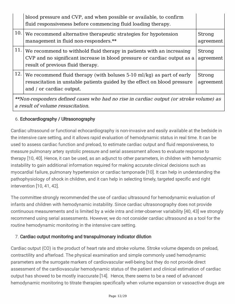
10. We recommend alternative therapeutic strategies for hypotensionmanagement in fluid non-responders.**
Strongagreement
11. We recommend to withhold fluid therapy in patients with an increasingCVP and no significant increase in blood pressure or cardiac output as aresult of previous fluid therapy.
Strongagreement
12. We recommend fluid therapy (with boluses 5-10 ml/kg) as part of earlyresuscitation in unstable patients guided by the effect on blood pressureand / or cardiac output.
Strongagreement
**Non-responders defined cases who had no rise in cardiac output (or stroke volume) asa result of volume resuscitation.
�. Echocardiography / Ultrasonography
Cardiac ultrasound or functional echocardiography is non-invasive and easily available at the bedside inthe intensive care setting, and it allows rapid evaluation of hemodynamic status in real time. It can beused to assess cardiac function and preload, to estimate cardiac output and �uid responsiveness, tomeasure pulmonary artery systolic pressure and serial assessment allows to evaluate response totherapy [10, 40]. Hence, it can be used, as an adjunct to other parameters, in children with hemodynamicinstability to gain additional information required for making accurate clinical decisions such asmyocardial failure, pulmonary hypertension or cardiac tamponade [10]. It can help in understanding thepathophysiology of shock in children, and it can help in selecting timely, targeted speci�c and rightintervention [10, 41, 42].
The committee strongly recommended the use of cardiac ultrasound for hemodynamic evaluation ofinfants and children with hemodynamic instability. Since cardiac ultrasonography does not providecontinuous measurements and is limited by a wide intra and inter-observer variability [40, 43] we stronglyrecommend using serial assessments. However, we do not consider cardiac ultrasound as a tool for theroutine hemodynamic monitoring in the intensive care setting.
7. Cardiac output monitoring and transpulmonary indicator dilution
Cardiac output (CO) is the product of heart rate and stroke volume. Stroke volume depends on preload,contractility and afterload. The physical examination and simple commonly used hemodynamicparameters are the surrogate markers of cardiovascular well-being but they do not provide directassessment of the cardiovascular hemodynamic status of the patient and clinical estimation of cardiacoutput has showed to be mostly inaccurate [14]. Hence, there seems to be a need of advancedhemodynamic monitoring to titrate therapies speci�cally when volume expansion or vasoactive drugs are

Page 13/29
delivered in order to improve cardiac output or systemic vascular resistance [44, 45]. In patients withrefractory shock, when an effective and accurate measurement of CO is needed, the following methodsmay be used depending upon available resources and expertise: measurement of CO using transthoracicultrasound (echocardiography) and transpulmonary dilution (TPD) [44-46]. Ultrasonography is non-invasive, easily available and can provide a fairly accurate and serial estimation of cardiac output at thebedside to monitor the initial response to therapy [40, 47]. However, it requires speci�c skills and it isoperator-dependent. Despite being the most reliable clinical method to measure cardiac output,application of TPD in the clinical practice may be challenging because of resources, technical di�cultiesor lack of expertise. It is invasive and not suited to emergency resuscitation. Only two methods areavailable in children below 40 Kg: transpulmonary thermodilution (TPTD) by PiCCO (Pulsion MedicalSystems, Germany) and transpulmonary ultrasound dilution (TPUD) by CO status (Transonic, USA) [44-46, 48]. Neither are in frequent use in ICU due to their intricate set-up, and particularly for PICCO the risk tochildren’s vessels from relatively large femoral arterial catheter required. Moreover, because of theirintermittent measurement technique TPD methods are not suitable for the detection of rapid, frequent,changes in hemodynamic status, as required in some critically-ill children.
In the clinical practice, we recommend to use cardiac output ultrasound/Doppler methods for estimatingCO in stable patients and the initial assessment of unstable patients. The committee did not reach to astrong agreement on the methods to estimate CO in children with refractory shock needing escalation oftreatment and hemodynamic monitoring. There was a strong agreement that TPD methods are the mostreliable but whether their use should be advised in situations needing escalation only reached a weakagreement. TPD methods also measure blood volumes and lung water but the committee recommendedagainst using these parameters for targeting hemodynamic goals. Measuring CO using PAC is notrecommended.
Cardiac output can also be estimated at the bedside using other non-invasive methods like bioimpedanceand bioreactance, pulse contour and Doppler. Validation of these methods in critically-ill children issparse and these methods are therefore not consensually viewed as accurate enough to estimateabsolute values of CO in the intensive care setting in children [49]. However, they might provide a trendover time. In general, the committee strongly agreed that at the current time no recommendationsregarding these methods can be given due to the limited experiences in critically-ill children.
�. Pulmonary artery pressure (PAP)
The pulmonary artery catheter (PAC) can provide continuous measurement of right atrial, PAP,measurement of CO and pulmonary arterial occlusion pressure (wedge pressure). However, because ofinvasiveness and size it is not used or recommended in intensive care clinical practice in children [50].Similarly, left atrial pressure can be measured using surgically inserted left atrial catheters (LAC) [51, 52].Still, alternative less invasive techniques are being used in children to estimate left atrial pressure inunstable patients and LAC or PAC are rarely used in today’s intensive care clinical practice [53, 54].
Page 14/29
Because of the above, the committee recommends not to use a PAC in children to measure cardiac outputor PAP in the ICU. Instead, transthoracic ultrasound echocardiography can be easily used to estimate PAPat the bedside non-invasively and it can provide serial assessment to monitor the response to therapy ordisease process (as above). However, it should not be used to estimate PAP in patients with rightventricular (RV) failure [55]. For precise measurement of PAP, we recommend using the PAC only at thecardiac catheterization laboratory.
Table 3: Recommendations on the use of cardiac ultrasound and other methods to estimate
cardiac output for hemodynamic monitoring in critically ill children
Page 15/29
SrNo
Recommendation Level ofagreement
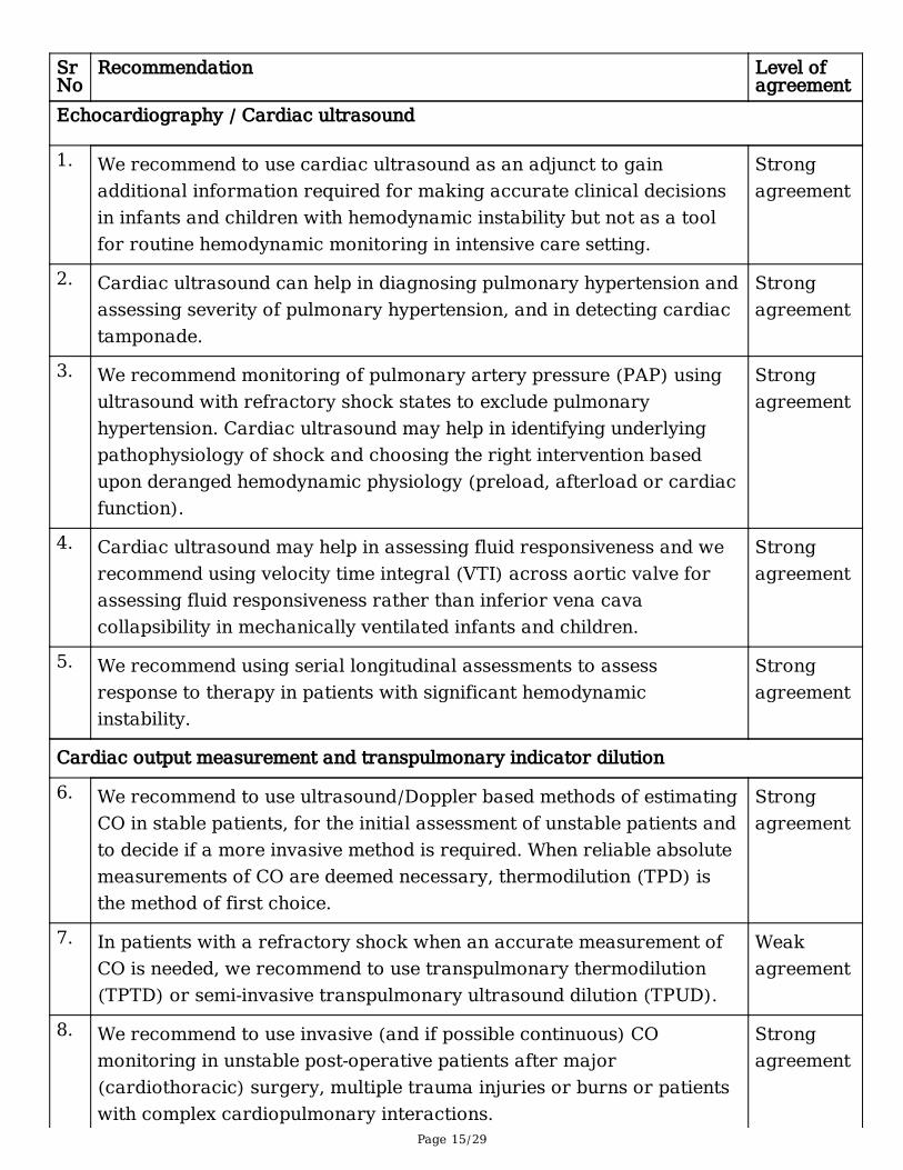
Echocardiography / Cardiac ultrasound
1. We recommend to use cardiac ultrasound as an adjunct to gainadditional information required for making accurate clinical decisionsin infants and children with hemodynamic instability but not as a toolfor routine hemodynamic monitoring in intensive care setting.
Strongagreement
2. Cardiac ultrasound can help in diagnosing pulmonary hypertension andassessing severity of pulmonary hypertension, and in detecting cardiactamponade.
Strongagreement
3. We recommend monitoring of pulmonary artery pressure (PAP) usingultrasound with refractory shock states to exclude pulmonaryhypertension. Cardiac ultrasound may help in identifying underlyingpathophysiology of shock and choosing the right intervention basedupon deranged hemodynamic physiology (preload, afterload or cardiacfunction).
Strongagreement
4. Cardiac ultrasound may help in assessing fluid responsiveness and werecommend using velocity time integral (VTI) across aortic valve forassessing fluid responsiveness rather than inferior vena cavacollapsibility in mechanically ventilated infants and children.
Strongagreement
5. We recommend using serial longitudinal assessments to assessresponse to therapy in patients with significant hemodynamicinstability.
Strongagreement
Cardiac output measurement and transpulmonary indicator dilution
6. We recommend to use ultrasound/Doppler based methods of estimatingCO in stable patients, for the initial assessment of unstable patients andto decide if a more invasive method is required. When reliable absolutemeasurements of CO are deemed necessary, thermodilution (TPD) isthe method of first choice.
Strongagreement
7. In patients with a refractory shock when an accurate measurement ofCO is needed, we recommend to use transpulmonary thermodilution(TPTD) or semi-invasive transpulmonary ultrasound dilution (TPUD).
Weakagreement
8. We recommend to use invasive (and if possible continuous) COmonitoring in unstable post-operative patients after major(cardiothoracic) surgery, multiple trauma injuries or burns or patientswith complex cardiopulmonary interactions.
Strongagreement

Page 16/29
9. We recommend against targeting fluid therapy based upon bloodvolumes measured with TPD or targeting hemodynamic therapy basedupon lung water measurement to assess pulmonary oedema in criticallyill children.
Strongagreement
10. Because of their intermittent measurement technique, TPD methods arenot suitable for the detection of fast changes in CO unless used inconjunction with continuous trend monitoring using pulse contouranalysis, calibrated by transpulmonary indicator dilution technology.
Strongagreement
Pulmonary artery pressure
11. We do not recommend to use pulmonary artery catheter (PAC) tomeasure CO in children. However, monitoring of left atrial pressureonly in selected cardiac surgery patients or patients after lungtransplant using a surgically inserted catheter can be helpful
Strongagreement
9. Lactate measurement
Determination of blood lactate concentration is a cheap, fast and easy bedside parameter that hasdemonstrated utility to predict the outcome or to trigger the need to intensify medical treatment [56]. Thecommittee showed some variation in their approach to the use of lactate in children since 5 out of 10recommendations needed a revision.
In critically-ill patients or children with shock, early and serial lactate blood sampling from a reliable sitesuch as a central venous or arterial indwelling catheter is recommended, though peripheral venoussampling with tourniquet time < 60 secs is possible [57]. This is speci�cally recommended when theinitial capillary lactate value is > 3.0 mmol/L [57-59]. Studies report an association between failure tonormalize lactate levels to a certain threshold (3.0±1.0 mmol/L) during the �rst 12 to 24 hours of ICUadmission, and adverse outcomes regardless of the reason for ICU admission [60, 61]. Experts could notagree on the use of lactate as part of a goal-directed approach and only weakly agreed on the approachto a persistent high lactate level. In the latter, lactate levels should always be used in conjunction withother clinical indicators of poor systemic perfusion and monitoring parameters. Persistently elevatedlactate levels may re�ect other mechanisms rather than those derived from poor tissue perfusion inshock, and instead re�ect aerobic glycolytic mechanisms including catecholamine administration orendogenous release [62].
10. Near-infrared spectroscopy
Near-infrared spectroscopy (NIRS) is a non-invasive, bedside technique to estimate regional capillary-venous hemoglobin saturation (rSO2). The mean baseline cerebral rSO2 is >70% in healthy children.Infants and children with cyanotic heart disease may have a cerebral rSO2 between 46-57% [63-67].

Page 17/29
Moreover, practitioners should be mindful about a considerable variability in NIRS values betweencommercially available devices. It has been observed that values measured in both monitors INVOS5100-C® (Medtronic; Boulder, CO, USA) and Foresight Elite® monitor (CAS Medical Systems; Branford,CN, USA) are not interchangeable [68]. Although NIRS is mainly used to measure rSO2 in the brain, thereare also reports of its use on other organs. In a study by Dabal et al. [69], it appears that renal NIRS andinferior vena cava desaturations precede rScO2 changes in the prediction of serious cardiovascularadverse events in patients after stage 1 Norwood palliation. Trend in NIRS values may provide valuablephysiological information in children with hemodynamic instability although clear (cut-off) values andevidence of bene�t are lacking.
The committee strived to de�ne recommendations with regard to this subject and 6 out of 7recommendation had to be rede�ned. As a result, the only strong recommendation was to advise againstroutine use of NIRS in all children with hemodynamic instability. Moreover, the committee agreed not tomake recommendations regarding the use of NIRS while treating children in shock, post-cardiac arrest,post traumatic brain injury and infants with hypoxic-ischemic encephalopathy. Lastly, there was noagreement on the clinical usefulness of a decline of cerebral rSO2 under 40-50% or a change in baselineof more than 20% [70 ].
11. Microcirculation
Microcirculatory assessment by videomicroscopy using side-stream or incident dark �eld is expensive,and not widely available. Currently it does not allow for assessment of rapid circulatory changes duringresuscitation [71]. No studies have de�ned the normal values of microcirculation in children outside theneonatal period but do report that vascular density seems to decrease with age [72]. So far, publishedstudies have not de�ned target values of microcirculatory parameters in critically ill children [72-77]. Atthis point in time, the committee recommends its use only for research purposes.
The committee also states that many routinely used parameters like capillary re�ll, peripheraltemperature, lactate, may re�ect aspects of the hemodynamic condition but do not adequately re�ect themicrocirculation and cannot be used as such. Although central venous to arterial CO2 difference couldprovide additional insight into the microcirculatory condition, we currently recommend against its use toguide resuscitation in critically ill children.
Table 4: Recommendations on use of serum lactate, near infrared spectroscopy (NIRS) andmicrocirculation assessment for hemodynamic monitoring in critically ill children
Page 18/29
SrNo
Recommendation Level ofagreement
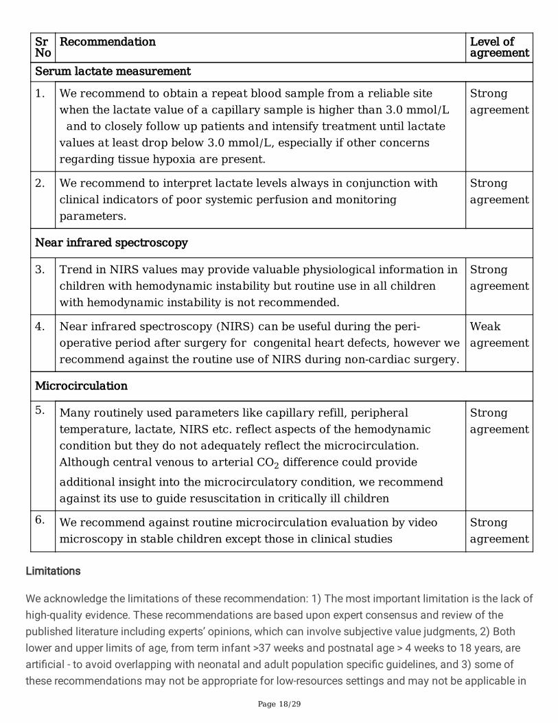
Serum lactate measurement
1. We recommend to obtain a repeat blood sample from a reliable sitewhen the lactate value of a capillary sample is higher than 3.0 mmol/L and to closely follow up patients and intensify treatment until lactatevalues at least drop below 3.0 mmol/L, especially if other concernsregarding tissue hypoxia are present.
Strongagreement
2. We recommend to interpret lactate levels always in conjunction withclinical indicators of poor systemic perfusion and monitoringparameters.
Strongagreement
Near infrared spectroscopy
3. Trend in NIRS values may provide valuable physiological information inchildren with hemodynamic instability but routine use in all childrenwith hemodynamic instability is not recommended.
Strongagreement
4. Near infrared spectroscopy (NIRS) can be useful during the peri-operative period after surgery for congenital heart defects, however werecommend against the routine use of NIRS during non-cardiac surgery.
Weakagreement
Microcirculation
5. Many routinely used parameters like capillary refill, peripheraltemperature, lactate, NIRS etc. reflect aspects of the hemodynamiccondition but they do not adequately reflect the microcirculation.Although central venous to arterial CO2 difference could provideadditional insight into the microcirculatory condition, we recommendagainst its use to guide resuscitation in critically ill children
Strongagreement
6. We recommend against routine microcirculation evaluation by videomicroscopy in stable children except those in clinical studies
Strongagreement
Limitations
We acknowledge the limitations of these recommendation: 1) The most important limitation is the lack ofhigh-quality evidence. These recommendations are based upon expert consensus and review of thepublished literature including experts’ opinions, which can involve subjective value judgments, 2) Bothlower and upper limits of age, from term infant >37 weeks and postnatal age > 4 weeks to 18 years, arearti�cial - to avoid overlapping with neonatal and adult population speci�c guidelines, and 3) some ofthese recommendations may not be appropriate for low-resources settings and may not be applicable in
Page 19/29
all settings requiring hemodynamic monitoring in children because of their limited availability orexpertise.
Nevertheless, despite these limitations the committee members believe that these are consensual expertrecommendations based upon literature review and rigorous standardized process of developing expertconsensus – followed DELPHI approach, a well-established standardized approach (DELPHI approach) –to reach consensus in such circumstances of limited published evidence to develop evidence-basedguidelines.
Future directions
The committee recognize that there is an important lack of knowledge and evidence concerninghemodynamic monitoring in children. There is a great need for: 1) studying the relationship betweenmeasured parameters and end-organ perfusion, and 2) evaluating the clinical e�ciency and patientoutcome when therapy is guided by speci�c monitoring technologies.
ConclusionsCardiovascular instability is common in children admitted to pediatric intensive care. Multiple-organdysfunction is commonly associated with cardiovascular derangements in patients with shock andcarries high mortality. Effective hemodynamic monitoring can help in identifying cardiovascularinstability early and choosing the appropriate targeted therapy timely. Currently, with the exception ofmanagement of shock there are no published HD monitoring guidelines for critically-ill children, and thepublished evidence remains scarce. These are therefore the �rst expert consensus recommendations forHD monitoring in critically-ill children with hemodynamic instability. These recommendations can helpclinicians in their clinical practice and may become the frame for future research aiming at providingstrong data for evidence-based guidelines in this �eld.
DeclarationsEthical Approval and Consent to participate:
Not applicable. All authors participated by accepting the invitation.
Consent for publication:
Not applicable. All authors have reviewed and agreed on the �nal manuscript for publication.
Availability of supporting data:
Supplementary material provided and attached with manuscript submission. Supporting data onconsensus development and voting available if required.
Page 20/29
Competing interests / Con�icts of interest: The authors declare no con�ict of interest or competinginterests.
Funding: None
Authors’ contributions:
JL, YS and JU conceptualized and designed the development of the whole project. JL took care of thewhole methodology and supervised the whole project. YS and JU wrote the �rst manuscript draft. Allauthors performed literature search and analysis, interpreted the literature data with their speci�cexpertise, participate in meetings discussions and voted on recommendations and manuscriptpreparation. Moreover, all authors critically reviewed the manuscript for important intellectual content,approved it in its �nal version and agreed to be accountable for all aspects of the work. The participationto the project did not entail any honorarium.
Acknowledgements: The authors acknowledge ESPNIC help in providing room and facilities for the Panelmeetings and anonymous voting for the consensus development.
References1. Antonelli M, Levy M, Andrews PJ, Chastre J, Hudson LD, Manthous C, et al . Hemodynamic
monitoring in shock and implications for management. International Consensus Conference, Paris,France, 27-28 April 2006. Intensive care med. 2007;33:575–590. https://doi.org/10.1007/s00134-007-0531-4
2. Watson RS, Crow SS, Hartman ME, Lacroix J, Odetola FO. Epidemiology and Outcomes of PediatricMultiple Organ Dysfunction Syndrome. Pediatr Crit Care Med. 2017;18:S4–S16.https://doi.org/10.1097/PCC.0000000000001047
3. Weiss SL, Fitzgerald JC, Pappachan J, Wheeler D, Jaramillo-Bustamante JC, Salloo A, et al. SepsisPrevalence Outcomes and Therapies (SPROUT) Study Investigators and Pediatric Acute Lung Injuryand Sepsis Investigators (PALISI) Network. Global epidemiology of pediatric severe sepsis: the sepsisprevalence, outcomes, and therapies study. Am J Respir Crit Care Med. 2015;191: 1147–1157.https://doi.org/10.1164/rccm.201412-2323OC
4. Weiss SL, Peters MJ, Alhzanni W, D Agus MS, Flori HR, Inwald DP, et al. Surviving sepsis campaigninternational guidelines for the management of septic shock and sepsis-associated organdysfunction in children. Pediatr Crit Care Med 2020;21:e52-e106https://doi.org/10.1097/PCC.0000000000002198
5. Davis AL, Carcillo JA, Aneja RK, Deymann AJ, Lin JC, Nguyen TC et al. American college of critical care medicine clinical practice parameters for hemodynamic support of pediatric and neonatal septicshock. Crit Care Med. 2017;45:1061-1093. https://doi.org/10.1097/CCM.0000000000002425

Page 21/29
�. Sethi, SK, Raghunathan V, Shah S, Dhaliwal M, Jha P, Kumar M, et al. Fluid Overload and RenalAngina Index at Admission Are Associated With Worse Outcomes in Critically Ill Children. FrontPediatr. 2018;6:118. https://doi.org/10.3389/fped.2018.00118
7. Selewski DT, Askenazi DJ, Bridges BC, Cooper DS, Fleming GM, Paden ML et al.The Impact of FluidOverload on Outcomes in Children Treated With Extracorporeal Membrane Oxygenation: AMulticenter Retrospective Cohort Study. Pediatr Crit Care Med. 2017;18:1126–1135.https://doi.org/10.1097/PCC.0000000000001349
�. Sivarajan VB, Bohn D. Monitoring of standard hemodynamic parameters: heart rate, systemic bloodpressure, atrial pressure, pulse oximetry, and end-tidal CO2. Pediatr Crit Care Med. 2011;12:S2-S11.https://doi.org/10.1097/PCC.0b013e318220e7ea
9. Kneyber M, de Luca D, Calderini E, Jarreau P H, Javouhey E, Lopez-Herce J, et al. Section RespiratoryFailure of the European Society for Paediatric and Neonatal Intensive Care. Recommendations formechanical ventilation of critically ill children from the Paediatric Mechanical Ventilation ConsensusConference (PEMVECC). Intensive Care Med. 2017;43:1764–1780. https://doi.org/10.1007/s00134-017-4920-z
10. Singh Y, Tissot C, Fraga MV, Yousef N, Cortes RG, Lopez J, et al. International evidence-basedguidelines on Point of Care Ultrasound (POCUS) for critically ill neonates and children issued by thePOCUS Working Group of the European Society of Paediatric and Neonatal Intensive Care (ESPNIC).Crit Care 2020;24:65. https://doi.org/10.1186/s13054-020-2787-9
11. Tume LN, Valla FV, Joosten K, Jotterand Chaparro C, Latten L, et al. Nutritional support for childrenduring critical illness: European Society of Pediatric and Neonatal Intensive Care (ESPNIC)metabolism, endocrine and nutrition section position statement and clinical recommendations.Intensive Care Med. 2020;46:411–425. https://doi.org/10.1007/s00134-019-05922-5
12. Atkins, D, Best, D, Briss, P A, Eccles, M, Falck-Ytter, Y, Flottorp, S, et al GRADE Working Group. Gradingquality of evidence and strength of recommendations. BMJ. 2004;328:1490.https://doi.org/10.1136/bmj.328.7454.1490
13. Brouwers MC, Kerkvliet K, Spithoff K, AGREE Next Steps Consortium. The AGREE Reporting Checklist:a tool to improve reporting of clinical practice guidelines. BMJ (Clinical research ed.) 2016;352:i1152. https://doi.org/10.1136/bmj.i1152
14. Tibby SM, Hatherill M, Marsh MJ, Murdoch IA. Clinicians’ abilities to estimate cardiac index inventilated children and infants. Arch Dis Child. 1997;7:516–518https://doi.org/10.1136/adc.77.6.516
15. Thompson M, Coad N, Harnden A, Mayon-White R, Perera R, Mant D. How well do vital signs identifychildren with serious infections in pediatric emergency care? Arch Dis Child. 2009;94:888–893.
Page 22/29
https://doi.org/10.1136/adc.2009.159095
1�. Erdem Ö, Kuiper JW, Tibboel D. Hemodynamic coherence in critically ill pediatric patients. Best PractRes Clin Anaesthesiol. 2016;30:499-510. https://doi.org/10.1016/j.bpa.2016.10.2002
17. Matettore A, Ray S, Harrison DA, Brick T, Macrae D, Peters MJ, Inwald DP. Intensive Care Med.2019;45:1482-1483 https://doi.org/10.1007/s00134-019-05638-6
1�. Gupta P, Dimple G. Blood Pressure Measurement in Children: Where do we stand? Indian Pediatr.2018;55:289-291.
19. Ray S, Rogers L, Noren DP, Dhar R, Nadel S, Peters MJ, Inwald DP. Risk of over‐diagnosis ofhypotension in children: a comparative analysis of over 50,000 blood pressure measurements.Intensive Care Med. 2017;43:1540–1541 https://doi.org/10.1007/s00134-017-4843-8
20. Michard F, Sessler DI, Saugel B. Non‐invasive arterial pressure monitoring revisited. Intensive CareMed. 2018;44:2213-2215. https://doi.org/10.1007/s00134-018-5108-x
21. Jansen TC, van Bommel J, Schoonderbeek FJ, et al. Early lactate-guided therapy in intensive careunit patients: a multicenter, open-label, randomized controlled trial. Am J Respir Crit Care Med2010;182: 752-761. doi:10.1164/rccm.200912-1918OC
22. Eytan D, Goodwin AJ, Greer R, Guerguerian AM, Mazwi M, Laussen PC. Distributions and Behavior ofVital Signs in Critically Ill Children by Admission Diagnosis. Pediatr Crit Care Med. 2018;19:115–124.https://doi.org/10.1097/PCC.0000000000001395
23. Eytan D, Goodwin AJ, Greer R, Guerguerian AM, Laussen PC. Heart Rate and Blood Pressure CentileCurves and Distributions by Age of Hospitalized Critically Ill Children. Front Pediatr. 2017;5:52.https://doi.org/10.3389/fped.2017.00052
24. Banker A, Bell C, Gupta-Malhotra M, Samuels J. Blood pressure percentile charts to identify high orlow blood pressure in children. BMC Pediatr. 2016;16:98. https://doi.org/10.1186/s12887-016-0633-7
25. Stergiou GS, Boubouchairopoulou N, Kollias A. Accuracy of Automated Blood Pressure Measurementin Children: Evidence, Issues, and Perspectives. Hypertension. 2017;69:1000-1006.https://doi.org/10.1161/HYPERTENSIONAHA.116.08553
2�. Mark JB. Central venous pressure monitoring: clinical insights beyond the numbers J CardiothoracVasc Anesth. 1991;5:163-173. https://doi.org/10.1016/1053-0770(91)90333-o
27. Magder S, Bafaqeeh F. The clinical role of central venous pressure measurements. J Intensive CareMed. 2007;22:44-51 https://doi.org/10.1177/0885066606295303

Page 23/29
2�. Stricker PA, Lin EE, Fiadjoe JE, et al. Evaluation of central venous pressure monitoring in childrenundergoing craniofacial reconstruction surgery. Anesth Analg 2013;116: 411-419.doi:10.1213/ANE.0b013e31827008e6
29. Gan H, Cannesson M, Chandler JR, Ansermino JM. Predicting �uid responsiveness in children: asystematic review. Anesth Analg. 2013;117:1380–92.https://doi.org/10.1213/ANE.0b013e3182a9557e
30. Renner J, Broch O, Duetschke P, Scheewe J, Höcker J, Moseby M, Jung O, Bein B . Prediction of �uidresponsiveness in infants and neonates undergoing congenital heart surgery. Br J Anaesth.2012;108:108–15 https://doi.org/10.1093/bja/aer371
31. Marik PE, Cavallazzi R. Does the central venous pressure predict �uid responsiveness? An updatedmeta-analysis and a plea for some common sense. Crit Care Med. 2013;41:1774-81.https://doi.org/10.1097/CCM.0b013e31828a25fd
32. Kumar A, Anel R, Bunnell E, Habet K, Zanotti S, Marshall S, et al. Pulmonary artery occlusion pressureand central venous pressure fail to predict ventricular �lling volume, cardiac performance, or theresponse to volume infusion in normal subjects. Crit Care Med; 2004;32:691-9.https://doi.org/10.1097/01.ccm.0000114996.68110.c9
33. Bronicki RA. Venous oximetry and the assessment of oxygen transport balance. Pediatr Crit CareMed. 2011;12:S21-S26. https://doi.org/10.1097/PCC.0b013e3182211667
34. Pérez AC, Eulmesekian PG, Minces PG, Schnitzler EJ. Adequate agreement between venous oxygensaturation in right atrium and pulmonary artery in critically ill children. Pediatr Crit Care Med.2009;10:76-79. https://doi.org/10.1097/PCC.0b013e318193699d
35. Tibby SM, Murdoch IA. Monitoring cardiac function in intensive care. Arch Dis Child. 2003;88:46-52https://doi.org/10.1136/adc.88.1.46
3�. de Oliveira CF, de Oliveira DS, Gottschald AF, Moura JD, Costa GA, Ventura AC, et al. ACCM/PALShaemodynamic support guidelines for pediatric septic shock: an outcomes comparison with andwithout monitoring central venous oxygen saturation. Intensive Care Med. 2008;34:1065-1075.https://doi.org/10.1007/s00134-008-1085-9
37. Arikan AA, Zappitelli M, Goldstein SL, Naipaul A, Jefferson LS, Loftis LL. Fluid overload is associatedwith impaired oxygenation and morbidity in critically ill children. Pediatr Crit Care Med. 2012;13:253-8. https://doi.org/10.1097/PCC.0b013e31822882a3
3�. Wang X, Jiang L, Liu S, Ge Y, Gao J. Value of respiratory variation of aortic peak velocity in predictingchildren receiving mechanical ventilation: a systematic review and meta-analysis. Crit Care.2019;23:372. https://doi.org/10.1186/s13054-019-2647-7.

Page 24/29
39. Lee JH, Kim EH, Jang YE, Kim HS, Kim JT. Fluid responsiveness in the pediatric population. Korean JAnesthesiol. 2019;72:429-440. https://doi.org/4097/kja.19305
40. Singh Y. Echocardiographic Evaluation of Hemodynamics in Neonates and Children. Front Pediatr.2017;5:201. https://doi.org/10.3389/fped.2017.00201
41. Fraga MV, Stoller JZ, Glau CL, De Luca D, Rempell RG, Wenger JL, et al. Seeing Is Believing:Ultrasound in Pediatric Procedural Performance. Pediatrics. 2019;144:e20191401.https://doi.org/10.1542/peds.2019-1401
42. Pereira de Souza Neto E, Grousson S, Du�o F, Ducreux C, Joly H, Convert J, et al. Predicting �uidresponsiveness in mechanically ventilated children under general anaesthesia using dynamicparameters and transthoracic echocardiography. Br J Anaesth. 2011;106:856-864.https://doi.org/10.1093/bja/aer090
43. de Boode WP, van der Lee R, Horsberg Eriksen B, Nestaas E, Dempsey E, Singh Y, et al. EuropeanSpecial Interest Group ‘Neonatologist Performed Echocardiography’ (NPE). The role ofNeonatologist Performed Echocardiography in the assessment and management of neonatalshock. Pediatric research 2018:84: 57–67. https://doi.org/10.1038/s41390-018-0081-1
44. Proulx F, Lemson J, Choker G, Tibby SM. Hemodynamic monitoring by transpulmonarythermodilution and pulse contour analysis in critically ill children. Pediatr Crit Care Med. 2011;12:459-466. https://doi.org/10.1097/PCC.0b013e3182070959
45. Nusmeier A, van der Hoeven JG, Lemson J. Cardiac output monitoring in pediatric patients. ExpertRev Med Devices. 2010;7:503-517. https://doi.org/10.1586/erd.10.19
4�. Wurzer P, Branski LK, Jeschke MG, Ali A, Kinsky MP, Bohanon FJ, et al. TranspulmonaryThermodilution Versus Transthoracic Echocardiography for Cardiac Output Measurements inSeverely Burned Children. Shock. 2016;46:249-253. https://doi.org/1097/SHK.0000000000000627
47. Singh Y, Katheria AC, Vora F. Advances in Diagnosis and Management of Hemodynamic Instability inNeonatal Shock. Front Pediatr. 2018;6:2. https://doi.org/10.3389/fped.2018.00002
4�. Lindberg L, Johansson S, Perez-de-Sa V. Validation of an ultrasound dilution technology for cardiacoutput measurement and shunt detection in infants and children. Pediatr Crit Care Med. 2014;15:139-147. https://doi.org/10.1097/PCC.0000000000000053
49. Suehiro K, Joosten A, Murphy LS, Desebbe O, Alexander B, Kim SH, Cannesson M. Accuracy andprecision of minimally-invasive cardiac output monitoring in children: a systematic review and meta-analysis. J Clin Monit Comput. 2016;30:603-620. https://doi.org/10.1007/s10877-015-9757-9
50. Perkin RM, Anas N. Pulmonary artery catheters. Pediatr Crit Care Med. 2011;12:S12-20.https://doi.org/10.1097/PCC.0b013e318220f079.
Page 25/29
51. Humphrey CB, Gibbons JA, Folkerth TL, Shapiro AR, Fosburg RG. An analysis of direct and indirectmeasurements of left atrial �lling pressure. J Thorac Cardiovasc Surg. 1976;71:643-647.
52. Flori HR, Johnson LD, Hanley FL, Fineman JR. Transthoracic intracardiac catheters in pediatricpatients recovering from congenital heart defect surgery: associated complications and outcomes.Crit Care Med. 2000;28:2997-3001. https://doi.org/10.1097/00003246-200008000-00053
53. Figueras-Coll M, Sanchez-de-Toledo J, Gran F, Abella R, Perez-Hoyos S, Rosés F. Echocardiography inthe Assessment of Left Atrial Pressure After Pediatric Heart Surgery: A Comparison Study WithMeasurements Obtained From Left Atrial Catheter. World J Pediatr Congenit Heart Surg. 2015;6:438-42. https://doi.org/10.1177/2150135115589999.
54. Goldberg DJ, Quartermain MD, Glatz AC, Hall EK, Davis E, Kren SA, et al. Doppler tissue imaging inchildren following cardiac transplantation: a comparison to catheter derived hemodynamics. PediatrTransplant. 2011;15:488-94. https://doi.org/10.1111/j.1399-3046.2011.01503.x.
55. Moceri P, Baudouy D, Chiche O, Cerboni P, Bouvier P, Chaussade C, Ferrari E. Imaging in pulmonaryhypertension: Focus on the role of echocardiography. Arch Cardiovasc Dis. 2014;107:261-71.https://doi.org/10.1016/j.acvd.2014.02.005
5�. Jansen TC, van Bommel J, Schoonderbeek FJ, Sleeswijk Visser SJ, van der Klooster JM, Lima AP, etal. LACTATE study group. Early lactate-guided therapy in intensive care unit patients: a multicenter,open-label, randomized controlled trial. Am J Respir Crit Care Med. 2010;182:752-761.https://doi.org/10.1164/rccm.200912-1918OC
57. Gallagher EJ, Rodriguez K, Touger M. Agreement between peripheral venous and arterial lactatelevels. Ann Emerg Med. 1997;29:479-483.
5�. Samaraweera SA, Gibbons B, Gour A, Sedgwick P. Arterial versus venous lactate: a measure of sepsisin children. Eur J Pediatr. 2017;176:1055-1060. https://doi.org/10.1007/s00431-017-2925-9
59. Stoll D, Englund E, Hillborg H, Vedin S, Larsson A. Capillary and venous lactate measurements with ahandheld device compared to venous blood-gas analysis for emergency patients. Scand J TraumaResusc Emerg Med. 2018;26:47 https://doi.org/10.1186/s13049-018-0510-5
�0. Kumar R, Kumar N. Validation of lactate clearance at 6 h for mortality prediction in critically illchildren. Indian J Crit Care Med. 2016;20:570-574. https://doi.org/10.4103/0972-5229.192040
�1. Hatherill M, McIntyre AG, Wattie M, Murdoch IA. Early hyperlactataemia in critically ill children.Intensive Care Med. 2000;26:314-318. https://doi.org/10.1007/s001340051155
�2. Allen M. Lactate and acid base as a hemodynamic monitor and markers of cellular perfusion PediatrCrit Care Med. 2011;12:S43-S49. https://doi.org/10.1097/PCC.0b013e3182211aed

Page 26/29
�3. Tina LG, Frigiola A, Abella R, Artale B, Puleo G, D'Angelo S, et al. Near Infrared Spectroscopy inhealthy preterm and term newborns: correlation with gestational age and standard monitoringparameters. Curr Neurovasc Res. 2009; 6:148-54. https://doi.org/10.2174/156720209788970090
�4. Bernal NP, Hoffman GM, Ghanayem NS, Arca MJ. Cerebral and somatic near-infrared spectroscopy innormal newborns. J Pediatr Surg. 2010;45:1306-10. https://doi.org/10.1016/j.jpedsurg.2010.02.110.
�5. Johnson BA, Hoffman GM, Tweddell JS, Cava JR, Basir M, Mitchell ME, et al. Near-infraredspectroscopy in neonates before palliation of hypoplastic left heart syndrome. Ann Thorac Surg.2009;87:571-7. https://doi.org/10.1016/j.athoracsur.2008.10.043.
��. Kurth CD, Steven JL, Montenegro LM, Watzman HM, Gaynor JW, Spray TL, Nicolson SC. Cerebraloxygen saturation before congenital heart surgery. Ann Thorac Surg. 2001;72:187-92.https://doi.org/10.1016/s0003-4975(01)02632-7
�7. Fenton KN, Freeman K, Glogowski K, Fogg S, Duncan KF. The signi�cance of baseline cerebraloxygen saturation in children undergoing congenital heart surgery. Am J Surg. 2005;190:260-3.https://doi.org/10.1016/j.amjsurg.2005.05.023.
��. Schmidt C, Heringlake M, Kellner P, Berggreen AE, Maurer H, Brandt S, et al. The effects of systemicoxygenation on cerebral oxygen saturation and its relationship to mixed venous oxygen saturation: Aprospective observational study comparison of the INVOS and ForeSight Elite cerebral oximeters.Can J Anaesth. 2018;65:766-775. https://doi.org/10.1007/s12630-018-1093-3.
�9. Dabal RJ, Rhodes LA, Borasino S, Law MA, Robert SM, Alten JA. Inferior vena cava oxygen saturationmonitoring after the Norwood procedure. Ann Thorac Surg. 2013;95:2114-20https://doi.org/10.1016/j.athoracsur.2013.01.076
70. Hoffman GM, Ghanayem NS, Tweddell JS. Noninvasive assessment of cardiac output. SeminThorac Cardiovasc Surg Pediatr Card Surg Annu. 2005;12-21.https://doi.org/10.1053/j.pcsu.2005.01.005.
71. Massey MJ, Larochelle E, Najarro G, Karmacharla A, Arnold R, Trzeciak S, et al. The microcirculationimage quality score: development and preliminary evaluation of a proposed approach to gradingquality of image acquisition for bedside videomicroscopy. J Crit Care. 2013;28:913-7.https://doi.org/10.1016/j.jcrc.2013.06.015.
72. Top AP, van Dijk M, van Velzen JE, Ince C, Tibboel D. Functional capillary density decreases after the�rst week of life in term neonates. Neonatology. 9 2011;9:73-7 https://doi.org/10.1159/000316945
73. González R, López J, Urbano J, Solana MJ, Fernández SN, Santiago MJ, López-Herce J. Evaluationof sublingual microcirculation in a pediatric intensive care unit: prospective observational studyabout its feasibility and utility. BMC Pediatr. 2017;17:75. https://doi.org/10.1186/s12887-017-0837-5
Page 27/29
74. Top AP, Ince C, de Meij N, van Dijk M, Tibboel D. Persistent low microcirculatory vessel density innonsurvivors of sepsis in pediatric intensive care. Crit Care Med. 2011;39:8-13https://doi.org/10.1097/CCM.0b013e3181fb7994
75. Scolletta S, Marianello D, Isgrò G, Dapoto A, Terranova V, Franchi F, et al. Microcirculatory changes inchildren undergoing cardiac surgery: a prospective observational study. Br J Anaesth. 2016;117:206-13. https://doi.org/10.1093/bja/aew187.
7�. Schinagl CM, Mormanova ZH, Puchwein-Schwepcke A, Schmid I, Genzel-Boroviczény O. The effect ofred blood cell transfusion on the microcirculation of anemic children. Eur J Pediatr. 2016;175:793-8.https://doi.org/10.1007/s00431-016-2704-z.
77. Paize F, Sarginson R, Makwana N, Baines PB, Thomson AP, Sinha I, et al. Changes in the sublingualmicrocirculation and endothelial adhesion molecules during the course of severe meningococcaldisease treated in the pediatric intensive care unit. Intensive Care Med. 2012;38:863-71.https://doi.org/10.1007/s00134-012-2476-5.
Figures

Page 28/29
Figure 1
Flow chart of the methodology used in consensus development.
Supplementary Files
This is a list of supplementary �les associated with this preprint. Click to download.

Page 29/29
Additional�le1Figure1.jpg
Additional�le2Table1.CSandBPrecommendations.pdf
Additional�le3Table2.CVPScvO2�uidresponse.pdf
Additional�le4Table3.USCOrecommendation.pdf
Additional�le5Table4.Endorganperfusion.pdf
Additional�le2Table12.pdf