
Inspire - ARTP
73
Inspire The ARTP Journal INSIDE THIS ISSUE: FIRST WORD 3 A WORD FROM THE CHAIR 6 THE UTILISATION OF CARDIOPULMONARY EXERCISE TESTING (CPET) TO PREDICT LENGTH OF HOSPITAL STAY (LOS) AFTER RADICAL CYSTECTOMY (RC) 8 THE ROLE OF RESPIRATORY AND SLEEP PHYSIOLOGY IN THE PREOPERATIVE RISK ASSESSMENT OF PATIENTS UNDERGOING ELECTIVE SURGERY. PART II—EXERCISE TESTING 14 ON THE BLOWER 46 FROM THE ARCHIVE 56 COMMUNICATIONS 58 JOIN ARTP 72 December 2016 VOLUME 17, ISSUE 3 www.artp.org.uk
Transcript of Inspire - ARTP

Inspire The ARTP Journal
INSIDE THIS ISSUE:
FIRST WORD 3
A WORD FROM THE CHAIR 6
THE UTILISATION OF CARDIOPULMONARY EXERCISE
TESTING (CPET) TO PREDICT LENGTH OF HOSPITAL
STAY (LOS) AFTER RADICAL CYSTECTOMY (RC)
8
THE ROLE OF RESPIRATORY AND SLEEP PHYSIOLOGY IN
THE PREOPERATIVE RISK ASSESSMENT OF PATIENTS
UNDERGOING ELECTIVE SURGERY. PART II—EXERCISE
TESTING
14
ON THE BLOWER 46
FROM THE ARCHIVE 56
COMMUNICATIONS 58
JOIN ARTP 72
Decem
ber 2
01
6
VO
LUM
E 17, ISSU
E 3 w
ww
.artp.o
rg.u
k

This Journal is published by the Association for Respiratory Technology and Physiology.
No part of it may be reproduced, stored in a retrieval system or transmitted in any form,
by any means, electrical, mechanical, photocopying, recording or otherwise, without prior
permission of the ARTP. The views expressed in this Journal are not necessarily those
of the Association for Respiratory Technology and Physiology.
ARTP EDITORIAL BOARD
Aidan Laverty Inspire Editor
Position vacant Inspire Deputy Editor
Alison Butler S-news Editor
Dr Andrew Robson
Dr Brendan Cooper
Keith Butterfield
Nigel Clayton
Dr Karl Sylvester
Chris Jones
Martyn Bucknall
Kimberley Jenkins
Katie Bayfield
ARTP EXECUTIVE Council
Martyn Bucknall President
Dr Karl Sylvester Chair
Julie Lloyd Vice Chair
Emma Spence Treasurer
Tracey Fleming Secretary
Ken Hutchinson Non-Executive Director (HR)
Mark Hubbocks Executive Director (Finance)
Robin Baldwin Non-Executive Director (Patient)
Dr James Hull Non-Executive Director
ARTP EXECUTIVE Board
Dr Karl Sylvester Chair
Julie Lloyd Vice Chair
Emma Spence Treasurer
Tracey Fleming Secretary
Chris Jones Communications
Dr Vicky Cooper Sleep
Kelly Pauley Events
Ian Cliff Standards
Joanna Shakespeare Education
Claire Stacey Workforce
Paul Burns Paediatrics
Page 2

Welcome to the Christmas issue of ‘Inspire’ where I have taken the liberty of replacing ARTP blue for festive red. Before you email and complain I want to stress this is strictly a limited edition! This issue sees the new ARTP logo in ’Inspire’ for the first time (the correct colour scheme is at the top of page 2), the final airing of the ARTP 40th anniversary logo and a final archive article from ’Breath’, this time from March 1977, a time when one could buy a scientific textbook for £9. The article looks at the relationship between chronic airflow obstruction and smoking and I noted while preparing the issue that the battle to make smoking less ‘glamorous’ continues to this day (https://www.theguardian.com/society/2016/nov/30/tobacco-firms-uk-introduction-plain-packaging-court-of-appeal). I hope you have enjoyed reading the archive issues as much as I have. Remember that ARTP members can read archive issues online by following the link at the top of the article.
I have added a ‘Communications’ section which compiles ARTP announcements since the last issue. This may be a useful way to keep track of some of the work ARTP is doing between issues, which Karl alludes to in his ’Word from the Chair’ We have part II of the “Role of Respiratory and Sleep Physiology in the preoperative risk assessment of patients undergoing elective surgery”, this time covering Exercise Testing. The role of exercise testing in predicting the length of hospital stay post-surgery is also addressed by another article, an ARTP grant winner for the author to attend the recent ERS conference. This article does not support the predictive value of CPET as previously found, which may be due to the exclusion criteria used but which highlights the need for larger, multicentre prospective studies. The presentations from the recent ARTP ‘National Strategy Day’ have just been released for members and make interesting reading. A Christmas cracker of ‘On the Blower’ is produced for the first time by Matthew Rutter (no pressure, Matt!), where they are seeking YOUR ideas on what the future may hold for lung function. I say a fond farewell and thank you to Paul Burns, who has stepped down as ’Inspire’ Deputy Editor to concentrate on his roles in ARTP Paediatrics and Education and who knows what else besides!
As usual, I end by requesting you contact me if there is anything you would like changed about ‘Inspire’, things you don’t like (the red cover, perhaps?!) and ideas for how it could be improved. I may try and release a survey about this in the near future. Best wishes for a Happy Christmas
and New Year and I look forward to meeting you at ARTP conference early in 2017.
Aidan Laverty [email protected].
A i d a n L a v e r t y P a u l B u r n s
FIRST WORD
VOLUME 17, ISSUE 3. DECEMBER 2016
Page 3
ALL CORRESPONDENCE TO:
ARTP Administrator, Executive Business Support Ltd.,
Unit E1 City Wharf,
Davidson Road,
Lichfield,
Staffordshire WS14 9DZ
Tel: 01543 442141
Fax: 0121 355 2420
e-mail: [email protected]
ADVERTISING RATES Please contact ARTP Administration for more
information on [email protected] or see the ARTP website

We regret to hear of the recent loss of
one of the world’s greatest respiratory
physiologists, Professor Neil Pride, aged
85. Neil Pride was a brilliant physician
and scientist who could think clearly,
design studies and perform research to
incredibly high standards. His work on
lung mechanics revolutionised our
understanding of how the lung changes
in disease. He wrote early seminal
papers on the static pressure–volume
curves with John Gibson and his
collaborations with Mike Hughes have
produced textbooks that are the
mainstay of our profession. His writing
was clear and always simplified complex
ideas into understandable concepts. He
was always a quiet, kind and modest
man and it was my enormous pleasure
to award him his ARTP Lifetime Award
at Conference. He seemed almost
embarrassed to receive it, such was his
modesty. He was a Fellow of the ERS,
Emeritus Professor at Imperial, London
and was a brilliant teacher of respiratory
physiology. ARTP wish his family and
colleagues our deepest sympathy for the
end of a great life, well spent for the
benefit of all.
OBITUARY
Page 5
Professor Neil Pride
Dr B G Cooper, Consultant Clinical Scientist,
Hon. Senior Research Fellow, Birmingham 2016

A W
OR
D F
RO
M T
HE C
HA
IRDr. Karl
Sylvester
ARTP
Honorary
Chair
Page 6
Festive greetings to you all. I hope you are
all set and ready to go for the holidays. This
is a bumper Christmas edition of Inspire in
a lovely new blood red colour. This is also
the last edition for 2016, ARTP’s 40th
celebration year. I hope it’s been a good
one for you all. It’s been a busy time within
ARTP, with a new branding exercise
completed, progress made towards our
next guidelines edition (watch this space), a
new spirometry handbook, production of
an NHS England spirometry competency
document, progress on a commissioning
guide for spirometry training and much,
much more. As Chair I am very proud of
what the ARTP committees have achieved
in a voluntary capacity. Remember that all
roles within ARTP are voluntary and people
give up their time to work tirelessly on
behalf of the ARTP membership, while
holding down extremely busy day jobs and
home lives.
I attended a joint Board meeting between
the RCCP and AHCS recently. At a previous
National Strategy Day I urged these two
organisations to begin to work more closely
together on the areas where there was
commonality. One of these areas is
regulation of the physiological workforce. I
am pleased to say they have heeded this
call to action and the first meeting went
very well. There does appear to be a drive
towards closer working on the common
areas. One point of discussion was a single
register to avoid the confusion of which
register to join. There appears to be a
desire to generate this one register, but
discussions are in the very early stages and
I will keep you updated of progress.
I was very fortunate to attend the Academy
for Healthcare Science conference in
Cardiff at the beginning of December. This
was held jointly with the Welsh
Government and I was very impressed with
how embedded healthcare scientists are
into the healthcare structure in Wales. The
Chief Scientific Adviser, Christine Morrell,
has done an amazing job in raising the
profile of healthcare science within the
Welsh health system. There is also an
impressive level of support from the
Deputy Chief Medical Officer too. The
theme in Wales is very much about Prudent
Healthcare. Only doing what you can do
and nothing more. This includes
streamlining the requests for diagnostic
investigations to only those that are
absolutely necessary for the diagnosis and
management of patients. Certainly I’m sure
within our practice we can identify
requests where they were “nice to have”
rather than essential. With the direction
the healthcare system is taking across the
UK I believe this is a strategy we all should
be adopting to ensure we can keep up with
the demands on our services without an
increase in capacity.
Our own conference is only round the
corner now and the excitement is building
for our trip to Belfast. ARTP conference’s
first visit to Northern Ireland, truly ensuring
we are a UK wide organisation. I hope you
have already registered to attend or are
planning to do so shortly. This will be an
amazing conference in terms of scientific
and educational content and evening
entertainment. As the prices of travel tend
to increase closer to the departure date I
would encourage you to book your flights
or boats early to ensure a good price.
All it leaves me to do is wish you all a
Merry Christmas and a successful and
prosperous New Year.
Karl

BELFAST19-20 JANUARY 2017
ANNUAL CONFERENCEARTP
We are delighted to announce that the 2017 ARTP Annual conference will be taking place at theWorld Famous Europa Hotel which is located in the heart of Belfast City Centre. This will be thefirst ever ARTP conference to be held in Northern Ireland and we have been truly overwhelmed bythe welcome and support we have received from ‘Visit Belfast’ in arranging the venue.
This promises to be a superb conference filled with engaging speakers, covering topics from a rangeof physiology and new innovations.
Some members have suggested that Belfast is too far away and difficult to travel to. Nothing couldbe further from the truth. Return flights from most UK airports are in the region of £70 return.There are flights on a number of airlines, multiple times per day from most UK airports to eitherBelfast City or Belfast International airport.
For more information on the programme and traveloptions, please visit
www.artp.org.uk

Introducing F&P Brevida™. Gaining patient confidence is key to successful CPAP therapy and patient
confidence begins with a mask that fits and is comfortable. Developed from extensive patient-centred research,
F&P Brevida features both a simple, adjustable headgear to deliver an individual fit, and the innovative AirPillow
cushion which inflates to form a ‘pillow’ of air in and around the nose for a gentle and effective seal. For patient
confidence and comfort, choose a Fisher & Paykel Healthcare nasal pillows mask. The Mask Matters Most™. www.fphcare.co.uk
Designed for Confidence. Built for Comfort.
Our most innovative nasal mask yet
DreamWear minimal contact nasal mask
Come to our stand at the ARTP Conference to see how quick and easy DreamWear is to fi t
– then get a fun photo taken and instantly printed wearing our award-winning mask!
DreamWear allows your patients
to sleep in any position they like,
unrestricted by their PAP tubing.
The under-the-nose cushion
prevents red marks, discomfort
or irritation on the nose bridge.*
DreamWear is part of our Dream
Family sleep therapy solution.
* Mask does not directly contact the bridge of the nose or nostrils

Page 8
Transitional Cell Bladder Cancer
(TCBC) is the most common type
of urothelial carcinoma, which has
been reported to exist in 90% of
all bladder cancer diagnoses1.
Radical Cystectomy (RC)
combined with pelvic lymph node
dissection and ileal conduit
reconstruction remains the ‘gold
standard’ and is indeed the most
common surgical intervention for
muscle invasive bladder cancer2.
Although regarded as the ‘gold
standard’, RC is related with
significant postoperative
complications (PC) in the short
and long term periods as well as
high rates of mortality (6-11%)3-7
and morbidity (19-64%)8-14. TCBC
is associated with smoking and
environmental exposures and
therefore this population of
patients are at an increased risk
of both cardiovascular and
respiratory disease15.
Over the past 5 years guidelines
published by the Royal College of
Surgeons and The Department of
Health16 have recommended that
patients who require major
abdominal surgical intervention
should undergo objective risk
stratification. This would allow
the most appropriate, consultant
led care and either admission to
high dependency unit (HDU) or
intensive therapy unit (ITU)
postoperatively. Current
approaches to discriminate
between high and low risk
patients lack the correct level of
sensitivity and specificity to
predict postoperative outcomes
after RC17-20. In addition, some
proposed methods of risk
stratification including dynamic
stress echocardiography are only
capable of identifying the risk of
postoperative complication that is
associated with a single organ or
organ group19-20.
Cardiopulmonary exercise testing
(CPET) is frequently used in
perioperative medicine and has
been shown to provide
information regarding post-
operative outcomes5, 21-27. Initial
research carried out in the 1990s
suggested that certain
parameters and thresholds,
derived from CPET, be used to
assess a patient’s fitness for major
abdominal surgery. Older23, 28 has
demonstrated that pre-operative
CPET can be utilised to assess a
patient’s ventilatory, ventilatory-
perfusion, cardiovascular and
metabolic responses to
incremental exercise. A patient’s
ability to increase their oxygen
consumption during an
incremental exercise test has
been shown to strongly correlate
with the ability to maintain an
increased organ oxygenation and
therefore functional organ status
during the post-operative period.
Older23, 28 has shown that the
uptake of O2 (VO2) at the
anaerobic threshold (AT) allows
identification of high-risk patients,
including those patients who have
normal cardiopulmonary function
at rest measured by pulmonary
and cardiac function testing.
The purpose of this study was to
investigate if any correlations
exist between the parameters
derived from the performance of
preoperative CPET and length of
hospital stay (LOS) in patients
who were admitted for elective
RC surgery for TCBC.
ERS CONFERENCE GRANT WINNER
The Utilisation of Cardiopulmonary Exercise Testing (CPET) to Predict
Length of Hospital Stay (LOS) after Radical Cystectomy (RC)
Edward Parkes1 and Vicky Moore2
1. Clinical Physiologist, 2. Clinical Scientist.
Department of Respiratory and Sleep Physiology, Heartlands Hospital, Birmingham, UK
Introduction
Page 9
Patients and Methods
Consecutive patients who were
referred to the CPET service from the
Urology Surgical team between July
2013 and July 2015 for preoperative
CPET were retrospectively reviewed.
The study received Local Ethics
Committee approval at the Heart of
England NHS Foundation Trust as an
audit. Patients were excluded if: 1)
they performed a submaximal CPET
2) CPET was terminated early due to
reasons that precluded safe
continuation 3) surgical intervention
was not a RC 4) patient did not
achieve an AT.
Cardiopulmonary Exercise Testing
CPET was performed according to
departmental protocol based on
American Thoracic Society (ATS)
guidelines29. Tests were performed
using a cycle ergometer (Ergoline
cycle ergometer) with breath –by-
breath gas analysis (Carefusion
Jaeger JLAB v5.7). Gas calibration was
performed before the
commencement of each clinic with
volume calibration being performed
before each test. The patient’s
incremental workload was calculated
according to Cooper and Storer30.
After saddle height adjustment,
patients were seated on the cycle
ergometer and connected to
continuous 12-lead
electrocardiography (ECG), blood
pressure and oxygen saturation
(SpO2) monitoring. Baseline data was
taken over a 2-minute resting period,
followed by the commencement of
cycling for 2 minutes in an unloaded
exercise phase. After this point a 1-
minute incremental workload phase
was initiated and the patient would
exercise until a symptom-limited end
point was reached. A pedal cadence
of 60rpm was required throughout
exercise. Test termination criteria
included but were not limited to:
abnormal ECG rhythm or ischaemic
changes; excessive BP increases or
decreases, significant arterial
desaturation and patient signs and
symptoms. The patient was
monitored during an unloaded
recovery phase for a minimum of 2
minutes. The AT was determined
manually after each test using the
‘gold standard’ V-slope (VCO2 vs VO2)
technique31 and confirmed using the
breathing equivalents for O2 and CO2
(Eq.O2 and Eq.CO2)30 Peak VO2 was
calculated as an average over the last
5 breaths of exercise.
Data Collection and Statistical
Analysis
Patient demographics (age, height
and weight) including the results of
the CPET were retrospectively
collected. The patient’s electronic
record (EPR) was accessed to record
LOS, which was calculated from the
date of surgery to the date of
discharge.
Statistical analysis was performed
using SPSS software (IBM).
Correlations between data sets were
made using Spearman's rank order
correlation and Mann Whitney U
statistical tests.
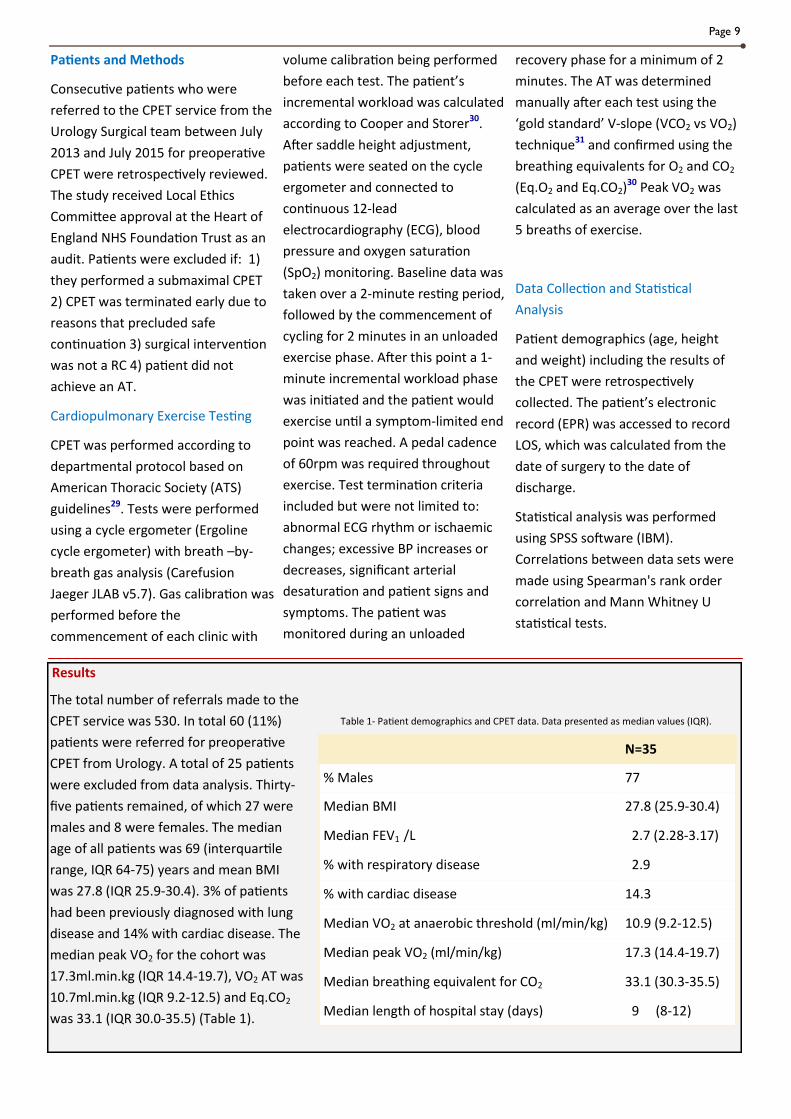
Results
N=35
% Males 77
Median BMI 27.8 (25.9-30.4)
Median FEV1 /L 2.7 (2.28-3.17)
% with respiratory disease 2.9
% with cardiac disease 14.3
Median VO2 at anaerobic threshold (ml/min/kg) 10.9 (9.2-12.5)
Median peak VO2 (ml/min/kg) 17.3 (14.4-19.7)
Median breathing equivalent for CO2 33.1 (30.3-35.5)
Median length of hospital stay (days) 9 (8-12)
The total number of referrals made to the
CPET service was 530. In total 60 (11%)
patients were referred for preoperative
CPET from Urology. A total of 25 patients
were excluded from data analysis. Thirty-
five patients remained, of which 27 were
males and 8 were females. The median
age of all patients was 69 (interquartile
range, IQR 64-75) years and mean BMI
was 27.8 (IQR 25.9-30.4). 3% of patients
had been previously diagnosed with lung
disease and 14% with cardiac disease. The
median peak VO2 for the cohort was
17.3ml.min.kg (IQR 14.4-19.7), VO2 AT was
10.7ml.min.kg (IQR 9.2-12.5) and Eq.CO2
was 33.1 (IQR 30.0-35.5) (Table 1).
Table 1- Patient demographics and CPET data. Data presented as median values (IQR).

Page 10
The median LOS for all patients was 9 (IQR 8-12) days. Spearman’s rank correlation coefficient showed that LOS did
not correlate with peak VO2 (p=0.269), Eq.CO2 (r=0.018 p=0.511) (Graph 1) and VO2AT (r=0.0018 p=0.323) (Graph
2). Furthermore, analysis using the Mann-Whitney U-test identified that when an Eq.CO2 threshold of < or > 33 was
applied there was no statistically significant relationship with LOS (p=0.082).
Graph 1. Relationship between LOS and Eq.CO2 in patients undergoing RC. RC=Radical cystectomy
LOS=Length of hospital stay Eq.CO2=Breathing equivalent for carbon dioxide.
Graph 2. Relationship between LOS and VO2AT in patients undergoing RC. RC=Radical cystectomy
LOS=Length of hospital stay VO2AT=Uptake of O2 at Anaerobic Threshold.
Length of Hospital Stay (LOS)/Days
Length of Hospital Stay (LOS)/Days

Page 11
Discussion
The utilisation of preoperative CPET to allow adequate risk stratification of perioperative care has been previously
documented21-28. The current study aimed to identify if any correlation’s existed between LOS and parameters
derived from the performance of preoperative CPET, namely peak VO2, VO2AT and Eq.CO2 in a cohort of patients
diagnosed with TCBC undergoing RC, but no relationships were found with any parameters.
Foundational research by Older23, 28 performed 20 years ago described a relationship between VO2AT and
postoperative mortality in a heterogeneous non-cardiac surgical population. A VO2AT threshold of < or >
11.0l.min.kg was identified as allowing discrimination between those patients who require more intensive and
specialised postoperative care. Older23 reported a 1% mortality rate in those patients achieving a VO2AT
>11ml.min.kg compared to an 18% mortality rate in those patients achieving a VO2AT <11ml.min.kg. Interestingly,
those patients who demonstrate cardiac ischaemia have an increased mortality rate of 42%, thus increasing the
likelihood of a postoperative myocardial infarction (MI). However, in a variety of different surgeries including
abdominal aortic aneurysm repair, nephrectomy and radical cystectomy (RC), the VO2AT threshold may not fully
translate into single surgical type cohorts.
Previous studies investigating preoperative CPET and its postoperative predictive value with homogeneous surgical
cohorts have described positive correlations between LOS and VO2AT after RC15, 22. Prentis22 identified a VO2AT
threshold of 12ml.min.kg as a major predictor of LOS while Tolchard15 identified that an Eq.CO2 >33 was the single
most important predictor of LOS. This study provides information that does not support the notions of neither
Tolchard15 nor Prentis22. Reasons for this inconsistency may include the exclusion of patients from RC whom, due to
a low preoperative fitness, as defined by a VO2AT value <11.ml.min.kg, would have required a greater LOS.
The results of this study also need to be accepted in context with its limitations. The small sample size from a single
centre cohort may explain the differences in the predictive power of both the Eq.CO2 and VO2AT that have been
previously reported in larger scale studies15, 22. As such, there is a recognised need to perform larger, multicentre,
prospective studies in order to further substantiate the usefulness of preoperative CPET in RC. LOS was allocated as
the main outcome variable in this study, however others have included postoperative complications as well as the
need for either HDU or ITU postoperative care. The assessment of these outcome measures and the relationship
between previously described CPET variables will be an avenue of future research.
In summary, preoperative CPET has been described to provide valuable clinical information that can be used to
tailor a patient’s perioperative care and predict the likelihood of a higher level of postoperative medical support in
either HDU or ITU21-28. Although this study does not support previous notions15, 22 regarding the relationship
between LOS and VO2AT or Eq.CO2 it should not be overlooked as it highlights the need for further research in this
area of perioperative medicine.

Page 12
1. Cancer Research UK (2016). Types of Bladder Cancer, retrieved September 21st 2016, from http://
www.cancerresearchuk.org/about-cancer/type/bladder-cancer/about/types-of-bladder-cancer
2. Stein, J.P & Skinner, D.G. (2006). Radical cystectomy for invasive bladder cancer: long-term results of a
standard procedure. World Journal of Urology 24: 296–304
3. Isbarn, H., Jeldres, C., Zini, L et al. (2009). A population-based assessment of perioperative mortality after
cystectomy for bladder cancer. Journal of Urology 182: 70–711
4. Quek, M.L., Stein, J.P., Daneshmand, S et al. (2006). A critical analysis of perioperative mortality from radical
cystectomy. Journal of Urology 175: 886–9012
5. Chahal, R., Sundaram, S.K., Iddenden, R., Forman, D.F, Weston, P.M., Harrison, S.C. (2003). A study of the
morbidity, mortality and long-term survival following radical cystectomy and radical radiotherapy in the
treatment of invasive bladder cancer in Yorkshire. European Urology 43: 246–5713
6. Weizer, A.Z., Joshi, D.D., Daignault, S.S et al. (2007). Performance status is a predictor of overall survival of
elderly patients with muscle invasive bladder cancer. Journal of Urology 177: 1287–9314
7. Liedberg, F. (2010). Early complications and morbidity of radical cystectomy. European Urology Supplement
9: 25–30
8. Chang, S.S., Cookson, M.S., Baumgartner, R.G et al. (2002). Analysis of early complications after radical
cystectomy: results of a collaborative care pathway. Journal of Urology 167: 2012–64
9. Smith, J.A. (2005). Early and late treatment-related morbidity following radical cystectomy. Journal of
Urology 173: 2033–45
10. Fairey, A.S., Jacobsen, N-E.B., Chetner, M.P et al. (2009). Associations between comorbidity, and overall
survival and bladder cancer specific survival after radical cystectomy: results from the Alberta Urology
Institute Radical Cystectomy database. Journal of Urology 182: 85–936
11. Shabsigh, A., Korets, R., Vora, K.C et al. (2009). Defining early morbidity of radical cystectomy for patients
with bladder cancer using a standardized reporting methodology. European Urology 55: 164–747
12. Meller, A.E., Nesrallah, L.J., Dall’Oglio, M.F et al. (2008). Complications in radical cystectomy performed at a
teaching hospital. International Brazilian Journal of Urology 28: 522–58
13. Thalmann, G.N & Stein, J.P. (2008). Outcomes of radical cystectomy. British Journal of Urology International
102: 1279–889
14. Buscarini, M., Pasin, E., Stein, J.P. (2007). Complications of radical cystectomy. Minerva Urologica e
Nefrologica 59:67–87
15. Tolchard, S., Angell, J., Pyke, M., Lewis, S., Dodds, N., Darweish, A., White, P and Gillatt, D (2015).
Cardiopulmonary reserve as determined by cardiopulmonary exercise testing correlates with length of
hospital stay and predicts complications after radical cystectomy. British Journal of Urology International 115
pp554-561
16. Leow, J, Reese, S., Jiang, W et al. (2014). Propensity-matched comparison of morbidity and costs of open and
robot-assisted radical cystectomies: a contemporary population-based analysis in the United States.
European Urology 66: 569–76
17. Clark, P.E., Stein, J.P., Groshen, S.G et al. (2005). Radical cystectomy in the elderly: comparison of clinical
outcomes between younger and older patients. Cancer104: 36–4316
References

Page 13
18. Horovitz, D., Turker, P., Bostrom, P.J et al. (2012). Does patient age affect survival after radical cystectomy?
British Journal of Urology International 110: E486–9317
19. Fairey, A., Chetner, M., Metcalfe, J et al. (2008). Associations among age, comorbidity and clinical outcomes
after radical cystectomy: results from the Alberta Urology Institute radical cystectomy database. Journal of
Urology 180:128–3418
20. Prasad, S.M., Ferreria, M.M., Berry, A.M et al. (2009). Surgical apgar outcome score: perioperative risk
assessment for radical cystectomy. Journal of Urology 181: 1046–53
21. Ting, S.M.S., Iqbal, H., Hamborg, T., Imray, C.H.E., Hewins, S., Banerjee, P., Bland, R., Higgins, R and Zehnder,
D (2013). Reduced functional measure of cardiovascular reserve predicts admission to critical care unit
following kidney transplantation. Public Library of Science ONE 8(5) e64335
22. Prentis, J.M., Trenell, M.I., Vasdev, N., French, R., Dines, G., Thorpe, A and Snowden, C.P (2013). Impaired
cardiopulmonary reserve in an elderly population is related to postoperative morbidity and length of
hospital stay after radical cystectomy. British Journal of Urology International 112(2) E13-19
23. Older, P., Smith, R., Courtney, P and Hone, R (1993). Preoperative Evaluation of Cardiac Failure and Ischemia
in Elderly Patients by Cardiopulmonary Exercise Testing. Chest 104(3) pp701-704
24. Goodyear, S.J., Yow, H., Saedon, M., Shakespeare, J., Hill, C.E., Watson, D., Marshall, C., Mahmood, A.,
Higman, D and Imray, C.H (2013). Risk stratification by pre-operative cardiopulmonary exercise testing
improves outcomes following elective abdominal aortic aneurysm surgery: a cohort study. Perioperative
Medicine (London) 2(10) pp1-13
25. Dunne, D.F., Jones, R.P., Lythgoe, D.T., Pilkington, F.J., Palmer, D.H., Malik, H.Z., Poston, G.J., Lacasia, C., Jack,
S and Fenwick, S.W (2014). Cardiopulmonary exercise testing before liver surgery. Journal of Surgical
Oncology 110(4) pp439-44
26. Colson, M., Baglin, J., Bolsin, S and Grocott, M.P.W (2012). Cardiopulmonary exercise testing predicts 5 yr
survival after major surgery. British Journal of Anesthesia 109(5) pp735-741
27. Ow ,M.M., Erasmus, P., Minto, G., Struthers, R., Joseph, M., Smith, A., Warshow, U.M., Cramp, M.E and
Cross, T.J (2014). Impaired functional capacity in potential liver transplant candidates predicts short-term
mortality before transplantation. Liver Transplant 20(9) pp1081-1088
28. Older, P., Hall, A and Hader, R (1999). Cardiopulmonary Exercise Testing as a Screening Test for Preoperative
Management of Major Surgery in the Elderly. Chest 116(2) pp355-360
29. American Thoracic Society (ATS)/American College of Chest Physicians (ACCP). 2003. Statement on
Cardiopulmonary Exercise Testing. American Journal of Respiratory and Critical Care Medicine, 167 (2) (2003)
pp211-277
30. Cooper, C.B & Storer, T.W. (2001). Exercise testing and interpretation. A practical approach. Cambridge
University Press.
31. Beaver, W.L., Wasserman, K., Whipp, B.J. (1985). A new method for detecting anaerobic threshold by gas
exchange. Journal of Applied Physiology 60(6):2020-7

The Role of Respiratory and Sleep Physiology in
the preoperative risk assessment of patients
undergoing elective surgery
Adrian H Kendrick, Department of Respiratory Medicine, University
Hospitals, Bristol, & Department of Applied Science, University of the
West of England, Bristol
Summary This review outlines the case for the role of both the lung function and sleep services in the assessment of
patients undergoing elective surgery.
Part I LUNG FUNCTION TESTING (‘Inspire’, August 2016) showed the FEV1 and the DLco are the two primary indices
used in the assessment of patients for lung resection. The evidence for other indices, including arterial blood
gases, non-invasive blood gases and measurements of static lung volumes is, in most cases, not supportive of their
routine use but may be useful in particular patient groups. For example, the measurement of static lung volumes
may be appropriate in patients who have significant obesity, and blood gas measures may be useful in patients
within known airflow obstruction.
In this issue EXERCISE TESTING. The tests of stair climbing, shuttle walk test, 6-minute walk test and CPET have all
been assessed as part of the overall assessment of patients, with the simpler tests generally being more available
away from specialist centres. In lung resection, there is strong evidence of the role of exercise testing using the
different modalities. New evidence from other surgical procedures, away from lung resection perhaps need further
investigation, but the available evidence points to the use of a number of indices, some of which might be specific
to a particular organ.
Finally, SLEEP (in the next ‘Inspire’, April 2017) constitutes about one-third of our day, and the prevalence of
obesity leading in many to obstructive sleep apnoea is an important component of the pre-operative assessment.
This should not be overlooked as there is evidence that sleep apnoea may present some difficulties in the post-
operative phase.
The role of respiratory and sleep departments in the pre-operative assessment of patients is here to stay, and will
increase the demands placed upon these services. Challenges will include the assessment of an increasingly older
population who wish to have surgery. None of the tests we undertake should be seen as preventing patients from
having surgery, more they should be seen as advising the patient about the likely risks of having the surgery and
possibly to explore appropriate alternatives.
Part II: Exercise Testing
Page 14

Page 15
From Part I. Cardiopulmonary Exercise Tests (CPET):
In the preoperative setting, CPET can be used to assess a
patients’ functional capacity and allow prediction as to
whether they will tolerate the physiological stress of
surgery. One key advantage of CPET is the integration of
the assessments of cardiac, respiratory and metabolic
variables in a situation that mimics surgery. A potential
downside of CPET is it involves facilities that not every
centre has available, essentially requires an hour-long
appointment, so may be regarded as time-consuming,
and requires a skilled practitioner (Senior Physiologist/
Scientist or anaesthesiologist) to perform and analyse
the test. There is a degree of uncertainty about the
predictive value of CPET on perioperative morbidity and
mortality, and about how CPET results should be used in
the clinical environment to inform preoperative
optimisation and perioperative management.
A single article provided low grade evidence from a
retrospective cohort analysis (Goodyear et al, 2013).
There was some evidence of a decreased length of stay
and reduced 30-day mortality, but it was noted that
there may be bias and imprecision in the data as this
was not a true RCT.
There were sixteen observational studies covering
abdominal aortic aneurysm (Barakat et al, 2015; Carlisle
& Swart, 2007; Grant et al, 2015; Hartley et al, 2012;
Prentis et al, 2012), lung resection surgery (Bruneli et al,
2009; Bruneli et al, 2012; Licker et al, 2011; Torchio et al,
2010), colorectal surgery (West et al, 2014),
pancreaticoduodenectomy (Ausania et al, 2012; Junejo
et al, 2014) and other surgery (Junejo et al, 2012;
McCullough et al, 2006; Prentis et al, 2013; Snowden et
al, 2010). These articles will be reviewed later under the
CPET testing section, with generally all the evidence
being classified as low grade. The evidence for using
anaerobic threshold (AT), oxygen uptake (VO2), peak VO2
and the ventilatory equivalent for carbon dioxide (VE/
VCO2) as predictors is unclear and is regarded across all
forms of surgery as of low quality for predicting
mortality at 30-days, 90-days or 3 years. Furthermore,
postoperative complications were not that well
predicted, based on the assessment criteria used. The
guideline group arrived at the conclusion that “...there is
not enough robust evidence to recommend or not
recommend CPET testing before surgery”.
Exercise Testing
The benefits of any cancer treatment must be balanced
against the potential harm, so the traditional risk
assessment requires a form of evaluation of the
patient’s performance status. Performance status is
known to be an independent predictor of survival in
patients receiving chemotherapy and/or radiotherapy
(Sculier, Chansky, Crowley, et al, 2008).
Patient-reported scoring systems rely very much on
subjective factors, and may not closely correlate with
the patients’ own perceptions of their functional status
(Dajczman, Kasymjanova, Kreisman, et al, 2008).
To overcome these limitations, we need an objective
methodology to evaluate the functional status and
exercise capacity of a patient - exercise testing
therefore provides that opportunity.
In lung cancer, or where there is concern regarding the
operability of a patient, exercise testing is generally used
in the preoperative assessment of patients for lung
resection to risk-stratify them, as exercise capacity is
associated with the perioperative risk for morbidity and
mortality. Exercise testing in those patients with
advanced lung cancer - nonsurgical patients - and in
survivors is also important. It is noted in the 1990’s that
exercise testing was regarded as controversial, as
consensus was in favour of some form of exercise
testing procedure as part of the work-up for patients
undergoing surgery for cancer.
Similarly to lung cancer surgery, aortic aneurysm surgery
will place significant metabolic demands upon the
patient in the perioperative period due to wound
healing, haemostasis, ventilation, intra-operative
haemodynamic changes and catecholamine stress
response to surgery (Attia et al, 1976; Silverstein et al,
1979; Waxman, 1987; Baxendale et al, 1996; Salatash et
al, 2001; Pearson et al, 2005).
Determination of functional capacity is a key component
in preoperative cardiac risk assessment. It is generally
measured in metabolic equivalents (METs), where 1
MET equals the basal metabolic rate (Ainsworth et al,
2011). Exercise testing provides an objective assessment
of functional capacity. If formal testing is not available,
the functional capacity may be estimated from the
ability to perform activities of daily living.

Page 16
One MET is the metabolic demand at rest and is
equivalent to a VO2 of 3.5 mL.min-1.kg-1 (Fleg et al, 2000).
Climbing two flights of stairs at a slow pace demands at
least 4 METs (Hlatky et al, 1989; Eagle et al, 2002), and is
regarded a good predictor of peri-operative
complications associated with major non-cardiac surgery
(Reilly et al, 1999; Girish et al, 2001). Strenuous sports,
such as swimming require > 10 METS (Figure 1).
Figure 1. Relationship of METs to activities, with 1 MET being equivalent to sleeping up towards greater than 10 METs
The inability to climb two flights of stairs or run a short
distance (< 4 METs) indicates poor functional capacity
and is associated with an increased incidence of post-
operative complications (POC’s) or events. After thoracic
surgery, a poor functional capacity has been associated
with an increased mortality (relative risk 18.7; 95% CI 5.9
– 59), but in comparison with thoracic surgery, a poor
functional status is not associated with an increased
mortality after noncardiac surgery (relative risk 0.47;
95% CI 0.09–2.5) (Biccard, 2005). This may reflect the
importance of pulmonary function as a major predictor
of survival after thoracic surgery, since resting
pulmonary function is related to functional capacity.
For example, in the study of Wiklund, Stein &
Rosenbaum (2001), who assessed 5939 patients
scheduled for non-cardiac surgery, the pre-operative
functional capacity measured in METs showed a
relatively weak association with post-operative cardiac
events or death. Where the functional capacity is high,
the prognosis is excellent, even in the presence of stable
Ischaemic Heart Disease or other risk factors (Morris et
al, 1991). However, where the functional capacity is
poor or indeed unknown, the presence of, and the
number of risk factors in relation to the risk of surgery
will likely determine the pre-operative risk stratification
and hence, the perioperative management.
When we undertake exercise testing in healthy subjects,
we know that a maximum exercise tolerance is limited
by the oxidative ability of skeletal muscle and/or cardiac
output. With increasing exercise intensity, VO2 increases
and reaches a point at which increasing exercise
intensity no longer leads to an increase in VO2 and a
plateau is observed – this is VO2,max. If an individual
becomes fatigued before this plateau is observed, this is
defined as the peak oxygen uptake - VO2,peak (Fleg et al,
2000). The anaerobic threshold (AT) is the point where
oxygen-independent (or ‘anaerobic’) metabolism is
required in addition to aerobic metabolism to sustain
exercise performance. This is usually due to increased
flux through the (oxygen independent) glycolytic
pathway, increasing pyruvate generation and lactate
production. AT is usually determined by lactate or
ventilatory thresholds and although the aetiology of
these thresholds may be different (Spurway, 1992), the
term ‘anaerobic threshold’ in pre-operative evaluation
usually refers to the ventilatory threshold. The
ventilatory threshold is usually reproducible and safe to
measure, even in patients with myocardial ischaemia
and heart failure (Older, Hall & Hader, 1999; Fleg et al,
2000).
A normal VO2,max will generally exclude significant
pulmonary, cardiovascular, haematological,
neuropsychological, and skeletal muscle disease
(American Thoracic Society & American College of Chest
Physicians, 2003) and hence this procedure is now
regarded as the standard measurement of
cardiopulmonary fitness (Arena & Sietsema, 2011).
However, in patients with cancer, exercise limitations
may be due to the effects of the cancer itself, coexisting
morbidities, and/or the effects of treatment. Cancer-
related anaemia (Bokemeyer, Oechsle, Hartmann, 2005),
muscle atrophy and dysfunction (Christensen et al,

Page 17
2014) may all limit oxygen transport through a reduction
in oxygen content (anaemia) and oxygen utilization. In
patients with coexisting lung disease limitations of
ventilation and gas exchange can occur, whilst in
patients with coexisting heart disease, chronotropic
incompetence and ventricular dysfunction due to
ischaemia and/or remodelling can limit cardiac output.
Cancer treatment itself may lead to impairments in
pulmonary and/or cardiovascular function. In time, the
inactivity that accompanies cancer, its comorbidities,
and treatment related effects can reduce muscle
strength and conditioning, further reducing exercise
capacity.
The four most common exercise test procedures used in
the pre-operative assessment of patients are 1) Stair
Climbing test (SCT), 2) 6-minute walk test (6MWT), 3)
Shuttle walk test (SWT) and 4) Cardiopulmonary Exercise
Testing (CPET). Current international and national
guidelines, make recommendations based on the use of
SCT, SWT and CPET, with no current guidelines including
the 6MWT (Brunelli A, et al, 2009; Lim et al, 2010;
Brunelli et al, 2013).
Stair Climbing
Whilst stair climbing may not normally be the province
of many lung function laboratories, there is a strong
body of literature outlining its use in clinical practice and
in the assessment of presurgical patients (Olsen et al,
1991; Brunelli et al, 2001; Brunelli et al, 2002; Bruneli et
al, 2008a; Bruneli et al, 2008b; Brunelli et al, 2010;
Berasconi et al, 2012; Brunelli et al, 2012a). This is not a
new test (Sounders, 1961; Van Norstrand, Kjeslberg &
Humphrey, 1968; Bolton et al, 1969) and is the most
used low-technology exercise test by thoracic surgeons
(Charloux et al, 2009).
In general, the inability of a patient to walk up two
flights of stairs predicts poor outcomes (Van Norstrand,
Kjeslberg & Humphrey, 1968), in relation to mortality,
longer postoperative intubation and hospital stay. The
odds ratio of death when walking 2 flights is 18.7 (5.9 –
58.6), based on a number of studies in patients
undergoing thoracic surgery (Colman et al, 1982; Holden
et al, 1992; Pate et al, 1996; Brunelli et al, 2002).
The use of the stair walk also allows an estimation of
stair climb VO2, peak (Olsen et al, 1991) as follows –
Work (watts) = Step height (m) x steps.min-1 x Wt (kg) x
0.1635 (kg-m.min-1 to watts).
VO2,peak (mL.min-1) = 5.8 x Wt (kg) + 151 + (10.1 x work)
Therefore, for a step height of 0.174 m, a body weight of
79 kg and completing 85 steps/min, the work is 191
watts and the estimated VO2,peak is 2539 mL.min-1 (97%
pred) or 32.1 mL.min-1.kg-1. This would equate to a METs
value of 32.1 ÷ 3.5 or 9.17, hence fit for surgery!
Holden et al (1992) compared the use of a stair climb to
the 6-minute walk test and a maximal CPET test, and
noted that being able to achieve > 44 steps was
predicted of a successful surgical outcome. They used
the relationship of Olsen et al (1991) to predict the
VO2,peak from the stair climb. In those patients who
survived (n = 11), the mean estimated VO2,peak
was 22.7 ± 1.6 mL.min-1.kg-1 compared to only
17.6 ± 3.8 mL.min-1.kg-1 in those who did not (n = 5),
albeit that there was not a perfect discrimination
between the two groups. The number of steps was 71 ±
23 versus 42 ± 24, which was statistically significantly
different (p < 0.05). In general, when the estimated
VO2,peak was > 20 mL.min-1.kg-1, the outcome at 90 days
was much better.
Girish et al (2001) assessed the use of stair-climbing in a
range of surgical patients undergoing high-risk surgery.
The authors reviewed the 30-day post-operative
complications (POC’s). 21/83 (25%) had POC’s. The FEV1
was significantly lower in those who had complications
(1.8 ± 0.77 vs 2.3 ± 0.82, p = 0.02), and these patients
had longer ICU days (6.5 ± 2.0 vs 1.3 ± 0.16, p = 0.03)
and spent more time in hospital (19 ± 3 vs 8 ± 1, p =
0.003). In terms of stair-climbing and POC’s, the inability
to climb two flights of stairs (18 steps/flight and 16.5 cm
height) was associated with a positive predicted value of
82% for developing a POC. Interestingly, stair climbing
did not predict the 30-day mortality rate well, with two
of three deaths occurring in patients who had climbed
three or more flights of stairs, whilst only one death
occurred in the nine patients (11%) unable to climb a
single flight of stairs.
Brunelli et al (2001) showed that in patients pre and post
lung resection, there was a significant decrease in stair
climbing capability, with the greater decrease (21.4%)
occurring in pneumonectomy patients as compared to a
14% reduction in lobectomy patients. In a subsequent

Page 18
paper, Brunelli et al (2002b) demonstrated the
usefulness of stair climbing in predicting
cardiopulmonary complications after lung resection. In
patients who had complications, it was observed that
they were significantly older (70.6 ± 5.9 vs 65.5 ± 9.9 yrs),
had lower FEV1%predicted (75.4 ± 16.7 vs 87.3 ± 19.3 %)
and climb fewer steps, as assessed by step altitude (Pate
et al, 1996) or distance of stairs climb (14.96 ± 5.5 vs
20.6 ± 4.62 m or 96 vs 133 steps with a step height of
0.155 m). Of note the Diffusion constant (Kco) was not
significantly different. Of all the measures undertaken in
this study, stair climbing was the only independent
predictor of cardiopulmonary complications after lung
resection.
In a further analysis, Brunelli et al (2008a) showed that
when patients achieved < 12 m altitude (75 steps),
cardiopulmonary complications, mortality and costs
were 2-fold, 13-fold and 2.5-fold higher than in patients
who achieved > 22 m (142 steps). The authors
recommended that a patient who was unable to achieve
12 m altitude (75 steps) should undergo a formal CPET
test with VO2 measurements in order to optimize their
perioperative management.
Nikolić et al (2008) wished to assess the usefulness of
stair climbing combined with pulse oximetry in relation
to postoperative complications in patients with an FEV1
of < 2 litres. Separating out those patients having
lobectomy in relation to complications, the pulse rate
and oxygen saturation at 40 steps were the most
discriminant indices in separating between minor/no
complications from severe complications/death.
Brunelli et al (2010) further studied this relationship. The
authors highlighted two key issues –
The test is difficult to standardise – stair height,
instructions to patients, monitoring information,
such as SpO2 and heart rate (HR).
It’s a low technology test that provides little useful
direct evidence of the physiological responses to
exercise, as compared to CPET testing.
To determine the accuracy of this test, in terms of
VO2,peak, the authors assessed 109 pre-surgical lung
resection candidates using the stair-climb test but with
the addition of a telemetric portable gas analyser
system. There was a high correlation between those
subjects who achieved a VO2,peak < 15 mL.min-1.kg-1 and
achieved < 14 m altitude on the stair-climb. On the other
hand, those achieving > 22 m altitude on the stair climb
had a VO2,peak > 15 mL.min-1.kg-1 and in some cases
> 20 mL.min-1.kg-1. There was a clear relationship
between a number of factors –
VO2,peak = 11.17 + (0.547 x altitude) + (0.413 x speed of
ascent) – 0.248 BMI
So for example -
VO2,peak = 11.17 + (0.547 x 35.2) + (0.413 x 15.2) – (0.248
x 27.34) = 29.9
This provides very similar results to the previous
calculations above for the same subject. The authors
noted in this group of patients, more than 50%
potentially had a VO2,peak < 15 mL.min-1.kg-1 who should
then be referred for a formal CPET test. Also of note, the
authors pointed out that in patients achieving > 22 m,
they would have developed a VO2,peak > 20 mL.min-1.kg-1
and so would have met the recently published European
guidelines by Brunelli et al (2009).
One question that remains to be answered is whether or
not stair-climbing can predict survival from cancer
surgery. In patients undergoing surgery for Stage 1 non-
small cell lung cancer (NSCLC), Brunelli et al (2012a)
observed that the threshold of altitude from stair-
climbing was 18 m, and any patient who climbed further
than this had a median survival of 97 months (95% CI: 89
to 105 months) versus 74 months (95% CI: 63 to 85
months) if less than 18 m. The equivalent 5-year survival
was 77% versus 54%. Even within the limitations of this
study, it is clear that survival is improved if a patient is
able to achieve a higher level of exercise, i.e. they are
fitter, before surgery. Those patients who are less fit,
may need to be considered for some form of pre-
rehabilitation in the weeks prior to operation, as an
improvement in fitness may have a better prognostic
outcome both in the short-term and longer term (Jones
et al, 2008).
In a more recent study, Lee et al (2014) showed that the
fitter an individual was, in terms of the amount of energy
expenditure per week, the lower their mortality was
(Figure 2).
Bernasconi et al (2012) compared a 20 m stair climb
against a treadmill exercise test and found no significant

Page 19
difference in VO2,peak (22.4 vs 22.7 mL.min-1.kg-1). The
speed of ascent was an important variable in that
achieving ≥15 m.min-1 accurately identified subjects for
who were fit for a pneumonectomy, with these patients
having a VO2,peak > 20 mL.kg-1.min-1. This group further
assessed the usefulness of a 20 m stair climb in a group
of patients in relation to the ACCP and ERS/ESTS
guidelines in lung cancer patients and concluded that
SCT has the potential to reduce both time and resource
usage in patients who are at moderate risk, based on
dividing the subjects in high, moderate and low risk
groups using ppoFEV1% and ppoDLco% (Bernasconi,
Diacon, Koegelenberg (2016).
In the most recent study, Reddy et al (2016) assessed
the time taken to complete a 20 m stair climb in patients
undergoing a range of abdominal surgery procedures.
The mean time taken to complete the 20 m stair climb
was 18.0 seconds (range 6 to 108). In reviewing their
data, univariable analysis showed that age and stair
climb time were two key factors in predicting
complications. In essence the longer taken to climb the
stairs, the higher the complication rate. It was also
observed that there was a relationship between length
of time to climb the stairs and length of stay and there
were also more complications in patients undergoing
colorectal surgery. The authors concluded that SCT
provided a measureable stress and predicted
postoperative complications, and was easy to
administer.
Clearly the ability of stair climbing to indicate potentially
increased mortality associated with thoracic surgery but
not with all major non-cardiac surgery may be related to
the different degree of importance of pulmonary
function to survival in the two groups. Stair climbing
capacity is known to correlate linearly with maximum
minute ventilation in thoracic patients (Pollock et al,
1993).
The FEV1 is a key measure of the prediction of survival
after lung resection surgery (Beckles et al, 2003).
Patients who climb three flights of stairs may be
expected to have an FEV1 > 1.7 L (Bolton et al, 1987),
and indeed the FEV1 x 40 gives an indication of the
maximum potential ventilation for a given individual. For
an FEV1 of 1.7 Litres, the estimated maximal ventilation
would therefore be 68 L.min-1, which should be
sufficient to achieve the level of exercise ventilation
required to climb three flights of stairs and still have
sufficient reserve ventilation.
< 2 1 0 0 2 1 0 0 - 4 1 9 9 4 2 0 0 - 8 3 9 9 8 4 0 0 - 1 2 5 9 9 > 1 2 5 9 90 .0
0 .2
0 .4
0 .6
0 .8
1 .0
E n e rg y E x p e n d itu re p e r w e e k
Mu
ltiv
aria
ble
Re
lati
ve
Ris
k
A ll - c a u s e M o r t a l i t y C a n c e r m o r ta l i t y C a r d io v a s c u la r m o r t a l i t y
N o n - c a n c e r , n o n - c a r d io v a s c u la r c a n c e r
Figure 2. Relative risks of mortality by physical level. Data from Lee et al, 2014.

Page 20
The observations in patients with lung cancer are in
contrast to those in major non-cardiac surgery, where
poor stair climbing capacity is not directly associated
with an increased risk of postoperative pulmonary
complications (Reilly et al, 1999).
Is Stair-Climbing a Useful Clinical Tool?
So, why are there differences between the two groups of
patients, and how can stair climbing be applicable for
the pre-operative assessment of patients requiring
surgery?
We know that poorer functional capacity can be
secondary to associated co-morbidities that are known
to increase peri-operative risk, these including diabetes,
congestive heart failure and a higher ASA score (Reilly et
al, 1999; Lee et al, 1999; American Society of
Anesthesiologists, 1963).
We also know that a minimum aerobic capacity is
needed to survive the metabolic stresses of the peri-
operative period. The metabolic requirements of the
postoperative stress response are generally regarded as
being less than the pre-operative aerobic capacity
required to predict the post-operative survival (Biccard,
2004). To sustain a postoperative VO2 of 5 mL.kg-1.min-1,
an anaerobic threshold of > 11 mL.kg-1.min-1 will be
needed (Older et al, 1993; Older et al, 1999). What is
interesting about both the studies of Older et al, is that it
suggests that aerobic capacity is a predictor of peri-
operative survival. Using the cut-off of 11 mL.kg-1.min-1
permits the separation of the patient’s postoperative
care. Older also showed that an AT > 4 mL.kg-1.min-1 is
associated with good peri-operative outcome. So why is
it then that the inability to climb two flights of stairs is
unable to predict mortality, especially when we know
that climbing a flight of stairs has been estimated to be
4.0 METs at a slow pace, and up to 8.0 METs at a fast
pace (Ainsworth et al, 2011)?
The differences between stair climbing and anaerobic
threshold in non-cardiac surgery would suggest that
there are significant physiological differences between
the two tests. The duration of the peri-operative stress
response after major surgery demands that it is
sustained by aerobic metabolism (Biccard, 2004). Simply
put, stair climbing is not a marker of maximal aerobic
capacity, whilst the anaerobic threshold is (Older et al,
1999; Bassett & Howley, 2000). There are a number of
physiological and technical reasons that support this
observation.
Test Duration: In undertaking a CPET test, we aim for a
test duration of 6 - 10 minutes as this determines the
contribution of aerobic and oxygen-independent
metabolic pathways to performance (Fleg et al, 2000),
and approximates to that of anaerobic threshold testing
(Older et al, 1999). Compare this to the time taken by
patients undergoing lobectomy who completed 91 ± 5.8
steps in an average time of 91.0 ± 15.9 seconds, and
those patients undergoing other types of surgery who
completed 83.8 ± 15.8 steps in an average time of 89.4 ±
19.1 seconds (Nikolić et al, 2008).
Metabolism at the start of exercise is principally oxygen-
independent, with oxidative phosphorylation lasting for
a short period of time (McArdle, Katch & Katch, 2014).
Heart failure, COPD, peripheral vascular disease and
diabetes, which many patients undergoing assessment
will have one or more of, will increase the time to
oxidative phosphorylation. So, despite stair climbing
having an apparently greater number of METs than the
AT required to survive major surgery, a significant
component of this is from oxygen-independent
metabolism, leading to an overestimate of aerobic
capacity (Spurway, 1992; Swinburn, Wakefield & Jones,
1985).
Inter-relationships: There is not a consistent relationship
between VO2,peak and AT in patients with ischaemic heart
disease and heart failure, with AT varying from between
50% to 100% of VO2,peak. This may explain why the
anaerobic threshold is a more sensitive predictor of
outcome than VO2,peak in patients with heart failure (Gitt
et al, 2002; Kavanagh et al, 2003; Sun et al, 2010; Sun et
al, 2012; Wasserman, et al, 2012). Indeed Kavanagh et al
(2003) noted that for each 1.0 mL.min-1.kg-1 increase in
VO2,peak there was generally a 10% reduction in cardiac
mortality.
The relationship between AT and VO2,peak may contribute
to the overestimation of perioperative aerobic capacity
using stair climbing as the METs achieved only relate to
VO2,peak (Pate et al, 1996) and not to AT, as AT may be
only 50% of the VO2,peak (Fleg et al, 2000).
Overestimation of aerobic capacity may, therefore
explain the unexpected deaths in patients who appear to
have good functional capacity.

Page 21
Performance: Exercise performance is, in part,
dependent on the mechanical efficiency with which the
subject performs exercise. Peri-operative survival should
solely depend on cardiorespiratory capacity, without
consideration of the musculoskeletal function of the
patient.
Patients with an acceptable aerobic capacity may be
included in the poor functional group due to
musculoskeletal factors. Although these patients are
unlikely to die of cardiovascular failure in the peri-
operative period, they are at increased risk of
postoperative complications (Reilly et al, 1999; Girish et
al, 2001). Anaerobic threshold testing may minimise
peripheral factors by limiting weight-bearing with
cycling, although this would not be so obvious where a
treadmill was being used.
Conclusions: It is clear that stair climbing is acceptable in
the assessment of patients undergoing thoracic surgery,
as it can be used to predict peri-operative survival and
post-operative complications.
However, in patients undergoing major non-cardiac
surgery, stair climbing may be less useful, as the
evidence suggests that the test is unable to predict the
aerobic capacity needed to survive the stress responses.
6-minute Walk Test (6MWT)
The 6MWT has not been commonly used in the pre-
operative assessment of patients with cancer. Previous
studies using the original 12-minute walk test
demonstrated a poor association between the distance
walked (12MWD) and pulmonary (Bagg, 1984) and
cardiopulmonary (Markos et al, 1989) complications.
The 6MWT has been used in a number of studies in lung
cancer. The 6-minute walking distance (6MWD) was
associated with respiratory failure (Pierce et al, 1994).
Where a 6MWD of > 300 m (1000 ft) was achieved, the
predicted survival at 90 days post-surgery was
significantly greater (Holden et al, 1992).
In a study involving patients with advanced lung cancer,
Kasymjanova et al (2009) assessed patients with stage III
to IV NSCLC. They observed that a 6MWD > 400 m was
the only variable associated with improved survival
using a multivariable analyses (HR = 0.44, p = 0.001).
In those patients who undertook a 6MWT before and
after chemotherapy, the mean 6MWD decreased after
two cycles of chemotherapy from 462 m to 422 m.
Jones et al (2012) assessed the prognostic value of the
6MWD in stage IIIB to IV NSCLC. The authors observed
that the 6MWD was an independent predictor of
survival, with each 50-m improvement in the 6MWD
associated with a 13% reduction in the risk for death.
Patients who achieved a 6MWD of > 450 m had a better
prognosis than those who achieved around 350 m.
Nakagawa et al. (2014) described significant correlations
between VO2max and the 6MWD and oxygen
desaturation in normal subjects. There was a significant
correlation between distance walked (m) and VO2,peak,
SpO2,rest, SpO2,min and DSpO2 4%. Using ROC curves, it
was noted that the distance walked from the 6MWT was
not the best predictor of a VO2,peak >15 mL.min-1.kg-1,
rather DSpO2 4% was, giving sensitivity and specificity
of 80.6% and 75% respectively. It was therefore
suggested that where a CPET is not available, or if the
patient is unable to perform CPET adequately, a 6MWT
would be an alternative. Furthermore, in the context of
the adopted Japanese Association for Chest Surgery
(JACS) guidelines (Colice et al, 2007), the authors suggest
that this would be potentially an acceptable alternative.
Granger et al (2015) compared the 6MWT to CPET in a
cohort of lung cancer patients, and whilst the 6MWT has
a logical appeal, their study showed that, in terms of
criterion validity the relationship between VO2,peak and
6MWD was poor (r = 0.24, p = 0.329).
Marjanski et al. (2015) retrospectively analysed data on
253 patients undergoing lobectomy, all of whom had
had a 6MWT. It was noted that for a walking distance of
≥500 m there were fewer postoperative complications
compared to those patients who walked < 500 m. The
authors conclude that – “the 6MWT seeks to identify a
high-risk group of patients in whom surgeon should
optimize monitoring, care and treatment in order to
reduce the complication rates”.
The conclusion from this limited number of studies
suggests that where the 6MWD is at least 300 m and ≥
500m, there appears to be a lower risk for perioperative
complications.

Page 22
Shuttle-Walk Test (SWT)
As with the SCT and the 6MWT, the SWT is a low-cost
form of exercise test that may be the only viable means
of assessing exercise performance in some centres
(Singh et al, 1992). The distance walked correlates well
with the VO2,peak (Singh et al, 1994). Where a more
complex test, such as CPET, is not available, the British
Thoracic Society (BTS) recommended the use of the SWT
as a screening tool before undertaking the more
complex CPET test (Lim et al, 2001).
The SWT is a maximal, progressive, paced test. The BTS
recommended the use of 25 shuttles, which equates to 5
minutes of exercise, a distance of 250 m, and
importantly, a VO2 of 15 mL.min-1.kg-1. How is this
related to actual testing?
Singh et al (1994) showed that there was a relationship
between VO2 on a treadmill and the SWT in patients with
COPD by directly measuring VO2 during exercise. The
studies of Booth et al (2001), Morales et al (1999),
Morales et al (2000) and Win et al (2006) demonstrated
an association between SWT distance and VO2, but did
not actually directly measure the VO2 during the SWT
itself. Therefore, apart from the original Singh study
(1994), there is no direct evidence of exactly what we
are measuring in relation to the VO2 at a chosen cut-off.
Win et al (2006) compared the shuttle walk and
measured VO2,peak in patients with lung cancer. All
patients completed both the SWT and a CPET using a
treadmill. The rationale of this study was to validate the
concept that failure to complete 250m should result in
surgery not being attempted (Lim et al, 2001; Beckles et
al, 2003). In those patients who failed to walk 400 m, a
significant proportion had a VO2,peak < 15 mL.min-1.kg-1,
and in those patients who achieved < 250 m, the
majority similarly exceeded the VO2 cut-off. The authors
recommended that in patients who failed to walk 400 m,
a CPET was required to highlight those patients who
would achieve a VO2,peak < 15 mL.min-1.kg-1.
Benzo & Sciurba (2010) using a simultaneously measured
VO2 throughout the SWT showed a clear relationship
between distance walked and VO2 (Figure 3). In their
analysis the authors noted that there was a degree of
discrepancy between the BTS recommendations of 250
m and 15 mL.min-1.kg-1. Using ROC analysis, the authors
shoed that for these precise cut-offs, the SWT had a
sensitivity of 94% with a false-positive rate of 23%
(Specificity – 77%). The more shuttles patient’s walked,
the lower the false-positive rate became, going from
17% (27 shuttles) to 0% (38 shuttles), the latter of which
coincides with a previous study (Win et al, 2006). One
important technical point that the authors make is that
the VO2 may have been higher in this study as it uses
walking, whilst if the subjects had used a cycle
ergometer, the VO2 when body mass is supported is
generally lower by around 10% (Hansen JE, 1984).
0 1 0 0 2 0 0 3 0 0 4 0 0 5 0 00
5
1 0
1 5
2 0
2 5
D is ta n c e (m )
VO
2(m
L/m
in/k
g)
Figure 3. Mean (95% CI) for VO2 vs distance walked during a shuttle walk test. Data from Benzo and Sciurba, 2010.

Page 23
In a study that compared the SWT, 6MWT and CPET,
Granger et al (2015) demonstrated that in NSCLC
patients, there was greater criterion validity of the SWT
test to VO2,peak from CPET (r = 0.61, p = 0.007) compared
to the 6MWD (r = 0.24, p = 0.329). This is perhaps not
surprising as the SWT is an incremental test, and hence
very similar to the incremental CPET test used in routine
practice. This study supports the findings of the study of
Win et al (2006).
Cardiopulmonary Exercise Testing (CPET)
The CPET test is basically a test of physiological status
and is used for risk stratification in patients being
considered for surgery (Brunelli et al, 2014). It is
regarded by many as the “gold standard” for testing, as
it will challenge the whole system under increasing
stress levels resulting from increasing the workload from
rest to maximal achievable by a patient at that given
point in time.
The importance of using a full CPET test is that it
provides a significant amount of information that may
be used to assess respiratory, cardiac and muscle
functional status in one test. The principal measure,
often quoted, is the VO2 either at peak exercise or at the
anaerobic threshold (AT).
In terms of the peak values, a value of > 20 mL.min-1.kg-1
is deemed fit for almost all surgery, and certainly for
lung cancer, whilst a value of < 10 mL.min-1.kg-1 may be
regarded as unsuitable for surgery in general (Fig.16).
The precise use of CPET in a range of different cancer
groups will be elucidated in this section of the review.
Abdominal Aortic Aneurysm
Nugent et al (1998) investigated the role of CPET in AAA
patients as part of pre-operative assessment. A treadmill
test was undertaken in 30 patients, with mean diameter
of 6.3 cm (3.8 – 8.7 cm) without difficulties. Although
there was a trend in relation to VO2,peak as being lower
overall in those patients who had complications, there
was no overall significant difference (18.6 ± 1.3 vs 21.8 ±
1.4 mL.min-1.kg-1). Four patients were deemed medically
unfit for surgery and all had a VO2,peak < 20 mL.min-1.kg-1.
The conclusion from this study was that although CPET
may be useful in identifying patients more likely to be at
risk of post-operative complications, albeit with limited,
strong evidence to support this from the data within this
study.
Carlisle & Swart (2007) assessed the mid-term survival of
patients following aortic aneurysm surgery. They noted
that VE/VCO2 was a key predictor of survival both at 30-
days and in the long-term. An increase of +1 in VE/VCO2
was associated with a hazard ratio of 1.13 for death. The
key value for differentiating those fit for surgery as
against those unfit was a VE/VCO2 > 42.
Thompson et al (2011) assessed the usefulness of CPET
in predicting 30-day and 30-month mortality in patients
with AAA. They used 4 variables – AT, VO2,peak, VEO2 and
VECO2. Fitness for surgery was based partly on CPET data
as well as co-morbidities and the size of the AAA. An AT
> 11 mL.min-1.kg-1 was considered ideal for surgery. Sixty
-three patients had open repair and were deemed fit for
surgery, whilst 36 patients were unfit for surgery. Those
in the fit group had a higher VO2,peak (15.1 vs 13.1
mL.min-1.kg-1; p < 0.001), a higher AT (12.0 vs 10.7
mL.min-1.kg-1; p < 0.001) and a lower VE/VCO2 (35 vs 37;
p = 0.005). In terms of the AT, the 30-month mortality
showed that patients with higher AT values survived
longer, and that AT was the best predictor of mortality.
Survival was 87.9% for the fit group, but only 61.1% for
the unfit group.
Hartley et al (2012) used CPET prospectively in a large
group of patients with AAA undergoing repair either via
EVAR or open repair. Using univariable analysis, in
relation to 30- and 90-day mortality showed that the
odds ratios for an AT < 10.2 mL.min-1.kg-1 (OR – 4.5 and
3.46), a VO2,peak < 15 mL.min-1.kg-1 (OR – 5.4 and 8.0) and
VE/VCO2 > 42 (OR – 3.11 and 3.06) were significant
contributors. Using multivariable analysis, in relation to
30-day mortality showed an increased OR for
AT < 10.2 mL.min-1.kg-1 (OR – 6.35), whilst for 90-day
mortality a VO2,peak < 15 mL.min-1.kg-1 had an OR of 8.59.
The authors suggest that the use of CPET variables
contribute to the risk management of patients
undergoing AAA surgery.
Prentis et al (2012a) provided the first evidence of the
use of CPET to predict outcome in abdominal aortic
aneurysm (AAA) patients undergoing endovascular
repair (EVAR). Of 185 patients, 101 underwent the EVAR
procedure whilst 84 had open repair. In those patients
Page 24
undergoing open repair, VO2,peak, AT and the workload
(Watts) were predictive of postoperative complications,
with the only significant independent variable being AT.
ROC curve analysis showed the optimal value of AT to be
10 mL.min-1.kg-1, and using this value, there was a
significantly longer overall critical care LOS in the
patients with values < 10, compared to those > 10
(median 4.1 vs 1.2, p = 0.02), as well as total hospital LOS
(median 16 vs 8, p < 0.01). Cox regression analysis
showed age and AT were predictors of hospital LOS, but
only age predicted critical care LOS. In the EVAR group,
AT predicted hospital LOS.
Goodyear et al (2013) assessed retrospectively, 188 AAA
who had had CPET, along with a control group of 128
AAA, who had not had CPET as it was not available at the
time. Patients having CPET were divided into three
groups – Pass (AT > 11 mL.min-1.kg-1), Fail (AT < 11) and
submaximal (no AT). Patients either underwent open
AAA surgery or had EVAR surgery. The primary outcomes
of this analysis were focused on total LOS and survival.
LOS (open) was lower in the CPET-pass group (median 10
days) compared to the CPET-fail group (median 11.5
days) and was also lower in both groups compared to
the control group (median 13 days). In EVAR patients
LOS was a median of 4 days regardless of CPET-pass or
fail, and was lower than the control group (median 6
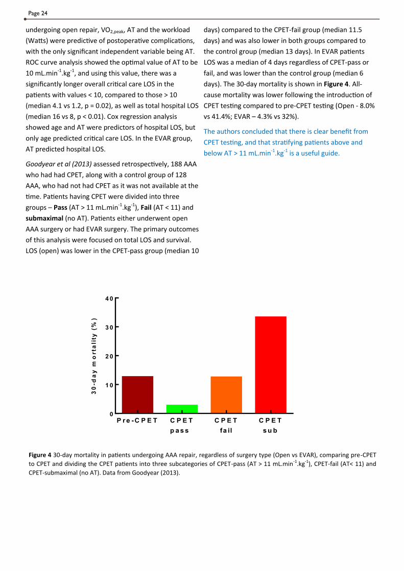
days). The 30-day mortality is shown in Figure 4. All-
cause mortality was lower following the introduction of
CPET testing compared to pre-CPET testing (Open - 8.0%
vs 41.4%; EVAR – 4.3% vs 32%).
The authors concluded that there is clear benefit from
CPET testing, and that stratifying patients above and
below AT > 11 mL.min-1.kg-1 is a useful guide.
P r e -C P E T C P E T
p a s s
C P E T
fa il
C P E T
s u b
0
1 0
2 0
3 0
4 0
30
-da
y m
orta
lity
(%
)
Figure 4 30-day mortality in patients undergoing AAA repair, regardless of surgery type (Open vs EVAR), comparing pre-CPET
to CPET and dividing the CPET patients into three subcategories of CPET-pass (AT > 11 mL.min-1.kg-1), CPET-fail (AT< 11) and
CPET-submaximal (no AT). Data from Goodyear (2013).

Page 25
Grant et al (2015) used CPET to assess patients
undergoing for AAA repair. Three indices were assessed
– AT < 10.2 mL.min-1.kg-1, VO2,peak < 15 mL.min-1.kg-1 or
VE/VCO2 > 42. Survival was better at 1-year and at 3-
years when either none or just one CPET variable was
abnormal (Figure 5). There were a number of global
indices that were independent predictors of survival, of
which VO2,peak < 15 mL.min-1.kg-1 (HR = 1.63) and
VE/VCO2 > 42 (HR = 1.68) were important. These results,
which included both open and EVAR, AAA repair were
deemed applicable to both types of surgery.
Abdominal & Upper GI Surgery
Older et al (1993) assessed cardiac failure and ischaemia
in elderly patients undergoing major intra-abdominal
surgery. The authors observed that where the AT
was < 11 mL.min-1.kg-1, there was a greater mortality,
than when the AT was > 11 mL.min-1.kg-1, this being 18%
(< 11) compared to 0.8% (> 11). In those patients with an
AT < 11 and myocardial ischaemia, the risk doubles. The
authors recommended that all patients with an AT < 11
should be admitted to an ITU postoperatively.
Older et al (1999) followed up on his previous study in
1993, using CPET as a screening test for perioperative
management of major intra-abdominal surgery in the
elderly. From this study, and reviewing the outcomes in
terms of ICU, HDU and Ward for the postoperative triage
site, a flow chart was created (Figure 6, overleaf). There
are some important points from this study. Firstly, age
should not be used to prevent patients from having
surgery. In this study, 62 patients were 80+ years of age,
yet the AT was within 1 SD covers the entire group. The
implication of using age only is that fit, older people may
be denied life-saving surgery, when they should not be.
The other key point is that CPET testing takes about one
hour and rarely results in an adverse event due to the
test procedure. Indeed the authors quote 3: >2000
(< 0.15%) possible adverse events, depending on the
definition used for adverse events.
0 - 1 2 30
2 0
4 0
6 0
8 0
1 0 0
N u m b e r o f s u b - th r e s h o ld C P E T in d ic e s
Su
rv
iva
l (%
)
1 - Y e a r 3 - Y e a r
Figure 5 Survival after elective AAA repair stratified by the number of sub-threshold indices from a CPET test. Data from Grant
et al, 2015.

Page 26
The final aspect of this study is that the number 11.0 for
AT has become fixated into the mind-set, but I’ll return
to that issue later.
Nagamatsu et al (2001) assessed the usefulness of CPET
and resting lung function in patients having
oesophagectomy.
Patients with cardiopulmonary complications, had a
VO2,peak/BSA significantly lower than patients with no
complications (0.79 ± 0.15 vs 0.97 ± 0.12 mL.min-1.kg-1, p
< 0.001). AT/BSA was not significantly different between
the two groups. None of the resting lung function data
was helpful separating out the two groups.
Using ROC analysis, the authors subdivided the patients
in different bands for VO2,peak/BSA and observed that
86% of patients with a value < 699 mL.min-1.m-2 had
cardiopulmonary complications, rising to 0%
complications with a value of ≥ 1100 mL.min-1.m-2. An
acceptable cut-off was deemed to be > 800 mL.min-1.m-2,
where only 10% of patients had complications.
Struthers et al (2008) reviewed the usefulness of the
incremental shuttle walk test and CPET as compared to a
questionnaire in a group of general surgical patients
undergoing intra-abdominal surgery. Using the Duke
Activity Status Index (DASI; Hlatky et al, 1989) and the
ISWT, the authors noted that where the
VO2,peak > 15 mL.min-1.kg-1 and an AT > 11 mL.min-1.kg-1,
there was good sensitivity using ROC curves.
However, using the CPET cut-offs, there were 36 patients
identified as “low risk”. The ISWT identified 13 where the
threshold as 360 m, and 9 patients where the DASI score
was 46. These results illustrated that even where DASI
and ISWT test data indicated poor outcomes, the use of
CPET indicated a potentially satisfactory outcome from
surgery, suggesting that simpler tests may not be as
good as CPET itself.
Figure 6 Flow chart showing the postoperative triage site and outcomes following major surgery. AT is expressed in units of
mL.min-1.kg-1, the VE/VO2 is the ventilatory equivalent for oxygen and CVS is the mortality due to the cardiovascular system.
Percentages are of the total group of n = 548. Modified from Older et al, 1999.

Page 27
Forshaw et al (2008) assessed the usefulness of CPET in
oesophagectomy patients. In patients who had
cardiopulmonary or noncardiopulmonary complications
or an unplanned ITU admission, there was minimal
difference between the AT or the VO2,peak compared to
those who did not have complications (Figure 7). The
authors concluded that CPET was of limited use.
Snowden et al (2010) assessed the usefulness of a CPET
test in 171 patients with a low level of subjective
function capacity (METs < 7.0). In those patients who
had > 1 complication at day 7 post surgery, there was a
longer LOS (26 vs 10 days, p < 0.001), AT was lower (9.1
vs 11.9 mL.kg-1.min-1, p = 0.001). Analysis using ROC
curves showed an optimal value of AT to be
10.1 mL.kg-1.min-1 and this measurement was one of the
independent predicators of complications.
Wilson et al (2010) assessed the effects of impaired
capacity on all-cause mortality following elective intra-
abdominal surgery. In a large cohort, it was noted that a
clinical history of ischaemic heart disease (RR 3.1; 95% CI
1.3 – 7.7), a VE/VCO2 > 34 (RR 4.6; 95% CI 1.4 – 14.8), and
an AT 10.9 mL.kg-1.min-1 (RR 6.8; 95% CI 1.6 – 29.5)
were significant predictors of all-cause hospital and 90-
day mortality. The effect of a reduced AT was most
noticeable in patients with no apparent history of
cardiac risk factors (RR 10.0; 95% CI 1.7 – 61.0).
Hightower et al (2010) assessed the usefulness of CPET
in relation to the ASA system in abdominal surgery. They
devised three new indices – %predicted AT (AT%), DHR
– change in HR from rest to AT and HR at AT. Using
univariate and multivariate analysis, the authors
demonstrated that AT% and DHR were useful predictors
of ASA outcome as was a combined index of (AT% +
DHR). The authors point out these new indices need
further exploratory studies to determine if they are
better than those currently use.
Swart & Carlisle (2012) undertook a case-controlled
study of critical care vs surgical ward in patients
undergoing open colorectal surgery. They used cut-off
points of ± 11 mL.min-1.kg-1. All patients with a threshold
> 11 were allocated to ward care, whilst those < 11 were
allocated either to ward care of critical care. The age of
the < 11 group was significantly higher compared to the
> 11 group. In the < 11, ward group there was a higher
0
5
1 0
1 5
2 0
2 5
3 0
mL
.min
-1.k
g-1
P r e s e n t A b s e n t P r e s e n t A b s e n t P r e s e n t A b s e n t
A T
V O 2 ,p e a k
C R E v e n ts N o n -C R e v e n ts IT U A d m is s io n
Figure 7 Outcomes predictions from CPET testing. Differences in VO2,peak and AT in patients with and without
cardiopulmonary events or an unplanned ITU admission, sowing the mean ± SD for each index with the events present or
absent. Data from Forshaw et al, 2008.

Page 28
incidence of adverse cardiac events. There was a shorter
overall stay in the > 11 group compared to either of the
< 11 groups. These results appear to be in broad
agreement with the previous studies of Older et al
(1993) and Older et al (1999).
Hennis et al (2012) assessed the potential usefulness of
CPET in predicting outcomes in gastric bypass surgery. At
this point in history, there was no study that had been
undertaken to specifically predict outcomes in this
growing group of patients, using cycle ergometry, the
only previous study using a treadmill (McCullough et al,
2006). The mean VO2,peak of the 106 patients was 15.7
(15.1 - 16.4) mL.min-1.kg-1. Eleven patients did not
however achieve an AT, giving a mean AT of
11.0 mL.min-1.kg-1 (10.6 – 11.3) and the VE/VCO2@AT of
25.9 (25.5 – 26.3). Postoperative morbidity (POMS) in
these patients was greater where the AT was
significantly lower (11.7 ± 1.7 vs 9.9 ± 1.5; p=0.049) with
VO2,peak and VE/VCO2@AT adding nothing. An AT of
11 mL.min-1.kg-1 was the optimal value, obtained from
ROC curves, for predicting morbidity. A similar AT – 11.4
was noted as the optimal value for predicting length of
stay (LOS) of > 3 days. One interesting component of the
analysis was the attempt to express the mean VO2,peak
and AT as a function of predicted adipose tissue free
mass – PATFM (mL.min-1.kg-1; Heymsfield et al, 2007),
which on this occasion did not add anything overall. The
overall conclusion was that AT was the best predictor of
LOS and POMS.
Colson et al (2012) undertook an extensive review of
1725 patients who underwent a range of surgical
procedure, but all having CPET testing. The aim was to
determine the CPET variables associated with 5-year
survival. Using single variable analysis and 30-day, one-
year and 5-year mortality showed none of the CPET
variables chosen were sufficiently strong to warrant any
further analysis. Categorizing patients into their surgical
group and assessing the 5-year outcome using Kaplan-
Meier curves, showed that vascular surgery had the best
long-term outcome (mean days survived – 1609),
whereas the worst outcome was for upper GI (mean
days survived – 1120). Whilst CPET variables did not
within the key indices, some of the derived variables
were noted to have a strong effect, including AT/HR, AT
and AT-RER. CPET variables are helpful in predicting 5-
year survival after major surgery.
Prentis et al (2013) assessed the usefulness of CPET in
patients undergoing radical cystectomy, where there are
known high levels of both morbidity and mortality.
Outcome measures included morbidity defined by the
Clavien-Dindo system (Dindo, Demartines & Clavien,
2004; Clavien et al, 2009) and LOS. It was observed that
in patients with major complications (Clavien score > 3)
the LOS was greater compared to those who did not (30
vs 16 days, p < 0.001; HR – 3.6). The AT was the only
index from CPET that differentiated between the major
(Clavien > 3) and minor (Clavien < 3) postoperative
complications (13.2 ± 3.6 vs 10.8 ± 1.8, p = 0.03; OR
0.74). Using an optimal AT of 12 mL.min-1.kg-1 showed a
significant relationship between fitness and LOS with
“unfit” staying 22 days as against the “fit” staying 16
days. The authors concluded that cardiopulmonary
reserve was related to major morbidity, increased LOS
and increased use of critical care services, so increasing
the overall cost of service provision for these patients.
Moyes et al (2013) studied 108 patients undergoing
resection for oesophagogastric cancer. Patients
underwent a CPET test and the AT and VO2,peak assessed
in relation to LOS and postoperative morbidity and
mortality. Where the AT was < 9 mL.min-1.kg-1, 42% of
patients had cardiopulmonary complications, reducing to
20% for an AT > 11 mL.min-1.kg-1. In the group with
cardiopulmonary complications, the mean AT was 9.9
mL.min-1.kg-1 compared to 11.9 in the group with no
complications. From ROC analysis, the AT that best
predicted cardiopulmonary complications was
9.0 mL.min-1.kg-1, which is lower than that proposed by
Older et al (1993).
Lee et al (2013) assessed the usefulness of the 6MWT
and CPET to predict complications at 30-days in patients
undergoing colorectal surgery. There was a significant
relationship between VO2,peak and the 6MWT distance
(r2 = 0.52, p < 0.0001) according to the equation -
VO2,peak = 0.042distance – 1.13
where VO2,peak is in mL.min-1.kg-1. Complications were
less likely to occur in patients achieving a longer walking
distance and increasing age. The authors concluded that
if CPET was not available, the a 6MWT could provide
acceptable prognostic information.
Ting et al (2013) in a further study in Chronic Kidney
Disease (CKD) wished to assess whether they could

Page 29
identify patients at high perioperative risk after kidney
transplant, and therefore likely to need admission to
CCU. As with the previous study, the use of AT was the
desired marker of outcome. 14/70 patients (20%)
needed CCU admission after transplant. These patients
had a lower AT (9.7 vs 12.5 mL.kg-1.min-1), lower VO2,peak
(16.8 vs 22.1 mL.kg-1.min-1) and O2,pulse (9.0 vs 11.8
mL.beat-1) and a higher VE/VCO2 (33.9 vs 31.2). The most
significant index was the AT, both independently and in
multivariable analysis. Using ROC analysis, the optimal
cut-off for AT was 11.31 mL.kg-1.min-1. This study, along
with the other study by the same group, indicates the
usefulness of CPET testing and in particular the
relevance of estimating AT for the management of
patients with CKD.
Ting et al (2014) assessed the CPET responses in CKD
patients who are known to have significant exercise
intolerance as a comorbid factor. The study of 240 CKD
patients included follow-up for 5 years. The key index
used was AT < 40% predicted VO2,peak. Of the patients,
124 (51.7%) received a transplant and in these patients
the AT was lower than in the non-transplant patients
(37.1% vs 40.4%, p = 0.01). 24 (10%) of the patients died
during follow-up. These patients, compared to the
survivors, had no difference in spirometry, but did have
lower VO2,peak (50.1% vs 65.2%, p < 0.001), lower AT (7.9
vs 10.7 mL.kg-1.min-1, p < 0.001) and lower AT%predicted
VO2,peak (29.9% vs 39.7%, p < 0.001). In terms of the
5-year survival, from all-cause mortality, those patients
with an AT ≥ 40%predicted VO2,peak had a survival rate of
94.4%, whilst those with a value < 40% had a survival of
46.6%. Using tertiles, survival was 38.5%, 67.1% and
100% for AT’s of < 9.4, 9.4 – 11.4 and
> 11.4 mL.kg-1.min-1 respectively. The authors concluded
that the use of AT was a good objective index of
cardiovascular reserve in patients with CKD.
Junejo et al (2014) assessed the usefulness of CPET in
patients undergoing pancreaticoduodenectomy for
cancer. CPET testing was undertaken on patient > 65
years or where comorbidities existed. Of 118 patients
who had surgery, 64 had CPET, the remainder being
classed as low-risk. In terms of in-hospital mortality and
30-day mortality, the VE/VCO2 was the best predictor.
VO2,peak and AT were unhelpful in this group.
Using ROC analysis, the optimal cut-off point was a VE/
VCO2 < 41 for indicating better long-term survival (Figure
8). CPET indices did not help in predicting morbidity.
Tolchard et al, (2014) assessed the usefulness of CPET in
patients undergoing radical cystectomy, either by open
surgery or robot guided. They assessed 105 patients with
outcomes being complications and LOS. Within the
study, at various points, staff looking after and following
up the patients were blinded to the CPET data. The
Clavien-Dindo scoring system was used to define
complications, the LOS was recorded in days and the 90-
day follow-up was recorded. Patients who had
1 2 30 .0
0 .2
0 .4
0 .6
0 .8
1 .0
S u r v iv a l ( Y e a r s )
Su
rv
iva
l F
ra
cti
on
V E/V C O 2 < 4 1V E/V C O 2 4 1
L o w R is k
Figure 8 Survival at 1, 2 and 3 years in patients undergoing Pancreaticoduodenectomy for cancer. Data from Junejo, 2014.

Page 30
complications had a significantly lower AT (10.6 vs 11.8,
p < 0.007), VO2,peak (14.3 vs 15.4, p = 0.022) and a higher
VE/VCO2 (33.3 vs 30.2, p = 0.007).
Multivariate logistic regression analysis showed that AT,
VE/VCO2 and presence of hypertension were significant
pre-operative factors with regards to postoperative
complications at ≤90 days. When 90-day complications
were stratified based on an AT ≤11 mL.kg-1.min-1 and/or
VE/VCO2 of ≥33 and/or presence of hypertension, a high-
risk and low-risk group was definable, with those at high
risk having a 5.55 greater risk of complications at 90
days, compared with those in the low risk group. Putting
this into context - the odds of a complication at ≤90 days
falls by 1.38 for each +1 rise in AT, whilst the odds rise by
1.16 for a +1 increase in VE/VCO2 and in those patients
with hypertension the odds rise by 3.44. For LOS, a VE/
VCO2 cut-off of 33 mL.kg-1.min-1 is important, with higher
values indicating potentially longer stays. This study
demonstrated that the use of both AT and the VE/VCO2,
which are less dependent on muscle mass and patient
effort, were the two principle indices from the CPET
testing in terms of LOS and 90-day outcomes.
West et al (2014) undertook a prospective and blinded
trial of CPET in patients undergoing major colonic
surgery. They assessed patients with CPET (reported
blind to clinical state) and recoded morbidity (assessed
blind to CPET) as well as post-operative outcome and
LOS. In comparing the patients who had complications to
those who did not, the former were older, and more had
heart failure, otherwise there was not real differences
between the groups.
In terms of the CPET data, the group with complications
had a lower AT and VO2,peak and a higher VE/VCO2 with
O2,pulse no different (Figure 9). In only 3 patients, all in the
complications group, was an AT not achieved. At the
optimized cut-off point of AT = 10.1 mL.kg-1.min-1, there
were generally fewer issues of morbidity when > 10.1
compared to < 10.1 using POMS defined at day 5. In a
multivariable regression model only sex and AT were
retained, with higher rates of complications in men than
women (OR = 4.19) for the same AT. Overall for a
1.0 mL.kg-1.min-1 increase in AT, there was » 20%
reduction in the odds of complications occurring and for
a 2.0 increase a » 40% reduction, when adjusted for sex.
This study, the first one to be blinded to some degree
confirms further the importance of using CPET to assess
patients for surgery and to highlight potential issues of
morbidity.
Chandrabalan et al (2014) assessed the usefulness of
CPET in patients undergoing pancreatic surgery. 100
patients were assessed, all having CPET before surgery.
In patients with an AT < 10 mL.kg-1.min-1, there were
A T V O 2 ,p e a k V E /V C O 2 O 2 ,p u l se
0
1 0
2 0
3 0
4 0
mL
.min
-1.k
g-1
or m
L.b
ea
t-1
p < 0 .0 1 p = 0 .0 1 p < 0 .0 1 p = 0 .2 2
Figure 9 Summary of CPET data comparing those patients with complications (red) and those without complications (green).
Data from West et al, 2014.

Page 31
significantly greater adverse outcomes compared to
those patients with an AT > 10 mL.kg-1.min-1. With the
same AT cut-off, there was an increased LOS in patients
with an AT < 10 mL.kg-1.min-1 (20 vs 16 days, p = 0.001),
In terms of receipt of adjuvant therapy post-surgery, the
only index that separated out those who did and those
who did not was AT, with the cut-off of 10 mL.kg-1.min-1.
CPET therefore proved useful is the pre-operative
assessment of patients undergoing major pancreatic
surgery.
Bariatric Surgery
McCullough et al (2006) assessed the cardiorespiratory
fitness and short-term complications after bariatric
surgery in a group of 109 patients with a BMI ranging
from 36.0 to 90.0 kg.m-2 (mean 48.7 ± 7.2). This was
potentially important as these authors had previously
reported that cardiorespiratory fitness levels in morbidly
obese patients were similar to patients with advanced
heart failure (Gallagher et al, 2004). The authors noted
that there was a graded inverse relationship between a
BMI > 25 kg.m-2 and VO2,peak. The subjects were divided
into tertiles, based on VO2,peak, which were 13.7 ± 2.1,
17.1 ± 0.80 and 21.3 ± 2.1 mL.kg-1.min-1 and at AT – 10.5
± 2.0, 12.3 ± 1.40 and 14.5 ± 2.2 mL.kg-1.min-1.
There were more complications, longer operating and
intubation times, and longer length of stay in patients in
the first tertile. There were no complications in those
with BMI < 45 kg.m-2 or peak VO2 > 15.8 mL.kg-1.min-1.
Multivariate analysis adjusting for age and sex showed
that VO2,peak was a significant predictor of complications.
Interestingly the authors also reported that 35/109
(32%) had sleep apnoea, although this was just reported
within the clinical characteristics of the patients, and no
further comment was made, including as to whether any
of the patients were treated!
Hepatic Surgery
One of the earliest studies in the hepatic transplant
group was by Epstein et al (2004). Of 156 patients
tested, 59 (38%) underwent hepatic transplantation. Of
these, 6 died within 100 days. There were no significant
differences in baseline data and resting lung function
data, including spirometry and ABG’s. Those that died
generally had a lower VO2,peak at < 60% predicted and a
lower AT%VO2,peak compared to the survivors. Overall, a
reduced aerobic capacity was considered a good guide
to 100-day mortality post-transplant. The authors
suggest that assessment of potential transplant
recipients may be important component of a limited
resource allocation.
In a further study by Prentis et al (2012b), this time in
relation to liver transplantation patients, which is
important as liver transplantation can have a high
mortality rate, especially in the early post-transplant
phase. None of the models for predicting outcome, at
the time of the study were able to discriminate between
survivors and non-survivors in the early post-transplant
phase (Jacob et al, 2005; Burroughs et al, 2006). 165/182
patients achieved a measurable AT and of these 60
underwent transplantation. AT was a component in an
attempt to predict the 90-day mortality, of which 6/60
patients had died within 90-days. In those patients who
died before 90-days, the AT was significantly lower (8.4 ±
1.3 vs 12.0 ± 2.4 mL.min-1.kg-1; p < 0.001), whilst VO2,peak
was not significantly different (12.5 ± 2.4 vs 14.8 ± 4.0
mL.min-1.kg-1; p = 0.17). Similarly, other CPET indices
were not significantly different. All these measures were
adjusted for actual body weight. However, it was noted
that in patients with refractory ascites, there was an
additional weight issue of the ascites, so the authors
adjusted the CPET indices to ideal body weight, based on
the Devine formula (Pai & Paloucek, 2000). Using ROC
analysis, the optimal value for AT to predict survival was
9.0 mL.min-1.kg-1. It was noted that an AT > 11.0 was
helpful in determining the critical care LOS, but not for
total hospital LOS.
Junejo et al (2012) highlights the issue of undertaking
liver resection in an increasingly older population who
are likely to have greater comorbidities. The age range
of the group of 94 patients who underwent surgery and
had CPET was 24 – 85 years (median 71). The group
median (range) for the key indices were: AT - 11.2 (7.4 –
21) mL.min-1.kg-1, VE/VCO2 - 32.0 (23.0 – 45.0), VO2,peak –
16.1 (9.5 - 28.1) mL.min-1.kg-1 and O2 Pulse – 9.0 (3.0 –
28.1) mL.beat-1. AT was the only marker related to post-
operative in-hospital mortality (5 deaths noted), with
ROC analysis giving a cut-off of 9.9 mL.min-1.kg-1. In
terms of complications, a cut-off of VE/VCO2 of 34.5,
from ROC analysis, was deemed a reasonable rule-in

Page 32
test. The relative risk of cardiopulmonary morbidity at
this cut-off was 2.17 (1.36 – 3.44). Multivariable
regression showed age (years) and the VE/VCO2 as the
two key predictors with OR’s of 1.07 and 3.45
respectively. In terms of predicting LOS,
an AT < 9.9 mL.min-1.kg-1 was associated with an
increased, unplanned ICU stay.
Survival of patients who had CPET, and divided by the AT
cut-off of 9.9 mL.min-1.kg-1 are shown in Figure 10. The
authors concluded that CPET was useful in this group of
patients, not least as it allows patients to make an
informed decision about the peri-operative risks.
Kaibori et al (2013) assessed the usefulness of patients
undergoing hepatectomy for hepatocellular carcinoma
(HCC). Sixty-one patients were studied. Post-operative
complications were classified according to the Clavien-
Dindo system. All surviving patients were followed up 3-
monthly for a median of 24 months post-operatively.
The Child-Pugh class was assessed at regular intervals
(Child & Turcotte, 1964).
In relation to CPET variables,
a VO2,peak ≥16.5 mL.min-1.kg-1 showed better survival at
both 1 year (87.1% vs 78.6%) and at 3-years (50.3% vs
10.8%) compared to those with a value
< 16.5 mL.min-1.kg-1. Survival was also better where the
AT ≥ 11.5 mL.min-1.kg-1 at 1-year (90.3% vs 75%) and at 3-
years (42.3% vs 33.4%) compared to those with
an AT < 11.5 mL.min-1.kg-1. In terms of maintenance of
the Child-Pugh class, AT was one of the factors from a
univariate analysis (Figure 11). The authors concluded
that hepatectomy can be safely performed in patients
with an AT ≥ 11.5 mL.min-1.kg-1 and that this is a marker
of better survival.
Bernal et al (2014) assessed CPET and survival with and
without transplantation in patients with chronic liver
disease. In a large group of 399 patients, 223 underwent
transplantation. The patients were monitored and the
primary outcome measure was mortality at 1-year,
either post CPET in those who did not transplantation
and 1-year in those after their transplant. In the
transplant group, LOS in ITU and total LOS were
assessed.
In those patients with ascites, significantly lower VO2,peak
(14.3 vs 17.5, p < 0.001) and AT (9.5 vs 11.4, p < 0.001)
were observed. It was also noted that in patients with
diabetes, VO2,peak vales were significantly lower, but this
was not similarly observed for AT. In the transplant
patients, survivors at 1-year had a higher AT (10.3 vs 9.3,
p < 0.05) and a higher VO2,peak (17.4 vs 13.8, p < 0.001).
The LOS was longer in those patients with an AT < 9.2
mL.min-1.kg-1 (21 vs 15 days), and in those patients with
a VO2,peak < 13.4 mL.min-1.kg-1 the ITU LOS was longer (4
days vs 3 days).
Comparing transplant to non-transplant patients,
mortality rates were lower in the transplant patients at
1 2 3 40 .0
0 .2
0 .4
0 .6
0 .8
1 .0
S u r v iv a l ( Y e a r s )
Su
rv
iva
l F
ra
cti
on
A T < 9 .9A T 9 .9L o w R is k
Figure 10 The survival fraction of patients deemed low risk compared to those who had an AT above and below
9.9 mL.min-1.kg-1. Data from Junejo et al 2012.

Page 33
90-days (1.8% vs 11.3%) and at 1 year (4.9% vs 34.6%).
Non-survivors had lower values of AT and VO2,peak
compared to survivors. The authors conclude that CPET
is important to assess patients, which is modifiable to
support and improve survival even if transplant is not
anticipated.
Neviere et al (2014) assessed the prognostic implications
of preoperative CPET testing and exercise oscillatory
ventilation (EOV) in patients undergoing liver transplant.
EOV is characterized by waxing and waning of tidal
volume leading to an oscillatory kinetics of breathing
with a period of 45 – 90 s. Mechanisms of ventilation
instability leading to EOV include prolonged circulatory
time, abnormal response of central and peripheral
controllers that drive ventilation and the impairment of
the efficiency of gas exchange system to damp out
changes in arterial gas tensions. In patients with heart
failure, EOV has been attributed to prolonged circulatory
time due to reduced cardiac output (Dhakal et al, 2012).
In patients with neurological disorders such as hepatic
encephalopathy, prolonged circulatory time may not be
responsible for EOV as normal or even hyperdynamic
circulatory status is typically present in advanced liver
diseases (Cherniak et al, 2005). Instead, in these clinical
conditions, ventilation instability has been related to
overstimulation of the ventilatory control centre that
will then present as periodic breathing – Cheyne-Stokes
pattern (Cherniak et al, 2005). Overstimulation of the
ventilatory drive has been reported in severe cirrhotic
patients and related to increased chemoreceptor
sensitivity in response to elevated catecholamine and
progesterone plasma concentrations (Lustik et al, 1997;
Passino et al, 2012).
Transplantation was undertaken in 263 patients.
Comparing those patients who died (n = 20) to those
who survived, resting lung function was no different.
Peak VO2 was significantly higher in the survivors (18.6
vs 17.1 mL.min-1.kg-1, p = 0.02) as was this index
expressed as %predicted (64% vs 55%, p = 0.04). The
only other index of significance was EOV, which was
observed in 6/20 non-survivors, and only 21/243 of the
survivors. In patients where EOV observed and died, the
VO2,peak, peak workload, VE/VCO2, peak and O2,pulse were all
significantly different.
EOV was independently associated with 1-year all-cause
mortality, whereas VO2,peak best predicted the primary
composite end-point of all-cause 1-year mortality and/or
LOS and early in-hospital mortality. Multivariable
analysis showed that EOV and VO2,peak were independent
predictors of mortality.
The authors conclude that CPET is useful and that the
use of EOV and a reduced VO2,peak may identify
transplant candidates who are at high risk.
Lung Cancer
Colman et al (1982) assessed 59 patients before
thoracotomy and observed that when compared to FEV1
and VC, CPET added little to predicting postoperative
complications, and noted only a weak correlation with
the results of pulmonary function tests. This rather
0 6 1 2 1 8 2 4 3 0 3 3 0 6 1 2 1 8 2 4 3 0 3 30 .0
0 .2
0 .4
0 .6
0 .8
1 .0
M o n t h s P o s t - O p e r a t iv e ly
Su
rv
iva
l F
ra
ctio
n
A T 1 1 .5
A T < 1 1 .5
E v e n t -f r e e s u r v iv a l C h ild -P u g h c la s s s u rv iv a l
Figure 11 Comparison of event-free survival and maintenance of Child-Pugh class after hepatectomy in patients with pre-
operative AT ≥11.5 mL.min-1.kg-1 and <11.5 mL.min-1.kg-1. Data from Kaibori et al, 2013

Page 34
negative study, given many of the reported
complications, perhaps reflected more a set of technical
difficulties rather than actual complications, and which
could not be reasonably predicted pre-operatively.
Additionally, the authors did not adjust VO2 for body
mass.
Smith et al (1984) reported in 22 patients that there was
a minimal risk of postoperative complications after
thoracotomy where the VO2,peak was 20 mL.min-1.kg-1.
However, there were postoperative complications
observed in those patients who were unable to exceed a
VO2,peak of 15 mL.min-1.kg-1. The authors concluded that
CPET testing, in patients undergoing thoracotomy was a
useful adjunct in the preoperative risk assessment for
such patients.
Bechard and Wetstein (1987) selected patients for lung
cancer resection on the basis of their FEV1, with > 1.7
Litres for pneumonectomy, > 1.2 Litres for lobectomy
and > 0.9 Litres for wedge resection. In patients with a
VO2,peak < 10 mL.min-1.kg-1, the mortality was 29% and
morbidity 43%, whereas with a value between 10 and 20
mL.min-1.kg-1 there was no mortality and 10.7%
morbidity. Patients > 20 mL.min-1.kg-1 had no morbidity
or mortality. These results are similar to those of Smith
et al (1984) and were as predicted by the work of
Vacanti et al (1970), who showed a relationship between
physical status and postoperative mortality.
Ussetti et al (1988) did not show any obvious relationship
between VO2,peak and postoperative complications.
However, there was no clear and systematic examination
of a range of alternative methodology in risk
stratification, such as < 10 mL.min-1.kg-1
or > 15 mL.min-1.kg-1.
Olsen et al (1989) assessed CPET performance using
submaximal exercise testing, but with invasive
catheterisation to measure pressures and cardiac output.
Exercise was repeated measurements of 25W and 40W
on a cycle ergometer. In those patients described as
tolerant, i.e. survived > 60 days the VO2/kg at 40W was
11.3 ± 2.1 mL.min-1.kg-1, compared to this who were
deemed intolerant (dead within 60 days) with a VO2/kg
of 7.8 ± 1.5 (p < 0.001). Most of their patients had COPD,
and it was therefore felt that exercising at this
submaximal level was helpful in uncovering issues before
surgery.
Markos et al (1989) undertook a comprehensive study in
patients requiring lung resection. They were aware of
the conflicting reports regarding the usefulness of CPET,
and the uncertainties that they raise. CPET, apart from
the change in SaO2 did not appear to contribute much to
the overall outcome. In patients who did not survive 90
days post-surgery, 2/3 had a value of VO2,peak
of > 10 mL.min-1.kg-1, but it was noted that 2/9 who had
no or minor complications similarly had values of
< 10 mL.min-1.kg-1 (Holden et al, 1992).
Boysen et al (1990) was aware of the controversy over
the issues of predicting post-thoracotomy complications.
Of 70 patients, 17 were studied using a treadmill
protocol to obtain the VO2,peak and associated variables.
2/17 patients had post-operative cardiopulmonary
complications and 6/17 had non-cardiopulmonary
complications. None of the exercise indices were related
to complications, but this may be as a result of such
small numbers.
Morice et al (1992) assessed 37 patients who were
deemed inoperable due to one or more criteria – FEV1%
40%, ppoFEV1% 33% or a PaCO2 >6 kPa. 8/37
patients had a VO2,peak 15 mL.min-1.kg-1 and proceeded
to surgery, whilst those who did not achieve this had
non-surgical management. In the 8 patients who had
resection, despite poor resting lung function, their
overall lung function showed severe airflow obstruction.
No relationship was observed between resting and
exercise data. None of the 8 patients died and all left
hospital within 22 days of surgery. The authors conclude
that CPET is a useful addition to resting lung testing in
the selection of patients undergoing lung resection.
Nakagawa et al (1992) assessed graded exercise
performance, and as with Olsen et al (1989) this was
done within cardiac catheterisation. Measures of blood
lactate were obtained and the index of VO2/BSA at a
lactate of 20 mg.dl-1 (La20) as well as the O2 delivery per
body surface area (O2D/BSA = CI x CaO2) at the same
lactate level. The patients were divided into 3 groups; 1
– no complications, 2 – nonfatal complications and 3 –
fatal complications.
The only indices that distinguished between groups 1+2
vs 3 were the O2D/BSA at La20 and the VO2/BSA (Figure
12). The authors concluded that the VO2/BSA at La20
should be > 400 mL.min-1.m-2 and the O2D/BSA should be

Page 35
> 500 mL.min-1.m-2 in patients proceeding to
thoracotomy.
Epstein et al (1993) assessed whether CPET could predict
complications after pulmonary resection, as previous
studies had not provided a clear-cut conclusion (Colman
et al, 1982; Smith et al, 1984; Bechard & Wetstein, 1987;
Ussetti et al, 1988; Markos et al, 1989). The authors
devised a cardiopulmonary risk index (CPRI) which
combined a cardiac risk score and a pulmonary risk
score, the latter of which included estimates of FEV1 and
PaCO2 > 6.0 kPa. By combining the two subscores, a total
score ranging from a minimum of 1 to a maximum of 10.
Indices of VO2,peak were adjusted for body mass (mL.min-
1.kg-1) and also body surface area (BSA: mL.min-1.m-2). A
cut-off of > 4 was observed to be indicative of increased
risk of complications, whilst a VO2,peak adjusted for BSA
of < 500 mL.min-1.m-2 was associated with a 6 times
greater risk of cardiopulmonary complications. There
was a relationship between a low VO2,peak and a raised
CPRI, which suggests that simply using the CPRI might
obviate the need for CPET testing, although that at this
time point would need further investigation.
In the study of Pierce et al (1994), it was noted that in
the 9 patients who died, 3/9 had a value > 20, with
values for the whole group (n = 52) ranging from 12.4 to
24.7 (mean 18.4 ± 3.1) mL.min-1.kg-1. The VO2,peak did
not, in this group, predict complications from the
surgery.
Bolliger et al (1995) attempted to predict postoperative
complications in lung resection patients by assessing
their VO2,peak in 80 patients. The patients were divided
into those who had complications (16/80, 20%) and
those who did not (64/80, 80%). In the patients who had
complications, 3/16 died (19% or 4% overall mortality).
The VO2,peak% was significantly greater in the group
without complications (84 ± 19% vs 61 ± 11%) and
proved the more appropriate marker of complications
within 30-days. If the VO2,peak > 75% the probability of
not having complications was 0.9, whereas when the
VO2,peak < 43%, the probability fell to 0.1.
Pate et al (1996) assessed resting lung function, stair
climbing and CPET and concluded in a small study of
lung resection patients (n = 12), that an FEV1 > 1.6 L
(40% predicted), a ppoFEV1 > 700 mL and
a VO2,peak > 10 mL.kg-1.min-1 would allow patients to
undergo resection safely.
Larsen et al (1997) prospectively assessed 97 patients
undergoing pneumonectomy or lobectomy, who were
included based on an FEV1 > 2.0 L or a ppoFEV1 > 1.0 L.
These authors noted that there were significant
differences for VO2,peak between those patients with any
complication, but not so in patients with
cardiopulmonary complications. Similarly the FEV1
tended to be higher in those patients without
complications. The authors provided a calculation based
on the VO2,peak% and FEV1 which allows an estimation of
VO 2 /B S A O 2 D /B S A0
2 5 0
5 0 0
7 5 0
1 0 0 0
mL
.min
-1.m
-2
N o
c o m p l ic a t io n s
N o n fa ta l
c o m p l ic a t io n s
D e a thN S
p < 0 .0 1
p < 0 .0 5
Figure 12. Comparison of two indices of exercise in lung cancer patients obtained at a blood lactate of 20 mg.dl -1 in three
groups of patients as defined. The levels of significance shown apply to both indices. Data from Nakagawa et al (1992).

Page 36
the risk of developing complications (Figure 13).
Brutsche et al (2000) further assessed the issue of
predicting operative risks using CPET in patients with
lung cancer. In patients with complications the VO2,peak/
kg% was significantly lower than in patients with no
complications (80 ± 17% vs 99 ± 21%; p < 0.0001) and,
along with the estimated extent of lung tissue resection,
were the only two independent predictors of
postoperative complications. The relationship of these
two variables permitted a range of probabilities to be
estimated (Figure 14) and within the limitations of this
study provide an estimate of potential difficulties that
may arise from the proposed surgery. The approach is
slightly different to that used by Larsen et al (1997), and
it would be interesting to compare such approaches
using new data, or by modelling the data to assess
outcomes.
Miyoshi et al (2000) assessed the effects of surgery on
exercise performance before and within and at 14 days
post-surgery. Whilst not using CPET to risk assess, this
study does provide an interesting insight into the
recovery, not only of resting lung function, but also
exercise function in the immediate post-operative
0 2 0 4 0 6 0 8 0 1 0 0 1 2 0 1 4 00
1
2
3
4
5
V O 2 %
FE
V1 (
litr
es
)
0 .2
0 .3
0 .4
0 .5
0 .60 .70 .80 .9
Figure 13. The relationship between absolute FEV1 and VO2,peak%. The isopleths are the risk (R) of developing of any
complications between 0.2 (Low risk) and 0.9 (High risk). The risks were calculated for FEV1’s from 1.0 to 4.0 litres and
VO2,peak% from 20 to 120%, using the equation from Larsen et al (1997) - ln(R/(1-R)) = 7.911 – 0.052VO2,peak% -
1.365FEV1. In the two patjent examples shown then for an FEV1 of 2.0 litres and achieving a VO2,peak of 60% predicted,
the risk is 0.88 (High risk), whilst for an FEV1 of 3 litres and achieving a VO2,peak of 95% predicted, the risk is 0.245 (Low
risk).
period. There were significant improvements in resting
lung function (Figure 15). In line with the observed
changes in resting lung function, exercise capacity
similarly significantly improved. From the observed
data, the authors were able to conclude that the
recovery in exercise capacity following surgery was a
combination of both circulatory and ventilatory
limitations. Ventilatory limitations may, in part, have
been due to the surgical injury to the chest wall
immediately following surgery.
Villani & Busia (2004) assessed which test – spirometry,
blood gases, lung scanning or CPET testing was the best
predictor of postoperative morbidity and mortality in
patients undergoing pneumonectomy. 47/150 patients
had complications postoperatively and 4 (2.7%) died
within one month. Those patients with complications
has a lower ppoFEV1%, a lower VO2,peak and in particular
those patients with a VO2,peak of < 50% predicted.
In patients with advanced lung and breast cancer, Jones
et al (2007) assessed the safety and feasibility of CPET.
In 85 patients who underwent CPET, three patients
(3.5%) were found to have positive ECG changes
suggestive of ischaemia, whereas asymptomatic,

Page 37
nonsignificant changes in ST segment developed in 12
patients with NSCLC (26.0%) and 17 patients with breast
cancer (43.6%); in two patients adverse events
developed during the test.
Baser et al (2006) assessed a cohort of 206 patients with
NSCLC. In general, and following the guidance regarding
CPET testing with abnormal FEV1% and DLco% showed
that CPET highlighted in a subgroup of 12 patients
overall poor VO2,peak.
Brunelli et al (2009) performed cycle ergometry on 204
patients undergoing lobectomy or pneumonectomy.
Logistic regression demonstrated that VO2,peak was an
independent predictor of pulmonary complications. The
conclusions from this study are important –
1. If the FEV1% and DLco% are both > 80%, CPET adds
very little to the outcome
2. If the FEV1% or DLco% are < 80%, but both are >
5 0 6 0 7 0 8 0 9 0 1 0 0 1 1 0 1 2 00 .0
0 .2
0 .4
0 .6
0 .8
1 .0
V O 2 ,p e a k /k g % p re d ic te d
Pro
ba
bil
ity
of C
om
pli
ca
tio
ns
S E /W e d g e LE BLE PE
Figure 14 Probabilities of postoperative complications based on exercise tests and lung resection according to stepwise lo-
gistic regression analysis. These were calculated independent of the localization assuming 50, 31, 20 and 5% tissue removed
for pneumonectomy (PE), bilobectomy (BLE), lobectomy (LE) and segmentectomy/wedge resection (SE/wedge) respectively.
Based on data from Brutsche et al (2000).
P r e P o s t-1 P o s t-2
0
1
2
3
4
5
0
5 0
1 0 0
1 5 0
2 0 0
Lu
ng
Vo
lum
es
(L
) a
nd
VO
2 (
L.m
in-1
)
HR
(bp
m) &
Ve
ntila
tion
(L.m
in-1
)
F E V 1 ( l )
F V C ( l )
V O 2,peak
H R
V E
Figure 15 Changes in resting lung function and exercise variables compared to pre-operative values. Post-1 were measured
within 14 days of surgery (9 ± 2 days) and Post-2 after 14 days (mean 26 ± 12 days). Data are mean ± 1SD. Data from Miyoshi
et al, 2000.

Page 38
40%, then CPET should be performed, and where
for a VO2,peak < 12 mL.kg-1.min-1, there was an
increase in mortality.
3. Where the ppoFEV1% ppoDLco% or both are <
40%, lung resection may be considered if the
VO2,peak is > 12 mL.kg-1.min-1.
4. Where the ppoFEV1 < 30% or the PPP < 1650
(Pierce et al, 1994; Figure 1), good surgical
outcome is possible where the VO2,peak is > 12
mL.kg-1.min-1. Where there is concern about
operability, then CPET should be performed.
5. In patients with coronary artery disease (CAD),
CPET should be performed to ensure that
operability and perioperative management are
appropriate.
6. CPET should be performed in patients with severe
comorbidities to determine whether sufficient
aerobic reserve exists to undertake lung-sparing
procedure.
The importance of this study cannot be understated as
this demonstrates the limitations of the guidelines of the
time (Colice et al, 2007; Lim et al, 2001) and should lead
to improvements in the pathway management of
patients requiring lung resection.
Bobbio et al (2009) assessed the usefulness of CPET
testing in patients undergoing lung resection on 71
patients. 54/71 patients had not complications, leaving
19/71 who did. It was noted that in the group with
pulmonary complications, FEV1%, VO2,peak/kg and VE%
was significantly reduced, which in terms of the latter is
perhaps not surprising since the FEV1 x 40 may be used
to estimate VE,max. In patients who had cardiac and/or
pulmonary complications, the DLco% was the one index
that showed a significant difference. There is one
interesting methodological issue with this study, which
was highlighted in the post-paper discussion. In patients
with a VO2,peak of between 10 to 15 mL.kg-1.min-1 (n = 9;
13%) each patient undertook a 4-week period of
pulmonary rehabilitation (Bobbio et al, 2008), although
no data is presented on the actual improvements in
VO2,peak.
Jones et al (2010) investigated the association between
VO2,peak and survival in patients with stage I to stage III
lung cancer. The patients were grouped according to
VO2,peak/kg as < 13.9, 14 – 17.3 and > 17.4 mL.kg-1.min-1.
The 5-year survival rate in those patients with a VO2,peak
< 13.9 was 30%, rising to 34% and 36% in those patients
with > 17.4. The adjusted hazard ratio (HR) were 0.79
and 0.76 for the middle and higher groups. Mean
survival for the group was 30, 34 and 36 months
respectively. Separating out the group into resected and
non-resected patients, the 5-year survival was 37, 37
and 40 months respectively in the resected group and
11.4, 23.1 and 19.5 in the non-resected group. The
authors concluded that VO2,peak was a strong,
independent predictor of survival in NSCLC patients.
Brunelli et al (2012b) moved away from AT and VO2,peak
in the assessment of respiratory complications and
death predictions in patients having lung resection. They
observed that of 225 patients, 51 (23%) had
cardiopulmonary morbidity and 5 (2.2%) died. Of the 25
patients who had respiratory complications, compared
to those without, the VE/VCO2 slope was higher (34.8 ±
5.5 vs 30.9 ± 6.1, p = 0.001). The VO2,peak/kg and
VO2,peak%, FEV1% and DLco% were not significantly
different. It was suggested that this index - VE/VCO2
slope might be a better predictor of respiratory
complications and death than VO2,peak. It is known that
the use of VE/VCO2 is a better predictor for poorer
outcomes in patients with chronic heart failure (Corrà et
al, 2004). A high VE/VCO2 reflects ventilatory inefficiency
and it is perhaps not surprising that in patients with
COPD who are undergoing lung resection surgery a
higher value is associated with increased mortality but
not with cardiopulmonary morbidity (Torchio et al,
2010). In this study it was noted that when using the cut
-off value of > 35, there was a 3-fold increase in the
incidence of respiratory complications and a 12-fold
increase in mortality. This cut-off of > 35 has been noted
to be predictive of an adverse outcome in patients who
have chronic heart failure (Corrà et al, 2002; Ferguson et
al, 2008). The VE/VCO2 slope appears to be a guide to
underlying ventilatory alterations that may become
more evident during exercise and may indeed be a more
useful or additional indicator for risk-stratification,
which is independent of VO2,peak.
Brunelli et al (2014) assessed VO2,max in patients with
Stage I NSCLC. In those patients with a VO2,max < 60%
survival at 48 months was 40% compared to 73% with a
VO2,max > 60% (p = 0.0004). Overall survival showed that

Page 39
age > 70 years (HR = 2.3) and VO2,max < 60% (HR = 2.4)
were both independent prognostic factors associated
with survival. Cancer specific survival was also greater
when the VO2,max > 60% (81% vs 61%). The authors
suggest that perhaps a presurgical rehabilitation
programme aimed at improving overall exercise
tolerance may be helpful in the long-term prognosis of
these patients.
Unable to achieve AT!
Lai et al (2013) reviewed a very important aspect of the
assessment and reporting of CPET results – what
happens when the patient is unable to perform a CPET
test, or fails to attain an AT? This is actually a really
important, but obvious question as there will be a group
of patients who simply are unable to complete the test
as required, for whatever reason. Furthermore, as
highlighted by the authors, many previous studies have
not included patients who were unable to complete the
test or in whom the AT was not attained (Older et al,
1993; Older et al, 1999; Wilson et al, 2010; Swart &
Carlisle, 2012; Carlisle et al, 2012; Challand et al, 2012).
The authors included all patients who agreed to be
assessed, and who were deemed as being “fit” with an
AT > 11.0 mL.min-1.kg-1, “Unfit” with an
AT < 11.0 mL.min-1.kg-1 and “Unable/No AT”. Overall the
patients deemed “Unable/No AT” had longer hospital
stays compared to “Unfit” and “Fit” patients (14.0 vs 9.9
vs 7.1 days) and similarly an increased mortality at 30-
days, 90-days and 2 years (42% vs 20% vs 11%). Overall
the authors suggest that where patients are classified as
“Unable/No AT” they had poorer outcomes after major
colorectal surgery. However, this is based on data from
26 patients only, compared to 174 deemed fit and 69
deemed unfit, but actually probably represents the
overall proportions of patients observed in clinical
practice. Indeed, with reference to the Hennis et al
(2012) study, 11/106 (10.4%) patients did not achieve an
AT.
The 4% Desaturation – meaning?
One question many people ask about is the 4%
desaturation that is observed on exercise, and why this
may be important.
In the study of Pierce et al (1994), it was noted that
during cycle ergometry 7/52 (13.5%) of patients had a
fall in SaO2 of > 4%, whilst in the same group, using a
step test 16/51 (31.4%) had a fall in SaO2 > 4%. This
suggests that there are differences in the responses to
the different exercise test methodology, producing
slightly differing end-points.
Brunelli et al (2008b) reviewed this and applied this to
his data in relation to maximal stair climbing. A decrease
in exercise O2 desaturation (EOD) and its association
with outcomes after lung resection was unclear, based
on a number of previous studies (Morice et al, 1992;
Kearney et al, 1994; Rao et al, 1995; Ninan et al, 1997;
Varela et al, 2001). In analysing 536 patients who
underwent surgery, 128 (24%) had postoperative
complications. Of these 128, it was observed that 27
(21%) had an EOD > 4%, as compared to the 48 (12%) of
the non-complications group. An alternative analysis was
to determine the number of patients in whom the SpO2
was > 90%. For this latter index, there was no difference
between the two groups.
The interesting observation is that the EOD > 4% was a
marker of adverse outcome particularly in patients with
underlying cardiac disease, where the morbidity was up
to 51%. The combination of an EOD > 4% and the
inability to stair climb > 12 m was clearly a strong marker
of postoperative complications. Add to this the cardiac
co-morbidity and you have a morbidity of 80% and a
mortality of 40%. Based on their analysis, the authors
recommended that all patients experiencing an exercise
EOD > 4% should be referred for CPET to assess VO2,peak
before surgery, to define the mechanisms impairing their
oxygen transport system, i.e. whether this is cardiac,
pulmonary or pulmonary vascular.
Nakagawa et al (2014) assessed patients using two
criteria – 1) SpO2 < 91% and 2) a DSpO2 > 4%. These two
criteria separated out a group of patients who required
more home oxygen set-ups and had greater
cardiopulmonary mortality at 30 and at 90 days.
So why may an EOD > 4% occur during exercise? This
may be explained by one or a combination of a number
of potential factors –
1. Alveolar ventilation does not increase to the same
relative degree as VO2
2. Cardiac output response to exercise is subnormal

Page 40
possibly where there is coexisting ischaemic heart
disease or pulmonary hypertension. The mixed
venous PO2 is very low, and, in the presence of
ventilation/perfusion inequality, this will decrease
the PaO2
3. Exercise could lead to ventilation/perfusion
mismatching, probably due to the temporary
accumulation of interstitial fluid in the lungs
caused by an increase in hydrostatic vascular
pressure in both the pulmonary artery and vein
(Wagner, 1992).
Whilst it is possible to estimate VO2,peak using a stair-
climb test, the question it raises is how accurate or how
closely approximating is the estimated value to actual
value, and should there by a cut-off value below which
we know that patients will not do well post-surgery?
In the paper by Ito et al (2015), they used two cut-offs of
1) walking 5 flights of stairs and 2) no > 4% EOD for
patients to have a lobectomy. In those who failed one or
the other cut-offs, the patients had sublobar resection.
Patients having a lobectomy had better overall outcomes
of survival.
Exercise testing is a useful tool to assess patients where
there is concern regarding the fitness of a patient for the
proposed surgery. Most studies have focused on
patients undergoing lung resection of lung cancer. The
body of evidence currently suggests that if a patient has
a VO2,peak of < 10 mL.min-1.kg-1 then there is an increased
risk of complications and death. Where the VO2,peak
of > 20 mL.min-1.kg-1, patients will be able to tolerate
surgery with an acceptable level of morbidity and
mortality (Figure 16).
Comparison studies between the various modalities of
exercise testing show the expected variations between
tests that one would expect. There are advantages to
non-laboratory field walking tests. In the clinical setting
6MWT, SCT and SWT can be performed quickly, simply
(with little expertise required) and relatively cheaply
(little equipment required). The results of the various
studies outlined above show that all three field exercise
tests are quick (< 16 min), safe (no adverse events) and
require only one trained clinician. This means, when set-
up properly, they can be relatively easily performed by
trained clinicians in their own setting, such as an
oncology ward, inpatient rehabilitation class or
community rehabilitation setting to assess, re-assess or
monitor patients. In a research study the ability to
perform measurement with few costs, training of
researchers and with little time burden for participants
(and potential drop-outs) is extremely advantageous to
seeking funding, and perhaps getting studies approved
by research ethics committees.
Bringing this all together, and incorporating all of the
test modalities – SCT, SWT, 6MWT and CPET, would
allow clear guidelines to be provided to those
undertaking the assessment of patients. In lung cancer
patients, the well established guidelines (Brunelli et al,
2013) could be inclusive of all such tests (Figure 16).
In other cancers, as outlined in this review similar
indices are used to assess outcomes for surgery. The
choice of indices within a specific surgical group should
be assessed on the basis of the evidence from the
studies reviewed in this article. There is much data
available and knowing the type of surgery, the level of
impairment before surgery should allow better risk
stratification of patients. To some extent, based on this
stratification, resources required post surgery can also
be determined with some idea of projected LOS and the
need for critical care access in high risk patients.
Whilst the field develops more, and with the
undertaking of more high quality research, better
definitions of outcomes will be become available.
Conclusions

Page 41
Figu
re 1
6.
Ph
ysio
logi
cal a
sses
smen
t o
f p
atien
ts u
nd
ergo
ing
lun
g re
secti
on
su
rger
y (B
run
elli
et a
l 20
13
), w
ith
a s
ugg
este
d m
od
ifica
tio
n o
f ad
din
g th
e o
utc
om
es o
f
the
6MW
T (H
a et
al,
2016
). T
he
algo
rith
m p
erm
its
pati
ents
to
be
asse
sse
d f
or
fitn
ess
of
surg
ery
incl
ud
ing
pn
eum
on
ecto
my,
lob
ect
om
y et
c. R
ead
ers
are
advi
sed
to o
bta
in a
co
py
of
the
Bru
nel
li p
aper
wh
ich
pro
vid
es a
fu
ll ex
pla
nati
on
of
the
risk
str
atifi
cati
on
an
d t
he
met
ho
do
logy
beh
ind
th
is a
lgo
rith
m.

Page 42
References
Ainsworth BE, Haskell WL, Herrmann SD, Meckes N, Bassett Jr DR, Tudor-Locke C, Greer JL, Vezina J, Whitt-Glover MC, Leon AS, (2011) Compendium of Physical Activities: a
second update of codes and MET values. Medicine and Science in Sports and Exercise, 43: 1575 - 1581. https://sites.google.com/site/compendiumofphysicalactivities/
American Thoracic Society & American College of Chest Physicians (2003). ATS/ACCP statement on cardiopulmonary exercise testing. Am J Respir Crit Care Med. 167: 211 –
277.
Arena R, Sietsema KE (2011). Cardiopulmonary exercise testing in the clinical evaluation of patients with heart and lung disease. Circulation. 123: 668 – 680.
Attia RR, Murphy JD, Snider M, Lappas DG, Darling RC, Lowenstein E (1976). Myocardial ischemia due to infrarenal aortic cross-clamping during aortic surgery in patients with
severe coronary artery disease. Circulation 53: 961 – 965.
Baser S, Shannon VR, Eapen GA, Jimenez CA, Onn A, Keus L, Lin E, Morice RC (2006). Pulmonary dysfunction as a major cause of inoperability among patients with non-small-
cell lung cancer. Clin Lung Cancer. 7: 344 - 349.
Bassett DR, Howley ET (2000). Limiting factors for maximum oxygen uptake and determinants of endurance performance. Med Sci Sports and Exerc 32: 70 – 84.
Baxendale BR, Baker DM, Hutchinson A, Chuter TA, Wenham PW, Hopkinson BR (1996). Haemodynamic and metabolic response to endovascular repair of infra-renal aortic
aneurysms. Br J Anaesth 77: 581 – 585.
Benzo RP, Sciurba FC (2010). Oxygen Consumption, Shuttle Walking Test and the Evaluation of Lung Resection. Respiration 80: 19 – 23.
Bechard D, Wetstein L, (1987). Assessment of exercise oxygen consumption as a preoperative criterion for lung resection. Ann Thorac Surg 44: 344 - 349.
Biccard BM (2005). Relationship between the inability to climb two flights of stairs and outcome after major non-cardiac surgery: implications for the pre-operative assessment of
functional capacity. Anaesthesia, 60: 588 – 593.
Bernal W, Martin-Mateos R, Lipcsey M, Tallis C, Woodsford K, McPhail MJ, Willars C, Auzinger G, Sizer E, Heneghan M, Cottam S, Heaton N, Wendon J (2014). Aerobic capacity
during cardiopulmonary exercise testing and survival with and without liver transplantation for patients with chronic liver disease. Liver Transpl. 20: 54 - 62.
Bernasconi M, Koegelenberg CFN, von Groote-Bidlingmaier F, Maree D, Barnard BJ, Diacon AH, Bolliger CT, (2012). Speed of Ascent During Stair Climbing Identifies Operable
Lung Resection Candidates. Respiration, 84: 117 – 122.
Biccard BM (2004). Peri-operative b-blockade and haemodynamic optimisation in patients with coronary artery disease and decreasing exercise capacity presenting for major
noncardiac surgery. Anaesthesia 59: 60 – 68.
Biccard BM (2005). Relationship between the inability to climb two flights of stairs and outcome after major non-cardiac surgery: implications for the pre-operative assessment of
functional capacity. Anaesthesia, 60: 588 – 593.
Bobbio A, Chetta A, Ampollini L, Primomo GL, Internullo E, Carbognani P, Rusca M, Olivieri D (2008). Preoperative pulmonary rehabilitation in patients undergoing lung resection
for non-small cell lung cancer. Eur J Cardiothorac Surg 33: 95 - 98.
Bobbio A, Chetta A, Internullo E, Ampollini L, Carbognani P, Bettati S, Rusca M, Olivieri D (2009). Preoperative pulmonary rehabilitation in patients undergoing lung resection for
non-small cell lung cancer. Eur J Cardiothorac Surg 35: 419 - 422.
Bokemeyer C, Oechsle K, Hartmann JT (2005). Anaemia in cancer patients: pathophysiology, incidence and treatment. Eur J Clin Invest. 35 (suppl 3): 26 – 31.
Bolliger CT, Jordan P, Soler M, Stulz P, Gradel E, Skarvan K, et al (1995). Exercise capacity as a predictor of postoperative complications in lung resection candidates. Am J
Respir Crit Care Med 151: 1472 – 1480.
Bolton JWR, Weisman DS, Haynes JL, et al, (1987). Stair climbing as an indicator of pulmonary function. Chest, 92: 783 – 788.
Booth S, Adams L (2001). The shuttle walking test: a reproducible method for evaluating the impact of shortness of breath on functional capacity in patients with advanced cancer.
Thorax 56: 146 - 150.
Boysen PG, Clark CA, Block AJ (1990). Graded exercise testing and post-thoracotomy complications. J Cardiothorac Anesth. 4: 68 - 72.
Brunelli A, Monteverde M, Salati M, Borri A, Al Refai M, Fianchini A, (2001). Stair-Climbing Test to Evaluate Maximum Aerobic Capacity Early After Lung Resection. Ann Thorac
Surg, 72: 1705 – 1710.
Brunelli A, Al Refai M, Monteverde M, Borri A, Salati M, Fianchini A (2002b). Stair Climbing Test Predicts Cardiopulmonary Complications After Lung Resection. Chest 121: 1106
– 1110.
Brunelli A, Al Refai M, Xiumé F, Salati M, Sciarra V, Socci L, Sabbatini A, (2008a). Performance at Symptom-Limited Stair-Climbing Test is Associated with Increased
Cardiopulmonary Complications, Mortality, and Costs after Major Lung Resection. Ann Thorac Surg 86: 240 – 248.
Brunelli A, Al Refai M, Xiumé F, Salati M, Marasco R, Sciarra V, Socci L, Sabbatini A, (2008b). Oxygen desaturation during maximal stair-climbing test and postoperative
complications after major lung resections. Eur J Cardio-thoracic Surg, 33; 77 – 82.
Brunelli A, Charloux A, Bolliger CT, Rocco G, Sculier JP, Varela G, Licker M, Ferguson MK, Faivre-Finn C, Huber RM, Clini EM, Win T, De Ruysscher D, Goldman L, (2009).
European Respiratory Society and European Society of Thoracic Surgeons joint task force on fitness for radical therapy: ERS/ESTS clinical guidelines on fitness for radical
therapy in lung cancer patients (surgery and chemoradiotherapy). Eur Respir J, 34: 17 –41.
Brunelli A, Belardinelli R, Refai M, Salati M, Socci L, Pompili C, et al (2010). Peak oxygen consumption during cardiopulmonary exercise test improves risk stratification in
candidates to major lung resection. Chest, 135: 1260 – 1267.
Brunelli A, Pompili C, Berardi R, Mazzanti P, Onofri A, Salati M, Cascinu S, Sabbatini A, (2012a). Performance at Preoperative Stair-Climbing Test is Associated with Prognosis
after Pulmonary Resection in Stage I Non-Small Cell Lung Cancer. Ann Thorac Surg 93: 1796 – 1801.
Brunelli A, Pompili C, Salati M, Refai M, Berardi R, Mazzanti P, Tiberi M (2014). Preoperative maximum oxygen consumption is associated with prognosis after pulmonary

Page 43
resection in stage I non-small cell lung cancer. Ann Thorac Surg. 98: 238 - 242.
Brutsche MH, Spiliopoulos A, Bolliger CT, Licker M, Frey JG, Tschopp JM (2000). Exercise capacity and extent of resection as predictors of surgical risk in lung cancer. Eur
Respir J 15: 828 – 832.
Bruyneel M, Van den Broecke S, Libert W, Ninane V, (2013). Real-time attended home-polysomnography with telematic data transmission. Int J Med Inform, 82: 696 - 701.
Challand C, Struthers R, Sneyd JR, et al (2012). Randomized controlled trial of intraoperative goal-directed fluid therapy in aerobically fit and unfit patients having major
colorectal surgery. Br J Anaesth 108: 53 – 62.
Chandrabalan VV, McMillan DC, Carter R, Kinsella J, McKay CJ, Carter CR, Dickson EJ (2014).Pre-operative cardiopulmonary exercise testing predicts adverse post-operative
events and non-progression to adjuvant therapy after major pancreatic surgery. HPB (Oxford). 15: 899 - 907.
Charloux A, Brunelli A, Bolliger CT, Rocco G, Sculier JP, Varela G, Licker M, Ferguson MK, Faivre-Finn C, Huber RM, Clini EM, Win T, De Ruysscher D, Goldman L, (2009).
Lung function evaluation before surgery in lung cancer patients: how are recent advances put into practice? A survey among members of the European Society of Thoracic
Surgeons and of the Thoracic Oncology Section of the European Respiratory Society. Interact Cardiovasc Thorac Surg, 9: 925 – 931.
Cherniack NS, Longobardo G, Evangelista CJ (2005). Causes of Cheyne-Stokes respiration. Neurocrit Care 3: 271 – 279.
Child CG, Turcotte JG (1964). Surgery and portal hypertension. In: The liver and portal hypertension. Edited by CG Child. Philadelphia: Saunders: 50 - 64.
Christensen JF, Jones LW, Andersen JL, et al (2014). Muscle dysfunction in cancer patients. Ann Oncol. 25: 947 – 958.
Clavien PA, Barkun J, de Oliveira ML et al (2009). The Clavien-Dindo classification of surgical complications: five-year experience. Ann. Surg. 250: 187 – 196.
Colice GL, Shafazand S, Griffin JP, Keenan R, Bolliger CT (2007). Physiologic evaluation of the patient with lung cancer being considered for resectional surgery: ACCP
evidenced-based clinical practice guidelines (2nd edition). Chest. 132 (suppl): 161S – 177S.
Colman NC, Schraufnagel DE, Rivington RN, Pardy RL (1982). Exercise testing in evaluation of patients for lung resection. Am Rev Respir Dis 125: 604 - 606.
Colson M, Baglin J, Bolsin S, Grocott MP (2012). Cardiopulmonary exercise testing predicts 5 yr survival after major surgery. Br J Anaesth. 109: 735 - 741.
Corrà U, Mezzani A, Bosimini E, Giannuzzi P (2004). Cardiopulmonary exercise testing and prognosis in chronic heart failure: a prognosticating algorithm for the individual
patient. Chest 126: 942 – 950.
Dajczman E, Kasymjanova G, Kreisman H, et al (2008). Should patient-rated performance status affect treatment decisions in advanced lung cancer? J Thorac Oncol. 3: 1133 –
1136.
Dhakal BP, Murphy RM, Lewis GD (2012). Exercise oscillatory ventilation in heart failure. Trends Cardiovasc Med 22: 185 – 191.
Dindo D, Demartines N, Clavien PA (2004). Classification of surgical complications: a new proposal with evaluation in a cohort of 6336 patients and results of a survey. Ann.
Surg. 240: 205 – 213.
Eagle KA, Berger PB, Calkins H, et al (2002). ACC⁄AHA guideline update for perioperative cardiovascular evaluation for noncardiac surgery-executive summary. A report of the
American College of Cardiology ⁄ American Heart Association Task Force on Practice Guidelines (Committee to update the 1996 guidelines on perioperative cardiovascular
evaluation for noncardiac surgery). Anesthesia and Analgesia 94: 1052 – 1064.
Epstein SK, Faling W, Daly BD, Celli BR (1993). Predicting complications after pulmonary resection: pre-operative exercise testing versus a multifactorial cardiopulmonary risk
index. Chest 104: 694 - 700.
Fleg JL, Pina IL, Balady GJ, et al (2000). Assessment of functional capacity in clinical and research applications. An advisory from the Committee on Exercise, Rehabilitation and
Prevention, Council on Clinical Cardiology, American Heart Association. Circulation 102: 1591 – 1597.
Forshaw MJ, Strauss DC, Davies AR, Wilson D, Lams B, Pearce A, Botha AJ, Mason RC (2008). Is cardiopulmonary exercise testing a useful test before esophagectomy? Ann
Thorac Surg. 85: 294 - 299.
Gallagher MJ, Franklin BA, deJong A, et al (2004). Cardiorespiratory fitness levels in obesity approximate those of heart failure patients: implications for exercise prescription.
Circulation 110: 822A
Girish M, Trayner Jr E, Dammann O, Pinto-Plata V, Celli B, (2001). Symptom-Limited Stair Climbing as a Predictor of Postoperative Cardiopulmonary Complications After High-
Risk Surgery. Chest 120: 1147 – 1151.
Gitt AK, Wasserman K, Kilkowski C, et al, (2002). Exercise anaerobic threshold and ventilatory efficiency identify heart failure patients for high risk of early death. Circulation 106:
3079 – 3084.
Hennis PJ, Meale PM, Hurst RA, O’Doherty AF, Otto J, Kuper M, Harper N, Sufi PA, Heath D, Montgomery HE,
Grocott MPW (2012). Cardiopulmonary exercise testing predicts postoperative outcome in patients undergoing gastric bypass surgery. Brit J Anaesth 109: 566 – 571.
Hightower CE, Riedel BJ, Feig BW, Morris GS, Ensor JE Jr, Woodruff VD, Daley-Norman MD, Sun XG (2010). A pilot study evaluating predictors of postoperative outcomes after
major abdominal surgery: Physiological capacity compared with the ASA physical status classification system. Br J Anaesth. 104: 465 - 471.
Hlatky MA, Boineau RE, Higginbotham MB, et al (1989). A brief self-administered questionnaire to determine functional capacity (The Duke Activity Status Index). American
Journal of Cardiology 64: 651 – 654.
Jacob M, Lewsey JD, Sharpin C, Gimson A, Rela M, van der Meulen JH (2005). Systematic review and validation of prognostic models in liver transplantation. Liver Transpl 11:
814 - 825.
Jones LW, Eves ND, Mackey JR, et al (2007). Safety and feasibility of cardiopulmonary exercise testing in patients with advanced cancer. Lung Cancer 55: 225 – 232.
Jones LW, Eves ND, Peterson BL, et al, (2008). Safety and feasibility of aerobic training on cardiopulmonary function and quality of life in postsurgical non-small cell lung cancer
patients: a pilot study. Cancer 113: 3430 – 3439.

Page 44
Jones LW, Watson D, Herndon JE 2nd, et al (2010). Peak oxygen consumption and long-term all-cause mortality in non small cell lung cancer. Cancer 116: 4825 – 4832.
Jones L, Hornsby WE, Goetzinger A, Forbes LM, Sherrard EL, Quist M, et al (2012). Prognostic significance of functional capacity and exercise behavior in patients with
metastatic non-small cell lung cancer. Lung Cancer 76: 248 – 252.
Junejo MA, Mason JM, Sheen AJ, Moore J, Foster P, Atkinson D, et al (2012). Cardiopulmonary exercise testing for preoperative risk assessment before hepatic resection. Brit J
Surg, 99: 1097 – 1104.
Kaibori M, Ishizaki M, Matsui K, Nakatake R, Sakaguchi T, Habu D, Yoshiuchi S, Kimura Y, Kon AH. (2013). Assessment of preoperative exercise capacity in hepatocellular
carcinoma patients with chronic liver injury undergoing hepatectomy. BMC Gastroenterology 13: 119
Kasymjanova G, Correa JA, Kreisman H, et al, (2009). Prognostic value of the six-minute walk in advanced non-small cell lung cancer. J Thorac Oncol, 4:602–7.
Kavanagh T, Mertens DJ, Hamm LF, et al (2003). Peak oxygen intake and cardiac mortality in women referred for cardiac rehabilitation. J Am Coll Cardiol. 42: 2139 - 2143.
Lai CW, Minto G, Challand CP, Hosie KB, Sneyd JR, Creanor S, Struthers RA (2013) Patients’ inability to perform a preoperative cardiopulmonary exercise test or demonstrate an
anaerobic threshold is associated with inferior outcomes after major colorectal surgery. Brit J Anaesthesia 111: 607 – 611.
Larsen KR, Svendsen UG, Milman N, Brenøe J, Petersen BN (1997). Exercise testing in the preoperative evaluation of patients with bronchogenic carcinoma. Eur Respir J. 10:
1559 - 1565.
Lee L, Schwartzman K, Carli F, Zavorsky GS, Li C, Charlebois P, Stein B, Liberman AS, Fried GM, Feldman LS (2013). The association of the distance walked in 6 min with pre-
operative peak oxygen consumption and complications 1 month after colorectal resection. Anaesthesia. 68:811 - 816.
Lim E, Baldwin D, Beckles M, et al (2010). Guidelines on the radical management of patients with lung cancer. Thorax. 65 (suppl 3): iii1 – iii27.
Lustik SJ, Chhibber AK, Kolano JW, et al (1997). The hyperventilation of cirrhosis: Progesterone and estradiol effects. Hepatology 5: 55–58.
Marjanski T, Wnuk D, Bosakowski D, et al (2015) Patients who do not reach a distance of 500 m during the 6-min walk test have an increased risk of postoperative complications
and prolonged hospital stay after lobectomy. Eur J Cardiothorac Surg. 47: e213 – e219.
Morales FJ, Martinez A, Mendez M, Agarrado A, Ortega F, Fernandez-Guerra J, Montemayor T, Burgos J (1999). A shuttle walk test for assessment of functional capacity in
chronic heart failure. Am Heart J 138: 291 - 298.
Morales FJ, Montemayor T, Martinez A (2000). Shuttle versus six-minute walk test in the prediction of outcome in chronic heart failure. Int J Cardiol 76: 101 - 105.
Morice RC, Peters EJ, Ryan MB, et al, (1992) Exercise testing in the evaluation of patients at high risk for complications from lung resection. Chest, 101: 356 - 361.
Morris CK, Ueshima K, Kawaguchi T, Hideg A, Froelicher VF, (1991). The prognostic value of exercise capacity: a review of the literature. Am Heart J, 122: 1423 – 1431.
Moyes LH, McCaffer CJ, Carter RC, Fullarton GM, Mackay CK, Forshaw MJ (2013). Cardiopulmonary exercise testing as a predictor of complications in oesophagogastric cancer
surgery. Ann R Coll Surg Engl. 95: 125 - 130.
Miyoshi S, Yoshimasu T, Hirai T, Hirai I, Maebeya S, Bessho T, Naito Y (2000). Exercise capacity of thoracotomy patients in the early postoperative period. Chest. 118: 384 - 390.
Nagamatsu Y, Shima I, Yamana H, Fujita H, Shirouzu K, Ishitake T (2001). Preoperative evaluation of cardiopulmonary reserve with the use of expired gas analysis during
exercise testing in patients with squamous cell carcinoma of the thoracic esophagus. J Thorac Cardiovasc Surg. 121: 1064 - 1068.
Nakagawa T, Chiba N, Saito M, et al (2014). Clinical relevance of decreased oxygen saturation during 6-min walk test in preoperative physiologic assessment for lung cancer
surgery. Gen Thorac Cardiovasc Surg. 62: 620 – 626.
Nakagawa K, Nakahara K, Miyoshi S and Kawashima Y (1992). Oxygen transport during incremental exercise load as a predictor of operative risk in lung cancer patients. Chest
101: 1369 - 1375.
Neviere R, Edme JL, Montaigne D, Boleslawski E, Pruvot FR, Dharancy S (2014). Prognostic implications of preoperative aerobic capacity and exercise oscillatory ventilation after
liver transplantation. Am J Transplant. 14: 88 - 95.
Nikolić I, Majerić-Kogler V, Plavec D, Maloča I, Slobodnjak Z, (2008). Stairs Climbing Test with Pulse Oximetry as Predictor of Early Postoperative Complications in Functionally
Impaired Patients with Lung Cancer and Elective Lung Surgery: Prospective Trial of Consecutive Series of Patient. Croat Med J. 49: 50 – 57.
Nugent AM, Riley M, Megarry J, O’Reilly MJ, MacMahon J, Lowry R (1998). Cardiopulmonary exercise testing in the pre-operative assessment of patients for repair of abdominal aortic aneurysm. Ir J Med Sci 167: 238 - 241. Older P, Smith R, Courtney P, Hone R (1993). Preoperative evaluation of cardiac failure and ischemia in elderly patients by cardiopulmonary exercise testing. Chest 104: 701 –
704
Older P, Hall A, Hader R (1999). Cardiopulmonary exercise testing as a screening test for perioperative management of major surgery in the elderly. Chest 116: 355 – 362.
Olsen GN, Weiman DS, Bolton JW, Gass GD, McLain WC, Schoonover GA (1989). Submaximal invasive exercise testing and quantitative lung scanning in the evaluation for
tolerance of lung resection. Chest 95: 267 – 273.
Olsen GN, Bolton JWR, Weisman DS, Hornung CA (1991). Stair climbing as an exercise test to predict the postoperative complications of lung resection: two years’ experience.
Chest 99: 587 – 590.
Pai MP, Paloucek FP (2000). The origin of the ‘‘ideal’’ body weight equations. Ann Pharmacother 34: 1066 - 1069.
Passino C, Giannoni A, Mannucci F, et al (2012). Abnormal hyperventilation in patients with hepatic cirrhosis: Role of enhanced chemosensitivity to carbon dioxide. Int J Cardiol
154: 22 – 26.
Pate P, Tenholder MF, Griffin JP, et al (1996). Preoperative assessment of the high-risk patient for lung resection. Ann Thorac Surg 61: 1494 – 1500.
Pearson S, Hassen T, Spark JI, Cabot J, Cowled P, Fitridge R (2005). Endovascular repair of abdominal aortic aneurysm reduces intraoperative cortisol and perioperative
morbidity. J Vasc Surg 41: 919 – 925.
Pollock M, Roa J, Benditt J, Celli B (1993). Estimation of ventilatory reserve by stair climbing. A study in patients with chronic airflow obstruction. Chest 104: 1378 – 1383.

Page 45
Prentis JM, Manas DM, Trenell MI, Hudson M, Jones DJ, Snowden CP (2012b). Submaximal cardiopulmonary exercise testing predicts 90-day survival after liver transplantation.
Liver Transpl 18: 152 - 159.
Rao V, Todd TRJ, Kuus A, Buth KJ, Pearson FG (1995). Exercise oximetry versus spirometry in the assessment of risk prior to lung resection. Ann Thorac Surg 60: 603 - 609.
Reddy S, Contreras CM, Singletary B, Bradford TM, Waldrop MG, Mims AH, Smedley WA, Swords JA, Wang TN, Heslin MJ (2016). Timed Stair Climbing Is the Single Strongest
Predictor of Perioperative Complications in Patients Undergoing Abdominal Surgery. J Am Coll Surg. 222: 559 - 566.
Reilly DF, McNeely J, Doerner D, et al (1999). Self-reported exercise tolerance and the risk of serious perioperative complications. Archives of Internal Medicine 159: 2185 –
2192.
Reilly DF, McNeely J, Doerner D, et al (1999). Self-reported exercise tolerance and the risk of serious perioperative complications. Archives of Internal Medicine 159: 2185 –
2192.
Salatash K, Sternbergh WC, York JM, Money SR (2001). Comparison of open transabdominal AAA repair with endovascular AAA repair in reduction of the postoperative stress
response. Ann Vasc Surg 15: 53 – 59.
Sculier JP, Chansky K, Crowley JJ, et al (2008). The impact of additional prognostic factors on survival and their relationship with the anatomical extent of disease expressed by
the 6th Edition of the TNM Classification of Malignant Tumours and the proposals for the 7th Edition. J Thorac Oncol. 3: 457 – 466.
Silverstein PR, Caldera DL, Cullen DJ, Davison JK, Darling RC, Emerson CW (1979). Avoiding the hemodynamic consequences of aortic cross-clamping and unclamping.
Anesthesiology 50: 462 – 466.
Singh SJ, Morgan MD, Scott S, Walters D, Hardman AE (1992). Development of a shuttle walking test of disability in patients with chronic airways obstruction. Thorax 47: 1019 -
1024.
Singh SJ, Morgan MD, Hardman AE, Rowe C, Bardsley PA (1994). Comparison of oxygen uptake during a conventional treadmill test and the shuttle walking test in chronic
airflow limitation. Eur Respir J. 7: 2016 - 2020.
Smith TP, Kinasewitz GT, Tucker WY, Spillers WP, George RB, (1984). Exercise capacity as a predictor of post-thoracotomy morbidity. Am Rev Respir Dis 129: 730 - 734.
Spurway NC (1992). Aerobic exercise, anaerobic exercise and the lactate threshold. British Medical Bulletin 48: 569 – 591.
Struthers R, Erasmus P, Holmes K, Warman P, Collingwood A, Sneyd JR (2008). Assessing fitness for surgery: a comparison of questionnaire, incremental shuttle walk, and
cardiopulmonary exercise testing in general surgical patients. Br J Anaesth 101: 774 - 780.
Sun XG, Hansen JE, Beshai JF, Wasserman K (2010). Oscillatory breathing and exercise gas exchange abnormalities prognosticate early mortality and morbidity in heart failure.
J Am Coll Cardiol. 55: 1814 - 1823.
Sun XG, Hansen JE, Stringer WW (2012). Oxygen uptake efficiency plateau best predicts early death in heart failure. Chest. 141: 1284 - 1294.
Swart M, Carlisle JB (2012). Case-controlled study of critical care or surgical ward care after elective open colorectal surgery. Br J Surg 99: 295 – 299.
Swinburn CR, Wakefield JM, Jones PW (1985). Performance, ventilation, and oxygen consumption in three different types of exercise test in patients with chronic obstructive
lung disease. Thorax 40: 581 – 586.
Thompson AR, Peters N, Lovegrove RE, Ledwidge S, Kitching A, Magee TR, et al (2011). Cardiopulmonary exercise testing provides a predictive tool for early and late
outcomes in abdominal aortic aneurysm patients. Ann R Coll Surg Engl 93: 474 - 481.
Ting SM, Iqbal H, Hamborg T, Imray CH, Hewins S, Banerjee P, Bland R, Higgins R, Zehnder D (2013). Reduced functional measure of cardiovascular reserve predicts
admission to critical care unit following kidney transplantation. PLoS One. 8: e64335.
Ting SM, Iqbal H, Kanji H, Hamborg T, Aldridge N, Krishnan N, Imray CH, Banerjee P, Bland R, Higgins R, Zehnder D (2014). Functional cardiovascular reserve predicts survival
pre-kidney and post-kidney transplantation. J Am Soc Nephrol. 25: 187 - 195.
Tolchard S, Angell J, Pyke M, Lewis S, Dodds N, Darweish A, White P, Gillatt D (2015). Cardiopulmonary reserve as determined by cardiopulmonary exercise testing correlates
with length of stay and predicts complications after radical cystectomy. BJU Int. 115: 554 - 561.
Ussetti P, Roca J, Agustı AGN, Rodriguez-Roisin R, Heras M, Catala M, et al (1988). Failure of exercise tolerance and hemodynamic studies to predict early post-thoracotomy
morbidity and mortality. Am Rev Respir Dis A94
Vacanti C, van Houten R, Hill R (1970). A statistical analysis of the relationship of physical status to postoperative mortality in 68,388 cases. Anesth Analg (Cleve) 49: 564 – 566.
Van Norstrand D, Kjeslberg MO, Humphrey EW, (1968). Preresectional evaluation of risk from pneumonectomy. Surg Gynecol Obstet, 127: 306 – 312.
Varela G, Cordovilla R, Jimenez MF, Novoa N, (2001). Utility of standardized exercise oximetry to predict cardiopulmonary morbidity after lung resection. Eur J Cardiothorac
Surg, 19: 351 - 354.
Villani F, Busia A (2004). Preoperative evaluation of patients submitted to pneumonectomy for lung carcinoma: role of exercise testing. Tumori. 90: 405 - 409.
Wagner PD, (1992). Ventilation-perfusion matching during exercise. Chest 101: 192S - 198S.
Wasserman K, Hansen JE, Sue DY, Stringer WW, Sietsema KE, Sun X-G, Whipp BJ (2012). Principles of Exercise Testing and Interpretation: Including Pathophysiology and
Clinical Applications, Lippincott Williams and Wilkins, Philadelphia.
Waxman K (1987). Hemodynamic and metabolic changes during and following operation. Crit Care Clin 3: 241 – 250.
Wiklund RA, Stein HD, Rosenbaum SH, (2001). Activities of daily living and cardiovascular complications following elective, noncardiac surgery. Yale J Biol Med, 74: 75 – 87.
Wilson RJT, Davies S, Yates D, Redman J, Stone M (2010). Impaired functional capacity is associated with all-cause mortality after major elective intra-abdominal surgery. Br J
Anaesth 105: 297 – 303.
Win T, Jackson A, Groves AM, Sharples LD, Charman SC, Laroche CM (2006). Comparison of shuttle walk with measured peak oxygen consumption in patients with operable
lung cancer. Thorax 61: 57 - 60.

Page 46
ON
TH
E B
LOW
ER
Matt Rutter
Alan Moore
Brendan Cooper
This edition of ‘On the blower’ has a past and future feel. Alan is
continuing his series of looking forward to new emerging products and
where we might be headed to with the diagnostic tests of the future,
while Vitalograph have kindly supplied a historical look back at spirometry
and how their company came to be involved.
Changes to the manufacturer liaison committee
It is with regret that I announce that Tom Kelly has had to step away from the
Manufacturers Liaison Committee (MLC). In his short term, he managed to have a
positive impact for both the ARTP and manufacturers. We are continuing in his footsteps
to update our processes and ensure everyone benefits from the close relationship that
the ARTP has with the manufacturers. Although Tom has left, he will not be completely
absent as he has taken up a new role with Philips Respironics and of course we wish him
all the best.
As a result of this I have stepped up from the role of deputy, albeit a lot sooner than I
expected, so bear with me while I find my feet. It has already been a steep learning
curve.
Manufacturers Survey
As many of you aware, the manufacturers survey was sent out having been presented at
the National Strategy Day. Please take the time to read the email and complete the
survey as both the ARTP and the manufacturers rely on your feedback. With only a few
weeks until the 1st of January deadline, time is running out. This years survey has the
added bonus that one lucky individual from the first 100 responders could win free
conference registration.
MR

Page 47
ON
TH
E B
LOW
ER
Carrying on from my new technology theme in the last issue I intend over the next couple of issues to bring details of new technologies in the spirometry field. For this issue I will concentrate on a recent effort from Scandinavia, Air Smart
Air Smart is a new concept in spirometer
design from Swedish company Pond
Healthcare Innovation AB. Well, what is so
different about this spirometer? Quite a
lot actually but, my goodness, as you will
see from the picture on the right, there
doesn't appear to be a lot to it.
It is nicely packaged in a swish box –
medical companies are swiftly learning
from companies like Apple that packaging
creates a lasting impression.
The turbine sensor with the cardboard mouthpiece looks a bit familiar though and, yes it
is. Pond Healthcare have not set out to reinvent the wheel. They have chosen to use
the FlowMIR single patient use disposable turbine from MIR which has been around
now for a few years and, to date, over 9 million of them have been sold. The cost of a
FlowMIR is around the £1 mark or less if you buy in larger quantities.
If you look carefully at the picture again you will see what looks like a headphone jack
plug and you'd be correct. So, where does that headphone jack plug into? Quite simply,
your iPhone or some Android devices (but not all Android devices). Well, it did plug into
all iPhones but, thanks to Apple Innovation, the iPhone 7 does not have a headphone
socket as they want to flog you very expensive and useless Wireless Headphones. I will
digress for a moment from medical technology but bluetooth simply cannot carry the
required frequency spectrum to give anywhere near as good a sound as wired
headphones. So, Wireless Beats sound even worse than Wired Beats.
I understand however that every iPhone 7 now comes with a Lightning connector to
3.5mm headphone jack adaptor. Pond Healthcare's web site does seem to need a little
updating and presently the device states compatibility from iPhone 4s through iPhone
6s so prospective purchasers need to check carefully.
The software comes from the usual App stores and is called 'Air Smart Spirometer'. It is
a free App with no in-App purchases. The current version for iPhone or iPad is 1.13
which gives you FEV1, FVC, FEV1/FVC and PEF. Earlier versions for some bizarre reason
didn't give PEF. So, no Relaxed VC, no LLN or Standardised Residuals, just good old %
Predicted and the reference set inbuilt in the App is NHANES III which is fine for the
American market but not for any other world sector. The manual states that it displays

Page 48
ON
TH
E B
LOW
ER
all FV curves performed. Maximum recording time
per trial is 15 seconds; which is the minimum
recording time recommended for a forced breath
by the ATS-ERS 2005 standard. Perversely if you do
not expire for at least 6 seconds the device will
automatically display an error message and ask you
to repeat the measurement. The interpretation
algorithm provided by Pond Healthcare is
outdated.
Is this a gimmick device or is it intended to be a
serious player? How does it work? Well, it uses the
microphone feature in the headphone socket and
the pulses are read in as a pulsed audio signal
which is then decoded by the software. Yes, it is
intended as a serious device but for patients to
monitor their own spirometry.
Yes, it is intended as
a serious device but
for patients to
monitor their own
spirometry.
Pond Healthcare state that the device is 'CE
certified as a Class II Medical Device and
developed according to the ISO-26782 standards',
which is the ISO standard for spirometers. It costs
€68 which is about £56 at current exchange rates.
The transducer is of the traditional infra-red beam
interruption type and is powered by an indwelling
CR2 Lithium battery.
Are there any snags with the device? Pond Healthcare claim that they have already
sold over 20,000 units which is considerable. But here comes the rub. Very cleverly
when you look at the specification, Pond Healthcare reckon the product life is only 2
years or around 1000 tests. The reason for this is that, very craftily, you cannot change
the CR2 Lithium battery. As the manual states 'If your device is more than 2 years old or
has been used over a thousand times, the battery could be drained. You can purchase a

Page 49
ON
TH
E B
LOW
ER
new spirometer at www.smartspirometry.com'. So, there you have it; a short life span
at a low cost device. 1000 tests actually means 1000 trials which equates as we know to
a rather smaller number of patient tests. Very clever indeed and this use of life limiting
features is starting to appear on a good number of previously low cost medical devices.
Indeed in the software setup you can select the number of trials permissible from 1 to 5
per event.
Does the device meet the requirements of the ATS-ERS 2005 standard? Well, actually it
doesn't as a minimum of 8 trials have to be permitted and it must also measure relaxed
VC. Funnily enough, the ISO standard for spirometers, ISO-26782, lets manufacturers off
the hook in some respects and that is why there are now a whole host of cheap
spirometers which don't measure everything required.
Pond Healthcare advise that the device compares favourably to the MIR MiniSPIR:
'To validate Air Smart Spirometer performance, we connected the device in parallel to
an ERS/ATS approved spirometer (MiniSpir®). In this set up, both devices received the
same air flow at the same time. The obtained results for FVC and FEV1 was plotted on a
graph and the correlation coefficient (r) was calculated. The correlation between Air
Smart Spirometer and MiniSpir® was excellent with R2 values of 0.999 for FEV1 and
0.991 for FVC.'
It is a real shame that Pond Healthcare has not evaluated the performance of the device
as recommended in the ATS-ERS 2005 standard. The method used is simply not
acceptable as a solid piece of science.
For every 10 devices sold the company will donate one to a needy cause.
For those tempted to rush out and buy one for testing patients in more remote
locations, there are huge information governance issues here as the data can be shared
publicly. Indeed that is regarded by Pond Healthcare as a key feature. Perhaps Sweden
is more liberal with medical data than we are in the UK. Further information can be
found at www.smartspirometry.com.

Page 50
ON
TH
E B
LOW
ER
As the old saying goes, 'Caveat Emptor'. What we have here is innovative is of technology let down badly by a lack of knowledge from the manufacturing company about the basics of spirometry. Things will improve, no doubt, with Version 2 if this version continues to sell well. We live in an increasingly consumer world and these low cost 'medical devices' are good earners.
AM
Well we were all impressed with Alan’s glimpse of the future in his last “Beam me up,
Scottie” article. With it being 50 years next year since its launch, I recently decided to
watch a DVD of Stanley Kubrick’s “2001: Space Odyssey”. For those not familiar
with this masterpiece of science fiction it shows the step-changes in the evolution of
man as “tool user” to “computer designer” to conquer the Universe. Many of the film’s
concepts and ideas were later used in Star Wars, Star Trek and even Red Dwarf! One of
the scenes in the middle of the film is a key character travelling on a Concorde-like
Space Shuttle (which has happened) to a manned orbiting space station (which has
sort of happened) but a shot down the cabin of this “plane” shows flat screen monitors
in every seat back! (normal for long haul now!). A bit later on in the film, two
astronauts are working at a desk , both with flat computer tablets! This was
1967! Whilst many projections into the future get things badly wrong, just every now
and then they are truly prophetic. I wonder how many visions of respiratory physiology
have come to fruition? For the next edition of On The Blower we are looking to ARTP
members to write what they anticipate to be the lung function and sleep physiology
service of the future. Why not be imaginative over Christmas and write your
thoughts and extrapolations down and send them in for this special article. Whilst this
may be an opportunity to have some fun exaggerating current issues, it may also be
good to see if we can look back in ten years and see if a few of them come to reality.
“2051: Spacer Physiology”, perhaps.
BC

Page 51
ON
TH
E B
LOW
ER
History of Spirometry—The Vitalograph Story
A new publication on lung function screening by a leading manufacturer of spirometry
equipment traces its history from Roman times to the latest hand-held technology.
Noel O’Reilly reports in the Journal of Occupational Health & Wellbeing.
Spirometry – the use of devices to measure lung capacity – is a frequent topic among
occupational health (OH) practitioners. Online forums host conversations about the
standards, guidance and evidence base for practice, the legal requirements for
recording results, the level and frequency of training needed to use the equipment and
the need to screen workers for new kinds of workplace exposures.
Now, one of the leading spirometry equipment suppliers has published a booklet on the
history of lung function testing, including, of course, its own part in the story.
“The Vitalograph Story”* covers the company’s first 50 years, but also touches on earlier
pioneers.
It might surprise you to discover that Greek philosopher and physician Claudius Galen
performed an experiment on human ventilation around 150AD, when he persuaded a
child to breathe in and out of a bladder. But it was the physician John Hutchinson who
built the first spirometer in 1846, an inverted bell floating on water. Hutchinson coined
the phrase “vital capacity”, which is well known to OH practitioners and OH journal
editors. Water-seal spirometers are still used today.
The next article was provided by Vitalograph, from a recent publication in the Journal of
Occupational Health & Wellbeing. For more details on the Vitalograph story booklet,
please contact Vitalograph.
MR
*Ed. This article does not indicate that ARTP condones all views expressed in the book. The reader is
pointed towards an independent and unbiased review of the many other spirometers available from
other companies ‘Spirometry then & now.’, Gibson GJ, ERS Breathe 2005; 1: 206-16.

Page 52
Tom Kelly
Matt Rutter
Alan Moore
ON
TH
E B
LOW
ER
The founders of Garthur, the company that was to become Vitalograph, were Dietmar
Garbe and Margaret Gardiner. They became distributors of German medical device
companies in the UK in the 1950s.
Their specialisation in cardio-respiratory diagnostics and therapy began when they
started distributing iron lungs and ventilators to help polio victims breathe. One of the
colourful incidents in the book relates to how, in 1961, a ventilator device was sent from
Buckingham Palace to the London Hospital to save Elizabeth Taylor from double
pneumonia, which she caught while filming the movie “Cleopatra”.
The current managing director is Bernard Garbe, whose parents were inspired to start
the company partly because Bernard suffered from asthma. In 1962, they were asked by
the Medical Research Council Pneumoconiosis Panel to develop a simple mobile
spirometer to screen coal miners for “black lung”. The invention that followed made it
possible to undertake spirometry in occupational health and primary care settings, and
set the scene for generations of OH nurses and doctors to carry out lung function
screening ever since.
The company only began manufacturing the equipment after failing to find an existing
medical devices company to produce it under licence. The portability of the new
spirometer, and its ease of use and relatively simple calibration, meant its use grew
rapidly. Physicians and technicians were able to instantly assess results and inform the
patient, which had not been possible with the more cumbersome spirometers in
laboratories.
The company exported equipment to Germany, Ireland and the US – there is a
Vitalograph office in Kansas City.
As technology developed, so did the spirometer, with the introduction of electronics in
the 1970s, before the widespread use of computers. The use of computer software
followed in 1980. In the early 1980s, Japanese companies began to innovate with flow-
measuring spirometers, which led Vitalograph to develop its own version after rejecting
many forms of flow-measuring technology. The booklet discusses the issue of accuracy in
flow-sensing spirometers and the failure of some equipment to pass the test of the
international spirometry standard ISO 26782:2009.
Innovations followed throughout the 1980s through to the new century, including the
first global, centralised data clinical trial in 2003. In 2009, the company launched a hand-
held spirometer and electronic diary, including automatic synchronisation with a
database, customised questionnaires, and remote data transmission by mobile phone or
a land line.
This, and other technology, means that lung function screening can move out of the
office to other locations.
The company has branched out beyond spirometry to electrocardiographs, audiometry,
pulse oximetry, chronic obstructive pulmonary disease (COPD) assessment, blood
pressure measurement and other forms of testing.

Page 53
ON
TH
E B
LOW
ER
From around the companies RemServe have a new
product called SoClean.
An Automated Cleaning Cycle,
sanitises by safely creating a
dry sealed flow pathway of
activated oxygen (O3) through
CPAP, Bilevel and similar
devices, including humidifiers,
hoses and mask. It fits all
popular mask types, CPAP and
NIV machines.
SoClean eliminates 99.9% of
CPAP bacteria, viruses, and
mould safely and naturally with
no water or chemicals. The
activated oxygen disinfects
equipment and naturally
breaks down to regular oxygen
within two hours. Additionally,
any excess activated oxygen
passes through a filter which
converts it back to regular
oxygen before release.
The same sanitising technology
is safely used on produce and
drinking water.
ResMed (UK) Ltd has recently
welcomed Emma Cooke
(Clinical Applications Specialist)
and Phil Stephens (Account
Manager) to the North West
territories, whilst being busy
bringing new products to
market.
December 2016 will see the
start of the roll-out of the
highly anticipated
enhancement to AirView in the
form of ‘Action Groups’ –
designed to help manage your
patient population more
efficiently, as well as some new
interesting insights into myAir,
ResMed’s patient engagement
application and how it can
improve patient compliance.
All this follows the successful
launch of the AirFit 20 series of
Masks in November which
have already started to make a
huge impression with clinicians
and patients alike. The new
InfinitySeal cushion used in the
AirFit F20 and N20 helps
deliver the fit range of 96.5%
and 99.4% respectively, and
are in fact the most tested
masks ever launched by
ResMed.
ResMed will once again be
supporting the annual
conference in Belfast this
January, so we invite everyone
who is attending to come along
to take a closer look at our new
products for real or by using
our Virtual Reality headsets!
nSpire Health have launched
Iris™ Integrated Respiratory
Information System (IRIS),
which brings relevant
respiratory data together in
one place, empowering
respiratory professionals to
efficiently analyse and
optimally diagnose and
manage respiratory disease.
Iris™ Decision a component of
Iris™ is a modular respiratory
care workstation designed to
provide actionable,
streamlined information to
consultants and physiologists
in order to better care for their
patients. The Interpretation
Module is the first released
component.
The Iris™ Decision
Interpretation Module uses
the power of Iris to present all
relevant clinical and diagnostic
respiratory data together in
one place for enhanced
interpretations with high
confidence of accuracy. View
relevant data from multiple
sources including PFT,
Spirometry, pre/post test
questionnaires, and test results
from other respiratory devices
to provide an interpretation
based on the complete picture
of your patient’s health.
Interpretation workflow is
simplified, allowing you to
quickly review, interpret,
digitally sign, and send test
results from any source to the
EMR/HIS using a single
interface.
http://socleansolutions.co.uk/
how-does-it-work/clinical-tests
http://
www.socleansolutions.co.uk/
wp-content/uploads/2015/09/
soclean2_manual_UK.pdf

Page 54
Tom Kelly
Matt Rutter
Alan Moore
ON
TH
E B
LOW
ER
Also launched are the next
generation of the HDpft systems
the KoKo® Px lung function
systems and KoKo® Sx
spirometers. They are designed
with the user and patient
experience in mind, making
them easier to use, faster and
more reliable. KoKo combines
the latest gas analysing and flow
sensing technologies with
robust reporting capabilities and
an intuitive user interface,
seamlessly integrated with
Iris™ delivering unparalleled
workflow and EMR/HIS
interoperability.
MR
At CareFusion, we have always been united in our vision to
improve the safety and lower the cost of healthcare for
generations to come. We are passionate about healthcare
and helping those that deliver it.
With that in mind CareFusion/BD and Apax Partners are
pleased to announce the launch of our joint venture as a
stand-alone, global respiratory company, Vyaire Medical.
Vyaire Medical will continue with our vision by becoming the
world’s largest pure-play “breathing company”. This pure
respiratory “startup” company comes with a 65-year proven
track record of pioneering, innovating, and advancing
respiratory diagnostics, ventilation, and anesthesia delivery
& patient monitoring.
Vyaire Medical, comprises all of CareFusion’s Respiratory
Solutions business lines, including Ventilation, Respiratory
Diagnostics, Vital Signs and AirLife. From industry-
pioneering brands like Jaeger, Micro Medical and
Sensormedics to respected industry leaders like AirLife,®
Vital Signs,® Viasys, and many others.
Vyaire Medical has more than 5,000 employees globally, all
focused on a shared purpose of closely collaborating with
healthcare providers and institutions to deliver world-class
products, support, and services that contribute to the overall
health and wellness of society.
As a singular, highly focused Respiratory company, Vyaire
Medical is fully dedicated to bringing new respiratory and
anaesthesia innovations and unparalleled customer service
to you and the patients you serve. We will continue to focus
on the needs of healthcare providers, investing in product
innovation to bring new ideas and concepts to market that
improve patient outcomes and address unmet clinical needs.
We are excited by the future of Vyaire Medical and the
possibilities this new company creates. We remain
committed to delivering the high quality products and
service that you have come to expect from CareFusion and
we look forward to continuing to serve you.
You will continue to be supported by our UK team of 14
individuals, including a dedicated clinical application and
IT/Connectivity specialist and a team of clinical physiologist
account managers, all committed to the UK Respiratory
Diagnostics division and focused on providing best in class
service for our customers.

102259r1 AirFit20 Series Advert ARTP.indd 1 30/11/2016 16:16:07
Designed to easily fi t them and everyone in between
Find out more at ResMed.com/AirFit20
Thanks to the InfinitySeal™ cushion, AirFit F20 fits 96.5% and
AirFit N20, 99.4% of patients tested.1-5
All are made to seal robustly regardless of facial shape or size.
1. ResMed internal study of 22 existing ResMed patients, conducted between 26/04/2016 – 27/05/2016 comparing the current market leading mask with AirFit F20. Preliminary patient study – data on file; ID A3810791. 2. ResMed AirFit F20 internal fitting study of 27 ResMed and non-ResMed patients, conducted between 30/03/2016 – 04/04/2016. Preliminary patient study – data on file; ID A3751086.3. ResMed AirFit F20 internal international fitting study of 34 ResMed and non-ResMed patients, conducted between 11/04/2016 – 15/04/2016. Preliminary patient study – data on file; ID A3774922.4 .ResMed AirFit F20 internal international fitting study of 90 ResMed and non-ResMed patients, conducted between 06/06/2016 – 22/06/2016. Preliminary patient study – data on file; ID A3830701.5. ResMed AirFit N20 internal international fitting study of 159 existing Resmed patients, conducted12/11/2015; ID A3697629.

Ass
ocia
tion
of
Res
pir
ato
ry T
ech
nic
ian
s &
Phy
siol
ogis
ts
Association of
Respiratory
Technicians
&
Physiologists ISSUE No. 4: March 1977
NEWSLETTER
What are the main causes of chronic airflow obstruction?
What proportion of smokers develop severe chronic lung disease?
Is there any point in trying to persuade people to give up
smoking?
- Just three questions among many one could ask on this subject.
There are no short cuts in trying to find the answers as one soon
becomes aware when studying the results of many years of
painstaking work by Professor Charles Fletcher and his colleagues
at the Hammersmith Hospital*. The authors describe a follow-up
study of 792 men over an eight year period, with a mass of
information including clinical symptoms, smoking habits and
measurements of lung function. The men, all volunteers, were in
full-time work either in a large office or in an engineering
works.
Cigarette smoking, as one perhaps might have expected, far
outweighed other factors as a cause of chronic airflow obstruction
in the group. In fact not a single man among 103 life-time non-
smokers developed significant airflow obstruction, and of
particular interest is the fact that about 80% of the smokers
themselves completely got away with it as well.
So it looks as if a minority of people have a special
susceptibility to cigarette smoke, the rate of decline of their
FEV over the years being twice as great as in their luckier work-
mates. There is no suggestion of course that this group is any
From the Archive:
Editors: DCS Hutchinson
MF Clay
Chest Unit
King’s College Hospital Medical School
Denmark Hill, London, SE5 6RX
CHRONIC AIRFLOW OBSTRUCTION
The Hammersmith Survey
Page 56
ARTP members can view archive issues of Inspire/
Breath at: http://artp.org.uk/en/members-area/
inspire-journal/inspire-archive/index.cfm

more or less likely to develop other cigarette-related diseases such as
lung cancer or coronary artery disease.
Is it any good giving up smoking once lung damage has developed? The
answer is YES! The rate of deterioration goes back to the level found
in non-smokers although little of the damage can be reversed.
This large study was naturally a team effort with technicians,
statisticians and clinicians all playing a part. From the technical
point of view, the 792 men attended six monthly for 8 years and 5
measurements of FEV were obtained at each attendance, making a grand
total of ^3,360! (The number of inches in a mil, you will have
noticed).
The FEV’s were measured by nurses and technicians, and it is worth
noting the attention to detail that went into this aspect. The
accuracy of the spirometers was checked every day; 21 different
observers took part during the study, and “before each new observer;s
observations were accepted, he or she and an established observer made
independent duplicate recordings of FEV and obtained answers to the
routine questionnaires on 40 subjects ….. And any technical errors thus
disclosed were discussed with the new observer and corrected”.
It would take some time to assimilate all the information in this book,
particularly the statistical sections! Each chapter is, however,
provided with a good summary and a final chapter setting out the main
conclusions rounds it all off. The price isn’t too bad for these days.
* The natural history of chronic bronchitis and emphysema: by Charles
Fletcher, Richard Peto, Cecily Tinker and Frank E. Speizer.
Pub. Oxford University Press 1976. Price £9.00
O-O-o-O-o-O-o
Manufacturers haven’t exactly been falling over themselves to place
advertisements in this Newsletter so we decided to put in one of our
own:
Page 57

Page 58
COMMUNICATIONS
Quality Assured Spirometry
September 12th 2016 saw the launch of a competency assessment framework ‘Quality Assured Spirometry’. This
document sets the minimum competency standards for practitioners performing spirometry. The ARTP spirometry
qualifications are now the recognised competency assessment qualifications for all practitioners performing
spirometry. The ARTP will also be responsible for holding the national register of spirometry accredited
practitioners at all levels. The document can be accessed via the link http://www.pcc-cic.org.uk/news/improving-
quality-diagnostic-spirometry-adults. The framework will be phased in commencing 1st April 2017 with full
implementation by 31.3.2021.
This document has been brought about by a collaboration between ARTP, Education for Health, ARNS, Asthma UK,
BLF, BTS and PCRS and has been endorsed by NHS England.
This is a really important day for Respiratory Physiology and the ARTP as we continue to promote quality standards
in the measurement of spirometry. We are looking to further support this initiative with additional spirometry
education resources such as the soon to be published Spirometry Handbook and online educational resources.
Joanna Shakespeare
Chair ARTP Education
ARTP Standards welcomes the publication of the competency framework for diagnostic spirometry in adults,
which is a direct result of the All Parliamentary Group (APPG) Report on premature mortality from respiratory
disease (2014). The document looks to implement many of the recommendations of the APPG, which includes a
common curriculum, quality training, and assessment of healthcare professionals against defined standards and
the creation of a national register.
ARTP Standards endorses this initiative which contributes to quality assured, accurate spirometry performed to a
national standard that ultimately aid accurate diagnosis of respiratory disease.
Ian Cliff
Chair ARTP Standards

Page 59
Representatives of the Registration Council for Clinical Physiologists (RCCP) Council and its professional
body members met with the Board members of the Academy of Healthcare Science (AHCS) on the 16th
November 2016.
The aims of the board to board meeting were to consider areas of mutual interest and to see where the
organisations might work together and in doing so look at where there may be benefit to patient safety,
public confidence and provide clarity for the professionals, profession, employers and educators.
Building on earlier discussions, at the conclusion of a successful meeting, both organisations requested
that the CEOs work together on developing plans and options which will create a robust, integrated
regulatory framework focused on maintaining and improving standards for all registrants of the relevant
professions and specialisms. This will involve a number of facilitated workshops and focussed discussions.
The Boards will meet again in the new year to formalise and agree the way forward.
Joint News Release
The AHCS, Academy for Health Care Science is the uniting voice of the diverse UK Healthcare
Science workforce, working alongside a diverse range of professional bodies to bring together a
wide range of scientific disciplines: AHCS takes a proactive role in the identification of issues of
concern to patients and the profession and supports the highest standards of patient care. It
provides an Accredited Register, equivalence and the Certificate of Attainment and Certificate of
Equivalence for the scientist training programme.
The RCCP campaigns for patients safety and holds a voluntary register for the disciplines of
Audiologists (including Hearing Therapists), Cardiac Physiologists, Gastro-intestinal Physiologists,
Neurophysiologists, Respiratory Physiologists and Sleep Physiologists.
For further information please contact:
Janet Monkman, CEO of AHCS on 07592 803609 [email protected] or
Paul Sharpe CEO of RCCP on 07973 545146 or chief [email protected]
COMMUNICATIONS

Page 60
COMMUNICATIONS
Rationale for a move to payment by treatment function code
For a number of years we have been striving to achieve appropriate coding, recognition of activity and attraction
of income for the important work we do in respiratory and sleep physiology. Under the expert guidance of Dr
Martin Allen, Consultant Respiratory and Sleep Consultant and lead of the Respiratory Expert Working Group at
The Information Centre, we are in a far better position than many of our physiological diagnostic colleagues.
However, there is still some work to do and in collaboration with the casemix office we are working on unbundling
respiratory and sleep physiological tariffs from respiratory medicine consultations. While this work progresses
there is an opportunity for greater recognition of the work we do via the utilisation of our dedicated treatment
function code. In the below document Dr Allen expertly outlines the rationale behind this change.
https://gallery.mailchimp.com/09570500787e3840eed674993/files/Rationale_for_TFC_final_MA_JC.pdf
I urge you all to follow this guidance, get in contact with your finance teams and ensure you are all being
appropriately recognised for the outstanding work you all do.
Dr. Karl Sylvester
ARTP Chair

Page 61
COMMUNICATIONS
Blue inhalers save lives – standard colour needed to prevent asthma deaths
Patients’ lives could be put at risk if pharmaceutical companies stray away from the widely recognised colour
coding of inhalers used to treat asthma.
That’s according to UK Inhaler Group (UKIG), which has published the findings of its survey of 3,000 healthcare
professionals and patients. The survey sought to determine the importance of coloured labelling on inhalers used
in the treatment of asthma and COPD (Chronic Obstructive Pulmonary Disease). Now the group is calling for an
official colour-code system for inhalers to prevent possible confusion over which inhaler to use in emergencies,
which it says could put patients’ lives at risk.
The survey of 2,127 patients with asthma and COPD and 596 healthcare professionals, is published 3 Nov in npj
Primary Care Respiratory Medicine.
‘Reliever’ inhalers, the ones used in an emergency for instant relief, are traditionally coloured blue. The survey
revealed 89 per cent of patients and 95 per cent of healthcare professionals frequently refer to the colour when
discussing reliever medication.
However, this remains an unofficial colour-coding system. With the increase in inhaler types available, there is
concern that blue may be used for inhalers not designed for emergency relief, and other colours used for quick
relief medications. UKIG is concerned this could mean patients reach for the wrong inhaler in an emergency.
Comments from patients who took part in the survey included:
“My seven year old knows the blue one is for when I am having an attack. It’s useful to tell people I need the blue
one and quicker therefore to get the medicine I need.”
“I am visually impaired and rely very much on the colour of my inhalers. It’s also great to be able to say to my kids
please fetch my blue inhaler for example and know they get the right one.”
Lead Author Monica Fletcher, CEO of UK charity Education for Health and Chair of the UKIG, said: “In an acute
emergency, inhalers that work rapidly to open up the airways are lifesaving, particularly for asthma.
“With a range of inhalers and new treatments available, it is vitally important for not only patients but their
families and carers such as schoolteachers to know which one to use in an emergency. Our survey revealed that it is
important to know what medications people take, but definition by colour is by far the preferred way to do this and
could save lives.”
Over the last two years two pharmaceutical companies that had planned to change the proposed colour of new
inhalers and break this ‘unofficial convention’ decided not to after considerable lobbying from the group. UKIG is
calling for all interested parties to agree a formal industry-wide approach to colour coding so that in the future it
would not be possible for a blue inhaler to obtain a licence unless it is a reliever and inhalers not for rapid
symptom relief will not be licensed if they are blue. Fletcher added: “These results highlight the importance of
colour and add to the debate about the need to formalise the colour coding of inhaled therapies. In particular, they
show the need for using the colour blue for inhalers for rapid relief of symptoms.
“We believe this survey should provide the impetus for all interested parties to discuss and agree a formal industry-
Page 62
COMMUNICATIONS
wide approach to colour coding of inhaled therapies for the benefit of patients, carers and healthcare
professionals.”
Dr Duncan Keeley, GP in Thame and policy lead for the Primary Care Respiratory Society UK, said clear
communication is important if clinicians and patients are going to work together to ensure the patient's lung
condition is as well controlled as possible.
He explained: “With so many different compounds in inhalers, and patients often having more than one
inhaler, referring to inhalers by their colour is obviously very helpful. And it also helps the people around the patient
to know that the blue inhaler is the one that needs to be used in an emergency. So this is about safety - in a real
emergency, when someone is struggling for breath, it is important that there is no room for confusion about which
inhaler will have a rapid effect to relieve symptoms.”
Toby Capstick, Lead Respiratory Pharmacist at Leeds Teaching Hospitals NHS Trust said: “There are a wide range of
inhalers available for the treatment of asthma and COPD. Our survey has shown that patients rely on the colour of
their inhalers to identify them rather than their names, which may be difficult to remember or pronounce.
“There was a strong opinion across patients and healthcare professionals that the blue colour is important to
identify reliever inhalers, so that they and friends or family can retrieve them in an emergency. However, there are
also concerns that if blue colouring is used for preventer inhalers, which are taken regularly to control asthma and
COPD, that this could cause patients to accidentally overuse their preventer inhalers potentially resulting in
significant side effects.”.
Useful background information:
Is the ‘blue’ colour convention for inhaled reliever medications important? A UK-based survey of healthcare
professionals and patients with airways disease, npj Primary Care Respiratory Medicine (2016) 26, 16081;
doi:10.1038/npjpcrm.2016.81; published online 03 Nov 2016 http://www.nature.com/articles/
npjpcrm201681
UK Inhaler Group is a coalition of not-for-profit organisations and professional societies with a common
interest in promoting the correct use of inhaled therapies. http://www.respiratoryfutures.org.uk/
programmes/uk-inhaler-group/
Education for Health is a leading UK-based educational charity, working to transform the lives of people
living with long term health conditions. http://www.educationforhealth.org
Further information
please contact Jules Gaughan, Project Manager, UK Inhaler Group
[email protected] +44 (0)7817398351

Page 63
COMMUNICATIONS
ARTP held the annual Nation Strategy Day in October. The slides from the presentations are now available for
ARTP members only from the website.

Page 64
COMMUNICATIONS
ARTP Upcoming Courses
Basic Sleep Course
1st February 2017
Jury's Inn, Birmingham
This course is ideal for band 3-6 staff that may be part of an existing basic sleep
service, or band 6 and above who wish to start a new service and want to cover all
the basics.
This one day course will focus on the theory and practical aspects of sleep breathing disorders and will cover the
physiology and pathophysiology of sleep breathing disorders, related diagnostics and their treatments.
The course will refer to ARTP SLEEP Guidelines on diagnosis and therapy and will also draw upon current and
established evidence base.
To register, please click HERE
Masterclass 6th and 7th March 2017 Hilton Belfast
This course is aimed at A4C Bands 3-5 level staff as a stand alone assessment of professional
competence. The course is designed to support the ARTP Practitioner and Associate professional examinations
providing the candidate with all the underpinning knowledge required for these examinations.
To register, please click HERE

Page 65
COMMUNICATIONS
Hospital treatment for children experiencing severe asthma attacks is ‘very effective’ - but
better education and review is needed on discharge to reduce re-admission – new audit
Please see below details of a new audit published by the British Thoracic Society (BTS) investigating treatment
during hospitalisation in children experiencing severe asthma attacks. ARTP are reassured by some of the outcomes
of this audit, in particular the appropriateness of first line treatment in most children with acute asthma. We are
pleased to see that evidence is provided of areas that can be improved. These include reducing environmental
smoke exposure in these vulnerable children, ensuring each child has an asthma plan in place and inhaler technique
is checked on every clinic visit. We strongly encourage all healthcare professionals working with children with
asthma to read this audit and to consider smoking cessation advice for parents and carers, along with more checks
regarding asthma care plans and inhaler technique.
Paul Burns
Chair, ARTP Paediatrics Committee
Children admitted to hospital with severe asthma attacks generally receive ‘very effective and efficient’ treatment
and care - but a greater level of asthma education and review is need on discharge from hospital, to help prevent
future attacks and readmission to hospital - according to a new national audit published Tuesday 29 November
2016 by the British Thoracic Society (BTS).
The BTS National Paediatric Asthma Audit reviewed over 5,500 sets of data in 153 hospitals and healthcare settings
across the UK in November 2015, probing the quality of emergency care and outcomes for children (over the age of
one) admitted to hospital with severe asthma attacks.
Positive areas highlighted in the audit include the following:
Medical care of children with acute wheezing and asthma continues to be highly efficient and effective.
Most children receive appropriate ‘first line’ rescue treatment and care.
Hospital stays are short – more than seven in ten children were in hospital for one day or less - with a
substantial proportion receiving care entirely within the emergency department.
Second line treatment used for children with more severe attacks such as intravenous therapies or
ventilation, and the use of the paediatric intensive care unit, were only required in a very small proportion of
children.
Key areas of concern highlighted in the audit include the following:
Exposure to environmental tobacco smoke (ETS) was reported in nearly a third (32%) of children. The audit
authors believe this level is worrying as tobacco smoke is known to be a key trigger for asthma attacks that

Page 66
COMMUNICATIONS
require hospital admission. A significant proportion of hospitals did not provide data on this question,
suggesting that clinical teams may need to pay greater attention to asking about exposure to tobacco
smoke.
The authors point to the need for healthcare professionals to always discuss the issue of environmental tobacco
smoke with parents or carers and, where appropriate, provide education about the role of tobacco smoke in
worsening asthma, and the pivotal need to minimise exposure in children. Smoking cessation support should also
be given as appropriate.
Chest X-rays and antibiotics were used more frequently than evidence suggests is appropriate.
Most aspects of discharge from hospital are less than optimal with fewer than six in ten (56%) children and
families/carers being given a personal asthma action plan. Furthermore, only four in ten (42%) of children
were reported to have had their asthma inhaler technique assessed.
The provision of, and adherence to, action plans has been shown to improve an individual’s control of their asthma
and reduce the number of ‘attacks’ needing admission to hospital. Experts believe inadequate discharge
procedures could be contributing to the current hospital re-admission rate of 15%.
• Contrary to national guidance, only 24% of families/carers and their children were advised to visit their GP
within two working days after discharge from hospital.
Commenting on the report, Dr James Paton, Reader in Paediatric Respiratory Medicine, University of Glasgow, who
led the Audit, said:
“The good news is that first line rescue care and treatment for children suffering from acute wheezing and
asthma is generally working extremely well in hospitals. This is very welcome.
“Overall, children are receiving the right treatments, and admission to paediatric intensive care is only needed
in a very small proportion of cases. Hospital stays are generally very short with a substantial number of children
receiving care entirely within the emergency department. This all points to good practice and adherence to
national guidance.
“However, there are some ‘red flags’ highlighted in the report. It’s very worrying that a third of children were
potentially exposed to tobacco smoke at home, although more data is needed here. When discharging children,
health professionals should take the opportunity to talk about the issue with their parents or carers - and offer
smoking cessation support as appropriate.
“Above all, it is paramount that children, families and carers leave hospital with a personal asthma action plan
that provides guidance and practical tools on managing and monitoring asthma effectively. For many people,
asthma is a long-term condition and should be treated as such. Provision of a proper long-term action plan at
the hospital discharge stage can help prevent further ‘attacks’ and readmission in the future.
At present this isn’t happening in the majority of cases. We should be doing better.”

Page 67
COMMUNICATIONS
UK hospitals fail to meet national standards in either helping patients quit smoking
or providing smoke-free environments – major new report
ARTP read with great interest the recent audit conducted by the British Thoracic Society (BTS) on smoking cessation
policies and practice within NHS hospitals. We agree that a more proactive use of time while patients attend our
hospitals to aid them to stop smoking needs to be introduced. It is disappointing to see so many areas where
opportunities have been missed to address the issue of smoking cessation. However, this report highlights the
areas for improvement and NHS Trusts I am sure will see this as a call to action. ARTP fully supports the Societies
pledge to use the report findings to call for all hospitals to deliver NICE Guidelines and to request that the Care
Quality Commission (CQC) hold Hospital Boards accountable for the delivery of smoke-free and smoking cessation
hospital policies. The report gives clear guidance on other activities Trusts should be adopting to deliver the help
patients need to stop smoking. We hope we will see a signifiant improvement in Trusts commitment to smoking
cessation for the benefit of patients, their relatives and friends.
Dr. Karl Sylvester
Chair, ARTP
According to a major new report launched Wednesday 7th December 2016, NHS hospitals across UK are falling
‘woefully short’ of national standards on helping patients who smoke to quit and enforcing smoke-free premises.
Experts presenting the findings at the British Thoracic Society (BTS) Winter Meeting, will state that many NHS
hospitals are missing out on a ‘golden opportunity’ to provide what is often the most effective front-line treatment
for smoking patients who are sick – support and medication to help them quit tobacco.
The BTS Report ‘Smoking cessation: policy and practice in NHS hospitals’ is unique in its scope and size; reviewing
the smoking cessation & smoke-free policies and practices of 146 hospitals across UK between April and May 2016
- including the analysis of 14,750 patient records.
The main findings of the report are as follows:
Over 7 in 10 (72%) hospital patients who smoked were not asked if they’d like to stop
Only 1 in 13 (7.7%) hospital patients who smoked were referred for hospital-based or community treatment
for their tobacco addiction
Over 1 in 4 (27%) hospital patients were not even asked if they smoke
Only 1 in 10 hospitals completely enforce their fully smoke-free premises. Rates of enforcement were even
lower for hospitals which provided areas where smoking was allowed. The report highlights the importance
of a smoke-free NHS – to trigger and support quit smoking attempts for patients and reduce second hand
smoke exposure for children, staff and the public
Provision of nicotine replacement therapies and other smoking cessation treatments were ‘poor’ in hospital
pharmacy formularies

Page 68
COMMUNICATIONS
Only 26% of hospitals had an identified consultant ‘lead’ overseeing their smoke-free and smoking cessation
plans
50% of frontline healthcare staff in hospitals were not offered training in smoking cessation
In the study, 25% of hospital patients were recorded as being ‘current smokers’ – which is higher than rates in the
general adult population (19%) Other studies have shown that approximately 1.1 million smokers are admitted to
NHS hospitals a year.
The Society is using the report findings to call for all hospitals to deliver NICE Guidelines in this area (PH48) and
that national regulators such as the Care Quality Commission (CQC) hold Hospital Boards accountable for the
delivery of smoke-free and smoking cessation hospital policies.
The report also highlights a number of key activities that all NHS hospitals should deliver to help more of their
patients quit smoking:
Offer a prescription for Nicotine Replacement Therapy to all patients who smoke to help them cope with
their tobacco dependence whilst in hospital
Refer all patients who smoke in hospital to specialised stop smoking support services to explore the option
of quitting smoking. Patients can opt out if they like – but the NHS should try to offer the most effective
treatment and support whatever the illness – and with many smoking-related conditions such as chronic
obstructive pulmonary disease (COPD), support and medication to help people quit smoking are the best
front line treatments
Employ an appropriately skilled senior clinician within the hospital to oversee, drive forward, and be
accountable for the hospital’s smoking cessation service
Employ smoking cessation practitioners in every hospital – this was recommended by NICE in 2013 but the
report shows patchy delivery across the country
Hospital Board involvement in delivering plans is key. Delivering smoke-free hospital grounds - as part of a
wider smoking cessation policy - requires Hospital Boards to work together including the chief executive,
director of human resources, director of facilities and the medical and nursing directors – in partnership with
the the ‘smoking cessation lead’ at the hospital
The Society is also encouraging more health professionals to become BTS ‘Stop Smoking Champions’ in their
hospital. There are over 160 at present and they deliver a range of vital activities to champion stop smoking service
provision. For further information, contact [email protected] or to see a video about
the initiative go to:
https://www.brit-thoracic.org.uk/standards-of-care/quality-improvement/smoking-cessation/bts-stop-smoking-
champions/
The Commissioning for Quality and Innovation (CQUIN) payment framework enables NHS commissioners to reward
excellence by linking a proportion of NHS providers’ income to the achievement of local quality improvement
goals. There is a new national CQUIN indicator ‘Preventing ill health from risky behaviours – alcohol and tobacco’,

Page 69
COMMUNICATIONS
which asks Hospital Trusts to identify and record the smoking status of all inpatients and to provide smokers with
very brief advice and an offer of medication and referral. The scheme applies to acute Trusts in 2018/19 and to
community and mental health trusts in both 2017/18 and 2018/19.
Dr Sanjay Agrawal, Consultant Lung Specialist & Chair of the British Thoracic Society’s Tobacco Group, who led the
audit said:
‘Our report shows that many NHS hospitals are woefully failing to meet national guidance on delivering smoking
cessation services and smoke-free premises. This is a dangerous situation that is costing the country dear in both
health and economic terms. We must do better.
Critically, hospitals are missing out on a golden opportunity to help supply often THE most effective treatment for
illnesses that smokers are admitted with – support and treatment for their tobacco dependence. If patients in
other disease areas were not offered, by default, the most effective way to treat their condition – there would
probably be an uproar. Nevertheless, this happens all too frequently with people with smoking-related illnesses.
Many hospital boards need to sort out their leadership, plans and resources on this issue – so they can deliver some
simple but life-changing steps: identify patients who smoke, ask them if they’d like to quit - and give effective
treatment and support to help them stop.’
Dr Lisa Davies, Consultant Respiratory Physician at Aintree University Hospital and Chair of the British Thoracic
Society Board, said:
‘Being admitted to hospital should be a real window of opportunity for smokers to quit – given that smoking should
be prohibited on the premises, tobacco use may be linked to their health condition, and expert stop smoking advice
and therapies are potentially ‘on tap.’ This report shows, however, that we need to fund, plan and deliver smoking
cessation work in hospitals far better – so we can fully deliver on this opportunity for our patients.
At a wider level, there is a real fight going on for the future of stop smoking support services in this country. Local
authorities, facing overall budget reductions, have cut funding for community-based stop smoking services –
meaning that people who need support may have nowhere to go.
The NHS must urgently work together, alongside local authorities, to plan and fund these vital services - to ensure
no-one who needs treatment and support to stop smoking falls through the net.’

Page 70
COMMUNICATIONS
For up to date information on course dates and exact
locations, please visit www.artp.org.uk
Course Name Dates Location
Basic Sleep 1st February Birmingham
Master Class 27th-28th February
Belfast
Respiratory Muscle 7th April Bristol
Practical Blood Gas
Sampling
28th April Birmingham
Lung Function
Reporting
15th-16th May Birmingham
Occupational Asthma May Birmingham
Research 9th June Bristol
Advanced Sleep 20th-21st June Birmingham
Basic Sleep 18th September Birmingham
Cardio Pulmonary 9th-11th October Manchester
Master Class 9th-10th October Birmingham
NIV 19th-20thOctober
Bristol
Practical Blood Gas
Sampling
November Birmingham
Please note that course details may be subject to change
2017 Course Dates and Locations

Pure. Clear. Safe.
www.gvs.com
New: Free Stowood Component and Accessories
Cards
Supporting sleep diagnostics
Looking for a part number? Need to re-order?
Available for all of our products, the new component cards detail commonly used
parts and accessories as well as the relevant product codes. These make
sourcing and maintaining your diagnostic equipment as easy as possible.
To request copies, to enquire about our range of sleep diagnostic equipment or for
pricing enquiries please contact
or call 01865 358860
All cards will be available from the new Stowood website (coming soon)

Page 72
The ARTP ...
is the sole professional organisation in the UK for
practitioners working in respiratory physiology and
technology.
develops training strategies, training materials,
organises and runs its own national training course and meetings for members.
holds a major national annual conference. (preferential rates for members).
provides the only national professional examinations for practitioners in; (1)
spirometry and (2) respiratory function testing.
produces ‘Inspire’ - the official journal of the ARTP.
circulates national job vacancies.
publishes guidelines and standards for good practice.
funds grants to enable members to attend important national / international
meetings and courses.
works closely with lung function equipment manufacturers and respiratory
pharmaceutical companies.
works in conjunction with the British Thoracic Society to produce national guidelines
and standards for good practice in the performance of respiratory measurement.
works closely with the NHS Executive & the Department of Health in formulating
policy and in the strategic direction of the profession.
is a founder member of the Conference of Clinical Scientist’s Organisation’s (CCSO)
and is a member of the Association of Clinical Scientist’s.
is a founder member of the Institiute of Physiological Sciences (IPS) and the
Federation of Healthcare Scientists (FedHCS).
has close involvement with Assembly 9 of the European Respiratory Society.
Free European Society membership.
GRANTS AVAILABLE ARTP currently offer grants for attendance at the following meetings:
Meeting Grant available Number available Application
Deadline
ARTP Conference Registration only 5 tbc
BTS Summer Meeting
Up to £300 5 tbc
ERS Congress Up to £1000 5 tbc
JOIN
AR
TP
MEMBERSHIP BENEFITS

Page 73
Year Date AGM Venue Town/City
1975 Inaugural Meeting King College Hospital London
1976 12/06/1976 “General Meeting" Brompton Hospital London
1977 NO MEETING?
1978 Spring Meeting, Derbyshire Royal Infirmary Derby
1978 AGM. Charing Cross Hospital London
1979 Spring:
1979 AGM;
1980 Spring: Harefield Hospital London
1980 04/10/1980 AGM, Walsgrave Hospital Coventry
1981 04/04/1981 Spring: Hope Hospital Manchester
1981 10/10/1981 AGM. Derbyshire Royal Infirmary Derby
1982 Spring:
1982 16/10/1982 AGM: Harefield Hospital London
1983 16/04/1983 Spring: Royal Liverpool Hospital Liverpool
1983 08/10/I 983 AGM: Kings College Hospital London
1984 06/04/1984 Spring: Stoke Mandeville Hospital Aylesbury
1984 06/10/1984 AGM: Lodge Moor Hospital Sheffield
1985 20/04/1985 Spring Leeds General Infirmary Leeds
1985 05/10/1985 AGM 10th Anniversary Papworth Hospital Cambridge
1986 Spring
1986 31/10/1986 AGM:York District Hospital York
1987 04/04/1987 Spring: City Hospital Edinburgh
1987 31/10/1987 AGM: Manor Hospital Walsall
1988 Spring ??? With BTS? Newcastle
1988 14/10/1988 AGM:City Hospital Edinburgh
1989 Spring Meeting, St Thomas’ Hospital London
1990 08/12/1990 AGM. Kensington Town Hall London
1991 30/11/1991 AGM: Queen Mary Westerfield Hall London
1992 Spring Stirling
1992 21/11/1992 AGM: B’ham General Hospital Birmingham
1993 NO MEETING
1994 18/02/1994 Spring:North Staffs Hospital Stoke on Trent
1994 26/11/1994 AGM: Stirling University Stirling
1995 Summer:QMC Nottingham
1995 24/11/1995 AGM: Pontefract General Infirmary Pontefract
1996 04/07/1996 Summer:University of Warwick Warwick
1996 22/11/1996 AGM: Park Hotel Fazakerley Liverpool
1997 03/07/1997 Univ of Loughborough Loughborough
1998 22/01/1998 AGM:ICC "25th Anniversary" Birmingham
1999 AGM:Racecourse/Moat House Doncaster
2000 10/02/2000 AGM:Hanover International Daventry
2001 22/02/2001 AGM:Hilton Blackpool
2002 17/01/2002 AGM:Hilton Blackpool
2003 16/01/2003 AGM:Moat House Stratford upon Avon
2004 28/01/2004. AGM:ICC Telford
2005 24/02/2005 AGM-Moat House 30th Anniversary Glasgow
2006 26/01/2006 AGM Hilton Metropole Brighton
2007 AGM-Moat House Glasgow
2008 AGM Hinckley Island Roundabout Hinckley
2009 AGM Hinckley Island Roundabout Hinckley
2010 28/01/2010 AGM Park Inn Hotel Heathrow
2011 03/03/2011 AGM Marriott Hotel Glasgow
2012 26/01/2012 AGM Hinckley Island Roundabout Hinckley
2013 07/02/2013 AGM Hinckley Island Roundabout Hinckley
2014 30/01/2014 AGM:Hilton Blackpool
2015 22/01/2015 AGM:Hilton Blackpool
2016 14/01/2016 AGM Russell Hotel 40th Anniversary London
ARTP MEETINGS