INSERTIONALACHILLESTENDINOPATHY - The … · INSERTIONALACHILLESTENDINOPATHY BonnieLin,DPM...
6
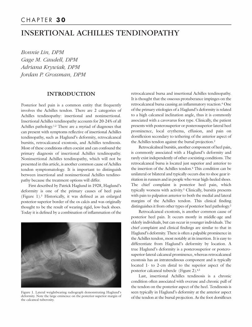
INTRODUCTION Posterior heel pain is a common entity that frequently involves the Achilles tendon. There are 2 categories of Achilles tendinopathy: insertional and noninsertional. Insertional Achilles tendinopathy accounts for 20-24% of all Achilles pathology. 1,2 There are a myriad of diagnoses that can present with symptoms reflective of insertional Achilles tendinopathy, such as Haglund’s deformity, retrocalcaneal bursitis, retrocalcaneal exostosis, and Achilles tendinosis. Most of these conditions often coexist and can confound the primary diagnosis of insertional Achilles tendinopathy. Noninsertional Achilles tendinopathy, which will not be presented in this article, is another common cause of Achilles tendon symptomatology. It is important to distinguish between insertional and noninsertional Achilles tendino- pathy because the treatment options will differ. First described by Patrick Haglund in 1928, Haglund’s deformity is one of the primary causes of heel pain (Figure 1). 3 Historically, it was defined as an enlarged posterior-superior border of the os calcis and was originally thought to be the result of wearing rigid, low-back shoes. Today it is defined by a combination of inflammation of the retrocalcaneal bursa and insertional Achilles tendinopathy. It is thought that the osseous protuberance impinges on the retrocalcaneal bursa causing an inflammatory reaction. 4 One of the primary etiologies of a Haglund’s deformity is related to a high calcaneal inclination angle, thus it is commonly associated with a cavovarus foot type. Clinically, the patient presents with posterosuperior or posterosuperior-lateral heel prominence, local erythema, effusion, and pain on dorsiflexion secondary to tethering of the anterior aspect of the Achilles tendon against the bursal projection. 2 Retrocalcalneal bursitis, another component of heel pain, is commonly associated with a Haglund’s deformity and rarely exist independently of other coexisting conditions. The retrocalcaneal bursa is located just superior and anterior to the insertion of the Achilles tendon. 4 This condition can be unilateral or bilateral and typically occurs due to shoe gear ir- ritation in runners and in people who wear high-heeled shoes. The chief complaint is posterior heel pain, which typically worsens with activity. 3 Clinically, bursitis presents with pain to palpation anterior to both the medial and lateral margins of the Achilles tendon. This clinical finding distinguishes it from other types of posterior heel pathology. 2 Retrocalcaneal exostosis, is another common cause of posterior heel pain. It occurs mostly in middle-age and elderly individuals, but can occur in younger individuals. The chief complaint and clinical findings are similar to that in Haglund’s deformity. There is often a palpable prominence in the Achilles tendon, most notably at its insertion. It is easy to differentiate from Haglund’s deformity by location. A true Haglund’s deformity is a posterosuperior or postero- superior-lateral calcaneal prominence, whereas retrocalcaneal exostosis has an intratendinous component and is typically located 1- to 2-cm distal to the superior aspect of the posterior calcaneal tubercle (Figure 2). 4,5 Last, insertional Achilles tendinosis is a chronic condition often associated with overuse and chronic pull of the tendon on the posterior aspect of the heel. Tendinosis is seen typically in Haglund’s deformity at the anterior aspect of the tendon at the bursal projection. As the foot dorsiflexes INSERTIONAL ACHILLES TENDINOPATHY Bonnie Lin, DPM Gage M. Caudell, DPM Adriana Krywiak, DPM Jordan P. Grossman, DPM CHAPTER 30 Figure 1. Lateral weightbearing radiograph demonstrating Haglund’s deformity. Note the large eminence on the posterior-superior margin of the calcaneal tuberosity.
Transcript of INSERTIONALACHILLESTENDINOPATHY - The … · INSERTIONALACHILLESTENDINOPATHY BonnieLin,DPM...
INTRODUCTION
Posterior heel pain is a common entity that frequentlyinvolves the Achilles tendon. There are 2 categories ofAchilles tendinopathy: insertional and noninsertional.Insertional Achilles tendinopathy accounts for 20-24% of allAchilles pathology.1,2 There are a myriad of diagnoses thatcan present with symptoms reflective of insertional Achillestendinopathy, such as Haglund’s deformity, retrocalcanealbursitis, retrocalcaneal exostosis, and Achilles tendinosis.Most of these conditions often coexist and can confound theprimary diagnosis of insertional Achilles tendinopathy.Noninsertional Achilles tendinopathy, which will not bepresented in this article, is another common cause of Achillestendon symptomatology. It is important to distinguishbetween insertional and noninsertional Achilles tendino-pathy because the treatment options will differ.
First described by Patrick Haglund in 1928, Haglund’sdeformity is one of the primary causes of heel pain(Figure 1).3 Historically, it was defined as an enlargedposterior-superior border of the os calcis and was originallythought to be the result of wearing rigid, low-back shoes.Today it is defined by a combination of inflammation of the
retrocalcaneal bursa and insertional Achilles tendinopathy.It is thought that the osseous protuberance impinges on theretrocalcaneal bursa causing an inflammatory reaction.4 Oneof the primary etiologies of a Haglund’s deformity is relatedto a high calcaneal inclination angle, thus it is commonlyassociated with a cavovarus foot type. Clinically, the patientpresents with posterosuperior or posterosuperior-lateral heelprominence, local erythema, effusion, and pain ondorsiflexion secondary to tethering of the anterior aspect ofthe Achilles tendon against the bursal projection.2
Retrocalcalneal bursitis, another component of heel pain,is commonly associated with a Haglund’s deformity andrarely exist independently of other coexisting conditions. Theretrocalcaneal bursa is located just superior and anterior tothe insertion of the Achilles tendon.4 This condition can beunilateral or bilateral and typically occurs due to shoe gear ir-ritation in runners and in people whowear high-heeled shoes.The chief complaint is posterior heel pain, whichtypically worsens with activity.3 Clinically, bursitis presentswith pain to palpation anterior to both the medial and lateralmargins of the Achilles tendon. This clinical findingdistinguishes it from other types of posterior heel pathology.2
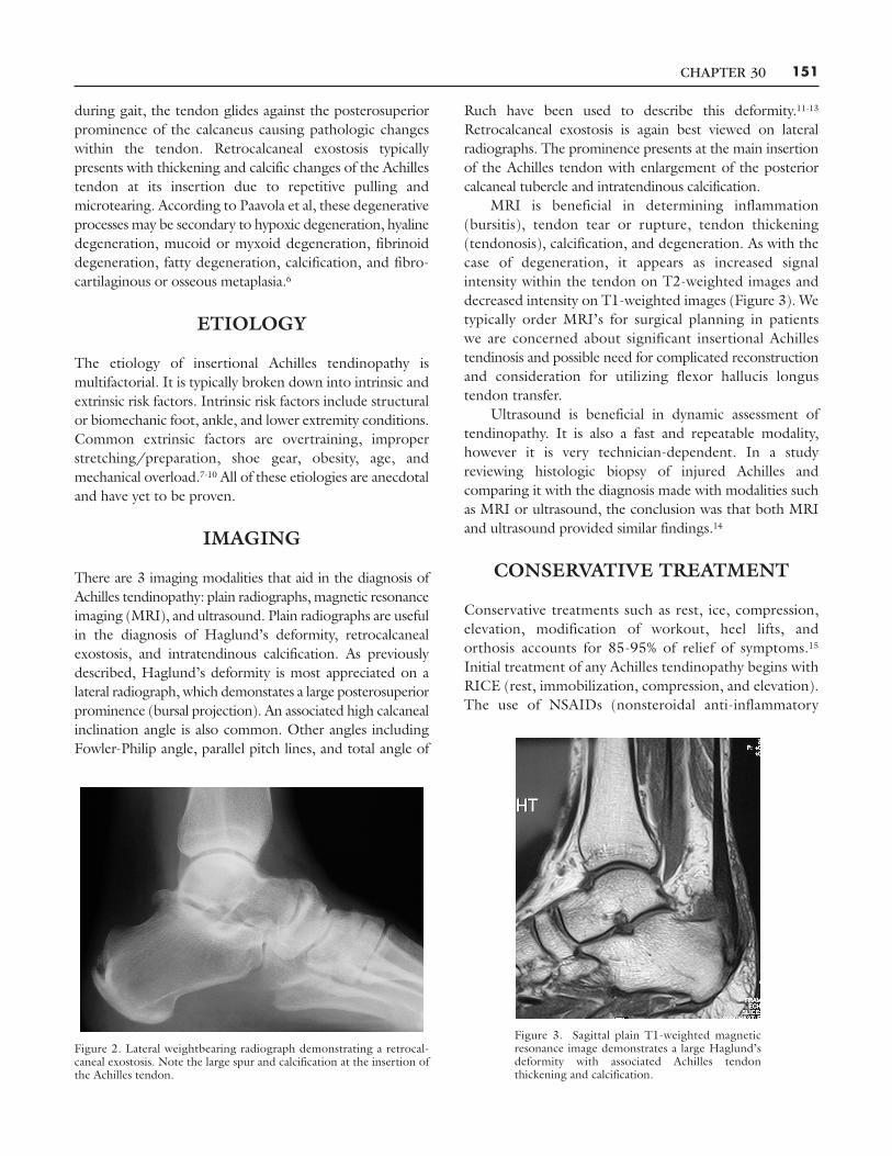
Retrocalcaneal exostosis, is another common cause ofposterior heel pain. It occurs mostly in middle-age andelderly individuals, but can occur in younger individuals. Thechief complaint and clinical findings are similar to that inHaglund’s deformity. There is often a palpable prominence inthe Achilles tendon, most notably at its insertion. It is easy todifferentiate from Haglund’s deformity by location. Atrue Haglund’s deformity is a posterosuperior or postero-superior-lateral calcaneal prominence, whereas retrocalcanealexostosis has an intratendinous component and is typicallylocated 1- to 2-cm distal to the superior aspect of theposterior calcaneal tubercle (Figure 2).4,5
Last, insertional Achilles tendinosis is a chroniccondition often associated with overuse and chronic pull ofthe tendon on the posterior aspect of the heel. Tendinosis isseen typically in Haglund’s deformity at the anterior aspectof the tendon at the bursal projection. As the foot dorsiflexes
INSERTIONAL ACHILLES TENDINOPATHY
Bonnie Lin, DPMGage M. Caudell, DPMAdriana Krywiak, DPMJordan P. Grossman, DPM
C H A P T E R 30
Figure 1. Lateral weightbearing radiograph demonstrating Haglund’sdeformity. Note the large eminence on the posterior-superior margin ofthe calcaneal tuberosity.
CHAPTER 30 151
during gait, the tendon glides against the posterosuperiorprominence of the calcaneus causing pathologic changeswithin the tendon. Retrocalcaneal exostosis typicallypresents with thickening and calcific changes of the Achillestendon at its insertion due to repetitive pulling andmicrotearing. According to Paavola et al, these degenerativeprocesses may be secondary to hypoxic degeneration, hyalinedegeneration, mucoid or myxoid degeneration, fibrinoiddegeneration, fatty degeneration, calcification, and fibro-cartilaginous or osseous metaplasia.6
ETIOLOGY
The etiology of insertional Achilles tendinopathy ismultifactorial. It is typically broken down into intrinsic andextrinsic risk factors. Intrinsic risk factors include structuralor biomechanic foot, ankle, and lower extremity conditions.Common extrinsic factors are overtraining, improperstretching/preparation, shoe gear, obesity, age, andmechanical overload.7-10 All of these etiologies are anecdotaland have yet to be proven.
IMAGING
There are 3 imaging modalities that aid in the diagnosis ofAchilles tendinopathy: plain radiographs, magnetic resonanceimaging (MRI), and ultrasound. Plain radiographs are usefulin the diagnosis of Haglund’s deformity, retrocalcanealexostosis, and intratendinous calcification. As previouslydescribed, Haglund’s deformity is most appreciated on alateral radiograph, which demonstates a large posterosuperiorprominence (bursal projection). An associated high calcanealinclination angle is also common. Other angles includingFowler-Philip angle, parallel pitch lines, and total angle of
Ruch have been used to describe this deformity.11-13
Retrocalcaneal exostosis is again best viewed on lateralradiographs. The prominence presents at the main insertionof the Achilles tendon with enlargement of the posteriorcalcaneal tubercle and intratendinous calcification.
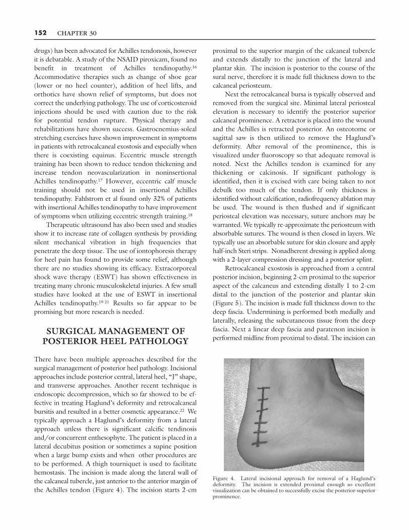
MRI is beneficial in determining inflammation(bursitis), tendon tear or rupture, tendon thickening(tendonosis), calcification, and degeneration. As with thecase of degeneration, it appears as increased signalintensity within the tendon on T2-weighted images anddecreased intensity on T1-weighted images (Figure 3). Wetypically order MRI’s for surgical planning in patientswe are concerned about significant insertional Achillestendinosis and possible need for complicated reconstructionand consideration for utilizing flexor hallucis longustendon transfer.
Ultrasound is beneficial in dynamic assessment oftendinopathy. It is also a fast and repeatable modality,however it is very technician-dependent. In a studyreviewing histologic biopsy of injured Achilles andcomparing it with the diagnosis made with modalities suchas MRI or ultrasound, the conclusion was that both MRIand ultrasound provided similar findings.14
CONSERVATIVE TREATMENT
Conservative treatments such as rest, ice, compression,elevation, modification of workout, heel lifts, andorthosis accounts for 85-95% of relief of symptoms.15
Initial treatment of any Achilles tendinopathy begins withRICE (rest, immobilization, compression, and elevation).The use of NSAIDs (nonsteroidal anti-inflammatory
Figure 2. Lateral weightbearing radiograph demonstrating a retrocal-caneal exostosis. Note the large spur and calcification at the insertion ofthe Achilles tendon.
Figure 3. Sagittal plain T1-weighted magneticresonance image demonstrates a large Haglund’sdeformity with associated Achilles tendonthickening and calcification.
CHAPTER 30152
drugs) has been advocated for Achilles tendonosis, howeverit is debatable. A study of the NSAID piroxicam, found nobenefit in treatment of Achilles tendinopathy.16
Accommodative therapies such as change of shoe gear(lower or no heel counter), addition of heel lifts, andorthotics have shown relief of symptoms, but does notcorrect the underlying pathology. The use of corticosteroidinjections should be used with caution due to the riskfor potential tendon rupture. Physical therapy andrehabilitations have shown success. Gastrocnemius-solealstretching exercises have shown improvement in symptomsin patients with retrocalcaneal exostosis and especially whenthere is coexisting equinus. Eccentric muscle strengthtraining has been shown to reduce tendon thickening andincrease tendon neovascularization in noninsertionalAchilles tendinopathy.17 However, eccentric calf muscletraining should not be used in insertional Achillestendinopathy. Fahlstrom et al found only 32% of patientswith insertional Achilles tendinopathy to have improvementof symptoms when utilizing eccentric strength training.18
Therapeutic ultrasound has also been used and studiesshow it to increase rate of collagen synthesis by providingsilent mechanical vibration in high frequencies thatpenetrate the deep tissue. The use of iontophoresis therapyfor heel pain has found to provide some relief, althoughthere are no studies showing its efficacy. Extracorporealshock wave therapy (ESWT) has shown effectiveness intreating many chronic musculoskeletal injuries. A few smallstudies have looked at the use of ESWT in insertionalAchilles tendinopathy.19-21 Results so far appear to bepromising but more research is needed.
SURGICAL MANAGEMENT OFPOSTERIOR HEEL PATHOLOGY
There have been multiple approaches described for thesurgical management of posterior heel pathology. Incisionalapproaches include posterior central, lateral heel, “J” shape,and transverse approaches. Another recent technique isendoscopic decompression, which so far showed to be ef-fective in treating Haglund’s deformity and retrocalcanealbursitis and resulted in a better cosmetic appearance.22 Wetypically approach a Haglund’s deformity from a lateralapproach unless there is significant calcific tendinosisand/or concurrent enthesophyte. The patient is placed in alateral decubitus position or sometimes a supine positionwhen a large bump exists and when other procedures areto be performed. A thigh tourniquet is used to facilitatehemostasis. The incision is made along the lateral wall ofthe calcaneal tubercle, just anterior to the anterior margin ofthe Achilles tendon (Figure 4). The incision starts 2-cm
proximal to the superior margin of the calcaneal tubercleand extends distally to the junction of the lateral andplantar skin. The incision is posterior to the course of thesural nerve, therefore it is made full thickness down to thecalcaneal periosteum.
Next the retrocalcaneal bursa is typically observed andremoved from the surgical site. Minimal lateral periostealelevation is necessary to identify the posterior superiorcalcaneal prominence. A retractor is placed into the woundand the Achilles is retracted posterior. An osteotome orsagittal saw is then utilized to remove the Haglund’sdeformity. After removal of the prominence, this isvisualized under fluoroscopy so that adequate removal isnoted. Next the Achilles tendon is examined for anythickening or calcinosis. If significant pathology isidentified, then it is excised with care being taken to notdebulk too much of the tendon. If only thickness isidentified without calcification, radiofrequency ablation maybe used. The wound is then flushed and if significantperiosteal elevation was necessary, suture anchors may bewarranted. We typically re-approximate the periosteum withabsorbable sutures. The wound is then closed in layers. Wetypically use an absorbable suture for skin closure and applyhalf-inch Steri strips. Nonadherent dressing is applied alongwith a 2-layer compression dressing and a posterior splint.
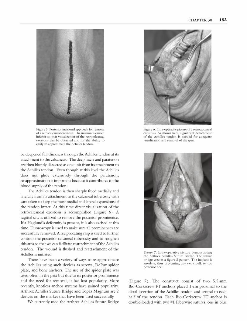
Retrocalcaneal exostosis is approached from a centralposterior incision, beginning 2-cm proximal to the superioraspect of the calcaneus and extending distally 1 to 2-cmdistal to the junction of the posterior and plantar skin(Figure 5). The incision is made full thickness down to thedeep fascia. Undermining is performed both medially andlaterally, releasing the subcutaneous tissue from the deepfascia. Next a linear deep fascia and paratenon incision isperformed midline from proximal to distal. The incision can
Figure 4. Lateral incisional approach for removal of a Haglund’sdeformity. The incision is extended proximal enough so excellentvisualization can be obtained to successfully excise the posterior-superiorprominence.
CHAPTER 30 153
be deepened full thickness through the Achilles tendon at itsattachment to the calcaneus. The deep fascia and paratenonare then bluntly dissected as one unit from its attachment tothe Achilles tendon. Even though at this level the Achillesdoes not glide extensively through the paratenon,re-approximation is important because it contributes to theblood supply of the tendon.
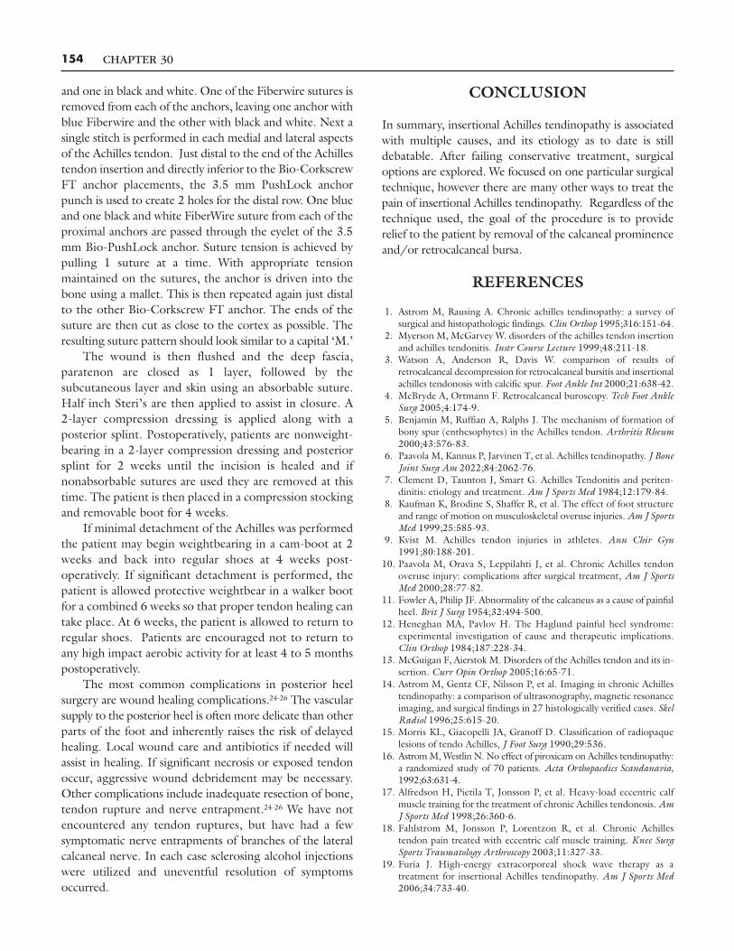
The Achilles tendon is then sharply freed medially andlaterally from its attachment to the calcaneal tuberosity withcare taken to keep the most medial and lateral expansions ofthe tendon intact. At this time direct visualization of theretrocalcaneal exostosis is accomplished (Figure 6). Asagittal saw is utilized to remove the posterior prominence.If a Haglund’s deformity is present, it is also excised at thistime. Fluoroscopy is used to make sure all prominences aresuccessfully removed. A reciprocating rasp is used to furthercontour the posterior calcaneal tuberosity and to roughenthis area so that we can facilitate reattachment of the Achillestendon. The wound is flushed and reattachment of theAchilles is initiated.
There have been a variety of ways to re-approximatethe Achilles using such devices as screws, DePuy spiderplate, and bone anchors. The use of the spider plate wasused often in the past but due to its posterior prominenceand the need for removal, it has lost popularity. Morerecently, knotless anchor systems have gained popularity.Arthrex Achilles Suture Bridge and Topaz Magnum are 2devices on the market that have been used successfully.
We currently used the Arthrex Achilles Suture Bridge
(Figure 7). The construct consist of two 5.5-mmBio-Corkscrew FT anchors placed 1-cm proximal to thedistal insertion of the Achilles tendon and central to eachhalf of the tendon. Each Bio-Corkscrew FT anchor isdouble-loaded with two #1 Fiberwire sutures, one in blue
Figure 5. Posterior incisional approach for removalof a retrocalcaneal exostosis. The incision is carriedinferior so that visualization of the retrocalcanealexostosis can be obtained and for the ability toeasily re-approximate the Achilles tendon.
Figure 6. Intra-operative picture of a retrocalcanealexostosis. As shown here, significant detachmentof the Achilles tendon is needed for adequatevisualization and removal of the spur.
Figure 7. Intra-operative picture demonstratingthe Arthrex Achilles Suture Bridge. The suturebridge creates a figure 8 pattern. The implant isknotless, thus preventing any extra bulk to theposterior heel.
CHAPTER 30154
and one in black and white. One of the Fiberwire sutures isremoved from each of the anchors, leaving one anchor withblue Fiberwire and the other with black and white. Next asingle stitch is performed in each medial and lateral aspectsof the Achilles tendon. Just distal to the end of the Achillestendon insertion and directly inferior to the Bio-CorkscrewFT anchor placements, the 3.5 mm PushLock anchorpunch is used to create 2 holes for the distal row. One blueand one black and white FiberWire suture from each of theproximal anchors are passed through the eyelet of the 3.5mm Bio-PushLock anchor. Suture tension is achieved bypulling 1 suture at a time. With appropriate tensionmaintained on the sutures, the anchor is driven into thebone using a mallet. This is then repeated again just distalto the other Bio-Corkscrew FT anchor. The ends of thesuture are then cut as close to the cortex as possible. Theresulting suture pattern should look similar to a capital ‘M.’
The wound is then flushed and the deep fascia,paratenon are closed as 1 layer, followed by thesubcutaneous layer and skin using an absorbable suture.Half inch Steri’s are then applied to assist in closure. A2-layer compression dressing is applied along with aposterior splint. Postoperatively, patients are nonweight-bearing in a 2-layer compression dressing and posteriorsplint for 2 weeks until the incision is healed and ifnonabsorbable sutures are used they are removed at thistime. The patient is then placed in a compression stockingand removable boot for 4 weeks.
If minimal detachment of the Achilles was performedthe patient may begin weightbearing in a cam-boot at 2weeks and back into regular shoes at 4 weeks post-operatively. If significant detachment is performed, thepatient is allowed protective weightbear in a walker bootfor a combined 6 weeks so that proper tendon healing cantake place. At 6 weeks, the patient is allowed to return toregular shoes. Patients are encouraged not to return toany high impact aerobic activity for at least 4 to 5 monthspostoperatively.
The most common complications in posterior heelsurgery are wound healing complications.24-26 The vascularsupply to the posterior heel is often more delicate than otherparts of the foot and inherently raises the risk of delayedhealing. Local wound care and antibiotics if needed willassist in healing. If significant necrosis or exposed tendonoccur, aggressive wound debridement may be necessary.Other complications include inadequate resection of bone,tendon rupture and nerve entrapment.24-26 We have notencountered any tendon ruptures, but have had a fewsymptomatic nerve entrapments of branches of the lateralcalcaneal nerve. In each case sclerosing alcohol injectionswere utilized and uneventful resolution of symptomsoccurred.
CONCLUSION
In summary, insertional Achilles tendinopathy is associatedwith multiple causes, and its etiology as to date is stilldebatable. After failing conservative treatment, surgicaloptions are explored. We focused on one particular surgicaltechnique, however there are many other ways to treat thepain of insertional Achilles tendinopathy. Regardless of thetechnique used, the goal of the procedure is to providerelief to the patient by removal of the calcaneal prominenceand/or retrocalcaneal bursa.
REFERENCES
1. Astrom M, Rausing A. Chronic achilles tendinopathy: a survey ofsurgical and histopathologic findings. Clin Orthop 1995;316:151-64.
2. Myerson M, McGarvey W. disorders of the achilles tendon insertionand achilles tendonitis. Instr Course Lecture 1999;48:211-18.
3. Watson A, Anderson R, Davis W. comparison of results ofretrocalcaneal decompression for retrocalcaneal bursitis and insertionalachilles tendonosis with calcific spur. Foot Ankle Int 2000;21:638-42.
4. McBryde A, Ortmann F. Retrocalcaneal buroscopy. Tech Foot AnkleSurg 2005;4:174-9.
5. Benjamin M, Ruffian A, Ralphs J. The mechanism of formation ofbony spur (enthesophytes) in the Achilles tendon. Arthritis Rheum2000;43:576-83.
6. Paavola M, Kannus P, Jarvinen T, et al. Achilles tendinopathy. J BoneJoint Surg Am 2022;84:2062-76.
7. Clement D, Taunton J, Smart G. Achilles Tendonitis and periten-dinitis: etiology and treatment. Am J Sports Med 1984;12:179-84.
8. Kaufman K, Brodine S, Shaffer R, et al. The effect of foot structureand range of motion on musculoskeletal overuse injuries. Am J SportsMed 1999;25:585-93.
9. Kvist M. Achilles tendon injuries in athletes. Ann Chir Gyn1991;80:188-201.
10. Paavola M, Orava S, Leppilahti J, et al. Chronic Achilles tendonoveruse injury: complications after surgical treatment, Am J SportsMed 2000;28:77-82.
11. Fowler A, Philip JF. Abnormality of the calcaneus as a cause of painfulheel. Brit J Surg 1954;32:494-500.
12. Heneghan MA, Pavlov H. The Haglund painful heel syndrome:experimental investigation of cause and therapeutic implications.Clin Orthop 1984;187:228-34.
13. McGuigan F, Aierstok M. Disorders of the Achilles tendon and its in-sertion. Curr Opin Orthop 2005;16:65-71.
14. Astrom M, Gentz CF, Nilsson P, et al. Imaging in chronic Achillestendinopathy: a comparison of ultrasonography, magnetic resonanceimaging, and surgical findings in 27 histologically verified cases. SkelRadiol 1996;25:615-20.
15. Morris KL, Giacopelli JA, Granoff D. Classification of radiopaquelesions of tendo Achilles, J Foot Surg 1990;29:536.
16. AstromM,Westlin N. No effect of piroxicam on Achilles tendinopathy:a randomized study of 70 patients. Acta Orthopaedics Scandanavia,1992;63:631-4.
17. Alfredson H, Pietila T, Jonsson P, et al. Heavy-load eccentric calfmuscle training for the treatment of chronic Achilles tendonosis. AmJ Sports Med 1998;26:360-6.
18. Fahlstrom M, Jonsson P, Lorentzon R, et al. Chronic Achillestendon pain treated with eccentric calf muscle training. Knee SurgSports Traumatology Arthroscopy 2003;11:327-33.
19. Furia J. High-energy extracorporeal shock wave therapy as atreatment for insertional Achilles tendinopathy. Am J Sports Med2006;34:733-40.
CHAPTER 30 155
20. Furia J, Rompe J. Extracorpeal shock wave therapy in the treatmentof chronic fasciitis and Achilles tendinopathy. Curr Opin Orthop2007;18:102-11.
21. Rees J, Wilson A, Wolman R. Current concepts in the managementof tendon disorders. Rheum 2006;45:508-21.
22. Maffulli N, Testa V, Capasso G, et al. Calcific insertional Achillestendinopathy: reattachment with bone anchors. Am J Sports Med2004;32:174-82.
23. Benthien, R A, Aronow, M S, Doran-Diaz , et al. Cyclic loading ofAchilles tendon repairs: a comparison of polyester and polyblendsuture. Foot Ankle Int 2006;27:512-8.
24. Gould J. Insertional tendinitis of the tendo Achilles. Tech Foot AnkleSurg 2005;4:222-9.
25. Wagner E, Gould J, Kneidel M, et al. Technique and results ofAchilles tendon detachment and reconstruction for insertionalAchilles tendonosis. Foot Ankle Int 2006;27:677-84.
26. Aronow MS. Posterior heel pain (retrocalcaneal bursitis, insertionaland noninsertional Achilles tendinopathy). Clin Podiatric Med Surg2005;22:19-43.