Innovation and Translation E orts in Wireless Medical...
26
Innovation and Translation Efforts in Wireless Medical Connectivity, Telemedicine and eMedicine: A Story from the RFID Center of Excellence at the University of Pittsburgh Ervin Sejdi´ c * Michael Rothfuss Joshua R. Stachel Nicholas G. Franconi Kara Bocan Michael R. Lovell † Marlin H. Mickle Abstract Translational research has recently been rediscovered as one of the basic tenants of engineer- ing. Although many people have numerous ideas of how to accomplish this successfully, the fundamental method is to provide an innovative and creative environment. The University of Pittsburgh has been accomplishing this goal though a variety of methodologies. The contents of this paper are exemplary of what can be achieved though the interaction of students, staff, fac- ulty and, in one example, high school teachers. While the projects completed within the groups involved in this paper have spanned other areas, the focus of this paper is on the biomedical devices, that is, towards improving and maintaining health in a variety of areas. The spirit of the translational research is discovery, invention, intellectual property protection, and the creation of value through the spinning off of companies while providing better health care and creating jobs. All but one of these projects involve wireless radio frequency energy for delivery. The remaining device can be wirelessly connected for data collection. Keywords: Translation research, University of Pittsburgh, roborat, wireless deep brain stim- ulation, vagus nerve stimulation, electroporation device, wireless Doppler, programmable pulse oximetry, implantable RFID tags, implantable blood flow monitor. * Ervin Sejdi´ c, Michael A. Rothfuss, Joshua R. Stachel, Nicholas G. Franconi, Kara Bocan, Marlin H. Mickle are with the Department of Electrical and Computer Engineering, Swanson School of Enginering, University of Pittsburgh, Pittsburgh, PA, 15261, USA. E-mails: [email protected], [email protected], [email protected], [email protected], [email protected], [email protected]. † Michael R. Lovell is the Chancellor of the University of Wisconsin-Milwauke, University of Wisconsin-Milwaukee, P.O. Box 413, Milwaukee, WI, 53201, USA. E-mail: [email protected]. 1
Transcript of Innovation and Translation E orts in Wireless Medical...

Innovation and Translation Efforts in Wireless Medical
Connectivity, Telemedicine and eMedicine: A Story from the RFID
Center of Excellence at the University of Pittsburgh
Ervin Sejdic∗ Michael Rothfuss Joshua R. Stachel Nicholas G. Franconi
Kara Bocan Michael R. Lovell† Marlin H. Mickle
Abstract
Translational research has recently been rediscovered as one of the basic tenants of engineer-
ing. Although many people have numerous ideas of how to accomplish this successfully, the
fundamental method is to provide an innovative and creative environment. The University of
Pittsburgh has been accomplishing this goal though a variety of methodologies. The contents of
this paper are exemplary of what can be achieved though the interaction of students, staff, fac-
ulty and, in one example, high school teachers. While the projects completed within the groups
involved in this paper have spanned other areas, the focus of this paper is on the biomedical
devices, that is, towards improving and maintaining health in a variety of areas. The spirit
of the translational research is discovery, invention, intellectual property protection, and the
creation of value through the spinning off of companies while providing better health care and
creating jobs. All but one of these projects involve wireless radio frequency energy for delivery.
The remaining device can be wirelessly connected for data collection.
Keywords: Translation research, University of Pittsburgh, roborat, wireless deep brain stim-
ulation, vagus nerve stimulation, electroporation device, wireless Doppler, programmable pulse
oximetry, implantable RFID tags, implantable blood flow monitor.
∗Ervin Sejdic, Michael A. Rothfuss, Joshua R. Stachel, Nicholas G. Franconi, Kara Bocan, Marlin H. Mickle
are with the Department of Electrical and Computer Engineering, Swanson School of Enginering, University of
Pittsburgh, Pittsburgh, PA, 15261, USA. E-mails: [email protected], [email protected], [email protected], [email protected],
[email protected], [email protected].†Michael R. Lovell is the Chancellor of the University of Wisconsin-Milwauke, University of Wisconsin-Milwaukee,
P.O. Box 413, Milwaukee, WI, 53201, USA. E-mail: [email protected].
1

1 Introduction
The dividing line between basic research and research that can be easily translated into value has
always been difficult and somewhat controversial. We assume engineering schools are traditionally
more on the translational side but not completely as have been the discussions (mostly friendly)
involving Engineering and Engineering Science. More recently, the interpretation of value has
extended the translational domain due to the medical value of many engineering developments.
The innovation and translation efforts are embedded in the core of our research culture at the
Swanson School of Engineering and the School of Medicine at the University of Pittsburgh. In
FY2012, there were 310 invention disclosures to the Office of Technology Management (OTM) for
commercial consideration, which represents a 21 % increase from previous year and a 319 % increase
over invention disclosure submissions in 2003. In 2003, the University of Pittsburgh researchers
submitted only 74 invention disclosures, which sparked a strategic shift at OTM aimed at attracting
more innovators and actively engaging them in the innovation commercialization process at the
University of Pittsburgh. Overall, there were 1,290 invention disclosures over the past five years.
The University’s patent portfolio continued to grow substantially in FY2012. The U.S. Patent and
Trademark Office awarded the University of Pittsburgh and its innovators 49 new U.S. patents in
FY2012, representing a 32.4 % increase from FY2011.
OTM’s ultimate goal is to move as many innovations as possible into the commercial market-
place, which led to the execution of 132 technology licenses or options to commercial partners in
FY2012. This represents a 26 % increase from FY2011. Total revenue for FY2012 rose to nearly
$6.8 million, up more than 10.2 percent from FY 2011. Total revenue includes licensing revenue,
equity sales, and legal fee reimbursement from licensees. Due to higher number of “platform” in-
novations developed at the University of Pittsburgh accompanied by more concentrated effort by
OTM and the Office of Economic Development (OED) to promote and facilitate start-up activities,
the university had nine start-ups this past year. Success was driven, in part, by a combination
that included OTM licensing managers and executives in residence, OEDs development and im-
plementation of the new Pitt Ventures initiative and start-up process, and a growing number of
innovations with more than one potentially profitable commercial application. Last, but not least,
the University of Pittsburgh is also a home to one of the six national Coulter Translational Research
Partners II Programs, which is accompanied by a $3.54 million five-year grant from the Wallace H.
Coulter Foundation.
This thriving innovation environment is well suited for the RFID (Radio Frequency Identifica-
2
tion) Center of Excellence at the University of Pittsburgh, which was formed in 2005. The mission
at the center is to be a unique workforce, rooted in a research and learning environment that is
strongly connected with industry. The RFID Center of Excellence and the earlier Swanson Center
for Product Innovation have spawned numerous spin off companies and licensed technologies where
the focus has shifted to biomedical applications. The spirit of the research environment has always
been in concert with both Wallace H. Coulter and Jerome H. Lemelson thus focusing on research
and technology that translates to generating value and jobs with the more recent emphasis on
biomedical applications.
This paper illustrates the range of products and technologies that have been translated into
seven companies and numerous patents that have been licensed to these and other companies to
create value through innovation in American industry.
The projects of the Center have won dozens of awards for both faculty and students over the
years with more than 25 named student inventors on US Patents. The student experience is one
of the most important issues for an educational research center. The projects covered in this
paper have made it possible for students to interact directly with clinicians in both engineering
and medical environments. These experiences have covered both translating medical problems into
technical solutions and taking part in numerous clinical trials involving animals (mice, Sprague
Dawley rats and swine) and cadavers. Needless to state, all projects described in this manuscript
had the necessary approvals from either the Institutional Review Board or the Institutional Animal
Care and Use Committee, if needed.
2 Our innovation efforts
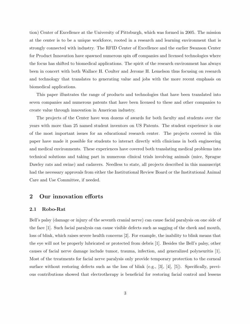
2.1 Robo-Rat
Bell’s palsy (damage or injury of the seventh cranial nerve) can cause facial paralysis on one side of
the face [1]. Such facial paralysis can cause visible defects such as sagging of the cheek and mouth,
loss of blink, which raises severe health concerns [2]. For example, the inability to blink means that
the eye will not be properly lubricated or protected from debris [1]. Besides the Bell’s palsy, other
causes of facial nerve damage include tumor, trauma, infection, and generalized polyneuritis [1].
Most of the treatments for facial nerve paralysis only provide temporary protection to the corneal
surface without restoring defects such as the loss of blink (e.g., [3], [4], [5]). Specifically, previ-
ous contributions showed that electrotherapy is beneficial for restoring facial control and lessens
3

synkinesis [6], [7].
The University of Pittsburgh originally began this work under the project title Blink Right. In
conjunction with the RFID Center, the ophthalmology department at the University of Pittsburgh
targeted ameliorating those suffering with the condition by stimulating the muscles associated with
blinking the eye on the affected side. This was accomplished by detecting a patient’s blink on
the unaffected side and then stimulating the muscles required to perform the blinking function
on the side affected with the Bell’s palsy. To demonstrate the capability to stimulate muscles
simultaneously and remotely, the Sprague Dawley rats were chosen. The mechanical movement of
of one rat’s leg produced a simultaneous and remote stimulation in the same leg on the remote rat.
One rat’s functional leg was outfitted with a probe, which feeds into a processing chain of analog
and digital electronics that detect the probed nerve’s electrical activity generated by mechanically
stimulating the functional leg. The post-processed nerve signal is transmitted via wireless radio
transmitter. On a second rat with a de-nervated leg, the transmitted wireless signal is detected by
a wireless radio receiver which feeds into a processing chain of analog and digital electronics which
generate the stimulation signal. The stimulation signal is applied to the de-nervated rat’s leg to
generate a contraction [1]. Thus, move one rat leg and the leg of the second rat moves with only
a wireless radio frequency connection between the two - hence the name Robo-Rat. A few of the
animal trials photos are shown in Figure 1.
Figure 1: Roborat in action
Several in vivo experiments were conducted involving the gastrocnemius leg muscles of our rats.
These experiments established (1) the magnitude of the stimulation signals required to generate
contraction in the de-nervated gastrocnemius leg muscle, (2) the magnitude of the electrical output
4

from functional gastrocnemius that could be sensed resulting from mechanical stimulation, and
(3) real-time remote stimulation of a de-nervated rat’s gastrocnemius leg muscle actuated by the
mechanical stimulation of a separate rat’s functional gastrocnemius leg muscle.
These experiments have shown that we can utilize radio-frequency sensors to initiate functional
electrical stimulation of a denervated muscle from a normally functioning muscle. Our results
showed that to produce a sufficient contraction, higher stimulation voltages were required with
longer pulse widths, which can be attributed to fatigue of the individual muscle fibers. The ex-
perimental results showed that 1V-2V stimulation voltages for 10 milliseconds were sufficient to
produce sufficient contraction without histopathologic damage to the tissue. The location of the
probe also played a role in the detection of voltage levels. Additional promising applications of this
system exist, not just in otolaryngology and neurology but also in rehabilitation.
Graduate students in the Department of Electrical and Computer Engineering at the Univer-
sity of Pittsburgh developed this technology. The Blink Right technology was licensed to a San
Francisco-based company, the same company that purchased licensing for Wireless Deep Brain
Stimulator from the university (described in the next section). This technology was one of the
many technologies that stemmed from the RFID Center’s generation of intellectual property in
the realms of wireless energy harvesting, low power devices, and wireless communication through
tissue.
2.2 Wireless Deep Brain Stimulation
Parkinsons disease is a neurodegenerative disorder, and its symptoms include stiffness, slowness of
movement, and resting tremor [8], [9]. Other common symptoms include depression and dementia
[10], [9]. Various treatments exist for Parkinsons disease, including numerous drugs (e.g., [11]) and
deep brain stimulation (DBS) (e.g., [12]). DBS usually involves surgery to implant a probe into
the brain and its corresponding controller into the body (e.g., [13], [14]). DBS is mainly used for
Parkinsons disease, but it can also be used to treat other diseases, such as Huntingtons disease (e.g.,
[15]), dystonia (e.g., [16]), and epilepsy (e.g., [17]). Both biological tests and computer simulations
support the idea that stimulation regulates neurons, improving data propagation over the irregular
neuronal activity present before DBS.
However, DBS devices can be cumbersome. First, they require a large implanted battery, which
have a limited life. Second, a control pack needs to be implanted in the chest. Such a pack would
then have subcutaneous wires threaded up through the neck to the top of the skull. These implanted
5

devices and wires can cause irritation and infection leading to removal of the device in some cases.
All of these drawbacks lead to device replacement surgeries every three to five years.
These drawbacks led the RFID Center of Excellence to design and test a small remotely pow-
ered device capable of driving a DBS probe from directly under the scalp as shown in Figure 2.
The device is remotely powered via inductive coupling, consuming just over 10 joules per day.
Figure 2: Wireless deep brain stimulation.
A 1.2-in. square printed circuit board with
maximum thickness of 0.25 inches is used
to fit the implantable circuitry. Our results
showed that we can power and program
the device through air, porcine skin and
a cadaver scalp. Additionally, the power
transmitting coil can be driven via a small,
cheap and easily replaceable external bat-
tery pack. A model has been made to
demonstrate a feasible realization of the
entire device which is both functional and
aesthetically pleasing.
The wireless DBS began when a San
Francisco-based company worked with the RFID Center of Excellence to develop a new technology
which targeted reducing the infection rates associated with the use of wired DBS devices. The
wireless DBS could run on power supplied by batteries and supercapacitors for several days before
needing to be recharged, and the recharging unit was affixed at the end of the patient’s bed to
provide an easy mode of recharging while the patient slept. The RFID Center developed the
wireless DBS and the University of Pittsburgh licensed the technology to the San Francisco-based
company. After the company ran into financial hardship, the license for the wireless DBS reverted
back to the University.
Later on, the same company worked with the RFID Center to expand the wireless DBS technol-
ogy to source it’s power from local wireless radio stations. These technologies have since garnered
interest from several companies. Both graduate and undergraduate students of the department of
Electrical and Computer Engineering at the University of Pittsburgh were responsible for devel-
oping these technologies, with many of the milestones in the development of the projects coming
from undergraduate senior design projects.
6

2.3 Vagus Nerve Stimulation
The deep brain stimulation concept was modified to provide stimulation to the vagus nerve which
is used to offset a seizure for patients with epilepsy. Shown in the figure below is the wired version
of the stimulator. In the wireless version, the stimulator is implanted in the neck with electronics
while the recharging (powering) circuitry is located in the patient’s collar. Such a device would
require a large battery and a control pack to be implanted in the patient’s body. Subcutaneous
wires would be running through. All these pieces represent a source of irritation and infection,
and as such could potentially require the patient to take antibiotics or to have the device removed.
Needless to say, the device is affected by magnetic interferences and a limited battery life, which
can require another surgery to replace the device. Therefore, it is desired to stimulate vagus nerve
but without the aforementioned shortcomings.
Figure 3: Vagus nerve stimulation.
Sponsored by the National Science Founda-
tion, a team of researchers from the Univer-
sity of Pittsburgh partnered with a number of
high school teachers to develop an implantable
device that would address the issues outlined
above. The implanted device provides a method
of electrically stimulating the vagus nerve of a
patient through the generation of current pulses
through one or more probes implanted in the
body of the patient. The implantable device
also includes control circuitry electrically con-
nected to the probe(s) that is structured to gen-
erate the current pulses. The power circuitry of the implant was designed to eliminate the need for
a battery and consequently the issues that go along with battery usage. The implant can receive
energy from a far-field source (e.g., a local radio station). The received energy is converted into a
DC power signal by the power circuitry that also provides this DC power to the control circuitry.
Thus, by creating a passively powered and implantable device, issues such as transcutaneous wires
as well as limited lifetime battery supplies are eliminated. The unique expertise of the RFID Center
of Excellence in the area of wireless communications and energy harvesting made it a clear choice
of partnership for the high school teachers who designed this device over a summer program.
7

2.4 Mass Immunization Device
Naked plasmid DNA (pDNA) is the simplest and safest gene delivery system and is primarily
used used for genetic immunization studies [18]. Gene delivery in vivo using electroporation is
an alternative approach [19], where naked DNA injection is combined with a pulsed electric-field
treatment. In electroporation, it is assumed that cell membranes are equivalent to an electrical
capacitor [20], and the lipidic membrane acts as a dielectric element. By applying an electric field to
cells, we introduce structural defects in the cell structure permitting the transit diffusion of nucleic
acids [21], [20]. The membrane permeability increases due to the electric pulses, which represents
the first step in the electro-gene transfer. Next, molecules are diffused through the membrane [21].
The advantage of electroporation is that its transfection efficiency is significantly greater than that
of naked DNA injection and has reduced inter-individual variability [22]. Electroporation based in
vivo gene transfer has been used for introducing DNA into rat hepatocellular carcinomas, mouse
testes, hepatocytes, skeletal muscle, melanoma, lung, skin, and rat skeletal muscle for correcting
anemia of renal failure (e.g., [18], [23], [24]).
Figure 4: A device for electroporation.
The genesis of this technology began when the pharmacology department at the University of
Pittsburgh received a contract to develop treatments to contain or manage epidemics caused by
infectious agents. The project started with the pharmacology department studying methods of
electro-gene transfer for genetic immunization and gene therapy. The Department of Electrical and
Computer Engineering at the University of Pittsburgh was responsible for developing the associated
hardware and electronics necessary to deliver the immunizations in a safe and repeatable manner
in an epidemic scenario.
The Swanson Center for Product Innovation, and later on the RFID Center of Excellence,
8

has designed a device that by alternating the polarity of the applied electric field elucidates the
mechanism of in vivo electro-gene transfer as shown in Figure 4. In other words, DNA is forced
into the cells at the anode during the first pulse. DNA is forced back to the cathode when the
polarity is reversed during the second pulse. This process is continuously repeated. Our results,
described in [18], enabled us to have better understanding of electro-gene transfer, that can allow
us to propose more efficient conditions for in vivo electroporation.
2.5 “Smart” Pill Dispenser
Medication adherence is essential to the effective treatment of serious and chronic illnesses. In
today’s settings, the patient is primarily responsible for following his or her medication regime
(e.g., [25], [26]). Treatments for chronic diseases especially rely on effective adherence, and missing
a scheduled dose can negatively impact the treatment plan. During clinical drug trials, for example,
researchers are attempting to understand the effectiveness and side effects of potential treatments.
These drug trials must be controlled and consistent in order to accurately represent the positive
and negative effects of the proposed drug, and to provide an adequate understanding of the drug
effects before the drug is released to the general public. Given that medication doses can be missed
or accidently taken multiple times, healthcare providers and researchers desire a reliable method of
verifying that prescribed treatment plans are being followed by patients and/or study participants
(e.g., [27], [28]). While self-reporting is the most common and sometimes the only feasible method,
its accuracy depends on the patient’s memory and reliability. Medication adherence becomes
especially problematic with age (e.g., [29]).
Therefore, there is a need for technologies that aid patients in remembering doses and record-
ing adherence for medical records. These technologies are important not only for extending and
improving the lives of patients, but also for enabling improvement of treatment plans based on
accurate logs of medication use. By improving adherence, medical resources can be more efficiently
allocated by eliminating the wasted resources resulting from inconsistent treatment plans [26]. The
RFID Center of Excellence has been increasingly involved in such medical and biomedical solutions.
The proximity of the university to medical centers enables the identification of relevant problems,
such as medication adherence, and development of applicable solutions. As such, design work in
our center often involves consideration of human factors in addition to basic functionality.
To address the issues surrounding medication adherence, a ”smart” pill box was developed in
the RFID Center of Excellence. The pill box keeps a time record of when a patient removes each
9

Figure 5: A “smart” pill box.
pill from the box as well as which pills are removed. The device then wirelessly communicates
the recorded data when the box is in range of an associated receiving station (server). The device
was designed around a typical blister pack, as shown in Figure 5, and was successfully integrated
with a commercial off the shelf pill dispensing box manufactured by West Pharmaceutical. The
device functions with common blister pack pill packages to increase the likelihood of adoption due
to ease of handling by the pharmacy and ease of use by the patients. The pill detection method
was implemented in a compact form factor that can easily be expanded and configured to other
blister pack dimensions.
A graphic user interface (GUI) was also developed in the RFID Center of Excellence, to display
information received from the pill box. The image in Figure 5 with the green indicator lights is a
screenshot from the GUI at the receiving station, visually indicating the contents of the associated
pill box. The box reports pill removal automatically via wireless communication, eliminating any
additional action by the user to properly operate the device. The user simply removes the pills as
they would with a traditional blister pack medication package, and the device records the action,
providing a simple and effective medication adherence tracking solution.
2.6 Avoiding Pulse Inhibition in Pacemakers and Implanted Cardiac Defibril-
lators
Radio Frequency Identification (RFID) is a rapidly growing technology finding applications in a
multitude of fields including in healthcare settings [30], [31], [32], [33], [34]. It has been suggested
that RFID technology holds the promise to contribute very significant improvements to the future
of the healthcare industry [35], [36]. Personnel and patient tracking systems, drug counterfeiting
protections, and asset tracking systems have all been proposed in order to improve quality of care
and patient safety as well as to provide significant time and cost savings. As RFID technologies
10

are becoming more commonplace in the healthcare environment, however, it becomes necessary to
address possible complications these systems may cause. Most significantly, the radio frequency
(RF) signals emitted by these systems have the possibility to cause electromagnetic interference
(EMI) in critical care equipment. For instance, it has been reported that RFID systems have the
capacity to interfere with the normal operation of pacemakers
A Pittsburgh based medical device company was recently interested in the incorporation of pas-
sive RFID technology into surgical equipment such as surgical sponges in order to solve the problem
of accidentally leaving this equipment in a surgical patient post-operatively. As a consequence, the
company sponsored a study conducted at the RFID Center of Excellence that was aimed at assess-
ing the extent and causes of interference between RFID systems and cardiac rhythm management
devices (CRMDs) including pacemakers and cardioverter defibrillators in order to ensure patient
safety [37]. A relatively large team of graduate student and post-doctoral researchers were involved
in this study that targeted three of the most commonly utilized RFID frequency bands, 143.75
kHz (LF), 13.56 MHz (HF) and 915 MHz (UHF). Additionally, multiple tissue simulation systems,
shown in Figure 6, were fabricated and tested in order to model the effects of the human body on
implantable CRMDs when exposed to RF energy.
Figure 6: Tissue simulation devices: from left to right - tissue interface circuit, horizontal saline
tank phantom, vertical saline tank phantom.
This large scale study [38], based on over 7400 laboratory tests, confirmed the capacity of RFID
systems to cause clinically significant interference on implantable CRMD systems mainly in the
lower frequency bands (LF, and HF) while UHF bands were shown to be relatively harmless to
these critical care devices. More importantly, however, this study identified the mechanism of the
most common form of CRMD interference, pacing inhibition, and proposes a possible solution.
Pacing inhibition is a scenario in which the CRMD system should pace the heart but fails to do
11
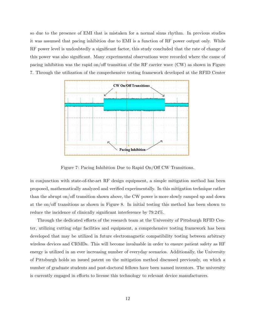
so due to the presence of EMI that is mistaken for a normal sinus rhythm. In previous studies
it was assumed that pacing inhibition due to EMI is a function of RF power output only. While
RF power level is undoubtedly a significant factor, this study concluded that the rate of change of
this power was also significant. Many experimental observations were recorded where the cause of
pacing inhibition was the rapid on/off transition of the RF carrier wave (CW) as shown in Figure
7. Through the utilization of the comprehensive testing framework developed at the RFID Center
Figure 7: Pacing Inhibition Due to Rapid On/Off CW Transitions.
in conjunction with state-of-the-art RF design equipment, a simple mitigation method has been
proposed, mathematically analyzed and verified experimentally. In this mitigation technique rather
than the abrupt on/off transition shown above, the CW power is more slowly ramped up and down
at the on/off transitions as shown in Figure 8. In initial testing this method has been shown to
reduce the incidence of clinically significant interference by 79.24%.
Through the dedicated efforts of the research team at the University of Pittsburgh RFID Cen-
ter, utilizing cutting edge facilities and equipment, a comprehensive testing framework has been
developed that may be utilized in future electromagnetic compatibility testing between arbitrary
wireless devices and CRMDs. This will become invaluable in order to ensure patient safety as RF
energy is utilized in an ever increasing number of everyday scenarios. Additionally, the University
of Pittsburgh holds an issued patent on the mitigation method discussed previously, on which a
number of graduate students and post-doctoral fellows have been named inventors. The university
is currently engaged in efforts to license this technology to relevant device manufacturers.
12
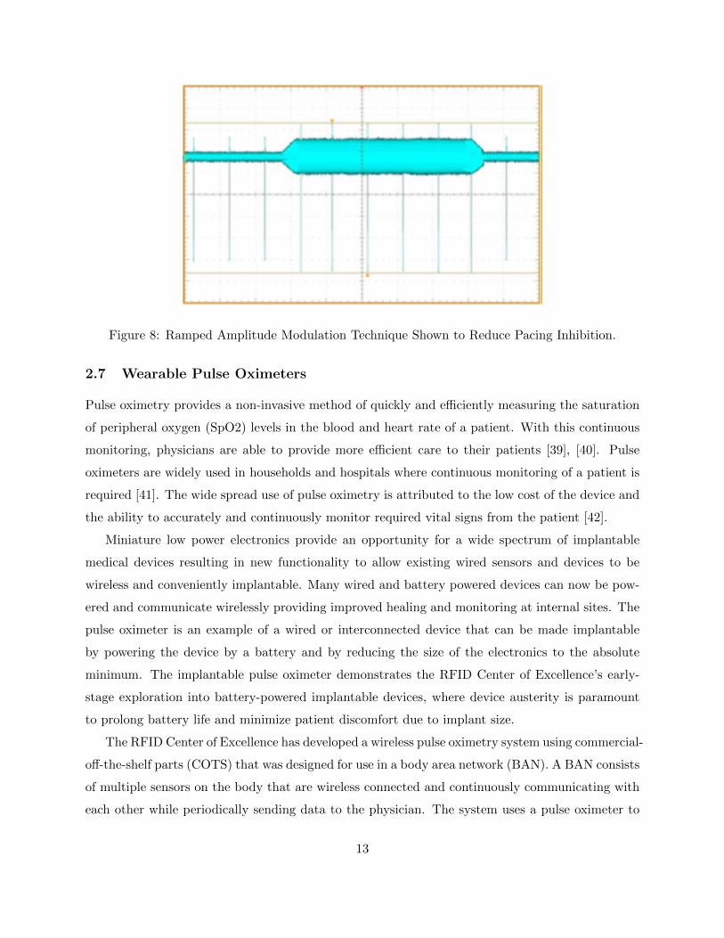
Figure 8: Ramped Amplitude Modulation Technique Shown to Reduce Pacing Inhibition.
2.7 Wearable Pulse Oximeters
Pulse oximetry provides a non-invasive method of quickly and efficiently measuring the saturation
of peripheral oxygen (SpO2) levels in the blood and heart rate of a patient. With this continuous
monitoring, physicians are able to provide more efficient care to their patients [39], [40]. Pulse
oximeters are widely used in households and hospitals where continuous monitoring of a patient is
required [41]. The wide spread use of pulse oximetry is attributed to the low cost of the device and
the ability to accurately and continuously monitor required vital signs from the patient [42].
Miniature low power electronics provide an opportunity for a wide spectrum of implantable
medical devices resulting in new functionality to allow existing wired sensors and devices to be
wireless and conveniently implantable. Many wired and battery powered devices can now be pow-
ered and communicate wirelessly providing improved healing and monitoring at internal sites. The
pulse oximeter is an example of a wired or interconnected device that can be made implantable
by powering the device by a battery and by reducing the size of the electronics to the absolute
minimum. The implantable pulse oximeter demonstrates the RFID Center of Excellence’s early-
stage exploration into battery-powered implantable devices, where device austerity is paramount
to prolong battery life and minimize patient discomfort due to implant size.
The RFID Center of Excellence has developed a wireless pulse oximetry system using commercial-
off-the-shelf parts (COTS) that was designed for use in a body area network (BAN). A BAN consists
of multiple sensors on the body that are wireless connected and continuously communicating with
each other while periodically sending data to the physician. The system uses a pulse oximeter to
13

measure the SpO2 and pulse rate of the patient while simultaneously providing a wireless up-link
to the physician’s office. The IR sensor electronics were evaluated and a decision was made to wire-
lessly transmit the complete LCD display, mainly to reduce the amount of re-engineering required,
as the pulse oximeter was invented over 35 years ago [43].
The proof of concept, shown in Figure 9, consists of two pulse oximeters; the transmitting pulse
oximeter is attached to the patient and actively monitors their SpO2 levels with the receiving pulse
oximeter mirroring the display. In this example, the pulse oximeters are interfaced to a Texas
Instruments CC2510 RF system-on-chip (SOC) that is designed for low power wireless applications
with an on-board 2.45 GHz RF transceiver. The transmitting CC2510 reads in serial display data
and packets it into frames for transmission. The receiving CC2510 decodes the data and transmits
the screen information to the receiving pulse oximeter. The proof of concept was developed as a
body area network for use in telemedicine applications.
Figure 9: Body area network proof of concept demo.
Multiple telemedicine proof of concepts were developed to provide a wide range of access to
user including 802.11b/g/n Wi-Fi networks, Cellular 3G networks and Bluetooth, as shown in
Figure 10. This provides a means of linking the body area network to the physician. The receiving
pulse oximeter and CC2510 were also replaced with a smartphone application that was capable
of recreating the display of the pulse oximeter. The wide range of connectivity options provides
a plug-and-play connectivity to the user, where no knowledge of the technology is required. The
idea being that a patient would have the ability to walk into a coffee shop with Wi-Fi and have
the body area network automatically update the physician on the current and past status of the
patient.
14
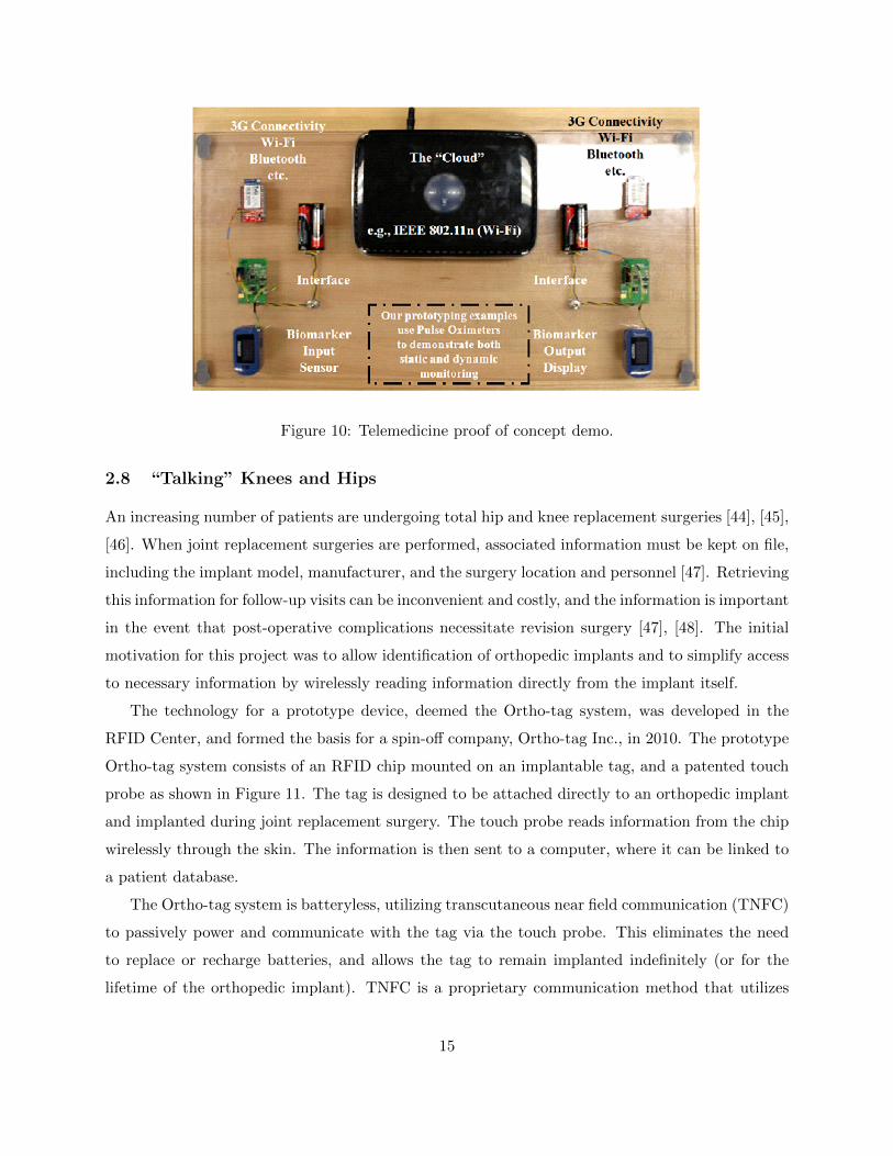
Figure 10: Telemedicine proof of concept demo.
2.8 “Talking” Knees and Hips
An increasing number of patients are undergoing total hip and knee replacement surgeries [44], [45],
[46]. When joint replacement surgeries are performed, associated information must be kept on file,
including the implant model, manufacturer, and the surgery location and personnel [47]. Retrieving
this information for follow-up visits can be inconvenient and costly, and the information is important
in the event that post-operative complications necessitate revision surgery [47], [48]. The initial
motivation for this project was to allow identification of orthopedic implants and to simplify access
to necessary information by wirelessly reading information directly from the implant itself.
The technology for a prototype device, deemed the Ortho-tag system, was developed in the
RFID Center, and formed the basis for a spin-off company, Ortho-tag Inc., in 2010. The prototype
Ortho-tag system consists of an RFID chip mounted on an implantable tag, and a patented touch
probe as shown in Figure 11. The tag is designed to be attached directly to an orthopedic implant
and implanted during joint replacement surgery. The touch probe reads information from the chip
wirelessly through the skin. The information is then sent to a computer, where it can be linked to
a patient database.
The Ortho-tag system is batteryless, utilizing transcutaneous near field communication (TNFC)
to passively power and communicate with the tag via the touch probe. This eliminates the need
to replace or recharge batteries, and allows the tag to remain implanted indefinitely (or for the
lifetime of the orthopedic implant). TNFC is a proprietary communication method that utilizes
15
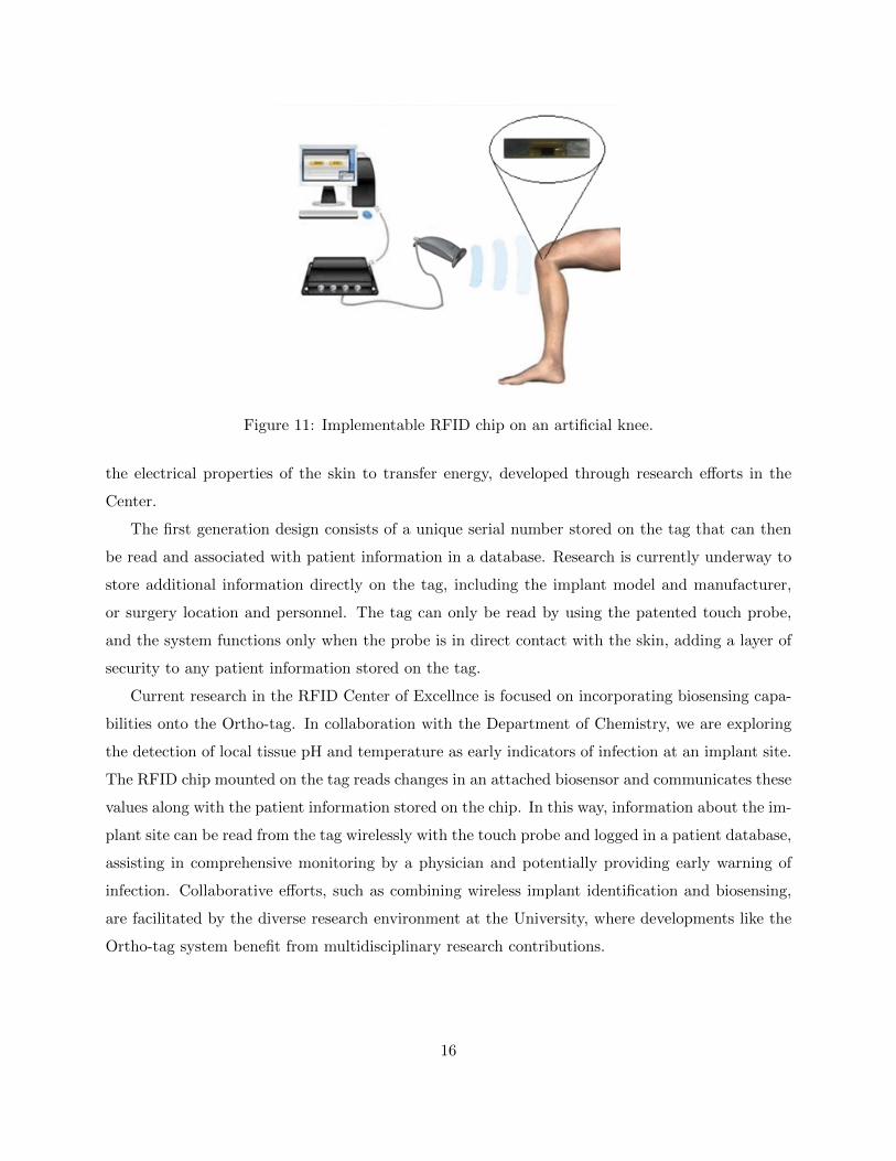
Figure 11: Implementable RFID chip on an artificial knee.
the electrical properties of the skin to transfer energy, developed through research efforts in the
Center.
The first generation design consists of a unique serial number stored on the tag that can then
be read and associated with patient information in a database. Research is currently underway to
store additional information directly on the tag, including the implant model and manufacturer,
or surgery location and personnel. The tag can only be read by using the patented touch probe,
and the system functions only when the probe is in direct contact with the skin, adding a layer of
security to any patient information stored on the tag.
Current research in the RFID Center of Excellnce is focused on incorporating biosensing capa-
bilities onto the Ortho-tag. In collaboration with the Department of Chemistry, we are exploring
the detection of local tissue pH and temperature as early indicators of infection at an implant site.
The RFID chip mounted on the tag reads changes in an attached biosensor and communicates these
values along with the patient information stored on the chip. In this way, information about the im-
plant site can be read from the tag wirelessly with the touch probe and logged in a patient database,
assisting in comprehensive monitoring by a physician and potentially providing early warning of
infection. Collaborative efforts, such as combining wireless implant identification and biosensing,
are facilitated by the diverse research environment at the University, where developments like the
Ortho-tag system benefit from multidisciplinary research contributions.
16

2.9 Doppler Flow Monitor
During free flap surgery, veins and arteries at the transfer site and of the free flap are connected
to facilitate perfusion of blood through the transferred tissue [49]. If the vascular connections fail,
the transferred tissue often dies and additional surgery will be required, with failure most likely
to occur in the first 48 hours after surgery [49], [50]. There are approximately 19,000 free flap
surgeries in United States each year [51]. Flap complication rates requiring reoperation can range
from 3-14% [51], [52].
Current in situ blood flow monitoring systems in free flap surgery require a transcutaneous
tether (i.e., wire) affixed to the vein or artery of a patient. The patient is tethered to a bulky
bedside monitor. These protruding wires limit patient mobility, and they increase the risk of vessel
trauma, false readings, and probe/tether dislodgement. A totally implantable blood flow monitor
can completely solve or significantly ameliorate all of the problems associated with the gold standard
for free flap monitoring.
A proof-of-concept wireless implantable blood flow monitor was developed at the University
of Pittsburgh as shown in Figure 12. The devices successfully detected occlusion in the femoral
arteries of swine, and the devices remained implanted for 3 days. The implanted devices’ clinical
metric is to detect the presence of blood flow; therefore, the accuracy demands are considerably
relaxed compared to commercial units. Even though the total implanted volume is larger in the case
of the wireless implant device than the tethered option (e.g., bedside monitor), clinicians deemed
this larger wireless implant volume an acceptable tradeoff for the proof-of-concept device.
The implantable flow monitor exploits the Doppler shift experienced by a traveling wave as it
passes from a transmitter transducer to a receiver transducer through a flowing blood stream within
a vessel. The implant’s design is a continuous-wave configuration, and the piezoelectric transducers
operate at 5MHz with a single element for the transmitter and a single element for the receiver.
Frequency translation to analog baseband is performed in a homodyne receiver configuration, which
precludes the need for precise frequency calibration when the transmitter and receiver are clocked
from the same time reference. The diameter of the electronics is currently about the size of a U.S.
50 Cent piece.
The blood velocity information is carried in analog baseband, which is wirelessly transmitted by
a radio transmitter onboard the implant. The transmitter operates in the 915MHz ISM band, and
the analog baseband directly modulates the carrier frequency. In this embodiment, the implantable
device is contained entirely within the test animal’s body; no wires protrude through the test
17

Figure 12: A prototype of a wireless Doppler device.
animal’s skin. Transmitted blood velocity information is wirelessly received by a remote receiver
located external to the test animal. The receiver converts the wireless information back to analog
baseband. Afterwards, the receiver amplifies the baseband information sufficiently to drive a speaker
for clinical blood flow assessment. Audible blood velocity information is a clinically familiar format.
The implantable wireless Doppler device represents another innovation that was possible only
through The University of Pittsburgh’s unique partnership with the University of Pittsburgh Med-
ical Center (UPMC). Along the same line as the RFID Center’s exploration into miniature pulse
oximeters, the wireless Doppler device represents another example of enabling existing sensors to
be conveniently implantable.
3 Our Translational Efforts
The origins of our translational efforts began with the desire to better acquaint students with
real engineering problems while also giving them the opportunity to see their solutions be used
by industry and, as a bonus, take part in the surrounding media coverage. The starting point
was an Engineering course in the Swanson School of Engineering at the University of Pittsburgh
where corporate sponsored projects became the student projects to be developed in terms of both
technology and business. A center was created initially by Professor Michael Lovell with funding
18
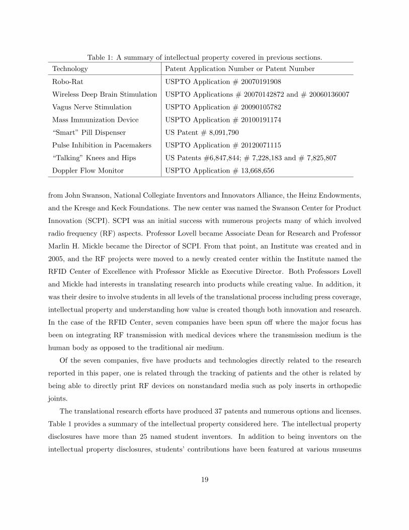
Table 1: A summary of intellectual property covered in previous sections.
Technology Patent Application Number or Patent Number
Robo-Rat USPTO Application # 20070191908
Wireless Deep Brain Stimulation USPTO Applications # 20070142872 and # 20060136007
Vagus Nerve Stimulation USPTO Application # 20090105782
Mass Immunization Device USPTO Application # 20100191174
“Smart” Pill Dispenser US Patent # 8,091,790
Pulse Inhibition in Pacemakers USPTO Application # 20120071115
“Talking” Knees and Hips US Patents #6,847,844; # 7,228,183 and # 7,825,807
Doppler Flow Monitor USPTO Application # 13,668,656
from John Swanson, National Collegiate Inventors and Innovators Alliance, the Heinz Endowments,
and the Kresge and Keck Foundations. The new center was named the Swanson Center for Product
Innovation (SCPI). SCPI was an initial success with numerous projects many of which involved
radio frequency (RF) aspects. Professor Lovell became Associate Dean for Research and Professor
Marlin H. Mickle became the Director of SCPI. From that point, an Institute was created and in
2005, and the RF projects were moved to a newly created center within the Institute named the
RFID Center of Excellence with Professor Mickle as Executive Director. Both Professors Lovell
and Mickle had interests in translating research into products while creating value. In addition, it
was their desire to involve students in all levels of the translational process including press coverage,
intellectual property and understanding how value is created though both innovation and research.
In the case of the RFID Center, seven companies have been spun off where the major focus has
been on integrating RF transmission with medical devices where the transmission medium is the
human body as opposed to the traditional air medium.
Of the seven companies, five have products and technologies directly related to the research
reported in this paper, one is related through the tracking of patients and the other is related by
being able to directly print RF devices on nonstandard media such as poly inserts in orthopedic
joints.
The translational research efforts have produced 37 patents and numerous options and licenses.
Table 1 provides a summary of the intellectual property considered here. The intellectual property
disclosures have more than 25 named student inventors. In addition to being inventors on the
intellectual property disclosures, students’ contributions have been featured at various museums
19

such as the Smithsonian Institution, the Boston Museum of Science and the Reuben H. Fleet
Science Center in San Diego. For example, the exhibition in San Diego was a part of the March
Madness for the Mind sponsored by the National Collegiate Inventors and Innovators Alliance and
the project presented was one of fourteen selected in the country for presentation. Furthermore,
over the years, there have been dozens of projects which have supported the core technologies of
these developments. These groups were typically composed of 2 to 4 students. In addition to
the citations, at least four students supported the University at functions in Chicago, Harrisburg
(PA), and Pittsburgh. These functions were for both alumni and legislators. Also, two students
now serve as Chief Technology Officer and Chief Scientific Officer for spin off companies licensing
technologies developed through the translational efforts. Last, but not least, there have been 103
joint publications with our trainees since the RFID Center of Excellence started with 107 students
named on the papers. The students also attended about a dozen of conferences.
4 Epilogue
In this paper, we overviewed innovation and translation efforts at the RFID Center of Excellence
at the University of Pittsburgh. In our experiences, the key ingredient for successful innovation
and translation is to set up an environment where translational efforts such as patents and start-
ups are encouraged as long as they are accompanied by traditional academic achievement such as
research manuscripts and training of highly qualified personnel. The RFID Center of Excellence
continues to be at the heart of innovation at Pitt, and an integral bridge between academics and
our always-expanding base of industrial and medical collaborators.
The next big challenge that our Center is already considering deals with the idea of Internet
of Things (IoT) and its effects on the health care system and patient care. We anticipate that
IoT will dramatically enhance the patient care as patients will be continuously monitored. This
ability to continuously monitor patients in real-life settings will enable physicians and researchers
to understand how the patient surroundings affect his/her health. This is essentially critical in
cases when a patient performs well during a clinical exam, but has a severe medical event in a
real-life scenario outside a hospital (e.g., patients perform well during a clinical gait assessment,
but fall around the corner of the hospital). Furthermore, IoT enables us to warn a patient that
she/he has missed medication, or that her/his blood glucose levels are dangerously high. From
the health care system point of view, IoT can significantly diminish the cost of the patient care,
as continuous monitoring of patients will enable clinicians to preemptively react to deteriorating
20
patient’s condition. Needless to say, to achieve this state of the art technology, there are a number
of engineering challenges ahead of us involving device miniaturization, signal processing, database
management, just to name a few.
References
[1] D. K. Jacob, S. T. Stefko, S. A. Hackworth, M. R. Lovell, and M. H. Mickle, “Communication
between functional and denervated muscles using radio requency,” Otolaryngology - Head and
Neck Surgery, vol. 134, no. 5, pp. 862–867, May 2006.
[2] E. Peitersen, “Bells palsy: The spontaneous course of 2,500 peripheral facial nerve palsies of
different etiologies,” Acta Oto-laryngologica, vol. 549, pp. 4–30, 2002.
[3] S. M. Gilbard and C. P. Daspit, “Reanimation of the paretic eyelid using gold weight implan-
tation: A new approach and prospective evaluation,” Ophthalmic Plastic and Reconstructive
Surgery, vol. 7, no. 2, pp. 93–103, Jun. 1991.
[4] B. J. Gantz, J. T. Rubinstein, P. Gidley, and G. G. Woodworth, “Surgical management of
Bell’s palsy,” Laryngoscope, vol. 109, no. 8, pp. 1177–1188, Aug. 1999.
[5] K. M. Abell, R. S. Baker, D. E. Cowen, and J. D. Porter, “Efficacy of gold weight implants in
facial nerve palsy: quantitative alterations in blinking,” Vision Research, vol. 38, no. 19, pp.
3019–3023, Oct. 1998.
[6] N. A. Syed, A. Delgado, F. Sandbrink, A. E. Schulman, M. Hallett, and M. K. Floeter, “Blink
reflex recovery in facial weakness: An electrophysiologic study of adaptive changes,” Neurology,
vol. 52, no. 4, p. 834, Mar. 1999.
[7] G. W. Cronin and R. L. Steenerson, “The effectiveness of neuromuscular facial retraining
combined with electromyography in facial paralysis rehabilitation,” Otolaryngology - Head
and Neck Surgery, vol. 128, no. 4, pp. 534–538, Apr. 2003.
[8] A. E. Lang and A. M. Lozano, “Parkinson’s disease,” New England Journal of Medicine, vol.
339, no. 15, pp. 1044–1053, Oct. 1998.
[9] D. Aarsland, J. Zaccai, and C. Brayne, “A systematic review of prevalence studies of dementia
in Parkinson’s disease,” Movement Disorders, vol. 20, no. 10, pp. 1255–1263, Oct. 2005.
21

[10] J. S. A. M. Reijnders, U. Ehrt, W. E. J. Weber, D. Aarsland, and A. F. G. Leentjens, “A
systematic review of prevalence studies of depression in Parkinson’s disease,” Movement Dis-
orders, vol. 23, no. 2, pp. 183–189, Jan. 2008.
[11] C. G. Goetz, W. Poewe, O. Rascol, and C. Sampaio, “Evidence-based medical review update:
Pharmacological and surgical treatments of parkinson’s disease: 2001 to 2004,” Movement
Disorders, vol. 20, no. 5, pp. 523–539, May 2005.
[12] A. Berney, F. Vingerhoets, A. Perrin, P. Guex, J.-G. Villemure, P. R. Burkhard, C. Benkelfat,
and J. Ghika, “Effect on mood of subthalamic DBS for Parkinsons disease: A consecutive
series of 24 patients,” Neurology, vol. 59, no. 9, pp. 1427–1429, Nov. 2002.
[13] P. Limousin, P. Krack, P. Pollak, A. Benazzouz, C. Ardouin, D. Hoffmann, and A.-L. Ben-
abid, “Electrical stimulation of the subthalamic nucleus in advanced Parkinson’s disease,” New
England Journal of Medicine, vol. 339, no. 16, pp. 1105–1111, Oct. 1998.
[14] R. Kumar, A. M. Lozano, Y. J. Kim, W. D. Hutchison, E. Sime, E. Halket, and A. E. Lang,
“Double-blind evaluation of subthalamic nucleus deep brain stimulation in advanced Parkin-
son’s disease,” Neurology, vol. 51, no. 3, pp. 850–855, Sep. 1998.
[15] A. Fasano, P. Mazzone, C. Piano, D. Quaranta, F. Soleti, and A. R. Bentivoglio, “GPi-DBS
in Huntington’s disease: Results on motor function and cognition in a 72-year-old case,”
Movement Disorders, vol. 23, no. 9, pp. 1289–1292, Jul. 2008.
[16] J. L. Ostrem and P. A. Starr, “Treatment of dystonia with deep brain stimulation,” Neurother-
apeutics, vol. 5, no. 2, pp. 320–330, Apr. 2008.
[17] P. Boon, K. Vonck, V. De Herdt, A. Van Dycke, M. Goethals, L. Goossens, M. Van Zandijcke,
T. De Smedt, I. Dewaele, R. Achten, W. Wadman, F. Dewaele, J. Caemaert, and D. Van Roost,
“Deep brain stimulation in patients with refractory temporal lobe epilepsy,” Epilepsia, vol. 48,
no. 8, pp. 1551–1560, Aug. 2007.
[18] F. Liu, S. Heston, L. M. Shollenberger, B. Sun, M. H. Mickle, M. Lovell, and L. Huang,
“Mechanism of in vivo DNA transport into cells by electroporation: electrophoresis across the
plasma membrane may not be involved,” The Journal of Gene Medicine, vol. 8, no. 3, pp.
353–361, Mar. 2006.
22

[19] D. J. Wells, “Gene therapy progress and prospects: Electroporation and other physical meth-
ods,” Gene Therapy, vol. 11, no. 18, pp. 1363–1369, Sep. 2004.
[20] E. Fattori, N. La Monica, G. Ciliberto, and C. Toniatti, “Electro-gene-transfer: A new ap-
proach for muscle gene delivery,” Somatic Cell and Molecular Genetics, vol. 27, no. 1/6, pp.
75–83, Nov. 2002.
[21] E. Neumann, S. Kakorin, and K. Toensing, “Fundamentals of electroporative delivery of drugs
and genes,” Bioelectrochemistry and Bioenergetics, vol. 48, no. 1, pp. 3–16, Feb. 1999.
[22] F. Andre and L. M. Mir, “DNA electrotransfer: its principles and an updated review of its
therapeutic applications,” Gene Therapy, vol. 11, pp. S33–S42, Oct. 2004.
[23] M. Dona, M. Sandri, K. Rossini, I. Dell’Aica, M. Podhorska-Okolow, and U. Carraro, “Func-
tional in vivo gene transfer into the myofibers of adult skeletal muscle,” Biochemical and
Biophysical Research Communications, vol. 312, no. 4, pp. 1132–1138, Dec. 2003.
[24] D. A. Dean, D. Machado-Aranda, K. Blair-Parks, A. V. Yeldandi, and J. L. Young, “Electro-
poration as a method for high-level nonviral gene transfer to the lung,” Gene Therapy, vol. 10,
no. 18, pp. 1608–1616, Sep. 2003.
[25] D. E. Morisky, L. W. Green, and D. M. Levine, “Concurrent and predictive validity of a
self-reported measure of medication adherence,” Medical Care, vol. 24, no. 1, pp. 67–74, Jan.
1986.
[26] K. C. Farmer, “Medication adherence in health care: Are we utilizing what we have learned?”
Clinical Therapeutics, vol. 33, no. 8, pp. 1081–1083, Aug. 2011.
[27] D. M. Cutler and W. Everett, “Thinking outside the pillbox - medication adherence as a
priority for health care reform,” New England Journal of Medicine, vol. 362, no. 17, pp. 1553–
1555, Apr. 2010.
[28] P. M. Ho, C. L. Bryson, and J. S. Rumsfeld, “Medication adherence: Its importance in car-
diovascular outcomes,” Circulation, vol. 119, no. 23, pp. 3028–3035, Jun. 2009.
[29] S. Baroletti and H. Dell’Orfano, “Medication adherence in cardiovascular disease,” Circulation,
vol. 121, no. 12, pp. 1455–1458, Mar. 2010.
23

[30] J.-Y. Kim, H.-J. Lee, N.-S. Byeon, H.-C. Kim, K.-S. Ha, and C.-Y. Chung, “Development
and impact of radio-frequency identification-based workflow management in health promotion
center: Using interrupted time-series analysis,” IEEE Transactions on Information Technology
in Biomedicine, vol. 14, no. 4, pp. 935–940, Jul. 2010.
[31] H. Ishihata, T. Tomoe, K. Takei, T. Hirano, K. Yoshida, S. Shoji, H. Shimauchi, and H. Hori-
uchi, “A radio frequency identification implanted in a tooth can communicate with the outside
world,” IEEE Transactions on Information Technology in Biomedicine, vol. 11, no. 6, pp.
683–685, Nov. 2007.
[32] C.-C. Lin, P.-Y. Lin, P.-K. Lu, G.-Y. Hsieh, W.-L. Lee, and R.-G. Lee, “A healthcare in-
tegration system for disease assessment and safety monitoring of dementia patients,” IEEE
Transactions on Information Technology in Biomedicine, vol. 12, no. 5, pp. 579–586, Sep.
2008.
[33] S.-Y. Lee, L.-H. Wang, and Q. Fang, “A low-power RFID integrated circuits for intelligent
healthcare systems,” IEEE Transactions on Information Technology in Biomedicine, vol. 14,
no. 6, pp. 1387–1396, Nov. 2010.
[34] C.-C. Lin, M.-J. Chiu, C.-C. Hsiao, R.-G. Lee, and Y.-S. Tsai, “Wireless health care ser-
vice system for elderly with dementia,” IEEE Transactions on Information Technology in
Biomedicine, vol. 10, no. 4, pp. 696–704, Oct. Oct.
[35] W. Yao, C.-H. Chu, and Z. Li, “The use of RFID in healthcare: Benefits and barriers,”
in 2010 IEEE International Conference on RFID-Technology and Applications (RFID-TA),
Guangzhou, China, Jun., 17–19, 2010, pp. 128–134.
[36] J. Sutherland and W.-J. van den Heuvel, “Towards an intelligent hospital environment: Adap-
tive workflow in the or of the future,” in Proceedings of the 39th Annual Hawaii International
Conference on System Sciences (HICSS ’06), vol. 5, Kauai, HI, USA, Jan., 4–7, 2006, pp.
100b–1–10.
[37] A. Ogirala, M. A. Rothfuss, J. Stachel, R. Yalamanchili, M. H. Mickle, and S. Saba, “Analysis
of electromagnetic interference to cardiac implantable electronic devices from RFID using tissue
interface circuit,” in Proc. ACES Conference on Applied Computational Electromagnetics,
Williamsburgh, Virginia, USA, Mar., 27–31, 2011.
24

[38] A. Ogirala, J. R. Stachel, and M. H. Mickle, “Electromagnetic interference of cardiac rhyth-
mic monitoring devices to radio frequency identification: Analytical analysis and mitigation
methodology,” IEEE Transactions on Information Technology in Biomedicine, vol. 15, no. 6,
pp. 848–853, Nov. 2011.
[39] J. A. Kline, J. Hernandez-Nino, C. D. Newgard, D. N. Cowles, R. E. Jackson, and D. Courtney,
“Use of pulse oximetry to predict in-hospital complications in normotensive patients with
pulmonary embolism,” The American Journal of Medicine, vol. 115, no. 3, pp. 203–208, Aug.
2003.
[40] R. I. Koppel, C. M. Druschel, T. Carter, B. E. Goldberg, P. N. Mehta, R. Talwar, and F. Z.
Bierman, “Effectiveness of pulse oximetry screening for congenital heart disease in asymp-
tomatic newborns,” Pediatrics, vol. 111, no. 3, pp. 451–455, Mar. 2003.
[41] M. R. Pinsky, L. Brochard, and J. Mancebo, Applied physiology in intensive care medicine.
Berlin: Springer, 2006.
[42] J. W. Severinghaus and J. Kelleher, “Recent developments in pulse oximetry,” Anesthesiology,
vol. 76, no. 6, pp. 1018–1038, Jun. 1992.
[43] K. K. Tremper, “Pulse oximetry,” Chest, vol. 95, no. 4, pp. 713–715, Apr. 1989.
[44] M. Kirksey, Y. Lin Chiu, Y. Ma, A. Gonzalez Della Valle, L. Poultsides, P. Gerner, and
S. G. Memtsoudis, “Trends in in-hospital major morbidity and mortality after total joint
arthroplasty: United States 1998-2008,” Anesthesia and Analgesia, vol. 115, no. 2, pp. 321–
327, Aug. 2012.
[45] A. Jain, B. E. Stein, R. L. Skolasky, L. C. Jones, and M. W. Hungerford, “Total joint arthro-
plasty in patients with rheumatoid arthritis: A United States experience from 1992 through
2005,” The Journal of Arthroplasty, vol. 27, no. 6, pp. 881–888, Jun. 2012.
[46] S. Kurtz, K. Ong, E. Lau, F. Mowat, and M. Halpern, “Projections of primary and revision
hip and knee arthroplasty in the United States from 2005 to 2030,” The Journal of Bone and
Joint Surgery, vol. 89, no. 4, pp. 780–785, Apr. 2007.
[47] E. W. Paxton, M. C. S. Inacio, M. Khatod, E. J. Yue, and R. S. Namba, “Kaiser permanente
national total joint replacement registry: Aligning operations with information technology,”
Clinical Orthopaedics and Related Research, vol. 468, no. 10, pp. 2646–2663, Oct. 2010.
25

[48] E. R. Bohm, M. J. Dunbar, and R. Bourne, “The Canadian joint replacement registry - what
have we learned?” Acta Orthopaedica, vol. 81, no. 1, pp. 119–121, Feb. 2010.
[49] I. Papel, Ed., Facial Plastic and Reconstructive Surgery. New York, NY, USA: Thieme
Medical Publishers, 2009.
[50] D. Novakovic, R. S. Patel, D. P. Goldstein, and P. J. Gullane, “Salvage of failed free flaps used
in head and neck reconstruction,” Head and Neck Oncology, vol. 1, pp. 33–1–5, Aug. 2009.
[51] K. G. Bowman and M. J. Carty, “Flap complications and thrombophilia: An evidence-based
model and cost analysis for preoperative screening,” ePlasty, vol. 11, pp. 278–289, Jul. 2011.
[52] J. J. Disa, Q. Y. Hu, and D. A. Hidalgo, “Retrospective review of 400 consecutive free flap
reconstructions for oncologic surgical defects,” Annals of Surgical Oncology, vol. 4, no. 4, pp.
663–669, Dec. 1997.
26