Inflammatory Bowel Disease: Clinics, Diagnosis ... · PDF fileInflammatory Bowel Disease:...
52
Inflammatory Bowel Disease: Clinics, Diagnosis, Diferential Diagnosis Jorge Amil Dias Porto, PORTUGAL [email protected]
-
Upload
nguyenkhuong -
Category
Documents
-
view
227 -
download
16
Transcript of Inflammatory Bowel Disease: Clinics, Diagnosis ... · PDF fileInflammatory Bowel Disease:...
Inflammatory Bowel Disease: Clinics, Diagnosis, Diferential Diagnosis
Jorge Amil Dias
Porto, PORTUGAL
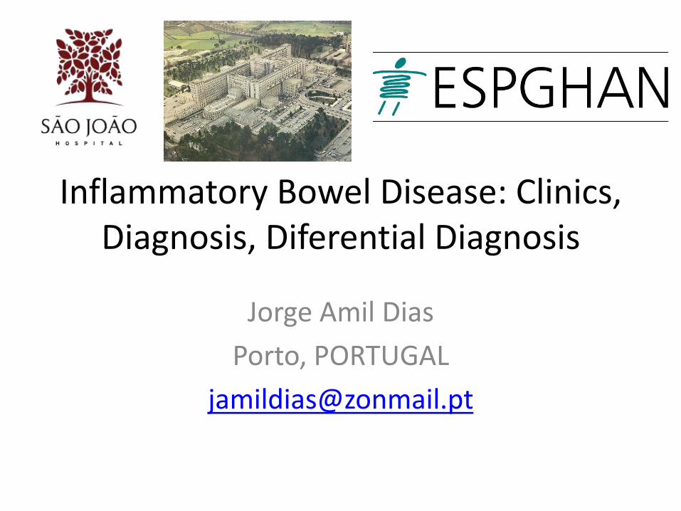
Cosnes J et al, Gastroenterology, 2011
<4/105
5-10/105
Global map of IBD
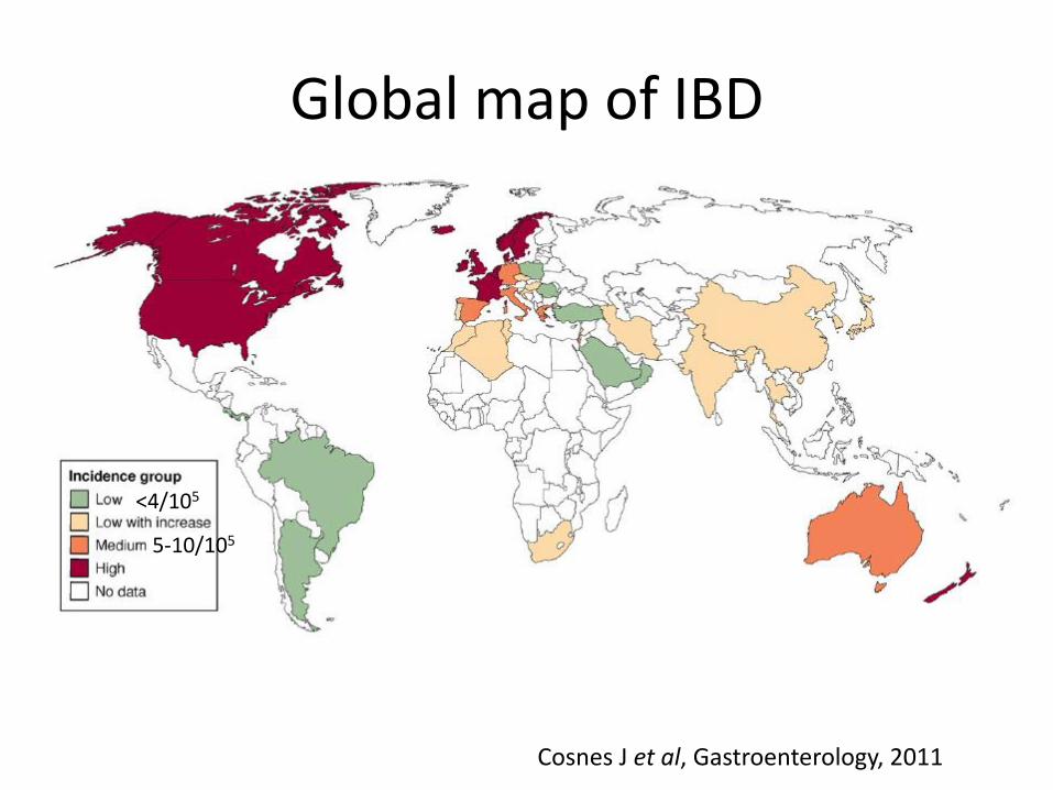
Crohn’s Disease
Ulcerative colitis
Benchimol et al Inflamm Bowel Dis, 2011
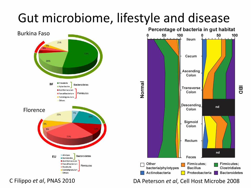
Burkina Faso
Florence
Gut microbiome, lifestyle and disease
C Filippo et al, PNAS 2010 DA Peterson et al, Cell Host Microbe 2008
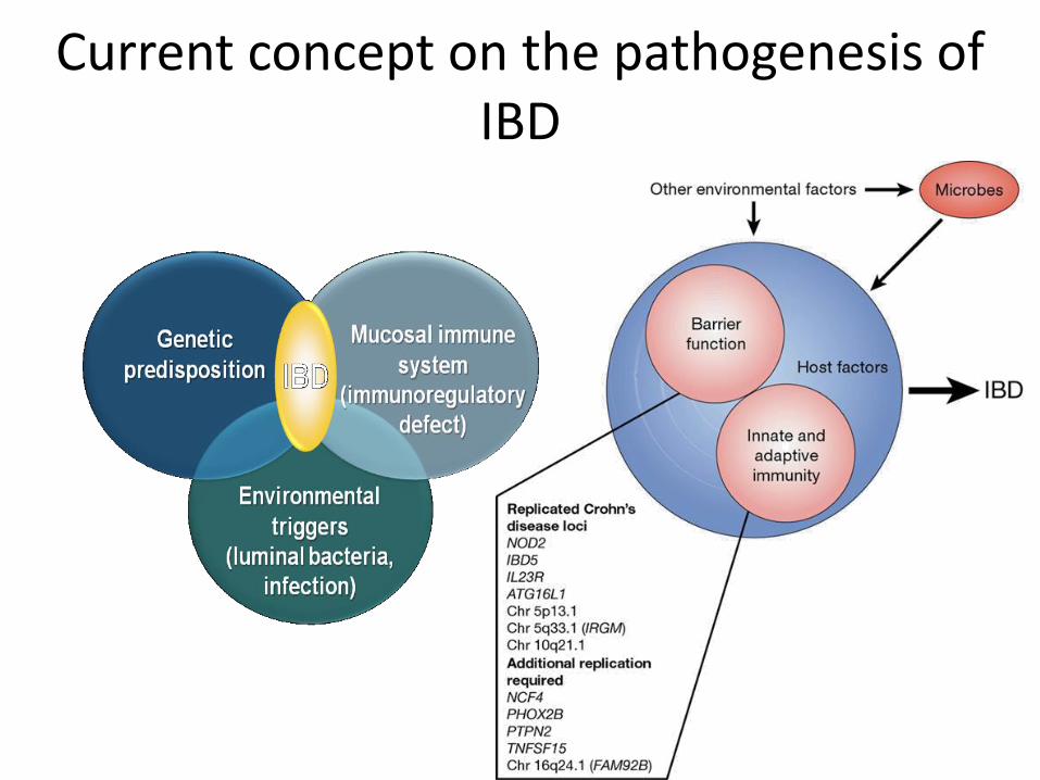
Current concept on the pathogenesis of IBD
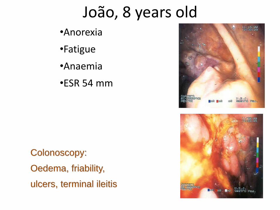
João, 8 years old •Anorexia
•Fatigue
•Anaemia
•ESR 54 mm
Colonoscopy:
Oedema, friability,
ulcers, terminal ileitis
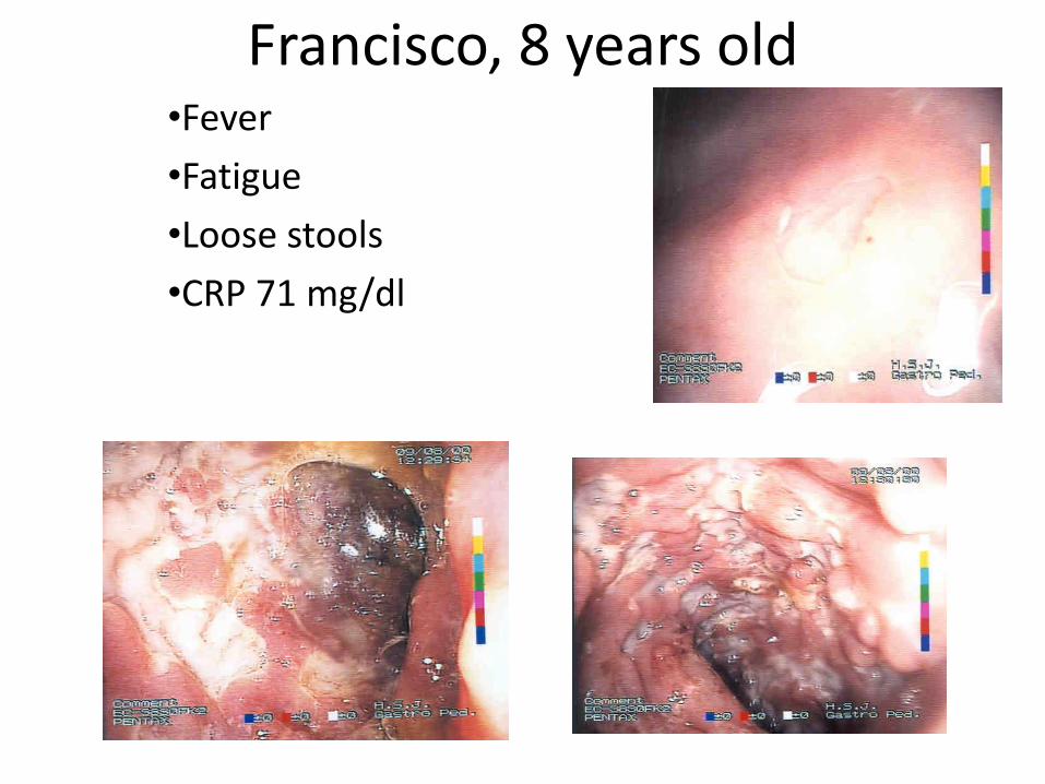
Francisco, 8 years old •Fever
•Fatigue
•Loose stools
•CRP 71 mg/dl
Apendicitis
? Rheumatoid arthritis
? Myeloproliferative syndr
? Neoplasia
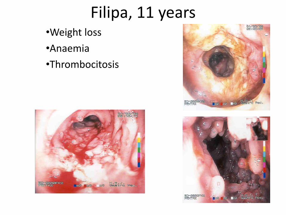
Filipa, 11 years •Weight loss
•Anaemia
•Thrombocitosis
Ricardo, 13 years
• Brought to medical care due to pain in both ankles
• For 8 months has had oral aphtae and occasional abdominal pain.
• Lost weight (2kg).
• Constipated.
• Recurrent anal fissures
• Family history irrelevant (no IBD)
Physical examination
• Weight 31kg (centile: <5); Height: 152cm (centile: 25-50); BMI 13.4kg/m2 (centile: <3)
• Oedema on both ankles.
• Swelling of lower lip with large aphtous ulcers
• Abdomen soft though tender in lower right quadrant.
• Liver and spleen not palpable.

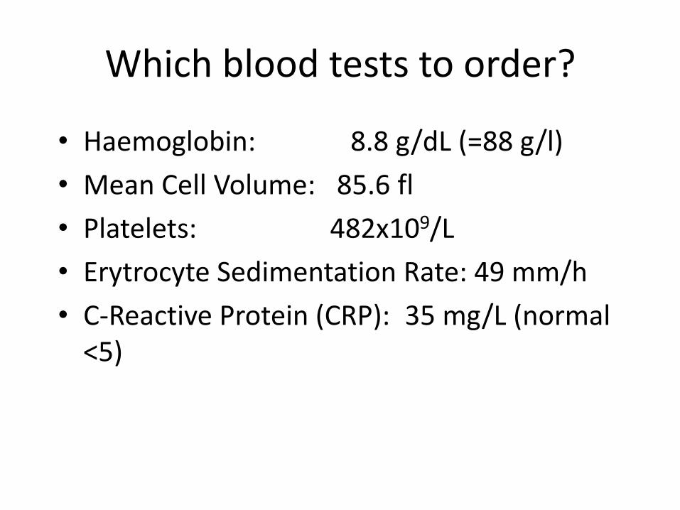
Which blood tests to order?
• Haemoglobin: 8.8 g/dL (=88 g/l)
• Mean Cell Volume: 85.6 fl
• Platelets: 482x109/L
• Erytrocyte Sedimentation Rate: 49 mm/h
• C-Reactive Protein (CRP): 35 mg/L (normal <5)
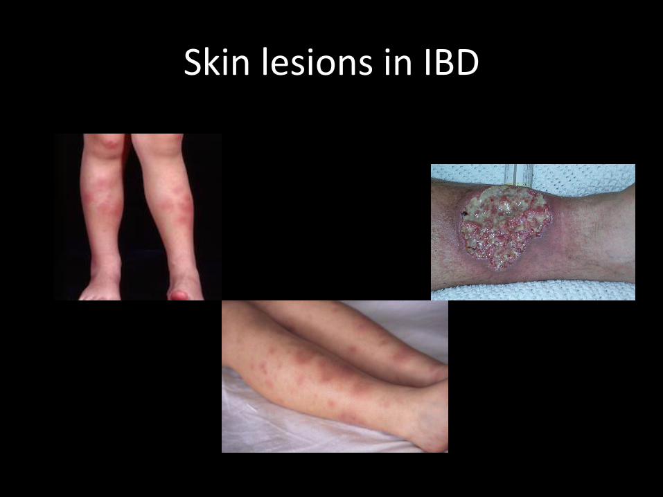
Skin lesions in IBD
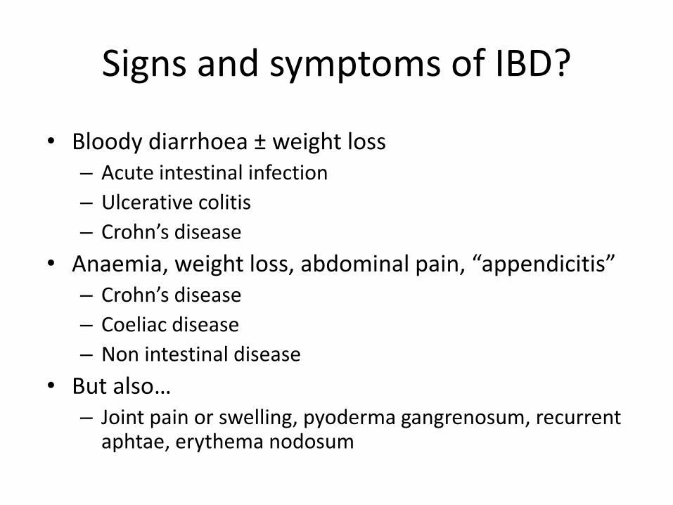
Signs and symptoms of IBD?
• Bloody diarrhoea ± weight loss – Acute intestinal infection
– Ulcerative colitis
– Crohn’s disease
• Anaemia, weight loss, abdominal pain, “appendicitis” – Crohn’s disease
– Coeliac disease
– Non intestinal disease
• But also… – Joint pain or swelling, pyoderma gangrenosum, recurrent
aphtae, erythema nodosum
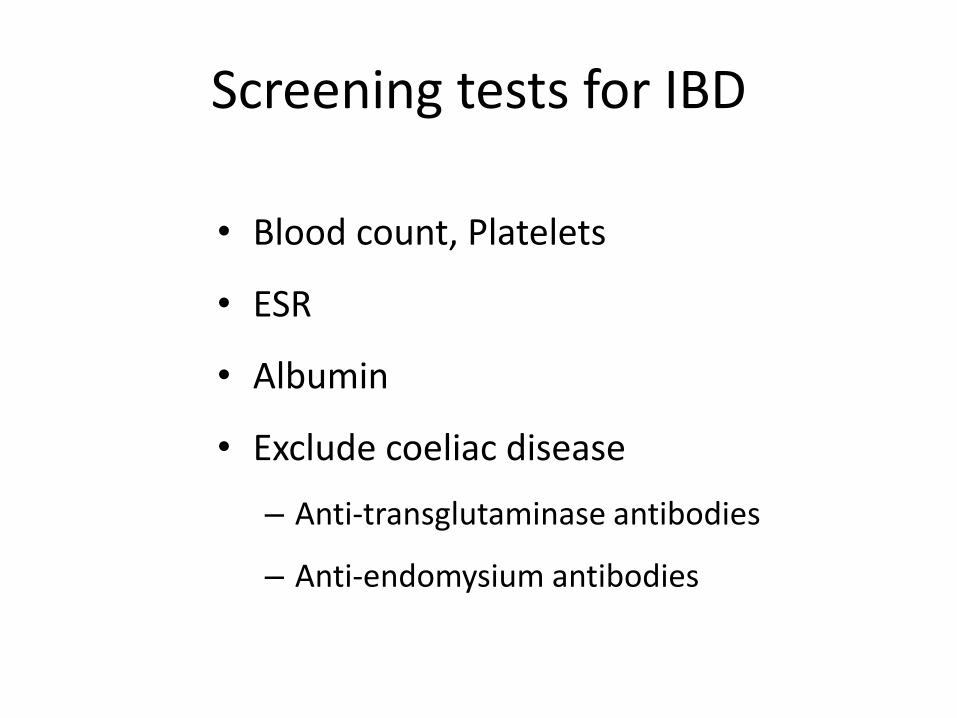
Screening tests for IBD
• Blood count, Platelets
• ESR
• Albumin
• Exclude coeliac disease
– Anti-transglutaminase antibodies
– Anti-endomysium antibodies
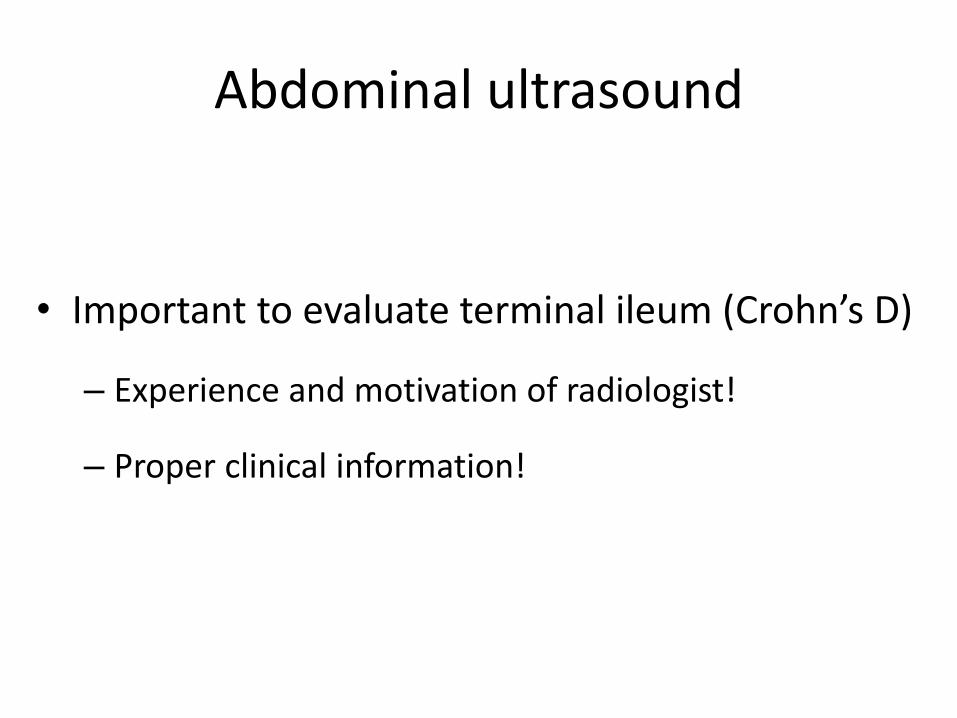
Abdominal ultrasound
• Important to evaluate terminal ileum (Crohn’s D)
– Experience and motivation of radiologist!
– Proper clinical information!
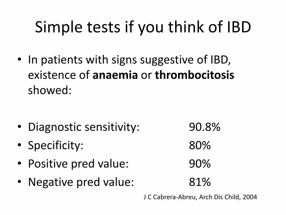
Simple tests if you think of IBD
• In patients with signs suggestive of IBD, existence of anaemia or thrombocitosis showed:
• Diagnostic sensitivity: 90.8%
• Specificity: 80%
• Positive pred value: 90%
• Negative pred value: 81% J C Cabrera-Abreu, Arch Dis Child, 2004
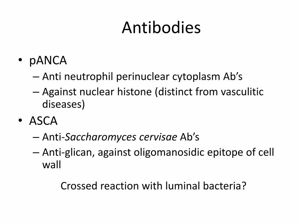
Antibodies
• pANCA – Anti neutrophil perinuclear cytoplasm Ab’s
– Against nuclear histone (distinct from vasculitic diseases)
• ASCA – Anti-Saccharomyces cervisae Ab’s
– Anti-glican, against oligomanosidic epitope of cell wall
Crossed reaction with luminal bacteria?
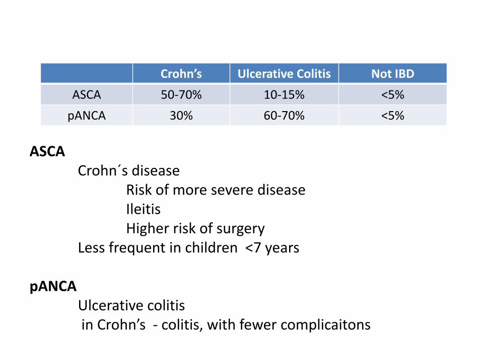
Crohn’s Ulcerative Colitis Not IBD
ASCA 50-70% 10-15% <5%
pANCA 30% 60-70% <5%
ASCA Crohn´s disease
Risk of more severe disease Ileitis Higher risk of surgery Less frequent in children <7 years
pANCA Ulcerative colitis in Crohn’s - colitis, with fewer complicaitons
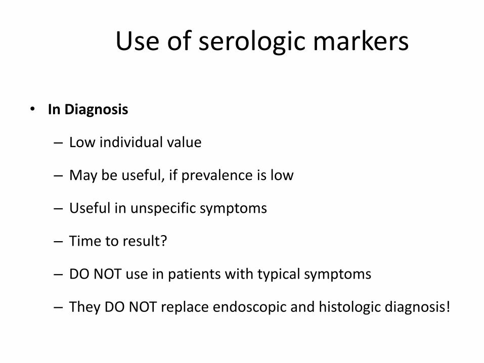
Use of serologic markers
• In Diagnosis
– Low individual value
– May be useful, if prevalence is low
– Useful in unspecific symptoms
– Time to result?
– DO NOT use in patients with typical symptoms
– They DO NOT replace endoscopic and histologic diagnosis!
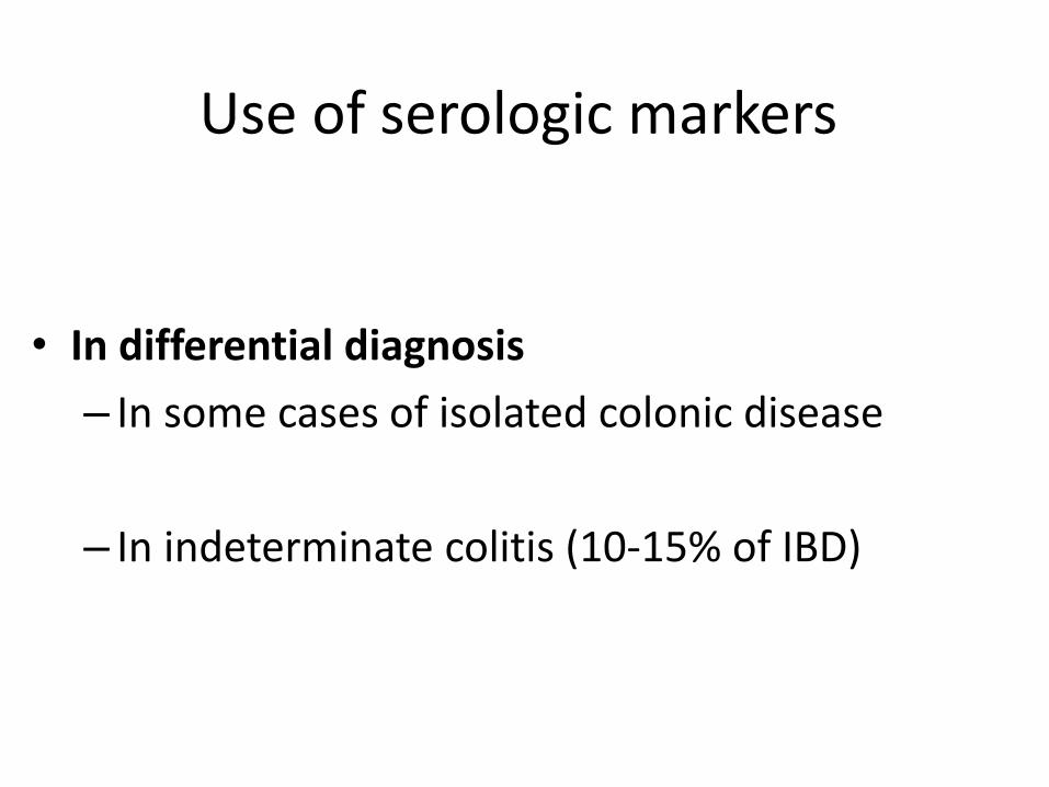
Use of serologic markers
• In differential diagnosis
– In some cases of isolated colonic disease
– In indeterminate colitis (10-15% of IBD)
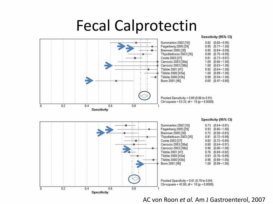
Fecal Calprotectin
AC von Roon et al. Am J Gastroenterol, 2007
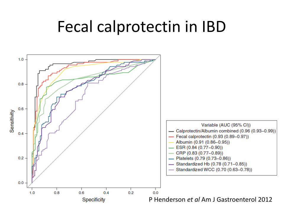
Fecal calprotectin in IBD
P Henderson et al Am J Gastroenterol 2012
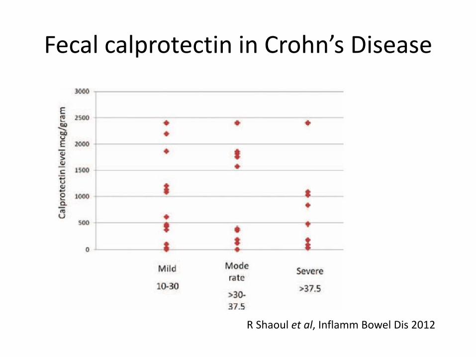
Fecal calprotectin in Crohn’s Disease
R Shaoul et al, Inflamm Bowel Dis 2012
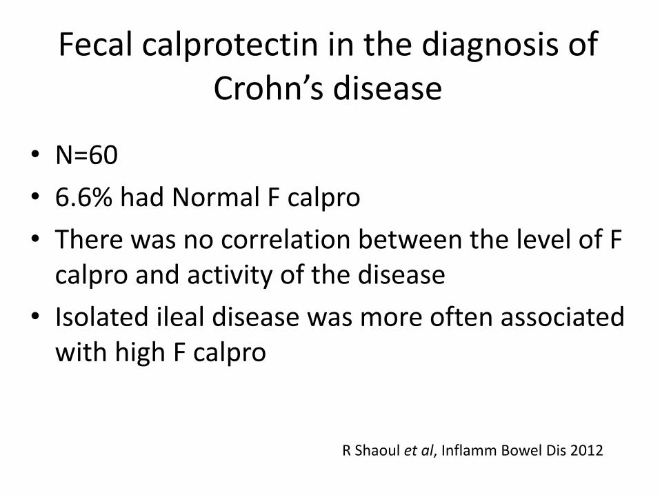
Fecal calprotectin in the diagnosis of Crohn’s disease
• N=60
• 6.6% had Normal F calpro
• There was no correlation between the level of F calpro and activity of the disease
• Isolated ileal disease was more often associated with high F calpro
R Shaoul et al, Inflamm Bowel Dis 2012
Fecal calprotectin in the diagnosis of Crohn’s disease
• N=60
• 6.6% had Normal F calpro
• There was no correlation between the level of F calpro and activity of the disease
• Isolated ileal disease was more often associated with high F calpro
R Shaoul et al, Inflamm Bowel Dis 2012
If you consider IBD, simple tests may help to identify patients for subsenquent endoscopic diagnosis
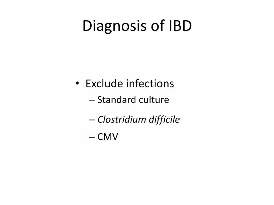
Diagnosis of IBD
• Exclude infections
– Standard culture
– Clostridium difficile
– CMV

Endoscopy
• Direct visualization of the mucosa.
• Possibility to obtain material for histology
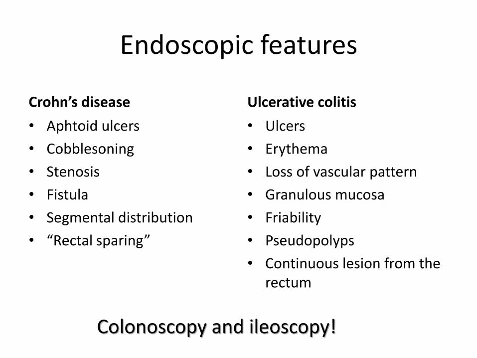
Endoscopic features
Crohn’s disease
• Aphtoid ulcers
• Cobblesoning
• Stenosis
• Fistula
• Segmental distribution
• “Rectal sparing”
Ulcerative colitis
• Ulcers
• Erythema
• Loss of vascular pattern
• Granulous mucosa
• Friability
• Pseudopolyps
• Continuous lesion from the rectum
Colonoscopy and ileoscopy!
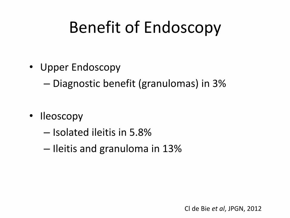
Benefit of Endoscopy
• Upper Endoscopy
– Diagnostic benefit (granulomas) in 3%
• Ileoscopy
– Isolated ileitis in 5.8%
– Ileitis and granuloma in 13%
Cl de Bie et al, JPGN, 2012
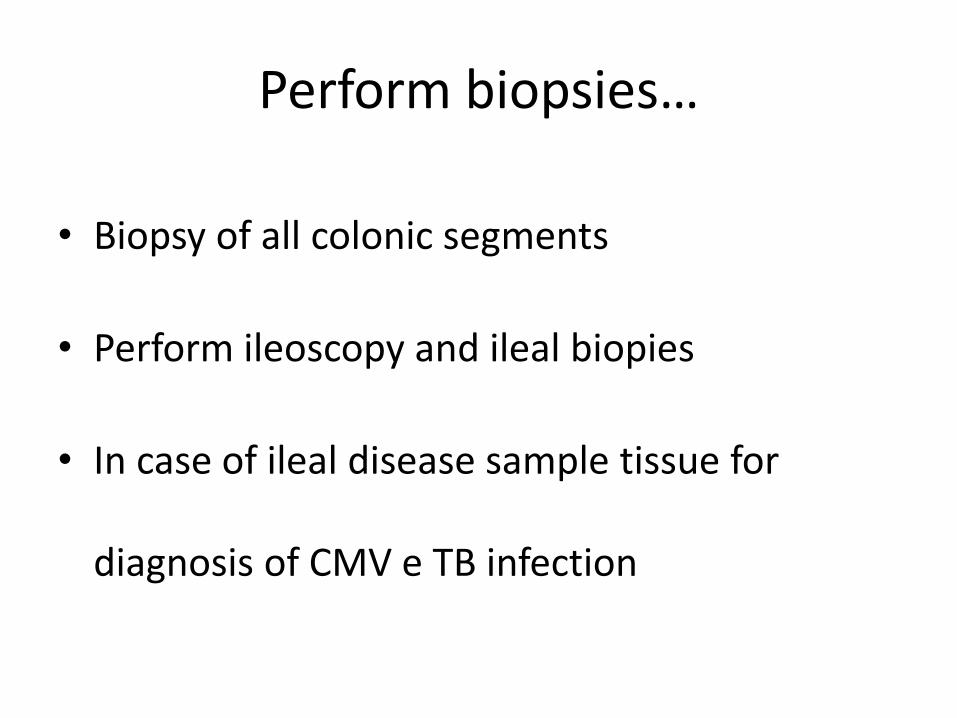
Perform biopsies…
• Biopsy of all colonic segments
• Perform ileoscopy and ileal biopies
• In case of ileal disease sample tissue for
diagnosis of CMV e TB infection
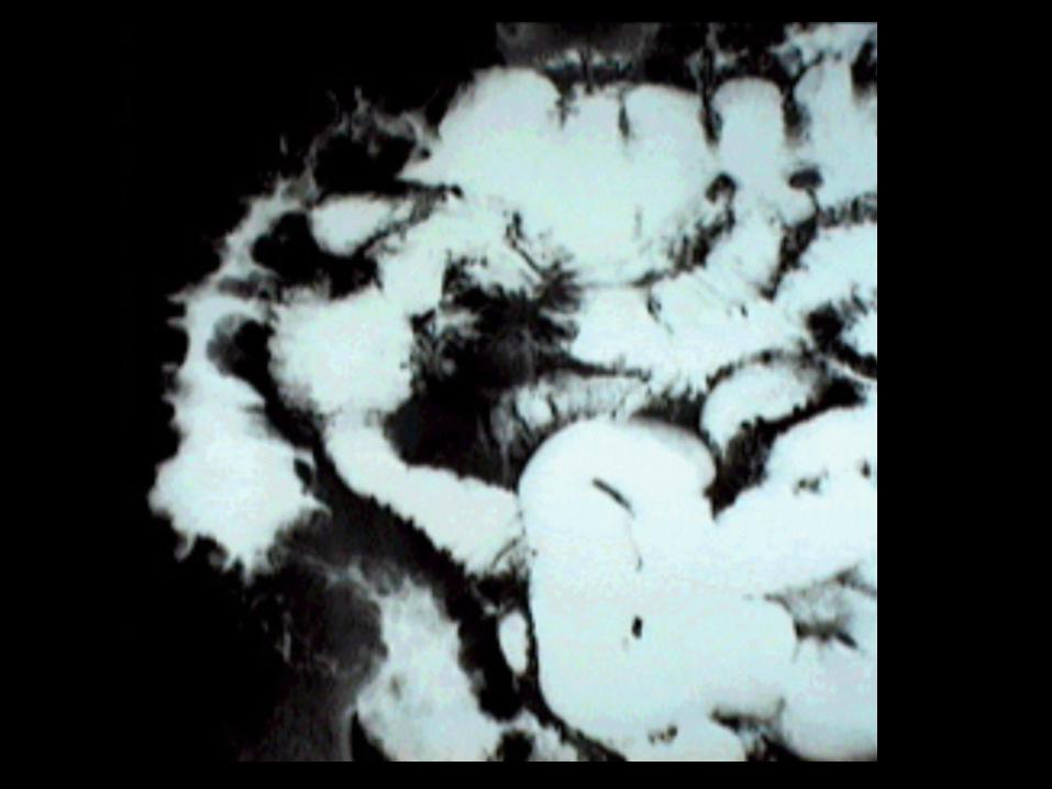
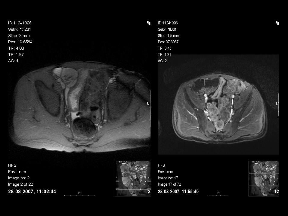
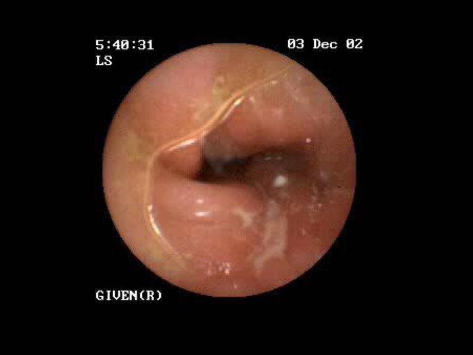
Visualization of small intestine
In Crohn’s and undetermined colitis
• Wireless capsule endoscopy
• Entero-CT
• Entero-MR
• Enteroclisis

Investigation of small Intestine in Eurokids
CI de Bie et al, JPGN, 2012
N=1404

Histologic features
Crohn’s disease
• Submucosal or transmural involvement
• Ulcers, distortion of crypts
• Crypt Abcessos
• Granuloma
• Focal lesions
Ulcerative colitis
• Mucosal involvement
• Distortion of crypts
• Crypt abcesses
• Goblet cell depletion
• Mucin granuloma (rare)
Talk to your pathologist! Provide adequade clinical information and feedback
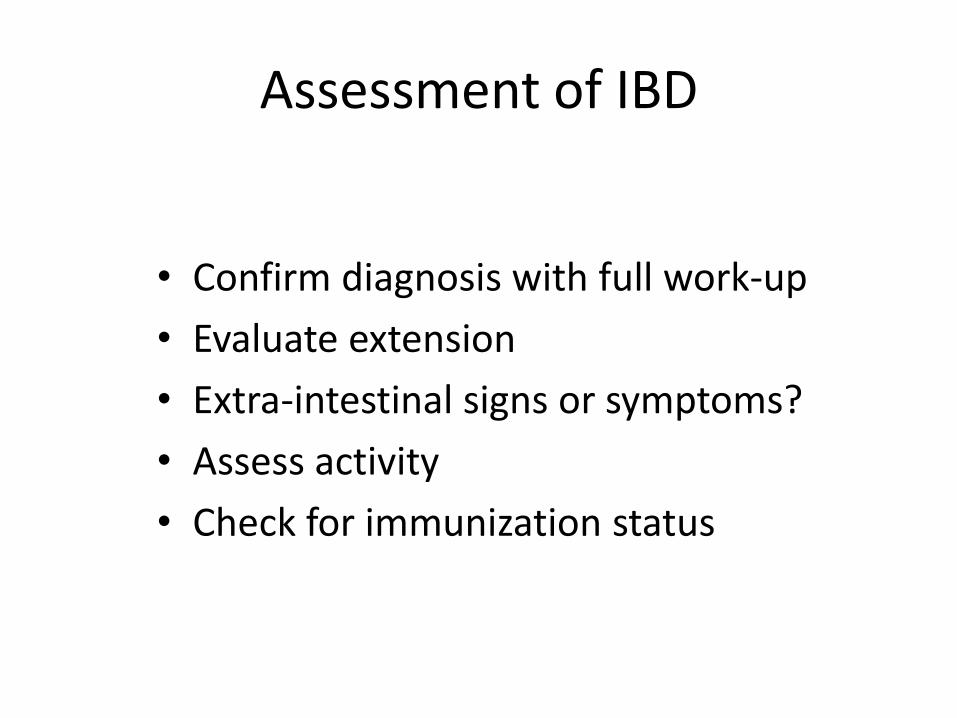
Assessment of IBD
• Confirm diagnosis with full work-up
• Evaluate extension
• Extra-intestinal signs or symptoms?
• Assess activity
• Check for immunization status
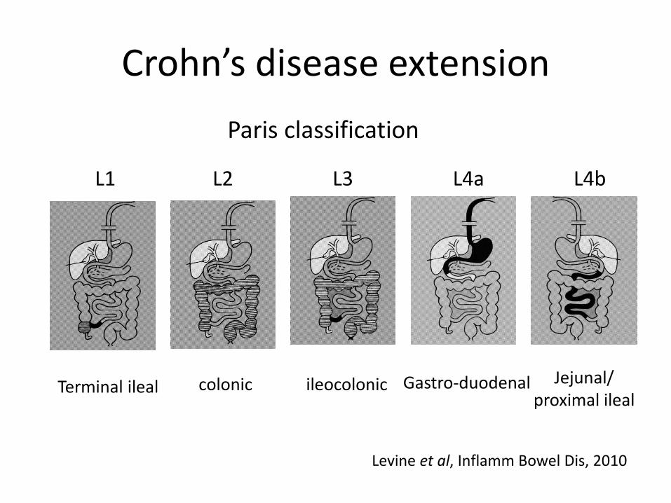
Crohn’s disease extension
Terminal ileal colonic ileocolonic Gastro-duodenal Jejunal/ proximal ileal
L1 L2 L3 L4a L4b
Levine et al, Inflamm Bowel Dis, 2010
Paris classification
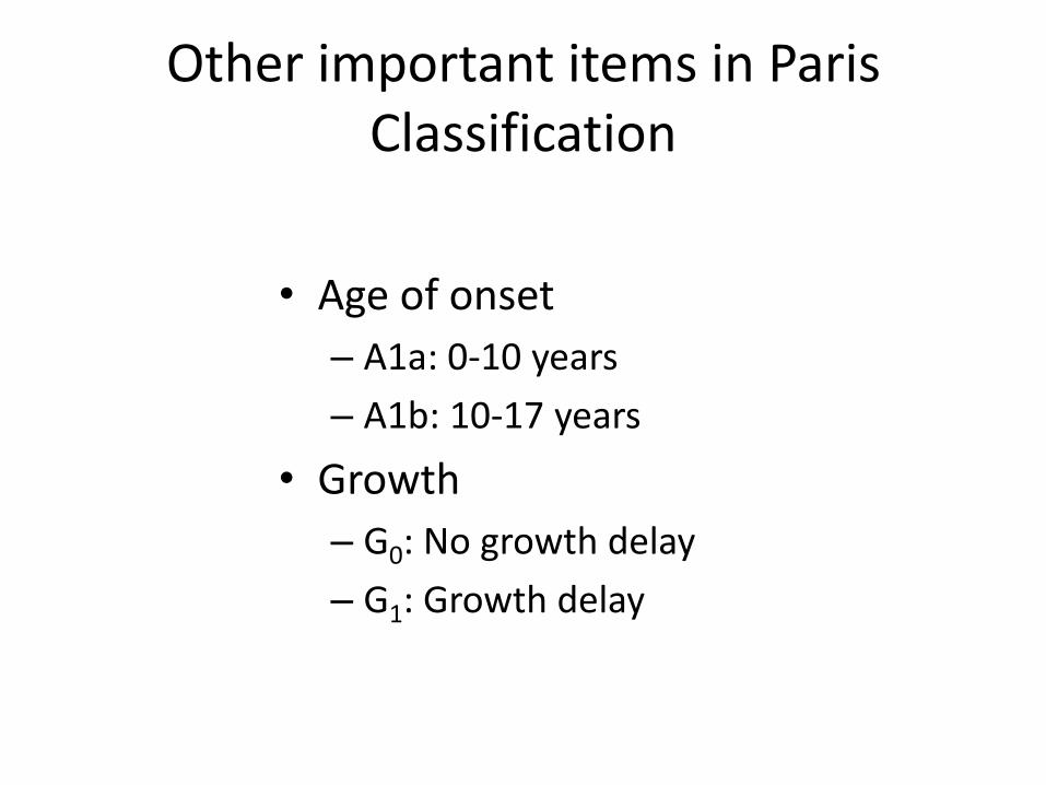
Other important items in Paris Classification
• Age of onset
– A1a: 0-10 years
– A1b: 10-17 years
• Growth
– G0: No growth delay
– G1: Growth delay
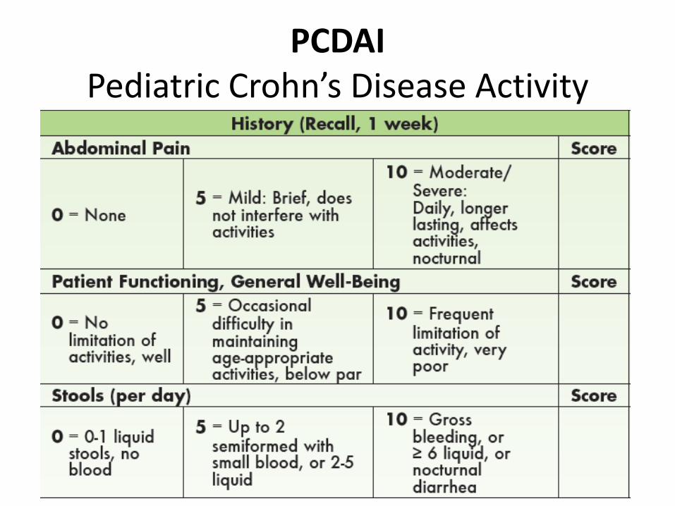
PCDAI Pediatric Crohn’s Disease Activity
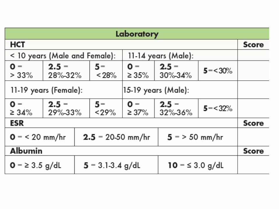
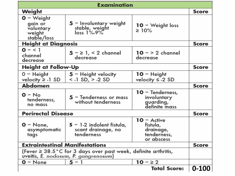
0-100
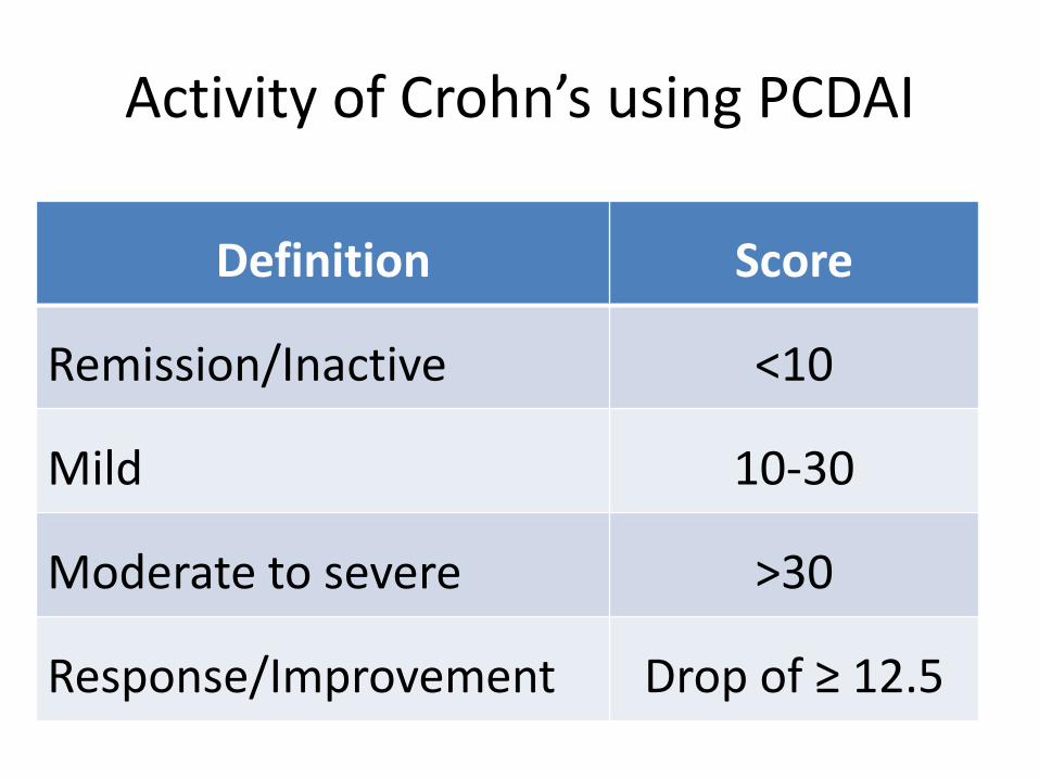
Activity of Crohn’s using PCDAI
Definition Score
Remission/Inactive <10
Mild 10-30
Moderate to severe >30
Response/Improvement Drop of ≥ 12.5
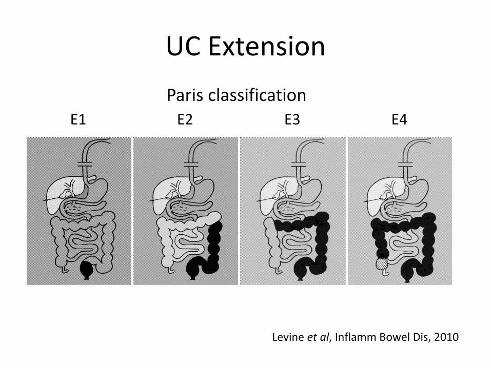
UC Extension
E1 E2 E3 E4
Paris classification
Levine et al, Inflamm Bowel Dis, 2010

Montreal and Paris classifications
A Levine et al. Inflamm Bowel Dis, 2011
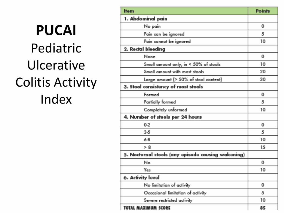
PUCAI Pediatric
Ulcerative Colitis Activity
Index
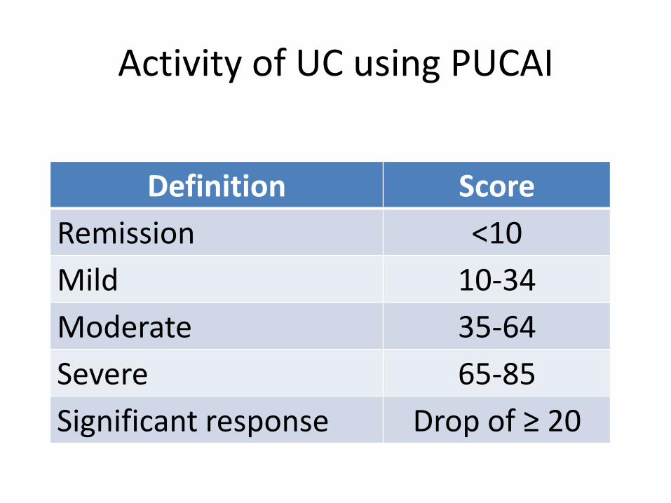
Activity of UC using PUCAI
Definition Score
Remission <10
Mild 10-34
Moderate 35-64
Severe 65-85
Significant response Drop of ≥ 20
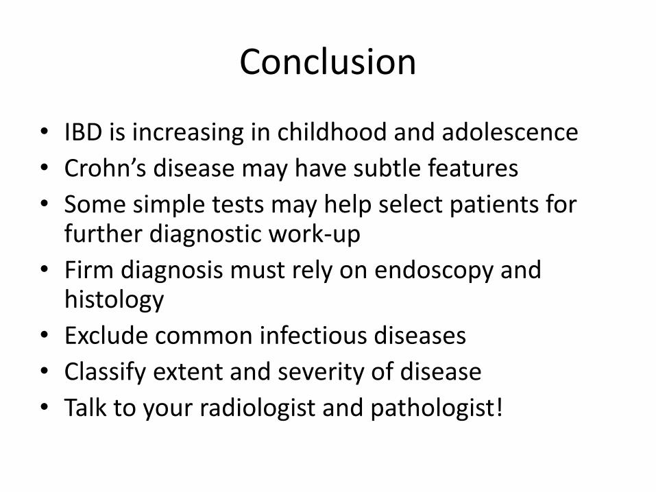
Conclusion
• IBD is increasing in childhood and adolescence
• Crohn’s disease may have subtle features
• Some simple tests may help select patients for further diagnostic work-up
• Firm diagnosis must rely on endoscopy and histology
• Exclude common infectious diseases
• Classify extent and severity of disease
• Talk to your radiologist and pathologist!