Induction of NAD(P)H:quinone reductase in murine hepatoma cells
5
Proc. Natl. Acad. Sci. USA Vol. 83, pp. 787-791, February 1986 Medical Sciences Induction of NAD(P)H:quinone reductase in murine hepatoma cells by phenolic antioxidants, azo dyes, and other chemoprotectors: A model system for the study of anticarcinogens (Sudan dyes/butylated hydroxyanisole/polycycfic aromatic hydrocarbons/cancer protection) MARY J. DE LONG, HANS J. PROCHASKA, AND PAUL TALALAY* Department of Pharmacology and Experimental Therapeutics, The Johns Hopkins University School of Medicine, Baltimore, MD 21205 Communicated by Charles Huggins, September 13, 1985 ABSTRACT Exposure of murine hepatoma (Hepa lclc7) cells to a variety of chemical agents known to protect animals against the neoplastic, mutagenic, and other toxic effects of chemical carcinogens results in dose- and time-dependent inductions of NAD(P)H:quinone reductase (EC 1.6.99.2). This enzyme protects against quinone toxicity by promoting oblig- atory two-electron reductions that divert quinones from oxi- dative cycling or direct interactions with critical nucleophiles. Quinone reductase levels are stable in culture, are easily measured, and are useful markers for the inductive effects of chemoprotective agents. The Hepa lclc7 system responds to chemoprotective compounds such as phenolic antioxidants {e.g., BHA [3(2)-tert-butyl-4-hydroxyanisole], BHT (3,5-di- tert-butyl-4-hydroxytoluene), and tert-butylhydroquinone}, lipophilic azo dyes belonging to the 1,1'-azonaphthalene, Sudan I (1-phenylazo-2-naphthol), and Sudan m [1-(4-phen- ylazophenylazo)-2-naphtholl families, polycyclic aromatic hy- drocarbons, coumarin and various other lactones, flavonoids, and certain sulfur compounds (e.g., benzylisothiocyanate, dithiolthiones, and dithiocarbamates), all of which are recog- nized enzyme inducers and chemoprotectors in vivo. Quinone reductase induction in Hepa lclc7 cells therefore provides a simple, versatile, and reliable system for the evaluation of the potency, kinetics, and mechanism of action of anticarcinogens. An astonishing variety of structurally unrelated compounds, including phenolic antioxidants, coumarins, azo dyes, flavonoids, polycycic aromatics, and certain sulfur com- pounds, protect laboratory animals against the neoplastic, mutagenic, and other toxic effects of chemical carcinogens (1, 2). Clarification of the molecular mechanisms underlying these protective actions is of importance in devising strate- gies for protecting man against cancer. Much evidence indicates that protection by these agents depends on altering the metabolism of carcinogens (3, 4). Some protectors (e.g., polycyclic hydrocarbons, 2,3,7,8-tetrachlorodibenzo-p-di- oxin, and azo dyes) are potent inducers of both phase I and phase II drug-metabolizing enzymest, whereas other protec- tive agents (e.g., phenolic antioxidants) induce phase II enzymes only (for review, see ref. 3). Although the balance between these two types of enzymes required for protective action remains unclear, current evidence indicates that in- duction of such phase II enzymes as glutathione S- transferases, quinone reductase, and epoxide hydrolase is important for protection (1-4, 6-8). We have developed a murine hepatic cell culture system (Hepa lclc7) that mimics many animal tissues in responding to a wide variety of chemoprotective agents by induction of cytosolic quinone reductase [NAD(P)H:(quinone-acceptor) oxidoreductase, EC 1.6.99.2; also known as menadione reductase, DT diaphorase, or vitamin K reductase]. These cells are a suitable model for studying chemoprotection because (i) they retain many characteristics of normal tissues, particularly the capacity for carcinogen activation and me- tabolism, and (ii) they are amenable to precise control of environmental, nutritional, and hormonal factors. Further, mutants defective in enzyme-induction pathways (9, 10) are available. Quinone reductase was selected as a marker for induction because (a) it protects against toxicity (3, 11-16), (b) its levels rise coordinately with other chemoprotective enzymes in many animal tissues in response to the adminis- tration of various anticarcinogens (4, 17, 18), and (c) it is induced by many chemically dissimilar substances that pro- tect against carcinogens (3, 19). An abstract of the work presented here has been published (20). MATERIALS AND METHODS Hepa lclc7 cells (a gift of J. P. Whitlock, Jr., Stanford University) were grown in alpha minimal essential medium minus nucleosides (GIBCO), supplemented with 10% fetal calf serum, at 37°C in an atmosphere of 5% CO2 in a humidified incubator. In our standard induction protocol, Hepa lclc7 cells were plated at 0.89 x 10⁶ cells per 75-cm2 plate, grown for 48 hr to late logarithmic phase, and then exposed (usually for 24 hr) to fresh medium containing the test compounds in dimethyl sulfoxide (0.1%, by volume). Cell layers were washed three times with 10 ml of cold 0.25 M sucrose, collected by scraping, frozen in liquid nitrogen, and stored at -80°C until enzyme activities were assayed. Preparation of cytosols and enzyme assays have been described (17, 18). The specific activities were obtained from spectrophotometric measurements of the reduction of 2,6- dichloroindophenol by NADH. Many azo compounds were generously provided by C. Huggins and J. Pataki of The University of Chicago (21) and purified by recrystallization when appropriate. 1-(4-Hydroxyphenylazo)-2-naphthol was synthesized by mixing 4-aminophenol (10 mmol) in 25 ml of H20 with 10 mmol of NaNO2. This solution was introduced dropwise into 25 ml of 0.4 M HCl, and the diazonium salt was added dropwise to 10 mmol of 2-naphthol in 25 ml of H20, Abbreviations: BHA, 3(2)-tert-butyl-4-hydroxyanisole; BHT, 3,5-di- tert-butyl-4-hydroxytoluene. *To whom reprint requests should be addressed at: Department of Pharmacology, Johns Hopkins School of Medicine, 725 N. Wolfe Street, Baltimore, MD 21205. tEnzymes involved in the metabolism of xenobiotics have been grouped into two broad classes (5): Phase I enzymes (which include the cytochromes P450) functionalize compounds by oxidation, reduction, or hydrolysis, whereas phase II enzymes carry out the conjugations of functionalized compounds with endogenous ligands (e.g., glucuronic acid, glutathione, and sulfate). We consider quinone reductase as a phase II enzyme, since it does not introduce new functional groups and is often induced coordinately with conjugation enzymes. 787 The publication costs of this article were defrayed in part by page charge payment. This article must therefore be hereby marked "advertisement" in accordance with 18 U.S.C. §1734 solely to indicate this fact.
Transcript of Induction of NAD(P)H:quinone reductase in murine hepatoma cells

Proc. Natl. Acad. Sci. USAVol. 83, pp. 787-791, February 1986Medical Sciences
Induction of NAD(P)H:quinone reductase in murine hepatoma cellsby phenolic antioxidants, azo dyes, and other chemoprotectors: Amodel system for the study of anticarcinogens
(Sudan dyes/butylated hydroxyanisole/polycycfic aromatic hydrocarbons/cancer protection)
MARY J. DE LONG, HANS J. PROCHASKA, AND PAUL TALALAY*Department of Pharmacology and Experimental Therapeutics, The Johns Hopkins University School of Medicine, Baltimore, MD 21205
Communicated by Charles Huggins, September 13, 1985
ABSTRACT Exposure of murine hepatoma (Hepa lclc7)cells to a variety of chemical agents known to protect animalsagainst the neoplastic, mutagenic, and other toxic effects ofchemical carcinogens results in dose- and time-dependentinductions of NAD(P)H:quinone reductase (EC 1.6.99.2). Thisenzyme protects against quinone toxicity by promoting oblig-atory two-electron reductions that divert quinones from oxi-dative cycling or direct interactions with critical nucleophiles.Quinone reductase levels are stable in culture, are easilymeasured, and are useful markers for the inductive effects ofchemoprotective agents. The Hepa lclc7 system responds tochemoprotective compounds such as phenolic antioxidants{e.g., BHA [3(2)-tert-butyl-4-hydroxyanisole], BHT (3,5-di-tert-butyl-4-hydroxytoluene), and tert-butylhydroquinone},lipophilic azo dyes belonging to the 1,1'-azonaphthalene,Sudan I (1-phenylazo-2-naphthol), and Sudan m [1-(4-phen-ylazophenylazo)-2-naphtholl families, polycyclic aromatic hy-drocarbons, coumarin and various other lactones, flavonoids,and certain sulfur compounds (e.g., benzylisothiocyanate,dithiolthiones, and dithiocarbamates), all of which are recog-nized enzyme inducers and chemoprotectors in vivo. Quinonereductase induction in Hepa lclc7 cells therefore provides asimple, versatile, and reliable system for the evaluation of thepotency, kinetics, and mechanism of action of anticarcinogens.
An astonishing variety of structurally unrelated compounds,including phenolic antioxidants, coumarins, azo dyes,flavonoids, polycycic aromatics, and certain sulfur com-pounds, protect laboratory animals against the neoplastic,mutagenic, and other toxic effects of chemical carcinogens(1, 2). Clarification of the molecular mechanisms underlyingthese protective actions is of importance in devising strate-gies for protecting man against cancer. Much evidenceindicates that protection by these agents depends on alteringthe metabolism of carcinogens (3, 4). Some protectors (e.g.,polycyclic hydrocarbons, 2,3,7,8-tetrachlorodibenzo-p-di-oxin, and azo dyes) are potent inducers of both phase I andphase II drug-metabolizing enzymest, whereas other protec-tive agents (e.g., phenolic antioxidants) induce phase IIenzymes only (for review, see ref. 3). Although the balancebetween these two types of enzymes required for protectiveaction remains unclear, current evidence indicates that in-duction of such phase II enzymes as glutathione S-transferases, quinone reductase, and epoxide hydrolase isimportant for protection (1-4, 6-8).We have developed a murine hepatic cell culture system
(Hepa lclc7) that mimics many animal tissues in respondingto a wide variety of chemoprotective agents by induction ofcytosolic quinone reductase [NAD(P)H:(quinone-acceptor)oxidoreductase, EC 1.6.99.2; also known as menadione
reductase, DT diaphorase, or vitamin K reductase]. Thesecells are a suitable model for studying chemoprotectionbecause (i) they retain many characteristics ofnormal tissues,particularly the capacity for carcinogen activation and me-tabolism, and (ii) they are amenable to precise control ofenvironmental, nutritional, and hormonal factors. Further,mutants defective in enzyme-induction pathways (9, 10) areavailable. Quinone reductase was selected as a marker forinduction because (a) it protects against toxicity (3, 11-16),(b) its levels rise coordinately with other chemoprotectiveenzymes in many animal tissues in response to the adminis-tration of various anticarcinogens (4, 17, 18), and (c) it isinduced by many chemically dissimilar substances that pro-tect against carcinogens (3, 19). An abstract of the workpresented here has been published (20).
MATERIALS AND METHODSHepa lclc7 cells (a gift of J. P. Whitlock, Jr., StanfordUniversity) were grown in alpha minimal essential mediumminus nucleosides (GIBCO), supplemented with 10% fetalcalf serum, at 37°C in an atmosphere of 5% CO2 in ahumidified incubator. In our standard induction protocol,Hepa lclc7 cells were plated at 0.89 x 10⁶ cells per 75-cm2plate, grown for 48 hr to late logarithmic phase, and thenexposed (usually for 24 hr) to fresh medium containing thetest compounds in dimethyl sulfoxide (0.1%, by volume). Celllayers were washed three times with 10 ml of cold 0.25 Msucrose, collected by scraping, frozen in liquid nitrogen, andstored at -80°C until enzyme activities were assayed.
Preparation of cytosols and enzyme assays have beendescribed (17, 18). The specific activities were obtained fromspectrophotometric measurements of the reduction of 2,6-dichloroindophenol by NADH. Many azo compounds weregenerously provided by C. Huggins and J. Pataki of TheUniversity of Chicago (21) and purified by recrystallizationwhen appropriate. 1-(4-Hydroxyphenylazo)-2-naphthol wassynthesized by mixing 4-aminophenol (10 mmol) in 25 ml ofH20 with 10 mmol of NaNO2. This solution was introduceddropwise into 25 ml of 0.4M HCl, and the diazonium salt wasadded dropwise to 10 mmol of 2-naphthol in 25 ml of H20,
Abbreviations: BHA, 3(2)-tert-butyl-4-hydroxyanisole; BHT, 3,5-di-tert-butyl-4-hydroxytoluene.*To whom reprint requests should be addressed at: Department ofPharmacology, Johns Hopkins School of Medicine, 725 N. WolfeStreet, Baltimore, MD 21205.tEnzymes involved in the metabolism of xenobiotics have beengrouped into two broad classes (5): Phase I enzymes (which includethe cytochromes P450) functionalize compounds by oxidation,reduction, or hydrolysis, whereas phase II enzymes carry out theconjugations of functionalized compounds with endogenous ligands(e.g., glucuronic acid, glutathione, and sulfate). We considerquinone reductase as a phase II enzyme, since it does not introducenew functional groups and is often induced coordinately withconjugation enzymes.
787
The publication costs of this article were defrayed in part by page chargepayment. This article must therefore be hereby marked "advertisement"in accordance with 18 U.S.C. §1734 solely to indicate this fact.
788 Medical Sciences: De Long et al.
resulting in the immediate formation of a red precipitate. Themixture was stirred for 30 min, filtered, and the product wasrecrystallized from hot ethanol (m.p. 192-1930C) (22).
RESULTSBaseline Studies. Under standardized conditions, the mean
basal specific activity of cytosolic quinone reductase ofHepalclc7 cells was 324 nmol/min per mg of protein (range 238 ±16 to 485 ± 31, for the mean values of four plates ± SEM) in10 experiments. The quinone reductase level of Hepa cells isthus about twice those of female mouse hepatic cytosols: 132± 15 (CD-1 mouse), 160 ± 16 (DBA/2J), and 213 ± 7(C57BL/6J) nmol/min per mg ofprotein (ref. 11; unpublishedobservations). The specific activity of quinone reductase ofHepa lclc7 cell cytosols is consistent from plate to platewithin a single experimental group, remains relatively con-stant with time during growth to confluence and for at least48 hr thereafter (Fig. 2), and is unaffected by 0.1% (byvolume) dimethyl sulfoxide.Dose Response and Kinetics of Induction of Quinone
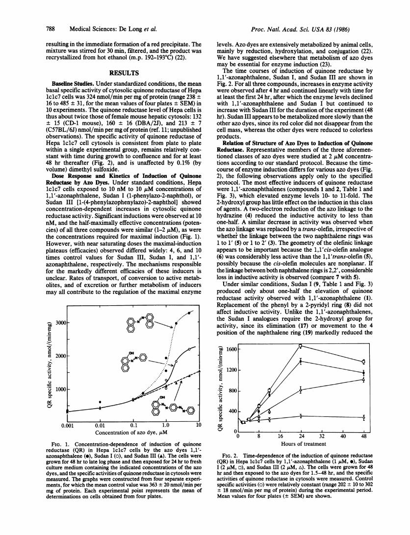
Reductase by Azo Dyes. Under standard conditions, Hepalclc7 cells exposed to 10 nM to 10 AM concentrations of1,1'-azonaphthalene, Sudan I (1-phenylazo-2-naphthol), orSudan III [1-(4-phenylazophenylazo)-2-naphthol] showedconcentration-dependent increases in cytosolic quinonereductase activity. Significant inductions were observed at 10nM, and the half-maximally effective concentrations (poten-cies) of all three compounds were similar (1-2 ,uM), as werethe concentrations required for maximal induction (Fig. 1).However, with near saturating doses the maximal-inductionplateaus (efficacies) observed differed widely: 4, 6, and 10times control values for Sudan III, Sudan I, and 1,1'-azonaphthalene, respectively. The mechanisms responsiblefor the markedly different efficacies of these inducers isunclear. Rates of transport, of conversion to active metab-olites, and of excretion or further metabolism of inducersmay all contribute to the regulation of the maximal enzyme
-3000
O .'~~~~~~Oe 2000
U~~~~~~~~~
U '1~~~~~~~~~~~~~~~~~~~~~~~~~~~~~0
I I0.001 0.01 0.1 1.0 10
Concentration of azo dye, uM
FIG. 1. Concentration-dependence of induction of quinonereductase (QR) in Hepa lclc7 cells by the azo dyes 1,1'-azonaphthalene (e), Sudan I (o), and Sudan III (A). The cells weregrown for 48 hr to late log phase and then exposed for 24 hr to freshculture medium containing the indicated concentrations of the azodyes, and the specific activities ofquinone reductase in cytosols weremeasured. The graphs were constructed from four separate experi-ments, for which the mean control value was 363 ± 20 nmol/min permg of protein. Each experimental point represents the mean ofdeterminations on cells obtained from four plates.
levels. Azo dyes are extensively metabolized by animal cells,mainly by reduction, hydroxylation, and conjugation (22).We have suggested elsewhere that metabolism of azo dyesmay be essential for enzyme induction (23).The time courses of induction of quinone reductase by
i,1'-azonaphthalene, Sudan I, and Sudan III are shown inFig. 2. For all three compounds, increases in enzyme activitywere observed after 4 hr and continued linearly with time forat least the first 24 hr, after which the enzyme levels declinedwith i,1'-azonaphthalene and Sudan I but continued toincrease with Sudan III for the duration of the experiment (48hr). Sudan III appears to be metabolized more slowly than theother azo dyes, since its red color did not disappear from thecell mass, whereas the other dyes were reduced to colorlessproducts.
Relation of Structure of Azo Dyes to Induction of QuinoneReductase. Representative members of the three aforemen-tioned classes of azo dyes were studied at 2 ,uM concentra-tions according to our standard protocol. Because the time-course of enzyme induction differs for various azo dyes (Fig.2), the following observations apply only to the specifiedprotocol. The most effective inducers of quinone reductasewere 1,1'-azonaphthalenes (compounds 1 and 2, Table 1 andFig. 3), which elevated enzyme levels 10- to 11-fold. The2-hydroxyl group has little effect on the induction in this classof agents. A two-electron reduction of the azo linkage to thehydrazine (4) reduced the inductive activity to less thanone-half. A similar decrease in activity was observed whenthe azo linkage was replaced by a trans-olefin, irrespective ofwhether the linkage between the two naphthalene rings was1 to 1' (5) or 1 to 2' (3). The geometry of the olefinic linkageappears to be important because the 1,1'cis-olefin analogue(6) was considerably less active than the 1,1'trans-olefin (5),possibly because the cis-olefin molecules are nonplanar. Ifthe linkage between both naphthalene rings is 2,2', considerableloss in inductive activity is observed (compare 7 with 5).Under similar conditions, Sudan I (9, Table 1 and Fig. 3)
produced only about one-half the elevation of quinonereductase activity observed with 1,1'-azonaphthalene (1).Replacement of the phenyl by a 2-pyridyl ring (8) did notaffect inductive activity. Unlike the 1,1'-azonaphthalenes,the Sudan I analogues require the 2-hydroxyl group foractivity, since its elimination (17) or movement to the 4position of the naphthalene ring (19) markedly reduced the
a 1600s
- 1200SE
*. 8005._ce
'O 4000
0.
I.
4
-$~~~~~~~~~~~~~~~~~~~~~~~~~~~~~-
I I I I
0 8 16 24 32 40 48Hours of treatment
FIG. 2. Time-dependence of the induction of quinone reductase(QR) in Hepa lclc7 cells by 1,1'-azonaphthalene (1 ,M, e), SudanI (2 ,uM, o), and Sudan III (2 uM, A). The cells were grown for 48hr and then exposed to the azo dyes for 1.5-48 hr, and the specificactivities of quinone reductase in cytosols were measured. Controlspecific activities (o) were relatively constant (range 202 ± 10 to 302+ 18 nmol/min per mg of protein) during the experimental period.Mean values for four plates (+ SEM) are shown.
Proc. Natl. Acad Sci. USA 83 (1986)

Proc. Natl. Acad. Sci. USA 83 (1986) 789
Table 1. Induction of cytosolic quinone reductase in Hepa lclc7hepatoma cells by 1,1'-azonaphthalene, Sudan I, and Sudan IIIanalogues
Quinonereductase Ratio of specific
Inducing specific activity, activitiesagent* nmol/(min mg) (treated/control)
1,1 '-Azonaphthalenes1 2960 ± 219 10.6 ± 0.82 2780 ± 211 9.91 ± 0.753 1280 ± 14 4.58 ± 0.054 1180 ± 43 4.20 ± 0.155 1160 ± 50 4.15 ± 0.186 600 ± 24 2.14 ± 0.097 426 ± 7 1.52 ± 0.02
Sudan I analogues8 1810 ± 54 6.47 ± 0.199 (Sudan I) 1720 ± 46 6.13 ± 0.1610 1620 ± 61 5.80 ± 0.2211 1550 ± 199 5.54 ± 0.7112 1250 ± 81 4.46 ± 0.2913 1230 ± 68 4.38 ± 0.2414 (Sudan II) 1220 ± 52 4.34 ± 0.1915 952± 31 3.40±0.1116 564 ± 25 2.64 ± 0.1217 523 ± 30 2.46 ± 0.1418 550 ± 21 1.96 0.0719 593 ± 86 1.83 ± 0.27
Sudan III analogues20 (Sudan IV) 1150 ± 88 4.11 ± 0.3121 (Sudan III) 1100 ± 39 3.92 ± 0.1422 682 ± 52 2.43 ± 0.1923 677 ± 43 2.42 ± 0.1524 671 ± 10 2.40 ± 0.0425 617 ± 8 2.20 ± 0.0326 555 ± 35 1.98 ± 0.1227 353 ± 14 1.26 ± 0.05
All enzyme specific activities are based on duplicate assays oncytosols of cells obtained from each of four plates and are expressedas means ± SEM. The treated/control ratios are based on a controlspecific activity of 280 ± 23 nmol/min per mg of protein for alldeterminations except for compounds 16 and 17 (specific activity 213± 21) and compound 19 (specific activity 324 ± 17). The SEM valuesof the treated/control ratios were obtained by dividing the SEM ofeach treated group by the control value.*Inducing agents were present at 2,4M in culture medium. Structuresare shown in Fig. 3.
inductive activity. Replacement of this hydroxyl group by anamino group (13) was far less damaging. Substituting thephenyl ring of Sudan I with an amino group at the 4' position(11) reduced inductive activity slightly, whereas the simul-taneous presence of 2'- and 4'-methyl substituents (14)resulted in a marked reduction of activity. Among theunsubstituted phenylazonaphthalenes, a 2,1' linkage (12)was a more effective inducer than a 1,1' linkage (17).Replacement of the azo linkage by a trans-olefin in thephenylazonaphthalenes showed a dichotomous effect; induc-tive activity was reduced if the ring linkage was from 2 to 1'(compare 15 and 12), whereas it was enhanced if the linkagewas from 1 to 1' (compare 17 and 10). A cis-olefin linkagemarkedly reduced the inductive capacity in comparison to thetrans-olefin (compare 10 and 18).Sudan III (21) and its analogues are of particular interest
because they were most potent in protecting rats againstmammary cancer, leukemia, and adrenal hemorrhage pro-duced by polycyclic aromatic compounds (19, 22, 24). In theHepa lclc7 system, Sudan III and its analogues were lessthan one-half as effective as 1,1'-azonaphthalene (1) and
ON~~~~ON12H
OH N OH
X 012 ON'No'"~ ~ 1
4
H lo H
H Ho v
H OH
H.10 t9"
1N NH3
~H HO N'N-
OH CH OH H
9/2oN 21P2221H ~~~OH
23
H
S03H 24 flH 25N-
H2N
26 27
FIG. 3. Structures of compounds 1-27.
about two-thirds as effective as Sudan I (9) in inducingquinone reductase. In this class of compounds, as in theSudan I analogues, the presence of a hydroxyl group ortho tothe azo linkage is important for induction. Thus, replacementof the 2-hydroxyl group by a 2-amino group (25) or by a4-amino group (26) reduced the inductive activity 50%. If theadditional phenylazo group is replaced by a cis- (23) or atrans-styrene (22) moiety, the inductive activity is diminishedeven further than by the presence of the phenylazo linkagealone. The presence of methyl groups on both phenyl rings ofSudan III (20) barely affected the inductive activity, whereasit was almost completely abolished by replacing the terminalphenylazo group by azonaphthol (27).
Induction of Quinone Reductase by Phenolic Antioxidants.Phenolic antioxidants, and in particularBHA [3(2)-tert-butyl-4-hydroxyanisole] and BHT (3,5-di-tert-butyl-4-hydroxy-toluene), which have been studied extensively for theirprotective effects (1, 2, 25), induce quinone reductase inHepa lclc7 cells (Table 2). However, substituted phenols(commercial BHA and its component isomers, BHT, and4-hydroxyanisole) are much less efficient inducers than thosecarrying unsubstituted phenolic groups, such as tert-butylhydroquinone and 3,5-di-tert-butylcatechol, both ofwhich are chemoprotectors and inducers of protective en-zymes in vivo (4, 7, 8, 26, 27). These findings suggest that theultimate inducers are probably hydroquinones or catechols(17, 18, 26). tert-Butylhydroquinone elevates the quinonereductase specific activity in Hepa lclc7 cells in a dose-dependent manner: 20 ,M concentrations doubled the en-zyme levels, and 50-60 uM more than tripled them.
Induction of Quinone Reductase by Coumarins, Flavonoids,and Other Lactones. Coumarin, a-angelicalactone, quercitin,and especially 5,6-benzoflavone all induce quinone reductasein the Hepa lclc7 system (Table 3). These compounds are all
Medical Sciences: De Long et al.
H
3

790 Medical Sciences: De Long et al.
Table 2. Induction of cytosolic quinone reductase in Hepa lclc7hepatocytes by phenolic antioxidants
Table 3. Induction of cytosolic quinone reductase in Hepa lclc7hepatocytes by coumarin, flavones, and other lactones
Quinone reduc-Inducing agent tase specific
Conc., activity,Compound* AM nmol/(min-mg)
A 10 583 + 30-650 + 17(n = 3)
30 689 + 35-1259 + 71(n = 6)
50 1106 + 35aB 30 388 + 16b
30 638 72c60 784 + 47c
C 15 570 + 34c30 673 74c60 789 + 37c
D 30 555 + 27c60 589 + 88c
E 2 345 + 17d10 734 ± 41010 755 ± 4
10 862 ± 38dF 10 268 ± 119
30 281 ± 12g50 516 ± 49a
Ratio of specificactivities
(treated/control)
1.37 ± 0.04-2.12 ± 0.13
2.60 ± 0.14-3.62
±0.22
4.64 ± 0.151.42 ± 0.061.32 ± 0.151.62 ± 0.101.18 ± 0.071.39 ± 0.151.63 ± 0.081.14 ± 0.061.21 ± 0.181.14 ± 0.062.18 ± 0.122.46 ± 0.012.85 ± 0.130.97 ± 0.041.01 ± 0.042.17 ± 0.21
All enzyme specific activities are based on duplicate assays oncytosols of cells obtained from each of four plates and are expressedas means ± SEM. The treated/control ratios are based on theindividual control values shown below, The SEM values of theseratios were obtained by dividing the SEM of each treated group bythe control value. Contrrol values: a238 ± 16; b274 + 22; C485 + 31;d302 ± 10; C337 ± 11; t307 ± 4; g227 ± 8.*A, tert-butylhydroquinone; B, 3-tert-butyl-4-hydroxyanisole (majorisomer of BHA); C, 2-tert-butyl-4-hydroxyanisole (minor isomer ofBHA); D, 4-hydroxyanisole; E, 3,5-di-tert-butylcatechol; F, 3,5-di-tert-butyl-4-hydroxytoluene (BHT). Structures are shown below.
OH OH OH OH OH OH
t e A O~~~~O
OH OCH3 OCH3 OCH3 CH3
A B C D E F
chemoprotective (1, 2), although to varying degrees, and arealso glutathione S-transferase inducers in vivo (7, 8).
Induction of Quinone Reductase by Polycyclic AromaticHydrocarbons. Low concentrations (2 ,uM) of benzo[a]py-rene, 7,12-dimethylbenz[a]anthracene, and 3-methylchol-anthrene induced quinone reductase in Hepa lclc7 cells to3.6-5.8 times control values, although 30 ,uM phenobarbitalwas completely inactive (Table 4). Although the polycyclichydrocarbons are themselves carcinogens, it is well recog-nized that under suitable conditions these compounds protectagainst their own carcinogenicity and toxicity or those ofother compounds (19, 28, 29).
Induction of Quinone Reductase by Sulfur Compounds. Anumber of sulfur-containing compounds (e.g., disulfiram,benzylisothiocyanate, dithiolthiones) are protectors againstchemical carcinogenesis, as well as inducers ofchemoprotec-tive enzymes such as glutathione S-transferases and quinonereductase in vivo (1, 2, 30-32). Under conditions of ourstandard protocol, quinone reductase activity was elevated inHepa lclc7 cells by benzylisothiocyanate (2.45-fold at 10,uM) and by oltipraz [5-(2-pyrazinyl)-4-methyl-1,2-dithiol-3-thione] and anethole dithiolthione [5-(4-methoxyphenyl)-1,2-dithiol-3-thione] (1.6- to 1.9-fold at 10 ,uM and 2.1- to 2.4-fold
Inducing agent
Coumarin10 ,uM50 ,uM
Quercitin10 AM30 ,uM50 ,IM
a-Angelicalactone10 ,uM50 ,uM
5,6-Benzoflavone2 ,uM10 JiM
Quinone reductasespecific activity,nmol/(min-mg)
235 ± 12a383 14b
451 ± 34C601± 14c607 ± 14b
303 ± 29c454 ± 10b
2270 ± 212d3070 ± 244e
Ratio of specificactivities
(treated/control)
1.10 ± 0.061.61 ± 0.06
1.63 ± 0.122.17 ± 0.052.55 ± 0.06
1.09 ± 0.101.91 ± 0.04
5.42 ± 0.519.50 ± 0.76
All enzyme specific activities are based on duplicate assays oncytosols of cells obtained from each of four plates and are expressedas means ± SEM. The treated/control ratios are based on theindividual control values shown below. The SEM values of theseratios were obtained by dividing the SEM of each treated group bythe control value. Control values: 1214 + 21; b238 ± 16; c277 + 8; d419± 10; C323 ± 17.
at 30 jM). In preliminary experiments, diethyldithiocarba-mate and bis(ethylxanthogen) [0, 0-diethyl dithiobis(thiofor-mate)], both chemoprotectors and enzyme inducers in vivo(2, 31), were also found to be inducers of quinone reductasein Hepa lclc7 cells.
DISCUSSION
Murine hepatic cells were chosen for these studies becauseinductions in vivo of chemoprotective enzymes by antioxi-dant anticarcinogens were most prominent in mouse liver (32,33). Hepa cells were established in culture from a transplant-able hepatoma (BW 7756) of the C57L/J mouse (34, 35), andthe lclc7 clone was selected for a high inducibility of arylhydrocarbon hydroxylase (36). This line retains many of thespecific morphological and biochemical characteristics ofliver (34, 35). The Hepa lclc7 cells also contain inducibleethoxyresorufin O-deethylase and cytochrome P-450reductase, as well as ornithine decarboxylase and epoxidehydrolase (37-39). These cells can therefore presumablydealkylate substituted phenols and reduce azo linkages. Such
Table 4. Induction of cytosolic quinone reductase in Hepa lclc7hepatocytes by polycyclic aromatic hydrocarbons
Quinone reductase Ratio of specificspecific activity, activities
Inducing agent* nmol/(min-mg) (treated/control)DMBAt 1360 ± 120a 4.21 ± 0.37Benzota]pyrene 1520 ± 76b 3.63 ± 0.18
1800 ± 41a 5.57 ± 0.133-Methylcholan-
threne 1670 ± 119b 3.99 ± 0.281890 ± 220a 5.85 ± 0.68
Phenobarbital 425 ± 79b 1.01 ± 0.19
All enzyme specific activities are based on duplicate assays oncytosols of cells obtained from each of four plates and are expressedas means ± SEM. The treated/control ratios are based on theindividual control values shown below. The SEM values of theseratios were obtained by dividing the SEM of each treated group bythe control value. Control values: ^323 ± 17; b419 ± 10.*Concentrations were 2 ,uM, except for phenobarbital (30 ,uM).t7,12-Dimethylbenz[alanthracene.
Proc. Natl. Acad Sci. USA 83 (1986)
Proc. Natl. Acad. Sci. USA 83 (1986) 791
metabolic functions are important for generating the signalsfor induction which depend on the formation of oxida-tion-reduction labile 1,2- or 1,4-diphenols or -diamines (23).The Hepa 1c1c7 cells have permitted analysis of the molec-ular control of the induction of aryl hydrocarbon hydroxyl-ase. A number of mutants defective in this pathway (10, 40)will be useful in uncovering the relationship between theinduction of phase I and phase II enzymes and their relativeimportance in chemoprotection.
Selection of quinone reductase as a marker of induction ofchemoprotective enzymes was stimulated by the discovery ofHuggins (19) that certain azo dyes (especially Sudan III),which protected rats against mammary cancer, leukemia, andadrenal hemorrhage produced by methylated benz[a]anthra-cene derivatives, also produced marked inductions ofquinone reductase, and that both effects required synthesis ofprotein (41). These observations implied a functional rela-tionship between induction of this enzyme and protectionagainst carcinogenesis and toxicity (19). Furthermore, Fujitaet al. (42) have demonstrated that, in rat liver, many azo dyesinduce quinone reductase coordinately with cytochromeP-450 and associated monooxygenase activities, glutathioneS-transferases, aldehyde dehydrogenase, and UDP-glucuro-nyltransferase. Thus, the azo dyes resemble polycyclicaromatics; both types of compounds bind to the receptorencoded by the Ah (aryl hydrocarbon) locus (43).The toxic effects of quinones (which are ubiquitous con-
stituents of human diet) have been ascribed to two mecha-nisms: first, direct interaction of electrophilic quinones withcritical cellular nucleophiles; and second, facile one-electronreductions (promoted by many flavoproteins) to semiquinonefree radicals which can participate in cyclic oxidation-reduc-tions to generate reactive oxygen species (44). Quinonereductase is unusual among flavoproteins in that it promotesobligatory two-electron reductions of quinones (45), thusdiverting quinones from oxidative cycling and preparing themfor conjugation with glucuronic acid (13, 46, 47). Manydifferent types of experiments attest to the importance ofquinone reductase as a cellular protector against the toxicitiesof quinones. Further, this enzyme can be induced by a widerange of compounds, including BHA, polycyclic aromatics,and 2,3,7,8-tetrachlorodibenzo-p-dioxin, which, like azodyes, can protect against chemical carcinogenesis (see ref. 3).For these reasons, measurement of the cytosolic levels of
quinone reductase of Hepa 1c1c7 hepatoma cells in cultureprovides a versatile, simple, and reliable method for identi-fying chemoprotective agents and for studying the mecha-nism of induction of chemoprotective enzymes.
Many of the azo dyes were generously donated by Charles Hugginsand John Pataki of The University of Chicago. We are grateful toHoward S. Bregman for assistance with synthesis of azo dyes and toAnnette B. Santamaria for skillful technical assistance. These studieswere supported by a Special Institutional Grant from the AmericanCancer Society (SIG-3). H.J.P. was supported by National Institutesof Health Training Grant GM07309.
1. Wattenberg, L. W. (1983) Cancer Res. 43, Suppl., 2448s-2453s.2. Wattenberg, L. W. (1985) Cancer Res. 45, 1-8.3. Talalay, P. & Benson, A. M. (1982) Adv. Enzyme Regul. 20,
287-300.4. De Long, M. J., Prochaska, H. J. & Talalay, P. (1983) in Protective
Agents in Cancer, eds. McBrien, D. C. H. & Slater, T. F. (Aca-demic, London), pp. 175-1%.
5. Williams, R. T. (1967) Fed. Proc. Fed. Am. Soc. Exp. Biol. 26,1029-1039.
6. Talalay, P., Batzinger, R. P., Benson, A. M., Bueding, E. & Cha,
Y.-N. (1979) Adv. Enzyme Regul. 17, 23-36.7. Sparnins, V. L., Chuan, J. & Wattenberg, L. W. (1982) Cancer
Res. 42, 1205-1207.8. Sparnins, V. L., Venegas, P. L. & Wattenberg, L. W. (1982) J.
Natl. Cancer Inst. 68, 493-496.9. Whitlock, J. P., Jr., Galeazzi, D., Israel, D. & Miller, A. G. (1983)
in Mechanism of Drug Action, eds. Mansour, T. E. & Ondarza,R. N. (Academic, New York), pp. 327-338.
10. Hankinson, O., Andersen, R. D., Birren, B. W., Sander, F.,Negishi, M. & Nebert, D. W. (1985) J. Biol. Chem. 260,1790-1795.
11. Benson, A. M., Hunkeler, M. J. & Talalay, P. (1980) Proc. Natl.Acad. Sci. USA 77, 5216-5220.
12. Chesis, P. L., Levin, D. E., Smith, M. T., Ernster, L. & Ames,B. N. (1984) Proc. Natl. Acad. Sci. USA 81, 1696-1700.
13. Lind, C., Hochstein, P. & Ernster, L. (1982) Arch. Biochem.Biophys. 216, 178-185.
14. Morrison, H., Jernstrwm, B., Nordenskjold, M., Thor, H. &Orrenius, S. (1984) Biochem. Pharmacol. 33, 1763-1769.
15. Thor, H., Smith, M. T., Hartzell, P., Bellomo, G., Jewell, S. A. &Orrenius, S. (1982) J. Biol. Chem. 257, 12419-12425.
16. Smart, R. C. & Zannoni, V. G. (1984) Mol. Pharmacol. 26,105-111.
17. De Long, M. J., Prochaska, H. J. & Talalay, P. (1985) Cancer Res.45, 546-551.
18. Prochaska, H. J., Bregman, H. S., De Long, M. J. & Talalay, P.(1985) Biochem. Pharmacol. 34, 3909-3914.
19. Huggins, C. B. (1979) Experimental Leukemia and MammaryCancer: Induction, Prevention, Cure (Univ. of Chicago Press,Chicago).
20. De Long, M. J., Prochaska, H. J. & Talalay, P. (1985) Fed. Proc.Fed. Am. Soc. Exp. Biol. 44, 500 (abstr. 518).
21. Huggins, C. & Pataki, J. (1965) Proc. Natl. Acad. Sci. USA 53,791-796.
22. Daniel, J. W. (1962) Toxicol. Appl. Pharmacol. 4, 572-594.23. Prochaska, H. J., De Long, M. J. & Talalay, P. (1985) Proc. NatI.
Acad. Sci. USA 82, 8232-8236.24. Kahl, R. (1984) Toxicology 33, 185-228.25. Wattenberg, L. W., Coccia, J. B. & Lam, L. K. T. (1980) Cancer
Res. 40, 2820-2823.26. Huggins, C. B., Ueda, N. & Russo, A. (1978) Proc. Natl. Acad.
Sci. USA 75, 4524-4527.27. Richardson, H. L., Stier, A. R. & Borsos-Nachtnebel, E. (1952)
Cancer Res. 12, 356-361.28. Miller, E. C., Miller, J. A., Brown, R. R. & MacDonald, J. C.
(1958) Cancer Res. 18, 469-477.29. Ansher, S. S., Dolan, P. & Bueding, E. (1983) Hepatology (Balti-
more) 3, 932-935.30. Benson, A. M. & Barretto, P. B. (1984) Fed. Proc. Fed. Am. Soc.
Exp. Biol. 43, 590 (abstr. 1781).31. Kensler, T. W., Egner, P. A., Trush, M. A., Bueding, E. &
Groopman, J. D. (1985) Carcinogenesis 6, 759-763.32. Benson, A. M., Batzinger, R. P., Ou, S.-Y. L., Bueding, E., Cha,
Y.-N. & Talalay, P. (1978) Cancer Res. 38, 4486-4495.33. Benson, A. M., Cha, Y.-N., Bueding, E., Heine, H. S. & Talalay,
P. (1979) Cancer Res. 39, 2971-2977.34. Bernhard, H. P., Darlington, G. J. & Ruddle, F. H. (1973) Dev.
Biol. 35, 83-96.35. Darlington, G. J., Bernhard, H. P., Miller, R. A. & Ruddle, F. H.
(1980) J. Natl. Cancer Inst. 64, 809-815.36. Hankinson, 0. (1979) Proc. Natl. Acad. Sci. USA 76, 373-376.37. Benedict, W. F., Gielen, J. E., Owens, J. S., Niwa, A. & Nebert,
D. W. (1973) Biochem. Pharmacol. 22, 2766-2769.38. Duthu, G. S. & Hankinson, 0. (1983) Cancer Lett. 20, 249-254.39. Duthu, G. S., Nestor, M. S., Berliner, J. A., Philpot, R. M. &
Hankinson, 0. (1983) Cancer Lett. 18, 237-243.40. Jones, P. B. C., Galeazzi, D. R., Fisher, J. M. & Whitlock, J. P.,
Jr. (1985) Science 227, 1499-1502.41. Huggins, C. & Fukunishi, R. (1964) J. Exp. Med. 119, 923-942.42. Fujita, S., Suzuki, M. & Suzuki, T. (1984) Xenobiotica 14, 565-568.43. Lubet, R. A., Connolly, G., Kouri, R. E., Nebert, D. W. &
Bigelow, S. W. (1983) Biochem. Pharmacol. 32, 3053-3058.44. Kappus, H. & Sies, H. (1981) Experientia 37, 1233-1241.45. Iyanagi, T. & Yamazaki, I. (1970) Biochim. Biophys. Acta 216,
282-294.46. Lind, C., Vadi, H. & Ernster, L. (1978) Arch. Biochem. Biophys.
190, 97-108.47. Lind, C. (1985) Biochem. Pharmacol. 34, 895-897.
Medical Sciences: De Long et al.