In vivo estimation of passive biomechanical properties of ...
17
ORIGINAL ARTICLE In vivo estimation of passive biomechanical properties of human myocardium Arnab Palit 1,2 & Sunil K. Bhudia 3 & Theodoros N. Arvanitis 2 & Glen A. Turley 1 & Mark A. Williams 1 Received: 21 March 2017 /Accepted: 13 December 2017 /Published online: 26 February 2018 # The Author(s) 2018. This article is an open access publication Abstract Identification of in vivo passive biomechanical properties of healthy human myocardium from regular clinical data is essential for subject-specific modelling of left ventricle (LV). In this work, myocardium was defined by Holzapfel- Ogden constitutive law. Therefore, the objectives of the study were (a) to estimate the ranges of the constitutive parameters for healthy human myocardium using non-invasive routine clinical data, and (b) to investigate the effect of geometry, LV end-diastolic pressure (EDP) and fibre orientations on estimated values. In order to avoid invasive measurements and additional scans, LV cavity volume, measured from routine MRI, and empirical pressure-normalised- volume relation (Klotz-curve) were used as clinical data. Finite element modelling, response surface method and genetic algorithm were used to inversely estimate the constitutive parameters. Due to the ill-posed nature of the inverse opti- misation problem, the myocardial properties was extracted by identifying the ranges of the parameters, instead of finding unique values. Additional sensitivity studies were carried out to identify the effect of LV EDP, fibre orientation and geometry on estimated parameters. Although uniqueness of the solution cannot be achieved, the normal ranges of the parameters produced similar mechanical responses within the physiological ranges. These information could be used in future computational studies for designing heart failure treatments. Keywords Ventricular diastolic mechanics . Finite element . Parameter estimation . Normal human subjects . Ventricular geometry . Fibre structure 1 Introduction Local myocardial wall stress is hypothesised to be responsible for many cardiac mechanisms, including ventricular remodel- ling, which is frequently associated with heart failure (HF). However, stress in the left ventricle (LV) cannot be measured directly [20]. Finite element (FE) method, in combination with advanced simulation tools and new cardiac imaging modali- ties can be used to analyse LV wall stress–strain distribution for providing a greater insight of the physiology of normal subjects and HF patients, and thereby, predict their responses to medical and surgical interventions [3, 6, 13, 14, 19, 25, 38, 39, 48, 51]. In such models, it is essential to accurately use in vivo passive biomechanical properties of the human myo- cardium in order to mimic the cardiac mechanics properly. Otherwise, the stress-strain prediction would be over or under estimated which will lead to inaccurate diagnostic information for surgical operation [61]. * Arnab Palit [email protected]; [email protected]; [email protected] Sunil K. Bhudia [email protected] Theodoros N. Arvanitis [email protected] Glen A. Turley [email protected] Mark A. Williams [email protected] 1 WMG, The University of Warwick, Coventry CV4 7AL, UK 2 Institute of Digital Healthcare, WMG, The University of Warwick, Coventry, UK 3 University Hospitals Coventry and Warwickshire, Coventry, UK Medical & Biological Engineering & Computing (2018) 56:1615–1631 https://doi.org/10.1007/s11517-017-1768-x
Transcript of In vivo estimation of passive biomechanical properties of ...

ORIGINAL ARTICLE
In vivo estimation of passive biomechanical propertiesof human myocardium
Arnab Palit1,2 & Sunil K. Bhudia3 & Theodoros N. Arvanitis2 & Glen A. Turley1 & Mark A. Williams1
Received: 21 March 2017 /Accepted: 13 December 2017 /Published online: 26 February 2018# The Author(s) 2018. This article is an open access publication
AbstractIdentification of in vivo passive biomechanical properties of healthy human myocardium from regular clinical data isessential for subject-specific modelling of left ventricle (LV). In this work, myocardium was defined by Holzapfel-Ogden constitutive law. Therefore, the objectives of the study were (a) to estimate the ranges of the constitutiveparameters for healthy human myocardium using non-invasive routine clinical data, and (b) to investigate the effectof geometry, LV end-diastolic pressure (EDP) and fibre orientations on estimated values. In order to avoid invasivemeasurements and additional scans, LV cavity volume, measured from routine MRI, and empirical pressure-normalised-volume relation (Klotz-curve) were used as clinical data. Finite element modelling, response surface method and geneticalgorithm were used to inversely estimate the constitutive parameters. Due to the ill-posed nature of the inverse opti-misation problem, the myocardial properties was extracted by identifying the ranges of the parameters, instead of findingunique values. Additional sensitivity studies were carried out to identify the effect of LV EDP, fibre orientation andgeometry on estimated parameters. Although uniqueness of the solution cannot be achieved, the normal ranges of theparameters produced similar mechanical responses within the physiological ranges. These information could be used infuture computational studies for designing heart failure treatments.
Keywords Ventricular diastolic mechanics . Finite element . Parameter estimation . Normal human subjects . Ventriculargeometry . Fibre structure
1 Introduction
Local myocardial wall stress is hypothesised to be responsiblefor many cardiac mechanisms, including ventricular remodel-ling, which is frequently associated with heart failure (HF).However, stress in the left ventricle (LV) cannot be measureddirectly [20]. Finite element (FE) method, in combination withadvanced simulation tools and new cardiac imaging modali-ties can be used to analyse LV wall stress–strain distributionfor providing a greater insight of the physiology of normalsubjects and HF patients, and thereby, predict their responsesto medical and surgical interventions [3, 6, 13, 14, 19, 25, 38,39, 48, 51]. In such models, it is essential to accurately usein vivo passive biomechanical properties of the human myo-cardium in order to mimic the cardiac mechanics properly.Otherwise, the stress-strain prediction would be over or underestimated which will lead to inaccurate diagnostic informationfor surgical operation [61].
* Arnab [email protected]; [email protected];[email protected]
Sunil K. [email protected]
Theodoros N. [email protected]
Glen A. [email protected]
Mark A. [email protected]
1 WMG, The University of Warwick, Coventry CV4 7AL, UK2 Institute of Digital Healthcare, WMG, The University of Warwick,
Coventry, UK3 University Hospitals Coventry and Warwickshire, Coventry, UK
Medical & Biological Engineering & Computing (2018) 56:1615–1631https://doi.org/10.1007/s11517-017-1768-x

Traditionally, ex vivomechanical testing was carried out onmyocardial tissue, harvested from a specific heart, to identifyits properties. Results from the biaxial tests of caninemyocardium [7, 21, 31, 63] and simple shear test of pig[9] and human [43, 44] myocardium clearly exhibited itsorthotropic behaviour. These experimental data not onlyprovided the understanding to define the constitutive lawsfor the myocardium material, but also helped in determin-ing the values of the parameters [41, 42]. These traditionalmethods involve invasive ex vivo procedures and result inthe destruction of the myocardial tissues. These methodsare therefore not ideal for in vivo clinical measurement[8]. Moreover, ex vivo experiments using cadaver heartsmay not be the true representative of the in vivo passiveproperties of the heart as these hearts do not representreal-life conditions due to the lack of homeostasis [56].
FE modelling, in combination with cardiac magneticresonance imaging (CMRI), was carried out to estimatepassive myocardial properties in a non-invasive manner[12, 13, 50, 56, 60–62]. Other work was accomplishedto assess the passive material properties in isolated heartswith FE analysis [2, 10, 29, 32, 33, 45]. Most of themused Fung-type transversely isotropic law [5, 16, 17, 22].In contrast, the orthotropic behaviour of myocardium wasevident from the simple shear test of pig’s [9] and humanmyocardium [44]. Moreover, Schmid, O’Callaghan [42]reported that a transversely isotropic law would not besuitable for modelling orthotropic responses of passivemyocardium under simple shear tests. Modified Fung-type law [1, 49] and pole-zero law [30, 45] were intro-duced to incorporate myocardial material orthotropy.However, in all the Fung-type material models, the mate-rial parameters were merely used as weighting factors,rather than providing any physical importance [15].Besides, some of the material parameters were highly cor-related in Fung-type and pole-zero law [15, 40, 45, 52].Recently, Holzapfel and Ogden [18] developed a consti-tutive law that considered the locally orthotropic tissuearchitecture and the parameters of this model were closelyrelated to the characteristic micro-structure of myocardi-um. In literature, the parameters of the material modelwere fitted to match the simple shear test results frompig myocardium [9] to define fully orthotropic nature.The values of these fitted parameters (Table 2) resultedin too stiff stress–strain relation in patient-specific model,and therefore, unable to produce measured end-diastolicvolume (EDV) of LV within the physiological LV end-diastolic pressure (EDP) [3, 11, 52]. Table 1 summarisedthe methods used to estimate the myocardial propertiesand limitation in research gap which is addressed in thispaper.
An inverse estimation method, which is typically for-mulated as a non-linear optimisation problem to minimise
the differences in the measurements with respect to theunknown parameters, was used to quantify passive prop-erties of human myocardium using Holzapfel-Ogden ma-terial law. Due to the highly non-linear nature of the in-verse optimisation problem, large design space and corre-lation amongst the material parameters, it is non-trivial toinversely estimate those parameters accurately anduniquely using limited clinical data. The trade-off is thatwhile fewer clinical data make the inverse problem moreill-posed, requirement of more subject-specific data (i.e.MRI tagging and in-vivo ventricular pressure) leads tomore complex and invasive clinical measurements withlonger processing times, which is not always possible.Therefore, one of the major challenges, addressed in thisstudy, was to extract the human myocardial propertiesinstead of finding unique solution using non-invasive rou-tinely used clinical data rather than using invasive andcomputationally expensive clinical data. Therefore, thestudy proposed a method to achieve the following objec-tives: (a) to estimate the normal ranges of Holzapfel-Ogden parameters for healthy human myocardium usingroutinely used non-invasive clinical data, and (b) to ex-plore the effect of geometry, fibre orientation and EDP onthe estimation of passive parameters.
2 Methods
2.1 FE modelling of ventricle
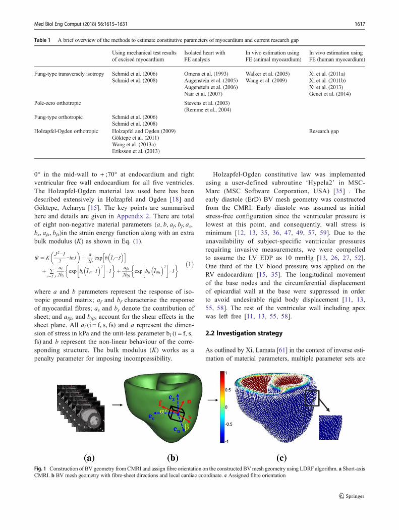
In the present study, ECG gated, breath hold, steady-statefree precession (SSFP) cine CMRI was used to capturethe images of five normal human ventricles at UHCW,UK. BSREC ethics approval (REGO-2012-032) and pa-tients’ consent were obtained to carry out the research onanonymised human da ta . Mimics and 3-ma t i c(Materialise, Belgium) were used to construct the bi-ventricular (BV) mesh geometry form CMRI as detailedin Palit, Turley [37] (Fig. 1a, b). The early diastolic vol-ume (ErDV), end diastolic volume (EDV), end systolicvolume (ESV) and, finally the ejection fraction (EF) foreach subject were calculated subsequently (Fig. 2). EachBV mesh geometry composed of at least 740,000 lineartetrahedral elements (tet4) to achieve accurate results asidentified from the mesh convergence study by Palit,Bhudia [35]. Details of the MRI scanning protocol anddemographic information of the subjects are enclosed inAppendix 1. Myocardial fibre structure was implementedby Laplace-Dirichlet-Region growing-FEM (LDRF) algo-rithm [37] (Fig. 1c). Based on previous histological stud-ies [1, 46], the fibre direction was defined by a linearvariation of helix angle (α) from − ;70° in the epicardiumand right ventricular (RV) septal endocardium to almost
1616 Med Biol Eng Comput (2018) 56:1615–1631
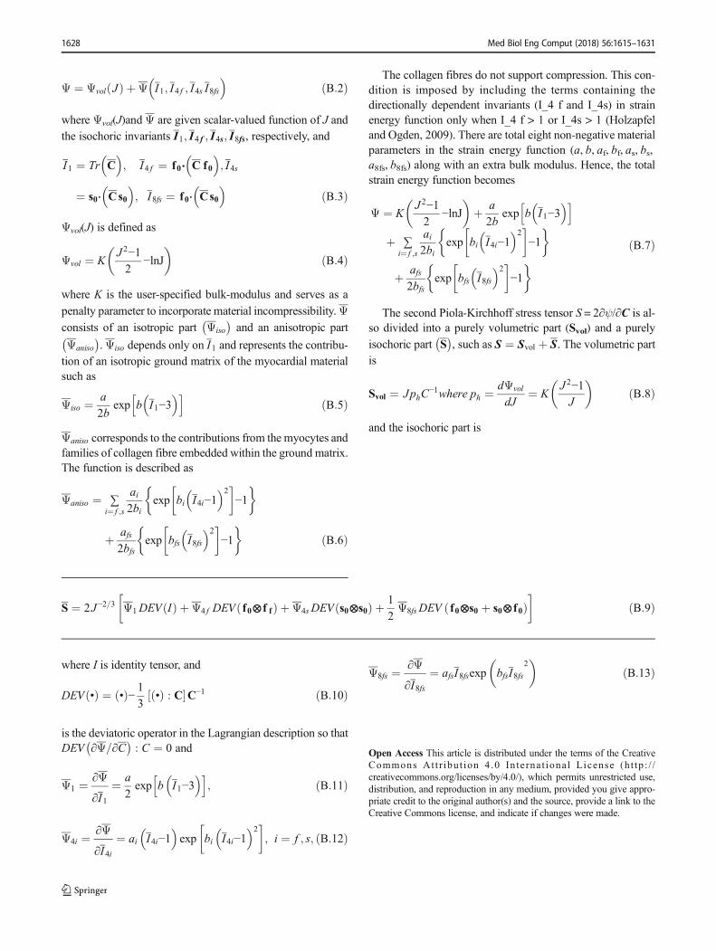
0° in the mid-wall to + ;70° at endocardium and rightventricular free wall endocardium for all five ventricles.The Holzapfel-Ogden material law used here has beendescribed extensively in Holzapfel and Ogden [18] andGöktepe, Acharya [15]. The key points are summarisedhere and details are given in Appendix 2. There are totalof eight non-negative material parameters (a, b, af, bf, as,bs, afs, bfs)in the strain energy function along with an extrabulk modulus (K) as shown in Eq. (1).
Ψ ¼ KJ2−12
−lnJ� �
þ a2b
exp b I1−3� �h i
þ ∑i¼ f ;s
ai2bi
exp bi I4i−1� �2
� �−1
� þ afs
2bfsexp bfs I8fs
� �2� �
−1� ð1Þ
where a and b parameters represent the response of iso-tropic ground matrix; af and bf characterise the responseof myocardial fibres; as and bs denote the contribution ofsheet; and a8fs and b8fs account for the shear effects in thesheet plane. All ai (i = f, s, fs) and a represent the dimen-sion of stress in kPa and the unit-less parameter bi (i = f, s,fs) and b represent the non-linear behaviour of the corre-sponding structure. The bulk modulus (K) works as apenalty parameter for imposing incompressibility.
Holzapfel-Ogden constitutive law was implementedusing a user-defined subroutine ‘Hypela2’ in MSC-Marc (MSC Software Corporation, USA) [35] . Theearly diastole (ErD) BV mesh geometry was constructedfrom the CMRI. Early diastole was assumed as initialstress-free configuration since the ventricular pressure islowest at this point, and consequently, wall stress isminimum [12, 13, 35, 36, 47, 49, 57, 59]. Due to theunavailability of subject-specific ventricular pressuresrequiring invasive measurements, we were compelledto assume the LV EDP as 10 mmHg [13, 26, 27, 52].One third of the LV blood pressure was applied on theRV endocardium [15, 35]. The longitudinal movementof the base nodes and the circumferential displacementof epicardial wall at the base were suppressed in orderto avoid undesirable rigid body displacement [11, 13,55, 58]. The rest of the ventricular wall including apexwas left free [11, 13, 55, 58].
2.2 Investigation strategy
As outlined by Xi, Lamata [61] in the context of inverse esti-mation of material parameters, multiple parameter sets are
Fig. 1 Construction of BV geometry fromCMRI and assign fibre orientation on the constructed BVmesh geometry using LDRF algorithm. a Short-axisCMRI. b BV mesh geometry with fibre-sheet directions and local cardiac coordinate. c Assigned fibre orientation
Table 1 A brief overview of the methods to estimate constitutive parameters of myocardium and current research gap
Using mechanical test resultsof excised myocardium
Isolated heart withFE analysis
In vivo estimation usingFE (animal myocardium)
In vivo estimation usingFE (human myocardium)
Fung-type transversely isotropy Schmid et al. (2006)Schmid et al. (2008)
Omens et al. (1993)Augenstein et al. (2005)Augenstein et al. (2006)Nair et al. (2007)
Walker et al. (2005)Wang et al. (2009)
Xi et al. (2011a)Xi et al. (2011b)Xi et al. (2013)Genet et al. (2014)
Pole-zero orthotropic Stevens et al. (2003)(Remme et al., 2004)
Fung-type orthotropic Schmid et al. (2006)Schmid et al. (2008)
Holzapfel-Ogden orthotropic Holzapfel and Ogden (2009)Göktepe et al. (2011)Wang et al. (2013a)Eriksson et al. (2013)
Research gap
Med Biol Eng Comput (2018) 56:1615–1631 1617
able to reproduce similar end-diastolic deformation states.Therefore, few strategic and logical assumptions were madeas follows:
2.2.1 Define the range of the parameters
From the definition of the material law, all the eightparameters are positive real number (i.e. a, b, af, bf, as,bs, afs, bfs > 0). On the other hand, it was reported thatthe human myocardium is less stiff than pig myocardium[13, 44]. Therefore, the shear stress for each shear mode[18] should be lower for human myocardium comparedto pig myocardium. Thus, the maximum value of eachparameter should not be higher than the value estimatedfrom the shear stress data of pig myocardium (Table 2).In this study, the average values, shown in Table 2, wasconsidered as the maximum value for the respective pa-rameters. Therefore,
amax ¼ 0:28; bmax ¼ 8:82;af
max ¼ 18:1; bfmax ¼ 16:5;
asmax ¼ 3:0; bsmax ¼ 9:5;a8fsmax ¼ 0:4; b8fsmax ¼ 10:9
9>>=>>; ð2Þ
2.2.2 Reduce the design space
Without changing the constitutive law, the design space of theproblem was reduced by selecting fewer parameters for theinverse estimation using similar method described in Xi,Lamata [61]. It was observed from the experiment of pigmyocardium that the order of shear responses in six shear
modes would follow as σ fsð Þ > σ fnð Þ > σ sfð Þ > σ snð Þ > σ nfð Þ;σ nsð Þ (where ij denoted the shear response in j direction of theplane containing i direction and i ≠ j ∈ {f, s, n}) [9, 18]. Theanalytical expressions of these shear stress modes, used to fitthe constitutive parameters to match the experimental data, areshown from Eq. (3) to Eq. (8) [15].
σ fsð Þ ¼ γ aexp γ2 b �þ 2γ3 a f exp γ4 b f
�þ γ afs exp γ2 bfs
� ð3Þσ fnð Þ ¼ γ aexp γ2 b
�þ 2γ3 a f exp γ4 b f � ð4Þ
σ sfð Þ ¼ γ aexp γ2 b �þ 2γ3 as exp γ4 bs
�þ γ afs exp γ2 bfs
� ð5Þσ snð Þ ¼ γ aexp γ2 b
�þ 2γ3 as exp γ4 bs � ð6Þ
σ nfð Þ ¼ γ aexp γ2 b � ð7Þ
σ nsð Þ ¼ γ aexp γ2 b � ð8Þ
where γ = amount of shear. From Eq. (3) to (8), it is observedthat the order of shear stiffness mode does not depend on theparameters a and b. The last six parameters are only respon-sible for maintaining the ordering as is evident from Eq. (3) to(6). The ordering can always be maintained if the ratiosamongst ai (and bi) are kept same (i.e. if af : as : afs = constantand bf : bs : bfs = constant). Therefore, all the ai and bi (wherei = f, s, fs) should be divided by Ka and Kb, respectively, sothat Eqs. (9) and (10) are maintained. Ka and Kb are positivereal number.
af
Ka¼ as
Ka¼ afs
Ka¼ constant ð9Þ
Fig. 2 Early diastolic volume (ErDv), end diastolic volume (EDV) andejection fraction (EF) extracted from CMRI for five healthy hearts (BV1to BV5)
Table 2 Values of Holzapfel-Ogden passive material parameters used in existing literature
Articles Passive material parameters
a (KPa) b af (KPa) bf as (KPa) bs afs (KPa) bfs
Holzapfel and Ogden (2009) 0.059 8.023 18.472 16.026 2.481 11.120 0.216 11.436
Göktepe et al. (2011) 0.496 7.209 15.193 20.147 3.283 11.176 0.662 9.466
Wang et al. (2013a) 0.236 10.81 20.037 14.154 3.724 5.164 0.411 11.3
Eriksson et al. (2013) 0.333 9.242 18.535 15.972 2.564 10.446 0.417 11.602
Average (approximated) 0.28 8.82 18.1 16.5 3 9.5 0.4 10.9
1618 Med Biol Eng Comput (2018) 56:1615–1631
bf
Kb¼ bs
Kb¼ bfs
Kb¼ constant ð10Þ
Thus, only four independent parameters (a, b, Ka, Kb) wererequired for inverse estimation to maintain the observed shearstiffness ordering [9]. The last six material parameters for humanmyocardiumwere then calculated using Eqs. (2), (9) and (10), as
ahumanf ¼ amaxf
Ka¼ 18:1
Ka; ahumans ¼ amaxs
Ka¼ 3
Ka;
ahumanfs ¼ amaxfs
Ka¼ 0:4
Ka
ð11Þ
bhumanf ¼ amaxf
Kb¼ 16:5
Kb; bhumans ¼ amax
s
Kb¼ 9:5
Kb;
bhumanfs ¼ amaxfs
Kb¼ 10:9
Kb
ð12Þ
If it was assumed that the af and bf values for human myo-cardium would not be less than 1, the maximum value of Kaand Kb should not be greater than 18.1 and 16.5, respectively.On the other hand, the minimum value of Ka and Kb should
not be less than 1 as ahumani <¼ amaxi and bhumani <¼ bmax
iwhere i = f, s, fs. The explicit definitions of the ranges areshown from Eqs. (17) to (20).
2.2.3 Problem formulation
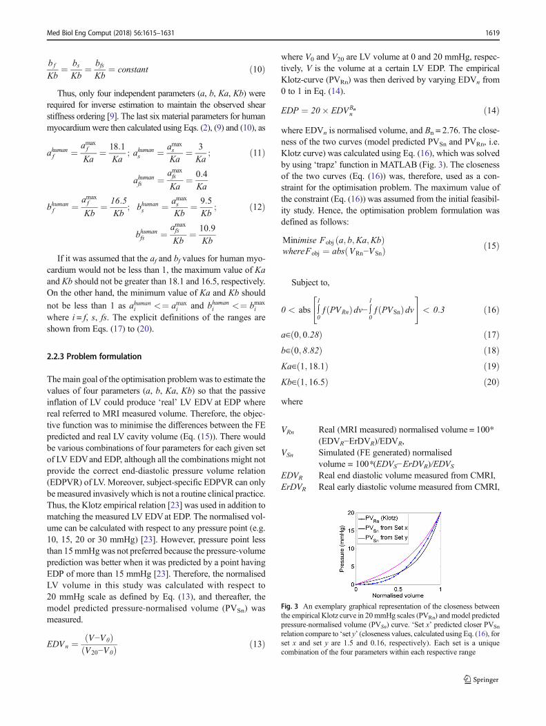
The main goal of the optimisation problem was to estimate thevalues of four parameters (a, b, Ka, Kb) so that the passiveinflation of LV could produce ‘real’ LV EDV at EDP wherereal referred to MRI measured volume. Therefore, the objec-tive function was to minimise the differences between the FEpredicted and real LV cavity volume (Eq. (15)). There wouldbe various combinations of four parameters for each given setof LV EDVand EDP, although all the combinations might notprovide the correct end-diastolic pressure volume relation(EDPVR) of LV. Moreover, subject-specific EDPVR can onlybemeasured invasively which is not a routine clinical practice.Thus, the Klotz empirical relation [23] was used in addition tomatching the measured LV EDVat EDP. The normalised vol-ume can be calculated with respect to any pressure point (e.g.10, 15, 20 or 30 mmHg) [23]. However, pressure point lessthan 15mmHgwas not preferred because the pressure-volumeprediction was better when it was predicted by a point havingEDP of more than 15 mmHg [23]. Therefore, the normalisedLV volume in this study was calculated with respect to20 mmHg scale as defined by Eq. (13), and thereafter, themodel predicted pressure-normalised volume (PVSn) wasmeasured.
EDVn ¼ V−V0ð ÞV20−V0ð Þ ð13Þ
where V0 and V20 are LV volume at 0 and 20 mmHg, respec-tively, V is the volume at a certain LV EDP. The empiricalKlotz-curve (PVRn) was then derived by varying EDVn from0 to 1 in Eq. (14).
EDP ¼ 20� EDVBnn ð14Þ
where EDVn is normalised volume, and Bn = 2.76. The close-ness of the two curves (model predicted PVSn and PVRn, i.e.Klotz curve) was calculated using Eq. (16), which was solvedby using ‘trapz’ function in MATLAB (Fig. 3). The closenessof the two curves (Eq. (16)) was, therefore, used as a con-straint for the optimisation problem. The maximum value ofthe constraint (Eq. (16)) was assumed from the initial feasibil-ity study. Hence, the optimisation problem formulation wasdefined as follows:
Minimise Fobj a; b;Ka;Kbð ÞwhereFobj ¼ abs VRn−VSnð Þ ð15Þ
Subject to,
0 < abs ∫1
0ƒ PVRnð Þdv− ∫
1
0ƒ PVSnð Þdv
" #< 0:3 ð16Þ
a∈ 0; 0:28ð Þ ð17Þb∈ 0; 8:82ð Þ ð18ÞKa∈ 1; 18:1ð Þ ð19ÞKb∈ 1; 16:5ð Þ ð20Þwhere
VRn Real (MRI measured) normalised volume = 100*(EDVR−ErDVR)/EDVR,
VSn Simulated (FE generated) normalisedvolume = 100*(EDVS−ErDVR)/EDVS
EDVR Real end diastolic volume measured from CMRI,ErDVR Real early diastolic volume measured from CMRI,
Fig. 3 An exemplary graphical representation of the closeness betweenthe empirical Klotz curve in 20mmHg scales (PVRn) andmodel predictedpressure-normalised volume (PVSn) curve. ‘Set x’ predicted closer PVSn
relation compare to ‘set y’ (closeness values, calculated using Eq. (16), forset x and set y are 1.5 and 0.16, respectively). Each set is a uniquecombination of the four parameters within each respective range
Med Biol Eng Comput (2018) 56:1615–1631 1619

EDVS End diastolic volume generated from simulation,PVRn Empirical Klotz curve in 20 mmHg pressure scale
(Eq. (14)),PVSn Pressure normalised volume curve generated from
FE resultsdv Infinitesimally small volumeabs Absolute value
2.2.4 Solving the optimisation problem
Initial sampling was carried out to create 50 ‘sets’ of parame-ters using the range of parameters defined in Eqs. (17)–(20).Each set is a unique combination of the four parameters withintheir respective ranges (Eq. (17)–(20)). In this study, Latinhypercube sampling (LHS) was used and was implementedin a customised script in MATLAB to generate uniformlydistributed sets of parameters. Simulation of passive inflationusing each set was then carried out using FE modelling andboundary conditions mentioned in Section 2.1. Two outputs(LV EDVand closeness of the PVSn to Klotz curve (PVRn), i.e.Eq. (16)) were measured from each simulation. Empiricalmodel of the two responses (Eqs. (15) and (16)) with respectto four independent material parameters were developed usingresponse surface method (RSM). Genetic algorithm (GA) wasthen used to solve the optimisation problem to identify theoptimal set of parameters. MATLAB GA function with allthe default parameter values was used in this study. Theoptimised parameters from GAwere used in FE model again,and the EDV and Klotz curve closeness was measured. If theabsolute differences betweenGA results and simulation resultswere greater than 3%, the new set with its simulation resultswas included in parameters set to modify the response surface.The process continued until the simulation results matchedwith GA results with in ± 3% range. A flow chart of the pro-posed inverse optimisation procedure is depicted in Fig. 4.
3 Results
3.1 In vivo estimations of the passive properties
Table 3 shows the values of four material parameters (a, b,Ka,Kb) for five human BVs using the proposed inverse optimisa-tion procedure (Fig. 4). The six material parameters corre-sponding to fibre-sheet (af, bf, as, bs, afs, bfs) were then derivedusing Eqs. (11) and (12). The values of all eight parameters aresummarised in Table 4 assuming LV EDP of 10 mmHg andfibre angle ± 70°. It was observed that the values of eachparameter were reduced with the increase in EF amongst theBVs. Moreover, the standard deviations of the parametersamongst the ventricles were not very high. A typical contourplot from the RSM and GA is shown in Fig. 5.
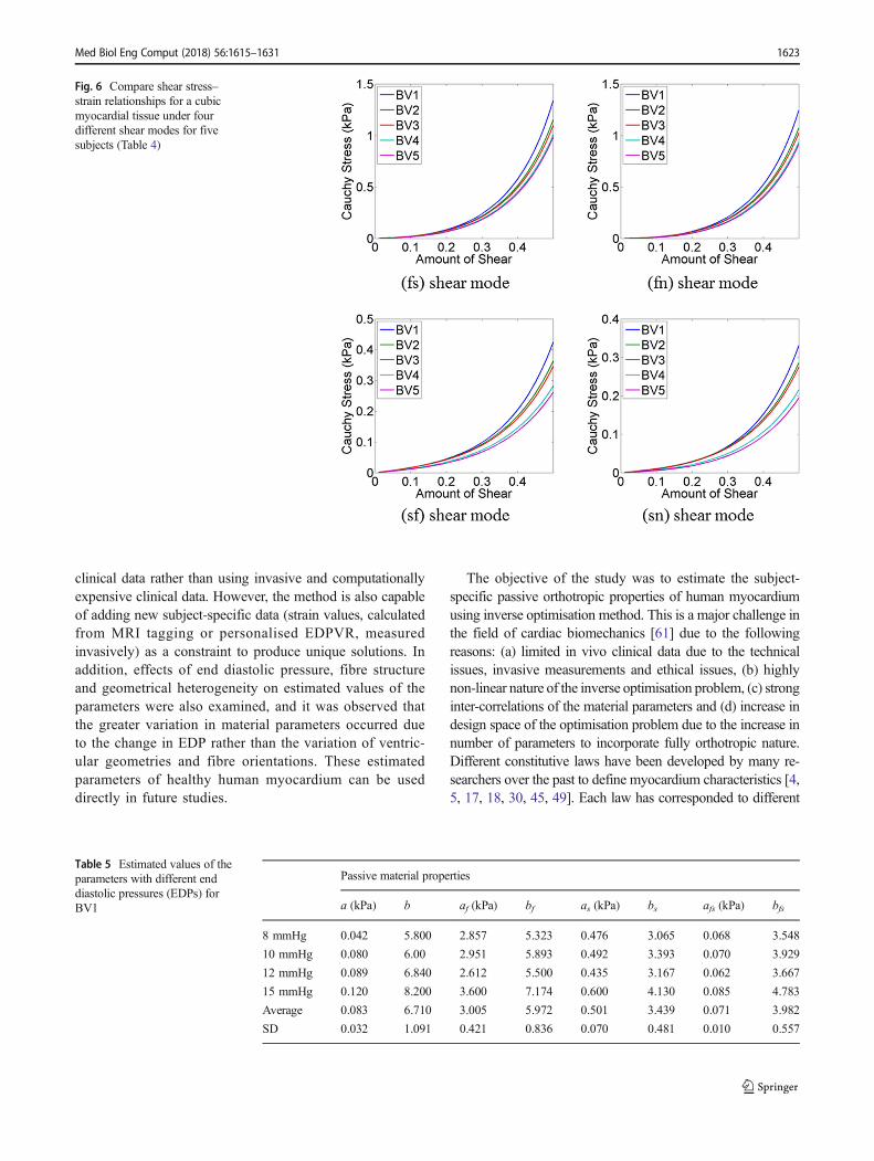
Shear stresses for four shear modes (σ(fs), σ(fn), σ(sf), σ(sn))were plotted (Fig. 6) using subject-specific values of the eightparameters, summarised in Table 4. It was observed that thepredicted myofibre stress–strain relationships under differentshear modes were reasonably similar for five subjects despiteof the differences in predicted values. The similarities weremore for shear in (fs) and (fn) plane compared to the shear in(sf) and (sn) plane.
3.2 Sensitivity study
LV EDP was assumed 10 mmHg during the parameter esti-mation due to the unavailability of in vivo pressure data.Therefore, a sensitivity study was carried out to explore thechanges in parameters if LV EDP was varied to 8, 12 and15 mmHg. The estimated values of material parameters aredepicted in Table 5. It was observed that the variation in pa-rameter bwas comparatively higher due to the change in EDP.
Another sensitivity study was accomplished to investigatethe effect of different fibre orientations on parameter estima-tion. BV1 mesh geometry and LV EDP of 10 mmHg wereutilised for the study. Table 6 shows the values of eight pa-rameters when fibre angle was ± 50°, ± 60°, ± 70° and ± 80°. Itwas observed that the effect of fibre orientation was compar-atively grater on parameter b.
Shear stresses for four shear modes (σ(fs), σ(fn), σ(sf), σ(sn))were plotted (Fig. 7) using the material parameters, predictedwith different EDP (Table 5) and with different fibre orienta-tions (Table 6). It was noticed that the predicted σ(fs) and σ(fn)
shear were quite similar except for 15 mmHg.When EDPwas15 mmHg, the myocardium became stiffer in fibre directions.The variation due to the fibre orientation was not very notablein (fs) and (fn) shear modes. However, the variation washigher for (sf) and (sn) shear modes although the predictedstress values were in the range from 0.4 to 0.8 kPa only.
Figure 8 shows variation in the material parameters due tothe change in the following: (a) ventricular geometry (BV1,BV2, BV3, BV4 and BV5), (b) EDP (8, 10, 12 and 15mmHg)for BV1 and (c) fibre orientation (± 50°, ± 60°, ± 70°, ± 80°)for BV1. It was observed that the effect of these variations ona, as, and afs were negligible whereas the variation in b param-eters was the highest. The other parameters af, bf, bs and bfsexperienced moderate variation. The effect of change in fibreorientation was the highest in parameter b. It was identifiedthat the greater variation in material parameters occurred dueto the change in EDP rather than the variation of ventriculargeometries and fibre orientations.
3.3 Compare with the state-of-the-art
The estimated values of the parameters in this study werefurther compared with the studies which inversely estimatedhuman myocardium parameters. The shear stress–strain
1620 Med Biol Eng Comput (2018) 56:1615–1631

relations under six different shear modes of a cubic myocar-dium tissue were derived using the parameters identified inthis study (for BV1) with the results from Gao, Li [12]. The
parameters identified with LV EDP of 10 mmHg were select-ed from both the studies. Figure 9 shows that there is a goodagreement between the predicted shear stress–strain relations,
Sampling
To define ‘set’ of parameters’ value
Response surface method
Create response surface for Fobj (Eq. (15) and PVSn
closeness to Klotz curve (Eq. (16))
Solve by GA
Identify the values of 4 parameters
Validation by simulation
Calculate LV EDV (Outcome 1) and
Klotz curve similarities (Outcome 2)
Final Parameter
Values
Simulation of passive inflation of LV using FE
Modelling to predict LV EDV and PVSn
No YesDifference
≤ 3%
Estimation of the Holzapfel-Ogden constitutive parameters
for healthy human myocardium
)f f s s fs fs(a,b,a ,b ,a ,b ,a ,b
Reduction in parameters to be estimated (a, b, Ka, Kb)
(Using ordering of shear stiffness)
Ka and Kb are defined by Eq. (9) and (10) respectively
gnil
pm
aS
evit
pa
dA
Fig. 4 Flow chart of the proposedinverse optimisation procedure toestimate the values of theHolzapfel-Ogden constitutiveparameters for healthy humanmyocardium
Table 3 Subject-specific fourmaterial parameter values ofhuman myocardium with10 mmHg LV EDP and helixangle = ± 70°
Subject Passive material properties EDVat 10 mmHg Klotz curve resemblances
a (kPa) b Ka Kb
BV1 0.080 6.00 6.10 2.80 83.01 0.09
BV2 0.092 4.80 6.80 3.10 118.89 0.20
BV3 0.089 4.76 6.98 3.30 98.08 0.15
BV4 0.060 4.45 7.20 3.40 138.03 0.16
BV5 0.048 4.38 7.30 3.30 101.04 0.12
Med Biol Eng Comput (2018) 56:1615–1631 1621

and therefore, between the estimated parameters values fromboth the studies. Clearly, it was observed that the shear stress–strain relations, measured from the traditional ex vivo exper-iment on excised human myocardium tissue [44], wereoverstiff. The fs and fn shear modes were in the range from6 to 7 kPa from ex vivo measurement [44] whereas it waswithin the range from 1.2 to 1.8 kPa from in vivo when theamount of shear was 0.5. These discrepancies might be due tothe tissue homoeostasis [8] in ex vivo conditions.
Another study was carried out by comparing the stress–strain relation of a cubic myocardial tissue under uniaxialstretch test using the estimated parameters in this study withthose from other studies, which inversely estimated humanmyocardial parameters. Xi, Lamata [62] estimated the valuesof four parameters for healthy human myocardium usingtransversely isotropic Fung-type law and LV EDP of13.6 mmHg (1.81 kPa).Wang, Young [55] calibrated the pres-sure scaling parameter (C1) only of transversely isotropicFung-type law for healthy human myocardium with LVEDP of 11 mmHg, and the values of other three parameterswere taken from canine studies published in Wang, Ennis[54]. Krishnamurthy, Villongco [24] reported the values offour parameters (a, b, af, bf) using transversely isotropic part
of Holzapfel-Ogden model for human LV with heart failure.Genet, Lee [13] reported the passive material properties ofhuman myocardium using transversely isotropic Fung-typelawwith 9 mmHgLVEDP. Furthermore, the parameter valuesestimated with different EDP in this study and by Gao, Li [12]were also used for uniaxial stress–strain relation comparison.Figure 10 summarises all the uniaxial stress–strain resultsusing the respective constitutive parameters. Although, dis-crepancies existed amongst the plotted results, which weredue to the subject variety (geometry, fibre orientation, EF)and different constitutive laws, the overall trend of the me-chanical responses were reasonably similar.
3.4 Differences in model predictions—pig vs humanmyocardium data
It was observed that true fibre strain in LV wall is more forhuman data, because it is less stiff than pig myocardium, andtherefore, inflated more at diastole (Fig. 11a). However, closeinspection indicated that the fibre strain and stress distribu-tions patterns were almost similar for both data sets, eventhough quantitative differences existed. For human data, lat-eral and posterior regions of endocardium were experiencedhigh fibre stress, whereas for pig data, the same regions wereexperienced high stress but in the middle of the wall (notexactly in endocardium) (Fig. 11b). Details of the slice posi-tions were described in Palit, Bhudia [35].
4 Discussion
The biomechanical properties of myocardium, derived fromex vivo cadaver heart, might be different from in vivo proper-ties due to tissue homeostasis. Therefore, estimation of in vivopassive orthotropic biomechanical properties of LV myocar-dium could provide improved insight of the physiology of theheart functionalities. This study introduced a novel methodconsisting of FE modelling, response surface method (RSM)and genetic algorithm (GA) to non-invasively estimate theHolzapfel-Ogden constitutive parameters using routinely used
Table 4 Subject-specific in vivopassive orthotropic eight materialparameters’ value of humanmyocardium with 10 mmHg LVEDP and helix angle = ± 70°
Subject Passive material properties
a (kPa) b af (kPa) bf as (kPa) bs afs (kPa) bfs
BV1 0.080 6.00 2.951 5.893 0.492 3.393 0.070 3.929
BV2 0.092 4.800 2.647 5.323 0.441 3.065 0.063 3.548
BV3 0.089 4.760 2.579 5.000 0.430 2.879 0.061 3.333
BV4 0.060 4.450 2.500 4.853 0.417 2.794 0.059 3.235
BV5 0.048 4.380 2.466 5.000 0.411 2.879 0.058 3.333
Average 0.074 4.878 2.628 5.214 0.438 3.002 0.062 3.476
SD 0.019 0.653 0.193 0.417 0.032 0.240 0.005 0.278
Fig. 5 Landscape of objective function (Fobj) related to the parameters aand b for BV1 when LV EDP was assumed as 10 mmHg
1622 Med Biol Eng Comput (2018) 56:1615–1631
clinical data rather than using invasive and computationallyexpensive clinical data. However, the method is also capableof adding new subject-specific data (strain values, calculatedfrom MRI tagging or personalised EDPVR, measuredinvasively) as a constraint to produce unique solutions. Inaddition, effects of end diastolic pressure, fibre structureand geometrical heterogeneity on estimated values of theparameters were also examined, and it was observed thatthe greater variation in material parameters occurred dueto the change in EDP rather than the variation of ventric-ular geometries and fibre orientations. These estimatedparameters of healthy human myocardium can be useddirectly in future studies.
The objective of the study was to estimate the subject-specific passive orthotropic properties of human myocardiumusing inverse optimisation method. This is a major challenge inthe field of cardiac biomechanics [61] due to the followingreasons: (a) limited in vivo clinical data due to the technicalissues, invasive measurements and ethical issues, (b) highlynon-linear nature of the inverse optimisation problem, (c) stronginter-correlations of the material parameters and (d) increase indesign space of the optimisation problem due to the increase innumber of parameters to incorporate fully orthotropic nature.Different constitutive laws have been developed by many re-searchers over the past to define myocardium characteristics [4,5, 17, 18, 30, 45, 49]. Each law has corresponded to different
Fig. 6 Compare shear stress–strain relationships for a cubicmyocardial tissue under fourdifferent shear modes for fivesubjects (Table 4)
Table 5 Estimated values of theparameters with different enddiastolic pressures (EDPs) forBV1
Passive material properties
a (kPa) b af (kPa) bf as (kPa) bs afs (kPa) bfs
8 mmHg 0.042 5.800 2.857 5.323 0.476 3.065 0.068 3.548
10 mmHg 0.080 6.00 2.951 5.893 0.492 3.393 0.070 3.929
12 mmHg 0.089 6.840 2.612 5.500 0.435 3.167 0.062 3.667
15 mmHg 0.120 8.200 3.600 7.174 0.600 4.130 0.085 4.783
Average 0.083 6.710 3.005 5.972 0.501 3.439 0.071 3.982
SD 0.032 1.091 0.421 0.836 0.070 0.481 0.010 0.557
Med Biol Eng Comput (2018) 56:1615–1631 1623

sets of parameters and behaviours, and therefore, specific in-verse optimisation approach was required. Structure-based,orthotropic Holzapfel-Ogden material law was used in thisstudy to define myocardial characteristics as it has been gainingpopularity in recent past over the other material laws (such asFung-type transversely isotropic or orthotropic law) for heartmodelling [12, 52]. However, unique estimation of eight mate-rial parameters from the limited in vivo clinical data is notpossible due to the ill-posed nature of the inverse problem.Therefore, in this study, human myocardial stiffness was ex-tracted by identifying the normal ranges of parameters withdifferent conditions, rather than finding unique solutions.
Due to the insufficient clinical data, solving the inverseoptimisation problem for fully orthotropic Hozapfel-Ogden
law became challenging. To overcome this, various constraintswere introduced to reduce the complexity of the problem.Inspired by the previous studies [13, 28, 60–62], the complex-ity of the problem was reduced by estimating a total of fourparameters (a, b, Ka, Kb) instead of eight. The decision wasbased (i.e. reduction in number of independent parameters) onthe shear test results of myocardium [9, 44]. It was observedfrom the experiment that the order of shear responses in sixshear modes would follow as σ(fs) > σ(fn) > σ(sf) > σ(sn) > σ(nf),σ(ns) [9, 18]. Comparing the analytical expression of these sixshear modes, it was concluded that only four parameterswould be enough to maintain such characteristics. Besides,initial feasibility study was conducted to narrow down theranges of four parameters so as to reduce the design space,
Table 6 Estimated values of theparameters with different fibreorientations with EDP =10 mmHg for BV1
Passive material properties
a (kPa) b af (kPa) bf as (kPa) bs afs (kPa) bfs
Fibre 50 0.060 4.090 2.647 5.500 0.441 3.167 0.063 3.667
Fibre 60 0.086 4.900 2.769 5.500 0.462 3.167 0.066 3.667
Fibre 70 0.080 6.000 2.951 5.893 0.492 3.393 0.070 3.929
Fibre 80 0.114 8.010 2.951 5.500 0.492 3.167 0.070 3.667
Average 0.085 5.750 2.829 5.598 0.472 3.223 0.067 3.732
SD 0.022 1.698 0.149 0.196 0.025 0.113 0.004 0.131
Fig. 7 Compare shear stress–strain relationships for a cubicmyocardial tissue under fourdifferent shear modes using thematerial parameter values,predicted with different EDP(Table 5) and with different fibreorientations (Table 6)
1624 Med Biol Eng Comput (2018) 56:1615–1631

and subsequently, to produce uniformly well-distributed Latinhypercube sample data.
Subject-specific LV EDP and fibre-orientations were notavailable due to invasive measurement and technical limita-tions [13, 26, 27, 52, 56]. Therefore, additional sensitivity stud-ies were performed in order to identify the changes in materialparameters due to the change in LV EDP and fibre orientations.Shear stress–strain relations under four different shear modesshowed that the mechanical responses were similar, and withinthe physiological range even though the values of the
parameters were changed due to the different EDP and fibre-orientations. These observations concurred excellently with thesensitivity study ofWang, Gao [52] using pig myocardium dataand Holzapfel-Ogden model. One possible explanation for thesame stress–strain relationship from different parameter valuesof the same constitutive law is that the law is designed in such away that I4f and I4s terms have major contributions in stressprediction. The estimated parameters that influence these terms(I4f and I4s) in the Holzapfel-Ogden constitutive law were rel-atively close for all the cases, and therefore, these parametersets yielded similar mechanical responses.
Validation of the inversely estimated parameters for eachhuman ventricle is not feasible as it is very challenging to per-form mechanical tests on in vivo human hearts. Therefore,stress–strain relations of a cubic myocardium under simpleshear and uniaxial stretch were compared with previous studies[12, 13, 24, 54, 60, 62]. It was observed that reasonably similarmechanical responses were predicted even with the differencesexisted in the used methods, material law, LV geometry, EDPand fibre orientations. Gao, Li [12] did not consider the effect ofdifferent fibre-orientation on material parameters estimation.However, it was shown that the passive inflation of LV in-creased with the increase in helix-angle [35]. Also, the distribu-tion of fibre stress altered due the change in fibre structure [35].In this study, the effect of different fibre orientations on estimat-ed material parameters were also explored for the first time.
SSFP cine CMRI was used to construct subject-specific bi-ventricular geometry, and subsequently, the FE modelling ofLV. Although MRI tagging is able to provide in vivo strainmeasurement, this requires additional scanning time, and sub-sequently, needs to perform complex image processing to cal-culate strain values. Besides, MRI tagging is not a routineclinical procedure whereas cine CMRI is performed routinely
Fig. 9 Comparison between shear stress-strain relationships for a cubicmyocardial tissue under different shear modes using the materialparameter values, predicted in this study and the values predicted byGao et al. (2015). The parameters value identified with LV EDP of10 mmHg was selected from both the studies. The parameters value forBV1, shown in Table 4, were used for this plot
Fig. 10 Comparison between stress–strain relationships for a cubicmyocardial tissue under uniaxial stretch using the material parametervalues, predicted in this study and in literature
Fig. 8 Effect on material parameters due to the changes in ventriculargeometry, EDP and fibre orientation
Med Biol Eng Comput (2018) 56:1615–1631 1625

and readily available. LV strain can be measured from 2D cineCMRI as described by Gao, Li [12]. However, it has severallimitations. These include difficulties in prediction of out-of-plane motion, and increase in uncertainties while estimatingpixel-wise strain due to lack of motion tracking algorithm[12]. To overcome this, end diastolic pressure volume relation(EDPVR) of LV was used in the study instead of using straincalculation. However, subject-specific measurement ofEDPVR requires invasive measurements which are not rou-tinely performed in the clinical setting. Empirical Klotz curvewas, therefore, incorporated to yield pressure-volume relationof LV. Klotz, Hay [23] reported that volume-normalisedEDPVRs of LV have a common shape, irrespective of
different species and diseased ventricles. Besides, comparingin vivo strain can only be possible at ED frame whereaspressure-normalise volume relation can be compared witheach pressure point with empirical Klotz curve. The trade-off is between fewer data making the inverse problem moreill-posed compared to the requirement of more subject-specif-ic, complex, invasive and time-consuming clinical data. CineMRIs are routinely performed in the clinical setting only.Therefore, one of the major challenges in the study was touse standard clinical data to estimate human myocardial pa-rameters. The estimated parameter values produced similarstress–strain results with those of other studies using MRItagging and invasive measurements. However, in vivo strain
(a)
(b)
kPa
Base
Equatorial
Apex
s-l
a-p
Human Pig Human Pig
Fig. 11 Comparison between themodel predictions using the dataset for pig and humanmyocardium. a Comparisonbetween EDPVRs of LV. bComparison between true fibrestrain and fibre stress (Cauchystress); details of the slice positionwere explained in Palit et al.(2015). The arrow sign in zoom inboxes show that the endocardiumexperiences high fibre stress forhuman ventricle whereas for pig,it starts after endocardium inposterior region of LV wall
1626 Med Biol Eng Comput (2018) 56:1615–1631

data and subject-specific EDPVR, if available in the future,can easily be incorporated in the proposed method by addingnew constraints while solving the optimisation.
One limitation of the study was the assumption of an initialstress-free state, which was present in all previous simulationsof the heart based on in vivo images [12, 13, 34, 35, 47, 49, 57,59]. Wang, Luo [53] reported that the effects of such initial(residual) stresses are relatively small in late diastole whenpressure is higher. In contrast, a recent study observed mea-surable effect (reduce the LV stiffness by 40% during passivefilling) of pre-stress during diastole [14]. Therefore, it is stillan open question and future studies will be carried out toconsider physiological pre-stress condition to identify the ef-fect of residual stress on parameter estimation.
5 Conclusions
In this study, subject-specific in vivo passive material proper-ties of human myocardium were estimated using inverse op-timisation procedure. MRI measured EDV and empiricalKlotz relation were used to scale the material parameters ofthe Holzapfel-Ogdenmodel for five healthy human ventricles.Anatomically realistic subject-specific models of five humanbi-ventricles (BVs), constructed from CMRI, that employedrule-based fibre-sheet orientation and a structure-basedorthotropic constitutive law (Holzapfel-Ogden model) wereused to simulate the passive diastolic mechanics. This studyintroduced a novel method consisting of FE modelling, re-sponse surface method (RSM) and genetic algorithm (GA)to non-invasively estimate the Holzapfel-Ogden constitutiveparameters using routinely used clinical data rather than usinginvasive and computationally expensive clinical data. Due tothe limited clinical data, two different sensitivity studies wereaccomplished to identify the changes in parameters with thechange in EDP and fibre orientations. Comparison of simpleshear and uniaxial stress–strain relations with other studies,which inversely estimated human myocardial parametersbased on different constitutive laws and EDP, showed thatthe estimated material parameters in this study generated sim-ilar stress–strain predictions. The study provided a wide rangeof parameter values due to the change in geometry, EDP andfibre orientations. These information could be useful for futurecomputational study to identify the normal ranges of myocar-dial wall stress and strain during cardiac cycle.
Acknowledgements Financial support was provided by WMG, TheUniversity of Warwick. Special mention goes to Dr. Pasquale Franciosafor his valuable suggestions in the work.
Compliance with ethical standards
Conflict of interest The authors have no conflict of interest.
Appendix 1
Appendix 2. Structure-based constitutive lawfor passive myocardium—Holzapfel-Ogdenmodel
The strain energy function developed byHolzapfel and Ogden(2009) was extended by Göktepe et al. (2011) usingdecoupled volumetric-isochoric formulation of finite elastici-ty. The deformation gradient (F) is multiplicativelydecomposed into a volumetric part Fvol and an isochoric part
F as,
F ¼ F Fvol with Fvol ¼ J1=3 I and F ¼ J−1=3 F ðB:1Þ
such that J = det (Fvol) and det F� ¼ 1. The right Cauchy-
Green tensor is defined asC ¼ J 2=3C, where C¼FTF de-
notes the modified tensor quantities. Themyocardium tissue isan orthotropic material with the fibre, sheet and sheet-normaldirections denoted by f0, s0, n0, respectively, in the Lagrangianframework. The strain energy function Ψ per unit referencevolume is additively decomposed into volumetric Ψvol(J) andisochoric Ψ parts,
Table 7 Typical Scanning parameters for SSFP CMRI
Short axis Long axis
FOV User adjustable(depends on patientsize) but typically~ 37 cm square
User adjustable(depends on patientsize) but typically~ 37 cm square
Acquisition matrix 192 × 200 pixels 200 × 200 pixels
Reconstructed imagematrix
512 × 512 pixels 512 × 512 pixels
Flip angle 50° 50°
Bandwidth 325.5 Hz/pixel 325.5 Hz/pixel
TR 4.4 ms 3.5 ms
TE 1.6 ms 1.5 ms
Slice thickness 8 mm 8 mm
Slice gap 0 0
Frames per cardiac cycle 30 30
Table 8 Demographicinformation Sex Age (years)
BV1 Female 39
BV2 Male 54
BV3 Male 28
BV4 Male 38
BV5 Male 36
Med Biol Eng Comput (2018) 56:1615–1631 1627
Ψ ¼ Ψvol Jð Þ þΨ I1; I4 f ; I4s I8fs� �
ðB:2Þ
where Ψvol(J)and Ψ are given scalar-valued function of J andthe isochoric invariants I1; I4 f ; I4s; I8fs, respectively, and
I1 ¼ Tr C� �
; I4 f ¼ f0⋅ C f0� �
; I4s
¼ s0⋅ Cs0� �
; I8fs ¼ f0⋅ Cs0� �
ðB:3Þ
Ψvol(J) is defined as
Ψvol ¼ KJ 2−12
−lnJ� �
ðB:4Þ
where K is the user-specified bulk-modulus and serves as a
penalty parameter to incorporate material incompressibility.Ψconsists of an isotropic part Ψiso
� and an anisotropic part
Ψaniso�
.Ψiso depends only on I1 and represents the contribu-tion of an isotropic ground matrix of the myocardial materialsuch as
Ψiso ¼ a2b
exp b I1−3� �h i
ðB:5Þ
Ψaniso corresponds to the contributions from the myocytes andfamilies of collagen fibre embedded within the ground matrix.The function is described as
Ψaniso ¼ ∑i¼ f ;s
ai2bi
exp bi I4i−1� �2
� �−1
�
þ afs2bfs
exp bfs I8fs� �2
� �−1
� ðB:6Þ
The collagen fibres do not support compression. This con-dition is imposed by including the terms containing thedirectionally dependent invariants (I_4 f and I_4s) in strainenergy function only when I_4 f > 1 or I_4s > 1 (Holzapfeland Ogden, 2009). There are total eight non-negative materialparameters in the strain energy function (a, b, af, bf, as, bs,a8fs, b8fs) along with an extra bulk modulus. Hence, the totalstrain energy function becomes
Ψ ¼ KJ 2−12
−lnJ� �
þ a2b
exp b I1−3� �h i
þ ∑i¼ f ;s
ai2bi
exp bi I4i−1� �2
� �−1
�
þ afs2bfs
exp bfs I8fs� �2
� �−1
� ðB:7Þ
The second Piola-Kirchhoff stress tensor S = 2∂ψ/∂C is al-so divided into a purely volumetric part (Svol) and a purely
isochoric part S�
, such as S ¼ Svol þ S. The volumetric partis
Svol ¼ JphC−1where ph ¼
dΨvol
dJ¼ K
J 2−1J
� �ðB:8Þ
and the isochoric part is
S ¼ 2J−2=3 Ψ1DEV Ið Þ þΨ4 f DEV f0⊗ f fð Þ þΨ4s DEV s0⊗s0ð Þ þ 1
2Ψ8fs DEV f0⊗s0 þ s0⊗ f0ð Þ
� �ðB:9Þ
where I is identity tensor, and
DEV •ð Þ ¼ •ð Þ− 1
3•ð Þ : C½ �C−1 ðB:10Þ
is the deviatoric operator in the Lagrangian description so thatDEV ∂Ψ=∂C
� : C ¼ 0 and
Ψ1 ¼ ∂Ψ
∂I1¼ a
2exp b I1−3
� �h i; ðB:11Þ
Ψ4i ¼ ∂Ψ
∂I4i¼ ai I4i−1
� �exp bi I4i−1
� �2� �
; i ¼ f ; s; ðB:12Þ
Ψ8fs ¼ ∂Ψ
∂I8fs¼ afsI8fsexp bfsI8fs
2� �
ðB:13Þ
Open Access This article is distributed under the terms of the CreativeCommons At t r ibut ion 4 .0 In te rna t ional License (h t tp : / /creativecommons.org/licenses/by/4.0/), which permits unrestricted use,distribution, and reproduction in any medium, provided you give appro-priate credit to the original author(s) and the source, provide a link to theCreative Commons license, and indicate if changes were made.
1628 Med Biol Eng Comput (2018) 56:1615–1631

References
1. Arts T, Costa KD, Covell JW, McCulloch AD et al (2001)Relating myocardial laminar architecture to shear strain andmuscle fiber orientation. Am J Physiol Heart Circ Physiol280(5):H2222–H2229. https://doi.org/10.1152/ajpheart.2001.280.5.H2222
2. Augenstein, K., et al. (2006) Estimation of cardiac hyperelasticmaterial properties from MRI tissue tagging and diffusion tensorimaging, in Medical Image Computing and Computer-AssistedIntervention–MICCAI 2006, R. Larsen, M. Nielsen, and J.Sporring, editors. Springer Berlin Heidelberg. p. 628–635
3. Baillargeon B, Rebelo N, Fox DD, Taylor RL, Kuhl E (2014) Theliving heart project: a robust and integrative simulator for humanheart function. Eur J Mech A Solids 48:38–47. https://doi.org/10.1016/j.euromechsol.2014.04.001
4. Costa KD, Holmes JW, McCulloch AD (2001) Modelling car-diac mechanical properties in three dimensions. Roy Soc 359:1233–1250
5. Costa KD, Hunter PJ, Rogers JM, Guccione JM, Waldman LK,McCulloch AD (1996) A three-dimensional finite element methodfor large elastic deformations of ventricular myocardium: I—cylin-drical and spherical polar coordinates. J Biomech Eng 118(4):452–463. https://doi.org/10.1115/1.2796031
6. de Vecchi A, Nordsletten DA, Razavi R, Greil G, Smith NP (2013)Patient specific fluid-structure ventricular modelling for integratedcardiac care. Med Biol Eng Comput 51(11):1261–1270. https://doi.org/10.1007/s11517-012-1030-5
7. Demer LL, Yin FCP (1983) Passive biaxial mechanical propertiesof isolated canine myocardium. J Physiol 339(1):615–630. https://doi.org/10.1113/jphysiol.1983.sp014738
8. Dokos S, LeGrice IJ, Smaill BH, Kar J, Young AA (2000) ATriaxial-measurement shear-test device for soft biological tissues.J Biomech Eng 122(5):471–478. https://doi.org/10.1115/1.1289624
9. Dokos S, Smaill BH, Young AA, LeGrice IJ (2002) Shear proper-ties of passive ventricular myocardium. Am J Physiol Heart CircPhysiol 283(6):H2650–H2659. https://doi.org/10.1152/ajpheart.00111.2002
10. Emery JL, Omens JH, McCulloch AD (1997) Biaxial mechanics ofthe passively overstretched left ventricle. Am J Physiol Heart CircPhysiol 272:H2299–H2305
11. Eriksson T et al (2013) Influence of myocardial fiber/sheet orienta-tions on left ventricular mechanical contraction. Math Mech Solids18(6):592–606. https://doi.org/10.1177/1081286513485779
12. Gao H et al (2015) Parameter estimation in a Holzapfel–Ogden lawfor healthy myocardium. J Eng Math:1–18
13. Genet M, Lee LC, Nguyen R, Haraldsson H, Acevedo-Bolton G,Zhang Z, Ge L, Ordovas K, Kozerke S, Guccione JM (2014)Distribution of normal human left ventricular myofiber stress atend diastole and end systole: a target for in silico design of heartfailure treatments. J Appl Physiol 117(2):142–152. https://doi.org/10.1152/japplphysiol.00255.2014
14. Genet M, Rausch MK, Lee LC, Choy S, Zhao X, Kassab GS,Kozerke S, Guccione JM, Kuhl E (2015) Heterogeneous growth-induced prestrain in the heart. J Biomech 48(10):2080–2089.https://doi.org/10.1016/j.jbiomech.2015.03.012
15. Göktepe S, Acharya SNS, Wong J, Kuhl E (2011) Computationalmodeling of passive myocardium. Int J Numer Methods BiomedEng 27(1):1–12. https://doi.org/10.1002/cnm.1402
16. Guccione JM, Costa KD, McCulloch AD (1995) Finite elementstress analysis of left ventricular mechanics in the beating dog heart.J Biomech 28(10):1167–1177. https://doi.org/10.1016/0021-9290(94)00174-3
17. Guccione JM, McCulloch AD, Waldman LK (1991) Passive mate-rial properties of intact ventricular myocardium determined from acylindrical model. J Biomech Eng 113(1):42–55. https://doi.org/10.1115/1.2894084
18. Holzapfel GA, Ogden RW (2009) Constitutive modelling of pas-sive myocardium: a structurally based framework for material char-acterization. Phil Trans R Soc A 367(1902):3445–3475. https://doi.org/10.1098/rsta.2009.0091
19. Horowitz A et al (1986) Comprehensivemodel for the simulation ofleft ventricle mechanics Part2 Implementation and results analysis.Med Biol Eng Comput 24:150–156
20. Huisman RM et al (1980) Measurement of left ventricular wallstress. Cardiovasc Res 14(3):142–153. https://doi.org/10.1093/cvr/14.3.142
21. Humphrey, J.D., R.K. Strumpf, and F.C. Yin (1990) Biaxial me-chanical behavior of excised ventricular epicardium. Vol. 259.H101-H108
22. Jhun C-S, Sun K, Cysyk JP (2014) Continuous flow left ventricularpump support and its effect on regional left ventricular wall stress:finite element analysis study. Med Biol Eng Comput 52(12):1031–1040. https://doi.org/10.1007/s11517-014-1205-3
23. Klotz S, Hay I, Dickstein ML, Yi GH, Wang J, Maurer MS, KassDA, Burkhoff D (2006) Single-beat estimation of end-diastolicpressure-volume relationship: a novel method with potential fornoninvasive application. Am J Physiol Heart Circ Physiol 291(1):H403–H412. https://doi.org/10.1152/ajpheart.01240.2005
24. Krishnamurthy A, Villongco CT, Chuang J, Frank LR, Nigam V,Belezzuoli E, Stark P, Krummen DE, Narayan S, Omens JH,McCulloch AD, Kerckhoffs RCP (2013) Patient-specific modelsof cardiac biomechanics. J Comput Phys 244:4–21. https://doi.org/10.1016/j.jcp.2012.09.015
25. Lee L et al (2014) Patient-specific finite element modeling of theCardiokinetix Parachute® device: effects on left ventricular wallstress and function. Med Biol Eng Comput 52(6):557–566.https://doi.org/10.1007/s11517-014-1159-5
26. Lee LC, Wall ST, Klepach D, Ge L, Zhang Z, Lee RJ, Hinson A,Gorman JH III, Gorman RC, Guccione JM (2013) Algisyl-LVRwith coronary artery bypass grafting reduces left ventricular wallstress and improves function in the failing human heart. Int JCardiol 168(3):2022–2028. https://doi.org/10.1016/j.ijcard.2013.01.003
27. Lee LC et al (2013) Analysis of patient-specific surgical ventricularrestoration: importance of an ellipsoidal left ventricular geometryfor diastolic and systolic function. J Appl Physiology (1985)115(1):136–144
28. Mojsejenko, D., et al. (2014) Estimating passive mechanical prop-erties in a myocardial infarction using MRI and finite elementsimulations. Biomech Model Mechanobiol
29. Nair AU, Taggart DG, Vetter FJ (2007) Optimizing cardiac materialparameters with a genetic algorithm. J Biomech 40(7):1646–1650.https://doi.org/10.1016/j.jbiomech.2006.07.018
30. Nash MP, Hunter PJ (2000) Computational mechanics of the heart.J Elas t 61(1/3) :113–141. ht tps : / /doi .org/10.1023/A:1011084330767
31. Novak VP, Yin FC, Humphrey JD (1994) Regional mechanicalproperties of passive myocardium. J Biomech 27(4):403–412.https://doi.org/10.1016/0021-9290(94)90016-7
32. Okamoto RJ, Moulton MJ, Peterson SJ, Li D, Pasque MK,Guccione JM (2000) Epicardial suction: a new approach to me-chanical testing of the passive ventricular wall. J Biomech Eng122(5):479–487
33. Omens JH, MacKenna DA, McCulloch AD (1993) Measurementof strain and analysis of stress in resting rat left ventricular myocar-dium. J Biomech 26(6):665–676. https://doi.org/10.1016/0021-9290(93)90030-I
Med Biol Eng Comput (2018) 56:1615–1631 1629

34. Palit, A., Bhudia S.K., Arvanitis T.N., Sherwood V., Wayte S.,Turley G.A., Williams M.A., Effect of fibre orientation on diastolicmechanics of human ventricle. Conf Proc IEEE EngMed Biol Soc,2015. 2015: p. 6523–6, DOI: 10.1109/EMBC.2015.7319887
35. Palit A, Bhudia SK, Arvanitis TN, TurleyGA,WilliamsMA (2015)Computational modelling of left-ventricular diastolic mechanics:effect of fibre orientation and right-ventricle topology. J Biomech48(4):604–612. https://doi.org/10.1016/j.jbiomech.2014.12.054
36. Palit A et al (2017) Passive diastolic modelling of human ventricles:effects of base movement and geometrical heterogeneity. JBiomech 52(Supplement C):95–105
37. Palit, A., et al., Assigning myocardial fibre orientation to a compu-tational biventricular human heart model, in The 15th InternationalConference on Biomedical Engineering, J. Goh, Editor. 2014,Springer International Publishing. p. 144–147
38. Panda SC, Natarajan R (1977) Finite-element method of stressanalysis in the human left ventricular layered wall structure. Med.Biol. Eng. Comput. 15:67–71
39. Perl M, Horowitz A (1986) Sideman, Comprehensive model for thesimulation of left ventricle mechanics. Part1 Model description andsimulation procedure. Med Biol Eng Comput 24:145–149
40. Remme EW, Hunter PJ, Smiseth O, Stevens C, Rabben SI, SkulstadH, Angelsen B (2004) Development of an in vivo method for de-termining material properties of passive myocardium. J Biomech37(5):669–678. https://doi.org/10.1016/j.jbiomech.2003.09.023
41. Schmid H, Nash MP, Young AA, Hunter PJ (2006) Myocardialmaterial parameter estimation—a comparative study for simpleshear. J Biomech Eng 128(5):742–750. https://doi.org/10.1115/1.2244576
42. SchmidH, O’Callaghan P, NashMP, LinW, LeGrice IJ, Smaill BH,Young AA, Hunter PJ (2008) Myocardial material parameter esti-mation: a non-homogeneous finite element study from simple sheartests. Biomech Model Mechanobiol 7(3):161–173. https://doi.org/10.1007/s10237-007-0083-0
43. Sommer G, Haspinger DC, Andrä M, Sacherer M, Viertler C,Regitnig P, Holzapfel GA (2015) Quantification of shear deforma-tions and corresponding stresses in the Biaxially tested humanmyo-cardium. Ann Biomed Eng 43(10):2334–2348. https://doi.org/10.1007/s10439-015-1281-z
44. Sommer G, Schriefl AJ, AndräM, Sacherer M, Viertler C,WolinskiH, Holzapfel GA (2015) Biomechanical properties and microstruc-ture of human ventricular myocardium. Acta Biomater 24:172–192.https://doi.org/10.1016/j.actbio.2015.06.031
45. Stevens C, Remme E, LeGrice I, Hunter P (2003) Ventricular me-chanics in diastole: material parameter sensitivity. J Biomech 36(5):737–748. https://doi.org/10.1016/S0021-9290(02)00452-9
46. Streeter DDJ et al (1969) Fiber orientation in the canine left ventri-cle during diastole and systole. Circ Res 24(3):339–347. https://doi.org/10.1161/01.RES.24.3.339
47. Sun K et al (2009) A computationally efficient formal optimizationof regional myocardial contractility in a sheep with left ventricularaneurysm. J Biomech Eng 131:111001/1–111001/10
48. Tang D, Yang C, Geva T, del Nido PJ (2010) Image-based patient-specific ventricle models with fluid–structure interaction for cardiacfunction assessment and surgical design optimization. Prog PediatrCardiol 30(1-2):51–62. https://doi.org/10.1016/j.ppedcard.2010.09.007
49. Usyk TP, Mazhari R, McCulloch AD (2000) Effect of laminarorthotropic myofiber architecture on regional stress and strain inthe canine left ventricle. J Elast 31:143–164
50. Walker JC, Ratcliffe MB, Zhang P, Wallace AW, Fata B, Hsu EW,Saloner D, Guccione JM (2005) MRI-based finite-element analysis
of left ventricular aneurysm. Am J Physiol-Heart Circ Physiol289(2):H692–H700. https://doi.org/10.1152/ajpheart.01226.2004
51. Walker JC, Ratcliffe MB, Zhang P, Wallace AW, Hsu EW, SalonerDA, Guccione JM (2008) Magnetic resonance imaging-based finiteelement stress analysis after linear repair of left ventricular aneu-rysm. J Thorac Cardiovasc Surg 135(5):1094–1102. https://doi.org/10.1016/j.jtcvs.2007.11.038
52. Wang HM, Gao H, Luo XY, Berry C, Griffith BE, Ogden RW,Wang TJ (2013) Structure-based finite strain modelling of the hu-man left ventricle in diastole. Int J Numer Methods Biomed Eng29(1):83–103. https://doi.org/10.1002/cnm.2497
53. Wang HM, Luo XY, Gao H, Ogden RW, Griffith BE, Berry C,Wang TJ (2014) A modified Holzapfel-Ogden law for a residuallystressed finite strain model of the human left ventricle in diastole.Biomech Model Mechanobiol 13(1):99–113. https://doi.org/10.1007/s10237-013-0488-x
54. Wang V et al (2012) Myocardial contractility and regional workthroughout the cardiac cycle using FEM and MRI, in StatisticalAtlases and Computational Models of the Heart. In: Camara Oet al (eds) Imaging and Modelling Challenges. Springer Berlin,Heidelberg, pp 149–159
55. Wang V et al (2013) In: Ourselin S, Rueckert D, Smith N (eds)Changes in in vivo myocardial tissue properties due to heart failure,in functional imaging and modeling of the heart. Springer Berlin,Heidelberg, pp 216–223
56. Wang VY, Lam HI, Ennis DB, Cowan BR, Young AA, Nash MP(2009) Modelling passive diastolic mechanics with quantitativeMRI of cardiac structure and function. Med Image Anal 13(5):773–784. https://doi.org/10.1016/j.media.2009.07.006
57. Wenk JF, Eslami P, Zhang Z, Xu C, Kuhl E, Gorman JH III, RobbJD, Ratcliffe MB, Gorman RC, Guccione JM (2011) A novel meth-od for quantifying the in vivo mechanical effect of material injectedinto a myocardial infarction. Ann Thorac Surg 92(3):935–941.https://doi.org/10.1016/j.athoracsur.2011.04.089
58. Wenk JF, Ge L, Zhang Z, Soleimani M, Potter DD, WallaceAW, Tseng E, Ratcliffe MB, Guccione JM (2012) A coupledbiventricular finite element and lumped-parameter circulatorysystem model of heart failure. Comput Methods BiomechBiomed Eng 16(8):807–818. https:/ /doi.org/10.1080/10255842.2011.641121
59. Wenk JF et al (2011) Regional left ventricular myocardial contrac-tility and stress in a finite element model of posterobasal myocardialinfarction. J Biomech Eng 133(4):044501–1–044501-6
60. Xi J, Lamata P, Lee J, Moireau P, Chapelle D, Smith N (2011)Myocardial transversely isotropic material parameter estimationfrom in-silico measurements based on a reduced-order unscentedKalman filter. J Mech Behav Biomed Mater 4(7):1090–1102.https://doi.org/10.1016/j.jmbbm.2011.03.018
61. Xi J, Lamata P, Niederer S, Land S, Shi W, Zhuang X, Ourselin S,Duckett SG, Shetty AK, Rinaldi CA, Rueckert D, Razavi R, SmithNP (2013) The estimation of patient-specific cardiac diastolic func-tions from clinical measurements. Med Image Anal 17(2):133–146.https://doi.org/10.1016/j.media.2012.08.001
62. Xi J et al (2011) In: Metaxas D, Axel L (eds) An Automatic DataAssimilation Framework for patient-specific myocardial mechani-cal parameter estimation, in functional imaging and modeling of theheart, vol 392-400. Springer Berlin, Heidelberg
63. Yin FCP, Strumpf RK, Chew PH, Zeger SL (1987) Quantificationof the mechanical properties of noncontracting canine myocardiumunder simultaneous biaxial loading. J Biomech 20(6):577–589.https://doi.org/10.1016/0021-9290(87)90279-X
1630 Med Biol Eng Comput (2018) 56:1615–1631

Arnab Palit PhD Arnab is aResearch Fellow at WMG,Warwick University. His researchinterests involve non-linear struc-tural and thermal FE modelling,multi-objective optimization, bio-mechanics within the Automotive,Aerospace and Healthcare.
Sunil K. Bhudia MBBS, FRCS(CTh), MD. Dr. Bhudia is a con-sultant cardiac surgeon in UHCWand an honorary associate clinicalProfessor at Warwick MedicalSchool. His research interests in-volve human factors, cardiacmodelling and clinical trials.
Theodoros N. Arvanitis RT,DPhil, CEng , FRSM Prof.Arvanit is is Head of IDH,WMG, at Warwick University.He is also an Affiliate Professorat Warwick Medical School andan Honorary Professor at theInstitute of Cancer and GenomicSciences, College of Medical andDental Sciences, University ofBirmingham. His research inter-ests span the areas of biomedicalengineering, neuroimaging andhealth informatics.
Glen A. Turley EngD Dr. Turleyis an experienced Engineer andResearcher with an expertise incoordinate metrology, and biome-chanics. He has successfully dem-onstrated these skills in a numberof different industries, in particu-lar automotive and medical engi-neering.
Mark A. Williams PhD, CEng,FIMechE Prof. Williams is headof the CIMAT & PVCIT groupin WMG, Warwick University.His research interests includeMetrology, 3D visualisation andimage processing working withinthe Automotive, Aerospace, andHealthcare and Defence sectors.
Med Biol Eng Comput (2018) 56:1615–1631 1631