In vitro detection of pathogenic Listeria monocytogenes ... · In vitro detection of pathogenic...
8
In vitro detection of pathogenic Listeria monocytogenes from food sources by conventional, molecular and cell culture method J.A. Khan 1,2 , R.S. Rathore 2 , S. Khan 3 , I. Ahmad 4 1 Department of Agricultural Microbiology, Faculty of Agricultural Sciences, Aligarh Muslim University, Aligarh, India. 2 Division of Veterinary Public Health, Indian Veterinary Research Institute, Izatnagar, Bareilly, India. 3 Division of Animal Biotechnology, Indian Veterinary Research Institute, Izatnagar, Bareilly, India. 4 Department of Agricultural Microbiology, Faculty of Agricultural Sciences, Aligarh Muslim University, Aligarh, India. Submitted: February 21, 2012; Approved: September 10, 2012. Abstract Among current in vitro methods for identification of pathogenic Listeria monocytogenes (L. monocytogenes) rely on growth in culture media, followed by isolation, and biochemical and serological identification. Now PCR (Polymerase Chain Reaction) has been used for the rapid, sensi- tive and specific detection of pathogenic L. monocytogenes. The pathogenicity of the organism is highly correlated with haemolytic factor known as listeriolysin O (LLO). A total of 400 samples from meat and 250 samples from raw milk and their products were collected from various local dairy farms, dairy units and butcheries in Bareilly, India. Pure isolates of L. monocytogenes obtained after enrichment in Buffered Listeria enrichment broth (BLEB) followed by plating onto Listeria oxford agar. The DNA extracted from pure isolates and used for the detection of bacterial pathogen. The oligonucleotide primer pairs (F: CGGAGGTTCCGCAAAAGATG; R: CCTCCAGAGTGATCGATGTT) complementary to the nucleotide sequence of the hlyA gene se- lected for detection of L. monocytogenes using polymerase chain reaction (PCR). PCR products of 234 bp generated with DNA from all of L. monocytogenes isolates. The highest occurrence of haemolytic L. monocytogenes isolates from various meat samples was in raw chicken (6.0%), fol- lowed by fish meat (4.0%), and then beef (2.5%). Among various milk and milk products, curd (2.0%) showed the highest prevalence, followed by raw milk (1.3%). The cytotoxic effects of haemolytic L. monocytogenes isolates were screened on vero cell lines. The cell lines with cell free culture supernatant (CFCS) examined at 1 min, 10 min, 30 min, and 60 min. The significant changes in vero cells were observed at 30 min with both 30 mL and 50 mL of volume. We conclude that appli- cation of PCR approaches can provide critical information on distribution of haemolytic strains of L. monocytogenes in food processing environments. Vero cell cytotoxicity assay (in vitro) resulted pos- itive in twenty four strong haemolysin producing L. monocytogenes isolates. The vero cytotoxicity assay could be suggested as a further step towards an alternative assay for detection of haemolytic strains of L. monocytogenes. Key words: L. monocytogenes, haemolysis, PCR, vero cytotoxicity. Introduction Food safety has emerged as an important global issue with international trade and public health implications (Sudershan et al., 2009). L. monocytogenes associated with outbreaks have been reported from a wide variety of food types (Swaminathan and Gerner-Smidt, 2007) while the bacterium has been isolated from meat, milk and milk prod- Brazilian Journal of Microbiology 44, 3, 751-758 (2013) Copyright © 2013, Sociedade Brasileira de Microbiologia ISSN 1678-4405 www.sbmicrobiologia.org.br Send correspondence to J.A. Khan. Division of Veterinary Public Health, Indian Veterinary Research Institute, Izatnagar, 243122 Bareilly, India. Email: [email protected]. Research Paper
Transcript of In vitro detection of pathogenic Listeria monocytogenes ... · In vitro detection of pathogenic...

In vitro detection of pathogenic Listeria monocytogenes from food sources by
conventional, molecular and cell culture method
J.A. Khan1,2, R.S. Rathore2, S. Khan3, I. Ahmad4
1Department of Agricultural Microbiology, Faculty of Agricultural Sciences, Aligarh Muslim University,
Aligarh, India.2Division of Veterinary Public Health, Indian Veterinary Research Institute, Izatnagar, Bareilly, India.
3Division of Animal Biotechnology, Indian Veterinary Research Institute, Izatnagar, Bareilly, India.4Department of Agricultural Microbiology, Faculty of Agricultural Sciences,
Aligarh Muslim University, Aligarh, India.
Submitted: February 21, 2012; Approved: September 10, 2012.
Abstract
Among current in vitro methods for identification of pathogenic Listeria monocytogenes (L.
monocytogenes) rely on growth in culture media, followed by isolation, and biochemical and
serological identification. Now PCR (Polymerase Chain Reaction) has been used for the rapid, sensi-
tive and specific detection of pathogenic L. monocytogenes. The pathogenicity of the organism is
highly correlated with haemolytic factor known as listeriolysin O (LLO). A total of 400 samples from
meat and 250 samples from raw milk and their products were collected from various local dairy
farms, dairy units and butcheries in Bareilly, India. Pure isolates of L. monocytogenes obtained after
enrichment in Buffered Listeria enrichment broth (BLEB) followed by plating onto Listeria oxford
agar. The DNA extracted from pure isolates and used for the detection of bacterial pathogen. The
oligonucleotide primer pairs (F: CGGAGGTTCCGCAAAAGATG; R:
CCTCCAGAGTGATCGATGTT) complementary to the nucleotide sequence of the hlyA gene se-
lected for detection of L. monocytogenes using polymerase chain reaction (PCR). PCR products of
234 bp generated with DNA from all of L. monocytogenes isolates. The highest occurrence of
haemolytic L. monocytogenes isolates from various meat samples was in raw chicken (6.0%), fol-
lowed by fish meat (4.0%), and then beef (2.5%). Among various milk and milk products, curd
(2.0%) showed the highest prevalence, followed by raw milk (1.3%). The cytotoxic effects of
haemolytic L. monocytogenes isolates were screened on vero cell lines. The cell lines with cell free
culture supernatant (CFCS) examined at 1 min, 10 min, 30 min, and 60 min. The significant changes
in vero cells were observed at 30 min with both 30 �L and 50 �L of volume. We conclude that appli-
cation of PCR approaches can provide critical information on distribution of haemolytic strains of L.
monocytogenes in food processing environments. Vero cell cytotoxicity assay (in vitro) resulted pos-
itive in twenty four strong haemolysin producing L. monocytogenes isolates. The vero cytotoxicity
assay could be suggested as a further step towards an alternative assay for detection of haemolytic
strains of L. monocytogenes.
Key words: L. monocytogenes, haemolysis, PCR, vero cytotoxicity.
Introduction
Food safety has emerged as an important global issue
with international trade and public health implications
(Sudershan et al., 2009). L. monocytogenes associated with
outbreaks have been reported from a wide variety of food
types (Swaminathan and Gerner-Smidt, 2007) while the
bacterium has been isolated from meat, milk and milk prod-
Brazilian Journal of Microbiology 44, 3, 751-758 (2013) Copyright © 2013, Sociedade Brasileira de Microbiologia
ISSN 1678-4405 www.sbmicrobiologia.org.br
Send correspondence to J.A. Khan. Division of Veterinary Public Health, Indian Veterinary Research Institute, Izatnagar, 243122 Bareilly, India. Email:
Research Paper

ucts in all over the world (Ryser and Marth, 1991; Jardat et
al., 2002; El-Malek et al., 2010; Ikeh et al., 2010). How-
ever, the published information on the status of L.
monocytogenes from these foods is very scattered and un-
systematic, both in the veterinary and public health sectors.
Listeria monocytogenes is a gram-positive rod-
shaped bacterium and has emerged as an important food-
borne pathogen known to be the causative agent of
listeriosis. Listeriosis is associated with the highest case fa-
tality rate of 30% approximately, unlike infection with
other common foodborne pathogens, such as Salmonella,
which rarely results in fatalities (Gouws and Liedmann,
2005). Immunocompromised patients, neonates and preg-
nant women are more susceptible to listeriosis. However,
the disease can also develop in seemingly healthy individu-
als (Parihar et al., 2008). The role of haemolysin called
listeriolysin (LLO) in pathogenicity has been well docu-
mented. The hlyA gene based detection for L.
monocytogenes has been frequently adopted by various in-
vestigators (Barbuddhe et al., 2000; Aznar and Alarcon,
2002; Dumen et al., 2008). The haemolytic factor from the
virulent strain EGD has been purified and characterized as
a 60 kDa thiol-activated exotoxin designated listeriolysin
O, which is a polypeptide of 529 amino acids, sharing
strong homologies with SLO and pneumolysin (Mangaud
et al., 1988; Karger and Ghasemi, 2009).
Thus, proficient and dependable techniques for the
isolation and identification of L. monocytogenes in food
samples are very important to facilitate clinical and epide-
miological studies. These include cultural methods and
rapid methods (based on the antibodies and nucleic acids)
can be categorised broadly into immunological (for exam-
ple, latex agglutination test, ELISA) and nucleic acid (poly-
merase chain reaction - PCR) based methods (Adzitey and
Huda, 2010). 1). L monocytogenes can be also identified by
mouse pathogenicity test, Anton’s test and toxicity of
chicken embryos, and these assays require whole animal
availability (Furrer et al., 1991).
Current microbiological methods of identification
rely on growth in culture media, followed by isolation, bio-
chemical and serological identification. Moreover, these
methods are laborious and time consuming, requiring a
minimum of five days to recognize pathogen (Khan et al.,
2011) and about ten days to identify L. monocytogenes by
confirmatory tests (Anon, 1996). On the otherside, immedi-
ate attention should be taken in case of contamination to en-
sure the safety of food products, especially in the case of
those food matrices having short shelf-lives, such as meat
or dairy products. In the past years, advancements in mo-
lecular methods have resulted in the development of rapid
methods that reduce analysis time and offer great sensitiv-
ity and specificity in the detection of pathogens. Among
these, PCR has been increasingly used for the rapid, sensi-
tive and specific detection of foodborne pathogens
(Amagliani et al., 2004). The PCR used in this study, for the
detection of L. monocytogenes, has been previously de-
scribed and this has been adopted by various investigators
for detecting haemolysin gene among L. monocytogenes
(Furrer et al., 1991). It has also been found to be applicable
in food sources, like meat and milk sources (Jaradat et al.,
2002; Aznar and Elizaquivel, 2008). The cell line methods
(tissue culture method) have been used to identify patho-
genic L. monocytogenes from non-pathogenic L.
monocytogenes earlier. However, very few literatures are
available on such activity, and those available are very scat-
tered and from clinical sources (Farber and Speirs, 1987;
Kathariou and Pine, 1991; Starvu et al., 2011).
Keeping things in mind, the published information on
status of L. monocytogenes in meat and milk sources and
methods available for the detection of pathogenic L.
monocytogenes, we investigated the prevalence of L.
monocytogenes in various samples of meat, milk and milk
products from local butcheries, dairy farms and confection-
eries in Bareilly, India, and examined in vitro LLO based
pathogenicity of L. monocytogenes by conventional, mo-
lecular and tissue culture method.
Materials and Methods
Sample collection and transportation
A total of 650 commercial food samples (400 from
raw meat and 250 from raw milk and milk products) were
randomly collected from butcheries (8), dairy farms (2) and
dairy/confectionery shops (3) at various locations in the
Bareilly city, Uttar Pradesh, India. The locations of sample
collection are shown in Table 1. The sampling was done in
winter (early December 2006), summer (end of May 2007)
and fall (early October 2007) seasons. Sampling visits were
made on every Monday for entire month. All the samples
were transported in ice packs containers within an hour to
the laboratory.
Isolation of L. monocytogenes
L. monocytogenes from different food samples were
isolated as per USFDA/BAM/CFSAN method, described
by Hitchins (1998). Briefly, 25 g of raw meat, cheese and
curd, while 25 mL of milk sample, was taken and homoge-
nized in 225 mL of Buffered Listeria Enrichment Broth
(BLEB), incubated at 30 °C for 4 h, and added selective
agent [0.5% (w/v) acriflavin and nalidixic acid and 1.0%
(w/v) cycloheximide, Hi-media, Mumbai, India]. Selective
enrichment for isolation was continued at 30 °C for 48 h.
Next, a loopful of each sample was streaked on Listeria Ox-
ford agar (Hi-media, Mumbai, India) and incubated at
37 °C for 20 h. Brown-green coloured colonies with a black
halo with varying radii were examined for their size, col-
our, consistency, shape and microscopic examination after
Gram staining. Those colonies showing typical characteris-
tics of Listeria were selected and purified by repeated
streaking, given an isolate number and maintained at 4 °C.
752 Khan et al.
The isolates were subjected to biochemical assays (utiliza-
tion of rhamnose, mannitol, catalase, motility at 25 °C and
37 °C and NO3 reduction) as recommended in
USFDA/BAM/CFSAN described by Hitchins (1998). The
standard methods described by Cruickshank et al. (1975)
were adopted for their identification as L. monocytogenes.
CAMP assay
The CAMP (Christie, Atkins, Munch-Peterson) assay
was performed according to standard procedure as de-
scribed by Hitchins (1998). Briefly, streak out the L.
monocytogenes isolates along with Staphylococcus aureus
and Rhodococcus equi on 5% sheep blood agar plates
(Merck, India) and observing zones of haemolysis after
24-48 h incubation at 35-37 °C.
Haemolysis on sheep blood agar
The characteristic beta (�) haemolysis was tested
with the standard procedure as described by Hitchins
(1998). Briefly, L. monocytogenes isolates were streaked
on 5% sheep blood agar plates (Merck, India) and
observing complete clear zones of haemolysis after 24-48 h
incubation at 35-37 °C.
Detection of L. monocytogenes isolates based onhaemolysin (hlyA) gene by PCR
The DNA extraction method was adopted as de-
scribed earlier by Sambrook and Russel (2001) from BHI
overnight culture of typical bacterial colonies on Listeria
Oxford agar. Briefly, 10 mL of overnight BHI broth culture
of L. monocytogenes centrifuged at 6000 rpm for 15 min at
4 °C. The pellet washed twice with TRIS-EDTA (TE)
buffer and finally suspended in 567 �L of TE buffer
(10 mM Tris, 1 mM EDTA) by gentle vortexing. To the
above suspension, 30 �L of 10% SDS and 3 �L of
20 �g/mL proteinase-K (100 �g/mL proteinase-K in 0.5%
SDS) added and mixed thoroughly by inversion and incu-
bated at 37 °C for 1 h in a water bath. To this, 100 �L of 5 M
NaCl added and mixed thoroughly followed by addition of
80 �L of CTAB/NaCl (10% CTAB dissolved in 0.7 M
Detection of pathogenic L. monocytogenes 753
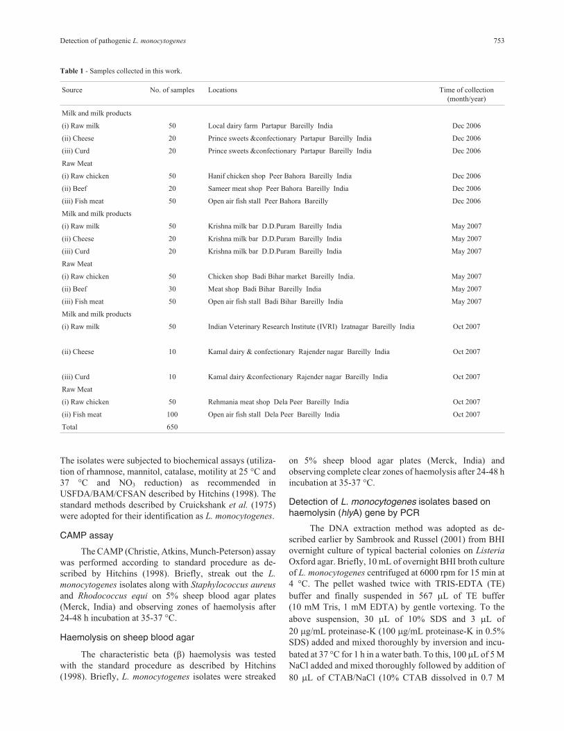
Table 1 - Samples collected in this work.
Source No. of samples Locations Time of collection
(month/year)
Milk and milk products
(i) Raw milk 50 Local dairy farm Partapur Bareilly India Dec 2006
(ii) Cheese 20 Prince sweets &confectionary Partapur Bareilly India Dec 2006
(iii) Curd 20 Prince sweets &confectionary Partapur Bareilly India Dec 2006
Raw Meat
(i) Raw chicken 50 Hanif chicken shop Peer Bahora Bareilly India Dec 2006
(ii) Beef 20 Sameer meat shop Peer Bahora Bareilly India Dec 2006
(iii) Fish meat 50 Open air fish stall Peer Bahora Bareilly Dec 2006
Milk and milk products
(i) Raw milk 50 Krishna milk bar D.D.Puram Bareilly India May 2007
(ii) Cheese 20 Krishna milk bar D.D.Puram Bareilly India May 2007
(iii) Curd 20 Krishna milk bar D.D.Puram Bareilly India May 2007
Raw Meat
(i) Raw chicken 50 Chicken shop Badi Bihar market Bareilly India. May 2007
(ii) Beef 30 Meat shop Badi Bihar Bareilly India May 2007
(iii) Fish meat 50 Open air fish stall Badi Bihar Bareilly India May 2007
Milk and milk products
(i) Raw milk 50 Indian Veterinary Research Institute (IVRI) Izatnagar Bareilly India Oct 2007
(ii) Cheese 10 Kamal dairy & confectionary Rajender nagar Bareilly India Oct 2007
(iii) Curd 10 Kamal dairy &confectionary Rajender nagar Bareilly India Oct 2007
Raw Meat
(i) Raw chicken 50 Rehmania meat shop Dela Peer Bareilly India Oct 2007
(ii) Fish meat 100 Open air fish stall Dela Peer Bareilly India Oct 2007
Total 650
NaCl), after proper mixing and incubation at 35 °C for
10 min in a water bath. An equal volume (0.7-0.8 �L) of
phenol; chloroform; isoamyl alcohol (25:24:1) was added,
mixed thoroughly and centrifuged at 13,000 rpm for
20 min. A thick white layer of protein and polysaccharide
was formed at the interface of the two layers. The upper
aqueous phase was transferred to a fresh microcentrifuge
tube. Equal volume of phenol; chloroform; isoamyl alcohol
was again added to the aqueous supernatant, re-extracted
by thorough mixing and spun at 13,000 rpm for 10-15 min.
The supernatant was transferred to a fresh microcentrifuge
tube. The procedure was repeated once again. To the final
supernatant, 0.6 volumes of isopropanol added at room
temperature and centrifuged at 13,000 rpm for 5 min then
rinsed with 400 �L of 70% ethanol. The DNA was har-
vested by spinning at 13,000 rpm for 5 min. The pellet was
dried and resuspended in 50 �L sterile triple glass distilled
water, added RNase (10 �g/mL) and incubated for 20 min
at room temperature.
The primers (synthesized by Genuine Chemical Cor-
poration, India) for the virulence associated gene hlyA were
used as described earlier (Furrer et al., 1991). The amplifi-
cation of hlyA gene (234 bp) by PCR was performed as fol-
lows: 2.5 �L of 10X PCR buffer (20 mM Tris HCl, pH 8.0
at 25 °C, 100 mM KCl, 0.1 mM EDTA, 1 mM DTT, 50%
glycerol, 0.5% tween 20 and 0.5% Nonidet-P40); 1.5 mM
MgCl2; 2.5 mM of dNTPs mix; 1.0 �L of both forward and
reverse primers (15 pmol); 1 U of Taq DNA polymerase en-
zyme; and 2 �L of DNA as template. Nuclease free water
was added to obtain a final volume of 25 �L. PCR condi-
tions included an initial denaturation step at 95 °C for
5 min, followed by 40 subsequent cycles consisting of heat
denaturation at 95 °C for 30 s, annealing at 54 °C for 1 min,
and extension at 72 °C for 1 min. A final extension was per-
formed at 72 °C for 5 min. PCR products were separated in
1.5% agarose gel electrophoresis, stained by ethidium bro-
mide (Sigma-Aldrich, USA), and visualized under UV
light.
Detection of L. monocytogenes isolates based onvero cell cytotoxicity assay
The cell free culture supernatant (CFCS) was pre-
pared as described by Farber and Speirs (1987). Briefly,
each L. monocytogenes isolate was inoculated in 5 mL of
Tryptic Soy Broth (TSB) (Himedia, Mumbai, India) and in-
cubated at 35 °C for 24 h. About 2 mL of broth culture was
re-inoculated into 20 mL of TSB and incubated at 35 °C for
16 h in a low speed shaking water bath. The bacterial cells
pelleted by centrifugation (10,000 x g) at 4 °C for 30 min,
and the resultant supernatant was filtered through 0.22 �m
pore size membrane filter (Nalgene, India). The sterility of
CFCS was checked by streaking on Tryptic Soy Agar
(TSA) plates (Hi-media, Mumbai, India), incubated at
35 °C for 24 h. The sterile CFCS were then stored at -20 °C.
The vero cell lines were propagated with a standard
method described by Konowalchuk et al. (1978). Glasgow
minimum essential medium (G-MEM) (Gibco, Mumbai,
India) of pH 7.4, containing sodium bicarbonate solution
(2.7%), was supplemented with New Born Calf Serum
(NBCS) (10% for growth and 20% for maintenance), used
for propagation and maintenance of the cell lines. The
trypsin-versene solution and Glasgow minimum essential
medium (G-MEM) were sterilized by Seitz filtration, and
incubated overnight at 37 °C, to check sterility. The cell
lines were maintained by regular sub-culturing.
The toxin assay was performed in 6-well microtiter
tissue culture plates (SPL life sciences, Korea) as the stan-
dard method described by Konowalchuk et al. (1978). Vero
cell culture in each well was seeded and allowed to grow as
confluent monolayers for 24-48 h. At the same time media
was discarded, monolayers were washed twice with Hank’s
Balanced Salt Solution (HBSS) (Difco, Haryana, India).
Volumes of 30 �L and 50 �L of CFCS were inoculated into
each well in duplicate, and 400 �L of the medium supple-
mented with New Born Calf Serum (NBCS) was then
added into each well. The microtiter plates were then incu-
bated at 35 °C under 5% CO2 atmosphere and examined
daily by inverted microscope (Olympus, Japan) up to
3 days, for changes in morphological characteristics of vero
cell lines.
Results
A total of 650 samples (400 from raw meat and 250
from raw milk and milk products) collected from various
locations were examined by cultural and biochemical
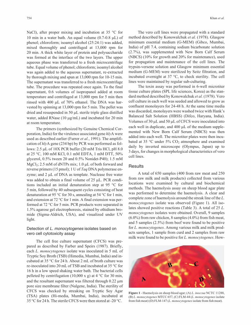
methods. The haemolysis assay on sheep blood agar plate
was performed to determine the haemolysis. A clear and
complete zone of haemolysis around the streak line of the L.
monocytogenes isolate was observed (Figure 1). All iso-
lates showed positive reactions (Table 3). A total of 25 L.
monocytogenes isolates were obtained. Overall, 9 samples
(6.0%) from raw chicken, 8 samples (4.0%) from fish meat,
and 5 samples (2.5%) from beef were found to be positive
for L. monocytogenes. Among various milk and milk prod-
ucts samples, 1 sample from curd and 2 samples from raw
milk were found to be positive for L. monocytogenes. How-
754 Khan et al.
Figure 1 - Haemolysis on sheep blood agar; (A) L. inoccua NCTC 11288,
(B) L. monocytogenes MTCC 657, (C) FLM-44 (L. monocytogenes isolate
from fish meat) (D) FLM-147 (L. monocytogenes isolate from fish meat).
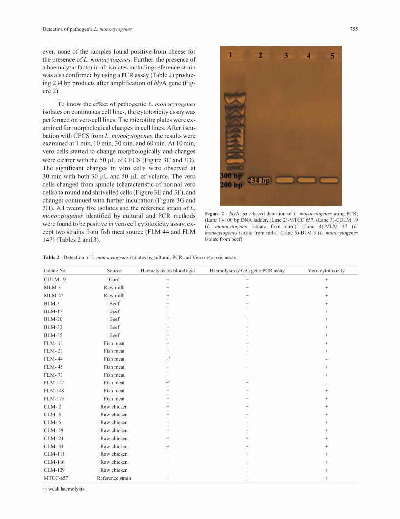
ever, none of the samples found positive from cheese for
the presence of L. monocytogenes. Further, the presence of
a haemolytic factor in all isolates including reference strain
was also confirmed by using a PCR assay (Table 2) produc-
ing 234 bp products after amplification of hlyA gene (Fig-
ure 2).
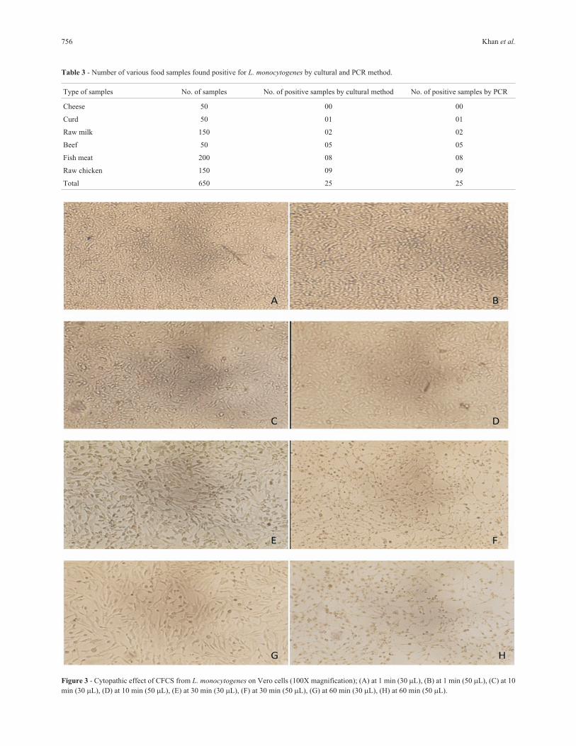
To know the effect of pathogenic L. monocytogenes
isolates on continuous cell lines, the cytotoxicity assay was
performed on vero cell lines. The microtitre plates were ex-
amined for morphological changes in cell lines. After incu-
bation with CFCS from L. monocytogenes, the results were
examined at 1 min, 10 min, 30 min, and 60 min. At 10 min,
vero cells started to change morphologically and changes
were clearer with the 50 �L of CFCS (Figure 3C and 3D).
The significant changes in vero cells were observed at
30 min with both 30 �L and 50 �L of volume. The vero
cells changed from spindle (characteristic of normal vero
cells) to round and shrivelled cells (Figure 3E and 3F), and
changes continued with further incubation (Figure 3G and
3H). All twenty five isolates and the reference strain of L.
monocytogenes identified by cultural and PCR methods
were found to be positive in vero cell cytotoxicity assay, ex-
cept two strains from fish meat source (FLM 44 and FLM
147) (Tables 2 and 3).
Detection of pathogenic L. monocytogenes 755
Table 2 - Detection of L. monocytogenes isolates by cultural, PCR and Vero cytotoxic assay.
Isolate No. Source Haemolysis on blood agar Haemolysin (hlyA) gene PCR assay Vero cytotoxicity
CULM-19 Curd + + +
MLM-31 Raw milk + + +
MLM-47 Raw milk + + +
BLM-3 Beef + + +
BLM-17 Beef + + +
BLM-20 Beef + + +
BLM-32 Beef + + +
BLM-35 Beef + + +
FLM- 13 Fish meat + + +
FLM- 21 Fish meat + + +
FLM- 44 Fish meat +a + -
FLM- 45 Fish meat + + +
FLM- 73 Fish meat + + +
FLM-147 Fish meat +a + -
FLM-148 Fish meat + + +
FLM-173 Fish meat + + +
CLM- 2 Raw chicken + + +
CLM- 5 Raw chicken + + +
CLM- 6 Raw chicken + + +
CLM- 19 Raw chicken + + +
CLM- 24 Raw chicken + + +
CLM- 43 Raw chicken + + +
CLM-111 Raw chicken + + +
CLM-116 Raw chicken + + +
CLM-129 Raw chicken + + +
MTCC-657 Reference strain + + +
+: weak haemolysis.
Figure 2 - hlyA gene based detection of L. monocytogenes using PCR;
(Lane 1)-100 bp DNA ladder, (Lane 2)-MTCC 657, (Lane 3)-CULM 19
(L. monocytogenes isolate from curd), (Lane 4)-MLM 47 (L.
monocytogenes isolate from milk), (Lane 5)-BLM 3 (L. monocytogenes
isolate from beef).
756 Khan et al.
Table 3 - Number of various food samples found positive for L. monocytogenes by cultural and PCR method.
Type of samples No. of samples No. of positive samples by cultural method No. of positive samples by PCR
Cheese 50 00 00
Curd 50 01 01
Raw milk 150 02 02
Beef 50 05 05
Fish meat 200 08 08
Raw chicken 150 09 09
Total 650 25 25
Figure 3 - Cytopathic effect of CFCS from L. monocytogenes on Vero cells (100X magnification); (A) at 1 min (30 �L), (B) at 1 min (50 �L), (C) at 10
min (30 �L), (D) at 10 min (50 �L), (E) at 30 min (30 �L), (F) at 30 min (50 �L), (G) at 60 min (30 �L), (H) at 60 min (50 �L).
Discussion
Various meat and milk products are frequently con-
sumed in all over the world, including India, and they can
act as transmission vehicles for L. monocytogenes. Several
reports revealed L. monocytogenes contamination in beef
(Bohaychuk et al., 2006; Stavru et al., 2011), chicken
(Genigeorgis et al., 1989; Bailey et al., 1990; Mahmood et
al., 2003; Paszkowska et al., 2005), fish (Furrer et al.,
1991; Kargar and Ghasemi, 2009), milk and milk products
(Barbuddhe et al., 2000; Jaradat et al., 2002; Sudershan et
al., 2009; Saikia and Joshi, 2010) in the present investiga-
tion. Among all the samples from all sources, the higher in-
cidence of L. monocytogenes was noticed with meat
samples. In developing countries like India, the possible
reason behind this may be the use of chopping boards,
mincing machines, cleaning clothes and carcasses, which
are frequently used in butcheries and kept for an indefinite
period of time. Moreover, methods of sterilization or disin-
fection with low efficacy are adopted to keep these equip-
ment and utensils sanitized (Nayak et al., 2010). Our
investigation provided an important insight about the L.
monocytogenes contamination in various meat, milk and
milk products in Bareilly, India, and other states where con-
sumers are regularly supplied with contaminated food. It
was observed that there is a need to implement HACCP in
the unorganised sector, as it accounts for a large amount of
food produced and processed in India.
The positive CAMP and haemolysis assay and the
presence of hlyA gene by PCR in all isolates suggest that
these virulent strains may have the potential to invade host
cells and cause listeriosis (Ryser and Marth, 2007). Due to
no confirmation of the pathogenic nature of the L.
monocytogenes isolates, the vero cell cytotoxicity assay
was performed. According to assay results, all twenty five
L. monocytogenes isolates, including the reference strain,
showed pathogenic effect on vero cell lines, except two
strains from fish meat (FLM 44 and FLM 147), that showed
weak haemolytic pattern. These two fish meat isolates were
also confirmed by PCR for the exact presence of a
haemolytic factor, and they were positive for the presence
of LLO. However, these two strains exhibited no
cytotoxicity in vero cell assay. L. monocytogenes is a stron-
ger producer of haemolysin and pathogenic for humans and
mice. It has also been described that the ability of L.
monocytogenes to produce haemolysin is strongly corre-
lated with its virulence. However, some strains produce
weak haemolysis reactions, that can appear to be negative
during culture base biochemical assay (Farber and Speirs,
1987). Thus, through the phenotypic interpretation, it is dif-
ficult to determine haemolysis of L. monocytogenes strains
(USFDA/BAM/CFSAN, 1998). In our knowledge, there
are very few literatures available on cytotoxic effect of L.
monocytogenes on vero cells, especially from food sources
(Farber and Speirs, 1987). The vero cell cytotoxicity assay
described in this work could be suggested as an alternative
or adjunct assay for haemolytic L. monocytogenes strains
identification. The test is easy to perform, and a positive re-
action can occur within 10 min of addition of undiluted bac-
terial culture filtrates. However, weak haemolytic strains
are matter for further investigation.
Acknowledgement
We express our thanks to the Director, IVRI and
Head, Department of Biochemistry and Department of Ani-
mal Biotechnology, IVRI, for providing facilities, Vero
cells and their constant encouragement.
References
Adzitey F, Huda N (2010) Listeria monocytogenes in foods: inci-
dences and possible control measures. Afr J Microbiol Res
4:2848-2855.
Amagliani G, Brandi G, Omiccioli E, Casiere A, Bruce IJ, Magna-
ni M (2004) Direct detection of Listeria monocytogenes
from milk by magnetic based DNA isolation and PCR. Food
Microbiol 21:597-603.
Anon (1996) Rapporti ISTISAN ISSN 1123-3117, 96/35. In
Amaglianni G, Giammarini C, Omiccioli E, Brandi G,
Magnani M (2007) Detection of Listeria monocytogenes us-
ing a commercial PCR kit and different DNA extraction
methods. Food Cont 18:1137-1142.
Aznar R, Alarcon B (2002) On the specificity of PCR detection of
Listeria monocytogenes in food: a comparison of published
primers. Syst Appl Microbiol 25:109-119.
Aznar R, Elizaquivel P (2008) Reliability of Listeria
monocytogenes identification by specific PCR assessed by
phenotypic and genotypic techniques. Food Anal Meth
1:243-251.
Bailey JS, Fletcher DL, Cox NA (1990) Listeria monocytogenes
colonization of broiler chickens. Poultry Sci 69:457-461.
Barbuddhe SB, Malik SVS, Bhilegaonkar KN, Kumar P, Gupta
LK (2000) Isolation of Listeria monocytogenes and anti-
listeriolysin O detection in sheep and goats. Small Rumi Res
38:151-155.
Bohaychuk VM, Gensler GE, King RK, Manninen KI, Sorensen
O, Wu JT, Stiles ME, Mcmullen LM (2006) Occurrence of
pathogens in raw and ready-to-eat meat and poultry products
collected from the retail market place in Edmonton, Alberta,
Canad J Food Prot 69:2176-2182.
Cruickshank R, Duguid JP, Marmion BP, Swain RHA (1975)
Medical microbiology- the practice of medical microbiol-
ogy. Churchill Livingstone, Edinberg, London and New
York.
Dumen E, Baca AU, Dumen E (2008) Comparative detection of
Listeria monocytogenes in raw milk by microbiological
method and PCR. Medycyna Wet 1:59-63.
El-Malek AMA, Ali SFH, Hassanein R, Mohamed MA, Elsayh
KI (2010) Occurrence of Listeria species in meat, chicken
products and human stools in Assiut city, Egypt with PCR
use for rapid identification of Listeria monocytogenes. Vet.
World 3:353-359.
Farber JM, Speirs JI (1987) Potential use of continuous cell lines
to distinguish between pathogenic and non pathogenic Liste-
ria spp. J Clin Microbiol 25:1463-1466.
Detection of pathogenic L. monocytogenes 757

Fuchs JA, Surendran PK (1989) Incidence of Listeria in tropical
fish and fishery products. Lett Appl Microbiol 9:49-51.
Furrer B, Candrian V, Hofelein C, Luthy J (1991) Detection and
identification of Listeria monocytogenes in cooked sausage
products and in milk by in vitro amplification of haemolysin
gene fragments. J Appl Bacteriol 70:372-379.
Genigeorgis CA, Dutulescu D, Garayzabel JF (1989) Prevalence
of Listeria spp. in turkey meat at supermarket and slaughter
house level. J Food Prot 52:618-24.
Gouws PA, Liedemann I (2005) Evaluation of Diagnostic PCR
for the Detection of Listeria monocytogenes in Food Prod-
ucts. Food Tech Biotech 43:201-205.
Hitchins AD (1998). Detection and enumeration of Listeria
monocytogenes in foods. Bacteriological Analytical Manual
Online. Available at:
http://www.cfsan.fda.gov/~ebam/bam-10.html. Accessed 6
March 2008.
Ikeh MAC, Obi SKC, Ezeasor, DN, Ezeonu IM, Moneke AN
(2010) Incidence and pathogenicity profile of Listeria sp.
isolated from food and environmental samples in Nsukka,
Nigeria. Afr J Biotechnol 9:4776-4782.
Jaradat ZW, Schutze GE, Bhunia AK (2002) Genetic homogene-
ity among Listeria monocytogenes strains from infected pa-
tients and meat products from two geographic locations de-
termined by phenotyping, ribotyping and PCR analysis of
virulence genes. Int J Food Microbiol 76:1-10.
Kargar M, Ghasemi A (2009) Role of Listeria monocytogenes
hlyA gene isolated from fresh cheese in human habitual
abortion in Marvdasht. Iran J Clin Infect Dis 4:214-218.
Karunasagar I, Segar K, Karunasagar I, Goebel W (1992) Inci-
dence of Listeria spp. in tropical sea foods. XI International
symposium on problems of listeriosis, Copenhagen Den-
mark, Abstract 155.
Kathariou S, Pine L (1991) The type strain(s) of Listeria
monocytogenes: A source of continuing difficulties. Int J
Syst Bacteriol 41:328-330.
Khan JA, Rathore RS, Ahmad I, Khan S (2011) Molecular Strat-
egies: Detection of Foodborne Bacterial Pathogens. In:
Ahmad, I., Ahmad F., Pitchel, J. (eds). Microbes and Micro-
bial Technology. Springer, NewYork, pp 189-206.
Konowalchuk J, Dickie N, Stavric S, Speirs J (1978) Properties of
an Escherichia coli cytototoxin. Infect Immun 20:575-577.
Mahmood MS, Ahmed AN, Hussain I (2003) Prevalence of Liste-
ria monocytogenes in poultry meat, poultry meat products
and other related inanimates at Faisalabad. Pak J Nut
2:346-349.
Mengaud J, Vicente MF, Chenevert J, Pereira JM, Geoffroy C,
Gicquel-Sanzey B, Baquero F, Perez-Diaz JC, Cossart P
(1988) Expression in Escherichia coli and sequence analysis
of the listeriolysin O determinant of L. monocytogenes. In-
fect Immun 56:766-772.
Nayak JB, Brahmbhatt MN, Savalia, CV, Bhong CD, Roy A,
Kalyani IH, Parmar BC (2010) Detection and characteriza-
tion of Listeria species from buffalo meat. Buff Bull 29:83-
94.
Parihar VS, Barbuddhe, SB, Danielsson-Tham, ML, Tham W
(2008) Isolation and characterization of Listeria species
from tropical seafoods. Food Cont 19:566-569.
Paszkowska KK, Bania J, Bystron J, Molenda J, Czerw M (2005)
Occurrence of Listeria spp. in raw poultry meat and poultry
meat products. Bull Vet Inst Pul 49:219-222.
Ryser ET, Marth EH (1991) Listeria, Listeriosis and Food Safety.
Marcel Dekker, New York (Basel).
Ryser ET, Marth EH (2007) Listeria, Listeriosis, and Food Safety.
Taylor and Francis, Boca Raton, FL.
Saikia P, Joshi SR (2010) Retail market poultry meats of North-
East India- A microbiological survey for pathogenic con-
taminants. Res J Microbiol 5:36-43.
Sambrook J, Russel DW (2001) Molecular Cloning: A Laboratory
Manual. Cold Spring Harbor Press, New York.
Stavru F, Frederic B, Anna S, Daniel R, Pascale C (2011) Listeria
monocytogenes transiently alters mitochondrial dynamics
during infection. PNAS.
www.pnas.org/cgi/doi/10.1073/pnas.1100126108.
Sudershan RV, Rao P, Polasa K (2009) Food safety research in In-
dia: a review. Asian J Food Ag Ind 2:412-433.
Swaminathan B, Gerner-Smidt P (2007) The epidemiology of hu-
man listeriosis. Microbes Infect 9:1236-1243.
USFDA/BAM/CFSAN (1998). Bacteriological analytical manual
online. Available at:
www.fda.gov/Food/ScienceResearch/LaboratoryMethods/
Bacter iologicalAnalyticalManualBAM/default.htm.
All the content of the journal, except where otherwise noted, is licensed under a
Creative Commons License CC BY-NC.
758 Khan et al.