Impact of cocoa flavanol intake on age dependent...
14
Impact of cocoa flavanol intake on age- dependent vascular stiffness in healthy men: a randomized, controlled, double- masked trial Article Published Version Creative Commons: Attribution 3.0 (CC-BY) Open Access Heiss, C., Sansone, R., Karimi, H., Krabbe, M., Schuler, D., Rodriguez Mateos, A., Krämer, T., Cortese-Krott, M. M., Kuhnle, G. G. C., Spencer, J. P. E., Schroeter, H., Merx, M. W. and Kelm, M. (2015) Impact of cocoa flavanol intake on age-dependent vascular stiffness in healthy men: a randomized, controlled, double-masked trial. AGE, 37 (3). 56. ISSN 0161-9152 doi: https://doi.org/10.1007/s11357-015-9794-9 Available at http://centaur.reading.ac.uk/40267/ It is advisable to refer to the publisher’s version if you intend to cite from the work. See Guidance on citing . To link to this article DOI: http://dx.doi.org/10.1007/s11357-015-9794-9 Publisher: Springer All outputs in CentAUR are protected by Intellectual Property Rights law, including copyright law. Copyright and IPR is retained by the creators or other copyright holders. Terms and conditions for use of this material are defined in
Transcript of Impact of cocoa flavanol intake on age dependent...

Impact of cocoa flavanol intake on agedependent vascular stiffness in healthy men: a randomized, controlled, doublemasked trial Article
Published Version
Creative Commons: Attribution 3.0 (CCBY)
Open Access
Heiss, C., Sansone, R., Karimi, H., Krabbe, M., Schuler, D., Rodriguez Mateos, A., Krämer, T., CorteseKrott, M. M., Kuhnle, G. G. C., Spencer, J. P. E., Schroeter, H., Merx, M. W. and Kelm, M. (2015) Impact of cocoa flavanol intake on agedependent vascular stiffness in healthy men: a randomized, controlled, doublemasked trial. AGE, 37 (3). 56. ISSN 01619152 doi: https://doi.org/10.1007/s1135701597949 Available at http://centaur.reading.ac.uk/40267/
It is advisable to refer to the publisher’s version if you intend to cite from the work. See Guidance on citing .
To link to this article DOI: http://dx.doi.org/10.1007/s1135701597949
Publisher: Springer
All outputs in CentAUR are protected by Intellectual Property Rights law, including copyright law. Copyright and IPR is retained by the creators or other copyright holders. Terms and conditions for use of this material are defined in

the End User Agreement .
www.reading.ac.uk/centaur
CentAUR
Central Archive at the University of Reading
Reading’s research outputs online

Impact of cocoa flavanol intake on age-dependent vascularstiffness in healthy men: a randomized, controlled,double-masked trial
Christian Heiss & Roberto Sansone & Hakima Karimi & Moritz Krabbe &
Dominik Schuler & Ana Rodriguez-Mateos & Thomas Kraemer &
Miriam Margherita Cortese-Krott & Gunter G. C. Kuhnle & Jeremy P. E. Spencer &
Hagen Schroeter & Marc W. Merx & Malte Kelm & for the FLAVIOLA Consortium,European Union 7th Framework Program
Received: 8 February 2015 /Accepted: 15 May 2015# The Author(s) 2015. This article is published with open access at Springerlink.com
Abstract Increased vascular stiffness, endothelialdysfunction, and isolated systolic hypertension arehallmarks of vascular aging. Regular cocoa flavanol(CF) intake can improve vascular function in healthyyoung and elderly at-risk individuals. However, themechanisms underlying CF bioactivity remain large-ly unknown. We investigated the effects of CF in-take on cardiovascular function in healthy youngand elderly individuals without history, signs, orsymptoms of cardiovascular disease by applyingparticular focus on functional endpoints relevant tocardiovascular aging. In a randomized, controlled,
double-masked, parallel-group dietary interventiontrial, 22 young (<35 years) and 20 elderly (50–80 year) healthy, male non-smokers consumed eithera CF-containing drink (450 mg CF) or nutrient-matched, CF-free control drink bi-daily for 14 days.The primary endpoint was endothelial function asmeasured by flow-mediated vasodilation (FMD).Secondary endpoints included cardiac output, vascu-lar stiffness, conductance of conduit and resistancearteries, and perfusion in the microcirculation.Following 2 weeks of CF intake, FMD improvedin young (6.1±0.7 vs. 7.6±0.7 %, p<0.001) andelderly (4.9 ± 0.6 vs. 6.3 ± 0.9 %, p< 0.001).Secondary outcomes demonstrated in both groupsthat CF intake decreased pulse wave velocity andlowered total peripheral resistance, and increasedarteriolar and microvascular vasodilator capacity,red cell deformability, and diastolic blood pressure,while cardiac output remained affected. In the elder-ly, baseline systolic blood pressure was elevated,driven by an arterial-stiffness-related augmentation.CF intake decreased aortic augmentation index(−9 %) and thus sys to l i c b lood pres sure(−7 mmHg; Clinicaltrials.gov: NCT01639781). CFintake reverses age-related burden of cardiovascularrisk in healthy elderly, highlighting the potential ofdietary flavanols to maintain cardiovascular health.
Keywords Age . Blood pressure . Arterial stiffness .
Endothelial function . Nutrition
AGE (2015) 37:56 DOI 10.1007/s11357-015-9794-9
Christian Heiss and Roberto Sansone contributed equally to thiswork.
C. Heiss (*) : R. Sansone :H. Karimi :M. Krabbe :D. Schuler :A. Rodriguez-Mateos : T. Kraemer :M. M. Cortese-Krott :M. W. Merx :M. KelmDivision of Cardiology, Pulmonology, and VascularMedicine, University Duesseldorf, Medical Faculty,Moorenstr. 5, 40225 Duesseldorf, Germanye-mail: [email protected]
G. G. C. Kuhnle : J. P. E. SpencerDepartment of Food and Nutritional Sciences, UniversityReading, Reading, UK
H. SchroeterMars Inc., McLean, VA, USA
M. KelmCardiovascular Research Institute Duesseldorf, UniversityDuesseldorf, Medical Faculty, Duesseldorf, Germany

Introduction
The mechanisms underlying vascular aging are largelyunknown but involve all segments of the arterial vascu-lar system. Hallmarks of vascular aging are not entirelyidentical to those of atherosclerosis, but increasing age isassociated with endothelial dysfunction and vascularstiffening, as well as isolated systolic hypertension,and a substantial excess of cardiovascular mortality(Kaess et al. 2012). Due to increased arterial stiffness,older humans exhibit an increased pulse wave velocity(PWV). In young individuals, the reflected pulse wavereturns to the heart and increases pressure during dias-tole and therefore contributes to the Bwindkessel effect^(i.e., elastic reservoir) of the aorta. In older individuals,the pulse wave propagates faster as compared to theyoung, and its return to the heart coincides with thesystolic forward flow, thus leading to aortic augmenta-tion of systolic blood pressure (SBP; Fig. 1).Furthermore, stiff arteries are characterized by a de-creased absorption of the pulse wave, resulting in in-creasedmechanical stress on the arterioles, which in turnis thought to cause damage to the microvasculature,leading to structural microvascular remodeling andrarefication (Mitchell 2008). In addition to an increasedperipheral total resistance, age-dependently reduced mi-crocirculatory perfusion, systemic endothelial dysfunc-tion, and intimal hyperplasia are observed in elderlysubjects (Celermajer et al. 1994; Rossi et al. 2002).There is great interest in dietary interventions to slowor even reverse the complex pattern of these age-associated vascular alterations, which increase cardio-vascular risk (Shai et al. 2010).
Diet is a major determinant of healthy aging, andcertain bioactives may be capable of contributing tothe maintenance of health, thus extending a healthy lifespan (Estruch et al. 2013; Williamson et al. 2009).Flavanols, a subgroup of dietary plant-derived bioac-tives, have gained increasing attention, as clinical die-tary interventions have shown that a higher intake offlavanol-containing foods can increase arterial functionin individuals at risk for cardiovascular disease (CVD),and in those with established CVD (Heiss et al. 2003;Heiss et al. 2010b). Although the mechanisms of actionunderlying the biological effects of flavanols are notcompletely understood, flavanols are one of few bioac-tives today, for which causality between the dietaryintake and an improvement in arterial function has beenestablished (Loke et al. 2008; Schroeter et al. 2006).
Whether or not the intake of cocoa flavanol (CF) has thepotential to improve cardiovascular function in healthyelderly individuals or to reverse age-related vasculardysfunction has not been specifically evaluated thus far.
The FLAVIOLA research project, funded by theEuropean Union’s 7th framework program, was createdto develop the BTargeted delivery of dietary flavanolsfor optimal human cell function: Effects on cardiovas-cular health.^ In order to understand the hemodynamicmechanisms that underlie the potential CF-intake-mediated reversal of age-associated changes in circula-tory function, the present FLAVIOLA AGE study aimsat elucidating in great detail the effects of cocoaflavanols in healthy individuals. We therefore studiedin young and elderly healthy individuals the effect of adietary CF intervention on hallmarks of cardiovascularaging in all segments of the cardiovascular system, suchas cardiac performance, endothelial dysfunction, in-creased systolic blood pressure, vascular stiffness, andmicrocirculatory functions.
Material and methods
Study participants and study design
We recruited YOUNG (<35 years) and ELDERLY (50–80 year) healthy male Caucasian adult subjects withouthistory, signs, or symptoms indicative of cardiovasculardisease, including previous myocardial infarction,stroke, and peripheral artery disease or current or previ-ous medication (Fig. 2). Participants were randomlyassigned to either the CF intake group (FLAVANOL;450 mg total flavanols two times daily) or a nutrient-matched CF-free group (CONTROL) based on a dou-ble-masked, parallel-group study design. All interven-tions were provided in anonymized sachets. Study par-ticipants were instructed to prepare the drinks by emp-tying the contents of each packet into ∼500 ml of water;drinks were prepared just prior to consumption. Drinkswere consume two times per day, one beverage in themorning with breakfast and one with the evening meal.This regimen was maintained for 14 days, with compli-ance assessed by the collection of empty sachets on thelast study day visit.
Measurements were taken fasted before (baseline)and 1 h after the first drink on day 1 and day 14.Endothelial function (primary end point) was measuredas flow-mediated vasodilation (FMD). Secondary
56 Page 2 of 12 AGE (2015) 37:56

Fig. 1 a Schematic of cardiovascular system and b physiologicalparameters reflecting basic components of the cardiovascular sys-tem. Cardiac function is determined by heart rate (HR), cardiacoutput (CO), stroke volume (SV), and total peripheral resistance(TPR); the aorta as an elastic artery is characterized by itsphysicomechanical properties: central blood pressure (BP), pulsewave velocity (PWV), and augmentation index (AIX); conduit
artery function is determined by flow-mediated andnitroglycerin-mediated vasodilation (FMD and NMD, respective-ly) as well as peripheral blood pressure that can be determined atthe upper arm and finger; arteriolar conductance is characterizedby forearm blood flow (FBF), cutaneous capillary blood flow bylaser Doppler perfusion imaging (LDPI), and blood rheology byred blood cell (RBC) deformability
AGE (2015) 37:56 Page 3 of 12 56

endpoints included measures of cardiac output, thephysicomechanical properties of the large conduit arter-ies, the dilatory capacity and tone of resistance arteries,and perfusion in the microcirculation (Fig. 1). Tertiaryendpoints included total plasma epicatechin metabolites.The study protocol was approved by the ethics commit-tee of the Heinrich-Heine-University; all subjects gave
written informed consent (Clinicaltrials.gov:NCT01639781).
Test materials
Both interventions used a low-calorie fruit-flavored bev-erage mix (provided by Mars, Inc.), standardized and
Fig. 2 a Study flow (CONSORT diagram) and b study protocol ll (RBC) deformability
56 Page 4 of 12 AGE (2015) 37:56

matched in composition. All beverage mixes were ag-glomerated powders, utilizing a maltodextrin base intowhich flavoring and sweeteners were incorporated. Thebeverage mixes were provided in sachets (7 g, equalsone serving) labeled with an alphanumeric identifier toenable a double-masked study design.
A high flavanol cocoa extract (Cocoapro®-processedcocoa extract, Mars Inc.) was the source of flavanols inthe CF-containing drink. The CF-containing drink(FLAVANOL) provided 450 mg of total cocoa flavanolsper serving (Adamson et al. 1999). The total amount ofCF in milligrams represents the sum of all monomericflavanols and their oligomers (i.e., procyanidins) with adegree of polymerization up to and including 10 (i.e.,DP 1–10). The predominant monomeric flavanol in thisdrink was (−)-epicatechin (see Table 1).
The control beverage mix did not contain any cocoaextract and thus provided 0 mg CF (CONTROL). Giventhe natural presence of theobromine and caffeine incocoa extract, both theobromine and caffeine wereadded to the control beverage mix in order to matchthe composition of alkaloids in the CF-containing testproduct. Coloring was also added so that the 0 mg CFdrink was also indistinguishable in appearance.Compositional details for the 0 mg (CONTROL) and450 mg CF (FLAVANOL) test drinks are provided inTable 2.
Hemodynamic monitoring
Cardiac function was determined by heart rate (HR),cardiac output (CO), stroke volume (SV), and total pe-ripheral resistance (TPR); the function of major conduitarteries, elastic aorta and muscular brachial artery, wascharacterized based on their physicomechanical proper-ties and capacity to dilate (Fig. 1). Physicomechanicalproperties of the aorta are central blood pressure (BP),pulse wave velocity (PWV), and augmentation index(AIX); dilatory capacity of brachial artery was measuredby flow-mediated and nitroglycerin-mediated vasodila-tion (FMD and NMD, respectively) as well as peripheralblood pressure that was determined at the upper arm andfinger; the conductance function of arterioles was evalu-ated as resting and maximal forearm blood flow (FBF)during reactive hyperemia. Important readouts of micro-vascular perfusion: Cutaneous perfusion was evaluatedby laser Doppler perfusion imaging (LDPI) at rest andmaximal perfusion during reactive hyperemia, and blood
rheology was determined as red blood cell (RBC)deformability.
FMD
Brachial artery FMD and nitroglycerin-mediated vasodi-lation (NMD)were measured by ultrasound (Vivid I, GE)in combination with an automated analysis system(Brachial Analyzer, MIA, Iowa City) as described(Heiss et al. 2010a). Brachial artery (BA) FMD wasmeasured by ultrasound (10-MHz transducer; Vivid I,GE) in combination with an automated analysis system(Brachial Analyzer, MIA, Iowa City, IO) in a 21 °C-temperature-controlled room after 15 min of supine rest.
Table 1 Baseline characteristics of study population (t test, mean[SEM])
YOUNG ELDERLY p value
A n 22 20
Age (years) 26±1 60±2 <0.001
BMI (kg/m2) 24.9±0.5 26.5±0.7 0.013
Height (m) 1.83±0.01 1.81±0.01 0.453
Weight (kg) 81±2 88±3 0.079
Creatinine (mg/dl) 1.0±0.03 1·0±0.03 0.991
Total cholesterol (mg/dl) 172±7 207±7 <0.001
LDL cholesterol (mg/dl) 129±7 157±6 0.005
HDL cholesterol (mg/dl) 53±4 54±2 0.900
Triglycerides (mg/dl) 97±44 118±39 0.104
Fasting plasma glucose(mg/dl)
89±2 95±2 0.027
HbA1c (%) 4.8±0.3 4.6±0.4 0.554
SBP (mmHg) 120±2 131±3 0.006
DBP (mmHg) 77±2 82±2 0.011
HR (bpm) 56±2 56±2 0.908
CRP (mg/dl) 0.1±0.03 0.1±0.03 0.692
Hb (mg/dl) 15.3±1.0 15.4±1.1 0.721
Leucocytes (1000/ul) 5.5±0.3 5.8±0.3 0.489
B Smoking history (n) 0 0Medication (n) 0 0
Hx of CVD (n) 0 0
Diabetes mellitus (n) 0 0
Hypercholesterolemia (n) 0 6
Arterial hypertension (n) 0 4
SEM standard error of the mean, BMI body mass index, LDL low-density lipoprotein, HDL high-density lipoprotein, SBP systolicblood pressure, DBP diastolic blood pressure, HR heart rate, CRPC-reactive protein, CVD cardiovascular disease
AGE (2015) 37:56 Page 5 of 12 56

Diameter and Doppler-flow velocity were measured atbaseline and immediately after cuff deflation, at 20, 40,60, and 80 s, and maximal diameter was used to calculateFMD. At the end of each study day, nitroglycerin-mediated vasodilation (NMD) was measured at 4 minafter 400μg sublingual nitroglycerin (Nitrolingual, Pohl).
Hemodynamic monitoring
The Task Force Monitor (CN-Systems, Graz, Austria)was used for continuous beat-to-beat assessment ofcardiovascular variables, including stroke volume, BP,heart rate, and total peripheral resistance by impedancecardiography, which included ECG, phonocardiogra-phy, Finapres (Finapres-Medical-Systems, Amsterdam,Netherlands), and BP monitoring system (Dynamap,Tampa, USA).
BP measurements
Office blood pressure was measured three times after10 min of rest using an automated clinical digital sphyg-momanometer (Dynamap, Tampa, FL, USA) with ap-propriately sized cuff placed around the upper arm atheart level. Furthermore, we determined 24-h ambula-tory BP measurements on the day before day 1 of thestudy and during the last 24 h of the study on day 13
until the clinical visit on day 14. Values are expressed asday, night, and total average. Furthermore, we deter-mined BP at the fingertip using a tonometric device(Finapres Medical Systems, Amsterdam, Netherlands).Values were taken over 5 min and results representaverage of measurements taken. Central BP was derivedfrom peripheral pulse wave analysis obtained withapplanation tonometry using a proprietary transfer func-tion (SphygmoCor®, AtCor medical, Australia).
Pulse wave analysis
Central blood pressure parameters including augmenta-tion index (AIX) were measured by applanation tonom-etry using the SphygmoCor® system. Via a transferfunction, the pressure waveform of the ascending aortawas synthesized. PWV was determined from measure-ments taken at the carotid and femoral artery as de-scribed (Van Bortel et al. 2012).
Forearm blood flow
Forearm blood flow (FBF) was measured by mercury-in-rubber strain gauge plethysmography (Periquant 833,Gutman, Eurasburg, Germany) according to standardtechniques at rest and during reactive hyperemia sec-ondary to 3 min of forearm ischemia.
Assessment of perfusion in cutaneous microcirculationusing laser doppler perfusion imaging
Perfusion of the cutaneousmicrocirculation of the forearmskin was measured at rest and during reactive hyperemiausing a scanning laser Doppler perfusion imager(PeriScan PIM III, Perimed, Sweden) as described(Keymel et al. 2010). The parameter obtained is a globalcirculatory index expressed in arbitrary units (au) integrat-ing the multidirectional average velocities in skin resis-tance arteries (lumen <100 m). After 15-min rest, the laserbeamwas positioned 15 cm above the forearm scanning afield of 200 mm2 on the volar site of the forearm.Microvascular reactivity was assessed duringpostocclusive reactive hyperemia. Following the baselineperfusion (1 min; 20 images), a blood pressure cuff locat-ed at the proximal forearm was inflated to suprasystolicpressure. After the cuff release, the microvascular re-sponse on reactive hyperemia was recorded. Data acqui-sition and analysis were performed by LDPIWin Software
Table 2 Composition of interventional vehicles ingested bi-daily(ND = not detectable)
FLAVANOL CONTROL
Total cocoa flavanols (mg) 450 ND
Monomers (mg) 73 ND
(−)-Epicatechin (mg) 64 ND
(−)-Catechin (mg) 7 ND
(+)-Catechin (mg) 2 ND
(+)-Epicatechin (mg) ND ND
Dimers-decamers (mg) 377 ND
Theobromine (mg) 44 46
Caffeine (mg) 10 6
Fat (g) 0 0
Carbohydrates (g) 6 6
Protein (g) 0.1 0.1
Energy (kcal) 25 25
Sodium (mg) 3 3
Potassium (mg) 95 85
56 Page 6 of 12 AGE (2015) 37:56

(Perimed, Sweden). Maximum perfusion, amplitude ofperfusion (maximum perfusion − baseline perfusion), ra-tio (maximum perfusion / baseline perfusion), percentageincrease ((maximum perfusion − baseline perfusion) /baseline perfusion × 100), time to peak response, and areaunder curve were evaluated without subtracting biologicalzero from the data (Keymel et al. 2010).
Measurement of RBC deformability
RBC deformability was measured by a laser-assistedoptical rotational cell analyzer (LORCA, R&RMechatronics, Hoorn, Netherlands) according to themanufacturers’ instructions as described (Keymel et al.2011). After a 15-min rest in a supine position, bloodwas drawn from the cubital vein, collected in a heparin-ized tube (10 IE/ml, Liquemin 5000 IE, Roche,Grenzach-Wyhlen, Germany), and RBC deformabilitywas measured by the laser-assisted optical rotational cellanalyzer (LORCA, R&R Mechatronics, Hoorn,Netherlands) according to the manufactures’ instruc-tions as described previously. Briefly, 20 μl of wholeblood was diluted 200 times in high-viscosity L–polyvinylpyrrollidone. One milliliters of the suspensionwas transferred into the LORCA device and automati-cally subjected to varying degrees of shear stress at 0.3to 10 Pa by stepwise increases of the rotation speed(Keymel et al. 2011).
Analysis of flavanols and their metabolites in plasma
(−)-Epicatechin and its related metabolites where ana-lyzed in plasma by HPLC-FLD/UVand electrochemicaldetection using authentic standards provided by MarsInc., as previously described (Ottaviani et al. 2012).Prior to analysis, plasma samples (0.5 ml) weredefrosted on ice and then subjected to β-glucuronidaseand sulfatase treatment (2000 units/ml; 40 min; 37 °C).Then, samples were mixed with 2 ml of acidified ice-cold methanol (0.5 % acetic acid in methanol, v/v)containing 3′-O-ethyl-(−)-epicatechin (500 nM) as arecovery standard. Samples were centrifuged at17,000×g for 15 min at 4 °C, and the supernatant wascollected. The pellet was extracted again with 2 ml ofacidified ice-cold methanol (0.5 % acetic acid in meth-anol, v/v) containing 3′-O-ethyl-(−)-epicatechin (500nM), and then with 1 ml of 50 % methanol acidifiedwith 0.5 % acetic acid and containing 3′-O-ethyl-(−)-
epicatechin (500 nM). Combined supernatantsunderwent concentration (to approximately 50 μl) usinga Speedvac system (Thermo Fisher Scientific Inc.,Basingstoke, UK) and were mixed with resorcinol(300 pmol) and catechol (300 pmol) prior to analysisby HPLC. Flavanol monomers and O-methylated me-tabolites were analyzed using a Hewlett-Packard 1200series HPLC (Hewlett-Packard, Palo alto, CA, USA)equipped with diode array and fluorescent detection.Samples (50 μl) were injected onto a reversed-phasePhenomenex Luna C18(2) column (4.6×150 mm) with3-μm particle size. The mobile phase consisted of (A)HPLC grade water, (B) 200mM sodium acetate, pH 4.4/Methanol (84/16), and (C) acetonitrile, and the follow-ing gradient protocol was run: 0 min, 75 % A, 25 % B;5 min, 75 % A, 25 % B; 20 min, 65 % A, 25 % B;28 min, 63 % A, 25 % B; 34 min, 55 % A; 25 % B;41 min, 45 % A, 25 % B; 45 min, 25 % B, 75 % C;55 min, 25% B, 75% C; 56.1 min 75%A, 25% B; and60 min, 75 % A, 25 % B. The flow rate was 0.8 ml/min.Detection of flavanols and their metabolites was per-formed using a fluorescent detector with excitationwavelength of 276 nm and emission wavelength of316 nm. The concentration of each compound wasdetermined using an external calibration curve producedwith the use of authentic standards.
Statistical methods
Results are expressed as mean ± standard error of themean (SEM). Baseline data represent data of first visit(day 1, 0 h). The primary test for an effect was a three-way repeatedmixedmodel measurements ANOVA (onewithin-subject factor: time [day 1, 0 h / day 1,1 h / day14, 0 h / day 14, 1 h]; two between-subject factors: age(YOUNG/ELDERLY) and intervention (CONTROL/FLAVANOL). ANOVA and confidence intervals forpairwise comparisons were computed with SPSS 20(IBM). Correlations were Pearson’s r. Values of p lessthan 0.05 were regarded as statistically significant.
Results
Baseline characteristics
A total of 60 male individuals underwent assessment foreligibility [see CONSORT flow diagram (Fig. 2)]; 22
AGE (2015) 37:56 Page 7 of 12 56

young and 20 elderly subjects were included. None ofthe participants had been previously diagnosed with, orhad clinical signs or symptoms of, cardiovascular dis-ease, such as myocardial infarction, coronary arterydisease, hypertension, diabetes, or hyperlipidemia.During the initial diagnostic workup, we observed mildelevations of systolic blood pressure (n=4) and choles-terol (n=6, see Table 1) in a few elderly individuals,which were in a line with the diagnosis of mild isolatedsystolic hypertension (grade 1) and hypercholesterol-emia. Importantly, none of these mild alterations wouldrequire medication according to current treatment guide-lines (Perk et al. 2012). Compared to the YOUNG, theELDERLY exhibited statistically significantly greaterBMI, total- and LDL-cholesterol, fasting plasma glu-cose, SBP, and diastolic blood pressure (DBP) at base-line, but all these parameters were within limits consid-ered normal, and none of the subjects had an indicationfor medical treatment according to current guidelines.The remaining demographic parameters did not signif-icantly differ between groups (Table 1). All subjectscompleted the study and all data were included in theanalysis. The test drinks were well tolerated and noadverse events were reported.
Baseline hemodynamic characterization of agedvasculature
At baseline on day 1, resting cardiac output was signif-icantly lower in ELDERLY as compared to YOUNG(6.4±0.2 and 5.2±0.3 l/min, p<0.05; Table 3). Giventhat there was no difference in heart rate (57±3 and 57±2 /min, p = n.s.), this was tenably driven by a signifi-cantly lower stroke volume in the ELDERY (107±4 and84±2 ml, p<0.05). Peripheral SBP (121±2 and 128±
2 mmHg, p<0.05) and DBP (76±2 and 81±2 ml,p<0.05) as assessed by standard office measurement,as well as total peripheral resistance (TPR), were signif-icantly greater in ELDERLY. Similarly, greater valueswere observed in 24-h ambulatory Holter monitoring(continuous measurement over 5 min at the fingertip),and central BP. ELDERLY participants exhibited stifferarteries with significantly greater pulse wave velocity(PWV; 5.9±0.2 and 9.4±0.4 m/s, p<0.05) and aorticaugmentation index (AIX; −11±2 and 20±2 %,p<0.05) as compared to YOUNG. Endothelium-dependent and endothelium-independent conduit arteryvasodilator function, asmeasured by FMD (6.3±0.2 and5.2±0.2 %, p<0.05) and nitroglycerin-mediated (NMD;15.0±3 and 12.6±0.5 %, p<0.05), respectively, waslower in the ELDERLY. The baseline diameter of thebrachial artery was greater in ELDERLY subjects (4.4±0.1 and 4.8±0.2 mm, p<0.05). As detailed in Table 3,resting forearm blood flow (FBF) and laser Dopplerperfusion, reflecting primarily skeletal muscle bloodflow (arteriolar cross-sectional area) and cutaneous cap-illary perfusion, respectively, were similar betweenYOUNG and ELDERLY. However, maximal hyper-emic blood flow was lower in ELDERLY as comparedto that in YOUNG, suggesting a lessened functionalcapacity to increase blood flow in response to ischemia.Red blood cell (RBC) deformability was similar inYOUNG and ELDERLY.
Flavanol intervention improves endothelial functionin YOUNG and ELDERLY
The FLAVANOL intervention led to an acute increase inbrachial artery FMD at 1 h after CF ingestion in bothYOUNG (6.1±0.2 to 7.4±0.2 %, p<0.05) and ELDE
Table 3 Summary of hemodynamic readouts (Mean [SEM])Group
Intervention
Day
Hour
CO (l/min) 6.1 ± 0.4 5.8 ± 0.5 6.1 ± 0.2 6.2 ± 0.3 6.4 ± 0.2 6.3 ± 0.2 6.4 ± 0.2 6.0 ± 0.1 5.2 ± 0.3 § 4.9 ± 0.2 § 4.9 ± 0.2 § 5.0 ± 0.2 § 5.2 ± 0.3 § 5.0 ± 0.2 § 5.1 ± 0.1 § 4.7 ± 0.1 §SV (ml) 107 ± 4 101 ± 7 111 ± 5 109 ± 6 109 ± 4 108 ± 7 108 ± 4 109 ± 4 82 ± 6 § 83 ± 6 § 80 ± 5 § 81 ± 5 § 84 ± 2 § 84 ± 3 § 83 ± 4 § 83 ± 4 §Heart rate (/min) 57 ± 3 57 ± 2 58 ± 2 56 ± 2 59 ± 1 57 ± 2 60 ± 3 58 ± 3 56 ± 3 58 ± 2 58 ± 2 55 ± 3 57 ± 2 58 ± 1 59 ± 2 58 ± 2TPR 1142 ± 71 1163 ± 100 1090 ± 47 1173 ± 64 1249 ± 28 940 ± 58 934 ± 86 944 ± 51 1781 ± 152 § 1763 ± 164 § 1812 ± 138 § 1784 ± 132 § 1901 ± 143 § 1527 ± 46 §*# 1485 ± 80 §*# 1473 ± 101 §*#
PWV (m/s) 5.9 ± 0.2 6.1 ± 0.2 5.8 ± 0.2 5.6 ± 0.1 6.0 ± 0.1 5.5 ± 0.2 *# 5.6 ± 0.1 *# 5.4 ± 0.2 *# 9.4 ± 0.4 § 9.5 ± 0.4 § 9.2 ± 0.3 § 9.2 ± 0.6 § 9.3 ± 0.5 § 8.6 ± 0.5 §*# 8.5 ± 0.4 §*# 8.2 ± 0.4 §*#AIX (%) -10.9 ± 2.3 -9.6 ± 3.2 -7.9 ± 3.6 -12.2 ± 3.9 -3.5 ± 2.8 -10.0 ± 2.8 -5.5 ± 2.1 -7.6 ± 2.1 20.4 ± 2.1 § 19.4 ± 1.9 § 20.6 ± 2.8 § 18.2 ± 2.6 § 25.0 ± 2.6 § 18.9 ± 2.3 §*# 16.1 ± 1.9 §*# 13.8 ± 2.8 §*#Central SBP (mmHg) 105 ± 2 104 ± 2 103 ± 2 101 ± 2 104 ± 3 102 ± 3 103 ± 4 104 ± 3 122 ± 2 § 121 ± 3 § 119 ± 4 § 118 ± 3 § 127 ± 6 §*# 123 ± 6 §*# 120 ± 5 §*# 120 ± 4 §*#Central DBP (mmHg) 78 ± 2 77 ± 2 76 ± 3 75 ± 2 78 ± 2 75 ± 2 *# 73 ± 2 *# 72 ± 2 *# 80 ± 2 § 80 ± 1 § 79 ± 1 § 81 ± 1 § 87 ± 3 §*# 83 ± 3 §*# 80 ± 3 §*# 82 ± 3 §*#
BA diameter (mm) 4.4 ± 0.1 4.4 ± 0.1 4.5 ± 0.1 4.0 ± 0.4 4.4 ± 0.1 4.4 ± 0.1 4.0 ± 0.4 4.4 ± 0.1 4.8 ± 0.2 § 4.8 ± 0.2 § 4.8 ± 0.2 § 4.8 ± 0.1 § 5.0 ± 0.9 § 4.9 ± 0.2 § 5.0 ± 0.2 § 5.0 ± 0.2 §FMD (%) 6.3 ± 0.2 6.2 ± 0.2 6.0 ± 0.2 6.3 ± 0.2 6.1 ± 0.2 7.4 ± 0.2 *# 7.8 ± 0.2 *# 7.8 ± 0.1 *# 5.2 ± 0.3 § 5.2 ± 0.32 § 5.0 ± 0.3 § 4.9 ± 0.3 § 4.9 ± 0.2 § 5.9 ± 0.2 §*# 6.3 ± 0.3 §*# 6.3 ± 0.2 §*#NMD (%) 15.0 ± 0.3 15,3 ± 0,3 14.7 ± 0.5 15.0 ± 0.4 12.6 ± 0.5 § 13.7 ± 0.6 § 13.0 ± 0.6 § 13.9 ± 0.3 §Office SBP (mmHg) 121 ± 2 119 ± 2 118 ± 2 119 ± 3 122 ± 1 122 ± 1 119 ± 2 120 ± 2 128 ± 2 § 127 ± 3 § 126 ± 3 § 125 ± 3 § 134 ± 4 § 129 ± 4 §*# 128 ± 3 §*# 127 ± 3 §*#Office DBP (mmHg) 76 ± 2 77 ± 2 73 ± 3 75 ± 2 78 ± 2 72 ± 2 *# 71 ± 2 *# 73 ± 2 *# 81 ± 2 § 79 ± 2 § 77 ± 2 § 78 ± 2 § 83 ± 3 § 78 ± 3 §*# 77 ± 3 §*# 76 ± 3 §*#24 h SBP (mmHg) 126 ± 2 125 ± 3 128 ± 3 124 ± 4 134 ± 2 § 133 ± 3 § 135 ± 3 § 130 ± 2 §#24 h DBP (mmHg) 76 ± 2 77 ± 2 77 ± 1 73 ± 3 # 86 ± 2 § 86 ± 2 § 91 ± 3 § 84 ± 3 §#Finger SBP (mmHg) 114 ± 2 115 ± 2 112 ± 2 114 ± 2 114 ± 4 112 ± 4 111 ± 4 111 ± 4 125 ± 3 § 126 ± 2 § 124 ± 3 § 124 ± 2 § 127 ± 5 § 120 ± 4 §*# 116 ± 4 §*# 119 ± 4 §*#Finger DBP (mmHg) 72 ± 2 72 ± 2 69 ± 2 71 ± 1 71 ± 2 66 ± 1 *# 64 ± 3 *# 64 ± 2 *# 82 ± 2 § 82 ± 2 § 79 ± 2 § 80 ± 2 § 84 ± 3 § 76 ± 3 §*# 72 ± 3 §*# 72 ± 3 §*#
FBF baseline (ml/100 ml*min) 1.5 ± 0.1 1.5 ± 0.1 1.5 ± 0.1 1.4 ± 0.1 1.7 ± 0.1 1.6 ± 0.1 1.6 ± 0.1 1.6 ± 0.1 1.1 ± 0.0 0.9 ± 0,1 0.8 ± 0.0 0.9 ± 0,0 1.5 ± 0.1 1.2 ± 0.1 1.1 ± 0.1 1.1 ± 0.0FBF maximal (ml/100 ml*min) 13.7 ± 1.6 14.3 ± 1.8 13.7 ± 1.7 13.8 ± 1.5 13.2 ± 0.6 16.2 ± 1.0 *# 16.9 ± 0.6 *# 16.9 ± 1.1 *# 11.3 ± 1.2 § 10.8 ± 1.5 § 11.6 ± 1.7 § 11.0 ± 1.5 § 10.9 ± 1.2 § 12.3 ± 1.5 §*# 14.0 ± 1.4 §*# 13.9 ± 1.1 §*#LDPI baseline (PU) 42 ± 1 42 ± 1 42 ± 1 39 ± 1 38 ± 1 40 ± 1 39 ± 1 40 ± 1 41 ± 1 44 ± 0 39 ± 1 40 ± 1 38 ± 1 39 ± 1 40 ± 1 39 ± 1LDPI max (PU) 259 ± 15 270 ± 17 260 ± 22 258 ± 17 257 ± 14 292 ± 15 *# 307 ± 14 *# 296 ± 17 *# 186 ± 5 § 186 ± 8 § 178 ± 5 § 180 ± 5 § 184 ± 15 § 200 ± 14 §*# 214 ± 12 §*# 213 ± 12 §*#RBC deformability 0.334 ± 0.011 0.326 ± 0.010 0.329 ± 0.007 0.329 ± 0.009 0.327 ± 0.008 0.334 ± 0.008 *# 0.332 ± 0.008 *# 0.336 ± 0.010 *# 0.321 ± 0.006 0,323 ± 0.007 § 0.319 ± 0.005 § 0,321 ± 0.007 § 0.325 ± 0.007 § 0.334 ± 0.009 §*# 0.341 ± 0.007 §*#0.341 ± 0.007 §*#
0 10 1 0 1 0 1
1 14
0 1 0 1 0 1 0 1
1 14 1 14 1 14
YOUNG ELDERLY
CONTROL FLAVANOL CONTROL FLAVANOL
p<0.05 versus individual baseline (day 1, 0 h); § p<0.05 versus YOUNG at the same time point and the same intervention group; # p<0.05versus CONTROL in the same age group
56 Page 8 of 12 AGE (2015) 37:56
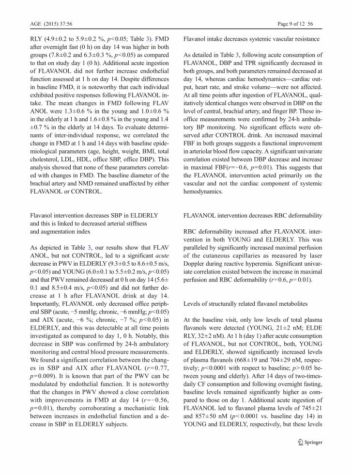
RLY (4.9±0.2 to 5.9±0.2 %, p<0.05; Table 3). FMDafter overnight fast (0 h) on day 14 was higher in bothgroups (7.8±0.2 and 6.3±0.3 %, p<0.05) as comparedto that on study day 1 (0 h). Additional acute ingestionof FLAVANOL did not further increase endothelialfunction assessed at 1 h on day 14. Despite differencesin baseline FMD, it is noteworthy that each individualexhibited positive responses following FLAVANOL in-take. The mean changes in FMD following FLAVANOL were 1.3±0.6 % in the young and 1.0±0.6 %in the elderly at 1 h and 1.6±0.8 % in the young and 1.4±0.7 % in the elderly at 14 days. To evaluate determi-nants of inter-individual response, we correlated thechange in FMD at 1 h and 14 days with baseline epide-miological parameters (age, height, weight, BMI, totalcholesterol, LDL, HDL, office SBP, office DBP). Thisanalysis showed that none of these parameters correlat-ed with changes in FMD. The baseline diameter of thebrachial artery and NMD remained unaffected by eitherFLAVANOL or CONTROL.
Flavanol intervention decreases SBP in ELDERLYand this is linked to decreased arterial stiffnessand augmentation index
As depicted in Table 3, our results show that FLAVANOL, but not CONTROL, led to a significant acutedecrease in PWVin ELDERLY (9.3±0.5 to 8.6±0.5 m/s,p<0.05) and YOUNG (6.0±0.1 to 5.5±0.2 m/s, p<0.05)and that PWVremained decreased at 0 h on day 14 (5.6±0.1 and 8.5±0.4 m/s, p<0.05) and did not further de-crease at 1 h after FLAVANOL drink at day 14.Importantly, FLAVANOL only decreased office periph-eral SBP (acute, −5 mmHg; chronic, −6 mmHg; p<0.05)and AIX (acute, −6 %; chronic, −7 %; p<0.05) inELDERLY, and this was detectable at all time pointsinvestigated as compared to day 1, 0 h. Notably, thisdecrease in SBP was confirmed by 24-h ambulatorymonitoring and central blood pressure measurements.We found a significant correlation between the chang-es in SBP and AIX after FLAVANOL (r=0.77,p=0.009). It is known that part of the PWV can bemodulated by endothelial function. It is noteworthythat the changes in PWV showed a close correlationwith improvements in FMD at day 14 (r=−0.56,p=0.01), thereby corroborating a mechanistic linkbetween increases in endothelial function and a de-crease in SBP in ELDERLY subjects.
Flavanol intake decreases systemic vascular resistance
As detailed in Table 3, following acute consumption ofFLAVANOL, DBP and TPR significantly decreased inboth groups, and both parameters remained decreased atday 14, whereas cardiac hemodynamics—cardiac out-put, heart rate, and stroke volume—were not affected.At all time points after ingestion of FLAVANOL, qual-itatively identical changes were observed in DBP on thelevel of central, brachial artery, and finger BP. These in-office measurements were confirmed by 24-h ambula-tory BP monitoring. No significant effects were ob-served after CONTROL drink. An increased maximalFBF in both groups suggests a functional improvementin arteriolar blood flow capacity. A significant univariatecorrelation existed between DBP decrease and increasein maximal FBF(r=−0.6, p=0.01). This suggests thatthe FLAVANOL intervention acted primarily on thevascular and not the cardiac component of systemichemodynamics.
FLAVANOL intervention decreases RBC deformability
RBC deformability increased after FLAVANOL inter-vention in both YOUNG and ELDERLY. This wasparalleled by significantly increased maximal perfusionof the cutaneous capillaries as measured by laserDoppler during reactive hyperemia. Significant univar-iate correlation existed between the increase in maximalperfusion and RBC deformability (r=0.6, p=0.01).
Levels of structurally related flavanol metabolites
At the baseline visit, only low levels of total plasmaflavanols were detected (YOUNG, 21±2 nM; ELDERLY, 32±2 nM). At 1 h (day 1) after acute consumptionof FLAVANOL, but not CONTROL, both, YOUNGand ELDERLY, showed significantly increased levelsof plasma flavanols (668±19 and 704±29 nM, respec-tively; p<0.0001 with respect to baseline; p>0.05 be-tween young and elderly). After 14 days of two-times-daily CF consumption and following overnight fasting,baseline levels remained significantly higher as com-pared to those on day 1. Additional acute ingestion ofFLAVANOL led to flavanol plasma levels of 745±21and 857±50 nM (p<0.0001 vs. baseline day 14) inYOUNG and ELDERLY, respectively, but these levels
AGE (2015) 37:56 Page 9 of 12 56

were not significantly different from values observedafter acute ingestion on day 1.
Discussion
The FLAVIOLA AGE study is the first study specificallyintended to investigate CF-associated improvements ofhallmarks of cardiovascular aging, on the level of car-diac performance, endothelial dysfunction, increasedsystolic blood pressure, vascular stiffness, and microcir-culatory functions. The outcomes demonstrate how aCF-based dietary intervention affects the cardiovascularsystem and reverses age-related increases of blood pres-sure together with vascular stiffness in healthy elderlyhumans. CF-intake-associated improvements in thecompliance of large arteries were accompanied by alowered pulse wave velocity and decreased aortic aug-mentation of systolic blood pressure. Endothelial func-tion in large conduit arteries was significantly improvedin healthy young and elderly individuals. These benefi-cial effects were associated with an improved dilatorycapacity of resistance arteries, decreased diastolic bloodpressure, and increases in microcirculatory perfusionand red blood cell deformability. These CF-intake-mediated modulations of circulatory function were in-dependent of changes in cardiac performance, sincecardiac output was not affected by CF (Table 3).
CF intake improves endothelial function in youngand elderly
Consistent with previous studies in other populations,ELDERLY exhibited significantly decreasedendothelium-dependent vasodilation at baseline as com-pared to YOUNG (Heiss et al. 2005). So far, no studyhas specifically investigated the vascular effects of CF inhealthy elderly and compared it to young individuals.CF-intake-related improvements in FMD occurredacutely following intake but, more importantly, werealso manifest when comparing baseline FMD (fasted;0 h) on days 1 and 14.Moreover, and despite differencesin baseline FMD, our results show that the absoluteeffect size of FMD improvements was similar in youngand elderly healthy men. It is also noteworthy that thebioavailability of flavanols in the healthy young menwas comparable to that in elderly individuals, as evi-denced by flavanol plasma concentrations.
CF intake attenuates age-related increase in bloodpressure and arterial stiffness
Our results show that the CF-intake-dependent de-crease in BP in ELDERLY was selective to SBP(Table 3). An endothelial function increase along witha decrease in PWV was also present in the YOUNG,but as would be expected from the initial absence ofBP augmentation in this group, these changes did notresult in a modulation of SBP. A similar relationshipbetween baseline BP and the effect size of BP-lowering therapeutic agents, which is in a similarrange as shown here for flavanols, has been reportedfor various drugs, including ACE inhibitors(O’Rourke and Hashimoto 2007). Tenable explana-tions of how flavanols could affect age-related chang-es in SBP and PWV may be found in the observationof improved compliance due to improved endothelialfunction as well as decreases in DBP, and thus chang-es in the distal reflection point of the pulse wave.
CF intake decreases diastolic blood pressureand increases conductance of small arteries
We observed significant decreases in DBP and also inTPR in both young and elderly participants. CFs (30–1080 mg total flavanols per day) have been shown toelicit a small, but significant DBP-reducing effect of−2.2 mmHg in a meta analysis of a heterogeneous studypopulation including patients with and without CVD(Ried et al. 2012). Resistance to blood flow is principal-ly mediated by arteriolar diameter, which is modified byarteriolar vasoconstriction and dilation, respectively. Weobserved a significant increase in the maximal forearmblood flow and cutaneous perfusion during reactivehyperemia. This suggests that CF intake may increasenot only the dilatory function of conduit arteries but alsothat of the arterioles. This effect, together with theincreased RBC deformability, contributes to the en-hanced perfusion of the microcirculation observed fol-lowing CF intake. Mechanistically, CF-related vasculareffects have been linked to an increase in nitric oxidesynthase (NOS) activity (Del Rio et al. 2013; Schroeteret al. 2006). RBCs have been demonstrated to express afunctional eNOS that may modulate RBC deformabilityindependent of vascular tone (Cortese-Krott et al. 2012;Horn et al. 2011). Consequently, our current data sup-port the supposition that CF intake improves tissue
56 Page 10 of 12 AGE (2015) 37:56

perfusion, at least in part, by increasing dilatory capacityof arterioles and increasing RBC deformability.
The FLAVIOLA AGE study and healthy vascular aging
Aging is considered to be an independent cardiovascularrisk factor in most methods of CV risk scoring. Thisraises the important question as to whether or not theregular intake of vasculoprotective bioactives not onlydoes reverse age-associated changes of circulatory func-tion but also has the potential to maintain vascularfunction with increasing age. It is tempting to speculatethat such an approach may ultimately support healthyvascular aging, extend the healthy life span, and lead toa lower rate of cardiovascular events. While such aproposition could only be finally verified throughlong-term longitudinal studies in healthy elderly indi-viduals, it is possible to meaningfully advance our un-derstanding of this question by way of smaller-scaleinvestigations like the FLAVIOLA AGE study, whichcan provide the necessary basis for larger-scale studiesin future. The present study extended our knowledgeregarding the potential role of CF intake in the mainte-nance of optimal cardiovascular function during normalaging. We demonstrated here that CF intake is effectivenot only in healthy young but also in elderly individuals,as evidenced by the CF-related reversal of age-specificalterations in circulatory function, vascular stiffness, andhemodynamics.
Limitations
Intending to specifically focus on mechanistic aspects ingreater detail than can be done practically in a larger-scale and longer-term study, the duration of this studywas chosen to be 2 weeks. This is based on the outcomesof our previous work showing that approximately2 weeks of CF intake at 450 mg two times daily issufficient to establish maximal and sustained improve-ments (plateau) in FMD in this study population.Consequently, it can be argued tenably that despite oflimitations with regard to study duration, our data arerelevant in furthering understanding of pharmacody-namics aspects of CF-mediated changes in endothelialfunction, cardiac output, conductance of conduit andresistance arteries, and perfusion in the microcirculation.An additional limitation regarding the design of this
study is the exclusion of female participants. The ratio-nale for doing so was based on complexities related tothe influence on FMD and other cardiovascular readoutsof hormonal changes that occur in women during themenstrual cycle and during peri-menopause and meno-pause. To assess detailed mechanistic aspects of CF-intake-related changes in cardiovascular function overthe background of menstrual-cycle-dependent modula-tions, it will require a specific and comprehensive studydesign, which was beyond the scope of this investiga-tion. Finally, the elderly subjects in our study presentedwith significantly lower cardiac output as compared tothe young subjects that was not significantly changed bythe flavanol intervention. It has been appreciated for along time that besides stiffening of the arteries, stiffen-ing of the heart occurs with advancing age (Tresch andMcGough 1995), leading to diastolic dysfunction(Upadhya et al. 2015). Whereas a lack of flavanol-related effects on cardiac output may be interpreted asa lack of major flavanol effect on cardiac function, moredetailed studies with echocardiography and potentiallyspiro-ergometry are needed to investigate the impact offlavanols on diastolic function and cardiac performancecapacity.
Conclusions
A CF-based dietary intervention in elderly individualsdecreases systolic blood pressure and improves vascularstiffness. These beneficial effects are associated withimproved endothelial function in large arteries and en-hanced vasodilator function in resistance arteries, aswell as with improved microcirculatory perfusion thatcan already be observed in young healthy participants.Our findings provide clear evidence and novel insightsthat scientifically underpin the potential of dietary CF toreverse, or at least to attenuate, features of circulatorydysfunction associated with vascular aging in healthymen, a major contributor to atherogenic risk indepen-dent of numeric age.
Conflict of interest CH, HS, JPES, GCGK, ARM, MWM, andMK are senior investigators in the FLAVIOLA research consor-tium of the European Union (FP7-KBBE-2008-2B). CH andARM are participants of the EU funded COST 236 ActionFA1403 POSITIVe (Interindividual variation in response to con-sumption of plant food 237 bioactives and determinants involved).Additional funding was provided by an unrestricted grant byMARS, Inc., the Deutsche Forschungsgemeinschaft (KE405/5-1
AGE (2015) 37:56 Page 11 of 12 56

to MK and FOR809 TP7 Me1821/3-1 to MWM & MK), and theForschungskommission of Medical Faculty of the Heinrich-HeineUniversity Duesseldorf to MCK and CH. MARS, Inc. providedthe standardized test drinks used in this investigation. HS isemployed by MARS, Inc., a member of the FLAVIOLA researchconsortium and a company engaged in flavanol research andflavanol-related commercial activities. None of the other authorshas a conflict of interest to declare other than stated above.
Open Access This article is distributed under the terms of theCreative Commons Attribution 4.0 International License(http://creativecommons.org/licenses/by/4.0/), which permitsunrestricted use, distribution, and reproduction in any medium,provided you give appropriate credit to the original author(s) andthe source, provide a link to the Creative Commons license, andindicate if changes were made.
References
Adamson GE et al (1999) HPLC method for the quantification ofprocyanidins in cocoa and chocolate samples and correlation tototal antioxidant capacity. J Agric Food Chem 47:4184–4188
Celermajer DS, Sorensen KE, Spiegelhalter DJ, GeorgakopoulosD, Robinson J, Deanfield JE (1994) Aging is associated withendothelial dysfunction in healthy men years before the age-related decline in women. J Am Coll Cardiol 24:471–476
Cortese-Krott MM et al (2012) Human red blood cells at work:identification and visualization of erythrocytic eNOS activityin health and disease. Blood 120:4229–4237. doi:10.1182/blood-2012-07-442277
Del Rio D, Rodriguez-Mateos A, Spencer JP, TognoliniM, BorgesG, Crozier A (2013) Dietary (poly)phenolics in humanhealth: structures, bioavailability, and evidence of protectiveeffects against chronic diseases. Antioxid Redox Signal 18:1818–1892. doi:10.1089/ars.2012.4581
Estruch R et al (2013) Primary prevention of cardiovascular dis-ease with a Mediterranean diet. N Engl J Med 368:1279–1290. doi:10.1056/NEJMoa1200303
Heiss C, Dejam A, Kleinbongard P, Schewe T, Sies H, Kelm M(2003) Vascular effects of cocoa rich in flavan-3-ols. JAMA290:1030–1031
Heiss C, Keymel S, Niesler U, Ziemann J, Kelm M, Kalka C(2005) Impaired progenitor cell activity in age-related endo-thelial dysfunction. J Am Coll Cardiol 45:1441–1448
Heiss C et al (2010a) Improvement of endothelial function withdietary flavanols is associated with mobilization of circulat-ing angiogenic cells in patients with coronary artery disease. JAm Coll Cardiol 56:218–224
Heiss C, Keen CL, KelmM (2010b) Flavanols and cardiovasculardisease prevention. Eur Heart J 31:2583–2592. doi:10.1093/eurheartj/ehq332
Horn P et al (2011) Nitric oxide influences red blood cell velocityindependently of changes in the vascular tone. Free RadicRes 45:653–661. doi:10.3109/10715762.2011.574288
Kaess BM et al (2012) Aortic stiffness, blood pressure progres-sion, and incident hypertension. JAMA 308:875–881. doi:10.1001/2012.jama.10503
Keymel S et al (2010) Characterization of the non-invasive assess-ment of the cutaneous microcirculation by laser Dopplerperfusion scanner. Microcirculation 17:358–366
Keymel S, Heiss C, Kleinbongard P, Kelm M, Lauer T (2011)Impaired red blood cell deformability in patients with coro-nary artery disease and diabetes mellitus. Hormone MetabolRes Hormon Stoffwechselfors Hormon Metabol 43:760–765. doi:10.1055/s-0031-1286325
Loke WM, Hodgson JM, Proudfoot JM, McKinley AJ, PuddeyIB, Croft KD (2008) Pure dietary flavonoids quercetin and(−)-epicatechin augment nitric oxide products and reduceendothelin-1 acutely in healthy men. Am J Clin Nutr 88:1018–1025
Mitchell GF (2008) Effects of central arterial aging on the structureand function of the peripheral vasculature: implications forend-organ damage. J Appl Physiol 105:1652–1660
O’Rourke MF, Hashimoto J (2007) Mechanical factors in arterialaging: a clinical perspective. J Am Coll Cardiol 50:1–13
Ottaviani JI, Momma TY, Kuhnle GK, Keen CL, Schroeter H(2012) Structurally related (−)-epicatechin metabolites inhumans: assessment using de novo chemically synthesizedauthentic standards. Free Rad Biol Med 52:1403–1412
Perk J et al (2012) European Guidelines on cardiovascular diseaseprevention in clinical practice (version 2012). The Fifth JointTask Force of the European Society of Cardiology and OtherSocieties on Cardiovascular Disease Prevention in ClinicalPractice (constituted by representatives of nine societies andby invited experts). Developed with the special contributionof the European Association for Cardiovascular Prevention& Rehabilitation (EACPR). Eur Heart J 33:1635–1701. doi:10.1093/eurheartj/ehs092
Ried K, Sullivan TR, Fakler P, Frank OR, Stocks NP (2012) Effectof cocoa on blood pressure. Cochrane Database Syst Rev 8:CD008893. doi:10.1002/14651858.CD008893.pub2
Rossi M, Cupisti A, Mariani S, Santoro G, Pentimone F (2002)Endothelium-dependent and endothelium-independent skinvasoreactivity in the elderly. Aging Clin Exp Res 14:343–346
Schroeter H et al (2006) (−)-Epicatechin mediates beneficial ef-fects of flavanol-rich cocoa on vascular function in humans.Proc Natl Acad Sci U S A 103:1024–1029
Shai I et al (2010) Dietary intervention to reverse carotid athero-sclerosis. Circulation 121:1200–1208. doi:10.1161/CIRCULATIONAHA.109.879254
Tresch DD, McGough MF (1995) Heart failure with normalsystolic function: a common disorder in older people. J AmGeriatr Soc 43:1035–1042
Upadhya B, Taffet GE, Cheng CP, Kitzman DW (2015) Heartfailure with preserved ejection fraction in the elderly: scopeof the problem. J Mol Cell Cardiol. doi:10.1016/j.yjmcc.2015.02.025
Van Bortel LM et al (2012) Expert consensus document on themeasurement of aortic stiffness in daily practice usingcarotid-femoral pulse wave velocity. J Hypertens 30:445–448. doi:10.1097/HJH.0b013e32834fa8b0
Williamson G et al (2009) Functional foods for health promotion:state-of-the-science on dietary flavonoids extended abstractsfrom the 12 Annual Conference on Functional Foods forHealth Promotion, April 2009. Nutr Rev 67:736–743
56 Page 12 of 12 AGE (2015) 37:56