Immunological characterization of Clonorchis sinensis...Immunological characterization of Clonorchis...
46
Immunological characterization of Clonorchis sinensis venom allergen-like protein 13 Hea Sun Woo Department of Medical Science The Graduate School of Yonsei University
Transcript of Immunological characterization of Clonorchis sinensis...Immunological characterization of Clonorchis...

Immunological characterization of Clonorchis
sinensis venom allergen-like protein 13
Hea Sun Woo
Department of Medical Science
The Graduate School of Yonsei University

Immunological characterization of Clonorchis
sinensis venom allergen-like protein 13
Directed by Professor Tai-Soon Yong
The Master’s Thesis
submitted to the Department of Medical Science,
the Graduate School of Yonsei University
in partial fulfillment of the requirements for the degree of
Master of Medical Science
Hea Sun Woo
December 2014

This certifies that the Master’s Thesis
of Hea Sun Woo is approved.
------------------------------------------------------------
Thesis Supervisor: Tai-Soon Yong
------------------------------------------------------------
Thesis Committee Member#1: Hyoung-Pyo Kim
------------------------------------------------------------
Thesis Committee Member#2: Jun-Young Seo
The Graduate School
Yonsei University
December 2014

Acknowledgements
어느덧 대학원 졸업을 앞두고 있습니다. 처음 대학원에 진학
했을 때는 무엇을 해야 하나 막하기만 했지만, 지금에서야
연구에 대한 눈이 떠지기 시작합니다.
제가 대학원 선택에 방황하던 시절 저를 밝게 받아 주신 용
태순 교수님. 바쁘신 와중에도 기생충 진단 실험을 팔 걷어
부치시며 함께 하자는 교수님의 모습이 아직도 아련합니다.
석사는 경험이 중요하다며 많은 학술적인 경험을 해외에서
쌓게 힘 써주셨습니다. 또한 2014년 멕시코 ICOPA 학회에
서 2018년 대구 ICOPA 개최 확정의 기쁨을 함께 할 수 있
어서 영광이었습니다. 지금까지 저를 성장 할 수 있도록 도
와주신 교수님께 깊이 감사 드립니다.
그리고 심사 과정 동안 제 논문의 부족한 점을 살펴주신 김
형표 교수님 그리고 서준영 교수님께도 감사 드립니다.
수년간 함께 해온 선생님들의 인연을 너무 소중하게 생각하
고 있으며 저를 도와주신 분 들게 쑥스럽지만 다시 한번 고
개 숙여 말씀 드리고 싶습니다. 과학에 대한 지식과 이해가
부족한 저에게 실험에 대한 맥락을 잡아 주시고 물고기를
낚아 주시는 것 보다 낚는 방법을 알려주신 김태윤 선생님,
동물실험에 대한 이해나 테크닉을 알려주시고 진로에 대한
비전 및 전망에 대해서 조언 해주신 이인용 선생님, 석사로
서의 갖추어야 할 기본 지식 및 실험에 대한 원리를 가르쳐
주신 김충렬 선생님, 학술 프레젠테이션에 관련된 테크닉에
대해서 가르쳐 주신 이명희 누나, 곤충에 관한 모든 지식을
전해주려고 저에게 힘쓴 남성현 형, 실험실 들어와서 처음부
터 끝까지 희로애락을 같이 한 곽유신, 항상 안부를 물어 주
며 격려해준 콜린 그리고 김주영, 수업과 시험공부를 함께
했던 김민정 선생님 그리고 노현진 선생님.
마지막으로 학위과정을 무사히 마칠 수 있도록 지원해주신
사랑하는 부모님에게 감사하다는 말을 전하고 싶습니다.
2014년12월
우해선

TABLE OF CONTENTS
ABSTRACT-------------------------------------------------------------------------- 1
Ⅰ. INTRODUCTION-------------------------------------------------------------- 3
Ⅱ. MATERIALS AND METHODS--------------------------------------------- 8
1. Parasite and animals----------------------------------------------------------- 8
2. RNA extraction and cDNA construction----------------------------------- 8
3. Molecular cloning------------------------------------------------------------- 8
4. Sequence analysis and homology modeling------------------------------- 9
5. Production of recombinant protein------------------------------------------ 10
6. Production of mouse immune sera------------------------------------------ 11
7. Immunolocalization of CsVAL13------------------------------------------- 11
8. Western blot analysis---------------------------------------------------------- 12
9. IgG-ELISA--------------------------------------------------------------------- 13
Ⅲ. RESULTS------------------------------------------------------------------------ 14
1. Cloning and sequence analysis----------------------------------------------- 14
2. Phylogenetic analysis--------------------------------------------------------- 19
3. Molecular modeling of CsVAL13------------------------------------------- 20
4. Production of a recombinant protein---------------------------------------- 22
5. Antigenicity of CsVAL13---------------------------------------------------- 24

6. Immune responses of C. sinensis-immunized mice to CsVAL13------- 27
7. Immunohistochemical localization------------------------------------------ 28
Ⅳ. DISCUSSION------------------------------------------------------------------- 28
Ⅴ. CONCLUSION----------------------------------------------------------------- 32
REFERENCES---------------------------------------------------------------------- 32
ABSTRACT (IN KOREAN)------------------------------------------------------ 36

LIST OF FIGURES
Figure 1. Cloning strategy for bacterial expression of recombinant
CsVAL13 protein------------------------------------------------------- 10
Figure 2. PCR and sequence analysis of CsVAL13---------------- 14
Figure 3. Sequence analysis of CsVAL13 using TMHMM and
SignalP 4.1 server------------------------------------------------------- 16
Figure 4. Multiple sequence alignment of SCP/TAPS domains of
various helminth VAL proteins--------------------------------------- 18
Figure 5. Phylogenetic analysis of SCP/TAPS domain sequences of
CsVAL13 and other VAL proteins----------------------------------- 19
Figure 6. Homology modeling of CsVAL13------------------------ 21
Figure 7. Purification of rCsVAL13---------------------------------- 22
Figure 8. Western blot analysis of rCsVAL13 with rCsVAL13-
immunized mouse sera------------------------------------------------- 23
Figure 9. Western blot analysis of rCsVAL13---------------------- 24
Figure 10. ELISA of rCsVAL13 with human sera----------------- 25
Figure 11. IgG1 and IgG2a levels in rCsVAL13-immunized mouse
sera------------------------------------------------------------------------ 26
Figure 12. Immunohistochemical localization of CsVAL13------ 27
LIST OF TABLES
Table 1. Primers for cloning of CsVAL13-------------------------- 9
1
ABSTRACT
Immunological characterization of Clonorchis sinensis venom allergen-like
protein 13
Hea Sun Woo
Department of Medical science
The Graduate School, Yonsei University
(Directed by Professor Tai-Soon Yong)
The SCP/TAPS (sperm coating protein/Tpx-1/Ag5/PR-1/Sc7) proteins are
multifunctional protein found in eukaryotes. Venom allergen-like (VAL) proteins,
members of the SCP/TAPS protein superfamily, have been reported from several
parasitic helminths. However, little is known about their biological and
immunological function. In this study, a VAL protein of the Chinese liver fluke
Clonorchis sinensis was cloned and characterized. A cDNA encoding 25 kDa protein
was identified from EST database of C. sinensis. A BLAST search revealed that the
protein shares 46% sequence identity with Schistosoma mansoni VAL 13 protein, and

2
thus, the protein was named CsVAL13. Multiple sequence alignment indicated that
SCP/TAPS domain of CsVAL13 shared 39~46% sequence identity with VAL
proteins from parasitic helminths. His and Tyr residues, which help to stabilize
protein structure, were highly conserved across the VAL protein sequences.
Phylogenetic analysis showed that SCP/TAPS domain of CsVAL13 sequence clusters
together with other group 2 VAL protein sequences. In the homology-modeled
structure of CsVAL13, an α-β-α sandwich structure and residues for a putative active
site were highly conserved. PCR-amplified cDNA sequence of CsVAL13 was
subcloned into pET32a bacterial expression plasmid vector. Recombinant CsVAL13
protein was produced bacterially and purified by Ni-NTA affinity chromatography.
Immune sera were obtained from BALB/c mice immunized with the recombinant
CsVAL13 protein. ELISA value of IgG1 in the immune sera against the recombinant
CsVAL13 protein was six-times higher than that of IgG2a suggesting that CsVAL13
can induce Th2-polarized immune response. Immunohistochemical localization using
the immune sera revealed that CsVAL13 was distributed mainly in the tegument and
intrauterine eggs of adult C. sinensis. These findings suggest that CsVAL13 may be
involved in host-parasite interactions and immune stimulation on the surrounding host
environments.
-------------------------------------------------------------------------------------------------------
Key words: Clonorchis sinensis, venom allergen-like protein, SCP/TAPS protein,
intrauterine eggs, tegument

3
Immunological characterization of Clonorchis sinensis venom allergen-like
protein 13
Hea Sun Woo
Department of Medical science
The Graduate School, Yonsei University
(Directed by Professor Tai-Soon Yong)
Ⅰ. INTRODUCTION
Clonorchis sinensis, the Chinese liver fluke, is the causative agent of
clonorchiasis1,2,3,4. It is widely distributed in Korea, China, Taiwan, Japan, eastern
Russia and northern Vietnam. About 20 million people are estimated to be infected
globally with C. sinensis. Humans can be infected with C . sinensis after consumption
of raw or undercooked freshwater fish3. Eggs produced by adult worms residing bile
ducts of its final hosts, mammalians eating raw fishes, are released via feces. First
intermediate host of C. sinensis is freshwater snails of Bithynia spp., Alocinma spp.
and Parafossarulus spp.. From the eggs eaten by the snails, miracidia hatch out in the
intestinal tract and develop to sporocysts and then to rediae. Cercariae produced from

4
rediae leave the snail and invade the second intermediate hosts, cyprinid fishes. In the
fishes, cercariae encyst to form metacercariae, which are infective forms to the final
host. When the final host ingests the metacercariae-infected fish, the metacercariae
excyst in the duodenum, migrate toward bile ducts and develop into adult worms4.
People infected with a little number of C. sinensis worms (<100) are usually
asymptomatic or show few clinical signs1. Moderate number of worms (<1,000) can
cause similar or more pronounced clinical signs. Fever, chills, anorexia, weight loss,
colic, fatigue, and abdominal distension may occur. When infected with a higher
number of worms (1,000-25,000), people present these signs more prominently with
an acute pain in the right upper quadrant of the abdomen. Chronic heavy infection
with C. sinensis can result in biliary stones, gastrointestinal symptoms, cholangitis,
jaundice and possibly cholangiocarinoma5. Recently, C. sinensis was classified as a
group I carcinogen by the International Agency for Research on Cancer (IARC) as
well as other parasitic trematodes, Opisthorchis viverrini and Schistosoma
haematobium6.
The sperm coating protein/Tpx-1/Ag5/PR-1/Sc7 (SCP/TAPS) superfamily is a large
group of proteins containing a distinctive 3-layer alpha-beta-alpha sandwich structure
domain called SCP/TAPS domain7. The SCP/TAPS proteins are present throughout
the eukaryotic kingdom8. Although the SCP/TAPS domain has yet to be ascribed its
activity, several superfamily members have provided strong evidence for the
importance of these proteins in a wide range of biological processes7.

5
The pathogenesis-related 1 (PR-1) family of the plant could be upregulated in
response to infection with tobacco mosaic virus8. Mammalian SCP/TAPS domain are
associated with biological processes such as lung development (rat lgl)9, immune
response (human CRISP-3)10 and testis/sperm development11. Antigen 5 (Ag5)
proteins, one of the 3 major allergens in hornet and yellow jacket venoms, was the
most frequently studied SCP/TAPS protein of arthropods12. Other SCP/TAPS proteins
were identified from the salivary gland of Aedes aegypti (yellow fever vector)13,
Glossina morsitans (sleeping sickness vector)14, and Anopheles gambiae (malaria
vector)15. SCP/TAPS proteins are also present in nematodes7. A number of parasitic
nematodes from different taxonomic clades are known to secrete SCP/TAP proteins
into the host during infection7. Several of these proteins possess immunomodulatory
effects such as platelet aggregation inhibition (Ancylostoma caninum HPI)16,
neutrophil chemotaxis alteration (Necator americanus ASP-2)17, and angiogenesis
stimulation (Onchocerca volvulus ASP-1)18. SCP/TAPS proteins identified from
nematodes and platyhelminths were named venom allergen-like (VAL) proteins, as
there is a wide range of naming conventions for SCP/TAPS proteins depending on the
species discussed7.
McCrisp1, 2, 3 and 4 of a cestode, Mesocestoides corti, were the first reported
platyhelminth VAL family members19. Among them, the full-length sequence was
determined only in McCrisp2. McCrisp2 gene encoded a protein containing a signal
peptide and complete SCP/TAPS domain. McCrisp2 expression in the apical region
(where the frontal gland develops) in tetrathyridia of M. corti suggested that cestode

6
VALs could be involved in host/parasite inter-relationships19. From the trematode
Schistosoma mansoni genome, 28 VAL proteins (SmVAL1 to 28) have been
identified, which encode complete SCP/TAPS domains20. Combination of genomic,
transcriptomic, phylogenetic and tertiary structural analyses discovered 2 distinct
types of SCP/TAPS proteins, group1 and 2. Group 1 VAL proteins contain a signal
peptide, 3 conserved disulfide bonds and an extended first loop region. Group 2 VAL
proteins do not possess these features but contain highly conserved His and Tyr
residues. These conserved amino acids are thought to help stabilize helices of
SCP/TAPS domains by intramolecular hydrogen bond formation20. Upregulation of
SmVAL gene family in the infective stage of S. mansoni suggests that they may be
involved in the establishment of chronic host/parasite interactions20
. Excreted/secreted
SmVAL9 protein is known to be involved in tissue reorganization/extracellular matrix
remodeling during intra-mammalian egg translocation, intra-molluscan sporocyst
development/migration and miracidia infection21. Until now, 14 different VAL
proteins of C. sinensis were found by intensive genomic search7. However, only a few
studies on the C. sinensis VAL proteins were performed. A fragment of a VAL
protein screened by C. sinensis EST database search showed an anti-allergic effect
suggesting that the protein is a promising effector for the inhibition of allergic and
inflammatory diseases22.
In these respects, studies on the structural and immunological bases of C. sinensis
VAL proteins are important. In this study, a VAL protein from C. sinensis was cloned.

7
To provide some insight into its biological role, tissue localization and immunological
characterization were performed as well as structural analysis.

8
Ⅱ. MATERIALS AND METHODS
1. Parasite and animals
Rats were orally infected with 200 C. sinensis metacercariae, which were obtained
from Pseudorasbora parva caught in Jinju, Gyeongsangnam-do, Korea. Six weeks
after infection, adult C. sinensis worms were harvested from the bile ducts of the rats
and stored at -70℃ or used immediately.
2. RNA extraction and cDNA construction
Total RNA was isolated from adult C. sinensis worms using Trizol reagent
(Invitrogen, Carlsbad, CA, USA), according to the manufacturer’s instruction. First-
strand cDNA was synthesized by reverse transcription-PCR.
3. Molecular cloning
The open reading frame (ORF) of CsVAL13 was amplified by polymerase chain
reaction (PCR) using the indicated primers (Table 1). The PCR was performed with
an initial denaturation at 94℃ for 5 min, 30 cycles of denaturation for 1 min at 94℃,
annealing for 1 min at 60℃, extension for 1min at 72℃, and a final extension for 5
min at 72℃. The PCR products were electrophoresed on 1.2 % agarose gel and

9
purified. Purified PCR product was cloned into pCR4-Topo plasmid vector
(Invitrogen), and sequenced (Genotech, Daejeon, Korea). After sequencing, the
plasmid was digested by BamHI and XhoI. Resulting fragment was cloned into
prokaryotic expression vector pET-32a(+) (Merck KGaA, Darmstadt, Germany) (Fig.
1).
4. Sequence analysis and homology modeling
The sequences of various VAL proteins were obtained by the GenBank. Multiple
sequence alignment of CsVAL13 was performed using a ClustalW 2.0. CLC Main
Workbench 6.5 (CLC bio, Aarhus, Denmark) was used for drawing a phylogenetic
tree. The homology model of CsVAL13 was generated in SWISS-MODEL using
Golgi-associated pathogenesis-related protein 1 (GAPPR1) of humans (PDB ID 4aiw)
as a template for modeling.
Table 1. Primers for cloning of CsVAL13
Primer Sequence
CsVAL13 (Forward) 5’-GGATCCACCGTATTAAATAACGAAGGAATT-3’
CsVAL13 (Reverse) 5’-CTCGAGCTTTTTATTTCCGCCTTTC-3’
Restriction enzyme sites are underlined. BamHI (GGATCC) and XhoI (CTCGAG)

10
5. Production of recombinant protein
pET-32a(+) plasmids containing CsVAL13 were introduced into Escherichia coli
BL21[DE3]. Overexpression of a recombinant protein (rCsVAL13) was induced by
adding 1 mM isopropyl-β-D-thiogalactopyranoside (IPTG) to the bacterial culture for
4 h at 37℃. Overexpressed rCsVAL13 was purified by Ni-NTA affinity
chromatography (Qiagen, Hilden, Germany) under denaturation condition. The
quality of the purified rCsVAL13 was examined by 10% SDS-PAGE.
Fig 1. Cloning strategy for bacterial expression of recombinant CsVAL13 protein.
11
6. Production of mouse immune sera
Six-week old female BALB/c mice were immunized intraperitoneally with 50 μg of
rCsVAL13 mixed with an equal volume of Freund’s complete adjuvant (Sigma-
Aldrich, St. Louis, MO, USA). Two weeks later, the mice were immunized again
intraperitoneally with 50 μg of rCsVAL13 mixed with Freund’s incomplete adjuvant
(Sigma-Aldrich). Two weeks later, immunization was boosted by injecting 20 μg of
rCsVAL13 into the tail vein of the mice. Three days later, the mice were sacrificed
and immune sera against rCsVAL13 were harvested.
7. Immunolocalization of CsVAL13
Adult C. sinensis worms were washed extensively with phosphate buffered saline
(PBS), fixed with 10% formalin, and embedded in paraffin blocks. Slide sections
mounted onto glass slides were deparaffinized and rehydrated. The glass slides were
blocked with 5% BSA in PBS-tween20 solution (PBST) for 1 h, and then incubated
with rCsVAL13-mmunized mouse sera or normal mouse sera at the indicated dilution
at room temperature for 1 h. After washing 3 times with PBST, sections were
incubated with horseradish peroxidase (HRP)-conjugated anti-mouse IgG (Dako,
Carpinteria, CA, USA) at room temperature for 1 h. The slides were washed 3 times
with PBST, immunoreactivity was visualized using DAB reagent (Dako, Carpinteria,

12
CA, USA), and sections were then counterstained with hematoxylin. Color
development on the slides was examined by light microscopy (Nikon, Tokyo, Japan).
8. Western blot analysis.
To determine the antigenic properties of CsVAL13, western blot analysis was
performed using sera from rats infected with C. sinensis (n=10) experimentally. Two
μg of purified rCsVAL13 was separated by 10% SDS-PAGE and subsequently
transferred onto nitrocellulose membrane (Bio-Rad, Hercules, CA, USA). The
membrane were cut into strips and blocked overnight with 5% skim milk in TBST (20
mM Tris, 150 mM NaCl, PH 7.4 with 0.05% Tween 20) at 4℃. After the washing 3
times with TBST, the membrane was incubated with 1:4,000-diluted pooled sera from
rats infected with C. sinensis, normal healthy controls (n=10) or 1:1,000-diluted anti-
His antibody (Qiagen) in TBST containing 5% skim milk at room temperature for 1 h.
After washing 3 times with TBST, the membrane strips were incubated with
1:20,000-diluted alkaline phosphatase (AP)-conjugated anti-rat IgG (Sigma-Aldrich)
or 1:20,000- diluted AP-conjugated anti-mouse IgG (Sigma-Aldrich). After 3 times of
washing, colors on the membrane strips were developed by incubating with
BCIP/NBT as a substrate (Promega, Madison, WI, USA).
13
9. IgG-ELISA
The wells of a microplate were coated with 2 μg/ml of rCsVAL13 at 4℃ overnight,
washed with PBST (135mM NaCl, 2.7 mM KCl, 4.3 mM Na2HPO4, 1.4mM KH2PO4,
pH 7.4 with 0.05% Tween 20) containing 3% skim milk. The plate incubated at 37℃
for 1 h with sera of patients with clonorchiasis (n=10) and normal human sera (n=10)
with PBS-T containing 3% skim milk (1:200) as a primary antibody. After the
microplate were washed, the secondary antibody, 1:10,000-diluted AP-conjugated
anti-mouse IgG (Sigma-Aldrich) was added to the wells. After washing with PBS-T
containing 3% skim milk, P-nitrophenyl phosphate substrate (Sigma-Aldrich) was
incubated at room temperature for 10 min. The reaction was stopped by adding 3 M
NaOH, and then absorbance values of each well were measured at wavelength of 405
nm by using a microplate reader (Tecan, Salzburg, Austria).

14
Ⅲ. RESULTS
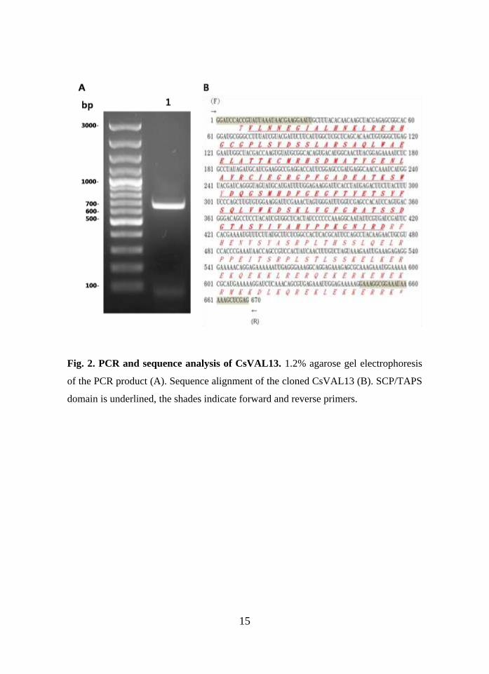
1. Cloning and sequence analysis
The Coding region of CsVAL13 cDNA was amplified by PCR and electrophoresed
(Fig.2A). A single band of 670 bp appeared by the electrophoresis. The PCR product
was purified and sequenced (Fig. 2B). Deduced sequence of CsVAL13 was composed
with 223 amino acids. Its calculated molecular mass was 25.1 kDa. Isoelectric point
of CsVAL13 protein was estimated to 9.602. Putative SCP/TAPS domain was found
in the sequence.
15
Fig. 2. PCR and sequence analysis of CsVAL13. 1.2% agarose gel electrophoresis
of the PCR product (A). Sequence alignment of the cloned CsVAL13 (B). SCP/TAPS
domain is underlined, the shades indicate forward and reverse primers.

16
There is no known signal peptide in the CsVAL13 sequence (Fig. 3A).
Transmembrane domain was not shown in the CsVAL13 sequence. All the residues of
CsVAL13 were predicted to be located outside of the cell membrane (Fig. 3B).
Fig. 3. Sequence analysis of CsVAL13 using TMHMM and SignalP 4.1 server.
Transmembrane domain was predicted using TMHMM 2.0 server (A). Signal peptide
was predicted using SignalP 4.1 server (B).

17
The open reading frame of CsVAL13 SCP/TAPS domain encodes 139 amino acids
(Fig. 4). The CsVAL13 SCP/TAPS domain sequence shares 42–61% identity with
group 2 VAL proteins in platyhelminths. His and Tyr residues known to contribute
structural stability were highly conserved. Among the residues of putative active site
in SCP/TAPS domain, Glu84 and His100 were replaced by Gln and Tyr, respectively.

18
Fig. 4. Multiple sequence alignment of SCP/TAPS domains of various helminth
VAL proteins. HmVAL Hymenolepis microstoma venom allergen protein
(CDS32832); SmVAL17 S. mansoni VAL 7 protein (AAZ04924), SmVAL13 S.
mansoni VAL 13 protein (CCD74793), ShGAPR1 S. haematobium Golgi-associated
plant pathogenesis-related protein 1 (KGB36935), SjGAPR1 S. japonicum Golgi-
associated plant pathogenesis-related protein 1 (CAX74107), EmVAL Echinococcus
multilocularis venom allergen protein (CDJ02618), CsGAPR1 C. sinensis Golgi-
associated plant pathogenesis-related protein 1 (GAA29512). Conserved amino acids
are shaded. Four residues comprising the putative SCP/TAPS domain of the active
site are boxed.
HmVAL KEKQELLDLHNQYRREVAGGKVPNQPGSSKLKDLEWSTELEASAQKHADSCIFEHDGSDDRKTAQWWWVGQ 121
SmVAL7 TQNSELLALHNAYRRNIKYGNVRDQPQAMSMLKLTWSHKLAEMAQEWALQCVPRRSNMTMRKGSKWTYVGQ 98 SmVAL13 QLNHDALNEHNRLRA--LHGCPP----------LKYDRRLAREAQAWAENLA----RLKIMKHSICDEYGE 59
ShGAPR1 QLNHDALNEHNRLRA--LHGCPP----------LKYDSRLAREAQAWADNLA----RMKIMKHSICDEYGE 59
SjGAPR1 KLNNEAIQAHNELRA--LHGCPK----------ISYDSKLASDSQKWAEHLA----SINCLQHSKADGYGE 59
EmVAL KLNEECIRAHNKLRA--LHGCAP----------LTYDAKLAKQAQKHAEYLL----KQNKMEHSTNRDYGE 60
CsGAPR1 ELNTQAIALHNQFRE--KHGSPP----------LVYDAKLAQTAQNWAEQLA----QTKCMRHSDMETYGE 59
CsVAL13 VLNNEGIALHNKLRE--RHGCGP----------LSYDSSLARSAQLWAEELA----TTKCMRHSDMATYGE 59
HmVAL NIAYSSSV-------AQNVKMWFDEYKDYNYNSNYCSG--VCGHYTQLVWADTTHVGCGVSRC-TFSGFNA 182 SmVAL7 SIAFVPKV-------RQAASVWFEQHKNYNFENNTCEANKTCADYKQLAFADTTHIGCGYAMCFNLTGLDK 162
SmVAL13 NLASAQSTGKAEMTGARATRNWYDEIHYHNFNKQFQSQ---SGHFTQLIWKNTSKAGFGIQHSV--DG-HH 124
ShGAPR1 NLATSQSTGKAELTGARATQNWYNEIHDHNFDKQFQSQ---TGHFTQVIWKNTSKAGFGIQYSN--DG-HH 124
SjGAPR1 NLAFQMSTAGASLNGREATRNWYDEISKHDFNGQNQPG---TGHFTQVIWKSTNKAGFGSAKSK--DG-MK 124
EmVAL NIALKGGTPGFQFTGYDASQMWYSEIRAYDFKGGDQLK---CGHFTQLVWSDTKQAGFGVAKSA--KG-DK 125
CsGAPR1 NLAYKGAWENATITGEEATKSWYAQGDYHDFNESFTYE---TSYFSQLIWKGSKNVGFGRAVSE--DG-EA 124
CsVAL13 NLAYRCIEGRGPFGADEATKSWYDQGSMHDFGEGFTYE---TSYFSQLVWKDSKLVGFGRATSS--DG-TA 124
HmVAL VYVVCNYGPGGNLNR 197 34% SmVAL7 VFVVCNYGPGGKYAN 177 36% SmVAL13 VFIVGRYEPPGNVNG 139 45%
ShGAPR1 VFVVGRYEPAGNVYG 139 45%
SjGAPR1 VYVVGRYKPAGNVIG 139 42%
EmVAL VIIVGQYKPPGNYMG 140 42%
CsGAPR1 AYIVAHYFPKGNIRS 139 61%
CsVAL13 SYIVAHYPPKGNIRD 139

19
2. Phylogenetic analysis
A phylogenetic tree was constructed with the same sequences by the NJ method and
revealed that CsVAL13 was clustered with CsGAPR1 (Fig. 5). These were classified
as group 2 VAL proteins along with EmVAL, ShGAPR1, SmVAL13, and SjGAPR1.
SmVAL7 and HmVAL were classified as Group group 1 VAL proteins.
Fig. 5. Phylogenetic analysis of SCP/TAPS domain sequences of CsVAL13 and
other VAL proteins. The phylogenetic tree was constructed using CLC Main
Workbench 6.5 program. Bootstrap values were obtained with 100 replications.
Group 2
Group 1
EmVAL
ShGAPR1
SmVAL13
SjGAPR1
CsGAPR1
CsVAL13
SmVAL7
HmVAL

20
3. Molecular modeling of CsVAL13
Homology model of CsVAL13 represented characteristic α-β-α sandwich structure
of SCP/TAPS domain in the molecule. The length of N-terminal α helix was shorter
than that of human GAPPRP1 used as template for modeling (Fig. 6A and B). Four
residues (His51, Glu59, Gln84, and Tyr100) known to comprise putative active site of
SCP/TAPS domain were closely associated and located on the surface of the main
cleft (Fig. 6C).

21
Fig. 6. Homology modeling of CsVAL13. Cartoon representations of CsVAL13 (A)
and human GAPPRP1 (B). α-β-α sandwich structures were represented blue and
yellow colors. N- and C-terminus of the sequence are indicated by N and C,
respectively. Meshed strcuture of CsVAL13 (C) represented the suface localization of
the four residues (His51, Glu59, Gln84, and Tyr100).

22
4. Production of a recombinant protein
The recombinant CsVAL13 protein (rCsVAL13) was produced bacterially and
purified. The purified rCsVAL13 showed a single band of 41.9 kDa on a 12 % SDS-
PAGE (Fig. 7A). Reactivity of rCsVAL13 to anti-His antibody was proved by
western blot (Fig. 7B).
Fig. 7. Purification of rCsVAL13. 12% SDS-PAGE of the protein fractions
collected during purification (A). M, pre-stained protein size marker; U, uninduced
bacterial lysate; I, induced bacterial lysate; P, purified recombinant protein. Western
blot analysis showing purified rCsVAL13 with anti-His antibody (B). M, pre-stained
protein size marker.
23
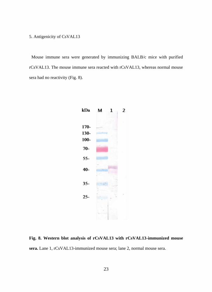
5. Antigenicity of CsVAL13
Mouse immune sera were generated by immunizing BALB/c mice with purified
rCsVAL13. The mouse immune sera reacted with rCsVAL13, whereas normal mouse
sera had no reactivity (Fig. 8).
Fig. 8. Western blot analysis of rCsVAL13 with rCsVAL13-immunized mouse
sera. Lane 1, rCsVAL13-immunized mouse sera; lane 2, normal mouse sera.

24
To examine antigenicity of rCsVAL13, western blot was performed with C. sinensis-
infected rat sera (Fig. 9). rCsVAL13 reacted with anti-His antibody as well as with
rCsVAL13-immunized mouse sera, but not with C. sinensis-infected rat sera.
Fig. 9. Western blot analysis of rCsVAL13. Lane 1, anti-His antibody; lane 2,
rCsVAL13-immunized mouse sera; lane 3~12, sera from each rat infected with C.
sinensis; lane 13; sera from healthy rats.
25
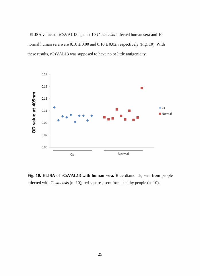
ELISA values of rCsVAL13 against 10 C. sinensis-infected human sera and 10
normal human sera were 0.10 ± 0.00 and 0.10 ± 0.02, respectively (Fig. 10). With
these results, rCsVAL13 was supposed to have no or little antigenicity.
Fig. 10. ELISA of rCsVAL13 with human sera. Blue diamonds, sera from people
infected with C. sinensis (n=10); red squares, sera from healthy people (n=10).

26
6. Immune responses of C. sinensis-immunized mice to CsVAL13
Levels of IgG1 and IgG2a in rCsVAL13-immunized mouse sera were examined by
ELISA. ELISA value of IgG1 and IgG2a in the immunized sera were 0.73 ± 0.04
and 0.12 ± 0.00 , respectively. (Fig. 11). This result indicates that CsVAL13 may
induce predominated Th2 immune responses in the host.
Fig. 11. IgG1 and IgG2a levels in rCsVAL13-immunized mouse sera.

27
7. Immunohistochemical localization
To localize native CsVAL13 protein in adult C. sinensis worm,
immunohistochemistry using rCsVAL13-immunized mouse sera was performed.
Brown colors were observed in the tegument and intrauterine eggs of an adult C.
sinensis worm stained with rCsVAL13-immunized mouse sera (Fig. 12B and D).
Normal mouse sera showed no colored signals (Fig. 12A and C).
Fig. 11. Immunohistochemical localization of CsVAL13. Adult C. sinensis
specimens were stained and visualized under a light microscope (a and b, ×40; c and
d, ×100). a and c, specimen reacted with normal mouse sera; b and d, specimen
reacted with rCsVAL13-immunized mouse sera. Scale bar 10 mm

28
Ⅳ. DISCUSSION
Studies on VAL proteins of trematodes have been mainly focused to S . mansoni7
and S. japonicum24. In this study, molecular structures and distribution in the adult
worm of the CsVAL13 protein, a newly identified VAL protein from C. sinensis,
were examined.
The CsVAL13 sequence had no known signal peptides and transmembrane domain.
However, its location was predicted to be outside of cell membrane (Fig. 3).
Therefore, the CsVAL13 might be secreted to the outside of cells by a non-classical
pathway like a secretion of nitric oxide synthase-interacting protein from C. sinensis23.
SCP/TAPS domain sequence of CsVAL13 had conserved His and Try residue at its
N-and C-terminus, respectively (Fig. 4). These conserved residues are supposed to be
associated with stabilizing SCP/TAP domains by hydrogen bond formation as
referred in SCP/TAPS domain of S. mansoni VAL proeins20. SmVAL13 protein has
four active site residues (His51, Glu59, Glu84, and His100) in its SCP/TAPS
domain20. In the CsVAL13 sequence, Glu84 and His100 were substituted by Gln and
Tyr, respectively, which had similar biochemical characteristics. As shown in
homology modeling, the active site residues were closely located on the surface of
main cleft in the molecule (Fig. 6), suggesting Ca2+ chelation activity25 are conserved.
SCP/TAPS domain of CsVAL13 was phylogenetically categorized as group 2 VAL
protein (Fig. 5). Among the 14 VAL proteins found by intensive genomic search in C.
sinensis, 6 VAL proteins were estimated as group 2 proteins7.

29
rCsVAL13 did not reacted with C. sinensis-infected rat sera in the western blot (Fig.
9). This result indicated that the molecular proportion and/or antigencity of CsVAL13
in the adult C. sinensis worm may relatively low (Fig. 9). This low antigeneicty was
also observed in the ELISA using C. sinensis-infected human sera (Fig. 10) implying
there is no potential as a diagnostic antigen.
IgG1 level in the rCsVAL13-immunized mouse sera was higher that IgG2a level
suggesting that CsVAL13 protein induce polarized Th2 immune responses in the host
(Fig. 11). VAL-1 protein of S. japonicum also increased Th2 responses24. Th2-
polarized immune responses are common in parasitic infection. However, further
immunological characterization of VAL proteins is required to provide information
on the helminth antigens contributing Th2 immune responses.
Native CsVAL13 proteins were found mainly in tegument and intrauterine eggs (Fig.
12). Tegument-specific localization implies that they might have a role in interaction
with its surrounding environment. Excretion of CsVAL13 protein with eggs from
adult worms may contribute immunological stimulation. McCrsp2 of M. corti was
expressed in the apical region in tetrathyridia of M. corti, suggesting that the cestode
VALs could be involved in host-parasite relationships19. VAL-1 protein of S.
japonicum was secreted by eggs, head glands and penetration glands of cercariae
suggesting its role in interaction with a host24.
Until now, only a few studies on the C. sinensis VAL protein have been performed.
There is no evidence that VAL proteins of trematodes including C. sinensis can
modulate the host immune response as observed for the nematode VAL proteins. The

30
present study provides information regarding the biological role of C. sinensis VAL
protein.

31
Ⅴ. CONCLUSION
1. Sequence of CsVAL13 had no known signal peptides and transmembrane domain.
CsVAL13 was predicted to be located outside of cell membrane indicating that it may
be translocated by non-classical pathways.
2. CsVAL13 protein was classified as a group 2 VAL protein.
3. CsVAL13 protein has four known putative active site residues located on the main
cleft in the molecular surface.
4. CsVAL13 protein induced polarized Th2 immune responses in the host, since IgG1
level in the rCsVAL13-immunized mouse sera was higher than IgG2a level.
5. The native CsVAL13 protein was mainly located in tegument and intrauterine eggs
suggesting that they might interact with environmental host tissues and stimulate
immune responses.
Collectively, CsVAL13 is the first cloned C. sinensis VAL protein of which
localization is egg and tegument-specific.

32
REFERENCES
1. Lun ZR, Gasser RB, Lai DH, Li AX, Zhu XQ, Yu XB, et al. Clonorchiasis: a key
foodborne zoonosis in China. Lancet Infect Dis. 2005;5(1):31-41.
2. Hong ST, Fang Y. Clonorchis sinensis and clonorchiasis, an update. Parasitol Int.
2012;61(1):17-24.
3. Zhou J, Sun J, Huang Y, Zhou C, Liang P, Zheng M, et al. Molecular identification,
immunolocalization, and characterization of Clonorchis sinensis calmodulin. Parasitol
Res. 2013;112(4):1709-17.
4. Liang P, Zhang F, Chen W, Hu X, Huang Y, Li S, et al. Identification and
biochemical characterization of adenylate kinase 1 from Clonorchis sinensis. Parasitol
Res. 2013;112(4):1719-27.
5. Rim HJ. Clonorchiasis: an update. J Helminthol. 2005;79(3):269-81.
6. Shin HR, Oh JK, Masuyer E, Curado MP, Bouvard V, Fang YY, et al.
Epidemiology of cholangiocarcinoma: An update focusing on risk factors. Cancer Sci.
2010;101(3):579-85.
7. Chalmers IW, Hoffmann KF. Platyhelminth Venom Allergen-Like (VAL) proteins:
revealing structural diversity, class-specific features and biological associations across
the phylum. Parasitology. 2012;139(10):1231-45.
8. van Loon LC, Gerritsen YA, Ritter CE. Identification, purification, and
characterization of pathogenesis-related proteins from virus-infected Samsun NN
tobacco leaves. Plant Mol Biol. 1987;9(6):593-609.

33
9. Oyewumi L, Kaplan F, Sweezey NB. Lgl1, a mesenchymal modulator of early lung
branching morphogenesis, is a secreted glycoprotein imported by late gestation lung
epithelial cells. Biochem J. 2003;376(Pt 1):61-9.
10. Udby L, Calafat J, Sørensen OE, Borregaard N, Kjeldsen L. Identification of
human cysteine-rich secretory protein 3 (CRISP-3) as a matrix protein in a subset of
peroxidase-negative granules of neutrophils and in the granules of eosinophils. J
Leukoc Biol. 2002;72(3):462-9.
11. Fitzpatrick JM, Johnston DA, Williams GW, Williams DJ, Freeman TC, Dunne
DW, et al. An oligonucleotide microarray for transcriptome analysis of Schistosoma
mansoni and its application/use to investigate gender-associated gene expression. Mol
Biochem Parasitol. 2005;141(1):1-13
12. LuG, Villalva M, Coscia MR, Hoffman DR, King TP. Sequence Analysis and
Antigenic Cross-reactivity of a Venom Allergen, Antigen 5, from Hornets, Wasps,
and Yellow Jackets. J Immunol. 1993;150(7):2823-30.
13. Valenzuela JG, Pham VM, Garfield MK, Francischetti IMB, Ribeiro JMC.
Toward a description of the sialome of the adult female mosquito Aedes aegypti.
Insect Biochem Mol Biol. 2002;9(32):1101-22.
14. Francischetti IMB, Valenzuela JG, Pham VM, Garfield MK, Ribeiro JMC.
Toward a catalog for the transcripts and proteins (sialome) from the salivary gland of
the malaria vector Anopheles gambiae. J Exp Biol. 2002;205(Pt 16):2429-51.
15. Li S, Kwon J, Aksoy S. Characterization of genes expressed in the salivary glands
of the tsetse fly, Glossina morsitans morsitans. Insect Mol Biol. 2001;10(1):69-76.
34
16. Del Valle A, Jones BF, Harrison LM, Chadderdon RC, Cappello M. Isolation and
molecular cloning of a secreted hookworm platelet inhibitor from adult Ancylostoma
caninum. Mol Biochem Parasitol. 2003;2(129):167-77.
17. Bower MA, Constant SL, Mendez S. Necator americanus: The Na-ASP-2 protein
secreted by the infective larvae induces neutrophil recruitment in vivo and in vitro.
Exp Parasitol. 2008;118(4):569-75.
18. Tawe W, Pearlman E, Unnasch TR, Lustigman S. Angiogenic activity of
Onchocerca volvulus recombinant proteins similar to vespid venom antigen 5. Mol
Biochem Parasitol. 2000;109(2);91-9.
19. Britos L, Lalanne AI, Castillo E, Cota G, Señorale M, Marín M. Mesocestoides
corti (syn. vogae, cestoda): Characterization of genes encoding cysteine-rich secreted
proteins (CRISP). Exp Parasitol. 2007;116(2):95-102.
20. Chalmers IW, McArdle AJ, Coulson RM, Wagner MA, Schmid R, Hirai H, et al.
Developmentally regulated expression, alternative splicing and distinct sub-groupings
in members of the Schistosoma mansoni venom allergen-like (SmVAL) gene family.
BMC Genomics. 2008;9:89.
21. Yoshino TP, Brown M, Wu XJ, Jackson CJ, Ocadiz-Ruiz R, Chalmers IW, et al.
Excreted/secreted Schistosoma mansoni venom allergen-like 9 (SmVAL9) modulates
host extracellular matrix remodelling gene expression. Int J Parasitol. 2014;44(8):551-
63.

35
22. Jeong YI, Kim YJ, Ju JW, Hong SH, Lee MR, Cho SH, et al. Identification of
anti-allergic effect of Clonorchis sinensis-derived protein venom allergen-like
proteins (CsVAL). Biochem Biophys Res Commun. 2014;445(3):549-55.
23. Bian M, Li S, Wang X, Xu Y, Chen W, Xhou C, et al. Identification,
immunolocalization, and immunological characterization of nitric oxide synthase-
interacting protein from Clonorchis sinensis. Parasitol Res. 2014;113(5):1749-57.
24. Chen J, Hu X, He S, Wang L, Hu D, Wang X, et al. Expression and immune
response analysis of Schistosoma japonicum VAL-1, a homologue of vespid venom
allergens. Parasitol Res. 2010;106(6):1413-8.
25. Cantacessi C, Campbell BE, Visser A, Geldhof P, Nolan MJ, Nisbet AJ, et al. A
portrait of the "SCP/TAPS" proteins of eukaryotes-developing a framework for
fundamental research and biotechnological outcomes. Biotechnol Adv.
2009;27(4):376-88.
36
ABSTRACT (IN KOREAN)
간흡충 venom allergen-like protein 13 의 면역학적 특성
< 지도교수 용태순 >
연세대학교 대학원 의과학과
우 해 선
SCP/TAPS (sperm coating protein/Tpx-1/Ag5/PR-1/Sc7) 단백질은 진핵생
물에서 발견되는 다기능 단백질이다. Venom allergen-like (VAL) 단백질은
주로 기생 연충에서 보고된 SCP/TAPS 단백질로서 그 생물학적, 면역학적
특성은 많이 알려지지 않았다. 본 연구에서는 간흡충의 VAL 단백질 한 종
류의 유전자를 클로닝하고 특징을 알아보았다. 간흡충 EST 데이터베이스로
부터 25 kDa 단백질을 코딩 하는 cDNA를 찾아냈다. 이 단백질은 BLAST 검
색에서 만손주혈흡충의 VAL13 단백질과 46%의 서열유사성을 보였기에
CsVAL13이라 명명하었다. Multiple sequence alignment 결과 CsVAL13 단백
질서열은 기생 연충의 VAL 단백질서열과 39~46% 유사하였다. 이들 기생 연
충 VAL 단백질서열에는 이 단백질의 구조적 안정화에 기여하는 히스티딘과
타이로신 잔기들이 잘 보존되어 있었다. CsVAL13 단백질 서열은 다른 기생

37
연충의 Group 2 VAL 단백질 서열과 분자계통분류학적으로 같은 분기에 속
했다. 사람 Golgi-associated plant pathogenesis-related protein 1 (PDB
ID 4aiw)을 주형으로 하여 SWISS-MODEL로 예측한 CsVAL13의 3차구조는 VAL
단백질 특유의 α-β-α sandwich를 포함하고 있었다. PCR로 증폭한 CsVAL13
cDNA를 대장균 발현 플라스미드 벡터 pET32a에 서브클로닝 하였다. 대장균
에서 생산된 재조합 CsVAL13 단백질(rCsVAL13)을 Ni-NTA 흡착 크로마토그
래피로 정제하였다. 재조합 CsVAL13 단백질로 BALB/c 마우스를 면역시켜
면역혈청을 얻었다. 이 면역혈청이 IgG2a 항체보다 IgG1 항체를 더 많이
갖고 있는 것으로 보아 CsVAL13은 Th1보다 Th2 면역반응에 더 영향을 주는
것으로 생각된다. 면역조직염색 결과 CsVAL13 단백질은 간흡충 성충의 표
피와 자궁 내 충란에 집중적으로 분포하였다. 이상의 결과는 간흡충 성충
표피에 특이하게 분포하는 CsVAL13이 숙주-기생충 상호작용의 한 부분을
담당하며, 주변 숙주조직에 대한 면역 자극에 기여할 가능성이 있음을 의
미한다.
---------------------------------------------------------------------
핵심되는 말: 간흡충, venom allergen-like 단백질, SCP/TAPS 단백질,
충란, 표피