Immunoelectronmicroscopic Localization Of Iga In Skin … herpetiformis (DH) ... In this paper we...
5
THE JOURNAL OF INVESTIGATIVE DERMATOLOGY. 67:502-506. 1976 Copyright © 1976 by The Williams & Wilkins Co. REPORTS Vol. 67. :-':0. 4 Printed in U.S.A. IMMUNOELECTRONMICROSCOPIC LOCALIZATION OF IgA IN SKIN OF PATIENTS WITH DERMATITIS HERPETIFORMIS HIDEO YAOITA, M.D., AND STEPHEN 1. KATZ, M.D., PH.D. Dermatology Branch, National Cancer Inst itute, National Institut es of Health, Bethesda, Maryland, U. S. A. Ultrastructural loca lization of in vivo-bound IgA in the skin of patients with dermatitis herpetiformis was determined by using a modified peroxidase - antiperoxidase multistep method. Three types of reaction product deposition are seen. The most common type of reaction product deposition , that which is identified by direct immunof1uorescen ce as the speckled type of I gA deposit , shows up as clumps and yarnlike fibrils. The seco nd type of IgA deposition , which is a linear band by direct immunot1uorescence, appears to be associ- ated with anchoring fibril s. The third type of IgA deposition, which is also linear by immuno- fluorescence, is confined to the lamina lucida . Dermatitis herpetiformis (DH) is a chronic sub- epidermal blistering disease in which many immu - nologic abnormalities have been demon st rated [I , 2 ]. The most prominent of these is the almost universal presence of in vivo-bound IgA deposited in a granular or fibrillar pattern in the dermal papillae or in a continuous or linear pattern along the dermal -e pidermal junction of normal and perilesional skin [3 ]. In this rega rd Seah and Fry [4] demonstrated in vivo-bound IgA in all 50 patients st udied and we hav e made this observa- tion in 38 of 40 patients di ag nose d by clinical, histologic, and treatment response (to sulfones or sulfapyridine) criteria. In order to determine the exact location of the IgA deposits in the skin of patients with DH and to gain insight into the structure(s) to which the IgA is bound, we have used an ultrastructural immuno - peroxidase method [5]. In this paper we describe the ultrastructural localization of in vivo-bound IgA in the skin of patients with DH who have the granular or fibrillar type of deposits and in those who have the continuous of linear type of depos- its. Manuscript received March 17, 1976; accepted for publication May 6, 1976. Reprint requests to: Dr. H. Yao ita, National Cancer Institute, National Institutes of Health, Building 10, Room 12N250, Bethesda, Maryland 20014. Abbreviations: BP: bullous pemphigoid BMZ: basement membrane zone DH: dermatitis herpetiformis DIF: direct immunofluoresce nce G/HA: goat antihuman IgA HG : herpes gestation is NGS: normal goat serum P- AP: peroxidase -anti peroxidase multistep (method) MATERIALS AND METHODS Patients. Each of the 6 patients st udi ed had clinical and hi stologic evidence of DH and each re sponded dramatically to sulfones or sulfapyridin e. There was a prompt recurrence of lesions when the medication was stopped. The normal and perivesicular le sional s kin of each patient demon st rated in vivo-bound IgA in the dermal papillary tips (3 patients-2 men aged 33 and 73 and 1 woman aged 56) or at the dermal-epidermal junction (3 pat ients-2 women aged 42 a nd 55 and 1 man aged 33) using standard direct immunonuoresce nt tech· niques. No circulating antiepithelial antibodies were detected in the sera of any of these patients. Normal skin was also obtained from 2 volunteers. Peroxidase-antiperoxidase (P-AP) multistep m eth od. The tissue preparation and staining procedur es were the same as described previously [5 J exce pt that a 1 :40 dilution of fluoresceinated goat antihuman IgA (G/HA, Hyland; specific antibody concentration 2.2 mg/ml) was used for the demonstration of in vivo-bound Ig. Normal goat se rum (NGS) was used as a control. RESULTS Light Microscopy Speckled type. When the P-AP method was used , the dark brown clumps of reaction products were seen beneath the epidermis, especially in the dermal papillae of both normal ' and perile s ional skin (Fig. O. These deposits were the same s hape , of equivalent size, and in the same distribution as those seen in the tissue stained by direct immuno- fluorescence (DIF) (Fig. 2). The epidermis and the rest of the dermis showed only very light yellowish brown staining which was considered to be back- ground. 502 Linear type. The dark brown linear bands of reaction products were seen along the basement membrane zone (BMZ) of the epidermis in the normal-appearing and perilesional skin of 3
Transcript of Immunoelectronmicroscopic Localization Of Iga In Skin … herpetiformis (DH) ... In this paper we...
THE JOURNAL OF INVESTIGATIVE DERMATOLOGY. 67:502-506. 1976 Copyright © 1976 by The Williams & Wilkins Co.
REPORTS
Vol. 67. :-':0. 4 Printed in U.S.A.
IMMUNOELECTRONMICROSCOPIC LOCALIZATION OF IgA IN SKIN OF PATIENTS WITH DERMATITIS HERPETIFORMIS
HIDEO YAOITA, M.D., AND STEPHEN 1. KATZ, M.D., PH.D.
Dermatology Branch, National Cancer Institute, National Institutes of Health, Bethesda, Maryland, U. S. A.
Ultrastructural loca lization of in vivo-bound IgA in the skin of patients with dermatitis herpetiformis was determined by using a modified peroxidase- antiperoxidase multistep method. Three types of reaction product deposition are seen. The most common type of reaction product deposition , that which is identified by direct immunof1uorescence as the speckled type of IgA deposit , shows up as clumps and yarnlike fibrils. The second type of IgA deposition, which is a linear band by direct immunot1uorescence, appears to be associated with anchoring fibril s. The third type of IgA deposition, which is also linear by immunofluorescence, is confined to the lamina lucida.
Dermatitis herpetiformis (DH) is a chronic subepidermal blistering disease in which many immunologic abnormalities have been demonstrated [I , 2 ]. The most prominent of these is the almost universal presence of in vivo-bound IgA deposited in a granular or fibrillar pattern in the dermal papillae or in a continuous or linear pattern along the dermal-epidermal junction of normal and perilesiona l skin [3 ]. In this rega rd Seah and Fry [4] demonstrated in vivo-bound IgA in all 50 patients studied and we have made this observation in 38 of 40 patients diagnosed by clinical, histologic, and treatment response (to sulfones or sulfapyridine) criteria.
In order to determine the exact location of the IgA deposits in the skin of patients with DH and to gain insight into the structure(s) to which the IgA is bound, we have used an ultrastructural immunoperoxidase method [5]. In this paper we describe the ultrastructural localization of in vivo-bound IgA in the skin of patients with DH who have the granular or fibrillar type of deposits and in those who have the continuous of linear type of deposits.
Manuscript received March 17, 1976; accepted for publication May 6, 1976.
Reprint requests to: Dr. H. Yaoita , National Cancer Institute, National Institutes of Health, Building 10, Room 12N250, Bethesda, Maryland 20014.
Abbreviations: BP: bullous pemphigoid BMZ: basement membrane zone DH: dermatitis herpetiformis DIF: direct immunofluorescence G/HA: goat antihuman IgA HG: herpes gestation is NGS: normal goat serum P- AP: peroxidase-anti peroxidase multistep (method)
MATERIALS AND METHODS
Patients. Each of the 6 patients studied had clinical and histologic evidence of DH and each responded dramatically to sulfones or sulfapyridine. There was a prompt recurrence of lesions when the medication was stopped. The normal and perivesicular lesional skin of each patient demonstrated in vivo-bound IgA in the dermal papillary tips (3 patients-2 men aged 33 and 73 and 1 woman aged 56) or at the dermal- epidermal junction (3 patients-2 women aged 42 and 55 and 1 man aged 33) using standard direct immunonuorescent tech· niques. No circulating antiepithelial antibodies were detected in the sera of any of these patients . Normal skin was also obtained from 2 volunteers.
Peroxidase- antiperoxidase (P-AP) multistep m ethod. The tissue preparation and staining procedures were the same as described previously [5 J except that a 1 :40 dilution of fluoresceinated goat antihuman IgA (G/HA, Hyland ; specific antibody concentration 2.2 mg/ml) was used for the demonstration of in vivo-bound Ig. Normal goat serum (NGS) was used as a control.
RESULTS
Light Microscopy
Speckled type. When the P-AP method was used , the dark brown clumps of reaction products were seen beneath the epidermis, especially in the dermal papillae of both normal' and perilesional skin (Fig. O. These deposits were the same shape , of equivalent size, and in the same distribution as those seen in the tissue stained by direct immunofluorescence (DIF) (Fig. 2). The epidermis and the rest of the dermis showed only very light yellowish brown staining which was considered to be background.
502
Linear type. The dark brown linear bands of reaction products were seen along the basement membrane zone (BMZ) of the epidermis in the normal-appearing and perilesional skin of 3

'Jet. 1976 IGA LOCALI ZATION IN DERMATITIS HERPETIFORMIS 503
FIG. 1. DH sk in with speckled-type IgA deposition stained by P - AP method. The dark brown clumps beneath the epidermis are t he reaction product deposits which indicate IgA deposition (x 220) .
FIG. 2. DH skin with speckled-type IgA deposition stained by immunofluorescent techn ique. The white clumps beneath the epidermis show the IgA deposits in t he sa me pattern as in Fig. 1 ( x 220).
FIG. 3. DH skin with linear (dermal) -type IgA deposit ion stained by P-AP method. The dark brown linear band along t he epidermal- dermal junction shows t he reaction products (x 220).
FIG. 4. DH skin with linear (dermal)-type IgA deposition stained by immunof1uorescent technique. The white lin ear band along the epidermal- dermal junction shows IgA deposits in t he sa me pattern as in Fig. 3 ( x 220) .
patients (Fig. 3) . This pattern of IgA deposition was the same as t hat seen using OrF (Fig. 4). The other parts of the t issue showed the same light yellowish brown background as seen in the speckled type .
Electron Microscopy
There were no reaction products in the intercellular spaces and only sparse dark deposits in the deep dermis of a ll G/HA- and NGS-treated OH and normal human sk in of the volunteers. G/HAsta ined normal human skin showed no spec ific reaction products .
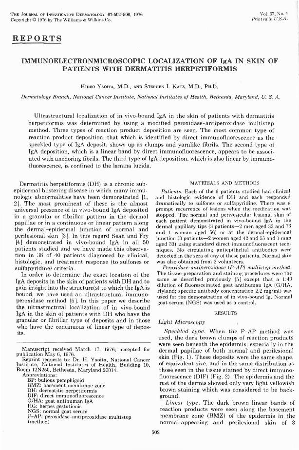
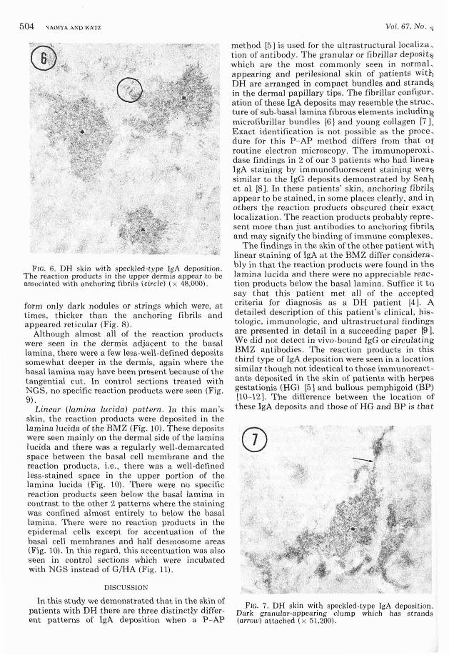
Granular (dermal) pattern. In the skin of patients with OH with the speckled pattern, all of the reaction products were seen in the dermis (Fig. 5). It was difficul t to identify the componen ts of t he stained substances because they were usually very densely sta ined (Fig. 6). However, some of t he stained structures had stringlike tails (80-130 A dia meter) and, in areas, looked like yarn (Fig. 7) . It was not possible to determine whether t he periodi city of the stringlike tails was regular since they were also stained very densely in most areas. Only a few of the specks in t he upper dermis appeared to be associated with anchoring fibrils (Fig. 6). There was almost no staining of the elastic fibers.
Linear (dermal) pattern. In skin of 2 of the 3 patients with linear IgA staining by OIF, the reaction products were seen beneath the basa l lamina in a nodu lar or re t icular pattern (Fig. 8). These deposits were limited to t he area where anchoring fibrils are usually seen. The periodicity of some of the a nchoring fibrils was clearly accen-
. f5\:' : .... _. :<t
·Y :;'· .,
FIG. 5. DH skin with spec kled-type IgA deposition. The dark, well-demarcated granular appea ring clumps (0) in the upper dermis are the reaction products which indicate IgA deposition. The stain ed clumps in the dermis are easi ly distinguished from the epidermis and epidermal components because of t heir high electron density. E = epidermis; D = dermis ( x 4,2(0). Sections in Figs. 5- 9 were not counterstained with uranyl acetate.
tuated when only small amounts of reaction products were seen a round the fib rils (Fig. 8). However, in many areas t he reaction products obscured t he period icity of the anchoring fibri ls a nd appeared to
504 YI\O ITI\ I\ND KI\TZ
.' ~ .,.,
FIG. 6. DH skin with speckl ed-type IgA deposit ion . The reaction products in the upper dermis appear to be associated with anchoring fibrils (ci rcle) (x 48,000).
form only dark nodules or strings which were, at times, thicker t han t he anchoring fibri ls and appeared reticul ar (Fig. 8).
Although a lmost a ll of t he reaction products were seen in the dermis adjacent to the basa l lamina, t here were a few less-well -defined deposits somewhat deeper in the dermis, again where the basal la mina may have been present because of the tangential cut. In control sections treated with NGS, no specific reaction products were seen (Fig. 9) .
Linear (lamina Lucida) pattern. In this man's skin , the reaction products were deposited in t he lamina lucida of the BMZ (Fig. 10). These depos its were seen mainly on the dermal side of the lamina I ucida and t here was a regularly weJl-demarcated space between the basa l ceJl membrane and the reaction products, i.e., t here was a weJl-defined less-sta ined space in t he upper por tion of t he lamina lucida (Fig. 10) . There were no specific react ion products seen below the basal lamina in COntrast to the other 2 patterns where the staining was confined a lmost ent irely to below the basal lamina. There were no reaction products in the epidermal cells except for accentuation of the basal cell membranes and half desmosome areas (Fig. 10). In t his regard, this accentuation was also seen in control sections which were incubated with NGS instead of G/HA (F ig. 11).
DISCUSSION
In this study we demonstrated t hat in the skin of patients with DH there are three distinctly different patterns of IgA deposition when a P - AP
Vol. 67, No . 'i
method [5 J is used for t he ultrastructural localiza , tion of antibody. The granula r or fibri lla r deposit~ which a re t he most commonly seen in normaL appearing and perilesional skin of patients wit!) DH are arranged in compact bundles and strand::, in the derma l papi llary t ips. The fibriJlar configur , ation of t hese IgA deposits may resemble t he struc, ture of sub-basal la mina fibrous elements includin~ microfibrillar bundles [6 ) and young collagen [7 L Exact iden t ification is not possible as t he proce , dure for t his P - AP method differs from t hat o~ routine electron microscopy . The immunoperox i , dase findings in 2 of our 3 patients who had li neal' IgA staining by immunofluorescent sta ining wer~ similar to the IgG deposits demonstrated by Sea!,. et a l. [8). In t hese patients' skin , a nchoring fibri l ~ a ppear to be stained , in some places clearly , and in. others the reaction products obscured t heir exact loca lization. The reaction products probably repre , sen t mOre t han just antibodies to anchoring fibril~ and may signify the binding of immune complexes.
The find ings in t he skin of t he other patient wit!,. linear staining of IgA at t he BMZ differ considera , bly in t hat the reaction products were found in t h e lamina lucida and there were no a ppreciab le reaction products b elow the b asa l la mina. Suffi ce it to say t hat t his patient met a ll of the accepted. criteria for diagnosis as a DH patient [4). A detailed description of th is patient's cl in ica l, histologic, immunologic, and ul trastructural findings are presented in detai l in a succeeding paper [9], We did not detect in vivo-bound IgG or circulating BMZ ant ibodies. The reaction products in t his third ty pe of IgA deposition were seen in a location s imilar t hough not ident ical to t hose immunoreactants deposited in the skin of patients with herpes gestationis (HG) [5 J and bullous pemphigoid (BP) [10- 12 J. The difference between t he location of t hese IgA deposits and those of HG and BP is that
CD . 1,
.\
F IG. 7. DH skin with speckled-type IgA deposit ion . Dark gra nula r-appearing clump wh ich has stra nds (arrow) attached (x 51,200).
Oct. 1976 IGA LOCALIZATION IN DERMATITIS HERPETIFORM IS 505
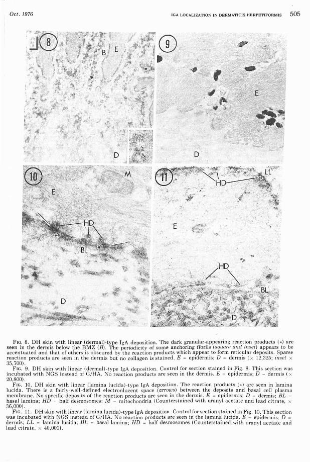
FIG. 8. DH skin with linear (dermal)·type IgA deposition. The dark granular-appearing reaction products (0) a re seen In the dermIS below the BMZ (8). The periodicity of some anchorIng flbnls (square and Inset) appears to be accentuated and that of others is obscured by the reaction products which appear to form reticular deposits . Sparse reaction products are seen in t he dermis but no collagen is stained. E = epidermis; D = dermis (x 12,325; inset x 35,700) .
FIG . 9. DH skin with li near (dermal )·type IgA deposition. Control for section stained in Fig. 8. This section was incubated with GS instead of G/ HA. No reaction products are seen in the dermis . E = epidermis; D = dermis (x 20,800).
FIG . 10. DH s kin with lin ear (la mina lucida)·type IgA deposition. The reaction products (0) are seen in lamina lucida. There is a fairly·well·defined elect ron lucent space (arrows) between the deposits and basal ce ll plasma membrane. No spec ific deposits of the reaction products a re seen in the dermis. E = epidermis; D = dermis; BL = basal lamina; HD = ha lf desmosomes; M = mitochondria (Counterstained with uranyl acetate and lead citrate, x 36,000).
FIG. 11. DH skin with linea r (lam ina lucida)·type IgA deposition. Control for section sta ined in Fig. 10. This sect ion was incubated with NGS instead of G/HA . No react ion products are seen in the lam ina lucida. E = ep iderm is; D = dermis; LL = lamina lucida ; BL = basal lamina; HD = half desmosomes (Coun tersta ined with urany l acetate and lead citrate, x 40,000).
506 YAOITA AND KATZ
in this patient's skin there is a well-defined space between the reaction products and the basal cell plasma membrane, whereas in HG the reaction products are seen throughout the lamina lucida and in BP a well-defined space is at times seen between the reaction products and the basal lamina. It is therefore suggested that there may be different antigenic structures in the lamina lucida. Jablonska et al [13 ] recently addressed themselves to the occasional difficulty in differentiating DH from BP. 1m munoelectronmicroscopic identification of the localization of the in vivo-bound antibodies in the skin of their patients would undoubtedly help to further categorize the disease .
Several possibilities exist as to why the patterns of IgA deposition differ from one patient's skin to another . First, it may be that in the three types of DH skin t here are different antigens or specific binding sites for IgA. Second, the skins of patients with DH may be antigenically similar to each other a nd there are different sites for the adherence of circulating immune complexes which deposit in various patterns. Both or other possibilities may be operating. Characterization of the antigen or of the specific IgA antibody will aid in understanding the importance and etiology of these IgA deposits.
We are grateful to Dr . M. A. Lutzner and Dr. B. Wetzel for their criticism, and to Mrs. M. Gullino, Miss L. Gazze, and Mr. H. Schaffer for their excellent assistance .
REFERENCES
1. Van der Meer JB: Dermatitis Herpetiformis: A Specific Entity. Leiden, NV Drukkerij.YH Batteljee and Terpstra, 1972
Vol. 67, No.4
2. Alexander JO: Dermatitis Herpetiformis. London/ TorontolPhiladelphia, Saunders, 1975
3. Fry L, Seah PP: Dermatitis herpetiformis, Immunological Aspects of Skin Diseases . Edited by L ~ry, I PP Seah. London, Medical and Technical PublIsh-ing, 1974, pp 22- 65 ..
4. Seah PP, Fry L: Immunoglobulins in dermatItis herpetiformis and their relevance in diagnosis. Br J Dermatol 92: 157- 166, 1975 t
5. Yaoita H , Gullino M , Katz SI: Herpes gestationis. Ultrastructural loca lizat ion' of in vivo-bound complement. J Invest Dermato1 66:382- 388, 1976
6. Briggaman RA, Wheeler CE: The epidermal-dermal junction. J Invest Dermatol 65:71-84, 1975
7. Hashimoto K : Fibroblast, collagen and elastin, Ultrastructure of Normal a nd Abnormal Skin. Edited by AS Zelickson . Philadelphia, Lea & Febiger, 1967, pp 228-260
8. Seah PP, Fry L, Stewart JS, Chapman BL, Hoffbrand AV, Holborow EJ: Immunoglobulins in the skin In
dermatitis herpetiformis and coeliac disease. Lancet 1:611- 614, 1972
9. Yaoita H, Hertz KC , Katz SI: Dermatitis herpetifor-mis: immunoelectronmicroscopic and ultrastruCtural studies of a patient with linear deposition of IgA. J Invest Dermatol 67 (in press) 1976
10. Schaumburg-Lever G, Rule A, Schmidt-Ullrich B, Lever WF: Ultrastructural localization of in vivobound immunoglobulins in bullous pemphigoid. A preliminary report. J Invest Dermatol 64:47-49, 1975
11. Holubar K, Wolff K , Konrad K, Beutner EH: Ultrastructura l localization of immunoglobulins in bul lous pemphigoid s kin. J Invest Dermatol 65:220-227, 1975
12. Schmidt-Ullrich B, Rule A, Schaumburg-Lever G, Lebla nc C: Ultrastructural localization of in vivobound complement in bullous pemphigoid. J Invest Dermatol 65:217- 219, 1975
13. Jablonska S, Chorzelski TP, Beutner EH, Maciejowska E, Rzeska G: Dermatitis herpetiformis and bullous pemphigoid: intermediate and mixed forms. Arch Dermatol 112:45-48, 1976