Imaging Systems 284 Gel Excision Instrument 297
16
www.bio-rad.com/imaging/ Imaging Systems 284 Gel Excision Instrument 297 Imaging Instruments
Transcript of Imaging Systems 284 Gel Excision Instrument 297

www.bio-rad.com/imaging/
Imaging Systems 284
Gel Excision Instrument 297
Imaging Instruments

284 | Imaging Instruments
www.bio-rad.com/imaging/
Imaging Systems
www.bio-rad.com
Bio-Rad offers imaging products for a variety of applications. Products include imaging systems, a specialized instrument for gel excision, and specialized image analysis software.
Imaging SystemsMolecular Imager® systems detect images and quantitate colorimetric, chemiluminescent, fluorescent, and radioisotopic signals. Software (pages 300–308) provides automation for image acquisition with data analysis and validation. Refer to the guide below to select the imaging system best suited to your applications.
Imaging System Selection Guide
Molecular Imager Systems Criterion Personal Stain GS-800™ Gel Doc™ ChemiDoc™ VersaDoc™ VersaDoc PharosFX™ Molecular Application Free™ Densitometer XR+ XRS+ MP 4000 MP 5000 PharosFX™ Plus Imager™
Nucleic Acid Detection Ethidium bromide stain — — 4 4 5 3 5 5 — SYBR® Green I stain — — 4 4 5 3 5 5 — SYBR® Safe stain — — 4 4 5 3 5 5 — Coumarin stain — — 4 4 5* 3* — — —
Protein Detection, 1-D Gels Stain-free gels 5 — — — — — — — — Coomassie Blue stain — 5 4 4 5 3 2 2 — Silver stain — 5 4 4 5 3 2 2 — SYPRO Ruby protein gel stain — — 4 4 5 3 5 5 — Flamingo™ fluorescent gel stain — — 4 4 4 3 5 5 — Oriole™ fluorescent gel stain — — 5 5 5 5 — — —
Protein Detection, 2-D Gels Coomassie Blue stain — 5 3 3 4 2 2 2 — Silver stain — 5 3 3 4 2 2 2 — SYPRO Ruby protein gel stain — — 3 3 4 3 5 5 — Flamingo fluorescent gel stain — — 3 3 4 3 5 5 — Pro-Q stain — — 3 3 4 3 5 5 — Cy2, Cy3, Cy5 label — — — — 5 4 5 5 —
Blot Detection Coomassie Blue stain — 5 4 4 5 3 2 2 — Silver stain — 5 4 4 5 3 2 2 — SYPRO Ruby protein blot stain** — — — — 5 5 5 5 — Immun-Star™ chemiluminescence — — — 4 3 5 — — — Chemifluorescence** — — 1 1 5 5 4 4 — Quantum dot** — — 1* 1* 5* 5* 5 5 —
Micro- and Macroarray*** Detection Radiolabel — — — — — — — 5 5 Fluorescence — — 2 2 5 3 5 5 — Immun-Star chemiluminescence — — — 4 5 3 — — —
Colony Counting Colorimetric detection — — 4 4 5 3 3 3 — Fluorescence detection — — 4 4 5 3 5 5 —
Isotopic Detection Radiolabel — — — — — — — 5 5 X-ray film — 5 4 4 5 3 3 3 —
— Not recommended; 1–5, recommendation level (5 = highest).* Custom filter required.** Optimal with low-fluorescence PVDF membrane.*** With spot diameters ≥400 µm.

Imaging Instruments | 285
www.bio-rad.com/imaging/
Imaging Systems
www.bio-rad.com
■■ Environmental friendliness — in contrast to Coomassie stain techniques, acetic acid and methanol are not used, reducing organic waste
■■ Ease of use — one-touch gel image and analysis processing
■■ Sensitivity — results are equal to or better than that of Coomassie stain
SDS-PAGE followed by protein visualization using Coomassie Blue stain is a standard, widely used method. However, it involves time-consuming staining and destaining procedures.
The Criterion Stain Free gel imaging system allows direct visualization, analysis, and documentation of protein samples in PAGE gels with the single touch of a button — without staining, destaining, or gel drying procedures. The complete system is composed of the Criterion Stain Free gels, Criterion Stain Free imager, and Image Lab™ software. It is a dedicated, small-format, easy to use system that uses standard SDS-PAGE conventions, making adoption easy because researchers do not have to change sample preparation procedures. This system is ideal for quick sample assessment prior to purification procedures and as a precursor to blotting and profiling workflows in which Coomassie stain is ordinarily used for detection in purity assessment, quantitation, protein characterization (western blotting), and profiling (MALDI-TOF, LC-MS/MS). Benefits include:
■■ Fast results — image the gel and complete the analysis in as few as 2.5 min
■■ Reproducibility — automation and standardization of the methods eliminate the background variability within a gel or between gels that is often seen with standard Coomassie stain techniques
Criterion Stain Free™ Gel Imaging System
Time Comparison: Colloidal Coomassie Blue Stain vs. Criterion Stain Free System Protocols
Steps Colloidal Coomassie Stain Protocol Criterion Stain Free Protocol
Washes in deionized water 15 minutes (3 x 5 minutes) 0 minutes Staining 60 minutes (30–60 minutes) 0 minutes Destaining 30 minutes 0 minutes Imaging and analysis 15–30 minutes 2.5–5 minutesTotal time ≤135 minutes 5 minutes
Sample Tray
Image Lab Software
Criterion Stain Free Gel
Criterion Stain Free Imager

286 | Imaging Instruments
www.bio-rad.com/imaging/
Imaging Systems
www.bio-rad.com
Criterion Stain Free GelsCriterion Stain Free gels have Tris-HCl formulation for PAGE applications and a proprietary compound that facilitates protein visualization by UV imaging. Criterion Stain Free gels are made without SDS. With SDS omitted from the running buffer, the gels can be used to run proteins under nondenaturing conditions for subsequent analysis of native conformation and activity. To run denaturing gels, simply use a running buffer that contains SDS to allow estimation of molecular weight of sample proteins.
Single-percentage resolving gels — Choose a single-percentage gel when your sample has proteins of a limited size range and your goal is to separate a single band from neighboring bands. These gels will produce the greatest separation between bands with similar molecular weights. Single-percentage gels are cast with a 4% stacking gel to further sharpen protein bands before they enter the resolving gel.
Linear gradient gels — Choose a linear gradient gel if your sample contains a wide range of molecular weights. These gels allow both high and low molecular weight bands to be visualized on the same gel.
Criterion Stain Free system vs. Coomassie Blue stain method. Complex protein samples were run in duplicate and visualized using the Criterion Stain Free system (A) and Coomassie stain method (B). The Coomassie stain method produces variable background despite the use of consistent staining and destaining times. Quantitation of duplicate serial dilutions of b-galactosidase show higher reproducibility with the Criterion Stain Free system (C) compared with the Coomassie stain method (D). Average %CV for Criterion Stain Free gels is 4.85%; for Commassie-stained gels it is 24.08%.
Criterion™ electrophoresis system. Features include locator slots to slide gels easily, lot information printed on each gel, and the built-in wedge on the lid to open the cassette.
B. Gel stained with the conventional Coomassie stain method.A. Gel visualized with the Criterion Stain Free system.
D. b-Galactosidase band, Bio-Safe™ Coomassie stain.
Ban
d d
ensi
ty, u
nits
0.120
0.100
0.080
0.060
0.040
0.020
0.000
1 10 100 1,000
Log (protein quantity, ng)
C. b-Galactosidase band, Criterion Stain Free system.
Sig
nal i
nten
sity
, uni
ts
35,000
30,000
25,000
20,000
15,000
10,000
5,000
0
1 10 100 1,000
Log (protein quantity, ng)
Imaging Instruments | 287
www.bio-rad.com/imaging/
Imaging Systems
www.bio-rad.com
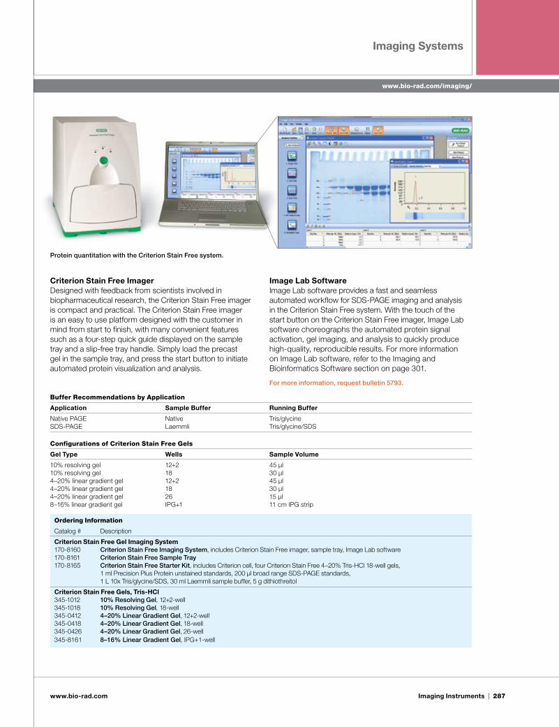
Criterion Stain Free ImagerDesigned with feedback from scientists involved in biopharmaceutical research, the Criterion Stain Free imager is compact and practical. The Criterion Stain Free imager is an easy to use platform designed with the customer in mind from start to finish, with many convenient features such as a four-step quick guide displayed on the sample tray and a slip-free tray handle. Simply load the precast gel in the sample tray, and press the start button to initiate automated protein visualization and analysis.
Image Lab SoftwareImage Lab software provides a fast and seamless automated workflow for SDS-PAGE imaging and analysis in the Criterion Stain Free system. With the touch of the start button on the Criterion Stain Free imager, Image Lab software choreographs the automated protein signal activation, gel imaging, and analysis to quickly produce high-quality, reproducible results. For more information on Image Lab software, refer to the Imaging and Bioinformatics Software section on page 301.
For more information, request bulletin 5793.
Protein quantitation with the Criterion Stain Free system.
Buffer Recommendations by Application
Application Sample Buffer Running Buffer
Native PAGE Native Tris/glycineSDS-PAGE Laemmli Tris/glycine/SDS
Ordering Information
Catalog # Description Price, U.S. $
Criterion Stain Free Gel Imaging System 170-8160 Criterion Stain Free Imaging System, includes Criterion Stain Free imager, sample tray, Image Lab software 12,500.00170-8161 Criterion Stain Free Sample Tray 500.00170-8165 Criterion Stain Free Starter Kit, includes Criterion cell, four Criterion Stain Free 4–20% Tris-HCl 18-well gels, 831.00
1 ml Precision Plus Protein unstained standards, 200 µl broad range SDS-PAGE standards, 1 L 10x Tris/glycine/SDS, 30 ml Laemmli sample buffer, 5 g dithiothreitol
Criterion Stain Free Gels, Tris-HCl345-1012 10% Resolving Gel, 12+2-well 15.00345-1018 10% Resolving Gel, 18-well 15.00345-0412 4–20% Linear Gradient Gel, 12+2-well 15.00345-0418 4–20% Linear Gradient Gel, 18-well 15.00345-0426 4–20% Linear Gradient Gel, 26-well 15.00345-8161 8–16% Linear Gradient Gel, IPG+1-well 15.00
Configurations of Criterion Stain Free Gels
Gel Type Wells Sample Volume
10% resolving gel 12+2 45 µl10% resolving gel 18 30 µl4–20% linear gradient gel 12+2 45 µl4–20% linear gradient gel 18 30 µl4–20% linear gradient gel 26 15 µl8–16% linear gradient gel IPG+1 11 cm IPG strip

288 | Imaging Instruments
www.bio-rad.com/imaging/
Imaging Systems
www.bio-rad.com
Ordering Information
Catalog # Description Price, U.S. $
170-7983 Molecular Imager GS-800 USB Calibrated Densitometer, PC or Mac, 100–240 V 16,826.00170-7956 Installation Qualification/Operational Qualification (IQ/OQ) 4,722.00170-9615 Quantity One Software CFR Module 3,385.00
Specifications
Light source Fluorescent white
Wavelength 400–750 nm
Absorbance range 0–3.0 OD (transmissive); 0–2.0 OD (reflective)
Operating modes Transmissive and reflective
Scanning area Transmissive 29 x 40 cm Reflective 30 x 40 cm
Sampling rate 800 x 1,600 dpi
Pixel data density 12-bit
Resolution 36.3 µm
Operating system Windows XP, Windows 2000; Mac OS X compatibility
Voltage 100–240 V
Dimensions 62.6 x 47.3 x 19.3 cm (24.6 x 18.6 x 7.6") (W x D x H)
Weight 20.8 kg (45.9 lb)
The Molecular Imager GS-800 USB calibrated densitometer offers superior accuracy, sensitivity, and data reproducibility. It automatically self-calibrates the optical density to optimize detection. Features include:
Imaging of a wide variety of samples such as 1-D and ■■
2-D gels, colorimetric dot and slot blots, film-based chemiluminescent blots, autoradiograms, slides, and photographs
■■ Transmissive and reflective imaging using red, green, and blue CCD technology to scan chromogenic samples at the optimal detection wavelength
■■ IQ/OQ for verification of the reflectance and transmittance calibration functions using NIST-traceable calibration targets
■■ High resolution and analysis of closest bands on a gel due to 12-bit precision and 36.3 µm resolution
■■ Scanning of larger gels for enhanced separation of proteins on oversized 29 x 40 cm sample platen
■■ Accurate quantitation of samples, such as Coomassie Blue– and silver-stained gels, over a large dynamic range (0–3.0 OD)
■■ Sealed imaging platen to accommodate wet samples of variable thickness
■■ Purity analysis and lane background tools for manufacturing QC
U.S. FDA 21 CFR Part 11 regulation compliance software ■■
features
Transmittance and ReflectanceUnlike laser densitometer systems, the GS-800 USB calibrated densitometer has both transmittance and true reflectance capabilities that allow accurate scans of both
transparent and opaque samples. The advantage of true reflectance scanning is accurate detection and analysis of molecules on the surface of membranes, rather than molecules distributed throughout the membrane matrix. True reflectance provides the most accurate detection and analysis of dot blots, slot blots, and other electrophoretic blots without quantitation error.
IQ/OQThe calibration of the GS-800 USB calibrated densitometer can be validated by following GS-800 IQ/OQ protocols, which are available as an accessory. The easy to follow protocols include instructions for installation of the hardware and software and for verification of the calibration functions.
Quantity One® Software CFR ModuleThe GS-800 USB densitometer can be used in a GMP/GLP-compliant environment. The CFR module assists users with meeting U.S. FDA 21 CFR Part 11 requirements.
For more information, request bulletins 2596 and 5474.
Molecular Imager® GS-800™ USB Calibrated DensitometerSee Also
Gel stains: pages 221–226.
Gel analysis software: pages 301–305.

Imaging Instruments | 289
www.bio-rad.com/imaging/
Imaging Systems
www.bio-rad.com
The Molecular Imager Gel Doc XR+ and ChemiDoc XRS+ systems are based on CCD high-resolution, high-sensitivity detection technology and modular options to accommodate a wide range of samples and support multiple detection methods including fluorescence, colorimetry, densitometry, chemiluminescence, and chemifluorescence. The systems are controlled by Image Lab™ software to optimize imager performance for fast, integrated, and automated image capture and analysis of various samples.
The systems accommodate a wide array of samples, from large handcast polyacrylamide gels to small ReadyAgarose™ gels and various blots. The systems are the ideal accompaniment to PCR, purification, and electrophoresis systems, enabling image analysis and documentation of restriction digests, amplified nucleic acids, genetic fingerprinting, RFLPs, and protein purification and characterization. Key benefits include:
Gel and blot imaging and analysis are quick and accurate■■
Automated, hands-off routines; no training is required ■■
Save and recall all the steps in the workflow for ■■
repeatable and reproducible results
Optimize the system at setup for image data that is ■■
always accurate, reproducible, and free of imaging artifacts
Wide range of applications with special accessories to ■■
preserve sample integrity for downstream research while ensuring user safety
Comprehensive, automated quantitative analysis of ■■
protein and DNA samples in seconds
Customize and organize data in reports■■
Obtain publication-quality results quickly■■
Gel Doc XR+ and ChemiDoc XRS+ systems optimize reproducibility and reliability of experimental data, enabling quantitative comparisons between different experiments. Using proprietary algorithms, these imaging systems are calibrated at setup to ensure:
Images are in focus at all times, regardless of zoom level ■■
or sample position
Appropriate flat fielding correction is automatically and ■■
consistently applied to image data for every application
Imaging artifacts are automatically corrected ■■
ChemiDoc XRS+ System With Image Lab Software
See Also
Gel stains: pages 221–226.
Gel analysis software: pages 301–305.
Agarose gel systems: pages 265–267.
Blotting: pages 226–248.
An alternative to UV illumination to better preserve DNA samples. Top, serial dilutions of precision molecular mass ruler (Bio-Rad) stained with ethidium bromide (EtBr) on agarose gel imaged with UV light; bottom, serial dilutions of precision molecular mass ruler stained with SYBR® Safe on agarose gel imaged with XcitaBlue™ conversion screen. Lane 1 of 51.2 bp has an initial load of 51.2 ng, and the Gel Doc XR+ system detects down to 100 pg. There is no loss in sensitivity when a combination of SYBR® Safe nucleic acid fluorescent stain and less harmful blue excitation is used instead of UV-excitable EtBr. The SYBR® Safe image was taken using the XcitaBlue conversion screen and SYBR® Safe/GFP emission filter.
Molecular Imager® Gel Doc™ XR+ and ChemiDoc™ XRS+ Systems New

290 | Imaging Instruments
www.bio-rad.com/imaging/
Imaging Systems
www.bio-rad.com
Immun-Star™ WesternC™ chemiluminescent kit (ChemiDoc XRS+ image).
SYBR® Safe stain (Gel Doc XR+ image).
Gel Doc XR+ SystemThe Gel Doc XR+ system is a fast, easy to use, high-resolution gel imaging and documentation system. It enables quick and easy visualization, documentation, and analysis of nucleic acid and protein gels, blots, and macroarrays with a few clicks of the mouse. The system supports fluorescence and colorimetric detection methods.
The Gel Doc XR+ system consists of a darkroom hood, CCD camera and software-controlled motorized lens, UV and white light illuminators, filter slider with standard filter, and UV-protection shield. The system enables you to:
Increase cloning efficiency and protein production ■■
by protecting DNA electrophoresis samples from UV exposure using the XcitaBlue conversion screen and blue light excitable stains such as GelGreen, SYBR® Safe, and SYBR® Green I
Maintain standard operating procedures or criteria for ■■
sample performance as there is no loss in sensitivity compared to UV and ethidium bromide staining
The Gel Doc XR+ system can be upgraded to the ChemiDoc XRS+ system.
ChemiDoc XRS+ SystemThe ChemiDoc XRS+ system offers sensitive chemiluminescence detection in addition to gel and blot documentation of fluorescent and colorimetric samples. The system includes a supersensitive 16-bit CCD camera that is supercooled for detection of faint samples. The ChemiDoc XRS+ system can be used for imaging with a wide variety of applications that require high resolution and sensitivity (such as chemiluminescent western blots).
The ChemiDoc XRS+ system eliminates the need to use costly and unreliable X-ray film technologies while providing quantitative and reproducable data in seconds. The system features a signal accumulation mode (SAM), which guides a user through determining optimum exposure time and capturing a desired image of a chemiluminescent sample.
For more information, request bulletins 5837 and 5838.
Western blot of human serum antitrypsin and transferrin serial dilutions detected using the Immun-Star WesternC chemiluminescent kit. The top blot was imaged on film for 300 sec and the bottom blot was imaged on the ChemiDoc XRS+ system for 60 sec. The ChemiDoc XRS+ system has a lower limit of detection despite overexposure of the blot on film.
Optimizing chemiluminescence detection with Image Lab software.
Oriole™ protein gel stain (Gel Doc XR+ image).
Coomassie Blue stain (Gel Doc XR+ image).

Imaging Instruments | 291
www.bio-rad.com/imaging/
Imaging Systems
www.bio-rad.com
Specifications
Gel Doc XR+ System ChemiDoc XRS+ System
Automation Capabilities Workflow automated selection Application driven, user selected, or recalled by a protocol Application driven, user selected, or recalled by a protocolWorkflow automated execution By protocol via application-specific settings By protocol via application-specific settingsWorkflow reproducibility 100% repeatability via recallable protocols 100% repeatability via recallable protocolsAutofocus Precalibrated focus for any zoom setting or sample height Precalibrated focus for any zoom setting or sample heightImage flat fielding Dynamic; precalibrated and optimized per application Dynamic; precalibrated and optimized per applicationAutoexposure 2 user-defined modes (intense or faint bands) 2 user-defined modes (intense or faint bands)
Hardware SpecificationsMaximum sample size 28 x 36 cm 28 x 36 cm
Maximum image size 25 x 26 cm 25 x 26 cm
Excitation source Trans-UV and epi-white standard (302 nm included; Trans-UV and epi-white standard (302 nm included; 254 and 365 nm options); optional trans white, 254 and 365 nm options); optional trans white, self-powered, or conversion screen; self-powered, or conversion screen; optional XcitaBlue UV/blue conversion screen optional XcitaBlue UV/blue conversion screen
Illumination control 3 modes (trans-UV, trans white, epi-white) 5 modes (trans-UV, epi-white, no illumination for chemiluminescence; optional trans white and XcitaBlue conversion screen)
Detector CCD Supercooled CCD
CCD resolution (H x V) 1,392 x 1,040 1,392 x 1,040
Pixel size (H x V in microns) 4.65 x 4.65 4.65 x 4.65
Cooling system — Peltier cooled
Camera cooling temperature — –30°C
Filter holder 3 positions (2 emissions filter, 1 no filter) 3 positions (2 filter, 1 no filter)
Emissions filters 1 included (standard), 3 optional 1 included (standard), 3 optional
Dynamic range >3.0 orders of magnitude >4.0 orders of magnitude
Pixel density (gray levels) 4,096 665,535
Dynamic flat fielding Application-specific, for all applications Application-specific, for all applications
Dimensions (L x W x H) 36 x 60 x 96 cm 36 x 60 x 96 cm
Weight 32 kg 32 kg
Operating RangesVoltage 110/115/230 VAC nominal 110/115/230 VAC nominalTemperature 10–28°C (21°C recommended) 10–28°C (21°C recommended)Humidity <70% noncondensing <70% noncondensing
Ordering Information
Catalog # Description Price, U.S. $
Molecular Imager Gel Doc XR+ and ChemiDoc XRS+ Systems With Image Lab Software Inquire170-8195 Molecular Imager Gel Doc XR+ System With Image Lab Software, PC or Mac, includes darkroom,
UV transilluminator, epi-white illumination, camera, cables, Image Lab software 170-8265 Molecular Imager ChemiDoc XRS+ System With Image Lab Software, PC or Mac, includes darkroom, Inquire
UV transilluminator, epi-white illumination, camera, power supply, cables, Image Lab software
Accessories170-8199 Molecular Imager Gel Doc XR+ Installation Kit Inquire170-8299 Molecular Imager ChemiDoc XRS+ Installation Kit Inquire170-7950 White Light Transilluminator, plugs into universal hood 583.00170-8001 White Light Conversion Screen, optional Inquire170-8074 Filter, 520DF30, 62 mm, for SYBR Green I/GFP/SYBR Gold/fluorescein stain 721.00170-8075 Filter, 560DF50, 62 mm, for Cy3/rhodamine stains 721.00170-8076 Filter, 630BP30, 62 mm, for SYPRO Ruby/Texas Red stains 721.00170-8081 Filter, standard emission, 62 mm 362.00170-8098 254 nm UV Lamps, 6, replacement lamps 341.00170-6887 365 nm UV Lamps, 6, replacement lamps 264.00170-8097 Standard 302 nm UV Lamps, 6 309.00170-8089 Mitsubishi P93DW Printer, optional thermal printer 1,443.00170-7581 Mitsubishi Thermal Printer Paper, 4 rolls, for use with Mitsubishi P93DW printer 132.00170-8183 XcitaBlue Conversion Screen Kit, includes viewing goggles and standard detection filter 1,537.00
continues

292 | Imaging Instruments
www.bio-rad.com/imaging/
Imaging Systems
www.bio-rad.com
Molecular Imager VersaDoc MP imaging systems are high quality, flexible instruments that allow imaging of a wide range of samples, including:
Samples from proteomic and genomic studies■■
■■ Protein gels (2-D) including DIGE and other multichannel differential electrophoresis techniques
Nucleic acid and protein gels (1-D)■■
Western, northern, and Southern blots■■
Quantum dots■■
Colony counting■■
Multiplex gels and blots■■
The standard configuration includes a supersensitive, deeply cooled CCD camera as well as darkroom, power supply, cables, epi-illumination (blue and white), transillumination (UV), fluorescence reference plate, focusing target, and Quantity One® 1-D analysis software. Optional accessories include green and red excitation sources for increased multiplexing capabilities (useful for DIGE and imaging of quantum dots). VersaDoc imaging systems offer:
Excellent sensitivity and resolution ■■
■■ Patented* flat fielding technology for highly uniform data, enabling accurate quantitation
Modularity and upgradability■■
VersaDoc MP 4000 System The VersaDoc MP 4000 system is an ideal imager for proteomic studies, offering maximum flexibility with the ability to image colorimetric and fluorescently stained gels and blots and to detect chemiluminescent and other low-light samples. It resolves the finest details of every band or spot. Features include:
A 3.2 megapixel CCD camera with 53 µm resolution■■
Accurate and quantifiable collection of data with a CV ≤5%■■
■■ Identification of differences in protein expression using optional PDQuest™ 2-D analysis software
VersaDoc MP 5000 SystemThe VersaDoc MP 5000 system is the ideal luminescent imager; it uses a back-thinned, blue-enhanced CCD camera with high quantum efficiency. Each camera is cooled to absolute temperatures to minimize background noise and enhance the signal-to-noise ratio. This high-performance digital camera produces luminescent images of very faint samples (due to either low-abundance samples or faint signal) at speeds faster than exposure times of film-based detection. Features include:
■■ Supercooling to –35°C (absolute) for optimal imaging with low-light applications ■■ True 16-bit data and a dynamic range that covers 5 orders of magnitude to maximize limit of detection ■■ Quantitation of differences in sample abundance using Quantity One 1-D analysis software ■■ Red, green, blue, and broadband UV illumination for multiplex quantitative imaging
For more information, request bulletins 5685 and 5723.
Molecular Imager® VersaDoc™ MP SystemsSee Also
Gel analysis software: pages 301–305.
Kits for protein sample preparation:
pages 11–19.
Protein standards: pages 161–169.
Catalog # Description Price, U.S. $
Accessories (cont.) 170-8008 Orange Fluorescence Reference Plate 99.00170-3759 Bio-Rad Fluorescent Ruler 21.00170-3760 Gel Cutter Ruler 21.00170-8184 Gel Alignment Templates, 3 45.00170-8026 Image Lab Focus Calibration Target Inquire170-8027 Image Lab Flat Fielding Disc Inquire
* U.S. patent 5,951,838.

Imaging Instruments | 293
www.bio-rad.com/imaging/
Imaging Systems
www.bio-rad.com
DIGE with Cy2/Cy3/Cy5.Cy5-labeled protein. Cy3/Cy5-labeled macroarray slide.
Cy3-labeled protein.Cy2-labeled protein. Cy3/Cy5-labeled multifluorescent western blot.
Qdot fluorescent blot.
Specifications
VersaDoc MP 4000 VersaDoc MP 5000
Excitation sources Red, green, blue, broadband UV, and white light Red, green, blue, broadband UV, and white light
Emission filters 530BP, 605BP, 695BP 530BP, 605BP, 695BPDetector Front-illuminated high-sensitivity CCD camera Back-illuminated, high-sensitivity, with microlens technology blue-enhanced CCD camera
Flat fielding Included Included
CV using flat fielding ≤5% ≤5%
Multichannel image collection Included Included
Optimized exposure Included Included
Camera cooling system Peltier Peltier
Cooling range (absolute) +10°C –35°C
Pixel size (H x V) 6.8 x 6.8 µm 24 x 24 µm
Pixel array size (H x V) 2,184 x 1,472 pixels 512 x 512 pixels
Pixel data density 16-bit (0–65,535 levels) 16-bit (0–65,535 levels)
Linear full well capacity 49,000 electrons/pixel 330,000 electrons/pixel
Read noise 11 electrons rms 3 electrons rms
Peak quantum efficiency 87% ~90%
Peak quantum efficiency, 425 nm >60% 80% for ECL and ECL Plus
Dynamic range 4.0 orders of magnitude 5.0 orders of magnitude
Illumination modes Trans- and epi-illumination Trans- and epi-illumination
Transillumination area 25 x 25 cm 25 x 25 cm
Operating system compatibility Windows XP, Windows 2000; Mac OS X Windows XP, Windows 2000; Mac OS X
Dimensions (W x D x H) 58 x 66 x 99 cm (22.8 x 26.0 x 39.0") 58 x 66 x 99 cm (22.8 x 26.0 x 39.0")
Ordering Information
Catalog # Description Price, U.S. $
Molecular Imager VersaDoc Systems170-8640 Molecular Imager VersaDoc MP 4000 System, PC or Mac, 100/240 V, includes CCD camera, darkroom, 49,450.00
power supply, cables, epi-illuminator, transilluminator, fluorescence reference plate, focusing target, Quantity One software
170-8650 Molecular Imager VersaDoc MP 5000 System, PC or Mac, 100/240 V, includes CCD camera, darkroom, 59,304.00 power supply, cables, epi-illuminator, transilluminator, fluorescence reference plate, focusing target, Quantity One software
continues

294 | Imaging Instruments
www.bio-rad.com/imaging/
Imaging Systems
www.bio-rad.com
Catalog # Description Price, U.S. $
Accessories170-7725 Fixed Lens, 50 mm 624.00170-7706 Fixed Lens, 105 mm, optional 680.00170-8001 White Light Conversion Screen, optional Inquire170-8089 Mitsubishi P93DW Printer, optional thermal printer 1,443.00170-7581 Mitsubishi Thermal Printer Paper, 4 rolls, for use with Mitsubishi P93DW printer 132.00170-8642 CCD Camera for VersaDoc MP 4000 System, replacement camera 27,288.00170-8052 CCD Camera for VersaDoc MP 5000 System, replacement camera Inquire170-8655 VersaDoc MP Sample Tray, for use with Molecular Imager VersaDoc MP systems 424.00170-3751 Mini-Transilluminator Fluorescent UV Lamp 81.00
Molecular Imager® PharosFX™ and Personal Molecular Imager™ (PMI™) Systems*
The Molecular Imager PharosFX, PharosFX™ Plus, and PMI systems support a wide range of imaging applications and can be used to image many types of samples such as gels, blots, and microplates. The product family offers a broad range of features, including:
■■ Enhanced sensitivity for multiplex fluorescence applications
■■ High sensitivity and precise quantitation for radioisotopes
Flexibility in the choice of emission filters, including ■■
custom filters
Ergonomic design for optimized workflow ■■
■■ USB2 interface for easy computer configurations and fast data transfer
For capabilities of specific systems, refer to the guide below.
Molecular Imager PharosFX SystemsMolecular Imager PharosFX systems are specially designed for imaging the most complex fluorescence applications with highest data accuracy. The Molecular Imager PharosFX Plus system combines the sophisticated fluorescence imaging capabilities of the PharosFX system with the ability to image radiolabeled samples using storage phosphor screens, all in a convenient, ergonomically designed unit.
PMI SystemThe PMI system is designed specifically for detection of radiolabeled samples using storage phosphor screens. The PMI system has all the storage phosphor detection capabilities and functionality of the top-of-the-line PharosFX Plus system.
Laser Scanner
Optional External Lasers
See Also
Gel stains: pages 221–226.
Gel analysis software: pages 301–305.
Configuration Guide
Features PharosFX PharosFX Plus PMI
Fluorescence detection Blue-excited (488 nm external laser) ° ° — Green-excited (532 nm internal laser) • • — Red-excited (635 nm external laser) ° ° — Multiplex applications • • —
Radioisotope detection — • • Kodak/Fuji phosphor screens (using internal laser) (532 nm) (635 nm)
Choice of emission filters (including custom filters) • • —
• Standard. ° Optional. — Not available.
* Class I laser products.

Imaging Instruments | 295
www.bio-rad.com/imaging/
Imaging Systems
www.bio-rad.com
Fluorescence DetectionThe PharosFX and PharosFX Plus systems use multiple lasers, which enhances application flexibility. Internal and external laser options allow optimal excitation of single- or multicolor fluorescent samples, permitting the detection of almost any fluorescent stain or label, including but not limited to ethidium bromide; Flamingo™ stain; SYBR® Green I; SYBR® Gold; SYPRO Orange; SYPRO Red; Nile Red; SYPRO Ruby; Deep Purple; Alexa Fluor 488, 532, 546, and 635; FITC; FAM; Cy2; Cy3; Cy5; HEX; R6G; TAMRA; Texas Red; Pro-Q Diamond; and Pro-Q Emerald.
Software-controlled filter wheels accommodate eight filter slots, allowing detection of combinations of dyes. Installation of a custom emission filter allows detection of novel fluorophores with the PharosFX or PharosFX Plus system. For applications requiring additional laser lines, an optional external laser module (up to two lasers) can be added for more flexibility. Optional external 488 nm and 635 nm lasers are available for both systems. These upgrades allow users to change research focus or take advantage of developments in fluorescence chemistry.
All laser and filter combinations are driven by user-friendly Quantity One® 1-D analysis software. Simply choose the application, and the software automatically selects the optimal hardware (laser and filter) settings. You can also create custom applications and apply custom emission filters for novel fluorescent probes.
Storage Phosphor Imaging for Radioisotope Detection PharosFX Plus and PMI systems apply storage phosphor technology that offers ultimate sensitivity, with exposure times typically 1/10 that of film, and quantitative accuracy that is far superior. The PharosFX Plus and PMI systems are compatible with Kodak storage phosphor screens that can be used with Bio-Rad exposure cassettes or with standard autoradiography cassettes. Screens from other suppliers can also be used.
Compatible Kodak phosphor screens include:
■■ Imaging screen-K — general-purpose screen designed for all common radioisotopic emitters, such as 32P, 33P, 35S, and 14C. Available in 35 x 43 cm and 20 x 25 cm formats, this screen is guaranteed for 1 year
■■ Imaging screen-K/tritium — specialty screen available for imaging 3H. This screen requires special care and handling and is reusable if cared for properly. The screen is 20 x 24 cm and is guaranteed for 6 months
For information on the PMI system, request bulletin 5475; for the PharosFX systems, request bulletins 5331 and 5476. For additional application information, request bulletin 5331. For help in selecting the appropriate laser combination and accessories for your application, contact your local Bio-Rad sales representative.
Images acquired with the PMI system. Top, 14C-labeled rat, longitudinal section (exposed to imaging screen-K); bottom, 35S-labeled 2-D PAGE gel (exposed to imaging screen-K).
Imaging Screens (35 x 43 cm and 20 x 25 cm)
SYBR® Green I stain. FITC/Cy3/Cy5-labeled DNA.
DIGE with Cy2/Cy3/Cy5.Flamingo fluorescent gel stain.

296 | Imaging Instruments
www.bio-rad.com/imaging/
Imaging Systems
www.bio-rad.com
Ordering Information
Catalog # Description Price, U.S. $
Molecular Imager PharosFX and PharosFX Plus Systems170-9460 Molecular Imager PharosFX Plus System, PC or Mac, 110–240 V, includes Quantity One software, 72,654.00
sample tray set, fluorescence filters (170-7866, 170-7896) and phosphor imaging filters, USB2 cable170-9450 Molecular Imager PharosFX System, PC or Mac, 110–240 V, includes Quantity One software, sample tray 64,890.00
set, fluorescence filters (170-7866, 170-7896), USB2 cable
Personal Molecular Imager (PMI) System170-9400 Personal Molecular Imager (PMI) System, PC or Mac, 110/240 V, includes Quantity One software, 41,104.00
sample tray set, USB2 cable
Accessories for PharosFX and PharosFX Plus Systems 170-7890 External Laser, 488 nm, includes 170-9459 filter 27,736.00170-7893 635 nm External Laser Upgrade, for 170-7890, includes 170-7865 filter 17,195.00170-7892 External Lasers, 488 nm and 635 nm, includes 170-7865 filter 32,304.00170-7896 Filter 640 nm BP, for Texas Red dye 201.00170-9459 Filter 530 nm BP, for ECL Plus, AttoPhos, SYBR Green I, Alexa Fluor 488, FITC, Cy2, and Pro-Q Emerald dyes 595.00170-7866 Filter 605 nm BP, for ethidium bromide, SYPRO Red, SYPRO Ruby, Alexa Fluor 532 and 546, and Cy3 dyes 419.00170-7865 Filter 695 nm BP, for Cy5 and Alexa Fluor 635 dyes 467.00170-7863 Filter 555 nm LP, for Texas Red dye 600.00170-7867 Blank Filter Holder Inquire
continues
Imaging System Specifications
PharosFX Plus PharosFX PMI
Detection limit Storage phosphor <0.95 dpm/mm2 for 1 hr exposure to 14C using imaging screen-K • — • <0.15 dpm/mm2 for 1 hr exposure to 32P using imaging screen-K • — •
Fluorescence 0.2 fmol of FITC end-labeled DNA using 488 nm laser • • — (depends on 6 pg of SYBR® Green I-stained DNA using 488 nm laser • • — experimental 0.4 fmol of FITC end-labeled DNA using 532 nm laser • • — conditions) 25 pg of SYBR® Green I-stained DNA using 532 nm laser • • — 0.2 fmol of Cy3 end-labeled DNA using 532 nm laser • • — 0.2 fmol of Cy5 end-labeled DNA using 635 nm laser • • —
Dynamic range 5 orders of magnitude (1:65,535) or 16-bit • • •
Linearity r2 > 0.99 • • •
Uniformity ±5% over entire scan area • • •
Scan resolution 800, 200, 100, and 50 µm (user-selectable) • • •
Scan time 20 x 25 cm area: 8.5 min at 100 µm, 15 min at 50 µm • • • 35 x 43 cm area: 8.5 min at 200 µm, 17 min at 100 µm • • •
Spatial resolution of 14C: 200 µm (2.5 line pairs/mm) using imaging screen-K • — • storage phosphor* 32P: 300 µm (1.5 line pairs/mm) using imaging screen-K • — •
Digital resolution 16-bit (65,536 gray scale) • • •
Excitation source 25 mW 532 nm (green) diode-pumped solid-state laser • • 10 mW 635 nm diode laser — — •
Optional external lasers 15 mW 488 nm (blue) external argon ion laser • • — 10 mW 635 nm (red) external diode laser • • —
Maximum power 65 W • • —
Input voltage range 110–240 VAC, 50–60 Hz • • •
Operating humidity 10–32°C, 30–80% • • • requirements
Computer interface USB2 • • •
Operating system Windows XP, Windows 2000, Windows Vista; Mac OS X • • • compatibility
Dimensions (W x D x H) 57 x 68 x 30 cm (22.4 x 26.8 x 11.8") • • •
Weight 32 kg (70.5 lb; scanner) • • •
* Dependent on radioisotope characteristics and storage phosphor crystal size coated on the screen.

Imaging Instruments | 297
www.expressionproteomics.com
Gel Excision Instrument
www.bio-rad.com
The EXQuest spot cutter complements Molecular Imager® systems and is designed to cut spots or bands from gels and blots with high efficiency, then deliver them to 96- and 384-well microplates or 96-tube racks. This fully integrated system is controlled through PDQuest™ 2-D analysis software or Quantity One® 1-D analysis software. It includes an enclosure, an imaging system, a fluidics system, robotics, sensors, a cutting head, a gel tray, a microplate rack, and a wash station. The spot cutter is also available with a PC.
Versatile Excision CapabilityThe EXQuest spot cutter allows cutting from:
Freestanding 2-D and 1-D SDS-PAGE gels■■
2-D and 1-D gels cast on plastic or glass backing■■
PVDF and nitrocellulose membrane blots■■
Hands-Free Spot Cutting (Multiple Gels) and Plate Processing
■■ Capability to image gels and blots that are visibly stained (for example, with silver or Coomassie Blue stain) or fluorescence stained (SYPRO Ruby or Flamingo™ fluorescent gel stains)
Resolution of 100 µm for unbeatable precision■■
■■ Ability to image and cut up to 4 gels at a time at up to 600 spots/hour
■■ High-throughput delivery with >99.5% accuracy to 96- and 384-well microplates or 96-tube racks
Software Features ■■ PDQuest software automatically selects spots in order from lowest to highest amount of protein, minimizing the chance of carryover contamination from spot to spot
■■ Quantity One software provides convenient tools for 1-D gel cutting
■■ Tracking of spot information is controlled from image analysis to protein annotation; analyses can be compiled to direct automated spot excision
For more information, request bulletin 3194.
EXQuest™ Spot Cutter
Gel Excision InstrumentThe EXQuest™ spot cutter is a precision instrument that accurately locates and excises protein bands or spots from 1-D and 2-D gels or blots and loads them into microplates or tubes for downstream processing and analysis.
Catalog # Description Price, U.S. $
Accessories for PharosFX, PharosFX Plus, and PMI Systems 170-7811 Sample Tray 576.00170-7813 Sample Holders, for gels, 4 136.00170-7812 Multi-Sample Tray I, for small aluminum-mounted screens and microplates 472.00170-7814 Microplate Adaptor, for multi-sample tray I 329.00170-7819 Multi-Sample Tray II, for scanning gels mounted to glass plates 570.00
Accessories for PharosFX Plus and PMI Systems 170-7845 Imaging Screen-K (Kodak)/Tritium, 20 x 24 cm 1,295.00170-7843 Imaging Screen-K (Kodak), 20 x 25 cm 1,295.00170-7841 Imaging Screen-K (Kodak), 35 x 43 cm 2,495.00170-7861 Exposure Cassette-K, for 20 x 25 cm Kodak screen 268.00170-7862 Exposure Cassette-K, for 35 x 43 cm Kodak screen 353.00170-7809 Eraser Screen-K, 110/120 V 2,234.00170-7806 Eraser Screen-K, 220/240 V Inquire
See Also
Gel stains: pages 221–226.
Gel analysis software: pages 301–305.

298 | Imaging Instruments
www.expressionproteomics.com
Gel Excision Instrument
www.bio-rad.com
Ordering Information
Catalog # Description Price, U.S. $
EXQuest Spot Cutter 165-7200 EXQuest Spot Cutter, includes enclosure, imaging system, fluidics system, robotics, sensors, 80,296.00
cutting head, gel tray, microplate rack, wash station 165-7201 EXQuest Spot Cutter With PC 82,972.00
Accessories 165-7202 Cutting Tip, 1.0 mm 198.00165-7203 Cutting Tip, 1.5 mm 198.00165-7204 Glass Bottle, 1 L 50.00165-7205 Calibration Pucks, 10 50.00165-7206 Membrane Cutting Head, with 1.0 mm tip 594.00165-7207 Membrane Cutting Tip, 1.0 mm 198.00165-7208 Gel Cutting Sheets, 15 83.00165-7209 Gel Holding Clips, 2 244.00165-7210 Calibration Target 56.00165-7211 Camera Target 50.00165-7212 Micro Tubes, 1.5 ml, 20 80.00165-7214 Bottle Holder 169.00165-7215 Gel Tray 296.00165-7216 Transilluminator Lamp 32.00165-7217 Round-Bottom Microplates, 96-well, 20 91.00165-7218 Ferrule, 10-32, 1/16" OD, 10 66.00165-7219 Bar Code Reader 425.00165-7220 Microplate Holder 312.00
Specifications
System accuracy ±0.1 mm (100 µm)
Excision speed 600 spots/hr
Excision platform Accommodates 1 large gel (PROTEAN® Plus gels) or up to 4 small gels (Criterion™ gels) without the need for trimming
Spot pickup rate >99.5% on first cut
CCD camera 1,600 x 1,200 imaging pixel resolution, 100 µm resolution, 12-bit, cooled to 0°C
Excision tip nominal internal diameter 1.5 mm, 1.0 mm, and optional membrane cutting tip
Excision surfaces Backed or free gels, 1-D and 2-D gels, 0.5–2.0 mm thick visible light applications (Coomassie Blue and silver stains), UV light applications (Flamingo and SYPRO Ruby stains), PVDF and nitrocellulose membrane blots
Operating temperature 15–30°C
Operating humidity range 12–85%, noncondensing
Dimensions (D x W x H) 69 x 91 x 66 cm; requires 38 cm clearance above and 25 cm on the left side of the instrument
Weight Instrument only, 76 kg (168 lb); crated weight for shipping, 122 kg (270 lb)
Minimum computer requirements Microsoft Windows 2000 or XP operating system (1.2 GHz), 2.1 GB hard drive, 512 MB RAM, (computer not included) screen resolution 1,024 x 768, 2 MB video RAM
Connection type USB