Transduodenal excision of ampullary tumours€¦ · Transduodenalexcision ofampullarytumours...
7
The Ulster Medical Journal, Volume 71, No. 2, pp. 121-127, November 2002. Transduodenal excision of ampullary tumours A K Bohra, L McKie, T Diamond Accepted 25 September 2002 SUMMARY The commonly recommended treatment for ampullary tumours - pancreaticoduodenectomy results in significant morbidity and mortality. This study is a retrospective evaluation of the procedure of transduodenal local excision of ampullary tumours. Demographics, symptoms, histological findings and outcomes were retrospectively analysed in 15 patients. Survival analysis was done by the method of Kaplan-Meier and log-rank test. The median age was 68 years (range 54-78). Endoscopic biopsy was accurate in only 41 % of cases. CT scan demonstrated a mass in 50% cases. Definitive histology reported 4 adenomas, 2 carcinomas-in-situ and 9 adenocarcinomas. Median hospital stay was 13 days. There was no operative mortality. Mean duration of follow-up was 31 months (range 7-70 months). The procedure appears curative for adenomas and in-situ carcinoma. Overall 3 year actuarial survival for ampullary tumours is 65% while that for moderately differentiated carcinomas is 50%. Pre-operative investigations provide inadequate histological information. Wide local excision is a safe operation with low morbidity and good survival in carefully selected cases. However, the role of local excision for carcinoma appears to be palliative rather than curative. INTRODUCTION Ampullary tumours are uncommon tumours arising from the surface of the papilla of Vater or from the inner epithelial lining of the ampulla itself. ' Carcinomas of the ampulla of Vater account for approximately 6% of all periampullary tumours.3 Villous adenomas of the ampulla have a reported incidence of 0.04-0.12 per cent.3 The reported incidence of malignancy varies widely but seems to be around 25 per cent.4'5 It is difficult to get an accurate preoperative histological grading of the tumour.",5 Villous adenomas of the ampulla are considered premalignant and there is controversy regarding optimum surgical management." 4 There are two main surgical options - local, transduodenal excision of the tumour (ampullectomy) or pancreaticoduoden- ectomy. This study is an evaluation of 15 patients with ampullary tumours who underwent local excision. PATIENTS AND METHODS Between April, 1995 and June, 2000 fifteen patients with tumours of the ampulla of Vater underwent transduodenal excision (TDE). The indications for TDE were either operative feasibility (i.e. tumour small enough macroscopically to allow confident complete local excision) or significant associated co-morbidity preventing a pancreaticoduodenectomy. Patients were identified from the hepatobiliary database and the records were reviewed for clinical presentation, preoperative investigations, surgery, immediate postoperative complications, pathological findings, hospital stay and follow- up. The histology reports were reviewed with regards to tumour size, resection margins, pancreatic and lymphovascular invasion, the T stage (UICC) and tumour differentiation. Survival analysis was Mater Hospital Trust, Crumlin Road, Belfast BT14 6AB. A K Bohra, MBBS, MS, FRCS, Specialist Registrar L McKie, MD, FRCS, Consultant Surgeon T Diamond, BSc, MD, FRCS, FRCSI, Consultant Surgeon Correspondence to Mr. Diamond t) The Ulster Medical Society, 2002.
Transcript of Transduodenal excision of ampullary tumours€¦ · Transduodenalexcision ofampullarytumours...
The Ulster Medical Journal, Volume 71, No. 2, pp. 121-127, November 2002.
Transduodenal excision of ampullary tumours
A K Bohra, L McKie, T Diamond
Accepted 25 September 2002
SUMMARYThe commonly recommended treatment for ampullary tumours - pancreaticoduodenectomyresults in significant morbidity and mortality. This study is a retrospective evaluation of theprocedure of transduodenal local excision of ampullary tumours. Demographics, symptoms,histological findings and outcomes were retrospectively analysed in 15 patients. Survival analysiswas done by the method of Kaplan-Meier and log-rank test.The median age was 68 years (range 54-78). Endoscopic biopsy was accurate in only 41% of cases.CT scan demonstrated a mass in 50% cases. Definitive histology reported 4 adenomas, 2carcinomas-in-situ and 9 adenocarcinomas. Median hospital stay was 13 days. There was nooperative mortality. Mean duration of follow-up was 31 months (range 7-70 months). Theprocedure appears curative for adenomas and in-situ carcinoma. Overall 3 year actuarialsurvival for ampullary tumours is 65% while that for moderately differentiated carcinomas is50%.Pre-operative investigations provide inadequate histological information. Wide local excision isa safe operation with low morbidity and good survival in carefully selected cases. However, therole of local excision for carcinoma appears to be palliative rather than curative.
INTRODUCTION
Ampullary tumours are uncommon tumoursarising from the surface of the papilla of Vater orfrom the inner epithelial lining of the ampullaitself. ' Carcinomas ofthe ampulla ofVater accountfor approximately 6% of all periampullarytumours.3 Villous adenomas of the ampulla havea reported incidence of 0.04-0.12 per cent.3 Thereported incidence of malignancy varies widelybut seems to be around 25 per cent.4'5 It is difficultto get an accurate preoperative histologicalgrading of the tumour.",5 Villous adenomas of theampulla are considered premalignant and there iscontroversy regarding optimum surgicalmanagement." 4 There are two main surgicaloptions - local, transduodenal excision of thetumour (ampullectomy) or pancreaticoduoden-ectomy. This study is an evaluation of 15 patientswith ampullary tumours who underwent localexcision.PATIENTS AND METHODS
Between April, 1995 and June, 2000 fifteenpatients with tumours of the ampulla of Vater
underwent transduodenal excision (TDE). Theindications for TDE were either operativefeasibility (i.e. tumour small enoughmacroscopically to allow confident complete localexcision) or significant associated co-morbiditypreventing a pancreaticoduodenectomy. Patientswere identified from the hepatobiliary databaseand the records were reviewed for clinicalpresentation, preoperative investigations, surgery,immediate postoperative complications,pathological findings, hospital stay and follow-up.
The histology reports were reviewed with regardsto tumour size, resection margins, pancreatic andlymphovascular invasion, the T stage (UICC)and tumour differentiation. Survival analysis was
Mater Hospital Trust, Crumlin Road, Belfast BT14 6AB.
A K Bohra, MBBS, MS, FRCS, Specialist RegistrarL McKie, MD, FRCS, Consultant SurgeonT Diamond, BSc, MD, FRCS, FRCSI, Consultant Surgeon
Correspondence to Mr. Diamond
t) The Ulster Medical Society, 2002.
The Ulster Medical Journal
done by the method of Kaplan-Meier. Differencein survival between subsets was compared by thelog-rank test.A B
/-1
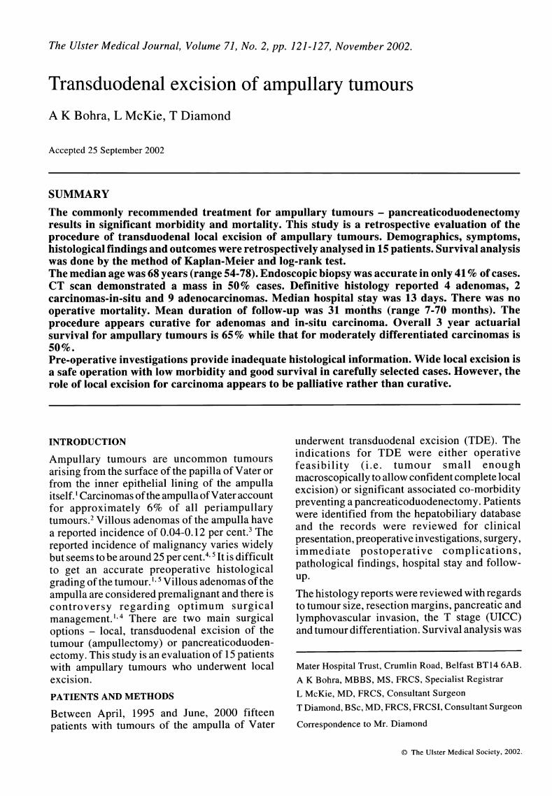
Fig 1. Stay sutures are inserted around the tumour asmarkers for wide diathermy excision (a) prior toexcision of the papilla of Vater, encompassingpart of biliary and pancreatic ducts and posteriorduodenal wall (b).
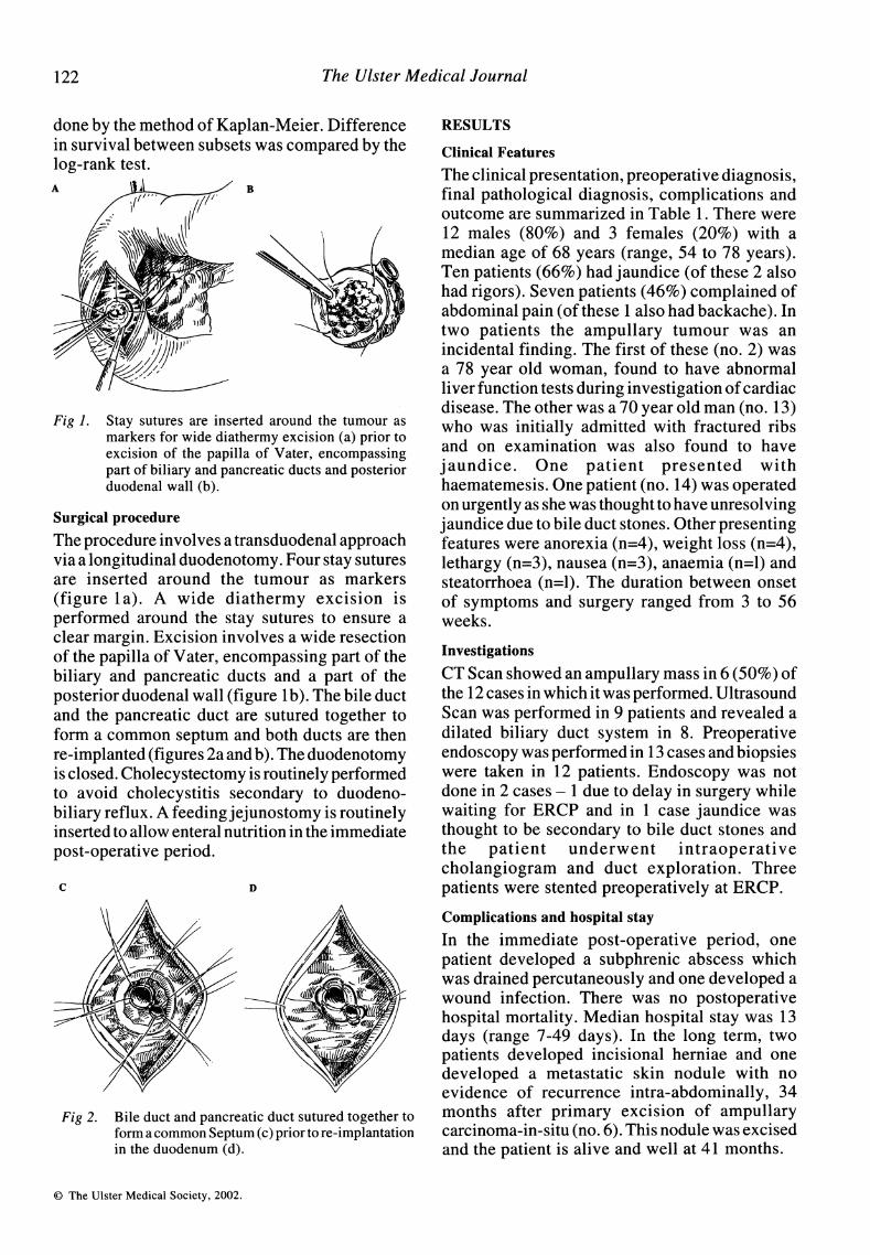
Surgical procedureThe procedure involves a transduodenal approachvia a longitudinal duodenotomy. Four stay suturesare inserted around the tumour as markers(figure la). A wide diathermy excision isperformed around the stay sutures to ensure aclear margin. Excision involves a wide resectionof the papilla of Vater, encompassing part of thebiliary and pancreatic ducts and a part of theposterior duodenal wall (figure Ib). The bile ductand the pancreatic duct are sutured together toform a common septum and both ducts are thenre-implanted (figures 2a and b). The duodenotomyis closed. Cholecystectomy is routinely performedto avoid cholecystitis secondary to duodeno-biliary reflux. A feeding jejunostomy is routinelyinserted to allow enteral nutrition in the immediatepost-operative period.
C D
Fig 2. Bile duct and pancreatic duct sutured together toform a common Septum (c) prior to re-implantationin the duodenum (d).
RESULTS
Clinical FeaturesThe clinical presentation, preoperative diagnosis,final pathological diagnosis, complications andoutcome are summarized in Table 1. There were12 males (80%) and 3 females (20%) with amedian age of 68 years (range, 54 to 78 years).Ten patients (66%) had jaundice (of these 2 alsohad rigors). Seven patients (46%) complained ofabdominal pain (of these 1 also had backache). Intwo patients the ampullary tumour was anincidental finding. The first of these (no. 2) wasa 78 year old woman, found to have abnormalliver function tests during investigation ofcardiacdisease. The other was a 70 year old man (no. 13)who was initially admitted with fractured ribsand on examination was also found to havejaundice. One patient presented withhaematemesis. One patient (no. 14) was operatedon urgently as she was thought to have unresolvingjaundice due to bile duct stones. Other presentingfeatures were anorexia (n=4), weight loss (n=4),lethargy (n=3), nausea (n=3), anaemia (n=l) andsteatorrhoea (n=l). The duration between onsetof symptoms and surgery ranged from 3 to 56weeks.
InvestigationsCT Scan showed an ampullary mass in 6 (50%) ofthe 12 cases in which it was performed. UltrasoundScan was performed in 9 patients and revealed adilated biliary duct system in 8. Preoperativeendoscopy was performed in 13 cases and biopsieswere taken in 12 patients. Endoscopy was notdone in 2 cases - 1 due to delay in surgery whilewaiting for ERCP and in 1 case jaundice wasthought to be secondary to bile duct stones andthe patient underwent intraoperativecholangiogram and duct exploration. Threepatients were stented preoperatively at ERCP.
Complications and hospital stayIn the immediate post-operative period, onepatient developed a subphrenic abscess whichwas drained percutaneously and one developed awound infection. There was no postoperativehospital mortality. Median hospital stay was 13days (range 7-49 days). In the long term, twopatients developed incisional herniae and onedeveloped a metastatic skin nodule with noevidence of recurrence intra-abdominally, 34months after primary excision of ampullarycarcinoma-in-situ (no. 6). This nodule was excisedand the patient is alive and well at 41 months.
i The Ulster Medical Society, 2002.
122
Transduodenal excision ofampullary tumours
TABLE
Demography, Clinicalfeatures, Pathology and outcome
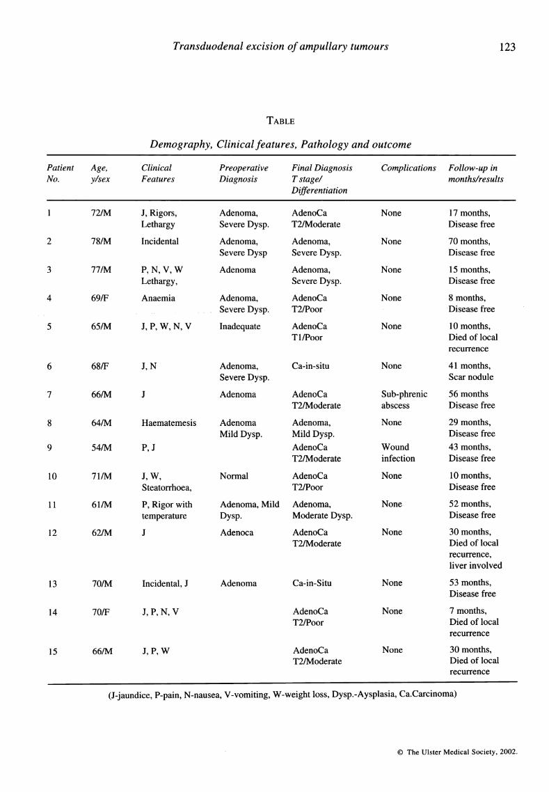
Patient Age, Clinical Preoperative Final Diagnosis Complications Follow-up inNo. y/sex Features Diagnosis T stage! months/results
Differentiation
J, Rigors,Lethargy
Incidental
P, N, V, WLethargy,
Anaemia
J, P, W, N, V
J, N
J
Haematemesis
Adenoma,Severe Dysp.
Adenoma,Severe Dysp
Adenoma
Adenoma,Severe Dysp.
Inadequate
Adenoma,Severe Dysp.
Adenoma
AdenomaMild Dysp.
P,J
AdenoCaT2/Moderate
Adenoma,Severe Dysp.
Adenoma,Severe Dysp.
AdenoCaT2/Poor
AdenoCaTI/Poor
Ca-in-situ
AdenoCaT2/Moderate
Adenoma,Mild Dysp.AdenoCaT2/Moderate
None
None
None
None
None
None
Sub-phrenicabscess
None
Woundinfection
17 months,Disease free
70 months,Disease free
15 months,Disease free
8 months,Disease free
10 months,Died of localrecurrence
41 months,Scar nodule
56 monthsDisease free
29 months,Disease free43 months,Disease free
10 71/M
11 61/M
12 62/M
J,W,
Steatorrhoea,
P, Rigor withtemperature
J
Normal
Adenoma, MildDysp.
Adenoca
AdenoCaT2/Poor
Adenoma,Moderate Dysp.
AdenoCaT2/Moderate
None
None
None
10 months,Disease free
52 months,Disease free
30 months,Died of localrecurrence,
liver involved
13 70/M
14 70/F
15 66/M
C The Ulster Medical Society, 2002.
1 72/M
2 78/M
3 77/M
4 69/F
5 65/M
6 68/F
7 66/M
8 64/M
9 54/M
AdenomaIncidental, J
J, P, N, V
J, P, W
Ca-in-Situ None
None
None
AdenoCa
T2/Poor
AdenoCaT2/Moderate
53 months,Disease free
7 months,Died of localrecurrence
30 months,Died of localrecurrence
(J-jaundice, P-pain, N-nausea, V-vomiting, W-weight loss, Dysp.-Aysplasia, Ca.Carcinoma)
123
The Ulster Medical Journal
Histo-pathology
Preoperative endoscopic biopsy was accurate inonly 5 patients (41%) of whom 4 had adenomasand 1 had an adenocarcinoma. Multiple biopsieswere routinely undertaken at endoscopy. Amongstthe remainder, preoperative biopsy revealed a
villous adenoma with severe dysplasia in 5patients ofwhom 2 eventually had carcinoma-in-situ, 2 had a moderately differentiatedadenocarcinoma and 1 had a poorly differentiatedadenocarcinoma. One preoperative biopsy was
insufficient and 1 was normal. Both turned out tobe poorly differentiated adenocarcinomas. In total,4 patients had adenomas, 2 had carcinoma-in-situand 9 had adenocarcinoma, of which 5 were
moderately differentiated and 4 poorlydifferentiated. Of the 9 adenocarcinomas 1 was
in stage TI and 8 were T2. Frozen section biopsyperformed in one patient (9) was reported as
benign though definitive histology revealedmoderately differentiated adenocarcinoma.Resection margins were clear in 14 patients. A re-
laparotomy could not be undertaken in the one
patient with involved resection margins due tosignificant associated co-morbidity. There was
no pancreatic invasion, though lympho-vascularinvasion was found in 3 cases. Mean tumourdiameter was 2.5 cms (range, 1.5 to 4.2 cms).Mean duration of follow-up was 31.4 months(range, 7 to 70 months).
Outcome
The longest survival in this study was 70 months(no. 2) with a total of 4 patients alive after 50months, 1 with a diagnosis of moderatelydifferentiated adenocarcinoma, 1 with carcinoma-in-situ and 2 with dysplastic adenomas. Fourpatients (26 percent) died in this study. All thepatients died of local recurrence. Two of thesehad T2, moderately differentiated tumours, andthe other 2 had poorly differentiated tumours instage T I and T2. Survival was worst in the poorlydifferentiated group - 1 patient who also hadinvolved resection margins surviving 7 monthsand another 10 months while both the patientswith moderately differentiated carcinomassurvived 30 months. Of the 3 patients withlymphovascular invasion 2 had poorlydifferentiated and 1 had a moderatelydifferentiated carcinoma. The latter is alive at 43months while of the patients with poorlydifferentiated carcinoma with lymphovascularinvasion 1 is alive at 10 months and the other diedat 7 months.
1.00 -
0.75 -
.0
0
° O 50
0.E
c0.25
0.00 - I -
0 12 24 36 48 60 72
Months
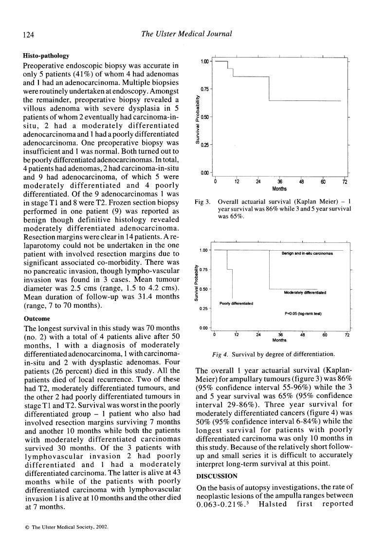
Fig 3. Overall actuarial survival (Kaplan Meier) 1
year survival was 86% while 3 and 5 year survivalwas 65%.
1.00
= 0.75D-0
2tL
X5 0.50.2-
0.25
0.000 12 24 36
Months48 60 72
Fig 4. Survival by degree of differentiation.
The overall 1 year actuarial survival (Kaplan-Meier) for ampullary tumours (figure 3) was 86%(95% confidence interval 55-96%) while the 3and 5 year survival was 65% (95% confidenceinterval 29-86%). Three year survival formoderately differentiated cancers (figure 4) was50% (95% confidence interval 6-84%) while thelongest survival for patients with poorlydifferentiated carcinoma was only 10 months inthis study. Because of the relatively short follow-up and small series it is difficult to accuratelyinterpret long-term survival at this point.
DISCUSSION
On the basis of autopsy investigations, the rate ofneoplastic lesions of the ampulla ranges between0.063-0.21 %.5 Halsted first reported
The Ulster Medical Society, 2002.
Benign and in-situ carcinomas
Moderately differentiated
Poorly differentiated
P<O.05 (log-rank test)
I I I~~~~~~~~~~~~~~ r-
124
Transduodenal excision of ampullary tumours
transduodenal excision of an ampullary mass in1899.6 In 1935 A. 0. Whipple reported a 2-stageresection of ampullary carcinoma.7
Although the ampulla is easily accessible byendoscopy there is a high reported incidence offalse negative results for biopsy of carcinoma,ranging from 25-60%.l,5, 89, 10, 11 The results inthis study are similar, with a false negative rate of59%. CT scan detection of a lesion is consideredeven less sensitive with a reported figure ofaround 20% in some studies.1 Endoscopicultrasonography aided diagnosis and staging hasbeen widely recommended. 12, 13, 14 However,Cahen et al reported an accuracy of only 44 percent with endoscopic ultrasonography assistedstaging and a false positive outcome for metastaticlymph nodes in 3 1% of their cases.3 Endoscopicultrasonography was not available for this study.Per-operative frozen sections are unreliable inexcluding malignancy and can also give falsepositive results.5 15 It seems nearly impossible toexclude, with certainty, the presence ofcarcinomain an adenoma without complete excision.5
While TDE is now accepted as the procedure ofchoice for benign adenomas, pancreatico-dudenectomy is the commonly recommendedsurgery for ampullary cancer.'0 There have beenrecommendations for radical surgery even forbenign lesions with some authors citing problemslike a high incidence of malignancy, difficulty inaccurate pre-operative confirmation, tendencyfor tumours to recur and concerns regarding theoncological adequacy of local resection forampullary cancers.8' 9 16 3-year survival around55 to 60 per cent and 5-year survival around 35 to55 per cent has been reported afterpancreaticoduodenectomy.171-9 Beger et alreported 3- and 5-year survival rates of 72% and52% respectively with RO resections after radicalsurgery, but RO resection was possible in only62% of the malignant cases in their study, with anintra-abdominal complication rate of 25%. Thepresent study with overall actuarial 3 and 5 yearsurvival projections of65% would tend to favourthe less radical approach.
Some reports also suggest that local resection isoncologically acceptable with comparable resultsand low morbidity in selective cases.3 5' ' Taraziet al reported a 2 year survival of 55 per cent anda 5-year survival of 41 per cent following TDEfor carcinomas.20 Newman and Pittam reported a5 year survival of 41 per cent and a surviving
patient 15 years after TDE for ampullarycarcinoma.20 Wise et al have reported a 3 yearsurvival at 50 per cent and 5 year survival at 37.5per cent.22 Knox and Kingston have reported abetter 1, 2, 3 and 5 year survivals following TDEcompared to radical procedures.23 Robertson andImrie reported a median survival of 57 monthswith44% five-year actuarial survival, afterTDE.24Fifty percent of their survivors developedrecurrent disease while there was a 25% incidenceof post-operative deaths. Talamini et al in a largestudy of 28 years experience from the JohnHopkins Institution reported an overall mortalityof 3.8% after radical resection with no deaths inthe last 5 years in consecutive 45 patients.25Morbidity in the latter part of their study hadreduced to 38%. Five-year survival was 38% andthey believed avoidance of transfusion improvedthe prognosis. Various studies have proposedthatTDE is justifiable when the ampullary tumouris pTl(UICC-staging) and graded GI or G2(highly or moderately differentiated) with nolymphatic infiltration and complete resection(RO).5'26 The present study would tend tocorroborate these suggestions. Unfortunatelyaccurate TNM staging pre- or peroperatively isnot feasible.
Howe et al, in a prospective study of thecorrelation between clinicopathological variablesand survival of 123 patients presenting withampullary carcinomas, found negative marginsand negative nodes as independent predictors ofimproved survival.2 Lymphatic drainage oftumours ofthe ampulla is distinct from pancreatictumours in that, even in advanced cases, theyseem to involve only a local group of lymphnodes near the ampulla, yielding a superiorprognosis if the infiltration does not involvepancreatic tissue.5'27 Pancreatic involvement isclosely related to extensive nodal involvementand ampullary cancers invading the pancreaticparenchyma may act more like pancreatic cancersresulting in a poor prognosis.5'27 In our studythere was no pancreatic involvement in any of thecases and resection margins appeared clear in allbut one case. Neither size nor duration ofsymptoms have been reported to accurately predictthe presence of malignancy.10 Interestingly, inthe present study all 10 patients withjaundice hadcarcinoma or carcinoma-in-situ while none of thepatients with villous adenoma had jaundice. Thiswould be in keeping with some reports whichsuggest that malignant lesions tend to present
C The Ulster Medical Society, 2002.
125
126 The Ulster Medical Journal
with jaundice while benign lesions do not."9' 1 0 Itwould be interesting to see from larger studies inthe future ifelevated bilirubin could be utilised asa guide to the general decision making regardingthe appropriate procedure and also as a predictorof survival in tumours of the ampulla.
In conclusion, our study suggests that obtainingan exact preoperative diagnosis for ampullarytumours is very difficult. None of the existinginvestigative tools seem to be entirelyconfirmative about the true histological nature ofthe tumour and the extent of the disease.Transduodenal excision of ampullary tumoursseems curative for benign tumours and for in-situcancers. It is a low risk alternative for patientswith carcinoma who are poor surgical risks due toage or co-morbidity. Oncologically, local excisionofcarcinoma appears to be a palliative rather thancurative procedure.
REFERENCES
1. Posner S, Colletti L, Knot J, Mulholland M, EckhauserF. Safety and long-term efficacy of transduodenalexcision for tumors of the ampulla of Vater. Surgery2000; 128(4): 694-701.
2. Howe J R, Klimstra D S, Moccia R D, Conlon K C,Brennan M F. Factors predictive of survival inampullary carcinoma. Ann Surg 1998; 228(1): 87-94.
3. Cahen D L, Fockens P, de Wit L T, Offerhaus G J A,Obertop H, Gourna D J. Local resection orpancreaticoduodenectomy for villous adenoma of theampulla of Vater diagnosed before operation. Br JSurg 1997; 84(7): 948-5 1.
4. Treitschke F, Beger H G. Local resection of benignperiampullary tumors. Ann Oncol 1999; 10(Suppl 4):212-4.
5. Beger H G, Treitschke F, Gansauge F, Harada N, HikiN, Mattfeldt T. Tumor of the Ampulla of Vaterexperience with local or radical resection in 171consecutively treated patients. Arch Surg 1999; 134(5):526-32.
6. Halsted W S. Contributions to the surgery of the bileduct passages, especially of the common bile duct.Boston Med Surg J 1899; 141: 645-54.
7. Whipple A, Parsons W, Mullins C. Treatment ofcarcinoma of ampulla of Vater. Ann Surg 1935; 102:763-79.
8. Chappuis C, Divincenti F C, Cohn I. Villous tumoursof the duodenum. Ann Surg 1989; 209(5): 593-9.
9. Ryan D P, Schapiro R H, Warshaw A L. Villoustumors of the duodenum. Ann Surg 1985; 203: 301-6.
10. Rattner D W, Fernandez-del Castillo C, BruggeW R,Warshaw A L. Defining the criteria for local resectionof ampuilary neoplasms. Arch Surg 1996; 131(4):366-71.
11. Yamaguchi K, Enjoji M, Kitamura K. Endoscopicbiopsy had limited accuracy in diagnosis of ampullarytumors. Gastrointest Endosc 1990; 36(6): 588-92.
12. Tio T L, Sie L H, Verbeek P C M, De Wit L T, TytgatG N J. Endosonography in diagnosing and stagingduodenal villous adenoma. Gut 1992; 33(4): 567-8.
13. Yasuda K, Mukai H, Cho E, Nakajima M, Kawai K.The use ofendoscopic ultrasonography in the diagnosisand staging of carcinoma of the papilla of Vater.Endoscopy 1988; 20(Suppl 1): 218-22.
14. Rosch T, Braig C, Gain T, Feuerbach S, Stewart J R,Schusdziorro V, et al. Staging of pancreatic andampullary carcinoma by endoscopic ultrasonography.Comparison with conventional sonography, computedtomography, and angiography. Gastroenterology1992; 10(1): 188-99.
15. Asbun H J, Rossi R L, Munson J L. Local resection forampullary tumors. Is there a place for it? Arch Surg1993; 128(5): 515-20.
16. Chareton B, Coiffic J, Landen S, Bardaxoglou E,Campion J P, Launois B. Diagnosis and therapy forampullary tumors: 63 cases. World J Surg 1996;20(6): 707-12.
17. Harada N, Treitschke F, Imaizumi T, Beger H G.Pancreatic invasion is a prognostic indicator afterradical resection for carcinoma of the ampulla ofVater. J Hepato-Biliary-Pancreatic Surg 1997; 4:215-19.
18. Hanyu F, Imaizumi T, Nakamura M, Yoshikawa T.Proposed definition of early cancer of the papilla ofVater. J Biliary Tract Pancreas 1984; 5: 847-52.
19. Willett C G, Warshaw A L, Convery K, Compton C C.Patterns of failure after pancreaticoduodenectomy forampullary carcinoma. Surg Gynecol Obstet 1993;176(1): 33-8.
20. Tarazi R Y, Hermann R E, Vogt D P, Hoerr S 0,Esselstyn C B, Coperman A M, et al. Results ofsurgical treatment of periampullary tumours: a 35years experience. Surgery 1986; 100: 716-23.
21. Newman R J, Pittam M R. Local excision in thetreatment of carcinoma of the ampulla of Vater. J RColl Surg Edinb 1982; 27(3):154-7.
22. Wise L, Pizzimbono C, Dehrier L P. Periampullarycancer. A clinico-pathologic study of 62 patients. AmJSurg 1976; 131(2): 141-8.
23. Knox R A, Kingston R D. Carcinoma of the ampullaof Vater. Br J Surg 1986; 73(1): 72-3.
24. Robertson J F, Imrie C W. Local excision of ampullarycarcinoma. Acta Chir Scand 1986; 152: 537-9.
C The Ulster Medical Society, 2002.
Transduodenal excision of ampullary tumours 127
25. Talamini M A, Moesinger R C, Pitt H A, Sohn T A,Hruban R H, Lillemoe K D. Adenocarcinoma of theampulla of Vater. A 28-year experience. Ann Surg1997; 225: 590-9.
26. Klein P, Reingruber B, Kastl S, Dworak 0,Hohenberger W. Is local excision of pTl -ampullaryCarcinomas justified? Eur J Surg Oncol 1996; 22(4):366-71.
27. Shirai Y, Ohtani T, Tsukada K, Hatakeyama K. Patternsof lymphatic spread of carcinoma of the ampulla ofVater. Br J Surg 1997; 84(7): 1012-6.
C The Ulster Medical Society, 2002.