Imaging of cardio-pulmonary treatment related damage · Imaging of cardio-pulmonary treatment...
16
Imaging of cardio-pulmonary treatment related damage Dr. Andrea Borghesi Dr. Emanuele Gavazzi Department of Radiology 2 – University of Brescia Radiotheraphy and Lung • The goal of radiation therapy (RT) is to reduce or eliminate the tumor with minimal toxicity to normal surrounding tissue • RT is used to treat many intrathoracic and chest wall malignacies (breast cancer, bronchogenic carcinoma, oesophageal neoplasm, malignant mesothelioma and lymphoma) Benveniste MFK et al Clinical Radiol;2013; Graves PR et al Semin Radiat Oncol 2010 NSCLC! IMRT Brest cancer! 3D-CRT Oesophageal cancer ! IMRT
Transcript of Imaging of cardio-pulmonary treatment related damage · Imaging of cardio-pulmonary treatment...

Imaging of cardio-pulmonary treatment related damage
Dr. Andrea Borghesi
Dr. Emanuele GavazziDepartment of Radiology 2 – University of Brescia
Radiotheraphy and Lung
• The goal of radiation therapy (RT) is to reduce or eliminate the tumorwith minimal toxicity to normal surrounding tissue
• RT is used to treat many intrathoracic and chest wall malignacies(breast cancer, bronchogenic carcinoma, oesophageal neoplasm, malignant mesothelioma and lymphoma)
Benveniste MFK et al Clinical Radiol;2013; Graves PR et al Semin Radiat Oncol 2010
NSCLC! IMRTBrest cancer! 3D-CRT Oesophageal cancer ! IMRT
• The lung is one of the most sensitive tissue to ionizing radiation and damage to normal lung tissue remains the main obstacle in RT
• Radiation-induced lung disease (RIDL) is a frequent complication of RT (40% of patients develop radiographic abnormalities - 7% develop symptomatic pneumonitis)
• Radiation-induced changes in the lung are dependent on a number of factors inclundig:– Patient-specific factors (age, smoking, pre-exsting lung disease)– RT technique (3D CRT, IMRT e SBRT)– Radiation dose– Volume of lung irradiated– Administration of chemotherapy
Radiation-induced lung injury (RILD)
Benveniste MFK et al Clinical Radiol 2013; Graves PR et al Semin Radiat Oncol 2010Mesurolle B et al Radiographics 2000; Park et al Radiographics 2000; Jennings FLAA 1962
Radiation-induced lung disease (RILD)
• Two distinct clinical, pathologic,and radiologic phases of RILD are recognized:
– Acute phase (Radiation Pneumonitis ! 4-12 weeks after RT)
– Chronic phase (Radiation Fibrosis ! several months after RT)
• Depending on the severity of lung injury, theseabnormalities may resolve completely, but they more oftenundergo progressive organization and eventually lead to fibrosis
Benveniste MFK et al Clinical Radiol 2013; Park et al Radiographics 2000; Larici AR et al Radiographics 2011

Usual Radiologic Findings after RT
• RILD is not generally seen with doses below 20Gy and it ismost commonly seen with doses > 40Gy.
• Radiologic manifestations of RILD, generally confined to the field of irradiation, are better detected on CT than chestradiographs
• Acute phase (Radiation Pneumonitis)
• Chest X-ray– Diffuse haziness– Consolidation– Pleural effusion– Elevation of diaphragm
Usual Radiologic Findings after RT

• Acute phase (Radiation Pneumonitis)
• CT (more sensitive)– Ground-glass opacity– Patchy or dense consolidation– Pleural effusion
Benveniste MFK et al Clinical Radiol 2013;
Usual Radiologic Findings after RT
• Chronic phase (Radiation Fibrosis)
• Chest X-ray– Linear opacities– Dense consolidation– Architectural distortion– Volume loss– Shift of the mediastinum– Elevation of diaphragm
Usual Radiologic Findings after RT
• Chronic phase (Radiation Fibrosis)
• CT (more sensitive)– Steaky opacities– Dense consolidation– Traction bronchiectasis– Architectural distortion– Volume loss– Pleural thickening– Shift of the mediastinum– Elevation of diaphragm
Benveniste MFK et al Clinical Radiol 2013; Graves PR et al Semin Radiat Oncol 2010
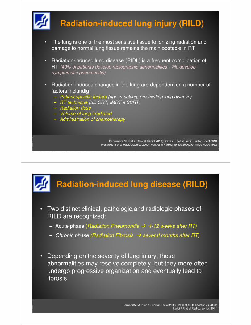
Usual Radiologic Findings after RT
• Acute and chronic phase– 53 year-old female with mucoepidermoid carcinoma of the tracheal
carina
Before RT 1 month after RT 1 year after RT
Usual Radiologic Findings after RT

Unusual Radiologic Findings after RT
• Necrosis: uncommon (0.6%), severe and late complicationafter RT (>60 Gy)
• Cavitation within the radiation fibrosis may also indicate an infectious process (including TBC) and recurrent tumour
Mesurolle B et al Radiographics 2000
Unusual Radiologic Findings after RT
• Pneumothorax (1%): usually occurs in patients with radiologic evidence of post-irradiation fibrosis
• BOOP (2.5%): patchy, bilateral and multifocal migratorylung opacities (consolidation and ground glass infiltration)
Mesurolle B et al Radiographics 2000; Kano et al. Jpn J Radiol 2012

Unusual Radiologic Findings after RT
• BOOP: patchy, bilateral and multifocal migratory lungopacities (consolidation and ground glass infiltration)
Kano et al. Jpn J Radiol 2012
4 months after RT (right breast cancer)
Differential diagnosis considerations
• When radiological manifestations of radiation damage are different from the expected patterns, other disease entities have to beconsidered
– Infection
• Before completion of RT
• Abrupt onset
• Lung opacities outside of the treated areas
• Tree in bud pattern
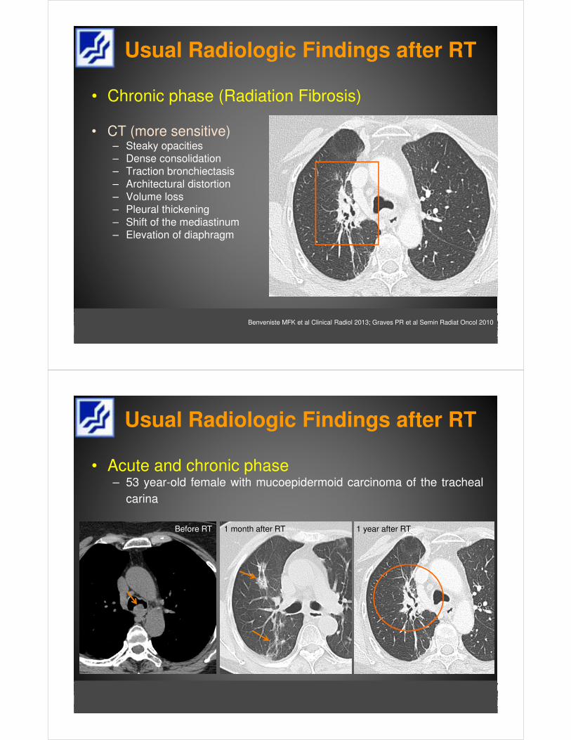
– Recurrent tumor
• Occurs within 2 years after RT is completed
• Development of a lobulate contour within the fibrosis
• PET-CT improved DD between recurrent tumour and radiation fibrosis(**PET-CT is best performed 6 months after RT is completed)
Benveniste MFK et al Clinical Radiol 2013; Munden RF et al Radiation-induced lung disease. 1sted. Philadelphia: Saunders-Elsevier; 2008.
Recurrent tumor within fibrosisPET-TC
recurrent tumor 18 months after RT
Focal FDG uptake is suggestive of residual or recurrent disease
Conclusions
• RT is an important modality in the treatment of patients with neoplasms.
• Knowledge of pulmonary abnormalities related to RT is important to recognize patterns of RILD and detect complications, such as recurrent malignancy or infection.
Benveniste MFK et al Clinical Radiol;2013; Graves PR et al Semin Radiat Oncol 2010; Park et al Radiographics 2000; Mesurolle B et al Radiographics 2000;

Heart and RT: epidemiology
• Cardiovascular disease is now the most common non-malignancy cause of death in radiation-treated cancer survivors (Hodgkin’s lymphoma and breast cancer), most often occurring decades after treatment (3 to 29 yearsafter treatment).
• The long-term effects on the heart still remain unclear, mandating longer follow-up.
• Increased risk of coronary artery disease (CAD), congestive heart failure, valvular heart disease, pericardial disease, and sudden death.
Aleman BM et al. J Clin Oncol 2003;21:3431–9. Ng AK. Br J Haematol 2011;154:23–31.Hoppe RT. Ann Oncol 1997; Suppl 1:115–8. Patnaik JL et al. Breast Cancer Res 2011;13:R64.
Jaworski et al. JACC Vol. 61, No. 23, 2013

Prevention and follow-up
• Before radiotherapy: comprehensive baseline evaluationincluding a detailed cardiovascular history, cardiac examination, risk factor profiling, and echocardiography (systolic and diastolic function).
• Prolonged cardiological follow-up and cardiac screeningis mandatory in cancer patients who have received irradiation to facilitate early identification of cardiac related complications.
• Control and minimize cardiac risk factors
Prevention and follow-up
• There is a paucity of data to support the optimal method and frequency of screening post-radiotherapy patients
• Development of a uniform approach to be potentially beneficial
• The focus of screening should ideally incorporate non-invasive, radiation-free modalities in the first instance

Cardiac imaging
• Rest and stress echocardiography
• Cardiac magnetic resonance imaging (CMRI)
• Coronary computerized topography (CCT)
Feng M et al. Int J Radiat Oncol Biol Phys. 2011 January 1; 79(1): 10–18.
CMRI: sequences
• Morfology
black blood (HASTE, TSE T1, STIR T2)
bright blood (True FISP)
• Functioncine SSFP, Gradient-Echo
• Tissue characterization (delayed enhancement)segmented IR fast GE
• Perfusion
• Valvular Flowphase contrast

Morfology: acute and chronic pericarditis
black blood (HASTE, TSE T1, STIR T2)bright blood (True FISP)
Morfology: acute and chronic pericarditis
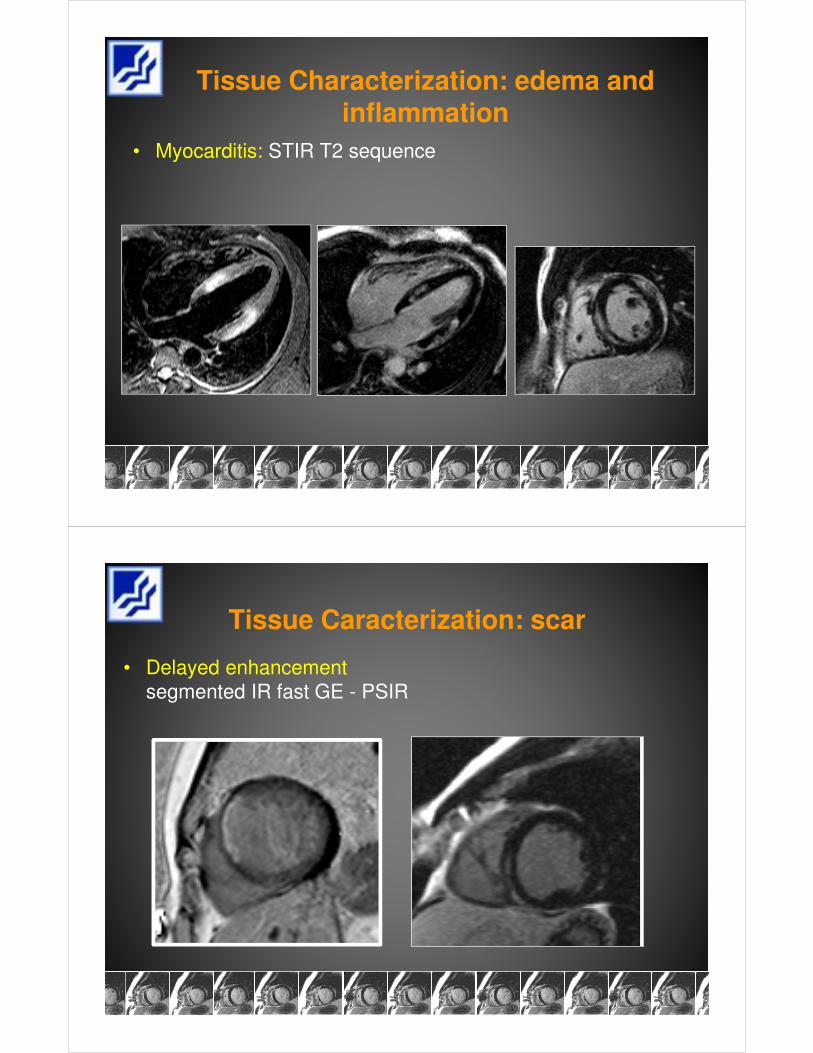
Tissue Characterization: edema and inflammation
• Myocarditis: STIR T2 sequence
Tissue Caracterization: scar
• Delayed enhancementsegmented IR fast GE - PSIR

Tissue characterization: T1 mapping
With or without contrast medium
T1 value and extracellular volume: correlate with myocardial fibrosis
• Cine and Phase contrast sequences
Valvular disease: thickening, stenosisand regurgitation

Coronary CT
• Calcium scoring: Agatstone score
Coronary CT
• Detection of asintomatic CAD
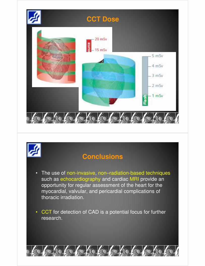
CCT Dose
Conclusions
• The use of non-invasive, non–radiation-based techniques such as echocardiography and cardiac MRI provide an opportunity for regular assessment of the heart for the myocardial, valvular, and pericardial complications ofthoracic irradiation.
• CCT for detection of CAD is a potential focus for further research.