Identification of the metabolic intermediates of phthalate by Tn5 mutants of Pseudomonas...
8
JOURNAL o r FERMENTATION AND BIOENGINEERING Vo1. 77, NO. 6, 583-590. 1994 Identification of the Metabolic Intermediates of Phthalate by Tn5 Mutants of Pseudomonas testosteroni and Analysis of the 4,5-Dihydroxyphthalate Decarboxylase Gene JONG-HOON LEE, 1 TOSHIO OMORI, 2 AND TOHRU KODAMA 1. Department of Agricultural Chemistry, The University of Tokyo, Bunkyo-ku, Tokyo 1131 and Biotechnology Research Center, The University of Tokyo, Bunkyo-ku, Tokyo 113, 2 Japan Received 7 February 1994/Accepted 14 March 1994 Phthalate-, 4,5-dihydro-4,5-dihydroxyphthalate (DDP)-, 4,5-dihydroxyphthalate (DHP)-, and protoca- techuate-accumulating mutants of Pseudomonas testosteroni M4-1 were isolated by transposon insertion mutagenesis. From the DDP-accumulating, transposon-inserted mutant strain M4-122, the 4,5-dihydro- xyphthalate decarboxylase gene which is involved in phthalate metabolism was cloned and its nucleotide sequence determined. The structural gene, designated phtD, was 990 bp, corresponding to a protein of 330 amino acid residues (calculated molecular weight 37,200). The phtD gene was expressed in Escherichia coli by its own promoter located upstream of the phtD open reading frame. The promoter shows high homology with the E. coil a 7° consensus sequence. The putative amino acid sequence has 77.6% homology with that of the pht5 gene from Pseudomonas putida. The nucleotide sequence of the upstream region of the phtD gene has high homology with that of phtl, the putative positive regulator for the other pht genes of P. putida. The deduced amino acid sequence of the upstream region of the phtD gene also shows high similarity with that of Phtl. From these results, the genes of P. testosteroni and P. putida involved in phthalate metabolism would seem to have evolved from the same origin but diversified to compose different gene orders in different organisms. Phthalate is a precursor of phthalate esters, which are used as plasticizers for cellulose and vinyl products. Because of their massive production in the plastics and textile industries, phthalate esters are considered to be environmental pollutants (1). Research into the biodegradation of phthalate esters and phthalate was initiated to solve the problem of toxic- ity, but following a 1983 report by Ballard et al. (2) the direction of research has changed towards the produc- tion and use of the metabolic intermediates of phthalate, which have potential for use as raw materials in the or- ganic synthetic chemical industry. Nakazawa and Hayashi reported the degradation of phthalate through 4,5-dihydro-4,5-dihydroxyphthalate (DDP), 4,5-dihydroxyphthalate (DHP) and protocate- chuate by using several mutants of Pseudomonas testoste- roni NH1000 blocked in phthalate catabolism (3). We isolated a 4,5-dihydro-4,5-dihydroxyphthalate (DDP)-ac- cumulating mutant, strain M4-122, from P. testosteroni M4-1 by transposon mutagenesis and reported the DDP production conditions of the M4-122 strain (4). The iden- tification of phthalate-, DHP- and protocatechuate- accumulating mutants has also reconfirmed the dissimi- latory pathway of P. testosteroni in the present study (Fig. 1). Some enzymatic research concerned with phthalate degradation has also been reported. The purification and characterization of phthalate oxygenase and phthalate oxygenase reductase from Pseudomonas cepacia has been carried out (5), and the key enzyme 4,5-dihydrox- yphthalate (DHP) decarboxylase (EC 4.1.1.55) that converts DHP to protocatechuate was purified from P. * Corresponding author. 583 testosteroni and Pseudomonas fluorescens and character- ized (6, 7). Nomura et al. reported the genes encoding the initial degradation pathway of phthalate in Pseudomonas puo tida (8). To produce metabolic intermediates of phthalate with a gene manipulated microorganism, we started genetic research on P. testosteroni, which shows high phthalate degrading activity. A gene level analysis of Tn5-inserted mutants ac- cumulating the metabolic intermediates of phthalate, genetic research on the DHP decarboxylase gene, and the flanked region cloned from the mutant strain M4-122 are described in this paper. MATERIALS AND METHODS Transposon mutagenesis Transposon mutagenesis was performed according to the method of Rella et al. (4, 9) using the suicide transposon Tn5 donor plasmid pSUP2021. Escherichia coli S17-1 (pSUP2021), the donor strain containing the mobilizable Tn5 carrier vec- tor pSUP2021, was used for Tn5 insertion mutagenesis. This strain was kindly provided by R. Simon, Univer- sit/it Bielefeld, FRG (10). Bacterial strains and plasmids E. coli JM109 (11) was used as a host for cloning and subcloning. Another E. coli strain, XL1-Blue, was used as the host for bacte- riophage R408 (Stratagene) in the preparation of single- stranded DNAs for DNA sequencing. The phthalate degrading strain P. testosteroni M4-1 and its Tn5-insert- ed mutant strain, M4-122, which accumulates DDP, were used as sources of total DNA (4). P. putida PpY101 (met, nal) was also used as a host (12). Plasmids pBR322, pUC18, and pUCI9 were used for cloning.
-
Upload
jong-hoon-lee -
Category
Documents
-
view
212 -
download
0
Transcript of Identification of the metabolic intermediates of phthalate by Tn5 mutants of Pseudomonas...

JOURNAL or FERMENTATION AND BIOENGINEERING Vo1. 77, NO. 6, 583-590. 1994
Identification of the Metabolic Intermediates of Phthalate by Tn5 Mutants of Pseudomonas testosteroni and Analysis
of the 4,5-Dihydroxyphthalate Decarboxylase Gene JONG-HOON LEE, 1 TOSHIO OMORI, 2 AND TOHRU KODAMA 1.
Department of Agricultural Chemistry, The University of Tokyo, Bunkyo-ku, Tokyo 1131 and Biotechnology Research Center, The University of Tokyo, Bunkyo-ku, Tokyo 113, 2 Japan
Received 7 February 1994/Accepted 14 March 1994
Phthalate-, 4,5-dihydro-4,5-dihydroxyphthalate (DDP)-, 4,5-dihydroxyphthalate (DHP)-, and protoca- techuate-accumulating mutants of Pseudomonas testosteroni M4-1 were isolated by transposon insertion mutagenesis. From the DDP-accumulating, transposon-inserted mutant strain M4-122, the 4,5-dihydro- xyphthalate decarboxylase gene which is involved in phthalate metabolism was cloned and its nucleotide sequence determined. The structural gene, designated phtD, was 990 bp, corresponding to a protein of 330 amino acid residues (calculated molecular weight 37,200). The phtD gene was expressed in Escherichia coli by its own promoter located upstream of the phtD open reading frame. The promoter shows high homology with the E. coil a 7° consensus sequence. The putative amino acid sequence has 77.6% homology with that of the pht5 gene from Pseudomonas putida. The nucleotide sequence of the upstream region of the phtD gene has high homology with that of phtl, the putative positive regulator for the other pht genes of P. putida. The deduced amino acid sequence of the upstream region of the phtD gene also shows high similarity with that of Phtl . From these results, the genes of P. testosteroni and P. putida involved in phthalate metabolism would seem to have evolved from the same origin but diversified to compose different gene orders in different organisms.
Phthalate is a precursor of phthalate esters, which are used as plasticizers for cellulose and vinyl products. Because of their massive production in the plastics and textile industries, phthalate esters are considered to be environmental pollutants (1).
Research into the biodegradation of phthalate esters and phthalate was initiated to solve the problem of toxic- ity, but following a 1983 report by Ballard et al. (2) the direction of research has changed towards the produc- tion and use of the metabolic intermediates of phthalate, which have potential for use as raw materials in the or- ganic synthetic chemical industry.
Nakazawa and Hayashi reported the degradation of phthalate through 4,5-dihydro-4,5-dihydroxyphthalate (DDP), 4,5-dihydroxyphthalate (DHP) and protocate- chuate by using several mutants of Pseudomonas testoste- roni NH1000 blocked in phthalate catabolism (3). We isolated a 4,5-dihydro-4,5-dihydroxyphthalate (DDP)-ac- cumulating mutant, strain M4-122, from P. testosteroni M4-1 by transposon mutagenesis and reported the DDP production conditions of the M4-122 strain (4). The iden- tification of phthalate-, DHP- and protocatechuate- accumulating mutants has also reconfirmed the dissimi- latory pathway of P. testosteroni in the present study (Fig. 1).
Some enzymatic research concerned with phthalate degradation has also been reported. The purification and characterization of phthalate oxygenase and phthalate oxygenase reductase from Pseudomonas cepacia has been carried out (5), and the key enzyme 4,5-dihydrox- yphthalate (DHP) decarboxylase (EC 4.1.1.55) that converts DHP to protocatechuate was purified from P.
* Corresponding author.
583
testosteroni and Pseudomonas fluorescens and character- ized (6, 7).
Nomura et al. reported the genes encoding the initial degradation pathway of phthalate in Pseudomonas puo tida (8). To produce metabolic intermediates of phthalate with a gene manipulated microorganism, we started genetic research on P. testosteroni, which shows high phthalate degrading activity.
A gene level analysis of Tn5-inserted mutants ac- cumulating the metabolic intermediates of phthalate, genetic research on the DHP decarboxylase gene, and the flanked region cloned from the mutant strain M4-122 are described in this paper.
MATERIALS AND METHODS
Transposon mutagenesis Transposon mutagenesis was performed according to the method of Rella et al. (4, 9) using the suicide transposon Tn5 donor plasmid pSUP2021. Escherichia coli S17-1 (pSUP2021), the donor strain containing the mobilizable Tn5 carrier vec- tor pSUP2021, was used for Tn5 insertion mutagenesis. This strain was kindly provided by R. Simon, Univer- sit/it Bielefeld, FRG (10).
Bacterial strains and plasmids E. coli JM109 (11) was used as a host for cloning and subcloning. Another E. coli strain, XL1-Blue, was used as the host for bacte- riophage R408 (Stratagene) in the preparation of single- stranded DNAs for DNA sequencing. The phthalate degrading strain P. testosteroni M4-1 and its Tn5-insert- ed mutant strain, M4-122, which accumulates DDP, were used as sources of total DNA (4). P. putida PpY101 (met, nal) was also used as a host (12). Plasmids pBR322, pUC18, and pUCI9 were used for cloning.

584 LEE ET AL. J. FERMENT. BIOENO.,
Plasmid pBluescript II KS(+) (Stratagene) was used for sequencing.
Media and growth conditions Bacterial cells were grown in L broth (11) at 37°C. Antibiotics were added when necessary at the following concentrations: 50 mg/l ampicillin, 100 mg/l kanamycin, and 15 mg/l tetracy- cline. Cultures of P. testosteroni M4-1 were grown at 30°C in basal medium (4) supplemented with 5 mM disodium phthalate. The mutant strains and P. putida PpY101 were grown in the basal medium containing 0.6% p-hydroxybenzoic acid instead of disodium phthal- ate (the pH was adjusted to 7.0 with 1 M NaOH).
Assay for conversion of snbstrates involved in phthal- ate catabolism Bacterial conversion of substrates con- cerned with phthalate catabolism (phthalate, DDP, and DHP) was demonstrated by high-performance liquid chromatography after resting cell reaction. Overnight cultivated (5 ml) cells were harvested and washed with 50raM Tris-acetate buffer (pH7.5). The washed cells were resuspended in 1 ml of the same buffer containing 5 mM of each substrate (the pH was adjusted to 7.5 with 1 M NaOH) and shaken at 30°C for 12 h. After filtration (0.2pm cellulose acetate filter), the samples were anal- yzed by HPLC (Shimadzu, Kyoto) with an STR ODS-H column (5-/~m pore size; 150 by 4 mm; Shimadzu) and a mobile phase consisting of water--methanol--phosphor- ic acid (75 : 24.5 : 0.5) at a flow rate of 0.5 ml/min. The peaks were detected by the absorbance at 260 nm.
Preparation of cell extracts and enzyme assay Five ml of overnight cultures of transformants grown in L broth supplemented with ampicillin were washed three times with 50mM Tris-acetate buffer (pH7.5); the washed cells were resuspended in 1 ml of the same buffer and disrupted by sonication in an Insonator 201M (Kubota, Tokyo) for 5 min at 4°C. Crude extracts were collected by centrifugation at 15,000rpm for 10min at 4°C. The protein concentration in the crude extracts were determined by using a Bio-Rad protein assay kit (Bio-Rad Lab., Richmond, USA) calibrated by bovine serum albumin.
The activity of DHP decarboxylase was assayed at 27°C by following the decrease in the absorbance at 230 nm due to the conversion of DHP to protocatechu- ate, as described by Nakazawa and Hayashi (6).
Analysis of cloned DHP decarboxylase Cloned DHP decarboxylase was purified from the cell extract of the transformant of pAL13 according to the procedure of Nakazawa and Hayashi (6) with modification. SDS- PAGE of the purified protein was performed by the method of Laemmli (13) with 7.5% gel. The gel was stained for protein by immersion in 0.25% Coomassie brilliant blue for 2.5 h and destained in 7.5% acetic acid.
The N-terminal amino acid sequence of the purified protein was obtained with protein bands blotted from SDS-PAGE gel onto a PVDF membrane (0.2/~m; Bio- Rad Lab.). Electroblotting was performed as described by Applied Biosystems and then the protein was applied to a gas-phase ABI protein/peptide sequencer (Applied Biosystems Inc., USA).
DNA manipulation Restriction enzyme digestion, ligation, plasmid isolation, and transformation of DNA into E. coli were carried out as described by Sambrook et al. (11). DNA fragments isolated from agarose gels by a Gene Clean kit (Bio 101 Inc., USA) were used for sub- cloning.
Southern hybridization DNA digested to comple-
tion with a restriction enzyme was separated by agarose gel electrophoresis and transferred to a nylon membrane (Hybond-N, Amersham, UK) as recommended by the manufacturer. Hybridization was performed at 68°C as described by the manufacturer with a Dig DNA labeling and detection kit, nonradioactive (Boehringer Mannheim Biochemicals Ind., Germany).
DNA sequencing The 2.1 kbp KpnI fragment of pAL89 was subcloned into the pBldescript II KS ( + ) vec- tor in both orientations and deleted by using a Kilo-Se- quence deletion kit (Takara Shuzo Co., Kyoto). The iso- lation of single-stranded DNA templates and dideoxy- DNA sequencing reactions were performed using a Taq Dye Primer Cycle sequencing kit (Applied Biosystems Inc,) according to the manufacturer's manual. The sam- ples were separated by an ABI 373A DNA sequencer (Applied Biosystems Inc.). DNA sequences were edited by the Genetyx program, vet. 8 (Software Development Co., Tokyo).
Identification of promoter The promoter probe vec- tor pTS1045 (IncQ, Ap r, Sm r, xylE) (14, 15) was used for the identification of the promoter. DNA restriction fragments of pAL89 were subcloned into pTS1045 and then transferred into E. coli JM109. Transfer of plas- mids into P. putida PpY101 was performed using the CeU-Porator electroporation system according to the instruction manual (BRL). Pseudomonas isolation agar (Difco) supplemented with 300/~g/ml carbenicillin was used for the isolation of P. putida transformants. The existence of a promoter was reported by the activity of catechol 2,3-dioxygenase (C230) (16).
Chemicals Authentic 4,5-dihydroxyphthalate was synthesized from veratric acid according to the method of Pujar and Ribbons (7). All other chemicals were com- mercially available.
Nucleotide sequence accession number The nucleo- tide sequence data reported in this paper will appear in the DDBJ, EMBL, and GenBank Nucleotide Sequence Databases with the accession number D16537.
RESULTS
Isolation of Tn5 mutants defective in phthalate degra- dation After Tn5 mutagenesis of strain M4-1, some 104 mutants were selected on basal medium plates sup- plemented with p-hydroxybenzoic acid and kanamycin (100ttg/ml). Eleven mutants were found to lack the ability to grow on phthalate from kanamycin-resistant strains. These 11 mutants were classified into three groups according to their ability to grow on carbon sources such as protocatechuate, m-hydroxybenzoate, p- hydroxybenzoate, and 4-hydroxyphthalate. The 11 mu- tants were assayed for the conversion of phthalate by high-performance liquid chromatography after resting cell reaction. Mutant strain M4-106 did not convert phthalate. Conversions of phthalate into DDP, DHP, and protocatechuate by mutant strains M4-122, M4-I12, and M4-111 was detected (Fig. 1).
Cloning of the EcoRI fragment containing Tn5 from mutant strain M4-122 Total DNA was extracted from mutant strain M4-122 and digested by EcoRI, an endonuclease that does not cleave Tn5. The digested DNA was ligated to EcoRI-cleaved pBR322. E. coil JM109 was transformed by the ligated DNA, and kanamycin-resistant clones were selected. Two kanamy- cin-resistant clones isolated contained an insert of the

Vor. 77, 1994 PHTHALATE METABOLISM 585
Ph DHP Ph PCA Ph
Ph
DDP
I I ) J I I I I I I I I I I I I I I I, I 0 4 8 0 4 8 0 4 8 0 4 8
Retention time (min)
~ C O O H h t A B H O ~ ' ~ r . C O O H H O.. j , " " " ~ . C O O H ~ ~ . C O O H
FIG. 1. HPLC profiles of accumulated intermediates of phthalate by the mutant strains. Abbreviations: Ph, phthalate; DDP, 4,5-dihydro- 4,5-dihydroxyphthaiate; DHP, 4,5-dihydroxyphthalate; PCA, protocatechuate. The genes of phthalate oxygenase, phthalate oxygenase reduc- tase, DDP dehydrogenase, and DHP decarboxylase from P. testosteroni are named phM, phtB, phtC, and phtD.
same structure: a 14.2 kb insert consisting o f an 8.4 kb EcoRI fragment originating f rom the P. testosteroni genome and a 5.8 kb long Tn5. One of the plasmids was designated pSMK122 (Fig. 2).
Assay o f the substrate conversion with E. coli JM109 harbor ing only pSMK122 showed the conversion o f D H P into protocatechuate , indicat ing that D H P decar- boxylase, which converts D H P into protocatechuate , was encoded in the 14.2 kb insert.
Subcloning of the DHP decarboxylase gene An EcoRI-SmaI 6 kb fragment, the left flanking region of Tn5, was subcloned into pUC18 and pUC19 and then
designated pAL89 and pAL94 (Fig. 2). Both o f the plas- mids converted D H P into protocatechuate . Several dele- t ion derivatives of pAL89 were constructed in two orien- tat ions with respect to the lac promote r by using pUC18 and pUC19 and their capaci ty to convert D H P into pro- tocatechuate was analyzed (Fig. 2). As a result o f dele- t ion analysis, the D H P decarboxylase structural gene was assigned to the 2.1 kb KpnI cloning site in pAL13. The D H P decarboxylase was not expressed f rom pAL194 and pAL182, which were further deleted in the left KpnI-HindIII region of pAL13.
Identification of the DHP decarboxylase polypeptide
p S M K 1 2 2
pAL89
pAL483
pAL13
p A L 1 9 4
pPL4
pPL3 1Kb
-gb
I
ql
phtC
pAL94
pAL492
p A L 1 0
p A L 1 8 2
pPL5
FIG. 2. Restriction maps of pSMKI22 and its deletion derivatives. The bold arrows indicate the spans and traascriptional directions of the phtR and phtD genes. The bold dotted line indicates the predicted position of DDP dehydrogenase. The arrowheads indicate the direction of the transcription from the lac promoter. The triangle on pSMK122 indicates the transposon Tn5 and the insertion site of Tn5 in the 8.4 kb genomic EcoRI fragment of mutant strain M4-122. The pSMKI22 fragment was cloned into pBR322. Fragments of pAL89, pAL94, pAL483, pAL492, pAL 13, pAL 10, pAL 194, and pAL 182 were cloned into pUC 18 or pUC 19. The plasmids pPL3, pPL4, and pPL5 were constructed by ligating the restriction fragments to the upstream ofxylE of the promoter probe vector pTSI045. The arrows indicate the orientations of the fragments to the upstream of xylE. E, EcoRI; H, HindIII; K, KpnI; S, Sacl; Sm, Sinai.

586 LEE ET AL. J. FERMENT. BIOENG.,
Mr A B
97,400
66,200
45,000
31,000
21,500
14,400
FIG. 3. SDS-PAGE of the purified DHP decarboxylase from E. coli (pAL13). Lanes: A, molecular weight standards (sizes are indicated on the left of lane A); B, purified DHP decarboxylase.
The purified enzyme preparation reduced in the presence of SDS gave one protein band when subjected to SDS- gel electrophoresis. By gel filtration, the activity eluted in a single protein peak with an apparent estimated molecular weight of 150,000. From SDS-gel electro- phoresis, a molecular weight of 37,500 was estimated (Fig. 3). Thus, it can be tentatively assumed that DHP decarboxylase of P. testosteroni M4-1 consists of four identical subunits. These results were nearly the same as those reported by Nakazawa and Hayashi (6).
The N-terminal amino acid sequence of the purified DHP decarboxylase from the transformant was Ser-Lys- Leu-Gln-Leu-Ser-Ile-Ala-Val-Gly-Asn-Tyr-Asp-Arg-Met- Arg-Pro-Leu-Ile-Asp.
Nucleotifle sequence determination of the DHP fie- carboxylase gene The determined nucleotide sequence is shown in Fig. 4. A potential translational start codon which is preceded by a potential ribosome binding site (AAAGGAA) was found at position 570. An open read- ing frame consisting of 990 nucleotides lay downstream
of this putative start codon. The molecular mass of the protein predicted from this open reading frame is 37.2 kDal, which is close to the size of DHP decarbox- ylase produced from E. coli (37.5 kDal). The N-terminal amino acid sequence predicted from this open reading frame was consistent with that of DHP decarboxylase purified from the transformed E. coli. Methionine corre- sponding to the initiation codon is supposed to be proc- essed to expose serine in E. coli because the N-terminus of native DHP decarboxylase from the E. coli started from serine. From these results, this ORF is considered to encode the DHP decarboxylase gene. In the light of the finding that the DHP decarboxylase is involved in phthalate catabolism, we proposed the name phtD for the gene encoding DHP decarboxylase.
The overall base content of the 2.1 kb fragment ( G + C = 5 9 . 9 % ) was a little lower than that (61.8%) re- ported previously for the Pseudomonas testosteroni ge- nome (17). However, the coding regions are G + C rich (63.1%) as a result of preferential usage of codons with G or C at the third position.
The sequences homologous to the - 3 5 (TTGACA) and - 1 0 (TATAAT) consensus sequences for E. coli promoters are found preceding the ribosome binding site.
A search for amino acid sequences similar to that of DHP decarboxylase in the protein library Swiss/Prot (release 22) using the fast homology search program in the Genetyx software package (Software Development Co., Tokyo) did not detect any protein which exhibits global sequence similarity. The deduced amino acid se- quence of the pht5 gene which encodes the DHP de- carboxylase of P. putida recently reported by Nomura et al. (8) shows 68% similarity with that of phtD and there is high DNA sequence homology between the two genes (Fig. 5). The G + C contents of the phtD and pht5 genes are 63.1% and 56.1% respectively. The phtD gene uses codons which result in a high G + C content.
Because the genes cloned by Nomura et al. (8) from P. putida consist of a gene cluster, we compared the nu- cleotide sequences of the upstream and the downstream regions of the open reading frame of phtD to elucidate the existence of other genes of P. testosteroni involved
Kpn X ~TA CCC ATT TGC GGC ACG ATC CTG AAC TAC TGG ACG CCC ACC ATC ATT AAG CAG ACG GGC 60
Val Pro Ile Cys Gly Thr Ile Leu Asn Tyr TrD Thr Pro Thr Ile Ile Lys Gln Thr Gly
GTT TCC AAC ATG CTG CAC ATC GGC TTT CTG TCG GCT CTG CCT TAC TTG GTC GGT GCC GTT 120
Val Ser Asn Met Leu His Ile Gly Phe Leu Ser Ala Leu Pro Tyr Leu Val Gly Ala Val
GCC ATG CTG TTT ATT GCC CGC AGC TCC GAC CTG CGA TTG GAG CGC CGC TGG CAT TTC GCC 180
Ala Met Leu Phe Ile Ala Arg Ser Ser Asp Leu Arg Leu Glu Arg Arg Trp His Phe Ala
CTG TCG ACC ACC GCT GGT GCC ATT GGC GCG TTG ATG CTG ACG TTG TTT ACA GAA AGC CCG 240
Leu Ser Thr Thr Ala Gly Ala Ile Gly Ala Leu Met Leu Thr Leu Phe Thr Glu Ser Pro
GTG GCT GCG ATT GTA TGC CTG AGC CTG GTT TCG GTG AGC TAT TTC GCT GCG GCG GCT ATT 300
Val Ala Ala Ile Val Cys Leu Ser Leu Val Ser Val Set Tyr Phe Ala Ala Ala Ala Ile
GTC TGG ACC ATT CCG CCG AAC TAT CTC ACG GGT GAG GCT GCA GCA GGC GGG ATT GGC GTC 360 Val Trp Thr Ile Pro Pro Asn Tyr Leu Thr Gly Glu Ala Ala Ala Gly Gly Ile Gly Val
ATC AGC AGT CTG GGG CAG GTC GGA GCT TTT TTT GCG CCC ATC GTC CTG GGT TGG GTG AAA 420
Ile Ser Ser Leu GIy Gln Val GIy Ala Phe Phe Ala Pro Ile Val Leu Gly Trp Val Lys
AGC GCT ACC GGC AGT TTC TCG GCT GGC ATT TTG CTG GTG GCT C-CG CTG GTC TTC ATC GGA 480
Ser Ala Thr Gly Ser Phe Ser Ala Gly Ile Leu Leu val Ala Ala Leu Val Phe Ile Gly

Vol,. 77, 1994 PHTHALATE METABOLISM 587
GGC GIy
TTC
GCG Ala
GTG Val
GCC Ala
ACC Thr
CTT Leu
CGA
GTG Val
GAC ASp
GAC Asp
TCG
Ser
TAC GTG Tyr Val
GTG CCC Val Pro
GGC GTG Gly Val
GAG AAG GIu Lys
ACC ATC Thr Ile
TCG TGC Ser Cys
GCG GCC Ala Ala
AAG ACC Lys Thr
TCC AAG Set Lys
TTC ATC Phe Ile
GGT TTT Gly Phe
CTG TCG Leu Ser
-35 GCC GTC TTC TTT GGC GTG CAC GCA GCA GGG TCA CAA ACA GGC GCA TGA Ala Val Phe Phe GIy Val His Ala Ala Gly Ser Gln Thr Gly Ala ***
- 10 rb8 HI~III ATC CAT TTT AAT CGT CAC AAA GGA ATC AAC ATG AGC AAG CT T CAA CTG
Met Set LyS Leu Gln Leu
GGC AAT TAC GAC CGG ATG CGT CCG CTG ATC GAC GGC GAC GTC CAG ATC
Gly Asn Tyr ASp Arg Met Arg Pro Leu Ile ASp GIy Asp Val Gln Ile
CCG GTG TTC ATG CTG CAG GAT CCG GAA GAA ATC TTC TTC CGT GCG TTC Pro Val Phe Met Leu Gln Asp Pro Glu Glu Ile Phe Phe Arg Ala Phe
TAC GAC ATC TGC GAG CTG TCG CTC AGC AGC TAC TCG GTG AAG ACC GCA Tyr Asp Ile Cys Glu Leu Set Leu Ser Ser Tyr Ser Val Lys Thr Ala
CCG TAC ATT GCC GTG CCG GTG TTC CCT TCG CGG GCA TTC CGC CAC AGC Pro Tyr Ile Ala Val Pro Val Phe Pro Ser Arg Ala Phe Arg His Ser
CGT GCC GAT CGC GGC ATC AAC AGC CCG GCC GAC CTC AAG GGC AAG CGC Arg Ala ASp Arg GIy Ile Ash Ser Pro Ala Asp Leu Lys Gly Lys Arg
GAA TAC CAG CTG ACC GCC AAT GTG TGG GTG CGC ATG TTC CTG GAA GAG
Glu Tyr Gln Leu Thr Ala ASh Val Trp Val Arg Met Phe Leu Glu Glu
AAG GCC TCC GAC ATC CAG TGG GTG CGC GGC GGC TAC GAA GAT CCG ACC Lys Ala Ser ASp Ile Gln Tip Val Arg GIy GIy Tyr Glu Asp Pro Thr
ATC TCG CTG AAG CTG CCC GAA GGT GTG TCG CTG GTG AAT GCG CCG GAA Ile Ser Leu Lys Leu Pro Glu Gly Val Ser Leu Val ASh Ala Pro Glu
TCG AAC CTG CTG GCC GAT GGC GAA ATC GAT GGC GTG ATC GGG CCG CGT Ser Asn Leu Leu Ala Asp Gly Glu Ile Asp GIy Val Ile Gly Pro Arg
TTC GAC CGC GGT CAC CCG CAG GTC AAG TAC CTG TTC GAG GAC CCG CAG Phe Asp Arg GIy His Pro Gln Val Lys Tyr Leu Phe Glu Asp Pro Gln
GAA TGG TAT GAG CGT CGC AAG CTG TTC CCG ATC ATG CAC ACG CTA GGC Glu Tip Tyr Glu Arg Arg Lys Leu Phe Pro Ile Met His Thr Leu GIy
CTG GCA GAG CAG CAT CCC TGG CTG CCC GGC GCA CTG GTC AAG GCC TTC Leu Ala Glu Gln His Pro Tip Leu Pro Gly Ala Leu Val Lys Ala Phe
GCG GTG GCA CTG ACG CGC CTG AGC GAC ACT TCG GCT ACC AAG GTC ACG Ala Val Ala Leu Thr Arg Leu Ser Asp Thr Ser Ala Thr Lys Val Thr
GAG GAC CAA CTG CGC AAT GCG CGC CGC CTG ATG GGC CAG GAT TTC TGG Glu Asp Gln Leu Arg Asn Ala Arg Arg Leu Met Gly Gln Asp Phe Tip
GCC GAG AAT GCC CAT GTG GTT GAC CGT TTC CTC GCG CGA GAC CAT GCC Ala Glu Asn Ala His Val Val Asp Arg Phe Leu Ala Arg Asp His Ala
AGC CGC CGC CTC CAG CCT GCT GAG CTG TTC CAC CCG GCC AGC CTG GAG Ser Arg Arg Leu Gln Pro Ala Glu Leu Phe His Pro Ala Ser Leu Glu
•a IZ
CAA TCG 540
TCC ATC 600 Set Ile
GAC GGT 660 Asp GIy
CGC ACC 720 Arg Thr
GCC GGC 780 Ala Gly
TCG GTC 840 Ser Val
ATC GGC 900 Ile GIy
GAG TAC 960 Glu Tyr
CGC ATC 1020 Arg Ile
GGC CGT 1080 Gly Arg
GCC CCG 1140 Ala Pro
AAG GCT 1200
Lys Ala
GTG CGC 1260 Val Arg
GAG CAT 1320 Glu His
CTT CCG 1380 Leu Pro
TCG TAT 1440 Ser Tyr
GAAGGA 1500
Glu Gly
AGC TTC 1560 Ser Phe
AAG ATC TGA TCTCATCAGCTGCTGCGGATATTCCCGCGCACTTGGTCGACCAAGTGCGCGACGTTTGTATGTACAG 1636 Lys Ile ***
TC TGCTTTTCC GTTAGCCGCAGCATCACGTCTACAATGCACTCGCCCTGCGATGGCGCAAGGTATCCGTACCGATACCG 1715
TGGTGCTCGGCTTGTTCGTATCGGTGTGGTTGGCATACCTTT TGAATTGACAAAGCATGAGTGCAGAACCGGGGAAGCG 1794
GTCCGTGCACTTGTGAAGGCACTATGATTTCAAGCAGTCTGACGATGGCCGTGAAGGAGAAAGCATGACAGCCAGACCC 1873
TCTTCCAGCAAGGCACCGGATTCCAAAGAGAACCCGATCCTGCGGCATTGGCGCGAGGCCGTACCGGATGATCGTCTCG 1952
CTCACCTGGTGAAGGATGCAGGCCGGGCGATGGTTCGGGGC~TGCAGATGCGCCTGGCTCAGTATGAAGTTTCTTTCGG 2031 Kpnl
TCACTGGGCGT TTCTGCGCATTCTGTGGGTCAC CGATGGACTGACGCAGAAGGAACTAAGTGACGAGGCAGGTACC 2107
FIG. 4. Nucleotide sequence of the 2.1 kb KpnI fragment and the deduced amino acid sequences of the DHP decarboxylase and its upstream region from P. testosteroni M4-1. The putative promoter sequences homologous to the - 3 5 and - 1 0 regions of E. coli and the putative ribosome binding site (rbs) are underlined. Relevant restriction sites are underlined and labeled.

588 LEE ET AL.
A PhtD
Pht5
PhtD
Pht5
PhtD
Pht5
PhtD
Pht5
PhtD
Pht5
PhtD
Pht5
J. FERMENT. BIOENG.,
1 MSKLQLSIAVGNYDRMRPLIDGDVQIDGVDPVFMLQDPEEIFFRAFRTADYDI
1 MAREPIIMNKLNLSIAVGNYVAIRPLVDGEVQIDGVDPIFMLQDPEEIFFRAFRHADYDI
54 CELSLSSYSVKTAAGTSPYIAVPVFPSRAFRHSSVYVRADRGINSPADLKGKRIGVPEYQ
61 CELSLSSYSVKTAAGTSPYIAVPVFPSRAFRHTSIYIRNDRGIESAADLKGKRIGVPEYQ
114 LTA/Tv-WVRMFLEEEYGVKASDIQWVRGGYEDPTRIEKISLKLPEGVSLVNAPEGRTISNL W * * * * * * * * * * * . • * * * * * * * * * * * * * • * * * * * * * • * * * * * * *
121 LTANVWVRLFLEEDHGLKASDVTWVRGGYEETGRLEKIVLKLPADVIVENAPETETLSGM
174 LADGEIDGVIGPRAPSCFDRGHPQVKYLFEDPQKAAAEWYERRKLFPIMHTLGVRKTLAE • ° ° • ° , • . . . . . . .
181 LASGELDAVIGPRAPSCFTQGHPKVSYLYRDPQGAASDWYSRTQLFPIMHVLGIRRTLAE A
234 QHPWLPGALVKAFEHSKAVALTRLSDTSATKVTLPFIEDQLRNARRLMGQDFWSYGFAEN • . . . . . . . . .
241 QHPWLPGAVAKAFEKSKSVALHKLSDTSATKVTLPFIEDQLRAARQCLGEDFWSYGFEAN
294 AHVVDRFLARDHAEGLSSRRLQPAELFHPASLESFKI
301 RHVLSRFLERHHAEGLSSRLLQPEELFHPASLESFKI
B
Phtl Phtl Phtl Phtl
1 MTTTAATDAVPHLLQRSHERIEKVYRKVTLRFMTFIFVAWVLNYLDRVNISFAQVYLKHD 61 LGMSDADLRTRRKLVFHRLHRIGNTQYAYLQKIGARLTITRIMVLWGLISASMAFMTTPT
121 EFYIARALLGAAEAGFWPGIILYLTYWYPGARRARITSRFLLAIAAAGIIGGPLSGWILT 181 HFVDVMGMKNWQWMFILEGLPAAVMGVMAYFYLVDKPEQAKWLDDEEKSIILDALAADRA
PhtR
Phtl
1 VPICGTILNYWTPTIIKQTGVSNMLHIGFL
241 GKKPVTDKRHAVLAALKDPRVYVLAAGWATVPLCGTILNYWTPTIIRNTGIQDVLHVGLL
PhtR
Phtl
31 SALPYLVGAVAMLFIARSSDLRLERRWHFALSTTAGAIGALMLTLFTESPVAAIVCLSLV . . . . ° • . ° • , • ° . ° . . . .
301 STVPYIVGAIAMILIARSSDIRLERRKHFFFSIAFGALGACLLPHVVDSAIISITCLAMI
PhtR
Phtl
91 SVSYFAAAAIVWTIPPNYLTGEAAAGGIGVISSLGQVGAFFAPIVLGWVKSATGSFSAGI • ° ° ° . , • . . . . . . ° . . . .
361 AVSYFGAAAIIWSIPPAYLNDESAAGGISAISSLGQIGAFCAPIGLGWINTVTGSLAIGL
PhtR 151 LLVAALVF I GGLAVFFGVHAAG SQTGA
, o ° * * * , * * ° * * , ° * . ~ ¢ o . Phtl 421 T I IGALVLAGGMAVL IAVPANALS EKPLTDE
FIG. 5. Comparison of the deduced amino acid sequences of the pht genes from P. testosteroni and P. putida. Alignments of the amino acid sequences of the putative (A) PhtD with Pht5, (B) PhtR with the deduced positive regulator ofP. putida, Phtl, are shown• Identical amino acids are marked with asterisks and similar ones with dots. The location of a DNA base addition in the pht5 open reading frame is indicated by an arrowhead.
in phthala te degradat ion. The D N A sequence of the up- stream region of the phtD gene has 61% homology with that o f the phtl gene o f P. putida, which was supposed to encode a positive regulatory protein stimulating the ex- pression o f the genes for phthalate oxygenase and DDP dehydrogenase (8) over a 552 bp D N A sequence, but se- quences of the downst ream region and of the genes o f P. putida show no noticeable homology• The nucleotide sequence of the upstream region of the phtD gene is
t ranslated without f rame shift and the translated amino acid sequence has 58% similarity with the deduced amino acid sequence of the phtl gene over a 172 amino acid overlap (Fig. 5). We named the upst ream region of the phtD gene, which f rom the results of Nomura et al. (8) is supposed to encode a positive regulatory protein in- volved in phthalate metabol ism, phtR.
Identification of the promoter of the D H P decarboxy- lase gene Restrict ion fragments of pAL89 were sub-
VOL 77, 1994 PHTHALATE METABOLISM 589
TABLE 1. The influence of substrates on the expression of DHP decarboxylase
C230 activity (U/rag protein) Plasmid Substrate
None Phthalate DDP DHP pTS1045 0.28 0.23 0.26 0.24 pPL3 0.26 0.20 0.22 0.25 pPL4 1.90 1.75 1.89 3.28 pPL5 0.25 0.22 0.24 0.25
The substrates (phthalate, DDP: 4,5-dihydro-4,5-dihydroxy- phthalate, and DHP: 4,5-dihydroxyphthalate) were added at a con- centration of 5 mM to each basal medium supplemented with p-hy- droxybenzoic acid, methionine, and carbeniciUin to test the influ- ence of the substrates on the activation of the promoter. The activa- tion of the promoter was monitored by the expression level of C230 activity in P. putida PpYI01.
cloned into pTS1045 and then transferred into E. coli JM109. The constructed plasmids pPL3, pPL4, and pPL5 (Fig. 2) were transferred to P. putida PpY101. The cells containing pPL4 expressed C230 activity (Table 1).
To test the influence of substrates on the expression of DHP decarboxylase, the substrates (phthalate, DDP, and DHP) were added to each basal medium supplement- ed with p-hydroxybenzoic acid, methionine and car- benicillin. A somewhat high expression was shown by DHP, but none of the substrates activated the expres- sion of xylE (Table 1). This result shows the existence of a promoter upstream of the DHP decarboxylase gene, but the promoter is not activated by PhtR of P. testosteroni in P. putida.
DISCUSSION
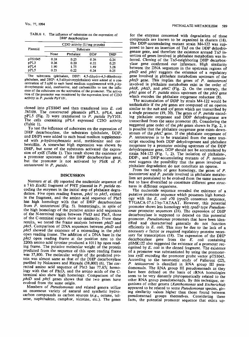
Nomura et al. (8) reported the nucleotide sequence of a 7 kb EcoRI fragment of PHT plasmid in P. putida en- coding the enzymes in the initial step of phthalate degra- dation. Five open reading frames, phtl to pht5, existed in the fragment, and the amino acid sequence of Pht5 has high homology with that of DHP decarboxylase from P. testosteroni (Fig. 5). Interestingly, in spite of the high homology of the putative amino acid sequences of the N-terminal region between PhtD and PhtS, those of the C-terminal region show no similarity. From these results, we tested the frame analysis between phtD and pht5. Comparison of DNA sequences between phtD and pht5 showed the existence of a misreading in the pht5 open reading frame. The addition of a DNA base in the pht5 open reading frame at the position next to the 220th amino acid tyrosine produced a 1011 bp open read- ing frame. The putative molecular weight of the protein predicted from the sequence of this open reading frame was 37,800. The molecular weight of the predicted pro- tein was almost same as that of the DHP decarboxylase purified by Nakazawa and Hayashi (38,000) (6). The cor- rected amino acid sequence of Pht5 has 77.6% homo- logy with that of PhtD, and the amino acids of the C- terminal also show high homology. Comparison of the phtD and pht5 genes shows that the two genes have evolved from the same origin.
Members of Pseudomonas and related genera utilize an enormous variety of natural and synthetic hydro- carbon compounds as carbon sources (e.g., octane, tol- uene, naphthalene, camphor, nicotine, etc.). The genes
for the enzymes concerned with degradation of these compounds are known to be organized in clusters (18). The DDP-accumulating mutant strain M4-122 was sup- posed to have an insertion of Tn5 on the DDP dehydro- genase gene, and therefore the existence around Tn5 in- sertion of genes involved in phthalate metabolism was in- ferred. Cloning of the Tn5-neighboring DHP decarbox- ylase gene confirmed our inference. High similarity between the DNA sequences in the upstream regions of phtD and phtl suggests the existence of a regulatory gene involved in phthalate metabolism upstream of the phtD gene. This implies the genes of P. testosteroni involved in phthalate metabolism exist in the order of phtR, phtD, and phtC (Fig. 2). On the contrary, the phtl gene of P. putida exists upstream of the pht2 gene which encodes the phthalate oxygenase reductase (8).
The accumulation of DDP by strain M4-122 would be unthinkable if the pht genes are composed of an operon similar to the nab and xyl genes which are transcribed by a single promoter (19, 20). The genes of P. putida encod- ing phthalate oxygenase and DDP dehydrogenase are transcribed from the same promoter (8). Considering the suggested gene order of the pht genes shown in Fig. 2, it is possible that the phthalate oxygenase gene exists down- stream of the phtC gene. If the phthalate oxygenase of P. testosteroni is to be translated from the same tran- script encoding both DDP dehydrogenase and phthalate oxygenase by a promoter existing upstream of the DDP dehydrogenase gene, DDP should not be accumulated by strain M4-122 (Fig. 1, 2). The isolation of phthalate-, DDP-, and DHP-accumulating mutants of P. testoste- roni suggests the possibility that the genes involved in phthalate degradation do not constitute an operon.
From the results of gene homology, the genes of P. testosteroni and P. putida involved in phthalate metabo- lism are postulated to be evolved from the same ancestor but to have diversified to constitute different gene struc- tures in different organisms.
The nucleotide sequence revealed the existence of a putative promoter sequence which possesses high homol- ogy with the E. coli a70 (rpoD) consensus sequence, TTGACA-17_+ 2 bp-TATAAT. However, this potential promoter shows less homology with rpoD type Pseudom- onas promoter sequences (21). The expression of DHP decarboxylase is supposed to depend on this potential promoter. Pseudomonas promoters that have been iden- tified and characterized generally do not function efficiently in E. coli. This may be due to the lack of a necessary ~ factor or required regulatory proteins neces- sary for transcription (18). The expression of the DHP decarboxylase gene from the E. coli containing pSMK122 also suggested the existence of a promoter rec- ognized by E. coli in the cloned fragment. The existence of a promoter was substantiated by using the promoter- less xylE encoding the promoter probe vector pTS1045. According to the taxonomic study of Palleroni (22), P. testosteroni is classified in RNA group III pseu- domonads. The RNA group III pseudomonads as they have been defined on the basis of rRNA homologies seem to be very distantly phylogenetically related to the other RNA group pseudomonads. By this technique, or- ganisms of other genera (Xanthomonas and Escherichia) appeared to be related to some Pseudomonas species, giv- ing similarity values higher than those found between pseudomonad groups themselves. Considering these facts, the potential promoter sequence that exists up-

590 LEE ET AL. J. FERMENT. BIOENO.,
s t ream of the D H P decarboxylase gene is though t to be the p romote r o f the D H P decarboxylase gene.
Ident i f ica t ion of the D D P dehydrogenase gene and the other genes of P. t e s tos t e ron i involved in phtha la te degrada t ion is necessary to elucidate the s t ructure o f other genes concerned with ph tha la te metabo l i sm.
REFERENCES
1. Mayer, F. L., Stalling, D. L., and Johnson, J. L.: Phthalate es- ters as environmental contaminants. Nature (London), 238, 411-413 (1972).
2. Ballard, D.G.H., Courtis, A., Shirley, I.M., and Taylor, S.C.: A biotcch route to polyphenylene. J. Chem. Sot., Chem. Commun., 954-955 (1983).
3. Nakazawa, T. and Hayashi, E.: Phthalate metabolism in Pseu- domonas testosteroni: accumulation of 4,5-dihydroxyphthalate by a mutant strain. J. Bacteriol., 131, 42-48 (1977).
4. Omori, T., Matsubara, M., Masuda, S., and Kodama, T.: Production of 4,5-dihydro-4,5-dihydroxyphthalate from phtha- late by a mutant strain of Pseudomonas testosteroni M4-1. Appl. Microbiol. Biotechnol., 35, 431-435 (1991).
5. Batie, C.J. , LaHaie, E., and Ballou, D.P. : Purification and characterization of phthalate oxygenase and phthalate oxy- genase reductase from Pseudomonas cepacia. J. Biol. Chem., 262, 1510-1518 (1987).
6. Naknzawa, T. and Hayashi, E.: Phthalate and 4-hydroxy- phthalate metabolism in Pseudomonas testosteroni: purifica- tion and properties of 4,5-dihydroxyphthalate decarboxylase. Appl. Environ. Microbiol., 36, 264-269 (1978).
7. Pu|ar, B.G. and Ribbons, D.W.: Phthalate metabolism in Pseudomonas fluorescens PHK: purification and properties of 4,5-dihydroxyphthalate decarboxylase. Appl. Environ. Micro- biol., 49, 374-376 (1985).
8. Nomnra, Y., Nakagawa, M., Ogawa, N., Harashima, S., and Oshima, Y.: Genes in PHT plasmid encoding the initial degra- dation pathway of phthalate in Pseudomonas putida. J. Fer- ment. Bioeng., 74, 333-344 (1992).
9. Rella, M., Mercenier, A., and Hags, D.: Transposon insertion mutagenesis of Pseudomonas aeruginosa with a TN5 deriva- tive: application to physical mapping of the arc gene cluster. Gene, 33, 293-303 (1985).
10. Simon, R., Priefer, U., and Piihler, A.: A broad host range mobilization system for in vivo genetic engineering: transposon mutagenesis in Gram-negative bacteria. Bio/Technology, 1, 784-791 (1983).
11. Samhrook, J., Fritseh, E. F., and Maniatis, T.: Molecular clon-
ing: a laboratory manual, 2nd ed. Cold Spring Harbor Labora- tory, Cold Spring Harbor, New York (1989).
12. Fnkuda, M. and Yano, K.: Construction of broad host range cloning vectors for Gram-negative bacteria. Agric. Biol. Chem., 49, 2719-2724 (1985).
13. Laemmli, U.K.: Cleavage of structural proteins during the assembly of the head of bacteriophage T4. Nature (London), 227, 680-685 (1970).
14. Arai, H., Igarashi, Y., and Kodama, T.: Anaerobically in- duced expression of the nitrite reductase cytochrome c-551 ope- ton from Pseudomonas aeruginosa. FEBS Lett., 280, 351-353 (1991).
15. Inouye, S., Asai, ¥., Nakazawa, A., and Nakazawa, T.: Nude- otide sequence of a DNA segment promoting transcription in Pseudomonas putida. J. Bacteriol., 166, 739-745 (1986).
16. Nozaki, M.: Metapyrocatechase (Pseudomonas). Methods En- zymol., 17A, 522-523 (1970).
17. Stanier, R. Y., Palleroni, N.J. , and Doudoroff, M.: The aero- bic pseudomonads: a taxonomic study. J. Gen. Microbiol., 43, 159-271 (1966).
18. Rothmel, R.K., Chakrabarty, A.M., Berry, A., and Darzins, A.: Genetic systems in Pseudomonas. Methods Enzymol., 204, 485-514 (1991).
19. Nakazawa, T., Inouye, S., and Nakazawa, A.: Regulatory sys- tems for expression of xyl genes on the TOL plasmid, p. 133- 140. In Silver, S., Chakrabarty, A.M., Iglewski, B., and Kaplan, S. (ed.), Pseudomonas: biotransformation, pathogene- sis, and evolving biotechnology. American Society for Microbi- ology, Washington, DC (1990).
20. Schell, M. A.: Regulation of the naphthalene degradation genes of plasmid NAH7: examples of a generalized positive control system in Pseudomonas and related bacteria, p. 165-176. In Silver, S., Chakrabarty, A.M., Iglewski, B., and Kaplan, S. (ed.), Pseudomonas: biotransformation, pathogenesis, and evolving biotechnology. American Society for Microbiology, Washington, DC (1990).
21. Ronald, S., Farinha, M.A., Allan, B.J., and gropinski, A.M.: Cloning and physical mapping of transcriptional regula- tory (sigma) factors from Pseudomonas aeruginosa, p. 249- 257. In Galli, E., Silver, S., and Witholt, B. (ed.), Pseudomo- has: molecular biology and biotechnology. American Society for Microbiology, Washington, DC (1992).
22. Palleroni, N.J.: Present situation of the taxonomy of aerobic pseudomonads, p. 105-115. In Galli, E., Silver, S,, and Witholt, B. (ed.), Pseudomonas: molecular biology and bio- technology. American Society for Microbiology, Washington, DC (1992).