Identification of genetic mutations in ... - ubibliorum.ubi.pt‚ngela... · transfecção de uma...
78
UNIVERSIDADE DA BEIRA INTERIOR Ciências Identification of genetic mutations in patients with familial central diabetes insipidus Ângela Sofia Fernandes Alves Francisco Dissertação para obtenção do Grau de Mestre em Bioquímica (2º ciclo de estudos) Orientador: Prof. Doutor Manuel Carlos Loureiro de Lemos Orientador: Prof.ª Doutora Isabel Maria Theriaga Mendes Varanda Gonçalves Covilhã, Outubro de 2012
Transcript of Identification of genetic mutations in ... - ubibliorum.ubi.pt‚ngela... · transfecção de uma...

Mutations in patients with neurohypophyseal diabetes insipidus
1
UNIVERSIDADE DA BEIRA INTERIOR Ciências
Identification of genetic mutations in patients
with familial central diabetes insipidus
Ângela Sofia Fernandes Alves Francisco
Dissertação para obtenção do Grau de Mestre em
Bioquímica (2º ciclo de estudos)
Orientador: Prof. Doutor Manuel Carlos Loureiro de Lemos Orientador: Prof.ª Doutora Isabel Maria Theriaga Mendes Varanda Gonçalves
Covilhã, Outubro de 2012

Mutations in patients with neurohypophyseal diabetes insipidus
ii
Para os meus pais
com imenso amor!

Mutations in patients with neurohypophyseal diabetes insipidus
iii
Acknowledgement
Em primeiro lugar, quero agradecer ao Prof. Manuel Lemos e à Prof.ª Isabel Gonçalves por
toda a dedicação a este projeto, pela ajuda e principalmente pelo incentivo a nunca desistir.
É importante ter sempre alguém que nos oriente, mas também é importante ter quem confie
no nosso trabalho e nos queira levar sempre para a frente, apesar de todos os desafios
presentes durante o desenvolvimento desta tese. Muito obrigado por esta jornada, e por
todos os conhecimentos que me transmitiram.
De seguida quero agradecer aos meus pais. Vocês sempre foram a minha âncora, o meu porto
de abrigo. Vocês suportaram todas as lágrimas, todos os gritos, e todos os sorrisos que fui
soltando durante este ano. Aconteça o que acontecer, sei que vão estar sempre lá para mim,
e que sempre terei o vosso ombro para me apoiar, independentemente de todos os obstáculos
que o futuro possa colocar no meu caminho. Tudo o que hoje sou e tenho, a vocês o devo, e
durante toda a minha vida estarei imensamente grata por todo o amor que me deram durante
a minha vida. Obrigado!
Ao meu querido irmão um muito obrigado por, mesmo longe, estar sempre presente, quer
com telefonemas ou com mensagens. Obrigado por todos os empurrões, todos os incentives e
todas as palavras que, tão queridas, me faziam sentir que estavas aqui comigo.
Tenho também de te agradecer Bruno. Este ano não foi fácil. Foram tantos os problemas,
dificuldade e complicações, mas também foram bastantes os sorrisos e a felicidade.
Simplesmente és a minha outra metade. Ajudaste-me a ultrapassar este longo ano, apoiaste-
me em tudo e nunca me deixaste cair. Os teus conselhos sempre foram tão importantes para
mim, e sempre me ajudaram a escolher o caminho certo. Obrigado por estares sempre aqui
comigo.
Não posso deixar de agradecer aos amigos que descobri no laboratório. Mais do que colegas de
trabalho, vocês foram uma força inesgotável de alegria e amizade. Eduarda, Catarina, Inês,
Susana, Marina e Fernando, vocês deram um ar completamente diferente ao laboratório que,
mais do que um local de trabalho, revelou-se como um local de amizade e entreajuda. O que
quer que acontecesse, vocês estavam sempre lá para tornar as coisas mais simples, para não
falar da quantidade de gargalhadas que os nossos momentos de convívio proporcionam.
Obrigado por tudo!
Tenho também de agradecer aos meus amigos, quer os que me acompanham já desde os meus
anos de infância, aos que conheci durante a adolescência e aos que criei já na universidade.
Podemos conhecer muitas pessoas, mas só algumas é que se destacam no meio de tantas, e é
a essas pessoas que quero agradecer por, mesmo longe, não permitirem que a distância
separe corações que em certos tempos foram tão felizes juntos.

Mutations in patients with neurohypophyseal diabetes insipidus
iv
Obrigado a todas as pessoas que, mesmo sem qualquer obrigação, dispensaram parte do seu
tempo para me ajudaram no desenvolvimento deste projeto, transmitindo-me conhecimentos
importantes, quer para este momento, quer para experiências futuras. Sempre que
precisarem podem também contar com a minha ajuda.
Por fim, quero agradecer o financiamento deste projeto por parte da FCT, através do
programa COMPETE (PTDC/SAU-GMG/098419/2008 e PEst-C/SAU/UI0709/2011).

Mutations in patients with neurohypophyseal diabetes insipidus
v
Resumo alargado
A diabetes insípida (DI) é uma doença rara, caracterizada principalmente pela excreção de
elevados volumes de urina na forma diluída podendo, entre várias causas possíveis, ter origem
num defeito genético.
O desenvolvimento da doença pode dever-se a quatro causas possíveis. A mais comum deve-se
a uma deficiência na secreção da hormona antidiurética arginina vasopressina (AVP), sendo
referida como DI central ou neurohipofisária. Outra possível causa da doença deve-se a uma
insensibilidade, por parte das células renais, aos efeitos da AVP, sendo neste caso designada
como DI nefrogénica. A DI pode também dever-se a uma excessiva ingestão de líquidos, que
conduz à supressão da libertação da hormona AVP, sendo referida como polidipsia primária.
Por fim, um aumento do metabolismo da hormona AVP durante a gravidez pode também ser
uma causa da doença, designada por DI gestacional.
A hormona AVP é sintetizada nos neurónios magnocelulares. Estes têm origem no núcleo
supra-óptico e para-ventricular do hipotálamo e os seus prolongamentos terminam na
neurohipófise. A destruição destes neurónios resulta numa deficiência na produção da
hormona, conduzindo à DI central. Esta destruição pode ter inúmeras causas, incluindo
acidentes, cirurgias, doenças autoimunes, entre outras. Contudo, a doença também
apresenta uma base familiar, correspondendo a 1% de todas as causas de DI central. A DI
central apresenta sintomas persistentes de poliúria, polidipsia e sede, que geralmente se
começam a manifestar vários meses ou anos após o nascimento.
A DI central familiar apresenta duas características principais: está associada a mutações num
único alelo do gene que codifica a hormona (gene AVP), apresentando assim uma transmissão
autossómica dominante; e é causada por uma deficiência progressiva pós-natal na secreção da
hormona AVP, que se pensa resultar da degeneração seletiva dos neurónios magnocelulares.
O gene AVP é composto por 3 mil pares de bases e encontra-se localizado no braço curto do
cromossoma 20. Este gene contém três exões que codificam para o péptido sinalizador, para a
hormona AVP, para a neurofisina II (transportador da hormona) e ainda para um glicopéptido,
conhecido como copeptina. Após sintetizados, a hormona, a neurofisina II e o glicopéptido são
armazenados em vesiculas secretoras, nos terminais axonais dos neurónios, e são libertados
após a ocorrência de estímulos. Após a entrada na corrente sanguínea, a hormona vai atuar a
nível das células renais de modo a aumentar a sua permeabilidade para as moléculas de água,
favorecendo assim a absorção de água no rim.
Até à data do início deste trabalho, a doença estava associada a 70 mutações diferentes no
gene AVP localizadas ao longo de todo o precursor proteico. Pensa-se que estas mutações são
a causa da doença uma vez que interferem na estabilidade da cadeia de aminoácidos,
alterando a sua estrutura primária. Teoricamente, mutações que afetem a conformação de
Mutations in patients with neurohypophyseal diabetes insipidus
vi
proteínas secretoras resultam no desenvolvimento de patologias devido ao seu impacto na
função da proteína não conseguindo alcançar o seu destino, ficando retidas no reticulo
endoplasmático. Contudo, a razão dos precursores AVP mutados serem tóxicos para os
neurónios produtores de AVP está ainda por esclarecer.
Existem, até ao momento, três teorias que tentam explicar o mecanismo da doença. O
mecanismo não tóxico defende que há uma expressão simultânea dos precursores “wild-type”
e dos precursores mutados resultando numa associação de ambos. Assim, o precursor “wild-
type” é alterado, uma vez que ambos ficam retidos no reticulo endoplasmático. Contudo,
este mecanismo não explica a morte dos neurónios magnocelulares. O mecanismo tóxico
defende que a constante acumulação de precursores com conformações alteradas pode
interferir com a produção de proteínas essenciais à sobrevivência celular, resultando assim na
morte neuronal. Recentemente, um novo mecanismo foi proposto para explicar a patogénese
da doença. Observou-se a formação de vesiculas autofágicas, após acumulação de precursores
mutados, que resultam na destruição dos retículos endoplasmáticos danificados, juntamente
com os agregados proteicos. Durante este processo, se as células forem expostas a insultos
metabólicos e ambientais, pode ocorrer apoptose dependente de autofagia, resultando na
destruição dos neurónios magnocelulares.
A DI central familiar apresenta uma natureza benigna, contudo é uma doença que apresenta
uma intensa pesquiza em torno dos seus mecanismos moleculares uma vez que se trata de um
modelo de interesse para o estudo de doenças neuro-endócrinas e de transmissões
autossómica dominante.
O presente estudo tem por objetivos fazer uma revisão das mutações descritas na literatura
científica para o gene AVP, aumentar o número de mutações descritas com a análise de novos
pacientes diagnosticados com DI central familiar e caracterizar as consequências funcionais
das novas mutações identificadas.
Para alcançar os objetivos descritos, utilizou-se a seguinte metodologia: a revisão de todas as
mutações descritas até à data, através de pesquisa bibliográfica de artigos científicos;
realização de estudos genéticos, baseados na amplificação por PCR e na posterior
sequenciação dos três exões do gene AVP de 9 pacientes diagnosticados com DI central
familiar; inserção das novas mutações num vector de expressão contendo o cDNA do gene
AVP, através de técnicas de clonagem, digestão enzimática e mutagénese dirigida; e
finalmente a realização de estudos funcionais, por otimização das técnicas de transfecção e
imunocitoquímica com o vector de expressão AVP “wild-type”.
Os resultados obtidos mostraram que as 3 famílias apresentam mutações no gene AVP. O
paciente III-1, da família A, apresenta a alteração de uma timina para uma citosina na
posição 154 do cDNA (c.154T>C) que origina a substituição de uma cisteína por arginina na
posição 52 da proteína (p.C52R). O paciente II-1, da família B, apresenta uma alteração de

Mutations in patients with neurohypophyseal diabetes insipidus
vii
citosina para guanina na posição 289 do cDNA (c.289C>G) que resulta na substituição de uma
arginina por glicina, na posição 97 da proteína. O paciente II-4 da família C apresenta a
alteração de uma guanina para uma timina na posição 343 do cDNA (c.343G>T) que resulta na
substituição de um ácido glutâmico por um codão de terminação na posição 115 da proteína.
As três mutações estão em heterozigotia e as duas mutações encontradas no exão 2
correspondem a mutações novas, enquanto a mutação presente no exão 3 já se encontra
descrita na literatura.
Um vector de expressão contendo o cDNA do gene AVP (pRc/RSV-AVP), foi-nos gentilmente
oferecido por investigadores da área. O cDNA do gene AVP contido no vector de expressão
(pRc/RSV-AVP) foi sub-clonado no vector pVAX/lacZ e, através de mutagénese dirigida, as
mutações desejadas (c.154T>C e c.289C>G) foram introduzidas no cDNA. Assim, o cDNA com
as mutações está pronto a ser inserido no plasmídeo de expressão. Os ensaios de transfecção
e imunocitoquímica foram otimizados para o vector de expressão “wild-type”, uma vez que
foi observada marcação para a neurofisina II nos prolongamentos dos neurónios após
transfecção de uma linha celular neuronal (N2A) e marcação com anticorpos específicos.
Com este estudo, o número de mutações descritas para o gene AVP aumentou de 70 para 72 e
mais três famílias fazem parte do número total de famílias estudadas com DI central familiar.
É importante continuar o desenvolvimento de estudos funcionais, de modo a obter respostas
sobre os mecanismos moleculares responsáveis pelo desenvolvimento da doença uma vez que
estas serão importantes não só para a DI central familiar, mas também para o esclarecimento
de outras doenças que apresentem mecanismos moleculares semelhantes.
Palavras chave
Diabetes insípida central, mutações AVP.

Mutations in patients with neurohypophyseal diabetes insipidus
viii
Abstract
Diabetes insipidus (DI) is associated with defects that involve the secretion and the action of
hormone arginine vasopressin (AVP) resulting in the excretion of abnormally large volumes of
diluted urine. The most common defect that results in disease development is the deficient
secretion of the hormone AVP and the disease is referred to as central or neurohypophyseal
DI. The AVP hormone is synthesized in magnocellular neurons, that originate in the
supraoptic and paraventricular nuclei of the hypothalamus and are projected to
neurohypophysis, and the destruction of these neurons leads to a deficiency of AVP hormone,
resulting in neurohypophyseal DI. The familial form of disease represents 1% of all causes of
neurohypophyseal DI and the main points of the disease are: it is associated with mutations in
one allele of the AVP gene, and it is caused by postnatal development of deficient AVP
secretion, proposed to result from selective degeneration of the magnocellular neurons.
The aims of this thesis are: to review AVP mutations described in the scientific literature, to
expand the spectrum of mutations through the analysis of additional patients with DI and to
characterize the functional consequences of identified novel AVP mutations. To achieve these
aims a bibliographic research was developed; genetic studies were performed to amplify and
to sequence the three exons of the AVP gene in 9 patients; an expression vector containing
the desired mutations was constructed by subcloning, site-directed mutagenesis and
enzymatic digestion; and the functional studies were initialized by optimization of
transfection and immunocytochemistry assays for WT AVP cDNA expression vector.
Three mutations were identified: c.154T>C, c.289C>G and c.343G>T. The first two mutations
are novel and the last mutation is already described in the scientific literature. The AVP cDNA
from the expression vector was subcloned in the pVAX/lacZ vector and the mutations were
inserted in the AVP cDNA by site-directed mutagenesis and enzymatic digestion. The mutated
AVP cDNAs were sequenced and have been prepared to be inserted in the expression vector.
The transfection and immunocytochemistry protocols have been optimized for WT AVP cDNA
expression vector.
This study allowed the increase in the number of mutations from 70 to 72 different
mutations, although further work is necessary in order to understand the molecular
mechanisms responsible for the development of the disease and to give help and information
to patients affected with this disease.
Keywords
Neurohypophyseal diabetes insipidus, AVP mutations.

Mutations in patients with neurohypophyseal diabetes insipidus
ix
Table of Contents
1. Introduction ............................................................................................... 1
1.1. Definition and classification of Diabetes Insipidus ............................................. 1
1.2. Clinical aspects of Familial Neurohypophyseal Diabetes Insipidus (FNDI) ................. 3
1.3. AVP gene and AVP processing ..................................................................... 5
1.4. Genetic basis of adFNDI ............................................................................. 9
1.5. Pathogenesis of adFNDI ........................................................................... 10
1.6. Diagnosis and treatment .......................................................................... 14
1.7. Future perspectives ............................................................................... 15
1.8. Aims of the thesis .................................................................................. 16
2. Methods .................................................................................................. 17
2.1. Literature search of AVP gene mutations ...................................................... 17
2.2. Subjects and clinical procedures ............................................................... 17
2.3. DNA extraction ..................................................................................... 17
2.4. Amplification of the AVP gene by polymerase chain reaction ............................. 19
2.5. Gel electrophoresis ................................................................................ 20
2.6. AVP gene sequencing .............................................................................. 21
2.7. pRc/RSV Sequencing ............................................................................... 22
2.8. Cloning of AVP cDNA into pVAX1/lacZ .......................................................... 23
2.9. Competent cells .................................................................................... 24
2.10. Transformation of competent cells with recombinant pVAX1/lacZ ....................... 25
2.11. Site – directed mutagenesis ...................................................................... 26
2.12. Cloning of Frag3 and Frag CB .................................................................... 30
2.13. Expression of the normal AVP gene in Neuro 2A cells ....................................... 31
2.14. Immunocytochemistry ............................................................................. 32
3. Results .................................................................................................... 35
3.1. Reported mutations in the AVP gene ........................................................... 35
3.2. Identification of kindred with mutations ...................................................... 41
3.3. Identification of mutations in the AVP gene ................................................ 41
3.4. Construction of pVAX/AVP vector ............................................................... 44

Mutations in patients with neurohypophyseal diabetes insipidus
x
3.5. Site-directed mutagenesis as a method to introduce desired mutations in AVP cDNA 46
3.6. Cloning of mutations in pVAX/AVP ............................................................. 48
3.7. Analysis of AVP WT gene expression in N2A cells ............................................ 49
4. Discussion ................................................................................................ 51
5. Conclusion ............................................................................................... 53
6. Bibliography ............................................................................................. 54
Mutations in patients with neurohypophyseal diabetes insipidus
xi
List of Figures
Fig. 1. Structural organization of the AVP gene and the protein vasopressin precursor .......... 5
Fig. 2. Structural organization of the AVP pro-hormone ................................................ 7
Fig. 3. Model of the regulation of water permeability in renal collecting duct cells .............. 8
Fig. 4. Synthesis and processing of secretory proteins like AVP ..................................... 11
Fig. 5. A proposed model for the molecular basis of adFNDI........................................ 13
Fig. 6. Expression vector pRc/RSV ........................................................................ 23
Fig. 7. Cloning vector ....................................................................................... 24
Fig. 8. AVP cDNA with primers used in site-directed mutagenesis for the first mutation
(c.154T>C) .................................................................................................... 28
Fig. 9. AVP cDNA with primers used in site-directed mutagenesis for the second mutation
(c.289C>G) .................................................................................................... 29
Fig. 10. Percentage of each type of protein change caused by the mutations in 104 reported
families. ....................................................................................................... 35
Fig. 11. Unique mutations described in the human AVP gene ....................................... 41
Fig. 12. Pedigrees of three families with adFNDI. Index cases are marked by an arrow........ 42
Fig. 13. Electrophoresis of PCR products for each AVP gene exon .................................. 43
Fig. 14. Electropherograms from fragments of the AVP gene of one healthy individual and
three clinically affected subjects with novel identified mutations ................................. 44
Fig. 15. Analysis of purified pDNA after enzymatic digestion with XbaI ........................... 45
Fig. 16. Confirmation of AVP cDNA in the recombinant pVAX/AVP ................................. 45
Fig. 17. First PCR reactions for site directed mutagenesis for both mutations ................... 46
Fig. 18. Subsequent PCR reactions for both mutations ............................................... 47
Fig. 19. Electropherograms from site-directed mutagenesis fragments ........................... 47
Fig. 20. SmaI/FseI digestion of FragCB, Frag3 and pVAX/AVP ....................................... 48
Fig. 21. Electropherograms from the sequences of the mutated pVAX/AVP vector ............. 49
Fig. 22. Cellular localization of NPII protein in transiently transfected N2A cells as visualized
by confocal laser microscopy .............................................................................. 50

Mutations in patients with neurohypophyseal diabetes insipidus
xii
List of Tables
Table 1. Sequence of the primers used for amplification of AVP exons. .......................... 20
Table 2. Sequence of primers used for site-directed mutagenesis. ................................ 27
Table 3. Summary of trasfection assay conditions. .................................................... 32
Table 4. Antibodies used for immunocytochemistry assays with dilutions and incubation times
.................................................................................................................. 33
Table 5. Immunocytochemistry assay. ................................................................... 34
Table 6. Reported mutations in the AVP gene causing FNDI ........................................ 36
Mutations in patients with neurohypophyseal diabetes insipidus
xiii
List of Acronyms
AC Adenylyl cyclase
adFNDI Autosomal dominant familial neurohypophyseal diabetes insipidus
AMP Adenosine monophosphate
AQP2 Aquaporin 2
AQP3 Aquaporin 3
AQP4 Aquaporin 4
AQP2-P Phosphorilated aquaporin 2
AVP Arginine vasopressin
bp Base pair
BSA Bovine serum albumin
cAMP cyclic
cDNA Complementary DNA
DDAVP Des-amino-D-arginin vasopressin
DI Diabetes insipidus
DMEM Dulbecco’s modified eagle’s medium
DNA Deoxyribonucleic acid
dNTP Deoxynucleotide
ddNTPs 2’,3’-dideoxyribonucleoside triphosphates
E. coli Escherichia coli
FBS Fetal bovine serum
Fig. Figure
Frag1 Fragment 1
Frag2 Fragment 2
Frag3 Fragment 3
FragAB Fragment AB
FragBB Fragment BB
FragCB Fragment CB
GP glycopeptide
HSP70 70 kilodalton heat shock proteins
kb Kilo bases
MRI Magnetic resonance image
N2A cells Neuro 2A cells
NPII Neurophysin II
PCR Polymerase chain reaction
pDNA Plasmid DNA
PKA Protein kinase A
Opti-MEM Opti-modified eagle’s medium

Mutations in patients with neurohypophyseal diabetes insipidus
xiv
SP Signal peptide
UV Ultra-violet
V2 Arginine vasopressin type 2 receptors
WT Wild-type
Mutations in patients with neurohypophyseal diabetes insipidus
1
1. Introduction
1.1. Definition and classification of Diabetes Insipidus
Blood osmolality in healthy individuals is maintained within restricted limits by a series of
complex mechanisms. Adjustments in water balance determine the level of that osmolality.
These adjustments are mediated by delicate alterations in the thirst mechanism plus the
capacity of the kidney to alter urine flow rate and its osmolality in response to small changes
in the plasma concentration of the hormone arginine vasopressin (AVP) [1]. Thus, through all
these mechanisms, healthy humans can conserve their osmotic level despite extreme climatic
conditions and, to a certain degree, when water supply is inadequate [1].
However, alterations in these mechanisms can occur and lead to one of two main states:
inappropriate accumulation of water in organism, which is recognized as hypoosmolar states,
and loss of renal water, which is recognized as hyperosmolar syndrome [1].
Diabetes insipidus (DI) is a rare disease and is characterized by excretion of abnormal large
volumes, known as polyuria (>50mL/Kg/day) of dilute urine (<300mmol/Kg) [2, 3]. This
definition allows the exclusion of osmotic diuresis, which occurs when an excess of solute is
being excreted, like in the case of glucose in patients with diabetes mellitus and this is the
main difference between the two disorders [3]. This disorder can be acquired as a result of
various injuries or diseases, but can also be idiopathic or have a genetic origin [2].
In DI, the magnitude of the abnormality in concentration and excretion of the urine varies
according to some factors like the severity of the defect which results in the disorder, the age
of the patient, and the rate of solute and water intake [2].
Four basic defects are responsible for the development of DI. The first and the most common
defect that occurs in this disorder is the deficient secretion of the hormone AVP, and in this
case the DI is referred as neurohypophyseal, neurogenic, central or hypothalamic [3]. This
form of DI can be completely controlled by administration of AVP or its analogue, des-amino-
D-arginin vasopressin (DDAVP) [4]. The second type of DI is caused by renal defects, where the
cells of the kidneys are insensitive to the antidiuretic effects of AVP and is referred as
nephrogenic DI. In this case, the patients are unresponsive or poorly responsive to the
administration of AVP or DDAVP [3, 4]. In both forms of disease, the thirst mechanism remains
normal to regulate water balance [1]. Another defect that causes DI is excessive water intake
(polydipsia) that leads to suppression of AVP release and consequent polyuria. This form of DI
is called primary polydipsia and may be due to defects in the thirst mechanism or to cognitive
impairment. Hormone supplements like AVP and DDAVP can reduce the symptoms of polyuria,
Mutations in patients with neurohypophyseal diabetes insipidus
2
although these treatments should not be used because they can originate water intoxication,
since they reduce polyuria more than polydipsia, which results in rapid retention of excess
water and development of hyponatremia [1, 3, 4]. The fourth type of DI is due to increased
metabolism of AVP during pregnancy resulting in gestational DI. This form of DI can be
treated with DDAVP but is unresponsive to AVP. This occurs because the analogue of AVP is
much less susceptible to degradation by placental vasopressinase [3, 4].
Depending on the cause that originates DI, the deficiency in vasopressin action or secretion
can be partial or nearly total. Thus, the deficiency may or may not be associated with
concentration of the urine after a fluid-deprivation test or in response to other strong stimuli
like in the case of nausea, severe hypovolemia or severe hypotension [2].
Differentiating between the forms of DI is relatively easy if patients have severe deficiency in
either the secretion or action of AVP. In both cases, the patients undergo dehydration
induced by fluid deprivation, but the urine remains dilute [4]. This first result excludes the
possibility of primary polydipsia since in this form of disease, a fluid deprivation results in
concentration of urine because the hormonal mechanism remains normal and the problem
resides in excess of water intake. The next step to differentiate nephrogenic DI from
neurohypophyseal and gestational DI is the injection of AVP and DDAVP and measurement of
the urinary response [4]. Patients with nephrogenic DI do not respond to treatment since their
problem resides in renal insensitivity to AVP and not in hormonal deficiency and their urine
remains dilute. However, patients with neurohypophyseal or gestational DI are able to
concentrate their urine when AVP or DDAVP are administered because of the increased
plasma levels of AVP. If fluid deprivation results in concentration of urine, other tests are
necessary to differentiate between primary polydipsia and a less severe deficiency in the
secretion or action of AVP [4]. The most reliable way to make this distinction is to measure
plasma AVP and to relate the results to the plasma and urine osmolality during a fluid
deprivation and/or hypertonic saline infusion test [4].
However, with time the diagnosis becomes more complicated and the forms of the disease
can be confused. After prolonged periods of polydipsia, a decrease in maximal urine-
concentration ability occurs in the kidneys, regardless of the primary cause [5]. The passage of
large amounts of dilute urine through the distal nephron removes existent solutes from the
renal medullary interstitium, a process known as washout phenomenon, and results in the
decrease of osmotic gradient across the collecting tubular cells [1]. Since this gradient is
essential for the antidiuretic action of AVP, any mechanism responsible for DI may lead to an
additional defect at the renal level that complicates the interpretation of diagnostic tests
based on indirect analyses of the antidiuretic action of AVP [1].

Mutations in patients with neurohypophyseal diabetes insipidus
3
1.2. Clinical aspects of Familial Neurohypophyseal Diabetes
Insipidus
AVP is synthesized in magnocellular neurons that originate in the supraoptic and
paraventricular nuclei of the hypothalamus, which project down through the diaphragma
sellae to form the neurohypophysis [3, 4]. In contrast to the adenohypophysis, the
neurohypophysis does not synthesize hormones but functions as a reservoir for the storage
and release of hormones synthesized in the hypothalamus [6].
Destruction of magnocellular neurons results in a deficiency of AVP, leading to
neurohypophyseal DI. This neuronal destruction can have a variety of causes, including
trauma from surgery or accident, infections, autoimmune disease, congenital brain
malformations, aneurysms, and others [6]. However, neurohypophyseal DI can also occur on an
inherited, or familial, basis representing 1% of all causes of neurohypophyseal DI [7]. Usually,
the disease has an autosomal dominant transmission, however in 1996 an X-linked recessive
form was discovered [4].
There is another type of neurons that can produce and segregate AVP, known as parvocellular
neurons [4]. The projections of these neurons are located in the median eminence of the
hypothalamus [4]. In some studies, it was observed that these projections are apparently
unaffected in patients with neurohypophyseal DI and this fact may explain the preservation of
normal circadian rhythm and pituitary-adrenal function in these patients [4] because these
neurons also produce corticotrophin releasing factor, which is thought to interact with AVP in
the regulation of adrenocorticotropic hormone secretion [8]. Since these neurons are not
affected by neurohypophyseal DI, it is believed that the two types of AVP-producing neurons
have very different susceptibilities to the cytotoxic effects of the genetic mutations that lead
to development of the disease [4].
Autosomal dominant familial neurohypophyseal DI (adFNDI) is a rare disease with persistent
symptoms of polyuria, polydipsia and thirst which usually manifest several months or years
after birth [9]. Studies performed with mice with AVP gene mutations revealed some
differences, when compared with mice with the normal AVP gene: mice with a mutated AVP
gene consumed larger volumes of water, they excreted much more urine and the volume of
urine excreted is worse over time, and their urine osmolality is lower [9]. Thus, like in humans
with adFNDI, mice with certain mutations produce excessive amounts of dilute urine but
compensate by increasing water intake and so they can avoid severe dehydration and this fact
demonstrates that thirst mechanisms remain intact in the presence of the disease [9]. Despite
these symptoms, adFNDI causes relatively few and well-tolerated symptoms. Nocturia
(elimination of urine at night, disturbing sleep) is common and in children may present as
Mutations in patients with neurohypophyseal diabetes insipidus
4
enuresis (urine during sleep) [4]. Physical exams and routine laboratorial analyses are usually
within normal limits and hypernatremia or signs of hypertonic dehydration are minimal or
absent, except in the case of patients that are comatose, have an impaired thirst mechanism
or if the patients are unable to increase fluid intake [4].
The symptoms of severe polyuria and polydipsia, which segregate in an autosomal-dominant
pattern and respond to exogenous DDAVP, show several intriguing features. First, the
affected family members show a completely normal water balance at birth and during early
infancy but develop progressive symptoms of compulsive drinking at some point in childhood
[10]. Second, in some studies it was demonstrated that during repetitive fluid-deprivation
tests, the secretion of AVP is normal before the onset of the disease but then starts
diminishing during early childhood [11]. Finally, once fully developed, the symptoms of
polyuria and polydipsia continue throughout life [10]. Occasionally, spontaneous remissions of
polyuria and polydipsia during middle-age are observed, even though the patients continue to
have a deficient AVP secretion as severe as in their symptomatic kin. However, this remission
mechanism remains unexplained [4].
Magnetic resonance image (MRI) exams have been used to investigate neurohypophysis
anatomy in patients with adFNDI [4]. Some authors found that anterior and posterior lobes of a
normal pituitary gland have different signal intensities in images of magnetic resonance. The
posterior lobe presents a well-defined oval or round area of hyperintensity in both normal and
abnormal pituitary glands, although there are some variations in size and shape of the signal
from one person to another [6]. Some patients with adFNDI lack the characteristic bright spot,
or high-intensity signal [4, 12], which is common in the posterior lobe of the pituitary in 52%-
100% of healthy adults [6]. Signal intensity seems to be correlated closely with posterior lobe
function as it is suspected to result from neurovesicles in axon endings of AVP-producing
neurons [7, 13]. If neurovesicles really are responsible for the bright spot, it is not clear why
oxytocin-containing vesicles or vesicles located in the hypothalamus do not cause a high-
intense signal, maybe their concentration is insufficient [7]. Thus, the absence of the bright
spot in the posterior pituitary lobe could result from a neurotoxic accumulation of precursor
proteins that consequently lead to cell death [7, 12, 13].
However, the significance of MRI results is uncertain because the exact cellular source of the
signal is not yet known [4]. It is believed that the bright spot is absent in all patients with
neurohypophyseal DI due to destructive or unidentified pathological processes [6], however to
date relatively few patients with adFNDI have been studied and the results have been
conflicting since in some affected individuals the bright spot has been observed [4]. A positive
bright spot in a patient with adFNDI can be caused by a defect in hormone release from the
posterior pituitary leading to accumulation of neurovesicles and, thus, to a normal MRI
instead of defects in intra-axonal transport or processing of proteins [7]. However, at present,
neither the presence nor the absence of the bright spot can be related with the presence or

Mutations in patients with neurohypophyseal diabetes insipidus
5
absence of AVP-producing neurons in the posterior pituitary [4]. Thus, it is important that
additional MRI studies including adFNDI patients are performed to clarify the importance and
significance of the high-intensity signal found in some people [4].
1.3. AVP gene and AVP processing
adFNDI is caused by mutations in one allele of the gene that encodes for AVP. The gene has
approximately 3 kb (Gene ID: ENSG00000101200, Esemble) and is located on the short arm of
chromosome 20 (20p13) [3]. It consists of 3 exons and 2 introns and encodes AVP and
neurophysin II (NPII), the carrier of AVP. The first exon encodes the signal peptide, the
hormone AVP, and the NH2-terminal region of NPII [3]. The second exon encodes the highly
conserved central region of NPII and the third exon encodes the COOH-terminal region of NPII
and the glycopeptide, which is known as copeptin [3, 14]. The small size of the AVP gene
facilitates the mutational analysis [3] and the study of the mutations at the protein level (Fig.
1).
Fig. 1. Structural organization of the AVP gene and the protein vasopressin precursor. The gene is
composed of 3 exons and 2 introns. The signal peptide contains 19 amino acids, AVP hormone contains 9
amino acids, NPII contains 93 amino acids and copeptin contains 39 amino acids. SP, signal peptide;
AVP, arginine vasopressin; NPII, neurophysin II; GP, glycopeptide.
Several studies to analyze the expression of the AVP gene were performed using AVP
transgenes derived from some animals. These results support the hypothesis that cell-specific
enhancers and/or silencers that restrict expression of the AVP gene to specific neuronal cell-
types in the hypothalamus are present in the regions either downstream or upstream of the
AVP gene [15]. Recently, some authors demonstrated that DNA sequences in a 178 bp region
immediately downstream of exon 3 of the AVP are necessary for cell-specific expression of
AVP in rat hypothalamus [16].

Mutations in patients with neurohypophyseal diabetes insipidus
6
AVP and NPII are synthesized as a single precursor, prepro-vasopressin. The prepro-hormone
presumably is translated on ribosomes in the cytosol and translocated across the membrane
of the rough endoplasmic reticulum [4]. Once inside the endoplasmic reticulum, it is supposed
that the signal peptide remains attached noncovalently to the inner surface of the membrane
via the positive charges at its N-terminal. This ligation is thought to facilitate accurate
cleavage of the signal peptide by ensuring proper alignment with the signal peptidase [4].
However, the presence of certain small and neutral amino acids at the -1 and -3 positions
immediately adjacent to the cleavage site of the signal peptide are required for efficient and
accurate cleavage [4]. In this case, the signal peptide of AVP gene has an alanine and serine at
-1 and -3 positions, respectively [4]. The pro-hormone is generated by removal of the signal
peptide from the prepro-hormone and from by addition of a carbohydrate chain to the
copeptin [3, 9]. There are no certainties that the glycosylation process and copeptin are
important for protein proper folding, trafficking or further processing, however, it seems
possible that copeptin glycosylation plays an important role by assisting refolding of misfolded
AVP pro-hormone monomers through its interaction with the calnexin-calreticulin system in
the endoplasmic reticulum [17]. This system monitors protein folding and interacts principally
with the sugars of glycosylated proteins and places these proteins into the proximity of a
glycoprotein-specific member of the protein disulfide isomerase family [17].
After removal of the signal peptide, the precursor generally must fold and dimerize correctly
in the lumen of the endoplasmic reticulum [4], where the unique oxidizing environment allows
the formation of disulphide bridges [10], before they can proceed through the Golgi apparatus
[4]. If folding is not correct, usually precursors are retained in the endoplasmic reticulum,
where they may be taken up by chaperones or heat shock proteins and degraded [4]. In vitro
studies suggest that the stability of folding is dependent on binding of the N-terminal of the
hormone to a specific site located in the N-terminal of NPII [4]. Also, in the case of AVP and
NPII, like in others proteins, the correct folding of AVP-NPII in the endoplasmic reticulum
probably also depends of the position of critically situated amino acids, like in the case of
glycine or proline residues that enable the molecule either to rotate freely or to form a rigid
bend. Cysteine positions are also very important because, under the action of a disulphide
isomerase found in endoplasmic reticulum, they form specific disulphide bridges, which also
serve to stabilize the molecule in the correctly folded conformation [4]. Binding of AVP to NPII
also facilitates self-association of the folded pro-hormone into dimmers which are then
transported to the Golgi apparatus [4]. Here, final glycosylation takes place and the correctly
folded pro-hormones are finally packaged into dense granules which are transported along the
axon to the posterior pituitary [4, 9]. The pro-hormone has different cleavage signal sequences.
Whereas the hormone is followed by a sequence of three residues, glycine-lysine-arginine,
NPII is followed by a monobasic cleavage site, an arginine residue (Fig. 2) [10]. During axonal
transport, additional posttranslation processing occurs inside granules yielding AVP, NPII and
the glycopeptides in separated forms [3]. This posttranslation process consists of two

Mutations in patients with neurohypophyseal diabetes insipidus
7
successive cleavages; the first occurs between the hormone and the NPII by the action of a
dibasic endopeptidase, and the second between NPII and copeptin by a monobasic
endopeptidase [18]. Then, these molecules are stored within neurosecretory vesicles in the
nerve terminals and released into the blood in response to osmotic stimuli [9]. Inside the
vesicles, reversible noncovalent interactions between AVP and NPII persist until these
complexes are secreted into the bloodstream and they dissociate into free hormone and NPII
[4]. NPII can be seen as a chaperone-like molecule facilitating intracellular transport in
magnocellular cells [3], protecting AVP from proteolytic degradation during axonal transport of
the secretory granule to the posterior pituitary [13].
Fig. 2. Structural organization of the AVP pro-hormone. Each rectangle represents the individual
domains of the pro-hormone. The amino acid sequence of the hormone and the cleavage sites are
represented.
AVP controls serum osmolality by altering renal water absorption. Its release is a calcium-
mediated process of exocytosis when the axon is depolarized by an appropriate stimulus [2, 19],
which is determined mainly by the osmotic pressure of the plasma and extracellular fluid of
the body [2]. There are specialized hypothalamic cells, called osmoregulatory neurons, which
mediate the secretion of the hormone by responding to extremely small alterations in the
plasma concentration of sodium and other exogenous solutes [2]. The secretion of AVP is
stimulated by increases in serum osmolality, like in the case of hypernatremia, and by more
pronounced decreases in extracellular fluid [3].
The antidiuretic function of AVP can be summarized in few steps. After AVP release into the
systemic circulation, it binds to arginine vasopressin type 2 receptors (V2) on the basolateral
membrane of the collecting ducts cells of the kidneys, initiating a signal-transduction cascade
[3, 10]. The V2 receptor is coupled to a Gs protein and when AVP is present, the V2 receptor
activates the α subunit of the G protein which stimulates the adenylyl cyclase leading to an
increase in cyclic AMP (cAMP) inside collecting ducts cells and to the consequent activation of
protein kinase A (PKA) [2, 3]. The activation of the cAMP-PKA pathway originates two related
mechanisms: it increases the expression of a specific water pore, known as aquaporin 2
(AQP2) [2, 20], and it leads to the phosporylation of homotetrameric AQP2, which results in the
fusion of AQP2-containing vesicles with the luminal membrane of these cells [2, 21]. When
these channels are incorporated into the luminal membrane, water molecules diffuse into the

Mutations in patients with neurohypophyseal diabetes insipidus
8
cells and exit through the basolateral sides via different water channels, called aquaporin 3
and 4 (Fig. 3) [21].
Fig. 3. Model of the regulation of water permeability in renal collecting duct cells. AVP binds to its
receptor (V2) which activates adenylyl cyclase (AC), increasing the cyclic AMP (cAMP) concentration.
This intermediate activates protein kinase A (PKA) which stimulates aquaporin 2 (AQP2) synthesis and its
phosporylation, leading these transporters to the apical membrane in renal cells. AQP3, aquaporin 3;
AQP4, aquaporin 4; AQP2-P, phosphorilated aquaporin 2.
The described process is the molecular basis of the vasopressin-induced increase in the water
permeability of the apical membrane of the collecting tubule leading to a decrease in renal
water excretion [3]. In the absence of AVP stimulation, the cells of the collecting duct remain
impermeable to water and the large volumes of diluted urine that enter the collecting
tubules pass unmodified [2]. Thus, the excretion of urine reaches high rates and low
osmolarity [2].
AVP also increases the water reabsorption capacity of the kidney by regulating the urea
transporter in the collecting duct and the permeability of principal collecting duct cells to
sodium [20]. Thus, in the absence of AVP stimulation, the collecting duct cells have very low
permeability to water, sodium and urea, allowing the excretion of large volumes of hypotonic
urine [10].
Some patients with adFNDI retain some limited capacity to secrete AVP during severe
dehydration, however in most cases the deficiency of AVP secretion progresses and eventually
becomes so severe that the organism can no longer concentrate urine, even during severe
hypertonic dehydration [3, 4]. Symptoms of the disease usually appear after the first year of
life, in contrast with nephrogenic DI, in which the defects result from mutations in V2

Mutations in patients with neurohypophyseal diabetes insipidus
9
receptors or in AQP2 and the symptoms are present during the first week of life [3]. In the first
years of life, AVP deficiency can be partial and patients can concentrate their urine during a
fluid deprivation test [4]. Thus, this result can lead to a misdiagnosis of primary polydipsia and
to a delay in effective treatment [4].
1.4. Genetic basis of adFNDI
Until now, adFNDI has been associated with several different mutations in the AVP gene and
all, except two, are located in the coding region [10] (Human Gene Mutation Database). Most
of the mutations are single base substitutions, few are dinucleotide substitutions and the
remaining are deletions of 1 or 3 nucleotides [10].
Although varied in location and nature, mutations appear to have several characteristics in
common. The first similarity is that mutations appear to result in a similar clinical phenotype.
Second, most of the mutations affect residues that are in hydrophilic regions of the molecule.
Finally, all except one of the mutations are predicted to alter or remove one or more residues
that are important for folding and self-association of the pro-hormone [4, 10, 22], changing its
primary structure. Production of an abnormal precursor caused by changes in its primary
structure may be due to three types of mutations: those predicted to interfere with binding
of the AVP and NPII, those predicted to alter the flexibility, rigidity and disulphide bridge
formation of the pro-hormone and mutations predicted to encode a truncated NPII by
introducing premature stop codons [4, 10]. Mutations that interfere with binding of AVP and
NPII can result from changes in the N-terminal of AVP, like mutations that impair or misdirect
cleavage of the signal peptide or mutations that alter any of the first three amino acids of
AVP, or can result from alterations in the shape of the NPII binding pocket [4]. The second
type of mutations is the most common, and these mutations modify pro-hormone
characteristics by replacing, deleting or creating de novo a glycine, proline or cysteine
residue [4]. Mutations that delete glycine residues would be expected to interfere with folding
of the pro-hormone due to a loss of flexibility at those sites, mutations that replace or create
proline residues increase the rigidity of the molecule and mutations that replace or delete
cysteine residues are likely to impair folding by eliminating or modifying one or more of the
eight disulphide bridges that normally stabilize the pro-hormone into its proper conformation
[4, 13].
The mutations responsible for disease development are distributed throughout the precursor
protein [12]. Several mutations modify the signal peptide but the substitution of a alanine for
threonine at position 19 (A19T) is the most common mutation described in adFNDI and has
been found in several unrelated families around the world [10, 14]. This mutation is caused by a
single base substitution (guanine to adenine) in exon 1 [14] and gives rise to an aberrant
prepro-hormone that is glycosylated but retains the signal peptide as a result of inefficient
Mutations in patients with neurohypophyseal diabetes insipidus
10
cleavage by the signal peptidase [9, 23]. In mouse studies, mice that had this mutation did not
develop an apparent DI phenotype and the authors did not detect loss of AVP-producing
neurons, even in homozygous mice. Thus, like in humans, this mutation originates a relatively
mild phenotype in mice [9]. As for mutations in the NPII domain, a number of different
mutations have been identified, including missense mutations, nonsense mutations and a
single amino acid deletion [9]. There is evidence suggesting that the age of onset of the
symptoms is lower in several kindreds with mutations in the NPII domain than in those with
the A19T mutation in the signal peptide [4]. This fact can be explained because mutations
affecting the signal peptide cleavage site would be expected to allow the formation of some
normal pro-hormone from the mutant alleles, whereas the NPII mutations would not [14].
Mutations that alter the AVP hormone were also found [10]. No mutations predicting changes in
the linker regions connecting the pro-hormone domains or in the copeptin domains have been
identified, apart from the premature stop codons, which also truncate copeptin together with
distal portions of the NPII domain [4, 10]. Recently, Hedrich et al. identified one variant in the
copeptin domain which predicts a replacement of guanine by adenosine. However, individuals
carrying this nucleotide substitution alone do not show disease symptoms and authors
concluded that this alteration seems to be a rare polymorphism and not a disease-causing
mutation [13]. To date, the intronic mutation, found by Tae and colleagues [24], is the only
described mutation that does not occur in the exon regions of the AVP gene and is predicted
to cause retention of intron 2 during mRNA splicing. This mutation causes a frameshift from
position +1 of intron 2 and the introduction of a premature stop codon in exon 3 [24]. The
aberrant protein formed consists of 167 amino acids that lack the C-terminal of NPII due to a
premature codon insertion, whereas the protein translated by normal mRNA sequence of AVP
gene consists of 164 amino acids that include signal peptide, the hormone AVP, NPII and
copeptin [24].
All of the mutations described seem to be completely penetrant, although a few mutations
might not result in appearance of adFNDI until late adolescence [2].
1.5. Pathogenesis of adFNDI
The pathogenesis of adFNDI has been studied in different model systems during the past few
years [10]. The main points of the disease are: the disease is associated with mutations in one
allele of the AVP gene and is caused by postnatal development of deficient AVP secretion
proposed to result from selective degeneration of the magnocellular neurons that produce the
hormone in normal conditions [10].
Theoretically, mutations that affect the folding of secretory proteins result in loss-of-function
phenotypes due to their direct impact on protein function because these mutant proteins are
prevented from reaching their final destination [3]. Thus, mutant proteins that fail to fold

Mutations in patients with neurohypophyseal diabetes insipidus
11
correctly are retained initially in the endoplasmic reticulum [3], as this organelle has the
ability to recognize, retain and degrade misfolded, incompletely folded or partially assembled
copies of the proteins in a mechanism known as endoplasmic reticulum quality control [25],
and subsequently the proteins are degraded either by proteasomes or by another degradation
mechanism (Fig. 4) [3]. However, why AVP misfolded mutants are cytotoxic to AVP-producing
neurons is a question without answer, for now [3].
Fig. 4. Synthesis and processing of secretory proteins like AVP. mRNA and respective ribosomes migrate
to endoplasmic reticulum. Then, ribosomes attach to endoplasmic reticulum by a signal recognition
peptide (SRP) and the SRP receptor (SR). The growing peptide passes through the membrane via a
translocon (TR). Proteins with the correct fold are stored in vesicles which will proceed to the Golgi
apparatus. Misfolded proteins are initially retained in the endoplasmic reticulum, but then they are
translocated to the cytosol and degraded by proteasomes. C, vesicle coat protein. Adapted from [3].
Mutant hormone precursors that do not fold and self-associate correctly probably do not move
from the endoplasmic reticulum to the Golgi apparatus and to neurosecretory granules,
finally, where processing mechanisms that leads to NPII, copeptin and active AVP normally
occur [4]. The block in trafficking and processing of the precursor could completely eliminate
AVP production from the mutant allele [4]. However, the other allele remains normal and a
simple block in processing of the mutant allele would be insufficient to cause the clinical
symptoms that patients with adFNDI develop, especially because the deficiency of AVP
secretion is much greater than 50% [4]. This means that the mutations also interfere with the
expression of the normal allele, in a mechanism known as dominant negative effect and this

Mutations in patients with neurohypophyseal diabetes insipidus
12
mechanism can occur at any level like in transcription, translation, precursor processing and
in molecule secretion [4]. Based on some studies, the processing and secretion of mutant
precursors are delayed relative to processing of the wild type (WT) precursors [12] and this
fact can explain the accumulation of the mutant precursor in the endoplasmic reticulum.
Evidence that proves the existence of misfolded proteins in the endoplasmic reticulum is the
induction of a molecular chaperone called BiP, a member of the 70 kilodalton heat shock
proteins (HSP70) family [9]. This chaperone binds to misfolded proteins whose transport from
the endoplasmic reticulum is blocked and BiP expression is increased as part of the unfolded
protein response [9].
There are at least two mechanisms by which retention of misfolded mutant precursors in the
endoplasmic reticulum could impair production of AVP from the normal allele [4]. In the first
place, there is a ‘nontoxic’ mechanism when the mutant precursor is expressed at the same
time as the WT precursor leading to the association of both precursors to form abnormal
heteroligomers [4]. Thus, the mutant precursor impairs the trafficking of the WT precursor and
both precursors are retained in the endoplasmic reticulum where they can be degraded or
otherwise eliminated by the cell, leading to a decrease in protein activity of the WT
precursors [4, 26]. With time and the high rate of mutant precursors/dimers accumulation in
endoplasmic reticulum, together with the rapid degradation by the cytosolic proteasome of
these heterodimers, this mechanism could easily result in the development of a severe AVP
deficiency, even though the normal allele remains to be expressed at its usual rate [4, 10]. The
formation of heterodimers and homodimers between mutant and WT AVP pro-hormones was
already shown, such as the impairment of WT precursor trafficking by the mutant precursor
during heterologous expression in cell cultures [26], resulting in formation of abnormally
configured heterodimers that are retained in the endoplasmic reticulum [10].
However, the nontoxic mechanism does not explain the autopsy evidence for selective
degeneration of AVP-producing magnocellular neurons [4]. Thus, it is postulated that the
continuous accumulation of unfolded or misfolded mutant precursors in the endoplasmic
reticulum prevents expression of the normal allele by interfering with the production of
essential proteins that are important for survival of these neurons leading to a toxic
mechanism [4]. However, there is little evidence of cell death caused by apoptosis, suggesting
that it may occur by other pathways [9]. Some studies using immunohistochemical analyses to
detect cell death of AVP-producing neurons were negative to apoptosis by using apoptosis
markers [9]. But, this observation does not mean that apoptosis really does not occur in these
neurons because given the small number of AVP-producing neurons and the progressive loss of
cells over weeks to months, these assays may not be sensitive enough to detect apoptosis of a
small number of neurons [9].
The nontoxic and the toxic theories are not mutually exclusive and together they could
explain some facts like the delayed onset of the disease and its occurrence despite the
Mutations in patients with neurohypophyseal diabetes insipidus
13
presence of a normal allele [9]. On the other hand, these two mechanisms can represent
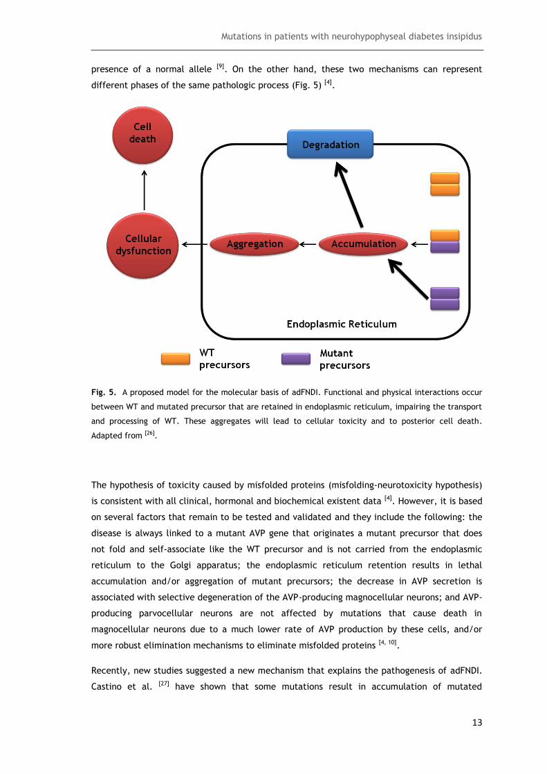
different phases of the same pathologic process (Fig. 5) [4].
Fig. 5. A proposed model for the molecular basis of adFNDI. Functional and physical interactions occur
between WT and mutated precursor that are retained in endoplasmic reticulum, impairing the transport
and processing of WT. These aggregates will lead to cellular toxicity and to posterior cell death.
Adapted from [26].
The hypothesis of toxicity caused by misfolded proteins (misfolding-neurotoxicity hypothesis)
is consistent with all clinical, hormonal and biochemical existent data [4]. However, it is based
on several factors that remain to be tested and validated and they include the following: the
disease is always linked to a mutant AVP gene that originates a mutant precursor that does
not fold and self-associate like the WT precursor and is not carried from the endoplasmic
reticulum to the Golgi apparatus; the endoplasmic reticulum retention results in lethal
accumulation and/or aggregation of mutant precursors; the decrease in AVP secretion is
associated with selective degeneration of the AVP-producing magnocellular neurons; and AVP-
producing parvocellular neurons are not affected by mutations that cause death in
magnocellular neurons due to a much lower rate of AVP production by these cells, and/or
more robust elimination mechanisms to eliminate misfolded proteins [4, 10].
Recently, new studies suggested a new mechanism that explains the pathogenesis of adFNDI.
Castino et al. [27] have shown that some mutations result in accumulation of mutated

Mutations in patients with neurohypophyseal diabetes insipidus
14
precursores in the endoplasmic reticulum, forming insoluble aggregates [27]. This accumulation
results in the development of a pathology characterized by a grossly deranged endoplasmic
reticulum which contains both mutated and WT protein [27]. With the aid of morphological
observations, Davies et al. suggest that these structures represent autophagic vesicles [28].
Autophagy results in organelle destruction together with the WT AVP, resulting in a
progressive AVP deficiency [27]. Under these circumstances, autophagy is a cell survival
mechanism that removes the deranged structures [27]. However, the cells are continuously
exposed to environmental and metabolic insults that can lead the cell to an autophagy-
dependent apoptosis once the neurons are frail, already undergoing autophagy in order to
clear mutant proteins [29]. This hypothesis does not exclude the misfolded-neurotoxicity
hypothesis as, although authophagy may be responsible for the initiation of adFNDI’s
symptoms, it does not exclude the possibility that degeneration of the AVP-producing
magnocellular neurons can be a long-term consequence [10].
The accumulation and cellular death caused by cytotoxicity of mutated precursors is a slow
and prolonged process, which explains some facts like the progressive onset of the symptoms
of the disease and the AVP deficiency [3, 14]. Autopsy studies performed in adFNDI patients
show a selective loss of AVP-producing magnocellular neurons in the supraoptic and, to a
lesser extent, in the paraventricular nucleus along with loss of their axonal extensions into
the neurohypophysis [4, 30]. These studies also show atrophy of the neurohypophysis and gliosis
[4, 30].
Some authors suggest that cell survival depends on its efficiency to degrade unfolded or
incompletely folded proteins. Thus, degradation-resistant proteins that accumulate in the
endoplasmic reticulum cause a more profound cytotoxic effect than proteins that are not
resistant to the degradation process [12].
It is difficult to determine significant differences in the severity of the disease produced by
the various AVP gene mutations [14]. The number of patients available for careful evaluation is
very limited and there is a high degree of variability, even for patients of the same family,
like the debut of symptoms, severity of polyuria and the degree of AVP deficiency [14]. Thus,
these factors result in a lack of genotype-phenotype correlation which could help to
determine the best treatment for the patients.
1.6. Diagnosis and treatment
The clinical diagnosis of DI can be made easily by measuring urine osmolality during a fluid-
deprivation test, at least when the disease is present in its complete form, as described
above [10]. However, with the development of knowledge related with the disease, some
authors suggested a new diagnosis based on molecular genetic evaluation that should be

Mutations in patients with neurohypophyseal diabetes insipidus
15
performed in all patients with familial occurrence of DI symptoms [10]. Once the molecular
diagnosis is established in adFNDI kindreds, it is easier to screen other family members for the
same mutations [10]. This fact has particular importance in infants at risk of inheriting the
mutation as this method allows the presymptomatic diagnosis, relieving years of parental
concern about the evolution of the disease in their offspring [10]. As adFNDI presents very few
symptoms and allows a normal quality of life, at least when offered an appropriable
treatment, and because there is little evidence for an associated risk of severe central
nervous system sequelae [31] compared with nephrogenic DI, a prenatal diagnosis seems not to
be indicated [10].
The treatment of adFNDI is relatively simple as the administration of the AVP analogue,
DDAVP, 2 to 3 times daily eliminates symptoms [32]. Patients with adFNDI have preservation of
the osmoregulation of thirst, thus only minor fluctuations in plasma osmolality are seen even
during irregular pharmacological treatment and the risk of inducing hyponatremia is very
small in these patients [10]. To date, no other V2 receptor agonist has been introduced in the
treatment of adFNDI but several delivery methods have been investigated and they are
available in nasal sprays, in common tablets and more recently in sublingual instant melting
tablet [10, 33]. However, it remains unknown if each delivery method results in better control
of polyuria and polydipsia or if it is only a matter of preference [10].
In an ideal perspective, the treatment of adFNDI should be able to provide a long-lasting
antidiuretic effect with the possibility to provide escape in case of higher-than-required fluid
intake, like in case of social reasons. This treatment can be obtained with gene therapy which
provides constantly high levels of AVP through the expression of the AVP gene contained in a
viral vector [10]. Many studies have shown the efficiency of gene therapy in AVP-deficient rats
using electroporation [34]. The next step in this treatment is the escape from the constant
antidiuresis induced by gene therapy and this can be achieved using the recently developed
V2 receptor antagonists [34]. However, there are several diverging opinions relative to the
safety of such viral approaches [10] and a further work is needed to clarify all the questions
around gene therapy.
1.7. Future perspectives
adFNDI is a disease with low morbidity and an effective treatment but, despite its benign
nature, the disease has been subject of intense research. This fact occurs due to its potential
value as a model for studies of neuroendocrinological diseases and for studies of dominant
negative mutations and due to its importance in the understanding of the effects of such
mutations on the folding of hormone precursors and the role of the protein quality control
machinery in the cellular handling of misfolded protein [35].

Mutations in patients with neurohypophyseal diabetes insipidus
16
Russel et al. [9] proposed that adFNDI could be considered a neurodegenerative disorder like
Alzheimer disease, Parkinson disease and others [9]. This suggestion is due to accumulation of
cytotoxic precursors inside neuronal cells in adFNDI, as in the above diseases, leading to the
posterior death of the cell.
A possible therapeutic approach to diseases caused by accumulation of misfolded proteins
inside the endoplasmic reticulum can be the use of pharmacologic chaperones to promote the
escape of proteins from this organelle [3]. Thus, the proteins can proceed their transport to its
target cells. In this case, without trafficking impairment, the mutant proteins could be
sufficiently functional if the problem resided in the transport of the proteins [3].
It is very important to proceed with genetic and molecular studies of adFNDI as the results
can give answers not only about adFNDI, but also help to explain other diseases that have the
same molecular mechanism like the case of neurodegenerative diseases or other pathologies
that involve protein misfolding or aggregates. On the other hand, it is necessary that patients
are informed about their state more deeply, principally in case of genetic diseases that are
transmitted through several generations.
1.8. Aims of the thesis
The present study is based on three main aims. First, to review AVP mutations described in
the scientific literature. Second, to expand the spectrum of mutations through the analysis of
additional patients with DI. Third, and last, to characterize the functional consequences of
identified novel AVP mutations.
Mutations in patients with neurohypophyseal diabetes insipidus
17
2. Methods
2.1. Literature search of AVP gene mutations
A database of the described and published AVP gene mutations was constructed by searching
the National Center Of Biotechnology Information Pubmed literature ~database for articles in
English, using the keywords AVP, mutation and Neurohypophyseal Diabetes Insipidus.
A total of 61 articles that described 70 different mutations were identified and evaluated.
The most relevant information was analyzed and a new nomenclature was assigned to each
mutation, based on recommendations from the authors Dunnen and Antonarakis [36]. Beyond
the alteration in AVP cDNA, others changes were also taken into account like the exon in
which mutations occur, the alterations caused at the protein level (amino acid changes),
protein domain, the population and the existence of functional studies.
2.2. Subjects and clinical procedures
A total of 9 patients diagnosed with neurohypophyseal DI, consisting of 3 familial cases and 6
sporadic cases, gave their informed consent for genetic studies of their AVP gene, in order to
identify possible mutations which could be responsible for their disease. Diagnosis of patients
was performed at the Endocrinology, Diabetes and Metabolism Service (University Hospital
from Coimbra, Portugal) and was based on a fluid deprivation test followed by DDAVP
administration.
The present study was approved by the Ethics Committee of the Faculty of Health Sciences at
the University of Beira Interior.
2.3. DNA extraction
When a blood sample is collected to perform molecular analysis, like the identification of
genetic mutations, the first step in laboratorial procedure is DNA extraction. The method
chosen for DNA extraction from peripheral blood is very important as it is necessary to obtain
a highly purified DNA without fragmentation. Some points are very important when a
particular technique is chosen like technical requirements, the time required to develop the
protocol, the efficiency of the method and its monetary cost [37]. Several methods are used
to extract DNA, including the use of organic solvents, but the contamination with proteins is a
frequent problem [38]. Miller et al. published, in 1987, a new method to extract DNA that

Mutations in patients with neurohypophyseal diabetes insipidus
18
involves the salting out of the cellular proteins by dehydration and precipitation with a
saturated NaCl solution [38].
A total of 10 mL of blood was collected from each patient with Neurohypophyseal DI and the
genomic DNA was extracted by the salting out method. The first stage in DNA extraction is
cell lysis in order to have the DNA in solution. To perform the red blood cells (RBC) lysis,
blood was transferred to 50 mL tubes and 30 mL of cold RBC lysis buffer (155 mM NH4Cl; 20
mM KHCO3; 0,1 mM Na2EDTA; pH 7,4), was added. This buffer is a hypotonic solution which
allows water intake into RBC, promoting their disruption. The mixture was incubated on ice
during 15 min and was centrifuged at 2500 rpm, during 10 min at 4ºC. It is very important to
remove hemoglobin, since its iron content can be a limitation to further downstream
applications, so the previous step is repeated as long as the pellet remains red. During
leucocyte lysis, 5mL of secondary extraction (SE) buffer (75mM NaCl; 25mM Na2EDTA; pH 8,0),
12,5µL of proteinase K (20mg/mL) and 500µL of 10% (w/v) sodium dodecyl sulfate (SDS) was
added to the mixture and it was incubated overnight at 55ºC in a thermal block (Star Lab).
Each reagent has a specific function: SE buffer contains chelating agents, which bind to
nuclease cofactors and prevent DNA degradation by these enzymes, SDS is a detergent so it
dissolves the cell membrane and denatures proteins and proteinase K digests proteins [39].
Protein precipitation was performed by adding 3mL of saturated NaCl (6M), since this reagent
decreases the solubility of proteins, followed by an incubation time of 10 min at 55ºC. The
mixture was vortexed during 25 sec and finally it was centrifuged at 4000 rpm, during 30 min
at 15ºC. The pellet was rejected and 100% (v/v) cold ethanol was added to the supernatant.
As DNA is insoluble in ethanol, when this reagent is added, DNA molecules form aggregates
that can be obtained in a pellet form upon centrifugation at 4500 rpm, during 5 min at 4ºC.
The pellet was washed with 70% (v/v) cold ethanol followed by a last centrifugation at 4500
rpm, during 5 min at 4ºC. In the end, the pellet was solubilized in 1mL of Tris-EDTA (TE)
buffer.
A most common method to quantify DNA samples is based on using a spectrophotometer, in a
wave length (λ) of 260 nm. This method permits to estimate the quantity (Beer-Lambert law:
A260=εbc, where A260 corresponds to absorbance, ε corresponds to molar absorbitivity with a
value of 20cm.mg.ml-1, b corresponds to path length of the cuvette in which the sample is
contained, and c corresponds to the concentration of the compound in solution) and the
relative purity of DNA samples (in the case of proteins or RNA contamination), since proteins
absorb light at 280 nm. A pure DNA sample will have a ratio (A260/A280) value of approximately
1.8 - 2.
The DNA was quantified using nanophotometer (IMPLEN).

Mutations in patients with neurohypophyseal diabetes insipidus
19
2.4. Amplification of the AVP gene by polymerase chain
reaction
The preparation of large amounts of specific DNA fragments is an indispensable tool in
experiments in molecular biology. Polymerase chain reaction (PCR) is an enzymatic
amplification technique that can be used, when the nucleotide sequences at the ends of a
particular DNA region are known, to prepare significant quantities of a specific DNA fragment
[40, 41].
The PCR procedure begins with heat-denaturation (95ºC) of a DNA sample into single strands
(denaturation step) so that in the next step, two synthetic oligonucleotides, added in great
excess, complementary to the 3’ ends of the DNA fragment of interest can hybridize with
their complementary sequences (annealing step). Annealing step occurs at lower
temperatures (50-60ºC). The hybridized oligonucleotides will serve as primers for synthesis of
a new DNA chain (extension step), in the presence of deoxynucleotides (dNTPs) and a
thermoresistant DNA polymerase, such as that from Thermus aquaticus (hence, its name Taq
polymerase). These three steps form a cycle, and when the extension step finishes, the whole
mixture is heated again to 95º C to denature the newly formed double stranded DNA and a
new annealing step occurs, since an excess of primers is present. Repeated cycles, each one
with a denaturation step, an annealing step and an extension step, quickly amplify the
sequence of interest [41].
The three exons of AVP gene were amplified separately by PCR, using specific primers
flanking each exon (Table 1). The PCR reaction was performed in a total volume of 25 µL with
100 ng of purified genomic DNA, 0.2 mM of deoxyribonucleotides (dNTPs) (nzytech), 1U of
Dream Taq DNA polymerase and complete Dream Taq buffer in a final MgCl2 concentration of
1 mM (Fermentas) and 0.25 µM of each primer (AVP_1F and AVP_1R for exon 1, AVP_2Fc and
AVP_2Rc for exon 2 and AVP_3Fd and AVP_2+3R for exon 3) (Stabvida). The PCR protocol was
initiated with a denaturation step at 95ºC during 5 min followed by 35 cicles, each cycle with
a denaturation step at 95ºC during 30 sec, an annealing step at 59ºC for exon 1, 75ºC for exon
2 and 66ºC for exon 3, during 30 sec and a extension step at 72ºC during 30 sec and finally a
longer extension step at 72ºC during 10 min so that Taq polymerase can synthesize the
remaining DNA chains. PCR protocols were performed in a T100 thermal cycler (Bio-Rad).
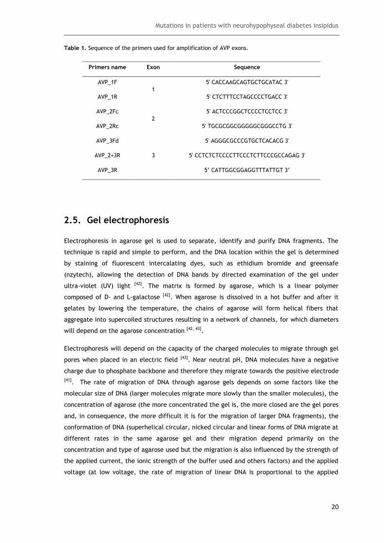
Mutations in patients with neurohypophyseal diabetes insipidus
20
Table 1. Sequence of the primers used for amplification of AVP exons.
Primers name Exon Sequence
AVP_1F
1
5' CACCAAGCAGTGCTGCATAC 3'
AVP_1R 5' CTCTTTCCTAGCCCCTGACC 3'
AVP_2Fc
2
5' ACTCCCGGCTCCCCTCCTCC 3'
AVP_2Rc 5' TGCGCGGCGGGGGCGGGCCTG 3'
AVP_3Fd
3
5' AGGGCGCCCGTGCTCACACG 3'
AVP_2+3R 5' CCTCTCTCCCCTTCCCTCTTCCCGCCAGAG 3'
AVP_3R 5’ CATTGGCGGAGGTTTATTGT 3’
2.5. Gel electrophoresis
Electrophoresis in agarose gel is used to separate, identify and purify DNA fragments. The
technique is rapid and simple to perform, and the DNA location within the gel is determined
by staining of fluorescent intercalating dyes, such as ethidium bromide and greensafe
(nzytech), allowing the detection of DNA bands by directed examination of the gel under
ultra-violet (UV) light [42]. The matrix is formed by agarose, which is a linear polymer
composed of D- and L-galactose [42]. When agarose is dissolved in a hot buffer and after it
gelates by lowering the temperature, the chains of agarose will form helical fibers that
aggregate into supercoiled structures resulting in a network of channels, for which diameters
will depend on the agarose concentration [42, 43].
Electrophoresis will depend on the capacity of the charged molecules to migrate through gel
pores when placed in an electric field [43]. Near neutral pH, DNA molecules have a negative
charge due to phosphate backbone and therefore they migrate towards the positive electrode
[41]. The rate of migration of DNA through agarose gels depends on some factors like the
molecular size of DNA (larger molecules migrate more slowly than the smaller molecules), the
concentration of agarose (the more concentrated the gel is, the more closed are the gel pores
and, in consequence, the more difficult it is for the migration of larger DNA fragments), the
conformation of DNA (superhelical circular, nicked circular and linear forms of DNA migrate at
different rates in the same agarose gel and their migration depend primarily on the
concentration and type of agarose used but the migration is also influenced by the strength of
the applied current, the ionic strength of the buffer used and others factors) and the applied
voltage (at low voltage, the rate of migration of linear DNA is proportional to the applied

Mutations in patients with neurohypophyseal diabetes insipidus
21
voltage but at higher voltages the mobility of high-molecular-weight DNA fragments increases
differentially) [43].
After the PCR reaction, the results were analyzed by electrophoresis. The agarose gel (1%
(w/v)) was prepared with TAE buffer (40mM Tris-acetate, 1mM EDTA, pH=8,0). Agarose was
dissolved in TAE buffer and, in order to visualize the PCR products, greensafe (nzytech) was
added (1 µL per mL of gel). After gel polymerization, it was placed in a TAE buffer-containing
electrophoresis tank with opposing immersed electrodes (Bio-Rad). The samples and the DNA
size standard (VC 100bp Plus DNA Ladder, Vivantis) were loaded in the slots along the top of
the gel and a 120 voltage was applied. At the end of the run, the gel was analyzed by UV
illumination (Uvitec) and it was photographed using FireReader software (Version 15.15,
Uvitec).
2.6. AVP gene sequencing
In order to identify mutations in genes, their sequence must be analyzed. The dideoxy chain-
termination method was described first in 1977 by Sanger F. and colleagues. The principal
aim of this method is to synthesize a set of daughter strands, from the DNA fragment of
interest, that are labeled at one end and differ in length by one nucleotide. When these
daughter strands are separated by gel electrophoresis, the nucleotide sequence of the
fragment can be established in automated DNA sequencing machines, since a fluorescence
detector that can distinguish the four fluorescent tags is located at the end of the gel.
Synthesis of truncated strands is due to the use of 2’,3’-dideoxyribonucleoside triphosphates
(ddNTPs) that, in contrast to normal dNTPs, lack a 3’-hydroxyl group. Due to the lack of this
group in ddNTPs, they can be incorporated into a growing DNA chain by the action of a DNA
polymerase, but once incorporated they cannot form a phosphodiester bond with the next
nucleotide leading to termination of the chain synthesis [41].
The method begins with the denaturation of a double-stranded DNA sample to generate the
template strands where a primer will hybridize for the polymerization reaction. In these
reactions, the ddNTPs, at lower concentrations when compared with dNTPs, are randomly
incorporated at the positions of the corresponding dNTP, causing termination of synthesis at
those positions in the sequence. The inclusion of different fluorochromes in each ddNTP
allows the identification of each truncated fragment after their migration through the
electrophoresis gel and thus, the DNA sequence can be determined by the order in which
each fragment passes through the fluorescence detector [41].
Before sequencing, the PCR products were purified by spin column technique following the
manufacturer’s instructions (JETQUICK PCR purification Spin Kit, Genomed). After

Mutations in patients with neurohypophyseal diabetes insipidus
22
purification, the presence of DNA was confirmed by electrophoresis in agarose gel (1% (w/v))
with DNA size standard HyperLadder II (Bioline).
DNA sequencing has three main steps: preparation of the DNA sequencing reaction, ethanol
precipitation and sample preparation for loading into the sequencing equipment (GenomeLab
GeXP, Beckman Coulter). In preparation of the DNA sequencing reaction, for one DNA sample,
two reactions were done. Each 20 µL reaction was prepared with approximately 14 ng of DNA
sample, 0.25 µM of the primer (Stabvida) and 8 µL of DTCS Quick Start Master Mix
(GenomeLab, Beckman Coulter). The primers used for sequencing reaction were the same
used in the PCR reaction. The reactions were placed in T100 thermal cycler with the following
protocol: 30 cycles, each cycle with one denaturating step at 96ºC during 20 sec, one
annealing step at 55ºC for exon 2 and 50ºC for exon 1 and 3 during 20 sec and one extension
step at 60ºC during 4 min. At the end of DNA sequencing reaction, ethanol precipitation and
sample preparation were performed according to manufacturer’s instructions (GenomeLab,
Beckman Coulter). The method used to sequence the samples comprised a capillary
temperature of 50ºC, a denature temperature of 90ºC during 120 sec, an injection voltage of
2.0 kV during 15 sec and a separation voltage of 4.0 kV during 70 min..
The results were analyzed using GenomeLab Genetic Analysis System software (Version
10.2.3, Beckman Coulter).
2.7. pRc/RSV Sequencing
The expression vector pRc/RSV (Fig. 6), which contains the human AVP cDNA, was a kind gift
from Dr. J. Larry Jameson (University of Pennsylvania, USA). The cDNA was sequenced by the
Sanger method. For preparation of DNA sequencing reaction, 190 ng of plasmid DNA (pDNA)
was initially denaturated at 95ºC during 3 min. Then, two reactions with a final volume of 20
µL were prepared. 0.25 µM of BHG-R primer (5’ GGCTGGCAACTAGAAGGCACAGTCGAGG 3’)
was added in one reaction and 0.25 µM of AVP_1F in the other reaction. Finally, 8 µL of DTCS
Quick Start Master Mix were added in both reactions. The reactions were placed in T100
thermal cycler with the following protocol: 30 cycles, each cycle with one denaturating step
at 96ºC during 20 sec, one annealing step at 50ºC during 20 sec and one extension step at
60ºC during 4 min. At the end of DNA sequencing reaction, ethanol precipitation and sample
preparation for loading into the instrument were performed according to manufacturer’s
instructions (GenomeLab, Beckman Coulter). The method used to sequence the pDNA was the
same used to sequence AVP exons with the exception of separation duration which was 150
min.
The results were analyzed using GenomeLab Genetic Analysis System software (Version
10.2.3, Beckman Coulter).

Mutations in patients with neurohypophyseal diabetes insipidus
23
Fig. 6. Expression vector pRc/RSV (Adapted from Invitrogen).
2.8. Cloning of AVP cDNA into pVAX1/lacZ
In order to study the effects of AVP mutations on gene expression at the molecular level, it is
important to have large quantities of the gene in a pure form. DNA cloning allows the
preparation of large numbers of identical DNA molecules [41]. The DNA fragment of interest,
AVP cDNA, is linked to a vector which allows the transport of foreign DNA and its replication
into a host cell, such as the bacterium Escherichia coli (E. coli). Once inside the host cell, the
inserted DNA is replicated along with the vector generating a large number of identical DNA
molecules [41, 43].
The aim of this experimental step was the transfer (subcloning) of AVP cDNA, present in the
expression vector pRc/RSV, to the pVAX1/lacz (Fig. 7) plasmid in order to insert the desired
mutations in cDNA AVP. This step was crucial since the expression vector had restriction sites
to the enzymes chosen for the insertion of the mutations. The XbaI (New England
BioLabs)/HindIII (Takara Biotechnology) cDNA fragment was transferred from pRc/RSV to
pVAX1/lacZ in the same sites.
A total of 2.7 µg of pRc/RSV were digested in a total volume of 50 µL with 40U of XbaI, 1x
NEBuffer 4 (New England Biolabs) and 100 µg/mL of bovine serum albumin (BSA). A total of 3
µg of pVAX1/lacZ were digested in a total volume of 50 µL with 40 U of XbaI, 1x NEBuffer 4
and 100 µg/mL of BSA. Both reactions were incubated during 2 hours (h) at 37ºC in a thermal
block. An electrophoresis in agarose gel (1% (w/v)) was undertaken to analyze the digestion
results. About 40-100 U of HindIII were added to pRc/RSV and pVAX1/lacZ. The reactions
were incubated at 37ºC during 2 h and 30 min in a thermal block. A final electrophoresis was
done in agarose gel (1% (w/v)) in order to analyze the digested fragments.

Mutations in patients with neurohypophyseal diabetes insipidus
24
Fig. 7. Cloning vector (Adapted from Invitrogen).
To purify the AVP cDNA (XbaI/HindIII) released from pRc/RSV, a low-melting point agarose
(2.5% (w/v)) (nzytech) was prepared. A 120 voltage was applied and at the end of migration
the corresponding bands were excised. DNA was extracted from gel by spin column technique
(JETQUICK Gel Extraction Spin Kit, Genomed) and the results analyzed by electrophoresis in
agarose gel (1% (w/v)). Regarding digested pVAX1/lacZ, the purification step was performed
by spin column technique (JETQUICK PCR purification Spin Kit, Genomed).
After purification, XbaI/HindIII AVP cDNA was ligated to XbaI/HindIII pVAX1/lacZ. Three
reactions were prepared: reaction A with a final volume of 16 µL, contained 200 ng of
XbaI/HindIII pVAX1/lacZ, 65 ng of XbaI/HindIII AVP cDNA, 3 U of T4 DNA ligase (pGEM-T Easy
Vector System I, Promega) and 1x Rapid Ligation Buffer (pGEM-T Easy Vector System I,
Promega); reaction B with a final volume of 20 µL, contained 200 ng of XbaI/HindIII
pVAX1/lacZ, 91 ng of XbaI/HindIII AVP cDNA, 3 U of T4 DNA ligase and 1x Rapid Ligation
Buffer; reaction C with a final volume of 14 µL, contained 100 ng of XbaI/HindIII pVAX1/lacZ,
65 ng of XbaI/HindIII AVP cDNA, 3 U of T4 DNA ligase and 1x Rapid Ligation Buffer. The
reactions were incubated during 1h at room temperature.
2.9. Competent cells
There are two methods that allow the cells to acquire competence in order to take up the
DNA from the medium: a chemical and a physical method. The chemical method is based on
washing E. coli cells with simple salt solutions, which allow cells to achieve the desired
competence state [42]. Then, DNA is added to the bacterial culture, which is subjected to a
brief heat shock that stimulates the cells to take up DNA from their surrounding medium [43].
Once inside the cell, the plasmid replicates autonomously and is passed to the next
generations during cell division [43].

Mutations in patients with neurohypophyseal diabetes insipidus
25
This simple chemical procedure generates enough transformed colonies four routine tasks but
the mechanisms by which these combinations of chemical agents and physical treatments
induce a state of competence remains unknown, such as the mechanism by which the plasmid
DNA enters and establishes itself in competent cells [42].
In order to prepare competent cells, the material and all solutions needed were sterile. The
environment in which cells were prepared was sterile too. Initially, 100 µL of E. coli (JM109)
cells were inoculated in 5 mL of LB – Broth medium at 37ºC with orbital shaking of 200 rpm
overnight. The following day, 2 mL of growing culture were inoculated in 250 mL of new LB-
Broth mediu at 37ºC with orbital shaking of 250 rpm until an optical density between 0.3 and
0.4 was obtained (λ = 600 nm). The absorbance was measured using a spectrophotometer
(Ultraspec 3000, Pharmacia Biotech). Then, the culture was centrifuged at 5000 rpm during
10 min at 4ºC. The pellet was resuspended in 62.5 mL of MgCl2 100 mM at 4ºC, during 3 min
and a new centrifugation was performed at 4000 rpm, during 10 min at 4ºC. Then, the pellet
was resuspended in 12.5 mL of CaCl2 100 mM at 4ºC and a further 112.5 mL of CaCl2 100mM at
4ºC were added. The mixture was maintained on ice during 30 min and one last centrifugation
was performed at 4000 rpm during 10 min at 4ºC. Finally, the cells were resuspended in 5 mL
of CaCl2 85 mM at 4ºC with 15% (v/v) of glycerol and aliquots of 100 µL were immersed in
liquid nitrogen and stored at -80ºC.
2.10. Transformation of competent cells with recombinant
pVAX1/lacZ
After 1h of ligation between XbaI/HindIII pVAX1/lacZ and XbaI/HindIII AVP cDNA, performed
in section 2.8, competent E. coli cells (JM109) were transformed with the ligation reaction
product. For each ligation, the total volume of reaction was added to 50 µL of competent
cells and the mixtures were incubated on ice during 30 min. Then, a heat shock at 42ºC
during 1 min was performed in a thermal block for each mixture and a new incubation on ice
during 2 min was performed. After this step, 200 µL of LB-Broth medium, without antibiotic,
was added and the cells were incubated during 2h at 37ºC with orbital shaking of 250 rpm. A
volume of 100 µL of the previous mixture was spread on LB-agar/Kanamycin plates (50
µg/mL) that were incubated at 37ºC overnight. A control was performed with 50 ng of initial
pVAX1/lacZ.
The following day, 6 colonies of each plate (except the control) were inoculated in 20 µL of
LB – Broth medium in order to performed PCR as a confirmation method of insertion of AVP
cDNA in the plasmid. The PCR were performed in a total volume of 25 µL using 2 µL of
inoculum, 0.2 mM of dNTPs, 1 U of Dream Taq DNA polymerase and complete Dream Taq
buffer in a final MgCl2 concentration of 1.5 mM and 0.25 µM of each primer (AVP_Mut1 (5’

Mutations in patients with neurohypophyseal diabetes insipidus
26
GCCCCGGGGGCAAAGGCCGCT 3’) and AVP_Mut4 (5’ CAAGGCCCCGGCCGGCCCGT 3’)). The PCR
protocol was initiated with a denaturation step at 95ºC during 5 min followed by 35 cicles,
each cycle with a denaturation step at 95ºC during 30 sec, an annealing step at 60ºC during 30
sec and a extension step at 72ºC during 30 sec and finally a longer extension step at 72ºC
during 10 min. PCR protocols were performed in T100 thermal cycler. An electrophoresis in
agarose gel (1% (w/v)) was performed to analyze the PCR results by comparer to a DNA size
standard (100bp DNA Ladder, New England Biolabs).
The positive colonies were inoculated in 4 mL of LB – Broth medium with kanamycin (50
µg/mL) and incubated overnight at 37ºC with orbital shaking of 250 rpm. After overnight
growth, the cells were harvested and the recombinant plasmids were purified using Wizard
Plus SV Minipreps DNA Purification System (Promega).
In order to confirm the recombinant plasmids, pDNA was subjected to digestion with XbaI. In
a final volume of 10 µL, 5 µL of each purified pDNA, 10 U of XbaI, 1x of respective buffer and
100 µg/mL of BSA were added. The reactions were incubated during 2 h at 37ºC and an
electrophoresis in agarose gel (1%(w/v)) was performed using a new DNA size standard (1 kb
DNA Ladder, New England Biolabs). The recombinant plasmid which presented the size
corresponding to recombinant pVAX1/lacZ (with XbaI/HindIII AVP cDNA) (pVAX/AVP) was also
digested with XbaI and HindIII to confirm the AVP cDNA presence in a final volume of 25 µL,
where 20 U of XbaI, 16-40 U of HindIII, 1x of XbaI buffer and 100 µg/mL of BSA were added.
The reaction was incubated at 37ºC during 3 h and the results analyzed by an electrophoresis
in agarose gel (1% (w/v)). Then, competent E. coli cells were transformed by the same
protocol with approximately 34 ng of pVAX/AVP. The recombinant plasmids were purified
using Wizard Plus SV Minipreps DNA Purification System. The pDNA (pVAX/AVP) was quantified
using nanophotometer, as in section 2.3.
2.11. Site – directed mutagenesis
Site-directed mutagenesis allows researchers to make small and specific changes in a DNA
sequence of interest. Alterations like substitutions of one base, deletions or insertions of a
very small number of bases can be performed by this technique [43].
The PCR site-directed mutagenesis required the synthesis of four DNA oligonucleotides
wherein two of them contained the desired mutation. Two primary PCR reactions produced
two overlapping DNA fragments and each fragment contained the same mutation introduced
by the primers, in the region of overlap. This overlap region allowed the recombination of
two fragments after their mixture, denaturation and renaturation, and could be extended by
DNA polymerase in order to produce a complete double-stranded DNA fragment which served
as template, for a second PCR reaction using only the outermost two primers to amplify the

Mutations in patients with neurohypophyseal diabetes insipidus
27
final sequence [40]. Finally, the modified DNA could be cloned in an expression vector and
their effect analyzed by introducing the DNA into a host cell.
In order to introduce the two desired mutations in the expression vector, site-directed
mutagenesis was used to create them. Seven primers were designed and synthesized
(Stabvida), one of them is common to both mutations (Table 2).
Table 2. Sequence of primers used for site-directed mutagenesis.
Name Sequence Mutation
AVP_Mut1 5’ GCCCCGGGGGCAAAGGCCGCT 3’ c.289C>G
AVP_Mut2B 5’ AAGGCGGCGCAGCCGCCCCCGCT 3’ c.289C>G
AVP_Mut3B 5’ AGCGGGGGCGGCTGCGCCGCCTT 3’ c.289C>G
AVP_Mut4 5’ CAAGGCCCCGGCCGGCCCGT 3’ c.289C>G c.154T>C
AVP_Mut5 5’GCAAAGGCCGCCGCTTCGGGCCCA 3’ c.154T>C
AVP_Mut6 5’TGGGCCCGAAGCGGCGGCCTTTGC 3’ c.154T>C
AVP1F_Mut7 5’ ATGCCTGACACCATGCTGCCCGCC 3’ c.154T>C
For the first mutation (c.154T>C) (Fig. 8), two initial PCR reactions were performed and
primers AVP_Mut6 and AVP1F_Mut7 were used to create fragment 1 (Frag1) and primers
AVP_Mut5 and AVP_Mut4 were used to create fragment 2 (Frag2). In a final volume of 25 µL,
for both reactions, the following reagents were added: 95 ng of expression vector (pRc/RSV),
0.2 mM of dNTPs, 1 U of Dream Taq DNA polymerase and complete Dream Taq buffer, in a
final MgCl2 concentration of 1 mM for both fragments and 0.25 µM of each primer. The PCR
protocol was the same for both reactions: a denaturation step at 95ºC during 5 min followed
by 35 cycles, each cycle with a denaturation step at 95ºC during 30 sec, an annealing step at
75ºC during 30 sec and a extension step at 72ºC during 30 sec and, finally, a longer extension
step at 72ºC during 10 min. The results were confirmed by an electrophoresis in agarose gel
(1% (w/v)). The PCR products were purified using Wizard SV Gel and PCR Clean-Up System
(Promega) and a new electrophoresis in agarose gel (1% (w/v)) was performed to analyze and
quantify the purified DNA. Finally, a third PCR reaction was prepared using primers
AVP1F_Mut7 and AVP_Mut4 yielding fragment 3 (Frag3). For a final volume of 25 µL,
approximately 10 ng of Frag 1 and 16 ng of Frag2 were added, together with 0.2 mM of
dNTPs, 1 U of Dream Taq and complete Dream Taq buffer, in a final MgCl2 concentration of
1mM, and 0.25 µL of each primer. The PCR protocol was initiated with a denaturation step at

Mutations in patients with neurohypophyseal diabetes insipidus
28
95ºC during 5 min followed by 40 cycles, each cycle with a denaturation step at 95ºC during
30 sec and an annealing/extension step at 72ºC during 1 min and finally a longer extension
step at 72ºC during 10 min. A final electrophoresis in agarose gel (1% (w/v)) was prepared to
analyze the PCR results.
Fig. 8. AVP cDNA with primers used in site-directed mutagenesis for the first mutation (c.154T>C).
Orange represents first exon, black represents second exon and blue represents third exon. Red
nucleotide represents nucleotide to be substituted and pink nucleotide, in the center of the primer,
represents the altered nucleotide. Surrounded by pink is the restriction site for SmaI and surrounded by
green is the restriction site for FseI.
The Frag 3 was sequenced in order to confirm the insertion of the desired mutation. PCR
products were purified by enzymatic digestion using exonuclease I (Fermentas) and alkaline
phosphatase (Thermo Scientific FastAP Thermosensitive Alkaline Phosphatase, Fermentas).
The manufacturer’s instructions for nucleotide purification were followed with an exception:
10 µL of PCR product were used instead of 5 µL. For DNA sequencing reaction, two reactions
with a final volume of 20 µL were prepared. 50 ng of purified PCR product were used in each
reaction and 0.25 µM of AVP_Mut4 was added in one reaction and 0.25 µM of AVP1F_Mut7 in
the other reaction. Finally, 4 µL of DTCS Quick Start Master Mix were added in both reactions.
The reactions were placed in the thermal cycler with the following protocol: a initial
denaturation step ate 96ºC during 3 min and 30 cycles, each cycle with one denaturating step
at 96ºC during 20 sec, one annealing step at 55ºC during 20 sec and one extension step at
60ºC during 4 min. At the end of the DNA sequencing reaction, ethanol precipitation and
sample preparation for loading into the instrument were performed according to
manufacturer’s instructions (GenomeLab, Beckman Coulter). The method used to sequence
the Frag3 was the same used to sequence the AVP exons with the exception of separation

Mutations in patients with neurohypophyseal diabetes insipidus
29
duration that was 120 min. The final results were analyzed using GenomeLab Genetic Analysis
System software (Version 10.2.3, Beckman Coulter).
For the second mutation (c.289C>G) (Fig. 9), primers AVP_Mut1 and AVP_Mut2B were used to
create fragment AB (FragAB) in a first PCR and primers AVP_Mut3B and AVP_Mut4 were used
to create fragment BB (FragBB) in a second PCR. In a final volume of 25 µL, for both
reactions, the following reagents were added: 95 ng of expression vector (pRc/RSV), 0.2 mM
of dNTPs, 1 U of Dream Taq DNA polymerase and complete Dream Taq buffer, in a final MgCl2
concentration of 1.5 mM for FragAB and 1 mM for FragBB, and 0.25 µM of each primer. The
PCR protocol used in both reactions was the same used for Frag1 and Frag2. An
electrophoresis in agarose gel (1% (w/v)) was performed to confirm the PCR reaction. The
products were purified using PCR & Gel Band Purification Kit (Grisp) and a new
electrophoresis was performed to analyze and quantify the purified DNA fragment. Finally, a
third PCR reaction was done using primers AVP1F_Mut7 and AVP_Mut4 yielding fragment CB
(FragCB). For a final volume of 25 µL, 16 ng of each fragment were added, together with 0.2
mM of dNTPs, 1 U of Dream Taq and complete Dream Taq buffer, in a final MgCl2
concentration of 1mM, and 0.25 µL of each primer. The PCR protocol was the same used to
amplify Frag3 and a final electrophoresis in agarose gel (1% (w/v)) was performed to analyze
the PCR results.
Fig. 9. AVP cDNA with primers used in site-directed mutagenesis for the second mutation (c.289C>G).
Orange represents first exon, black represents second exon and blue represents third exon. Red
nucleotide represents nucleotide to be substituted and pink nucleotide, in the center of the primer,
represents the altered nucleotide. Surrounded by pink is the restriction site for SmaI and surrounded by
green is the restriction site for FseI.
Mutations in patients with neurohypophyseal diabetes insipidus
30
FragCB was sequenced in order to confirm the insertion of the desired mutation. PCR
products were purified as previously. For preparation of DNA sequencing, two reactions with a
final volume of 20 µL were prepared. 10 ng of purified PCR product were used in each
reaction and 0.25 µM of AVP_Mut1 was added in one reaction and 0.25 µM of AVP_Mut4 in the
other reaction. Finally, 4 µL of DTCS Quick Start Master Mix were added in both reactions.
The reactions were placed in T100 thermal cycler with the same protocol used for Frag3. At
the end of the DNA sequencing reaction, ethanol precipitation and sample preparation for
loading into the instrument were performed according to manufacturer’s instructions
(GenomeLab, Beckman Coulter). The method used to sequence FragCB was the same used to
sequence Frag3 and the results were analyzed using the same software.
2.12. Cloning of Frag3 and Frag CB
Fragments containing the desired mutation were digested with restricted enzymes in order to
be introduced in pVAX/AVP. Both mutagenesis fragments and pVAX/AVP were digested using
SmaI (New England Biolabs) and FseI (New England Biolabs). For a final volume of 50 µL, 2.5
µg of pVAX/AVP and 1 µg of PCR product (Frag3 and FragCB), 20 U of SmaI and 1x of
respective buffer were added and the mixtures were incubated during 1 h and 30 min at 25ºC
in a thermal cycler. An electrophoresis in agarose gel (1% (w/v)) was performed to analyze
the results and the digested continued with addition of 10 U of FseI and 100 µg/mL of BSA.
The reactions were incubated during 1 h and 30 min at 37ºC in a thermal block and an
electrophoresis in agasore gel (1% (w/v) was performed to analyze the digest.
An electrophoresis in low melting point agarose (2.5% (w/v)) was performed in order to purify
the desired fragments. The excised fragments were purified using Wizard SV Gel and PCR
Clean-Up System (Promega) and the results analyzed by electrophoresis in agarose gel (1%
(w/v)). Purified fragments were quantified.
A molar ratio of 1:3 was used to ligate SmaI/FseI Frag CB to SmaI/FseI pVAX/AVP and to ligate
SmaI/FseI Frag CB to SmaI/FseI pVAX/AVP. In each reaction, 50 ng of vector and 15 ng of
insert were used. T4 DNA ligase (DNA ligation kit, Takara) was used to ligate fragments. The
reactions were incubated at 16ºC overnight in a T100 thermal cycler. The following day, NEB
5-alpha Competent E. coli (High Efficiency cells) (New England Biolabs) were transformed
with ligated vectors according to manufacturer’s instructions. Different colonies were
incubated in LB-Broth/kanamycin medium overnight, at 37ºC with 250 rpm and pDNA were
purified using Wizard Plus SV Minipreps DNA Purification System. The pDNA was quantified.
The pDNA of each colony was sequenced using the Sanger method. For preparation of the DNA
sequencing reaction, 150 ng of pDNA was initially denaturated at 95ºC during 3 min. Then,
two reactions for each colony were prepared to a final volume of 20 µL. 0.25 µM of
Mutations in patients with neurohypophyseal diabetes insipidus
31
AVP1F_Mut7 primer was added in one reaction and 0.25 µM of AVP_Mut4 in the other
reaction. Finally, 4 µL of DTCS Quick Start Master Mix were added in both reactions. The
reactions were placed in a thermal cycler with the following protocol: a denaturing step at
95ºC during 3 min and 30 cycles, each cycle with one denaturating step at 96ºC during 20 sec,
one annealing step at 50ºC during 20 sec and one extension step at 60ºC during 4 min. At the
end of the DNA sequencing reaction, ethanol precipitation and sample preparation for loading
into the instrument were performed according to manufacturer’s instructions (GenomeLab,
Beckman Coulter). The method used to sequence the pDNA was the same used to sequence
the AVP exons with exception of separation duration that was 110 min. The results were
analyzed using the same software.
2.13. Expression of the normal AVP gene in Neuro 2A cells
Several authors published functional studies where they show the effect of adFNDI mutations
in neuronal cell lines, allowing an advancement in the understanding of consequences of the
AVP gene mutations at the level of protein expression [12, 13, 44]. Several of these studies used
neuroblastoma cells, known as neuro2A (N2A), as these cells are from neuronal origin and
provide adequate conditions for the expression of the AVP gene [12].
N2A cells were kindly provided by Prof. Luis Pereira de Almeida (Center for Neurosciences and
Cell Biology & Faculty of Pharmacy, University of Coimbra, Portugal). The cells were seeded
in 12-well plates and growth in Dulbecco’s modified eagle’s medium (DMEM) (Biochrom AG)
with 10% of fetal bovine serum (FBS) and 5% of antibiotics (penicillin and streptomycin) at
37ºC in an atmosphere of 5% of CO2. One day before transfection, the medium was changed to
DMEM with 10% of FBS and without antibiotics. To optimize the optimum ratio of DNA to
lipofectamine reagent, the following day, cells were transfected with 5 µg or 10 µg of wild
type AVP cDNA construct (pRc/RSV-AVP) and 4 µL or 8 µL of Lipofectamine 2000 (Invitrogen)
per well (table 3). Two transfection controls were performed, one to analyze endogenous
protein production, where no pRc/RSV-AVP neither lipofectamine were added to cells, and
another to monitor cytotoxicity from the transfection reagent, where 8 µL of lipofectamine
were added to cell culture. Another two controls for immunocytochemistry assays were
performed where 5 µg of pRc/RSV-AVP and 4 µL of lipofectamine were added at each assay.
All assays were duplicated and were summarized in table 3. Lipofectamine was diluted in 100
µL of Opti-modified eagle’s medium (Opti-MEM) (Gibco) and incubated during 5 min at room
temperature. The expression vector was diluted in 100 µL of Opti-MEM. The diluted DNA and
lipofectamine were combined, mixed and incubated during 20 min at room temperature.
Finally, this combined mixture was added to cell culture and incubated at 37ºC in an
atmosphere of 5% of CO2. 24 h latter, the medium was changed to DMEM with 5% of antibiotic
without FBS and remained at 37ºC during 72 h.
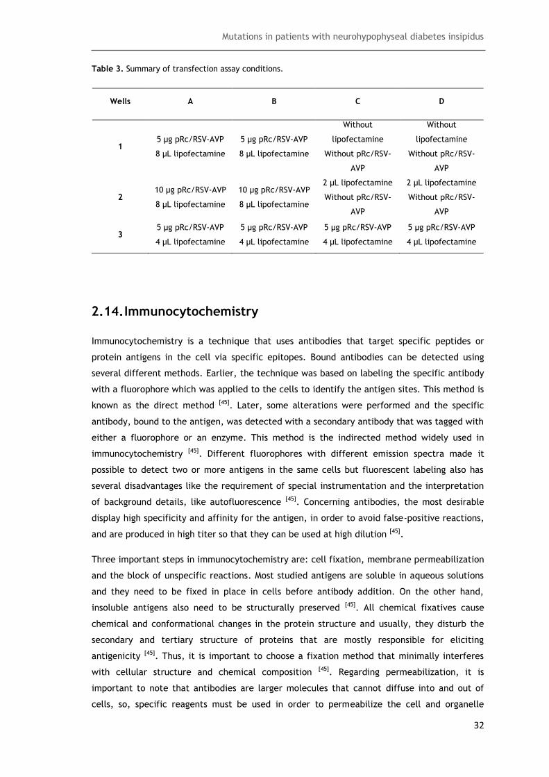
Mutations in patients with neurohypophyseal diabetes insipidus
32
Table 3. Summary of transfection assay conditions.
Wells A B C D
1 5 µg pRc/RSV-AVP
8 µL lipofectamine
5 µg pRc/RSV-AVP
8 µL lipofectamine
Without
lipofectamine
Without pRc/RSV-
AVP
Without
lipofectamine
Without pRc/RSV-
AVP
2 10 µg pRc/RSV-AVP
8 µL lipofectamine
10 µg pRc/RSV-AVP
8 µL lipofectamine
2 µL lipofectamine
Without pRc/RSV-
AVP
2 µL lipofectamine
Without pRc/RSV-
AVP
3 5 µg pRc/RSV-AVP
4 µL lipofectamine
5 µg pRc/RSV-AVP
4 µL lipofectamine
5 µg pRc/RSV-AVP
4 µL lipofectamine
5 µg pRc/RSV-AVP
4 µL lipofectamine
2.14. Immunocytochemistry
Immunocytochemistry is a technique that uses antibodies that target specific peptides or
protein antigens in the cell via specific epitopes. Bound antibodies can be detected using
several different methods. Earlier, the technique was based on labeling the specific antibody
with a fluorophore which was applied to the cells to identify the antigen sites. This method is
known as the direct method [45]. Later, some alterations were performed and the specific
antibody, bound to the antigen, was detected with a secondary antibody that was tagged with
either a fluorophore or an enzyme. This method is the indirected method widely used in
immunocytochemistry [45]. Different fluorophores with different emission spectra made it
possible to detect two or more antigens in the same cells but fluorescent labeling also has
several disadvantages like the requirement of special instrumentation and the interpretation
of background details, like autofluorescence [45]. Concerning antibodies, the most desirable
display high specificity and affinity for the antigen, in order to avoid false-positive reactions,
and are produced in high titer so that they can be used at high dilution [45].
Three important steps in immunocytochemistry are: cell fixation, membrane permeabilization
and the block of unspecific reactions. Most studied antigens are soluble in aqueous solutions
and they need to be fixed in place in cells before antibody addition. On the other hand,
insoluble antigens also need to be structurally preserved [45]. All chemical fixatives cause
chemical and conformational changes in the protein structure and usually, they disturb the
secondary and tertiary structure of proteins that are mostly responsible for eliciting
antigenicity [45]. Thus, it is important to choose a fixation method that minimally interferes
with cellular structure and chemical composition [45]. Regarding permeabilization, it is
important to note that antibodies are larger molecules that cannot diffuse into and out of
cells, so, specific reagents must be used in order to permeabilize the cell and organelle

Mutations in patients with neurohypophyseal diabetes insipidus
33
membranes allowing antibody access to intracellular and intraorganellar antigens [45]. With
respect to blocking of unspecific reactions, secondary antibodies can react with charged
groups like unbound aldehydes from the fixative reagents or cell components such as
histones. Thus, in order to minimize these reactions, it is important to block the charged
groups with proteins containing no important antigens [46].
After transfection, the medium was aspirated and the cells were washed with 1x phosphate-
buffered saline (PBS) buffer. The fixation and permeabilization were performed using a 1:1
ratio of methanol:acetone with both solutions at -20ºC. Then, a 12-well plate was incubated
at -20ºC during 10 min. Solvents such as alcohols and acetone are strong coagulant fixatives
and they act by displacing water, which causes cellular shrinkage and the destruction of most
organelles, breaking hydrogen bonds and thus disrupting the tertiary structure of proteins [45].
On the other hand, these solvents also dissolve membrane lipids leading to their
permeabilization [45]. Cells were washed again with 1x PBS buffer and blocked with blocking
buffer containing: 1x PBS, 0.1% Tween and 1% BSA for 1 h at room temperature. Then, cells
were washed with 1x PBS and 0.1% Tween (PBST) and incubated with respective antibodies
according to tables 4 and 5. The antibodies dilutions were performed with dilution solution
containing PBST and 1% FBS. After incubation with each antibody, cells were washed with PBST
during 15 min. Post-stained cell cultures on cover-slips were mounted using Dako fluorescent
mounting medium (Sigma). According to table 3 (3C and 3D), two more controls for
immunocytochemistry were performed: the first was used as labeling control (to analyze the
contribution of endogenous fluorescence) and the second was used as secondary antibody
control (to eliminate the nonspecific binding hypothesis).
The results were observed by Confocal laser scanning microscopy and the images were
obtained using the software Zen 2011 (Zeiss).
Table 4. Antibodies used for immunocytochemistry assays with dilutions and incubation times.
Antibody type Antibody Species Dilution Incubation time
1º Anti-NPII
(Neurophysin II, goat polyclonal IgG, Santa Cruz Biotechnology)
Goat 1/50 2 h
1º Anti-endoplasmic reticulum
(anti-protein disulfide isomerase, mouse IgG2b monoclonal, Invitrogen)
Mouse 1/1000 1h30
2º Anti-goat
(Alexa Fluor 350 donkey anti-goat IgG, Invitrogen)
Donkey 1/1000 1 h
2º Anti-mouse
(Alexa Fluor 488 goat anti-mouse IgG, Invitrogen)
Goat 1/1000 30 min
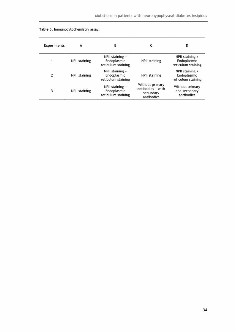
Mutations in patients with neurohypophyseal diabetes insipidus
34
Table 5. Immunocytochemistry assay.
Experiments A B C D
1 NPII staining NPII staining + Endoplasmic
reticulum staining NPII staining
NPII staining + Endoplasmic
reticulum staining
2 NPII staining NPII staining + Endoplasmic
reticulum staining NPII staining
NPII staining + Endoplasmic
reticulum staining
3 NPII staining NPII staining + Endoplasmic
reticulum staining
Without primary antibodies + with
secundary antibodies
Without primary and secondary
antibodies

Mutations in patients with neurohypophyseal diabetes insipidus
35
3. Results
3.1. Reported mutations in the AVP gene
After a literature search, a table was drawn with all the mutations so far described in the AVP
gene (Table 6). The first mutation was described in 1991 and since then, numerous families
with a FNDI diagnosis have been studied and found to have mutations in the AVP gene.
So far, there are 70 different mutations described in the scientific literature and 104
different families reported with one of these mutations (families and mutations reported in
this study were not taken into account). In these 70 mutations, 60 correspond to the
substitution of a single nucleotide, 3 correspond to the substitution of 2 nucleotides, 2
correspond to the deletion of a single nucleotide, 3 correspond to the deletion of 3
nucleotides and 1 mutation corresponds to an almost complete deletion of the AVP gene (with
the exception of exon 1). For one mutation it was not possible to determine the exact
nucleotide change due to insufficient data. According to these variants, 54 mutations cause
missense changes, 2 mutations cause deletion of a single amino acid residue, 3 mutations
cause deletion of 4 amino acids residues, 7 mutations lead to the insertion of a premature
stop codon (nonsense), 1 mutation causes a frameshift, 1 mutation causes an indel
(insertion/deletion) and two mutations have an unknown effect at protein (p.?). Several
families shared the same mutations; the most relevant was the c.55G>A mutation,
corresponding to 8% of studied families. Regarding the type of alteration at the protein level,
Fig. 10 presents their distribution between all reported families (families reported in this
study were not taken into account).
Fig. 10. Percentage of each type of protein change caused by the mutations in 104 reported families.
The mutations are distributed throughout the gene (Fig. 11)
77%
8%
3% 8%
1% 1% 2% Missense
Deletion of a single amino acid
Deletion of 4 amino acids
Nonsense
Frameshift
Indel
p.?
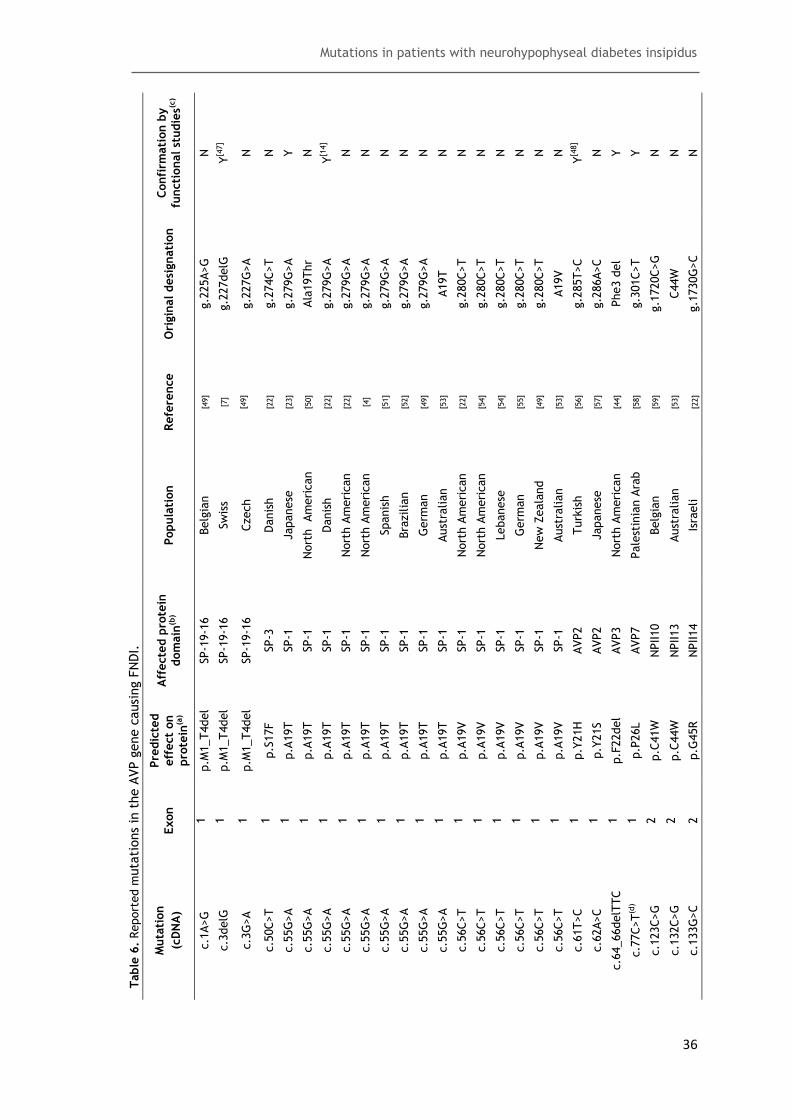
Mutations in patients with neurohypophyseal diabetes insipidus
36
Table
6.
Report
ed m
uta
tions
in t
he A
VP g
ene c
ausi
ng F
ND
I.
Confi
rmati
on b
y
functi
onal st
udie
s(c)
N
Y[4
7]
N
N
Y
N
Y[1
4]
N
N
N
N
N
N
N
N
N
N
N
N
Y[4
8]
N
Y
Y
N
N
N
Ori
gin
al desi
gnati
on
g.2
25A>G
g.2
27delG
g.2
27G
>A
g.2
74C>T
g.2
79G
>A
Ala
19Thr
g.2
79G
>A
g.2
79G
>A
g.2
79G
>A
g.2
79G
>A
g.2
79G
>A
g.2
79G
>A
A19T
g.2
80C>T
g.2
80C>T
g.2
80C>T
g.2
80C>T
g.2
80C>T
A19V
g.2
85T>C
g.2
86A>C
Phe3 d
el
g.3
01C>T
g.1
720C>G
C44W
g.1
730G
>C
Refe
rence
[49]
[7]
[49]
[22]
[23]
[50]
[22]
[22]
[4]
[51]
[52]
[49]
[53]
[22]
[54]
[54]
[55]
[49]
[53]
[56]
[57]
[44]
[58]
[59]
[53]
[22]
Popula
tion
Belg
ian
Sw
iss
Czech
Danis
h
Japanese
Nort
h
Am
eri
can
Danis
h
Nort
h A
meri
can
Nort
h A
meri
can
Spanis
h
Bra
zilia
n
Germ
an
Aust
ralian
Nort
h A
meri
can
Nort
h A
meri
can
Lebanese
Germ
an
New
Zeala
nd
Aust
ralian
Turk
ish
Japanese
Nort
h A
meri
can
Pale
stin
ian A
rab
Belg
ian
Aust
ralian
Isra
eli
Aff
ecte
d p
rote
in
dom
ain
(b)
SP-1
9-1
6
SP-1
9-1
6
SP-1
9-1
6
SP-3
SP-1
SP-1
SP-1
SP-1
SP-1
SP-1
SP-1
SP-1
SP-1
SP-1
SP-1
SP-1
SP-1
SP-1
SP-1
AVP2
AVP2
AVP3
AVP7
NPII10
NPII13
NPII14
Pre
dic
ted
eff
ect
on
pro
tein
(a)
p.M
1_T4del
p.M
1_T4del
p.M
1_T4del
p.S
17F
p.A
19T
p.A
19T
p.A
19T
p.A
19T
p.A
19T
p.A
19T
p.A
19T
p.A
19T
p.A
19T
p.A
19V
p.A
19V
p.A
19V
p.A
19V
p.A
19V
p.A
19V
p.Y
21H
p.Y
21S
p.F
22del
p.P
26L
p.C
41W
p.C
44W
p.G
45R
Exon
1
1
1
1
1
1
1
1
1
1
1
1
1
1
1
1
1
1
1
1
1
1
1
2
2
2
Muta
tion
(cD
NA
)
c.1
A>G
c.3
delG
c.3
G>A
c.5
0C>T
c.5
5G
>A
c.5
5G
>A
c.5
5G
>A
c.5
5G
>A
c.5
5G
>A
c.5
5G
>A
c.5
5G
>A
c.5
5G
>A
c.5
5G
>A
c.5
6C>T
c.5
6C>T
c.5
6C>T
c.5
6C>T
c.5
6C>T
c.5
6C>T
c.6
1T>C
c.6
2A>C
c.6
4_66delT
TC
c.7
7C>T
(d)
c.1
23C>G
c.1
32C>G
c.1
33G
>C

Mutations in patients with neurohypophyseal diabetes insipidus
37
Table
6.
Report
ed m
uta
tions
in t
he A
VP g
ene c
ausi
ng F
ND
I (c
onti
nuati
on).
Confi
rmati
on b
y
functi
onal st
udie
s(c)
Y[6
0]
N
N
N
N
N
N
N
N
N
N
N
Y[6
1]
N
N
N
N
Y[6
2]
Y[1
2]
N
N
N
N
N
N
N
Ori
gin
al desi
gnati
on
NPII G
ly17Val
g.1
748C>T
c.1
54T>C
g.1
757G
>C
c.1
60G
>C
g.1
757G
>C
g.1
757G
>A
g.1
758G
>T
g.1
537G
>A
g.1
761C>T
g.1
770G
>T
g.1
772T>C
g.1
773G
>A
C59del/
A60W
g.1
785T>C
c.1
92C>A +
c.1
93T>A
Cys6
5Phe
g.1
797T>C
g.1
824 -
1829delA
GG
g.1
824 -
1829delA
GG
g.1
824 -
1829delA
GG
g.1
827 -
29delA
GG
g.1
827 -
29delA
GG
g.1
827 -
29delA
GG
c.2
32 -
234delG
AG
g.1
824-1
829del
Refe
rence
[63]
[22]
Pre
sent
study
[55]
[64]
[49]
[65]
[66]
[67]
[68]
[69]
[4]
[70]
[71]
[72]
[73]
[74]
[49]
[75]
[7]
[11]
[49]
[49]
[49]
[76]
[53]
Popula
tion
Dutc
h
Aust
rian
Port
uguese
Germ
an
Nort
h A
meri
can
Germ
an
Spanis
h
Italian
Nort
h A
meri
can
Nort
h A
meri
can
Germ
an
Nort
h A
meri
can
Cypri
ot
Sw
iss
Italian
Chin
ese
Dutc
h
Nort
h A
meri
can
Japanese
Nort
h A
meri
can
Nort
h A
meri
can
Nort
h A
meri
can
Nort
h A
meri
can
Aust
rian
Kore
an
Aust
ralian
Aff
ecte
d p
rote
in
dom
ain
(b)
NPII17
NPII20
NPII21
NPII23
NPII23
NPII23
NPII23
NPII23
NPII23
NPII24
NPII27
NPII28
NPII28
NPII28_N
PII29
NPII32
NPII34
NPII34
NPII36
NPII47
NPII47
NPII47
NPII47
NPII47
NPII47
NPII47
NPII47
Pre
dic
ted
eff
ect
on
pro
tein
(a)
p.G
48V
p.R
51C
p.C
52R
p.G
54R
p.G
54R
p.G
54R
p.G
54R
p.G
54V
p.G
54E
p.P
55L
p.C
58F
p.C
59R
p.C
59Y
p.C
59_A60delinsW
p.L
63P
p.C
65S
p.C
65F
p.V
67A
p.E
78del
p.E
78del
p.E
78del
p.E
78del
p.E
78del
p.E
78del
p.E
78del
p.E
78del
Exon
2
2
2
2
2
2
2
2
2
2
2
2
2
2
2
2
2
2
2
2
2
2
2
2
2
2
Muta
tion
(cD
NA
)
c.1
43G
>T
c.1
51C>T
c.1
54T>C
c.1
60G
>C
c.1
60G
>C
c.1
60G
>C
c.1
60G
>A
c.1
61G
>T
c.1
61G
>A
c.1
64C>T
c.1
73G
>T
c.1
75T>C
c.1
76G
>A
c.1
77_179delC
GC
c.1
88T>C
c.1
92_193CT>AA
c.1
94G
>T
c.2
00T>C
c.2
32_234delG
AG
c.2
32_234delG
AG
c.2
32_234delG
AG
c.2
32_234delG
AG
c.2
32_234delG
AG
c.2
32_234delG
AG
c.2
32_234delG
AG
c.2
32_234delG
AG

Mutations in patients with neurohypophyseal diabetes insipidus
38
Table
6.
Report
ed m
uta
tions
in t
he c
ausi
ng F
ND
I (c
onti
nuati
on).
Confi
rmati
on b
y
functi
onal st
udie
s(c)
N
N
N
N
Y[7
7]
N
N
N
Y[1
2]
N
N
N
N
N
N
N
N
Y
N
N
N
N
N
N
N
N
Ori
gin
al desi
gnati
on
g.1
829G
>A
g.1
829G
>A
g.1
830A>G
g.1
830A>G
g.1
839T>C
g.1
848C>T
g.1
857C>T
Ser8
7Tyr
g.1
859G
>A
g.1
859G
>A
g.1
859G
>A
G88V (
GG
C>G
TC)
Cys9
2Ser
g.1
872G
>C
g.1
872G
>C
g.1
873G
>A
c.2
75G
>A
g.1
873C>G
g.1
873C>A
g.1
873C>A
g.1
874G
>T
g.1
883G
>T
g.1
883G
>T
g.1
884G
>T
g.1
884G
>T
g.1
884G
>T
Refe
rence
[78]
[59]
[22]
[49]
[22]
[79]
[80]
[81]
[82]
[22]
[22]
[83]
[84]
[22]
[85]
[80]
[64]
[59]
[22]
[49]
[86]
[22]
[49]
[87]
[88]
[89]
Popula
tion
Japanese
Moro
ccan
English
Norw
egia
n
Danis
h
Sw
edis
h-N
orw
egia
n
Nort
h A
meri
can
Dutc
h
Japanese
Nort
h A
meri
can
Nort
h A
meri
can
Bra
zilia
n
Fre
nch
Nort
h A
meri
can
Germ
an
Nort
h A
meri
can
Nort
h A
meri
can
Belg
ian
Norw
egia
n
Czech
Japanese
Nort
h A
meri
can
Aust
rian
Japanese
Japanese
Germ
an
Aff
ecte
d p
rote
in
dom
ain
(b)
NPII47
NPII47
NPII47
NPII47
NPII50
NPII53
NPII56
NPII56
NPII57
NPII57
NPII57
NPII57
NPII61
NPII61
NPII61
NPII61
NPII61
NPII61
NPII61
NPII61
NPII62
NPII65
NPII65
NPII65
NPII65
NPII65
Pre
dic
ted
eff
ect
on
pro
tein
(a)
p.E
78K
p.E
78K
p.E
78G
p.E
78G
p.L
81P
p.P
84L
p.S
87F
p.S
87Y
p.G
88S
p.G
88S
p.G
88R
p.G
88V
p.C
92S
p.C
92S
p.C
92S
p.C
92Y
p.C
92Y
p.C
92W
p.C
92X
p.C
92X
p.G
93W
p.G
96C
p.G
96C
p.G
96V
p.G
96V
p.G
96V
Exon
2
2
2
2
2
2
2
2
2
2
2
2
2
2
2
2
2
2
2
2
2
2
2
2
2
2
Muta
tion
(cD
NA
)
c.2
32G
>A
c.2
32G
>A
c.2
33A>G
c.2
33A>G
c.2
42T>C
c.2
51C>T
c.2
60C>T
c.2
60C>A
c.2
62G
>A
c.2
62G
>A
c.2
62G
>C
c.2
63G
>T
c.2
74T>A
c.2
75G
>C
c.2
75G
>C
c.2
75G
>A
c.2
75G
>A
c.2
76C>G
c.2
76C>A
c.2
76C>A
c.2
77G
>T
c.2
86G
>T
c.2
86G
>T
c.2
87G
>T
c.2
87G
>T
c.2
87G
>T

Mutations in patients with neurohypophyseal diabetes insipidus
39
Table
6.
Report
ed m
uta
tions
in t
he A
VP g
ene c
ausi
ng F
ND
I (c
onti
nuati
on).
Confi
rmati
on b
y
functi
onal st
udie
s(c)
N
Y
N
N
N
N
Y
N
N
Y[1
2]
N
N
N
N
N
N
N
N
Y
Y[7
7]
N
N
N
N
Y[9
0]
N
Ori
gin
al desi
gnati
on
g.1
884G
>A
g.1
884G
>A
c.2
89C>T
c.2
89C>G
g.1
887G
>C
g.1
665T>G
g.1
889T>G
g.1
665T>A
Cys9
8Ser
g.1
891C>A
g.1
892G
>C
g.1
907T>G
g.1
684G
>T
Cys1
04Tyr
C105S
c.3
13T>C
g.1
911G
>A
c.3
22G
>T
IVS2+1delG
g.2
094C>A
g.2
101G
>T
g.2
106-0
7 C
G>G
T
g.2
107G
>T
c.3
43G
>T
Cys1
16G
ly
Cys1
16Arg
Refe
rence
[49]
[13]
[91]
Pre
sent
study
[92]
[93]
[13]
[94]
[95]
[86]
[96]
[49]
[97]
[98]
[53]
[64]
[99]
[100]
[24]
[22]
[51]
[22]
[85]
Pre
sent
study
[101]
[101]
Popula
tion
Sw
edis
h
Polish
Nort
h A
meri
can
Port
uguese
Germ
an
Nort
h A
meri
can
Polish
Italian
Wels
h
Japanese
Bra
zilia
n
Czech
Nort
h A
meri
can
Dutc
h
Aust
ralian
Nort
h A
meri
can
Japanese
Dutc
h
Kore
an
Danis
h
Spanis
h
Fre
nch
Germ
an
Port
uguese
Dutc
h
Dutc
h
Aff
ecte
d p
rote
in
dom
ain
(b)
NPII65
NPII65
NPII66
NPII66
NPII66
NPII67
NPII67
NPII67
NPII67
NPII67
NPII68
NPII73
NPII73
NPII73
NPII74
NPII74
NPII74
NPII74
NPII77
NPII79
NPII82
NPII84
NPII84
NPII84
NPII85
NPII85
Pre
dic
ted
eff
ect
on
pro
tein
(a)
p.G
96D
p.G
96D
p.R
97C
p.R
97G
p.R
97P
p.C
98G
p.C
98G
p.C
98S
p.C
98S
p.C
98X
p.A
99P
p.C
104G
p.C
104F
p.C
104Y
p.C
105S
p.C
105R
p.C
105Y
p.E
108X/p.?
p.E
108fs
X167
p.C
110X
p.E
113X
p.E
115X
p.E
115X
p.E
115X
p.C
116G
p.C
116R
Exon
2
2
2
2
2
2
2
2
2
2
2
2
2
2
2
2
2
2
Intr
on 2
3
3
3
3
3
3
3
Muta
tion
(cD
NA
)
c.2
87G
>A
c.2
87G
>A
c.2
89C>T
c.2
89C>G
c.2
90G
>C
c.2
92T>G
c.2
92T>G
c.2
92T>A
c.2
93_294G
C>CT
c.2
94C>A
c.2
95G
>C
c.3
10T>G
c.3
11G
>T
c.3
11G
>A
n/a
c.3
13T>C
c.3
14G
>A
c.3
22G
>T
c.3
22+1delG
c.3
30C>A
c.3
37G
>T
c.3
42_343CG
>G
T
c.3
43G
>T
c.3
43G
>T
c.3
46T>G
c.3
46T>C
Mutations in patients with neurohypophyseal diabetes insipidus
40
Table
6.
Report
ed m
uta
tions
in t
he A
VP g
ene c
ausi
ng F
ND
I (c
onti
nuati
on).
Confi
rmati
on b
y
functi
onal st
udie
s(c)
N
N
N
Ori
gin
al desi
gnati
on
g.2
112C>G
g.2
116G
>T
Dele
tion 1
0.3
96 b
p
Refe
rence
[49]
[22]
[102]
Popula
tion
Italian
Nort
h A
meri
can
Bri
tish
Aff
ecte
d p
rote
in
dom
ain
(b)
NPII85
NPII87
Pre
dic
ted
eff
ect
on
pro
tein
(a)
p.C
116W
p.E
118X
p.?
Exon
3
3
Muta
tion
(cD
NA
)
c.3
48C>G
c.3
52G
>T
Gro
ss g
ene
dele
tion
(d)
(a)
Num
bers
repre
sent
the p
osi
tion in t
he A
VP p
recurs
or.
Lett
ers
corr
esp
ond t
o u
niv
ers
al abbre
via
tions
for
am
ino a
cid
s.
(b)
Num
bers
refe
r to
the p
osi
tion in indiv
idual pepti
des
ori
gin
ati
ng f
rom
the p
repro
-vaso
pre
ssin
. SP,
signal pepti
de;
AVP,
arg
inin
e v
aso
pre
ssin
; N
PII,
neuro
physi
n II.
(c)
Y,
yes;
N,
no.
Cit
ed r
efe
rences
refe
rences
repre
sent
late
r st
udie
s w
here
the f
uncti
onal st
udie
s w
ere
perf
orm
ed.
(d)
Muta
tion a
ssocia
ted w
ith a
uto
som
al re
cess
ive inheri
tance o
f FN
DI.
n/a,
exact
nucle
oti
de c
hange n
ot
available
in t
he o
rigin
al re
port
.

Mutations in patients with neurohypophyseal diabetes insipidus
41
Fig. 11. Unique mutations described in the human AVP gene. Black arrows represent described
mutations in scientific literature. Pink arrows represent the novel mutations studied in this thesis and
the orange arrow represents the third analyzed mutation, which as previously been described.
3.2. Identification of kindreds with mutations
Nine patients were diagnosed with neurohypophyseal DI. A blood sample of each patient
arrived to the Health Sciences Research Centre and the DNA of each individual was extracted
from leucocytes, in order to analyze the AVP gene and detect possible mutations.
In all these patients, only the three familial cases presented mutations in the AVP gene.
Although several individuals, in each family, were reported to have disease symptoms, only
patients indicated by arrows (Fig. 12) were available for genetic studies, with the exception
of family B (Fig. 12) in which both the index cass and his mother were studed and found to
have an AVP mutation. None of the 6 sporadic cases were found to have mutations in the AVP
gene.
3.3. Identification of mutations in the AVP gene
All three exons of the patients were amplified by PCR. Exon 1 amplification resulted in a 240
bp fragment, exon 2 amplification resulted in a 268 bp fragment and exon 3 amplification
resulted in a 322 bp fragment (Fig 13).

Mutations in patients with neurohypophyseal diabetes insipidus
42
Fig. 12. Pedigrees of three families with adFNDI. Index cases are marked by an arrow. Closed symbols
represent individuals with historical or laboratory evidence of DI and open symbols represent healthy
individuals.
The subsequent sequencing revealed three different mutations of which two of them are
novel mutations. Patient III-1 from family A showed a heterozygous T to C transition at
position 154 of AVP cDNA (c.154T>C) (Fig. 14 A). This alteration occurs in the second exon and
results in an amino acid substitution of cysteine to arginine at position 52 in the protein
(p.C52R), affecting NPII in amino acid position 21. Patient II-1 from family B showed a
heterozygous C to G transition at position 289 of AVP cDNA (c.289C>G) (Fig. 14 B). The
mutation also occurs in the second exon and results in an amino acid substitution of Arg to
glycine (Gly/G) at position 97 in protein (p.R97G) which corresponds to amino acid 66 of NPII.
The AVP gene of the patient’s mother (I-2, family B) was found to have the same mutation.
Patient II-4 from family C showed a heterozygous G to T transition at position 343 of AVP
cDNA (c.343G>T) (Fig. 14 C). The transition occurs in the third exon and results in a nonsense
mutation as the amino acid glutamic acid is substituted by a termination codon at position
115 in the protein (p.E115X). This mutation results in a truncated protein which ends at
amino acid 84 in NPII.

Mutations in patients with neurohypophyseal diabetes insipidus
43
Fig. 13. Electrophoresis of PCR products for each AVP gene exon. A, B and C correspond to first, second
and third exon, respectively. Lane 1 corresponds to DNA size standard, lane 2 corresponds to the
negative control of PCR reaction (without DNA), lane 3 corresponds to the positive control of PCR
reaction, which contains DNA of a healthy individual and the remaining lanes correspond to nine
analyzed patients.
The mutations found in the patients from family A and B are novel mutations, whereas the
mutation found in patient from family C has been already described [85].

Mutations in patients with neurohypophyseal diabetes insipidus
44
Fig. 14. Electropherograms from fragments of the AVP gene of one healthy individual and three
clinically affected subjects with novel identified mutations. A, missense mutation founded in patient III-
1 (family A) compared to a healthy individual in exon 2. B, missense mutation found in patients II-1
(family B) compared to a healthy individual in exon 2. C, nonsense mutation found in patient II-4 (family
C) compared to a healthy individual in exon 3.The heterozygous mutations are indicated by black
arrows.
3.4. Construction of the pVAX/AVP vector
pRc/RSV-AVP was sequenced to confirm the AVP cDNA sequence. There were no divergences
compared with the Ensembl (Gene ID: ENSG00000101200) sequence. Therefore, the pRc/RSV-
AVP was appropriate to be used as the wild-type form of AVP gene for further functional
studies.
The transfer of AVP cDNA from pRc/RSV-AVP to pVAX1/lacz was not an easy process and
required optimization. The transformation protocol of E. coli cells with pVAX/lacZ was only
achieved with a stabilization step of 2 h with agitation, after transformation. The best yields
of the three ligation ratios (of XbaI/HindIII AVP cDNA and XbaI/HindIII pVAX/lacZ) were
obtained with reactions A and B, refered in section 2.8.
PCR reaction is a convenient method to confirm the presence of an insert in bacteria’s pDNA.
However, in this study, we observed the existence of several false positives when the
pVAX/AVP was analyzed by PCR reaction, from LB-agar/ Kanamycin clones. When pVAX1/lacZ,
with 6100 bp, was digested with XbaI and HindIII, two fragments were obtained: a larger
fragment of 3125 bp, which represents the polylinker zone, and a smaller one with 2975 bp
where the AVP cDNA will be inserted, leading to a 3594 bp plasmid. Thus, both plasmids

Mutations in patients with neurohypophyseal diabetes insipidus
45
would be present in transformed colonies and in order to select the recombinants of interest,
a screening of pDNA from some colonies was performed with XbaI, revealing some pVAX/AVP
with 3594 bp length (Fig. 15, lanes 5,6,7,8,12,14,16 and 19).
Fig. 15. Analysis of purified pDNA after enzymatic digestion with XbaI. Lane 1 corresponds to 1 kb DNA
ladder and the remaining lanes correspond to several purified and digested pDNA. The bands with
greater molecular size correspond to digested pVAX/lacZ and the bands with lower molecular size
correspond to digested pVAX/AVP. The enzymatic digestion represented an efficient method to analyze
the presence of different pDNA.
Some of the purified pDNAs were also analyzed by XbaI and HindIII digestion, to confirm the
cDNA presence, and the correct fragments were obtained: XbaI/HindIII pVAX/AVP with 2975
bp and XbaI/HindIII AVP cDNA with 619 bp (Fig. 16).
Fig. 16. Confirmation of AVP cDNA in the recombinant pVAX/AVP. Lane 1 corresponds to 1 kb DNA
ladder, lanes 2 and 3 correspond to two different purified pDNAs, XbaI and HindIII digested, and lane
number 4 corresponds to 100 bp DNA ladder.
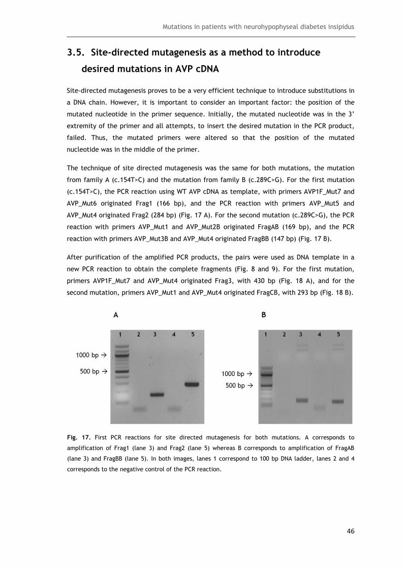
Mutations in patients with neurohypophyseal diabetes insipidus
46
3.5. Site-directed mutagenesis as a method to introduce
desired mutations in AVP cDNA
Site-directed mutagenesis proves to be a very efficient technique to introduce substitutions in
a DNA chain. However, it is important to consider an important factor: the position of the
mutated nucleotide in the primer sequence. Initially, the mutated nucleotide was in the 3’
extremity of the primer and all attempts, to insert the desired mutation in the PCR product,
failed. Thus, the mutated primers were altered so that the position of the mutated
nucleotide was in the middle of the primer.
The technique of site directed mutagenesis was the same for both mutations, the mutation
from family A (c.154T>C) and the mutation from family B (c.289C>G). For the first mutation
(c.154T>C), the PCR reaction using WT AVP cDNA as template, with primers AVP1F_Mut7 and
AVP_Mut6 originated Frag1 (166 bp), and the PCR reaction with primers AVP_Mut5 and
AVP_Mut4 originated Frag2 (284 bp) (Fig. 17 A). For the second mutation (c.289C>G), the PCR
reaction with primers AVP_Mut1 and AVP_Mut2B originated FragAB (169 bp), and the PCR
reaction with primers AVP_Mut3B and AVP_Mut4 originated FragBB (147 bp) (Fig. 17 B).
After purification of the amplified PCR products, the pairs were used as DNA template in a
new PCR reaction to obtain the complete fragments (Fig. 8 and 9). For the first mutation,
primers AVP1F_Mut7 and AVP_Mut4 originated Frag3, with 430 bp (Fig. 18 A), and for the
second mutation, primers AVP_Mut1 and AVP_Mut4 originated FragCB, with 293 bp (Fig. 18 B).
Fig. 17. First PCR reactions for site directed mutagenesis for both mutations. A corresponds to
amplification of Frag1 (lane 3) and Frag2 (lane 5) whereas B corresponds to amplification of FragAB
(lane 3) and FragBB (lane 5). In both images, lanes 1 correspond to 100 bp DNA ladder, lanes 2 and 4
corresponds to the negative control of the PCR reaction.
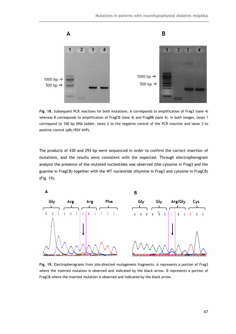
Mutations in patients with neurohypophyseal diabetes insipidus
47
Fig. 18. Subsequent PCR reactions for both mutations. A corresponds to amplification of Frag3 (lane 4)
whereas B corresponds to amplification of FragCB (lane 4) and FragBB (lane 4). In both images, lanes 1
correspond to 100 bp DNA ladder, lanes 2 to the negative control of the PCR reaction and lanes 3 to
positive control (pRc/RSV-AVP).
The products of 430 and 293 bp were sequenced in order to confirm the correct insertion of
mutations, and the results were consistent with the expected. Through electropherogram
analysis the presence of the mutated nucleotides was observed (the cytosine in Frag3 and the
guanine in FragCB) together with the WT nucleotide (thymine in Frag3 and cytosine in FragCB)
(Fig. 19).
Fig. 19. Electropherograms from site-directed mutagenesis fragments. A represents a portion of Frag3
where the inserted mutation is observed and indicated by the black arrow. B represents a portion of
FragCB where the inserted mutation is observed and indicated by the black arrow.
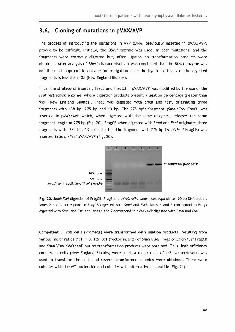
Mutations in patients with neurohypophyseal diabetes insipidus
48
3.6. Cloning of mutations in pVAX/AVP
The process of introducing the mutations in AVP cDNA, previously inserted in pVAX/AVP,
proved to be difficult. Initially, the BbvcI enzyme was used, in both mutations, and the
fragments were correctly digested but, after ligation no transformation products were
obtained. After analysis of BbvcI characteristics it was concluded that the BbvcI enzyme was
not the most appropriate enzyme for re-ligation since the ligation efficacy of the digested
fragments is less than 10% (New England Biolabs).
Thus, the strategy of inserting Frag3 and FragCB in pVAX/AVP was modified by the use of the
FseI restriction enzyme, whose digestion products present a ligation percentage greater than
95% (New England Biolabs). Frag3 was digested with SmaI and FseI, originating three
fragments with 138 bp, 275 bp and 13 bp. The 275 bp’s fragment (SmaI/FseI Frag3) was
inserted in pVAX/AVP which, when digested with the same enzymes, releases the same
fragment length of 275 bp (Fig. 20). FragCB when digested with SmaI and FseI originates three
fragments with, 275 bp, 13 bp and 5 bp. The fragment with 275 bp (SmaI/FseI FragCB) was
inserted in SmaI/FseI pVAX/AVP (Fig. 20).
Fig. 20. SmaI/FseI digestion of FragCB, Frag3 and pVAX/AVP. Lane 1 corresponds to 100 bp DNA ladder,
lanes 2 and 3 correspond to FragCB digested with SmaI and FseI, lanes 4 and 5 correspond to Frag3
digested with SmaI and FseI and lanes 6 and 7 correspond to pVAX/AVP digested with SmaI and FseI.
Competent E. coli cells (Promega) were transformed with ligation products, resulting from
various molar ratios (1:1, 1:3, 1:5, 3:1 (vector:insert)) of SmaI/FseI Frag3 or SmaI/FseI FragCB
and SmaI/FseI pVAX/AVP but no transformation products were obtained. Thus, high efficiency
competent cells (New England Biolabs) were used. A molar ratio of 1:3 (vector:insert) was
used to transform the cells and several transformed colonies were obtained. There were
colonies with the WT nucleotide and colonies with alternative nucleotide (Fig. 21).

Mutations in patients with neurohypophyseal diabetes insipidus
49
Fig. 21. Electropherograms from the sequences of the mutated pVAX/AVP vector. A represents
pVAX/AVP with mutation c.154T>C and the mutated nucleotide is indicated by the black arrow. B
represents pVAX/AVP with mutation c.289C>G and the mutated nucleotide is indicated by the black
arrow.
3.7. Analysis of AVP WT gene expression in N2A cells
Immunocytochemistry was the chosen method to analyze the expression of the WT AVP gene
in the transfected N2A cells. The transfection protocol was optimized. Initially, after
transfection, the cells remained only 6 hours in medium with DNA and lipofectamine and the
incubation step with DMEM with 5% of antibiotics and without FBS lasted 48 h. However, after
the immunocytochemistry assay, several aggregates of poorly differentiated cells were
observed and no staining for NPII was obtained. Thus, we increased the time in medium with
DNA and lipofectamine to 24 h, and the incubation time with DMEM with 5% of antibiotics and
without FBS to 72 h. The resulting cells were much more differentiated and some NPII staining
was observed, mostly in transfection assays containing 5 µg of DNA and 8 µL of lipofectamine
(Fig. 22).
The immunocytochemistry protocol was also optimized. After the transfection protocol, cells
were fixed with paraformaldehyde and permeabilized with a solution containing 1x PBS and
1% Triton and were blocked with blocking buffer containing 1x PBS, 0.1% Tween and 20% FBS.
The results obtained revealed a high green fluorescence, even without staining (Fig. 22). This
fluorescence indicated that N2A cells presented autofluorescence when the
immunocytochemistry was performed according to the previously protocol. Thus, the
immunocytochemistry protocol was altered to that described in section 2.13 but some
autofluorescence remained.

Mutations in patients with neurohypophyseal diabetes insipidus
50
Fig. 22. Cellular localization of NPII protein in transiently transfected N2A cells as visualized by confocal
laser microscopy. NPII was detected by incubation with a goat anti-NPII and an Alexa Fluor 350 donkey
anti-goat (blue staining). NPII is localized in cellular axons and it is indicated by the white arrows. The
green staining represents cells’ autofluorescence.
After transfection and immunocytochemistry optimization, the WT AVP gene was expressed in
N2A cells, as the NPII protein was observed in cells’ axons using confocal laser scanning
microscopy. However, a double staining of NPII and endoplasmic reticulum was not possible
with the use of antibodies referred in table 4. This was due to a possible cross-reactivity
between antibodies since there are two goat antibodies (anti-NPII and anti-mouse) and one
anti-goat antibody which can react with both goat antibodies and, thus, eliminate the
staining for NPII. To confirm this hypothesis, several double stainings were performed, for
NPII and endoplasmic reticulum, and no staining for NPII was observed.
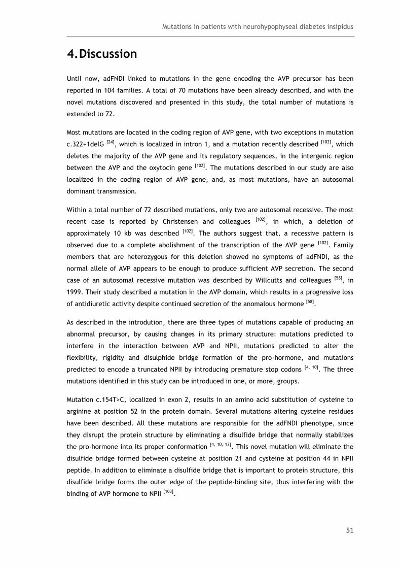
Mutations in patients with neurohypophyseal diabetes insipidus
51
4. Discussion
Until now, adFNDI linked to mutations in the gene encoding the AVP precursor has been
reported in 104 families. A total of 70 mutations have been already described, and with the
novel mutations discovered and presented in this study, the total number of mutations is
extended to 72.
Most mutations are located in the coding region of AVP gene, with two exceptions in mutation
c.322+1delG [24], which is localized in intron 1, and a mutation recently described [102], which
deletes the majority of the AVP gene and its regulatory sequences, in the intergenic region
between the AVP and the oxytocin gene [102]. The mutations described in our study are also
localized in the coding region of AVP gene, and, as most mutations, have an autosomal
dominant transmission.
Within a total number of 72 described mutations, only two are autosomal recessive. The most
recent case is reported by Christensen and colleagues [102], in which, a deletion of
approximately 10 kb was described [102]. The authors suggest that, a recessive pattern is
observed due to a complete abolishment of the transcription of the AVP gene [102]. Family
members that are heterozygous for this deletion showed no symptoms of adFNDI, as the
normal allele of AVP appears to be enough to produce sufficient AVP secretion. The second
case of an autosomal recessive mutation was described by Willcutts and colleagues [58], in
1999. Their study described a mutation in the AVP domain, which results in a progressive loss
of antidiuretic activity despite continued secretion of the anomalous hormone [58].
As described in the introdution, there are three types of mutations capable of producing an
abnormal precursor, by causing changes in its primary structure: mutations predicted to
interfere in the interaction between AVP and NPII, mutations predicted to alter the
flexibility, rigidity and disulphide bridge formation of the pro-hormone, and mutations
predicted to encode a truncated NPII by introducing premature stop codons [4, 10]. The three
mutations identified in this study can be introduced in one, or more, groups.
Mutation c.154T>C, localized in exon 2, results in an amino acid substitution of cysteine to
arginine at position 52 in the protein domain. Several mutations altering cysteine residues
have been described. All these mutations are responsible for the adFNDI phenotype, since
they disrupt the protein structure by eliminating a disulfide bridge that normally stabilizes
the pro-hormone into its proper conformation [4, 10, 13]. This novel mutation will eliminate the
disulfide bridge formed between cysteine at position 21 and cysteine at position 44 in NPII
peptide. In addition to eliminate a disulfide bridge that is important to protein structure, this
disulfide bridge forms the outer edge of the peptide-binding site, thus interfering with the
binding of AVP hormone to NPII [103].
Mutations in patients with neurohypophyseal diabetes insipidus
52
Mutation c.289C>G, present also in exon 2, results in an amino acid substitution of arginine to
glycine at position 97 in protein domain. This mutation is similar to other described mutations
that also introduce glycine residues, leading to an increase in protein flexibity and, thus
impairing its correct folding [4, 13].
Mutation c.343G>T, located in exon 3, results in a substitution of glutamic acid to a stop
codon, resulting in a truncated protein without copeptin and some residues in the COOH-
terminal of NPII. The role of copeptin is uncertain, howerever, it is possible that copeptin
glycosylation plays an important role by assisting refolding of misfolded AVP pro-hormone
monomers [17], as described in the introduction. Functional studies have not yet been
performed, however, protein expression can be expected for the same reason pointed for the
recessive pattern of the deletion described by Christensen and colleagues [102]. Thus, this
mutation can cause adFNDI phenotype by eliminating important residues that help in the
folding of the precursor.
Although the novel mutations identified in this study are likely to be pathogenic, further
studies are needed to demonstrate the functional effects of these mutations. For this
purpose, these mutations are already been cloned into an expression vector (pRc/RSV) and
the cells transfection protocol as already been optimized for the WT AVP gene. This WT AVP
gene is a crucial control for immunocytochemistry assays measurement of AVP levels, western
blot and apoptosis assays. It will be used as a control in all future functional studies in order
to clarify the cellular mechanisms that are altered by the mutations.
The immunocytochemistry assay has also been completely optimized for the WT AVP cDNA. It
will allow the analysis of NPII protein localization in N2A cells, since an accumulation of NPII
protein arround the cell nucleous, and in the endoplasmic reticulum is expected to be
observed. This may occur due to the aggregation of mutated precursors in the endoplasmic
reticulum, since they cannot continue to the Golgi apparatus, in contrast with the WT AVP
protein, which is transported along the cell axons [14].

Mutations in patients with neurohypophyseal diabetes insipidus
53
5. Conclusion
adFNDI is a rare disease, however, with time, more studies about this disease are being
reported, more mutations are being found and more cases are being studied. But, the
understanding of the molecular basis underlying the disease faces a lack of answers around
some questions. Why is the number of magnocellular neurons decreased in autopsies of
adFNDI patients? Why do so many mutations originate so similar clinical phenotypes?
Further work will be necessary to explain the points that remain without clarification and
functional studies are a useful tool in order to create the most similar environment to
neuronal cells, in this case, magnocellular neurons. Thus, it may be possible to identify the
mechanisms responsible for the progression of the disease and, once the cause of cellular
death is identified, a therapeutic approach can be developed in order to avoid this
progressive neuronal death.
Although functional studies are relevant, genetic studies also deserve a greater attention
since they can be used as a preventing tool. When a family presents an adFNDI history and a
genetic alteration is confirmed, the AVP gene of the younger members, despite the absence
of symptoms, can be analyzed for mutations. Thus, a child without symptoms can be a
potential patient, and face a dangerous situation, which can be avoided.
The understanding of the disease molecular mechanisms can be used not only in adFNDI, but
in other diseases with similar mechanisms like the case of neurodegenerative diseases and
other diseases that develop due to protein aggregates. Thus, it is important to continue the
research work, not only to enrich scientific knowledge, but also with the intention of
providing help and information to patients.
Mutations in patients with neurohypophyseal diabetes insipidus
54
6. Bibliography
1. Baylis, P. H. and Thompson, C. J., "Diabetes insipidus and hyperosmolar syndromes",
in Principles and practice of endocrinology and metabolism, Becker, K. L., (Ed). 2001,
Lippincott Williams & Wilkins: Philadelphia, USA. p. 285-292.
2. Babey, M.; Kopp, P.; and Robertson, G. L., "Familial forms of diabetes insipidus:
clinical and molecular characteristics". Nature reviews. Endocrinology, 2011. 7(12):
p. 701-714.
3. Fujiwara, T. M. and Bichet, D. G., "Molecular biology of hereditary diabetes
insipidus". Journal of the American Society of Nephrology : JASN, 2005. 16(10): p.
2836-2846.
4. Hansen, L. K.; Rittig, S.; and Robertson, G. L., "Genetic basis of familial
neurohypophyseal diabetes insipidus". Trends in endocrinology and metabolism: TEM,
1997. 8(9): p. 363-372.
5. De Wardener, H. E. and Herxheimer, A., "The effect of a high water intake on the
kidney's ability to concentrate the urine in man". The Journal of physiology, 1957.
139(1): p. 42-52.
6. Colombo, N.; Berry, I.; Kucharczyk, J.; Kucharczyk, W.; de Groot, J.; Larson, T.;
Norman, D.; and Newton, T. H., "Posterior pituitary gland: appearance on MR images
in normal and pathologic states". Radiology, 1987. 165(2): p. 481-485.
7. Rutishauser, J.; Boni-Schnetzler, M.; Boni, J.; Wichmann, W.; Huisman, T.; Vallotton,
M. B.; and Froesch, E. R., "A novel point mutation in the translation initiation codon
of the pre-pro-vasopressin-neurophysin II gene: cosegregation with morphological
abnormalities and clinical symptoms in autosomal dominant neurohypophyseal
diabetes insipidus". The Journal of clinical endocrinology and metabolism, 1996.
81(1): p. 192-198.
8. Makara, G. B., "The relative importance of hypothalamic neurons containing
corticotropin-releasing factor or vasopressin in the regulation of adrenocorticotropic
hormone secretion". Ciba Foundation symposium, 1992. 168: p. 43-51; discussion 51-
43.
9. Russell, T. A.; Ito, M.; Yu, R. N.; Martinson, F. A.; Weiss, J.; and Jameson, J. L., "A
murine model of autosomal dominant neurohypophyseal diabetes insipidus reveals
progressive loss of vasopressin-producing neurons". The Journal of clinical
investigation, 2003. 112(11): p. 1697-1706.
10. Christensen, J. H. and Rittig, S., "Familial neurohypophyseal diabetes insipidus--an
update". Seminars in nephrology, 2006. 26(3): p. 209-223.
11. Mahoney, C. P.; Weinberger, E.; Bryant, C.; Ito, M.; and Jameson, J. L., "Effects of
aging on vasopressin production in a kindred with autosomal dominant
neurohypophyseal diabetes insipidus due to the DeltaE47 neurophysin mutation". The
Journal of clinical endocrinology and metabolism, 2002. 87(2): p. 870-876.
Mutations in patients with neurohypophyseal diabetes insipidus
55
12. Ito, M. and Jameson, J. L., "Molecular basis of autosomal dominant neurohypophyseal
diabetes insipidus. Cellular toxicity caused by the accumulation of mutant vasopressin
precursors within the endoplasmic reticulum". The Journal of clinical investigation,
1997. 99(8): p. 1897-1905.
13. Hedrich, C. M.; Zachurzok-Buczynska, A.; Gawlik, A.; Russ, S.; Hahn, G.; Koehler, K.;
Malecka-Tendera, E.; and Huebner, A., "Autosomal dominant neurohypophyseal
diabetes insipidus in two families. Molecular analysis of the vasopressin-neurophysin II
gene and functional studies of three missense mutations". Hormone research, 2009.
71(2): p. 111-119.
14. Siggaard, C.; Rittig, S.; Corydon, T. J.; Andreasen, P. H.; Jensen, T. G.; Andresen, B.
S.; Robertson, G. L.; Gregersen, N.; Bolund, L.; and Pedersen, E. B., "Clinical and
molecular evidence of abnormal processing and trafficking of the vasopressin
preprohormone in a large kindred with familial neurohypophyseal diabetes insipidus
due to a signal peptide mutation". The Journal of clinical endocrinology and
metabolism, 1999. 84(8): p. 2933-2941.
15. Murphy, D. and Wells, S., "In vivo gene transfer studies on the regulation and function
of the vasopressin and oxytocin genes". Journal of neuroendocrinology, 2003. 15(2):
p. 109-125.
16. Fields, R. L.; House, S. B.; and Gainer, H., "Regulatory domains in the intergenic
region of the oxytocin and vasopressin genes that control their hypothalamus-specific
expression in vitro". The Journal of neuroscience : the official journal of the Society
for Neuroscience, 2003. 23(21): p. 7801-7809.
17. Barat, C.; Simpson, L.; and Breslow, E., "Properties of human vasopressin precursor
constructs: inefficient monomer folding in the absence of copeptin as a potential
contributor to diabetes insipidus". Biochemistry, 2004. 43(25): p. 8191-8203.
18. Acher, R.; Chauvet, J.; and Rouille, Y., "Dynamic processing of neuropeptides:
sequential conformation shaping of neurohypophysial preprohormones during
intraneuronal secretory transport". Journal of molecular neuroscience : MN, 2002.
18(3): p. 223-228.
19. Di Iorgi, N.; Napoli, F.; Allegri, A. E.; Olivieri, I.; Bertelli, E.; Gallizia, A.; Rossi, A.;
and Maghnie, M., "Diabetes insipidus--diagnosis and management". Hormone research
in paediatrics, 2012. 77(2): p. 69-84.
20. Ward, D. T.; Hammond, T. G.; and Harris, H. W., "Modulation of vasopressin-elicited
water transport by trafficking of aquaporin2-containing vesicles". Annual review of
physiology, 1999. 61: p. 683-697.
21. van Balkom, B. W.; Savelkoul, P. J.; Markovich, D.; Hofman, E.; Nielsen, S.; van der
Sluijs, P.; and Deen, P. M., "The role of putative phosphorylation sites in the targeting
and shuttling of the aquaporin-2 water channel". The Journal of biological chemistry,
2002. 277(44): p. 41473-41479.

Mutations in patients with neurohypophyseal diabetes insipidus
56
22. Rittig, S.; Robertson, G. L.; Siggaard, C.; Kovacs, L.; Gregersen, N.; Nyborg, J.; and
Pedersen, E. B., "Identification of 13 new mutations in the vasopressin-neurophysin II
gene in 17 kindreds with familial autosomal dominant neurohypophyseal diabetes
insipidus". American journal of human genetics, 1996. 58(1): p. 107-117.
23. Ito, M.; Oiso, Y.; Murase, T.; Kondo, K.; Saito, H.; Chinzei, T.; Racchi, M.; and Lively,
M. O., "Possible involvement of inefficient cleavage of preprovasopressin by signal
peptidase as a cause for familial central diabetes insipidus". The Journal of clinical
investigation, 1993. 91(6): p. 2565-2571.
24. Tae, H. J.; Baek, K. H.; Shim, S. M.; Yoo, S. J.; Kang, M. I.; Cha, B. Y.; Lee, K. W.;
Son, H. Y.; and Kang, S. K., "A novel splice site mutation of the arginine vasopressin-
neurophysin II gene identified in a kindred with autosomal dominant familial
neurohypophyseal diabetes insipidus". Molecular genetics and metabolism, 2005.
86(1-2): p. 307-313.
25. Si-Hoe, S. L.; De Bree, F. M.; Nijenhuis, M.; Davies, J. E.; Howell, L. M.; Tinley, H.;
Waller, S. J.; Zeng, Q.; Zalm, R.; Sonnemans, M.; Van Leeuwen, F. W.; Burbach, J.
P.; and Murphy, D., "Endoplasmic reticulum derangement in hypothalamic neurons of
rats expressing a familial neurohypophyseal diabetes insipidus mutant vasopressin
transgene". FASEB journal : official publication of the Federation of American
Societies for Experimental Biology, 2000. 14(12): p. 1680-1684.
26. Ito, M.; Yu, R. N.; and Jameson, J. L., "Mutant vasopressin precursors that cause
autosomal dominant neurohypophyseal diabetes insipidus retain dimerization and
impair the secretion of wild-type proteins". The Journal of biological chemistry,
1999. 274(13): p. 9029-9037.
27. Castino, R.; Davies, J.; Beaucourt, S.; Isidoro, C.; and Murphy, D., "Autophagy is a
prosurvival mechanism in cells expressing an autosomal dominant familial
neurohypophyseal diabetes insipidus mutant vasopressin transgene". FASEB journal :
official publication of the Federation of American Societies for Experimental Biology,
2005. 19(8): p. 1021-1023.
28. Davies, J. and Murphy, D., "Autophagy in hypothalamic neurones of rats expressing a
familial neurohypophysial diabetes insipidus transgene". Journal of
neuroendocrinology, 2002. 14(8): p. 629-637.
29. Castino, R.; Isidoro, C.; and Murphy, D., "Autophagy-dependent cell survival and cell
death in an autosomal dominant familial neurohypophyseal diabetes insipidus in vitro
model". FASEB journal : official publication of the Federation of American Societies
for Experimental Biology, 2005. 19(8): p. 1024-1026.
30. Bergeron, C.; Kovacs, K.; Ezrin, C.; and Mizzen, C., "Hereditary diabetes insipidus: an
immunohistochemical study of the hypothalamus and pituitary gland". Acta
neuropathologica, 1991. 81(3): p. 345-348.
31. Bruins, J.; Kovacs, G. L.; Abbes, A. P.; Burbach, J. P.; van den Akker, E. L.; Engel,
H.; Franken, A. A.; and de Wied, D., "Minor disturbances in central nervous system
Mutations in patients with neurohypophyseal diabetes insipidus
57
function in familial neurohypophysial diabetes insipidus". Psychoneuroendocrinology,
2006. 31(1): p. 80-91.
32. Fukuda, I.; Hizuka, N.; and Takano, K., "Oral DDAVP is a good alternative therapy for
patients with central diabetes insipidus: experience of five-year treatment".
Endocrine journal, 2003. 50(4): p. 437-443.
33. Vande Walle, J. G.; Bogaert, G. A.; Mattsson, S.; Schurmans, T.; Hoebeke, P.; Deboe,
V.; and Norgaard, J. P., "A new fast-melting oral formulation of desmopressin: a
pharmacodynamic study in children with primary nocturnal enuresis". BJU
international, 2006. 97(3): p. 603-609.
34. Yoshida, M.; Iwasaki, Y.; Asai, M.; Nigawara, T.; and Oiso, Y., "Gene therapy for
central diabetes insipidus: effective antidiuresis by muscle-targeted gene transfer".
Endocrinology, 2004. 145(1): p. 261-268.
35. Christensen, J. H.; Siggaard, C.; and Rittig, S., "Autosomal dominant familial
neurohypophyseal diabetes insipidus". APMIS. Supplementum, 2003(109): p. 92-95.
36. den Dunnen, J. T. and Antonarakis, S. E., "Nomenclature for the description of human
sequence variations". Human genetics, 2001. 109(1): p. 121-124.
37. Chacon-Cortes, D.; Haupt, L. M.; Lea, R. A.; and Griffiths, L. R., "Comparison of
genomic DNA extraction techniques from whole blood samples: a time, cost and
quality evaluation study". Molecular biology reports, 2012. 39(5): p. 5961-5966.
38. Miller, S. A.; Dykes, D. D.; and Polesky, H. F., "A Simple Salting out Procedure for
Extracting DNA from Human Nucleated Cells". Nucleic Acids Res, 1988. 16(3): p. 1215-
1215.
39. Nasiri, H.; Forouzandeh, M.; Rasaee, M. J.; and Rahbarizadeh, F., "Modified salting-
out method: high-yield, high-quality genomic DNA extraction from whole blood using
laundry detergent". Journal of clinical laboratory analysis, 2005. 19(6): p. 229-232.
40. Higuchi, R.; Krummel, B.; and Saiki, R. K., "A general method of in vitro preparation
and specific mutagenesis of DNA fragments: study of protein and DNA interactions".
Nucleic Acids Res, 1988. 16(15): p. 7351-7367.
41. Lodish, H.; Berk, A.; Zipursky, S. L.; Matsudaira, P.; Baltimore, D.; and Darnell, J. E.,
Molecular Cell Biology. 5th ed. 2000, W. H. Freeman and Company.
42. Sambrook, J. and Russell, D. W., Molecular cloning: a laboratory manual. 3rd ed.
2001, Cold Spring Harbor Laboratory Press: New York.
43. Karp, G., Cell and molecular biology: concepts and experiments. 6th ed. 2010, John
Wiley & Sons, Inc.: Unites States of America.
44. Wahlstrom, J. T.; Fowler, M. J.; Nicholson, W. E.; and Kovacs, W. J., "A novel
mutation in the preprovasopressin gene identified in a kindred with autosomal
dominant neurohypophyseal diabetes insipidus". The Journal of clinical endocrinology
and metabolism, 2004. 89(4): p. 1963-1968.
45. Javois, L. C., "Immunocytochemical methods and protocols", in Methods in molecular
biology, Walker, J., (Ed). 1999, Humana Press: New York.

Mutations in patients with neurohypophyseal diabetes insipidus
58
46. Burry, R. W., "Controls for immunocytochemistry: an update". The journal of
histochemistry and cytochemistry : official journal of the Histochemistry Society,
2011. 59(1): p. 6-12.
47. Beuret, N.; Rutishauser, J.; Bider, M. D.; and Spiess, M., "Mechanism of endoplasmic
reticulum retention of mutant vasopressin precursor caused by a signal peptide
truncation associated with diabetes insipidus". The Journal of biological chemistry,
1999. 274(27): p. 18965-18972.
48. Christensen, J. H.; Siggaard, C.; Corydon, T. J.; Robertson, G. L.; Gregersen, N.;
Bolund, L.; and Rittig, S., "Differential cellular handling of defective arginine
vasopressin (AVP) prohormones in cells expressing mutations of the AVP gene
associated with autosomal dominant and recessive familial neurohypophyseal diabetes
insipidus". The Journal of clinical endocrinology and metabolism, 2004. 89(9): p.
4521-4531.
49. Christensen, J. H.; Siggaard, C.; Corydon, T. J.; deSanctis, L.; Kovacs, L.; Robertson,
G. L.; Gregersen, N.; and Rittig, S., "Six novel mutations in the arginine vasopressin
gene in 15 kindreds with autosomal dominant familial neurohypophyseal diabetes
insipidus give further insight into the pathogenesis". European journal of human
genetics : EJHG, 2004. 12(1): p. 44-51.
50. McLeod, J. F.; Kovacs, L.; Gaskill, M. B.; Rittig, S.; Bradley, G. S.; and Robertson, G.
L., "Familial neurohypophyseal diabetes insipidus associated with a signal peptide
mutation". The Journal of clinical endocrinology and metabolism, 1993. 77(3): p.
599A-599G.
51. Calvo, B.; Bilbao, J. R.; Urrutia, I.; Eizaguirre, J.; Gaztambide, S.; and Castano, L.,
"Identification of a novel nonsense mutation and a missense substitution in the
vasopressin-neurophysin II gene in two Spanish kindreds with familial
neurohypophyseal diabetes insipidus". The Journal of clinical endocrinology and
metabolism, 1998. 83(3): p. 995-997.
52. Boson, W. L.; Sarubi, J. C.; d'Alva, C. B.; Friedman, E.; Faria, D.; De Marco, L.; and
Wajchenberg, B., "A signal peptide mutation of the arginine vasopressin gene in
monozygotic twins". Clinical endocrinology, 2003. 58(1): p. 108-110.
53. Chitturi, S.; Harris, M.; Thomsett, M. J.; Bowling, F.; McGown, I.; Cowley, D.; Leong,
G. M.; Batch, J.; and Cotterill, A. M., "Utility of AVP gene testing in familial
neurohypophyseal diabetes insipidus". Clinical endocrinology, 2008. 69(6): p. 926-
930.
54. Repaske, D. R.; Medlej, R.; Gultekin, E. K.; Krishnamani, M. R.; Halaby, G.; Findling,
J. W.; and Phillips, J. A., 3rd, "Heterogeneity in clinical manifestation of autosomal
dominant neurohypophyseal diabetes insipidus caused by a mutation encoding Ala-1--
>Val in the signal peptide of the arginine vasopressin/neurophysin II/copeptin
precursor". The Journal of clinical endocrinology and metabolism, 1997. 82(1): p. 51-
56.
Mutations in patients with neurohypophyseal diabetes insipidus
59
55. Heppner, C.; Kotzka, J.; Bullmann, C.; Krone, W.; and Muller-Wieland, D.,
"Identification of mutations of the arginine vasopressin-neurophysin II gene in two
kindreds with familial central diabetes insipidus". The Journal of clinical
endocrinology and metabolism, 1998. 83(2): p. 693-696.
56. Rittig, S.; Siggaard, C.; Ozata, M.; Yetkin, I.; Gregersen, N.; Pedersen, E. B.; and
Robertson, G. L., "Autosomal dominant neurohypophyseal diabetes insipidus due to
substitution of histidine for tyrosine(2) in the vasopressin moiety of the hormone
precursor". The Journal of clinical endocrinology and metabolism, 2002. 87(7): p.
3351-3355.
57. Kobayashi, H.; Fujisawa, I.; Ikeda, K.; Son, C.; Iwakura, T.; Yoshimoto, A.; Kasahara,
M.; Ishihara, T.; and Ogawa, Y., "A novel heterozygous missense mutation in the
vasopressin moiety is identified in a Japanese person with neurohypophyseal diabetes
insipidus". Journal of endocrinological investigation, 2006. 29(3): p. 252-256.
58. Willcutts, M. D.; Felner, E.; and White, P. C., "Autosomal recessive familial
neurohypophyseal diabetes insipidus with continued secretion of mutant weakly
active vasopressin". Human molecular genetics, 1999. 8(7): p. 1303-1307.
59. Brachet, C.; Birk, J.; Christophe, C.; Tenoutasse, S.; Velkeniers, B.; Heinrichs, C.;
and Rutishauser, J., "Growth retardation in untreated autosomal dominant familial
neurohypophyseal diabetes insipidus caused by one recurring and two novel mutations
in the vasopressin-neurophysin II gene". European journal of endocrinology /
European Federation of Endocrine Societies, 2011. 164(2): p. 179-187.
60. Olias, G.; Richter, D.; and Schmale, H., "Heterologous expression of human
vasopressin-neurophysin precursors in a pituitary cell line: defective transport of a
mutant protein from patients with familial diabetes insipidus". DNA and cell biology,
1996. 15(11): p. 929-935.
61. Birk, J.; Friberg, M. A.; Prescianotto-Baschong, C.; Spiess, M.; and Rutishauser, J.,
"Dominant pro-vasopressin mutants that cause diabetes insipidus form disulfide-linked
fibrillar aggregates in the endoplasmic reticulum". Journal of cell science, 2009.
122(Pt 21): p. 3994-4002.
62. Christensen, J. H.; Siggaard, C.; Corydon, T. J.; Robertson, G. L.; Gregersen, N.;
Bolund, L.; and Rittig, S., "Impaired trafficking of mutated AVP prohormone in cells
expressing rare disease genes causing autosomal dominant familial neurohypophyseal
diabetes insipidus". Clinical endocrinology, 2004. 60(1): p. 125-136.
63. Bahnsen, U.; Oosting, P.; Swaab, D. F.; Nahke, P.; Richter, D.; and Schmale, H., "A
missense mutation in the vasopressin-neurophysin precursor gene cosegregates with
human autosomal dominant neurohypophyseal diabetes insipidus". The EMBO journal,
1992. 11(1): p. 19-23.
64. Rutishauser, J.; Kopp, P.; Gaskill, M. B.; Kotlar, T. J.; and Robertson, G. L., "Clinical
and molecular analysis of three families with autosomal dominant neurohypophyseal
diabetes insipidus associated with a novel and recurrent mutations in the vasopressin-

Mutations in patients with neurohypophyseal diabetes insipidus
60
neurophysin II gene". European journal of endocrinology / European Federation of
Endocrine Societies, 2002. 146(5): p. 649-656.
65. Calvo, B.; Bilbao, J. R.; Rodriguez, A.; Rodriguez-Arnao, M. D.; and Castano, L.,
"Molecular analysis in familial neurohypophyseal diabetes insipidus: early diagnosis of
an asymptomatic carrier". The Journal of clinical endocrinology and metabolism,
1999. 84(9): p. 3351-3354.
66. Gagliardi, P. C.; Bernasconi, S.; and Repaske, D. R., "Autosomal dominant
neurohypophyseal diabetes insipidus associated with a missense mutation encoding
Gly23-->Val in neurophysin II". The Journal of clinical endocrinology and metabolism,
1997. 82(11): p. 3643-3646.
67. Stephen, M. D.; Fenwick, R. G.; and Brosnan, P. G., "Polyuria and polydipsia in a
young child: diagnostic considerations and identification of novel mutation causing
familial neurohypophyseal diabetes insipidus". Pituitary, 2010.
68. Repaske, D. R. and Browning, J. E., "A de novo mutation in the coding sequence for
neurophysin-II (Pro24-->Leu) is associated with onset and transmission of autosomal
dominant neurohypophyseal diabetes insipidus". The Journal of clinical endocrinology
and metabolism, 1994. 79(2): p. 421-427.
69. Wolf, M. T.; Dotsch, J.; Metzler, M.; Holder, M.; Repp, R.; and Rascher, W., "A new
missense mutation of the vasopressin-neurophysin II gene in a family with
neurohypophyseal diabetes insipidus". Hormone research, 2003. 60(3): p. 143-147.
70. Skordis, N.; Patsalis, P. C.; Hettinger, J. A.; Kontou, M.; Herakleous, E.; Krishnamani,
M. R.; and Phillips, J. A., 3rd, "A novel arginine vasopressin-neurophysin II mutation
causes autosomal dominant neurohypophyseal diabetes insipidus and morphologic
pituitary changes". Hormone research, 2000. 53(5): p. 239-245.
71. Fluck, C. E.; Deladoey, J.; Nayak, S.; Zeller, O.; Kopp, P.; and Mullis, P. E.,
"Autosomal dominant neurohypophyseal diabetes insipidus in a Swiss family, caused
by a novel mutation (C59Delta/A60W) in the neurophysin moiety of prepro-
vasopressin-neurophysin II (AVP-NP II)". European journal of endocrinology / European
Federation of Endocrine Societies, 2001. 145(4): p. 439-444.
72. Birkegaard, C.; Christensen, J. H.; Falorni, A.; Marzotti, S.; Minarelli, V.; Gregersen,
N.; and Rittig, S., "A novel variation in the AVP gene resulting in familial
neurohypophyseal diabetes insipidus in a large Italian kindred". Pituitary, 2012.
73. Luo, Y.; Wang, B.; Qiu, Y.; Zhang, C.; Jin, C.; Zhao, Y.; Zhu, Q.; and Ma, X., "Clinical
and molecular analysis of a Chinese family with autosomal dominant
neurohypophyseal diabetes insipidus associated with a novel missense mutation in the
vasopressin-neurophysin II gene". Endocrine, 2012. 42(1): p. 208-213.
74. Abbes, A. P.; Engel, H.; Bruggeman, E. J.; and Franken, A. A., "Gene symbol: AVP.
Disease: Diabetes insipidus, neurohypophyseal. Accession #Hm0558". Human genetics,
2006. 118(6): p. 783.

Mutations in patients with neurohypophyseal diabetes insipidus
61
75. Yuasa, H.; Ito, M.; Nagasaki, H.; Oiso, Y.; Miyamoto, S.; Sasaki, N.; and Saito, H.,
"Glu-47, which forms a salt bridge between neurophysin-II and arginine vasopressin, is
deleted in patients with familial central diabetes insipidus". The Journal of clinical
endocrinology and metabolism, 1993. 77(3): p. 600-604.
76. Lee, Y. W.; Lee, K. W.; Ryu, J. W.; Mok, J. O.; Ki, C. S.; Park, H. K.; Kim, Y. J.; Kim,
S. J.; Byun, D. W.; Suh, K. I.; Yoo, M. H.; Shin, H. B.; Lee, Y. K.; and Kim, C. H.,
"Mutation of Glu78 of the AVP-NPII gene impairs neurophysin as a carrier protein for
arginine vasopressin in a family with neurohypophyseal diabetes insipidus". Annals of
clinical and laboratory science, 2008. 38(1): p. 12-14.
77. Siggaard, C.; Christensen, J. H.; Corydon, T. J.; Rittig, S.; Robertson, G. L.;
Gregersen, N.; Bolund, L.; and Pedersen, E. B., "Expression of three different
mutations in the arginine vasopressin gene suggests genotype-phenotype correlation
in familial neurohypophyseal diabetes insipidus kindreds". Clinical endocrinology,
2005. 63(2): p. 207-216.
78. Miyakoshi, M.; Kamoi, K.; Murase, T.; Sugimura, Y.; and Oiso, Y., "Novel mutant
vasopressin-neurophysin II gene associated with familial neurohypophyseal diabetes
insipidus". Endocrine journal, 2004. 51(6): p. 551-556.
79. Jendle, J.; Christensen, J. H.; Kvistgaard, H.; Gregersen, N.; and Rittig, S., "Late-
onset familial neurohypophyseal diabetes insipidus due to a novel mutation in the AVP
gene". Clinical endocrinology, 2012. 77(4): p. 586-592.
80. Grant, F. D.; Ahmadi, A.; Hosley, C. M.; and Majzoub, J. A., "Two novel mutations of
the vasopressin gene associated with familial diabetes insipidus and identification of
an asymptomatic carrier infant". The Journal of clinical endocrinology and
metabolism, 1998. 83(11): p. 3958-3964.
81. Abbes, A. P.; Engel, H.; and Franken, A. A., "Gene symbol: AVP. Disease: Diabetes
insipidus, neurohypophyseal. Accession #Hm0559". Human genetics, 2006. 118(6): p.
783.
82. Ito, M.; Mori, Y.; Oiso, Y.; and Saito, H., "A single base substitution in the coding
region for neurophysin II associated with familial central diabetes insipidus". The
Journal of clinical investigation, 1991. 87(2): p. 725-728.
83. Melo, M. E.; Marui, S.; Brito, V. N.; Mancini, M. C.; Mendonca, B. B.; and
Knoepfelmacher, M., "Autosomal dominant familial neurohypophyseal diabetes
insipidus caused by a novel mutation in arginine-vasopressin gene in a Brazilian
family". Arquivos brasileiros de endocrinologia e metabologia, 2008. 52(8): p. 1272-
1276.
84. Abbes, A. P.; Engel, H.; Klomp, C.; and Bruggeman, E. J., "Gene symbol: AVP.
Disease: Diabetes insipidus, neurohypophyseal. Accession #Hm0561". Human genetics,
2006. 118(6): p. 784.
85. Bullmann, C.; Kotzka, J.; Grimm, T.; Heppner, C.; Jockenhovel, F.; Krone, W.; and
Muller-Wieland, D., "Identification of a novel mutation in the arginine vasopressin-
Mutations in patients with neurohypophyseal diabetes insipidus
62
neurophysin II gene in familial central diabetes insipidus". Experimental and clinical
endocrinology & diabetes : official journal, German Society of Endocrinology [and]
German Diabetes Association, 2002. 110(3): p. 134-137.
86. Nagasaki, H.; Ito, M.; Yuasa, H.; Saito, H.; Fukase, M.; Hamada, K.; Ishikawa, E.;
Katakami, H.; and Oiso, Y., "Two novel mutations in the coding region for
neurophysin-II associated with familial central diabetes insipidus". The Journal of
clinical endocrinology and metabolism, 1995. 80(4): p. 1352-1356.
87. Ueta, Y.; Taniguchi, S.; Yoshida, A.; Murakami, I.; Mitani, Y.; Hisatome, I.; Manabe,
I.; Sato, R.; Tsuboi, M.; Ohtahara, A.; Nanba, E.; and Shigemasa, C., "A new type of
familial central diabetes insipidus caused by a single base substitution in the
neurophysin II coding region of the vasopressin gene". The Journal of clinical
endocrinology and metabolism, 1996. 81(5): p. 1787-1790.
88. Kubota, T.; Yamamoto, T.; Ozono, K.; and Shimotsuji, T., "Hyperintensity of posterior
pituitary on MR T1WI in a boy with central diabetes insipidus caused by missense
mutation of neurophysin II gene". Endocrine journal, 2001. 48(4): p. 459-463.
89. Rauch, F.; Lenzner, C.; Nurnberg, P.; Frommel, C.; and Vetter, U., "A novel mutation
in the coding region for neurophysin-II is associated with autosomal dominant
neurohypophyseal diabetes insipidus". Clinical endocrinology, 1996. 44(1): p. 45-51.
90. Nijenhuis, M.; van den Akker, E. L.; Zalm, R.; Franken, A. A.; Abbes, A. P.; Engel, H.;
de Wied, D.; and Burbach, J. P., "Familial neurohypophysial diabetes insipidus in a
large Dutch kindred: effect of the onset of diabetes on growth in children and cell
biological defects of the mutant vasopressin prohormone". The Journal of clinical
endocrinology and metabolism, 2001. 86(7): p. 3410-3420.
91. Rutishauser, J.; Kopp, P.; Gaskill, M. B.; Kotlar, T. J.; and Robertson, G. L., "A novel
mutation (R97C) in the neurophysin moiety of prepro-vasopressin-neurophysin II
associated with autosomal-dominant neurohypophyseal diabetes insipidus". Molecular
genetics and metabolism, 1999. 67(1): p. 89-92.
92. Mundschenk, J.; Rittig, S.; Siggaard, C.; Hensen, J.; and Lehnert, H., "A new mutation
of the arginine vasopressin-neurophysin II gene in a family with autosomal dominant
neurohypophyseal diabetes insipidus". Experimental and clinical endocrinology &
diabetes : official journal, German Society of Endocrinology [and] German Diabetes
Association, 2001. 109(8): p. 406-409.
93. DiMeglio, L. A.; Gagliardi, P. C.; Browning, J. E.; Quigley, C. A.; and Repaske, D. R.,
"A missense mutation encoding cys(67) --> gly in neurophysin ii is associated with
early onset autosomal dominant neurohypophyseal diabetes insipidus". Molecular
genetics and metabolism, 2001. 72(1): p. 39-44.
94. Baglioni, S.; Corona, G.; Maggi, M.; Serio, M.; and Peri, A., "Identification of a novel
mutation in the arginine vasopressin-neurophysin II gene affecting the sixth intrachain
disulfide bridge of the neurophysin II moiety". European journal of endocrinology /
European Federation of Endocrine Societies, 2004. 151(5): p. 605-611.

Mutations in patients with neurohypophyseal diabetes insipidus
63
95. Abbes, A. P.; Davies, J. H.; Penney, M.; Engel, H.; and Gregory, J. W., "Gene symbol:
AVP. Disease: Diabetes insipidus, neurohypophyseal. Accession #Hm0560". Human
genetics, 2006. 118(6): p. 783.
96. Elias, P. C.; Elias, L. L.; Torres, N.; Moreira, A. C.; Antunes-Rodrigues, J.; and Castro,
M., "Progressive decline of vasopressin secretion in familial autosomal dominant
neurohypophyseal diabetes insipidus presenting a novel mutation in the vasopressin-
neurophysin II gene". Clinical endocrinology, 2003. 59(4): p. 511-518.
97. Santiprabhob, J.; Browning, J.; and Repaske, D., "A missense mutation encoding
Cys73Phe in neurophysin II is associated with autosomal dominant neurohypophyseal
diabetes insipidus". Molecular genetics and metabolism, 2002. 77(1-2): p. 112-118.
98. Bruggeman, E. J., "Gene symbol: AVP. Disease: Diabetes Insipidus,
neurohypophyseal". Human genetics, 2008. 123(5): p. 545.
99. Fujii, H.; Iida, S.; and Moriwaki, K., "Familial neurohypophyseal diabetes insipidus
associated with a novel mutation in the vasopressin-neurophysin II gene".
International journal of molecular medicine, 2000. 5(3): p. 229-234.
100. de Fost, M.; van Trotsenburg, A. S.; van Santen, H. M.; Endert, E.; van den Elzen, C.;
Kamsteeg, E. J.; Swaab, D. F.; and Fliers, E., "Familial neurohypophyseal diabetes
insipidus due to a novel mutation in the arginine vasopressin-neurophysin II gene".
European journal of endocrinology / European Federation of Endocrine Societies,
2011. 165(1): p. 161-165.
101. Abbes, A. P.; Bruggeman, B.; van Den Akker, E. L.; de Groot, M. R.; Franken, A. A.;
Drexhage, V. R.; and Engel, H., "Identification of two distinct mutations at the same
nucleotide position, concomitantly with a novel polymorphism in the vasopressin-
neurophysin II gene (AVP-NP II) in two dutch families with familial neurohypophyseal
diabetes insipidus". Clinical chemistry, 2000. 46(10): p. 1699-1702.
102. Christensen, J.; Kvistgaard, H.; Knudsen, J.; Shaikh, G.; Tolmie, J.; Cooke, S.;
Pedersen, S.; Corydon, T.; Gregersen, N.; and Rittig, S., "A novel deletion partly
removing the AVP gene causes autosomal recessive inheritance of early-onset
neurohypophyseal diabetes insipidus". Clinical genetics, 2011.
103. Huang, H. B. and Breslow, E., "Identification of the unstable neurophysin disulfide and
localization to the hormone-binding site. Relationship to folding-unfolding pathways".
The Journal of biological chemistry, 1992. 267(10): p. 6750-6756.

Mutations in patients with neurohypophyseal diabetes insipidus
64