Identification of a novel population of muscle stem cells in mice
14
The Rockefeller University Press, 0021-9525/2002/05/851/14 $5.00 The Journal of Cell Biology, Volume 157, Number 5, May 27, 2002 851–864 http://www.jcb.org/cgi/doi/10.1083/jcb.200108150 JCB Article 851 Identification of a novel population of muscle stem cells in mice: potential for muscle regeneration Zhuqing Qu-Petersen, 1 Bridget Deasy, 1 Ron Jankowski, 1 Makato Ikezawa, 1 James Cummins, 1 Ryan Pruchnic, 1 John Mytinger, 1 Baohong Cao, 1 Charley Gates, 1 Anton Wernig, 2 and Johnny Huard 1 1 Growth and Development Laboratory, Children’s Hospital of Pittsburgh, Department of Orthopaedic Surgery, University of Pittsburgh, Pittsburgh, PA 15260 2 Departments of Physiology and Neurophysiology, University of Bonn, D-53111 Bonn, Germany hree populations of myogenic cells were isolated from normal mouse skeletal muscle based on their adhesion characteristics and proliferation behaviors. Although two of these populations displayed satellite cell characteristics, a third population of long-time proliferating cells expressing hematopoietic stem cell markers was also identified. This third population comprises cells that retain their phenotype for more than 30 passages with normal karyotype and can differentiate into muscle, neural, and endothelial lineages both in vitro and in vivo. In contrast to the other two populations of myogenic cells, the trans- T plantation of the long-time proliferating cells improved the efficiency of muscle regeneration and dystrophin delivery to dystrophic muscle. The long-time proliferating cells’ ability to proliferate in vivo for an extended period of time, combined with their strong capacity for self-renewal, their multipotent differentiation, and their immune-privileged behavior, reveals, at least in part, the basis for the improve- ment of cell transplantation. Our results suggest that this novel population of muscle-derived stem cells will significantly improve muscle cell–mediated therapies. Introduction Growth and repair of skeletal muscle in the postnatal stage are normally initiated by the activation of a population of mononu- cleated muscle precursors called satellite cells, which are located beneath the basement membrane of muscle fibers (Schultz et al., 1985; Bischoff, 1986). Based on their ability to repair in- jured or damaged muscle fibers in the postnatal stage, satellite cells were proposed as a population of muscle stem cells (Bischoff, 1986; Stockdale, 1990; Schultz and McCormick, 1994). The differential behavior of the satellite cells both in vitro and in vivo supports the evidence that satellite cells are heterogeneous in nature (Schultz and McCormick, 1994; Rantanen et al., 1995; Molnar et al., 1996). At least two popu- lations of satellite cells, “fusing” and “nonfusing” satellite cells, have been isolated from skeletal muscle tissue of different species (Baroffio et al., 1996). A small population of satellite cells that divide more slowly than the main population has also been reported in mouse (Beauchamp et al., 1999) and rat muscle cell cultures (Schultz, 1996). By using a quantitative marking technique and the dynamics of myoblast transplanta- tion, it was found that the majority of transplanted muscle cells was eliminated shortly after transplantation and the newly regenerated myofibers in the host were formed by the progeny of a small percentage of the injected cells (Huard et al., 1994b; Beauchamp et al., 1999). This result suggests that there may be a unique subpopulation of stem cells within the satellite cell population. Similarly, previous studies have reported that some myoblasts become quiescent muscle precursors after implanta- tion into the skeletal muscle of mdx mice, and persist for 1 yr after implantation (Morgan et al., 1994; Gussoni et al., 1997; Smith and Schofield, 1997). These results, taken together, suggest that satellite cells are heterogeneous and that a small subpopulation of these cells displays stem cell characteristics. More substantial evidence of the existence of pluripotent stem cells in mouse skeletal muscle was recently reported. A side population (SP)* of cells was isolated from mouse muscle by Address correspondence to J. Huard, Growth and Development Laboratory, Children’s Hospital of Pittsburgh, Department of Orthopaedic Surgery, University of Pittsburgh, 3705 5th Ave., Pittsburgh, PA 15213-2582. Tel.: (412) 692-7807. Fax: (412) 692-7095. E-mail: jhuard@pitt.edu Key words: muscle-derived stem cells (MDSC); satellite cells; cell trans- plantation; dystrophin; mdx mice *Abbreviations used in this paper: CNPase, 2,3-cyclic nucleotide 3- phosphohydrolase; EP, early preplate; EPa, activated early preplate; EPq, quiescent early preplate; GFP, green fluorescent protein; I, injected; LP, late preplate; MDSC, muscle-derived stem cells; MHC-1, major histo- compatibility complex class 1; NGF, nerve growth factor; NI, nonin- jected; PE, phycoerythrin; PM, proliferation medium; pp1, pp2, and pp3, preplate 1, 2, and 3; SP, side population; TA, tibialis anterior; VEGF, vascular endothelial growth factor; vWF, von Willebrand factor. on January 23, 2019 jcb.rupress.org Downloaded from http://doi.org/10.1083/jcb.200108150 Published Online: 20 May, 2002 | Supp Info:
Transcript of Identification of a novel population of muscle stem cells in mice
The Rockefeller University Press, 0021-9525/2002/05/851/14 $5.00The Journal of Cell Biology, Volume 157, Number 5, May 27, 2002 851–864http://www.jcb.org/cgi/doi/10.1083/jcb.200108150
JCB
Article
851
Identification of a novel population of muscle stem cells in mice: potential for muscle regeneration
Zhuqing Qu-Petersen,
1
Bridget Deasy,
1
Ron Jankowski,
1
Makato Ikezawa,
1
James Cummins,
1
Ryan Pruchnic,
1
John Mytinger,
1
Baohong Cao,
1
Charley Gates,
1
Anton Wernig,
2
and Johnny Huard
1
1
Growth and Development Laboratory, Children’s Hospital of Pittsburgh, Department of Orthopaedic Surgery, University of Pittsburgh, Pittsburgh, PA 15260
2
Departments of Physiology and Neurophysiology, University of Bonn, D-53111 Bonn, Germany
hree populations of myogenic cells were isolatedfrom normal mouse skeletal muscle based on theiradhesion characteristics and proliferation behaviors.
Although two of these populations displayed satellite cellcharacteristics, a third population of long-time proliferatingcells expressing hematopoietic stem cell markers was alsoidentified. This third population comprises cells that retaintheir phenotype for more than 30 passages with normalkaryotype and can differentiate into muscle, neural, andendothelial lineages both in vitro and in vivo. In contrastto the other two populations of myogenic cells, the trans-
T
plantation of the long-time proliferating cells improvedthe efficiency of muscle regeneration and dystrophin deliveryto dystrophic muscle. The long-time proliferating cells’ abilityto proliferate in vivo for an extended period of time,combined with their strong capacity for self-renewal, theirmultipotent differentiation, and their immune-privilegedbehavior, reveals, at least in part, the basis for the improve-ment of cell transplantation. Our results suggest that this novel
population of muscle-derived stem cells will significantlyimprove muscle cell–mediated therapies.
Introduction
Growth and repair of skeletal muscle in the postnatal stage arenormally initiated by the activation of a population of mononu-cleated muscle precursors called satellite cells, which are locatedbeneath the basement membrane of muscle fibers (Schultz etal., 1985; Bischoff, 1986). Based on their ability to repair in-jured or damaged muscle fibers in the postnatal stage, satellitecells were proposed as a population of muscle stem cells(Bischoff, 1986; Stockdale, 1990; Schultz and McCormick,1994). The differential behavior of the satellite cells both invitro and in vivo supports the evidence that satellite cells areheterogeneous in nature (Schultz and McCormick, 1994;Rantanen et al., 1995; Molnar et al., 1996). At least two popu-lations of satellite cells, “fusing” and “nonfusing” satellite cells,have been isolated from skeletal muscle tissue of differentspecies (Baroffio et al., 1996). A small population of satellitecells that divide more slowly than the main population has alsobeen reported in mouse (Beauchamp et al., 1999) and ratmuscle cell cultures (Schultz, 1996). By using a quantitative
marking technique and the dynamics of myoblast transplanta-tion, it was found that the majority of transplanted muscle cellswas eliminated shortly after transplantation and the newlyregenerated myofibers in the host were formed by the progenyof a small percentage of the injected cells (Huard et al., 1994b;Beauchamp et al., 1999). This result suggests that there may bea unique subpopulation of stem cells within the satellite cellpopulation. Similarly, previous studies have reported that somemyoblasts become quiescent muscle precursors after implanta-tion into the skeletal muscle of mdx mice, and persist for
�
1 yrafter implantation (Morgan et al., 1994; Gussoni et al., 1997;Smith and Schofield, 1997). These results, taken together,suggest that satellite cells are heterogeneous and that a smallsubpopulation of these cells displays stem cell characteristics.
More substantial evidence of the existence of pluripotentstem cells in mouse skeletal muscle was recently reported. A sidepopulation (SP)* of cells was isolated from mouse muscle by
Address correspondence to J. Huard, Growth and Development Laboratory,Children’s Hospital of Pittsburgh, Department of Orthopaedic Surgery,University of Pittsburgh, 3705 5th Ave., Pittsburgh, PA 15213-2582. Tel.:(412) 692-7807. Fax: (412) 692-7095. E-mail: jhuard
�
@pitt.edu
Key words: muscle-derived stem cells (MDSC); satellite cells; cell trans-plantation; dystrophin; mdx mice
*Abbreviations used in this paper: CNPase, 2
�
,3
�
-cyclic nucleotide 3
�
-phosphohydrolase; EP, early preplate; EPa, activated early preplate; EPq,quiescent early preplate; GFP, green fluorescent protein; I, injected; LP,late preplate; MDSC, muscle-derived stem cells; MHC-1, major histo-compatibility complex class 1; NGF, nerve growth factor; NI, nonin-jected; PE, phycoerythrin; PM, proliferation medium; pp1, pp2, andpp3, preplate 1, 2, and 3; SP, side population; TA, tibialis anterior;VEGF, vascular endothelial growth factor; vWF, von Willebrand factor.
on January 23, 2019jcb.rupress.org Downloaded from http://doi.org/10.1083/jcb.200108150Published Online: 20 May, 2002 | Supp Info:

852 The Journal of Cell Biology
|
Volume 157, Number 5, 2002
FACS
®
, which previously was used to isolate hematopoieticstem cells (Gussoni et al., 1999). SP cells derived from bothmuscle and bone marrow were shown to be capable of differen-tiating into multiple lineages after intravenous transplantationinto host mice (Gussoni et al., 1999). Jackson et al. (1999) alsoshowed that a population of muscle-derived cells exhibited a re-markable capacity for hematopoietic differentiation after intra-venous implantation into lethally irradiated mice. Importantly,a population of muscle cells derived from mice that were defi-cient in Pax7, a transcription factor expressed in satellite cells,exhibited a strong capacity for differentiation into the hemato-poietic lineage but lacked the ability to differentiate into themyogenic lineage (Seale et al., 2000). This suggests that satellitecells and the muscle-derived SP cells displaying a hematopoieticpotential are distinct cell populations. Additionally, recentstudies have indicated that the progenitor cells isolated frombone marrow (Ferrari et al., 1998; Gussoni et al., 1999), theembryonic vasculature (De Angelis et al., 1999), the neuronalcompartment (Clarke et al., 2000; Galli et al., 2000), and vari-ous mesenchymal tissues (Young et al., 2001a,b) can also dif-ferentiate into the myogenic lineage, suggesting that stem cellsfrom other sources could contribute to muscle regeneration.Therefore, additional experiments are required to elucidatewhether multiple types of muscle-derived stem cells (MDSC)exist and whether they display a differential ability to regener-ate skeletal muscle.
Separation and characterization of muscle stem cells re-main critical because of the cells’ rarity (Schultz, 1996;Beauchamp et al., 1999; Gussoni et al., 1999; Lee et al.,2000) and the lack of specific markers to identify myogeniccells at very early stages of development (Dominov et al.,1998; Miller et al., 1999). The CD34 antigen, a transmem-brane cell surface glycoprotein, has been identified and char-acterized as a hematopoietic stem cell marker in differentspecies, including mice (Krause et al., 1994; Fennie et al.,1995; Morel et al., 1996). More recently, a population ofCD34
�
satellite cells was found in mouse skeletal muscle(Beauchamp et al., 2000; Lee et al., 2000), suggesting thatthe CD34 antigen is also expressed in some myogenic cells.If early myogenic cells display stem cell characteristics, theymight also express similar surface antigens, as observed inmurine hematopoietic stem cells. Sca-1, a protein expressedin murine hematopoietic stem cells (Nakauchi et al., 1999),may also be used as a muscle stem cell marker. The identifi-cation of the markers expressed in various types of muscle-derived cells will likely be helpful in delineating this smallpopulation of MDSC in mice.
We recently reported (Qu et al., 1998) the isolation (usingthe preplate technique) of a specific population of muscle-derived cells that could avoid early cell death after transplan-tation into skeletal muscle. We have also reported (Lee et al.,2000) the presence of two populations of myogenic cells,cells expressing m-cadherin and cells expressing CD34, inthe skeletal muscle of normal mice, residing between thebasal lamina and plasma membrane. Cloned cells isolatedfrom the CD34
�
population could differentiate into differ-ent lineages and, more importantly, could improve bothmuscle regeneration and bone healing in vivo (Lee et al.,2000). In the present study, three populations of muscle-derived cells were isolated from normal mice according to
their adhesion characteristics and proliferation behaviors: theearly preplate cells (EP); the late preplate cells (LP); and arare population of long-time proliferating cells that were de-rived from primary LP cultures (see Materials and methods).The levels of expression of stem cell and myogenic cellmarkers in these various populations of purified myogeniccells were investigated. The cell populations’ ability to im-prove the efficiency of myoblast transplantation in dystro-phin-deficient mdx mice was tested by investigating their ca-pacity for self-renewal, long-time proliferation, multipotentdifferentiation, and immune-privileged behaviors. Our re-sults suggest that the EP and LP cells are two populations ofsatellite cells, whereas the rare population of cells capable ofhigh proliferation represents a novel type of pluripotentMDSC.
Results
Proliferation ability of MDSC and EP cells
We have only characterized the proliferation behavior ofMDSC and EP cells, because most of the LP cells did notproliferate and died within 1–2 wk of culturing. These twopopulations of muscle-derived cells (EP and MDSC) dis-played differences in cell culture proliferation behaviors. EPcells remained quiescent (EPq) during the first 1–3 d afterinitial seeding and then divided three to five times in prolif-eration medium (PM) (activated EP cells [EPa]), with an av-erage division time of 12.6 h (
�
3.9, SD;
n
�
3 separatedcultures). Afterward,
�
80% of the EP cells in the culturefused into myotubes, even when cultivated in PM or at a lowconfluence (
�
30%). As mentioned above, the LP cells aredistinguished from EP cells by their slow division and theirlack of fusion. Due to the continuity of the preplate tech-nique (see Materials and methods), we cannot exclude thepossibility that some of the LP cells were isolated from anonfusing progeny of EP cells, as previously described forsatellite cells (Baroffio et al., 1996). The MDSC could di-vide for more than 30 passages in PM with an average divi-sion time of 15.1 h (
�
2.3, SD;
n
�
3 separated cultures).In addition, we have observed that MDSC preserve theirnormal karyotype with culturing (30 passages). The normaldiploid number of chromosomes (2
n
) seen in mice is 40. Werandomly examined 28 cells in MDSC culture and foundthat the majority of cells counted (24/28) had a diploidnumber of 40, with three missing a single chromosome andone gaining one chromosome. The few aneuploid cellsfound within the MDSC are probably due to normal tissueculture artifact (Wenger et al., 1984; Barch, 1991).
The expression of stem cell and early myogenic cell markers in the populations of muscle-derived cells
Immunostaining to colocalize the stem cell antigen Sca-1, themyogenic antigen desmin, and Hoechst (which reveals nuclei)was performed on EP and MDSC cultures in vitro (Fig. 1, aand b). In the EP culture (Fig. 1 a),
�
90% of the cells expresseddesmin (green), and 30–40% of the cells coexpressed a hetero-geneous, high level of Sca-1 (red, arrow). In the MDSC culture(Fig. 1 b), only 30–40% of the MDSC were desmin
�
(green,arrow), whereas
�
90% expressed Sca-1 (red, arrowhead). Toevaluate the relative percentages of CD34
�
and Sca-1
�
cells in

Identification of muscle stem cells in mice |
Qu-Petersen et al. 853
the two cell populations, flow cytometry was also performed(Fig. 1, c and d). In the EP culture (Fig. 1 c), 83% of the cellsexpressed CD34, and 55% of the cells coexpressed Sca-1/CD34. In the MDSC culture (passage 10), 79% of the cells ex-pressed CD34, and 60% of the cells coexpressed Sca-1/CD34
(Fig. 1 d). In contrast to the EP culture, the MDSC culturecontained a population (10%) of cells that were Sca-1
�
andCD34
�
. Finally, RT-PCR for CD34 (Fig. 1 e) and MyoD, anearly myogenic marker (Fig. 1 f), further validated that both EPcells and MDSC expressed CD34 and MyoD.
The phenotypes of these different populations of muscle-derived cells determined by immunochemistry, RT-PCR,and flow cytometry are summarized in Table I. Two differ-ent populations of EP cells were characterized: relatively qui-escent nondividing (EPq) cells and dividing (EPa) cells. Thenondividing EP cells were a population that had been culti-vated for 3 d from initial seeding, whereas the dividing EPcells had been cultivated for 5 d. The EPq cells displayed thephenotype desmin
�
, CD34
�
, Bcl2
�
, Sca-1
�
, MyoD
�
/
�
,myogenin
�
/
�
, and m-cadherin
�
/
�
, whereas the EPa cells dis-played the same characteristics for desmin, MyoD, myoge-nin, Bcl2, and m-cadherin, but were CD34
�
/
�
and Sca-1
�
/
�
.This result demonstrates that the cultivation of a similarpopulation of cells for an extra 2 d may alter the expressionpattern of myogenic and stem cell markers. The LP cellsdisplayed the phenotype desmin
�
/
�
, CD34
�
/
�
, Sca-1
�
/
�
,MyoD
�
/
�
, myogenin
�
/
�
, and m-cadherin
�
/
�
, whereas theMDSC were desmin
�
/
�
, CD34
�
, Bcl2
�
, Sca-1
�
, and m-cad-herin
�
. All of the cell populations were found by flow cy-tometry to be negative for both c-Kit and CD45, eliminat-ing their potential hematopoietic origin.
Although the two populations of proliferating myogeniccells (EP cells and MDSC) displayed similar patterns of ex-pression of myogenic and stem cell markers, differences inthe levels of Sca-1 and CD34 expression and the relative per-
Table I.
The phenotypes of EP, LP, and long-time proliferating cells (MDSC) investigated by immunocytochemistry, flow cytometry, and RT-PCR
Markers EPq EPa LP MDSC
Imm Imm Flow RT Imm Imm Flow RT
CD34
� �� �� � �� � �� �
Sca-1
� �� ��
ND
�� � ��
NDc-Kit ND ND
�
ND
� � �
NDCD45 ND ND
�
ND ND ND
�
NDDesmin
� �
ND ND
�� ��
ND NDBc12
� �
ND
�
ND
�
ND
�
MNF ND
�
ND
�
ND
�
ND
�
MyoD
�� ��
ND
� ��
ND ND
�
Myogenin
�� ��
ND
� ��
ND ND NDm-cadherin
�� �
ND ND
�� �
ND ND
EP and LP cells were dissociated from the hindlimb muscle of normalnewborn mice and separated by their adhesion characteristics (seeMaterials and methods for details). Two populations of EP cells were usedhere: the EP cells after 3 d of culturing (EPq, relatively quiescent) and the EPcells after 5 d of culturing (EPa, activated). The LP cells took 5–7 d to attachto flasks and were cultured for an additional 4 d. MDSC were isolated froma small population of long-time proliferating cells derived from the LP cells.In immunochemistry and flow cytometry,
�
,
�
95% of the cells werepositive;
��
, 40–80% of the cells were positive;
��
, 10–30% of the cellswere positive;
�
,
�
5% of the cells were positive. Flow, flow cytometry;Imm, immunochemistry; RT, RT-PCR.
Figure 1. Phenotypes of EP cells and MDSC in cultures and in normal muscle sections. (a and b) Immunostaining revealed that in the EP culture (a), most of the cells are desmin� (green, arrowhead), and some cells coexpressed a high level of Sca-1 (red, arrow). In contrast, many cells in the MDSC culture (b) expressed Sca-1 (red, arrowhead), but only 30–40% of these cells expressed desmin (green, arrow). (c and d) Flow cytometry showed that in the EP culture (c), 83% of the cells expressed CD34, and 55% of the cells coexpressed CD34/Sca-1. In the MDSC culture (d), 79% of the cells expressed CD34, and 60% of the cells coexpressed CD34/Sca-1. Unlike the EP culture (c), the MDSC culture (d) contained a popu-lation (10%) of cells that were Sca-1� and CD34�. (e and f) RT-PCR for CD34 (e) and MyoD, an early stage marker of myo-genesis (f), showed that the two popula-tions (EP cells and MDSC) express both CD34 and MyoD. M, markers; C, control MDSC without reverse transcriptase. (i and j) Immunostaining of muscle cross sections prepared from normal mice showed that Sca-1–expressing cells
(g, red) were found beneath the basement membrane, as revealed by laminin staining (h–j, green). The arrowhead in j is showing the Sca-1–expressing cells, whereas the arrow is showing the basal lamina expressing laminin. The nuclei were revealed by Hoechst staining (g, i, and j). (k–m) Colocalization of m-cadherin (Cy5, red), Sca-1 (PE, red), laminin (FITC, green), and nuclei (blue) confirmed that the Sca-1–expressing cells (k, arrow) did not colocalize with m-cadherin� cells (l, arrow). Similarly, the m-cadherin� cells (m, arrowhead) did not colocalize with Sca-1–expressing cells (n, arrowhead). Notice that the capillaries are also Sca-1� (m and n, arrows). Bars: (a, b, and k–n) 50 m; (g–i) 25 m; (j) 100 m.

854 The Journal of Cell Biology
|
Volume 157, Number 5, 2002
centages of desmin-expressing cells were still observed be-tween the populations. Unlike EP cells, most MDSC(
�
60%) do not express the myogenic marker desmin, and asmall population (10%) of these cells is Sca-1
�
/CD34
�
; thislatter characteristic also is found in primary hematopoieticstem cells (Nakauchi et al., 1999).
Based on these in vitro results, we hypothesized that thedifferent populations of myogenic cells expressing these par-ticular phenotypes might also be found in skeletal muscle invivo. Because the quiescent EP cells were m-cadherin
�
/
�
,CD34
�
, Sca-1
�
, and Bcl2
�
, whereas the MDSC werem-cadherin
�
, CD34
�
, Sca-1
�
, and Bcl2�, we investigatedthese markers by immunohistochemistry to detect the pres-ence of these two types of myogenic cells in normal musclesections. We already have reported a population of CD34�
and Bcl2� cells beneath the basement membrane in normalskeletal muscle (Lee et al., 2000); in this study, we have fur-ther investigated whether Sca-1� cells also could be locatedbeneath the basal lamina of muscle fibers. We colocalized byimmunohistochemistry Sca-1, laminin, and Hoechst/nuclei(Fig. 1, g–j). Sca-1 was revealed by Cy3 immunofluores-cence (Fig. 1 g, red), whereas laminin was detected by FITCimmunofluorescence (Fig. 1 h, green). As with CD34� andBcl2� cells in normal muscle cross sections (Lee et al.,2000), the colocalization of Sca-1/laminin/nuclei (Hoechst/blue) revealed that the cells expressing Sca-1 were also lo-cated beneath the basement membrane (Fig. 1, i and j). Fur-ther staining to colocalize Sca-1(phycoerythrin [PE], red),m-cadherin (Cy5, red), laminin (FITC, green), and nuclei(Hoechst, blue) confirmed that the Sca-1� cells (Fig. 1 k,red, arrow) did not colocalize with m-cadherin (Fig. 1 l, ar-row), and the m-cadherin–expressing cells (Fig. 1 m, arrow-head) did not colocalize with Sca-1 (Fig. 1 n, arrowhead). Inmuscle tissue, the majority of blood vessels and capillaries
were also Sca-1�, were often surrounded by basement mem-brane, and did not colocalize with Hoechst (Fig. 1, m and n,arrow). Some Sca-1� cells were observed outside of the base-ment membrane (unpublished data), and they probably de-rived from circulating blood. We also counted the numberof m-cadherin and Sca-1� cells beneath the basal lamina in15 gastrocnemius muscle sections from three normal mice.The ratio of Sca-1� to m-cadherin� cells was found to beless than 1:100 (4:600). These results support our previousfindings (Lee et al., 2000), indicating the existence of dis-tinct populations of muscle progenitor cells located beneaththe basal lamina of normal myofibers.
Long-term restoration of dystrophin by MDSC transplantationThe purification of different populations of muscle-derivedcells led us to investigate their role in muscle regeneration.Because both EP cells and MDSC showed the ability to pro-liferate in culture, an important criterion for cell transplan-tation, we focused our cell transplantation experiments onthese two populations of cells. We transplanted the samenumber of EP cells and MDSC (3–4 � 105) from normalnewborn mice (C57BL/6J) into the m. gastrocnemius ofmdx mice (C57BL/10ScSn DMDmdx/J; 6–8 wk old). Wedetected a large number of dystrophin� myofibers in theMDSC-injected muscle (Fig. 2 a), in contrast to the EP cell–injected muscle (Fig. 2 b), at 10 d after transplantation. TheMDSC-injected muscle contained many more small myofi-bers (Fig. 2 c) when compared with the EP cell–injectedmuscle (Fig. 2 d), suggesting that the injected MDSC maypossess a higher proliferative ability in vivo than EP cells, asobserved in vitro. Moreover, we observed many dystrophin�
myofibers at 30 d and 90 d (Fig. 2 e) after transplantation ofthe MDSC, but only a few dystrophin� myofibers were de-
Figure 2. Dystrophin expression in mdx skeletal muscle after MDSC and EP cell transplantation. (a–g) Many dystrophin� myofibers were detected in both MDSC-injected muscle (a) and the EP cell–injected muscle (b) at 10 d after injection; however, the number of dystrophin� myofibers was significantly higher in the MDSC-injected muscle (g). Boxed regions in a and b are enlarged in c and d, and show that the MDSC-injected muscle contained many more small dystrophin� myofibers (c) than the EP cell–injected muscle (d). Many dystrophin� myofibers were still observed at 90 d after injection of MDSC (e), whereas very few were observed in EP cell–injected muscles (f). The number of dystrophin� myofibers was significantly higher in the MDSC-injected muscle than in the EP cell–injected muscle at all time points analyzed (g; *P � 0.01,n � 3–5 animals/group). (h–j) MDSC-injected muscle sections containing dystrophin staining (red) were also counterstained with Hoechst (blue) to reveal the location of nuclei. Many dystrophin� myofibers were centronucleated at 30 d after implantation (h), but a significant decrease in the number of centronucleated myofibers occurred by 90 d after injection (i). We observed no difference in the percentage of centronucleated myofibers between injected and noninjected areas at 10 d after injection (j). However, at 30 and 90 d after transplantation, the number of centronucleated myofibers was significantly lower (*P � 0.01, n � 3 muscles/condition) at the injected site than at the noninjected site. Bars: (a and b) 500 m; (c and d) 50 m; (e and f) 150 m; (h and i) 25 m.

Identification of muscle stem cells in mice | Qu-Petersen et al. 855
tected in EP cell–injected muscle at 30 d and 90 d after in-jection (Fig. 2 f). Despite injection of the same number oftransplanted cells, the number of dystrophin� myofibers wasapproximately five times higher in the MDSC-injected mus-cle than in the EP cell–injected muscle at 10 d after injection(2178.8 � 628.5, n � 4, in the MDSC-injected musclevs. 420.4 � 152.7, n � 5, in the EP cell–injected muscle)(Fig. 2 g). At 30 and 90 d after transplantation, 10 timesmore dystrophin� myofibers were found in MDSC-injectedgroups than in EP cell–injected groups (1435.8 � 312.5,n � 4, vs. 128.7 � 37.8, n � 3 at day 30; 1834.0 � 489.9vs. 139.0 � 20.0, n � 3 at day 90) (Fig. 2 g).
To further examine the effect of MDSC transplantation onthe histology of the dystrophic muscle, we investigated thenumber of dystrophin� myofibers that were centronucleated at10, 30, and 90 d after transplantation. The percentage of cen-tronucleated myofibers revealed by Hoechst (blue) was investi-gated in both the injected and noninjected areas of the trans-planted muscles; 250–950 myofibers per site were analyzed(Fig. 2, h–j). The injected area was easily recognizable by thepresence of numerous dystrophin� myofibers. The percentageof centronucleated myofibers was not significantly different be-tween the noninjected (NI) and injected (I) sites at 10 d aftertransplantation (Fig. 2 j), but a significant decrease in the per-centage of centronucleated myofibers was observed betweennoninjected and injected areas at 30 d (NI, 67.1% � 7.6, vs. I,46.5% � 7.3, n � 4) (Fig. 2, h and j) and 90 d after injection(NI, 71.3% � 1.1, vs. I, 16.3% � 4.1, n � 3) (Fig. 2, i and j).These results suggest that the delivery of dystrophin via trans-plantation of MDSC can restore, at least in part, the histologyof dystrophic muscle for up to 3 mo after transplantation.
The unique ability of the novel population of MDSC toimprove both the extent of muscle regeneration and the per-sistence of dystrophin delivery within dystrophic mice whencompared with EP cells led us to investigate the mechanismby which these cells improve cell transplantation. We there-fore tested (a) the self-renewal and multipotent differentia-tion ability of these muscle-derived cells and (b) the cells’immune-privileged behavior after transplantation in vivo.
The self-renewal ability of MDSC in vitro and in vivoTo examine the self-renewal ability of MDSC, we first inves-tigated whether the high percentages of CD34� and Sca-1�
cells in the MDSC population could be maintained for a pe-riod of time with culturing in vitro (10 and 30 passages) andwhether this phenotype could be preserved between clonedand subcloned MDSC. High percentages of CD34� and Sca-1� cells in the MDSC culture (at passage 30) were observedby flow cytometry; 77% of the cells were CD34�, 57% wereCD34�/Sca-1�, and 8% were CD34�/Sca-1� (Fig. 3 a),which is very similar to the MDSC at passage 10 (Fig. 1 d).Moreover, these similarities also were observed in the MDSCclone cells (unpublished data), of which 73% were CD34�,62% were CD34�/Sca-1�, and 11% were CD34�/Sca-1�, aswell as in some subclones of MDSC cloned cells (Fig. 3 b), ofwhich 91% were CD34�, 59% were CD34�/Sca-1�, and 5%were CD34�/Sca-1�. These results indicate that MDSC havethe capacity for self-renewal in vitro.
The self-renewal capacity of MDSC in vivo was tested fur-ther by transducing both MDSC and EP cells with a retrovi-
rus carrying the lacZ reporter gene and injecting them intothe m. gastrocnemius of mdx mice. LacZ� muscle-derivedcells were isolated from the EP cell– (unpublished data) and
Figure 3. Self-renewal ability of MDSC and EP cells. (a and b) Flow cytometric analysis of CD34 and Sca-1 expression of MDSC (at passage 30) (a) and subcloned MDSC (b). Of the MDSC at passage 30, 77% were CD34�, 57% were CD34�/Sca-1�, and 8% were CD34�/Sca-1�, which is very similar to observations of the MDSC at passage 10 (Fig. 2 d). A subclone culture displayed a similar pattern of phenotypes: 91% of the cells were CD34�; 59% were CD34�/Sca-1�; and 5% were CD34�/Sca-1� (b). (c and d) EP cells and MDSC were transduced with a retrovirus encoding for the lacZ reporter gene and injected into mdx hindlimb muscle. LacZ� muscle-derived cells (c, arrow) were isolated from the injected dystrophic muscles 30 d after transplantation. Up to five times more lacZ� cells were observed in the cultures prepared from the MDSC-injected muscle than in those prepared from the EP cell–injected muscle (d; *P � 0.05, n � 3 cultures). (e–g) To reveal the donor-derived satellite cells, some MDSC-injected muscles were sectioned, and �-galactosidase (red) was colocalized with m-cadherin (green) and Hoechst (blue) by immunohistochemistry. �-Galactosidase� cells (e, arrowheads) expressing m-cadherin (f, arrowheads), which colocalized with nuclei (g, arrowheads; triple exposure), were detected in transplanted muscle. (h) We further performed immunochemistry to evaluate the number of m-cadherin� and Sca-1� cells in MDSC-injected muscle at 90 d after injection. The number of m-cadherin� and Sca-1� cells was higher (*P � 0.01, n � 3 muscles/experiment) in the injected site (dystrophin� myofibers) than in the noninjected site (dystrophin� myofibers). (Panel g) B, �-galactosidase staining; M, m-cadherin staining; H, Hoechst staining. Bars: (c) 25 m; (e–g) 50 m.
856 The Journal of Cell Biology | Volume 157, Number 5, 2002
MDSC-injected muscles (Fig. 3 c) of dystrophic mice at 1mo after transplantation. The number of lacZ� cells wascounted in each culture in 20 fields at 200� magnification(three animals per group). The number of lacZ� cells de-rived from the MDSC-injected muscles was significantlyhigher than the number of lacZ� cells derived from the EPcell–injected muscles (226.3 � 81.0 vs. 43.0 � 8.7, n � 3,P � 0.05) (Fig. 3 d). This suggests that transplantation ofboth EP cells and MDSC resulted in the formation of do-nor-derived muscle progenitors, but that the MDSC weremore efficient than the EP cells at generating muscle pro-genitors in the transplanted mdx muscles. To further con-firm the ability of MDSC to replenish muscle progenitors,some of the MDSC-injected muscles were sectioned andstained with �-galactosidase and m-cadherin by immunocy-tochemistry (Fig. 3, e–g). The �-galactosidase� cells (Fig. 3e, red) expressing m-cadherin (Fig. 3 f, green) were indeedcolocalized within the injected muscles at 3 mo after trans-plantation (Fig. 3 g), demonstrating that the MDSC con-tributed to the replenishment of the satellite cell compart-ment in the injected skeletal muscle.
Finally, the number of m-cadherin� and Sca-1� cells inthe injected areas was monitored at 90 d after injection andcompared with the noninjected areas. Sca-1� cells lying be-neath the basal lamina were determined by colocalizingSca-1, laminin, and nuclei. The injected sites were revealedby dystrophin� myofiber; the numbers of Sca-1� andm-cadherin� cells in five fields of the injected and non-injected areas were monitored at 200� magnification. In-terestingly, the injected areas contained five times morem-cadherin� cells than the noninjected areas (I, 26.7 � 1.2,vs. NI, 5.3 � 1.5, P � 0.05, n � 3 per group) (Fig. 3 h). Sim-ilarly, Sca-1� cells were found in the injected areas, whereasno Sca-1� cells could be detected in the noninjected areas an-alyzed (5.0 � 1.0 vs. 0, n � 3). These results provide furtherevidence that the injected MDSC contribute to the replenish-ment of myogenic progenitors (i.e., both m-cadherin– andSca-1–expressing cells) in the transplanted muscles.
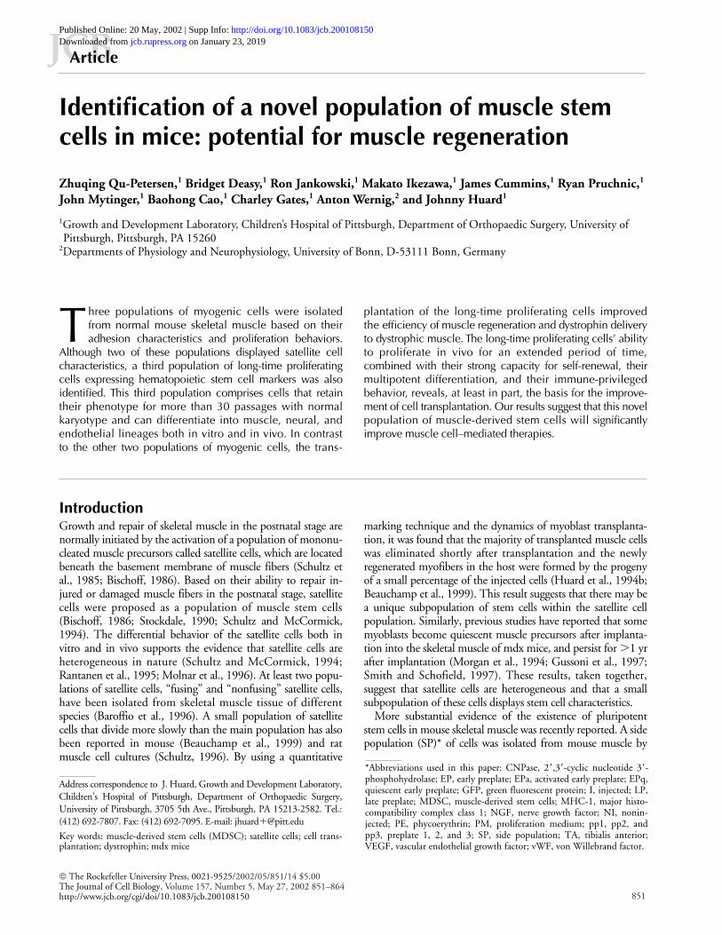
Immune-privileged behavior of MDSC in transplanted muscleTo investigate whether the improved transplantation capacityof the MDSC was related to a potential immune-privileged be-havior of the injected cells, immunohistochemical staining toreveal CD4 and CD8 T lymphocytes was performed on mus-cles injected with EP cells and MDSC. By 10 d after injectionof the EP cells, both CD4 (Fig. 4 b, arrowhead) and CD8 (Fig.4 c, arrowheads) lymphocytes had infiltrated the injected site,which was revealed by the detection of dystrophin� myofibersand green fluorescent microspheres (Fig. 4 a) in the trans-planted muscles (n � 4). We incubated the fluorescent micro-spheres with the cells before injection in order to follow theirfate after transplantation. At 30 d after injection, we still ob-served some CD4� cells (Fig. 4 d, arrowhead) in the muscles(n � 4) into which EP cells were transplanted; consequently,we observed a dramatic decrease in the number of dystrophin�
myofibers (Fig. 4 e). In contrast, at 30 and 90 d after injection,we detected CD4� and CD8� cells in only half of the MDSC-injected muscles (3:6). More importantly, we observed a com-plete absence of CD4� and CD8� cells in the injected area of
some MDSC-injected muscles, despite the presence of a largenumber of dystrophin� myofibers at 1 mo after injection (Fig.4, f and g).
We quantitated the number of infiltrating lymphocytes inthe EP cell– and MDSC-injected muscle and compared theresults among the different groups. The number of CD4
Figure 4. Detection of CD4 and CD8 lymphocytes in transplanted muscles and characterization of MHC-1 expression on MDSC and EP cells. (a–c) Immunostaining for dystrophin (dys; a), CD4 (b), and CD8 (c) cells was performed in muscle serial sections prepared from EP cell–injected muscles. By 10 d after injection (EP/D10), we detected both CD4 (b, arrowhead) and CD8 (c, arrowheads) lymphocytes in the injected area, as revealed by the green beads and the presence of numerous dystrophin� myofibers (a–c). Stars in a–c indicate the same muscle fiber in serial muscle sections. (d–g) We also performed immunostaining to colocalize CD4 (red) and dystrophin (green) in MDSC- and EP cell–injected muscles, which were counterstained with Hoechst (blue) at 30 d after injection (D30). We observed some CD4� cells in the EP cell–transplanted areas (d, arrowhead) in mdx muscles. In these areas, we also ob-served a dramatic decrease in the number of dystrophin� myofibers (e). In contrast, we detected an absence of CD4� cells in the MDSC-injected muscle (f) despite the presence of a large number of dystrophin� myofibers at 30 d after transplantation (g). In e and g, the letters C, D, and H represent the colocalization of CD4, dystrophin, and Hoechst. The stars in f and g show the same myofibers. (h and i) We analyzed the percentage of MHC-1–expressing cells on the MDSC and EP cell populations by flow cytometry. The MDSC were almost completely negative (0.5%) for the MHC-1 (i), whereas 69.3% of the EP cells were positive for MHC-1 (h). Bars, 100 m.

Identification of muscle stem cells in mice | Qu-Petersen et al. 857
and CD8 lymphocytes was monitored in four muscle sec-tions of each injected animal at 30 d after injection. We ob-served that the number of CD4 and CD8 lymphocytes wassignificantly lower in the MDSC-injected muscle than in theEP cell–transplanted muscles (CD4, 10.0 � 3.7 in MDSC,n � 4, vs. 48 � 13.3 in EP, n � 3; CD8, 5 � 3.5 inMDSC, n � 4, vs. 48.7 � 13.3 in EP, n � 3). There was asignificantly lower number of CD4 and CD8 lymphocyteswithin the injected MDSC than within the EP cells.
In an attempt to elucidate the mechanism by whichthe MDSC display this immune-privileged behavior, theMDSC and EP cells were tested for the expression of themajor histocompatibility complex class 1 (MHC-1), the an-tigens that are mainly responsible for rejection (Hall et al.,1984; Milton and Fabre, 1985; Mason and Morris, 1986).It has been observed that the MHC-1 is strongly expressedin myoblasts (Roy et al., 1991) and that the MHC-1 expres-sion on target cells is a prerequisite for antigen-specificT cell–mediated cytotoxicity (Emslie-Smith et al., 1989).Therefore, the expression of MHC-1 on the MDSC and EPcells was determined by flow cytometry. Surprisingly, theMDSC were almost completely negative (0.5%) for theMHC-1 (Fig. 4 i), whereas 69.3% of the EP cells werefound to be positive for the MHC-1 (Fig. 4 h). These resultssuggest that the improved transplantation capacity of theMDSC may be attributed to their inability to trigger infil-tration of activated lymphocytes because of their low expres-sion of MHC-1, which would eventually play a role in theimmune rejection of the transplanted cells.
The influence of growth factors on the MDSCTo investigate whether the MDSC could differentiate intoother lineages, the MDSC and two MDSC subclones(msc1and msc3) were stimulated with nerve growth factor(NGF) for 5 d, and their expression of 2�,3�-cyclic nucle-otide 3�-phosphohydrolase (CNPase) was analyzed by im-munochemistry. CNPase is a myelin-associated enzyme thatis a constituent of both the oligodendrocyte progenitor cellsin the central nervous system and Schwann cells in the pe-ripheral nervous system (Sprinkle et al., 1985). The numberof CNPase� cells was counted at 200� magnification in fivefields, and a total of 452–1020 cells were counted for eachcondition. An absence of CNPase-expressing cells was ob-served in the MDSC culture before stimulation (Fig. 5 a).However, 1.1% of the cells became CNPase� when incu-bated in NGF-supplemented medium (10 ng/ml) for 5 d(Fig. 5, b and c). Interestingly, CNPase� cells were detectedin msc1 and msc3 cultures without NGF stimulation. How-ever, after stimulation with NGF, the number of CNPase�
cells increased by 0.7% in the msc3 culture and by 2.1% inthe msc1 culture (Fig. 5 c).
A similar method was used to analyze the expression ofvon Willebrand factor (vWF), an endothelial cell marker(Mukai et al., 1980), in the MDSC after stimulation withvascular endothelial growth factor (VEGF). vWF� cells weredetected in the MDSC culture without VEGF stimulation,but their number increased in the presence of this growthfactor (by 16% with 15 ng/ml and by 21% with 25 ng/ml)(Fig. 5 d). At the same time, the number of desmin� cellsdecreased in the MDSC culture when stimulated with
VEGF (Fig. 5 d). This is in accordance with our recent ob-servation that a subclone of MDSC (mc13), when cultivatedin medium supplemented with rhBMP-2 (an osteogenic fac-tor) for 6 d (Lee et al., 2000), displayed a significant decreasein the number of desmin� cells while acquiring the ability toexpress alkaline phosphatase (a preosteoblast marker).
Multipotential differentiation of MDSC in vivoTo further validate these in vitro findings, cloned MDSCdevoid of NGF or VEGF stimulation were genetically en-gineered to express the lacZ reporter gene (nuclear loca-tion), and were injected into the m. gastrocnemius of mdxmice. By 10 d after injection, a large number of myofiberscontaining lacZ� nuclei was observed in the injected dys-trophic muscle (Fig. 6 a-1). Additionally, some of thelacZ� nuclei were found in vascular-like (Fig. 6 a-2, ar-
Figure 5. Multipotent differentiation of MDSC in vitro. (a–c) Immunostaining was performed on MDSC and the subclones of MDSC with or without stimulation by NGF. MDSC without stimulation did not express CNPase (a), whereas some cells became CNPase� in the presence of NGF-supplemented (10 ng/ml) medium for 5 d (b). Hoechst staining showed the total number of cells in culture (a and b). Interestingly, we detected CNPase� cells in two of the subclone cultures, msc1 and msc3, before NGF stimulation (c). However, the percentage of CNPase� cells was increased in all cell populations when incubated with NGF-supplemented medium for 5 d (c). (d) We also detected vWF� MDSC without VEGF stimulation, and the percentage of vWF� cells increased in the presence of VEGF-supplemented medium (15 and 25 ng/ml). We also observed that the number of desmin� cells decreased after stimulation with VEGF. Bar, 50 m.

858 The Journal of Cell Biology | Volume 157, Number 5, 2002
rowheads) and peripheral nerve–like structures (Fig. 6 a-3,arrow). Arrowheads show the border of the nerve-likestructure (Fig. 6 a-3). By 25 d after transplantation, lacZ�
nuclei were found in the endothelium of well-differenti-ated blood vessels (Fig. 6 b-1, arrow), as defined by bothmorphology and vWF immunostaining (Fig. 6 b-2).LacZ� nuclei were also found in the Schwann cells ofwell-differentiated peripheral nerve (Fig. 6 c, arrow) intransplanted muscle at 25 d after implantation. To con-firm the nature of these various differentiated cell types,colocalization of �-galactosidase/vWF/Hoechst by immu-nochemistry was performed on muscle section preparedfrom MDSC-injected muscles at 25 d after transplanta-tion (Fig. 6, d-1–d-3). �-Galactosidase� nuclei (greenfluorescence) were observed in vWF� (red fluorescence)structure (Fig. 6 d-1), and costained with Hoechst (Fig. 6,d-2 and d-3). Moreover, after transplantation of green flu-orescent protein (GFP)–expressing MDSC (Fig. 6, e-1–e-3), some peripheral nerve structures displaying CNPaseimmunoreactivity (Fig. 6 e-1) were colocalized with GFP-expressing cells (Fig. 6 e-2) and Hoechst (Fig. 6 e-3) at 25 dafter transplantation. These results demonstrate thatMDSC differentiate into both endothelial and peripheralnerve cells after implantation in mdx skeletal muscle. Theability of the MDSC to differentiate into blood vesselsand neural lineages may contribute to the regeneration offunctional muscle tissue with adequate neurogenic andvascular supplies.
DiscussionThree populations of muscle-derived cells, EP cells, LP cells,and MDSC, were isolated in the present study according totheir adhesion characteristics and proliferation behaviors. EPand LP cells represent two populations of satellite cells basedon their patterns of myogenic marker expression and theirbehavior in vitro. However, the MDSC displayed uniquecharacteristics that are usually associated with noncommit-ted progenitor cells. First, the detection of similar pheno-types in MDSC, as well as in cloned and subcloned MDSC,and the maintenance of the MDSC phenotype for a long pe-riod of time in vitro (30 passages) indicate that the MDSCare capable of self-renewal in vitro. Importantly, these highlyproliferating cells can be expanded in vitro for over 30 pas-sages while preserving a normal karyotype and are incapableof developing tumors in immunodeficient scid mice. Sec-ond, the ability of the MDSC to replenish myogenic pro-genitors (m-cadherin– and Sca-1–expressing donor-derivedcells) within the injected dystrophic muscle suggests that theMDSC also are capable of self-renewal in vivo. Third, theability of the MDSC to highly proliferate in vitro and tocontribute to a persistent restoration of dystrophin� myofi-bers within the transplanted muscle, in which a large num-ber of small regenerating myofibers can be observed for upto 90 d after injection, demonstrates that the MDSC possessa high capacity for long-term proliferation in vitro and invivo. Fourth, the detection of donor-derived cells in myofi-
Figure 6. Multipotent differentiation of MDSC in the injected skeletal muscle in vivo. 10 (a) and 25 d (b–e) after transplantation. (a-1–a-3) MDSC that were genetically engineered to express the lacZ reporter gene (nuclear localization) were injected into the m. gastrocnemius of mdx mice. At 10 d after injection, we detected many lacZ� cells in the transplanted muscle (a-1). In the injected area, a vessel-like structure containing lacZ� nuclei was also found (a-2, arrowheads). Some peripheral nerve–like structures (a-3, arrowheads) with lacZ� nuclei (a-3, arrow) were also found in the injected site. (b) At 25 d after transplantation, we observed some lacZ� nuclei (b-1, arrow) in the endothelium of well-differentiated blood vessels (b-1, star), which was confirmed by vWF staining in adjacent sections (b-2, star). (c) LacZ� nuclei (arrow) were also found in well-differentiated peripheral nerves in the injected skeletal muscle at 25 d after transplantation. (d) Colocalization of �-galactosidase, vWF, and Hoechst by immunochemistry revealed �-galactosidase� nuclei (d-1, green, arrowhead) in vWF� structure (d-1, red) and costained with Hoechst (d-2 and d-3, arrowhead). (e) After transplantation of MDSC isolated from GFP transgenic mice, some peripheral nerve structures expressing CNPase immunoreactivity, a Schwann cell marker (e-1, red), were colocalized with GFP-expressing cells (e-2) and Hoechst (e-3) in the transplanted mdx TA muscle. Triple exposure of CNPase, GFP, and Hoechst (blue) is shown in e-3. Bars: (a-1) 500 m; (b, c, and e) 50 m; (d) 25 m.

Identification of muscle stem cells in mice | Qu-Petersen et al. 859
bers, peripheral nerves, and blood vessels within the MDSC-injected muscle provides evidence of the multipotent natureof MDSC in vivo, as observed in vitro, when appropriatelystimulated with growth factors. Based on this evidence, theMDSC were confirmed as a novel population of musclestem cells with the capacities for high self-renewal, long-term proliferation, and multipotent differentiation.
Based on the relative number of cells isolated in the differ-ent fractions of muscle-derived cells (EP, LP, and MDSC),EP cells represent the main population of myogenic cells de-rived from skeletal muscle. More than 90% of the EP cellsexpress the myogenic marker desmin and markers of thelater stages of myogenesis, such as m-cadherin and myoge-nin (Miller et al., 1999), suggesting that EP cells represent alate myogenic precursor. The EP cells exhibited a very fastbut limited ability to proliferate, as well as a strong capacityto differentiate into myotubes. These EP cell characteristicsare similar to previous findings in satellite cells (Bischoff,1986; Baroffio et al., 1996; Schultz, 1996). A main popula-tion of m-cadherin� satellite cells was also detected in nor-mal muscle sections beneath the basement membrane, indi-cating that the EP cells were derived from the commonsatellite cell population. Surprisingly, although the trans-planted EP cells were highly purified and 95% of them ex-pressed desmin, they still displayed a limited capacity to re-generate skeletal muscle after cell implantation. We believethat the poor ability of the EP cells to proliferate and to self-renew in the regenerating mdx muscles explained, at least inpart, their limited regeneration capabilities in vivo.
The LP population represents �1% of satellite cells, andtheir function in skeletal muscle remains unclear becausethey displayed a very limited capacity to proliferate and dif-ferentiate. In a preliminary experiment, transplantation ofquiescent LP cells that were cultured for only 1–2 d did notshow the same efficiency of muscle regeneration at 30 d aftertransplantation, when compared with the MDSC. This isintriguing because the primitive MDSC were isolated fromthe LP cultures (see Materials and methods). It is possiblethat the primitive MDSC within LP cultures need specificunknown conditions to be activated before they become ef-fective in contributing to muscle regeneration. Therefore,activation of primitive MDSC by appropriate growth factorsin LP cultures may play a major role in the activation pro-cess and should be investigated in future experiments.
Difficulty was encountered when evaluating the relativenumber of cells in the MDSC populations within themuscle cell culture. Culturing LP cells at high confluence(60–70% confluence) often resulted in large numbers ofdifferentiated cells with typical fibroblast morphology.The fibroblast-like cells may come from either the LPculture or from the differentiation of MDSC daughtercells, which often grow faster than their parent cells.When the LP cells were cultured at low confluence(�30%), most of the LP cells died within 1–2 wk, andonly rare primary MDSC clones appeared. According toour findings in muscle cultures prepared from newbornmice, the ratio of the three populations of myogenic cellsis as follows: 105 relatively quiescent EP cells contained102 relatively quiescent LP cells, in which one MDSCclone could be obtained.
Additionally, it is difficult to investigate the source ofMDSC, considering their relatively low number in skeletalmuscle. Given recent studies in which pluripotent bonemarrow stem cells (Ferrari et al., 1998; Gussoni et al.,1999), blood vessel progenitors (De Angelis et al., 1999),neural stem cells (Clarke et al., 2000; Galli et al., 2000), andmesenchymal stem cells isolated from interstitial connectivetissue (Young et al., 2001a,b) have all shown the capacity todifferentiate into muscle lineage, these MDSC can be de-rived from various sources. We found that MDSC expressthe hematopoietic stem cell markers CD34 and Sca-1, aswell as neural and endothelial markers (when appropriatelystimulated). Various features suggest, however, that theMDSC derived from skeletal myofibers. These features in-clude (a) the potential of MDSC to differentiate into skeletalmuscle lineage both in vitro and in vivo, a potential which iseven higher than that of satellite cells; (b) their ability tospontaneously express myogenic markers, such as desminand MyoD, without the requirement of myogenic stimula-tion; and (c) their similarity in phenotype to a subpopula-tion of cells that has been identified within the basal laminaof myofibers, i.e., Sca-1�/m-cadherin� (Fig. 1, g–l), as wellas CD34� and Bcl2� (Lee et al., 2000).
Although �95% of cells in expanded MDSC cultures ex-pressed desmin, an early stage myogenic marker, only 30–40% of MDSC at earlier passages (10–12 passages) werefound to be desmin�. It is reasonable to speculate that theprimitive MDSC do not express desmin. Therefore, the pri-mary MDSC may be characterized phenotypically by myo-genic lineage�, CD34��, and Sca-1�. In the transplantationexperiment, long-term engraftments were observed in themuscle injected with the MDSC (myogenic lineage�/�,CD34�/�, Sca-1�), but not in the muscle injected with theEP cells (myogenic lineage�, CD34�, Sca-1�/�). In contrastto the EP culture, a small population (10%) of MDSC isSca-1�/CD34�. It is very interesting that murine hemato-poietic stem cells with long-term reconstituting capabilityare found to be in the hematopoietic lineage�, CD34low/�,Sca-1� populations (Osawa et al., 1996; Sato et al., 1999). Itis possible that different organ-specific stem cells may sharesome similarities in terms of surface antigen expression. Theidentification of surface antigens specific to muscle stemcells will help to enhance the rapid isolation of this minorityof long-time proliferating stem cells from muscle, as thiswould result in a more efficient isolation technique. Indeed,we recently demonstrated that the use of magnetic cell sort-ing to rapidly isolate and transplant phenotypically pureSca-1� myogenic cells (CD45�) resulted in the regenerationof skeletal muscle and restoration of dystrophin expressionin host mdx mice (Jankowski et al., 2001).
An important point of the current investigation is the dem-onstration of the functional ability of this specific populationof MDSC to regenerate dystrophic muscle (mdx) after trans-plantation. Transplantation of normal muscle precursors fortreatment of inherited myopathies, such as Duchenne muscu-lar dystrophy, has been widely studied (Partridge et al., 1989;Partridge, 1991; Morgan et al., 1990, 1993, 1994; Miller etal., 1997; Qu and Huard, 2000). The results from animal andhuman clinical trials have suggested that although myoblasttransplantation is feasible (Partridge, 1991; Morgan et al., 1993;
860 The Journal of Cell Biology | Volume 157, Number 5, 2002
Miller et al., 1997), it is rather inefficient (Huard et al.,1992a,b; Karpati et al., 1992; Tremblay et al., 1993; Mendellet al., 1995; Miller et al., 1997). The low survival rate, poorspreading of transplanted cells, and immunorejection of thedonor cells are still major problems facing myoblast transplan-tation (Huard et al., 1994a,b; Fan et al., 1996; Guerette et al.,1997; Beauchamp et al., 1999; Hodgetts et al., 2000; Skuk etal., 2000; Smythe et al., 2000). In animal experiments, im-munodeficient animals and/or immune-suppressive regimens(Huard et al., 1992a, 1994a; Morgan et al., 1993, 1994; Ki-noshita et al., 1994; Pavlath et al., 1994), preirradiation of theinjected muscle (Morgan et al., 1993), and myonecroticagents (Huard et al., 1994a; Vilquin et al., 1995) have beenused extensively to improve the success of this technique. Al-though these approaches may be used to improve the restora-tion of dystrophin in mdx mice, the success of this techniqueremains rather limited and largely clinically impractical.
In the present study, the cell transplantation was successfulfrom 10 to 90 d after injection of the MDSC into mdx muscle.The number of dystrophin� myofibers observed at 10 d afterinjection (2,178 myofibers) was not significantly differentfrom the number of dystrophin� myofibers observed at 90 d(1,851 myofibers) after transplantation. Interestingly, the mdxmice used as recipients in this experiment were not immuno-suppressed, and the injected muscles were not preirradiated orinjured with a myonecrotic agent. The injection of MDSC re-sulted in 10 times more dystrophin� myofibers in the injectedmuscle than did the transplantation of EP cells (1,435 vs. 136fibers) by 30 d after transplantation, although the same num-ber of cells (3–4 � 105) was injected in both groups. These re-sults illustrate that the MDSC display an improved transplan-tation capacity in skeletal muscle when compared with the EPcells. Therefore, we have attempted to elucidate the mecha-nism by which the MDSC display improved transplantationcapacity by characterizing their self-renewal ability, along withtheir immune-privileged behavior and their multipotent differ-entiation, after implantation in skeletal muscle.
Unlike the MDSC, the transplanted EP cells exhibited adramatic decrease in the number of dystrophin� myofibersfrom day 10 to day 30. In view of the previous report thatdystrophin is antigenic in dystrophin-deficient mice (Oht-suka et al., 1998) and also the reports in which serum anti-body formation has been found to be triggered by donormyoblasts (Huard et al., 1992a; Tremblay et al., 1993;Vilquin et al., 1995), we have investigated the immune re-jection against the injected cells. Detection of CD4 andCD8 T lymphocytes in all EP-injected muscles at 10 and30 d after injection provides evidence to support the im-mune rejection of the EP cells. Surprisingly, an absence ofinfiltration with CD4 or CD8 T-lymphocytes was ob-served in many of the MDSC-injected muscles at 30 and90 d after transplantation, despite the large numbers ofdystrophin� myofibers found in the injected muscles.These results suggest that MDSC are less immunogenicthan EP cells. The lack of expression of MHC-1 by theMDSC further supports this contention. These latter re-sults suggest that the improved transplantation capacity ofMDSC may be attributed to their inability to trigger an in-filtration of activated lymphocytes, which would partici-pate in the immune rejection of the transplanted cells.
We also have observed that the MDSC can self-renew bothin vitro and in the injected skeletal muscle in vivo. Indeed,MDSC can be expanded in vitro for up to 30 passages whilemaintaining their phenotype in terms of both myogenic andstem cell marker expression profiles. In addition, subclones ofMDSC display a phenotype almost identical to the MDSCclone from which they were derived; this fact provides evi-dence of the in vitro self-renewing capability of these cells. Fi-nally, five times more lacZ� cells were detected in the musclecultures derived from MDSC-injected muscle, when com-pared with the EP cell–injected muscle. Moreover, the detec-tion of Sca-1� and m-cadherin� cells in MDSC-injected mus-cle suggests that MDSC give rise to myogenic precursor cellsmore effectively than do EP cells. The high self-renewal abilitydisplayed by MDSC both in vitro and in vivo may also con-tribute to the persistent dystrophin delivery into mdx mice.
We also investigated whether the injected cells can differen-tiate into blood vessels and nerve cells with the potential tocontribute to the regeneration of functional muscle tissue withadequate vascular and nerve supplies. Indeed, our in vitro re-sults suggest that the stimulation of the MDSC with NGFand VEGF increased the number of CNPase- and vWF-expressing cells, respectively. More importantly, in the in vivoexperiments, although the MDSC were not stimulated ortransduced with NGF and VEGF, the injected cells could stilldifferentiate into endothelial and peripheral nerve lineages.Based on these results, we believe that the release of environ-mental stimuli (e.g., growth factors) within the dystrophicmuscle triggers the differentiation of the MDSC toward otherlineages. Such results suggest that multipotent differentiationof MDSC may contribute to the regeneration of functionalmuscle tissue in transplanted mdx mice. The ability of theMDSC to restore the function of the dystrophic mdx muscleis important and will be investigated further. It was also foundthat primary MDSC (passage 10) showed much higher poten-tial than later passages (after 30 passages) to differentiate intomuscle and other lineages. In extended cultures (after 40 pas-sages), some of the MDSC gradually lost their ability to re-generate muscle fibers or differentiate into other lineages, sug-gesting that the mechanism that governs the differentiation ofmultipotent muscle stem cells toward a specific lineage is yetto be determined. Further investigation of the gene expressionprogram, as well as the proper culture condition by which theproliferation and differentiation of multipotent stem cellscould be controlled, is paramount for the development ofmuscle stem cell transplantation for Duchenne muscular dys-trophy patients.
In summary, three populations of myogenic cells havebeen isolated by a modified version of the preplate tech-nique. The EP and LP cells are derived from the main popu-lation of satellite cells and are a type of committed muscleprecursor. The MDSC, derived from the subpopulation ofLP cells, are characterized by myogenic lineage�/�, CD34�/�,Sca-1�, c-Kit�, and CD45�. The use of MDSC can cir-cumvent hurdles facing myoblast transfer therapy and, con-sequently, improve the efficiency of muscle regeneration anddystrophin delivery to dystrophic muscle. The uniquefeatures of the MDSC, including (a) their long-time prolif-erating ability, (b) their strong self-renewal, (c) their mul-tipotent differentiation, and (d) their immune-privileged be-

Identification of muscle stem cells in mice | Qu-Petersen et al. 861
havior after implantation, at least partially elucidate the basisof the improved transplantation capacity of this novel popu-lation of MDSC.
Materials and methodsAnimalsNormal mice (C57 BL/6J) and mdx mice (C57BL/10ScSn DMDmdx/J) usedin this experiment were purchased from Jackson ImmunoResearch Labo-ratories. All animal protocols used for these experiments were approvedby the Children’s Hospital of Pittsburgh’s IACUC committee (protocolnos. 2/00 and 7/00).
Preparation of different populations of muscle-derived cell culturesPrimary muscle cultures were prepared from newborn (3–5 d) normalmice, and the MDSC were purified from the primary culture using amodified version of a previously described preplate technique (Richlerand Yaffe, 1970; Rando and Blau, 1994; Qu et al., 1998; Qu and Huard,2000). The hindlimb muscles of neonatal mice were removed, and thebones were dissected. The muscle was then minced into a coarse slurryusing scalpels. The muscle tissue was enzymatically dissociated at 37 Cin 0.2% collagenase-type XI (Sigma-Aldrich) for 1 h, and then centri-fuged at 3,500 rpm for 5 min. The cells were collected, incubated in dis-pase (GIBCO BRL), prepared as 2.4 units/1 ml HBSS (GIBCO BRL), for 45min, and then incubated for 30 min in 0.1% trypsin-EDTA (GIBCO BRL)diluted in HBSS. After the enzymatic dissociation, the muscle cells werecentrifuged and resuspended in proliferation medium (PM). PM consistsof DME containing 10% horse serum, 10% FBS, 0.5% chick embryo ex-tract, and 1% penicillin–streptomycin (all reagents purchased fromGIBCO BRL). Different populations of muscle-derived cells were isolatedbased on their adhesion characteristics. A flow chart for the isolation ofmuscle-derived cells (EP, LP, and MDSC) is shown in Fig. 7. The musclecells were plated on collagen-coated flasks (collagen type 1; Sigma-Aldrich) for 2 h (preplate 1 [pp1]). The nonadherent cells were thentransferred to other flasks (pp2), and the adherent cells in pp1 were dis-carded. It has been reported that the cells that rapidly attach are highly fi-broblastic in nature (Richler and Yaffe, 1970; Rando and Blau, 1994; Quet al., 1998; Qu and Huard, 2000). After 24 h, the floating cells in pp2 werecollected, centrifuged, and plated on new flasks (pp3). These procedureswere repeated at 24-h intervals until serial preplates (pp4–6) were ob-tained. All cell populations (pp2–6) were maintained in PM with dailychanges. Based on the previous report (Qu et al., 1998), 30–40% of the
cells in pp2 and pp3 are known to be nonmyogenic, whereas up to 95%desmin� cells can be found in pp4 and pp5. To further purify thedesmin� cells within pp2 and pp3, the cells were subsequently tryp-sinized and replated in fresh collagen-coated flasks using a previouslydescribed protocol (Qu and Huard, 2000). After 30 min, the supernatantcontaining mostly nonadherent cells (�90% of which were shown to bedesmin-expressing cells) were plated in new flasks. These nonadherentcells from pp2 and pp3 were combined with the cells from pp4 and pp5and used in the transplantation experiment as the EP population. To iso-late MDSC, pp6 cells (also termed LP cells) were plated in collagen-coated 12-well dishes at 50–100 cells/well (Fig. 7). Though most of theLP cells died within 1–2 wk of culturing, the MDSC formed clonal colo-nies during the first 2 wk of culturing. The MDSC take 5–7 d to attach tocollagen-coated flasks, and require �1–2 wk to develop clones, from 1to �300–500 cells. The doubling time of these cells is �24–36 h at theearly stage of culturing, which means that these cells divided eight tonine times in the first 2 wk of culturing. The wells containing multipleMDSC colonies were trypsinized (0.25% trypsin) and the detached cellswere transferred into a collagen-coated 6-well dish. The MDSC werethen grown in PM to �30% confluence and subsequently passaged. Thewell containing a single colony was selected and these cells were calledMDSC clones. These MDSC clones were expanded and used to isolatesubclones via a protocol previously described (Lee et al., 2000). TheMDSC cloned and subcloned cells were also expanded in PM as de-scribed above. We have also isolated MDSC, by using the same tech-nique as described above, from the skeletal muscle of GFP transgenicmice (C57BL/6-TgN[ACTbEGFP]) (Okabe et al., 1997).
Cell cycle durationThe EP cells and MDSC were plated in 24-well collagen-coated plates ata density of 450 cells/well in PM. Using the CytoWorks platform, visibleimaging was obtained for individual cells. This system uses a biobox in-cubator mounted to the stage of a Nikon microscope, which is linked toa CCD camera (Automated Cell Technologies, Inc.). Stage movementwas computer controlled, allowing for images of each view field to beacquired at 10-min intervals for 5 d. For each population, 100 cellswere selected and tracked. The division time of each cell was deter-mined by direct observation of the cells through the CytoWorks plat-form as previously described (Deasy et al., 2002). The initiation of celldivision was marked at the time when two daughter cells were formed,and these cells were subsequently followed until their respective divi-sion. The lapsed time between those two division events was recordedas the length of the cell division cycle.
Figure 7. Isolation of three populations of muscle-derived cells. Muscle-derived cells were enzymatically dissociated from neonatal mouse skeletal muscle and separated by their adhesion characteristics to collagen-coated flasks (modified preplate technique). After the enzymatic dissociation, the muscle cell extract was resuspended in PM and preplated on collagen-coated flasks. Different populations of muscle-derived cells were isolated based on adhesion characteristics. pp1 represented a population of primary fibroblasts that adhered in the first 2 h after isolation; subsequent preplates, containing a mixture of myogenic and nonmyogenic cells, were obtained at 24-h intervals (pp2–6). The nonmyogenic cells in pp2 and pp3 were removed from the cultures by replating the cells, and the resulting enriched pp2 and pp3 desmin� cells were combined with pp4 and pp5 cells and were termed the EP population. Cells in the pp6 cell population took an
additional 24–72 h to attach to collagen-coated dishes after transfer from pp5 and were termed LP cells. Most of the LP cells died during the first 1–2 wk of the cultivation period, with very few of the adherent surviving cells proliferating and forming clonal colonies. The surviving clones are called MDSC. We also isolated subclones from a single clone of MDSC, as shown in the flow chart.

862 The Journal of Cell Biology | Volume 157, Number 5, 2002
Immunocytochemistry on culturesThe primary antibodies used in this study were mouse anti-desmin (1:200;D-1033; Sigma-Aldrich), rabbit anti–mouse m-cadherin (1:50; obtainedfrom A. Wernig), rabbit anti–mouse Bcl2 (1:1,000; 15616E; BD PharMin-gen), biotin anti–Sca-1 (1:200; BD PharMingen), biotin anti–mouse CD34(1:200; 09432D; BD PharMingen), mouse anti-myogenin (1:100; 65121A;BD PharMingen), and mouse anti-MyoD (1:200; 13941A; BD PharMin-gen). EP cell, LP cell, and MDSC (cloned and subcloned) cultures werefixed and stained as previously described (Lee et al., 2000).
RNA analysesRT-PCR analyses of EP and MDSC cultures were performed as previously de-scribed (Lee et al., 2000). Total RNA was isolated using TRIzol reagent (LifeTechnologies). Reverse transcription was performed using SuperScriptTM First-Strand Synthesis System (Life Technologies) according to the manufacturer’sinstructions. PCR primer sequences and reaction parameters are from refer-ences as follows: Bcl2 (Dominov et al., 1998); CD34 (Lee et al., 2000); myo-genin and MyoD (Rohwedel et al., 1995); and MNF (Yang et al., 1997). PCRproduct sizes were analyzed on agarose gel. The expected product sizes wereBcl-2, 480 bp; CD34, 147 bp; myogenin, 86 bp; MyoD, 147 bp; and MNF,305 bp. Genomic DNA contamination was excluded by (a) primers spanningan intron and (b) reverse transcription reactions without reverse transcriptase.
Flow cytometryThe percentages of CD34� and Sca-1� cells in the MDSC and EP cultureswere analyzed by flow cytometry as recently described (Jankowski et al.,2001). Cultured cells were trypsinized, spun, washed in cold PBS (Dulbeccophosphate-buffered salt solution 1�; Mediatech) containing 0.5% BSA (ICNBiomedicals), and counted. The cells were then divided into equal aliquotsand spun to a pellet. A 1:10 mouse serum (Sigma-Aldrich) in PBS solutionand Fc Block (rat anti–mouse CD16/CD32; BD PharMingen) was used to re-suspend each pellet, and the suspensions were incubated for 10 min on ice.Optimal amounts of both direct and biotin-conjugated rat anti–mouse mono-clonal antibodies (c-kit, CD34, Sca-1, and CD45; all from BD PharMingen)were predetermined and added directly to each tube for 30 min. A similarprotocol was used to separately analyze MDSC and EP cells for MHC-1 ex-pression using a biotin-conjugated H-2kb antibody (BD PharMingen).Streptavidin–allophycocyanin conjugate was added to tubes containingcells labeled with biotinylated antibodies for 20 min. Just before analysis,7-amino-actinomycin D (Via-Probe; BD PharMingen) was added to each tubefor dead cell exclusion. Live cell events were collected and analyzed on aFACSCalibur® (Becton Dickinson) flow cytometer using Cell Quest software.
Stimulation of MDSC with growth factorsMDSC and two subclones of the MDSC clones (msc1 and msc3) werestimulated with NGF (10 ng/ml; Sigma-Aldrich) or VEGF (15 and 25 ng/ml;Sigma-Aldrich) for 5 d. Cells were plated at 20–30% confluence on col-lagen-coated flasks. PM containing growth factors was added to the wells,and PM without NGF and VEGF was added to the control wells. Immuno-cytochemistry was performed to examine the expression of CNPase andvWF. For the CNPase immunostaining, the primary antibody used was amouse anti-CNPase (1:400; Sigma-Aldrich), and the secondary antibodywas an anti-mouse conjugated to Cy3 (1:200; Sigma-Aldrich). For vWF im-munostaining, the primary antibody was a rabbit anti-vWF (1:400; Dako),and the secondary antibody was an anti–rabbit-conjugated Cy3 (1:200;Sigma-Aldrich). Desmin expression was also monitored in both the treatedand untreated wells using the protocol described above.
Cell transplantationMDSC and EP cells were transplanted as previously described (Qu et al.,1998; Qu and Huard, 2000). In brief, 3–4 � 105 viable cells suspended in20 l of HBSS were transplanted (single-point injection) in the gastrocne-mius muscle of mdx mice (C57BL/10ScSn DMDmdx/J). The mdx mice usedas recipients in this experiment were not immunosuppressed, and the in-jected muscles were not preirradiated or injured with a myonecrotic agentbefore transplantation. EP cells were cultivated for only 5 d after the initialseeding, whereas the MDSC were at passages 10–12 at the time of trans-plantation. The animals were killed at 10, 30, and 90 d after injection. Theinjected muscles were isolated and then frozen in 2-methylbutane pre-cooled in liquid nitrogen.
EP and MDSC cultures also were transduced with a retrovirus carryingthe gene encoding for �-galactosidase (nuclear localization) for 24 h be-fore transplantation, using a protocol previously described (Lee et al.,2000). We injected 5 � 105 cells into each gastrocnemius muscle. By 30 dafter injection, the animals were killed; some of the injected muscleswere frozen, sectioned, and stained for lacZ expression (histochemistry),
whereas others were used to prepare muscle cell cultures using a protocolsimilar to that described above (Fig. 7). The GFP-expressing MDSC (1 �105 cells for each population) were injected into the tibialis anterior (TA)muscles of mdx mice (9 wk old). By 25 d after injection, the animals werekilled; TA muscles were fixed with 1% paraformaldehyde, frozen, and sec-tioned for immunohistochemistry.
Immunohistochemical staining of muscle sections: normal muscleNormal muscle samples were obtained from 4-wk-old mice and frozen in2-methylbutane precooled with liquid nitrogen. Serial cryosections of 10m in thickness were prepared from the frozen muscles. The sections werefixed with cold acetone (�20 C) for 2 min, preincubated in 5% horse se-rum diluted with PBS for 30 min, and then rinsed thoroughly with PBS. Thefollowing is a list of stainings performed on these muscle sections. Controlstainings without primary and secondary antibodies were also performed.
Colocalization of Sca-1 and laminin. The sections were incubated with abiotin anti–mouse Sca-1 antibody (1:200 dilution) for 2 h and streptavidin-conjugated Cy3 (1:200) for 90 min. The sections were then incubated withrabbit anti–mouse laminin (1:100) for 1 h and anti–rabbit IgG-conjugatedfluorescein for 1 h (1:100). The nuclei were revealed by Hoechst 33342staining.
Colocalization of Sca-1, m-cadherin, and laminin. After preincubation,the sections were incubated with PE-conjugated anti–mouse Sca-1 (1:150;BD PharMingen) for 2 h, rabbit anti–m-cadherin (1:50) for 3 h, biotinylatedanti–rabbit IgG (1:100; Vector Laboratories) for 90 min, and FluoroLinkTM
CyTM5–labeled streptavidin (1:1,000, Amersham Pharmacia Biotech) for 90min, respectively. The sections were then incubated with rabbit anti–mouselaminin (1:100) for 30 min and anti–rabbit IgG-conjugated fluorescein(1:200) for 30 min. The nuclei were revealed by Hoechst 33342 staining.
Immunohistochemical staining of muscle sections: transplanted mdx muscleDystrophin staining. The staining was performed on muscle sections aspreviously described (Qu and Huard, 2000), and the sections were coun-terstained with Hoechst to visualize the locations of the nuclei.
Colocalization of m-cadherin and dystrophin. The sections were incu-bated with a rabbit anti–m-cadherin antibody (1:50) for 2 h and anti–rabbitIgG-conjugated fluorescein for 90 min. The sections were then stained fordystrophin and counterstained with Hoechst using the same protocol de-scribed above.
CD4 and CD8 staining. Sections were incubated with rat anti–mouseCD4 or CD8 (1:400; BD PharMingen) for 2 h and then with biotinylatedanti–rat IgG (1:200; Vector Laboratories) and streptavidin-conjugated Cy3(1:200; Sigma-Aldrich) for 90 min. The number of CD4 and CD8 lympho-cytes was also monitored and compared between the EP- and MDSC-injected muscles (n � 4). Some of the sections were also stained for dystro-phin (see “Dystrophin staining” above).
vWF/�-galactosidase staining. Muscle sections were incubated withmouse anti–�-galactosidase (1:100; Chemicon), and the next steps wereperformed in accordance with the instructions of the MOM kit manufac-turers (FMK-2201; Vector Laboratories). The sections were then incubatedwith rabbit anti-vWF (1:400; Dako) followed by a 1-h incubation with ananti–rabbit IgG-conjugated fluorescein (1:100). Finally, the sections werestained with Hoechst as described above.
CNPase staining. The TA muscle sections (injected with GFP-expressingMDSC) were incubated with mouse anti-CNPase (1:400; Sigma-Aldrich),and the next steps were performed in accordance with the MOM kit man-ufacturer’s instructions, except for using streptavidin-conjugated Cy3 to re-place fluorescein-conjugated avidin. The sections were consequentlystained with Hoechst as described above.
HistochemistryLacZ staining for culture. The muscle cells isolated from the EP- andMDSC-injected muscles were fixed in 1% glutaraldehyde and incubatedovernight with X-gal substrate at 37 C.
LacZ staining for muscle sections. The sections were fixed in 1% glu-taraldehyde, incubated overnight with X-gal substrate at 37 C, and thencounterstained with eosin (Sanes et al., 1986).
Karyotype analysis of MDSCCytogenetic analysis of the MDSC was performed using a technique previ-ously described (Barch, 1991; Lee et al., 2000). Cells were grown to�40% confluency in a T75 collagenated flask in DME (GIBCO BRL) sup-plemented with 10% horse serum (GIBCO BRL), 10% FBS (GIBCO BRL),0.5% chick embryo extract (GIBCO BRL), and 1% Pen-Strep solution(GIBCO BRL). Colcemid solution (GIBCO BRL) was added to the flask at a

Identification of muscle stem cells in mice | Qu-Petersen et al. 863
final concentration of 0.05 g/ml and allowed to incubate for 3 h at 37 C.Cells were then harvested by adding 0.1% trypsin-EDTA to the cells untilthey lifted. Cells were spun down (3,200 rpm, 5 min), resuspended in 5 ml0.75 M KCl, and incubated for 10 min at 37 C. Next, 3 ml of Carnoy’s fix-ative (3:1 methanol to glacial acetic acid) was added to the solution,which was vigorously pipetted. The cells were then pelleted as before.Fresh fixative was added and cells were again pelleted. This rinsing andpelleting was performed three additional times. Cells were then droppedon glass slides and allowed to dry overnight at 55 C. Metaphase cells wereGTG banded following a protocol outlined by Invitrogen. 28 metaphasecells were then counted for modal number using a magnification of 630�.Images for karyotype analysis were captured using AKS software (Image-netics, Inc.).
Statistical analysisThe data in this manuscript are presented as means � SD. Means werecompared using an unpaired t test for the number of dystrophin� myofibersbetween EP cell– and MDSC-transplanted groups. A paired t test was usedto compare the numbers of centronucleated myofibers, m-cadherin� cells,and Sca-1� cells between injected and noninjected areas in transplantedmdx muscle.
The authors thank Marcelle Pellerin for her technical assistance, and Lau-ren Rudick, Melissa Haas, and Ryan Sauder for their assistance with themanuscript. The authors also wish to thank Dr. Paul D. Robbins (Univer-sity of Pittsburg) for the retroviral vector, Dr. Joel Greenberger (Universityof Pittsburgh) for assistance with the combinatorial cell culture system tomeasure cell division time, and Dr. Lou Kunkel (Children’s Hospital, Bos-ton, MA) for the 6-10 dystrophin antibody.
This work was supported by grants to Dr. Johnny Huard from the Mus-cular Dystrophy Association, the Parent Project (USA), the National Insti-tutes of Health (NIH 1P01 AR45925-01), and the William F. and Jean W.Donaldson Chair at Children’s Hospital of Pittsburgh.
Submitted: 30 August 2001Revised: 25 March 2002Accepted: 2 April 2002
ReferencesBaroffio, A., M. Hamann, L. Bernheim, M.L. Bochaton-Piallat, G. Gabbiani, and
C.R. Bader. 1996. Identification of self-renewing myoblasts in the progenyof single human muscle satellite cells. Differentiation. 60:47–57.
Barch, M.J. 1991. Chromosome analysis. In The ACT Cytogenetics LaboratoryManual. 2nd ed. Raven Press, Ltd., New York. 349.
Beauchamp, J.R., J.E. Morgan, C.N. Pagel, and T.A. Partridge. 1999. Dynamics ofmyoblast transplantation reveal a discrete minority of precursors with stemcell–like properties as the myogenic source. J. Cell Biol. 144:1113–1122.
Beauchamp, J.R., L. Heslop, D.S. Yu, S. Tajbakhsh, R.G. Kelly, A. Wernig, M.E.Buckingham, T.A. Partridge, and P.S. Zammit. 2000. Expression of CD34and Myf5 defines the majority of quiescent adult skeletal muscle satellitecells. J. Cell Biol. 151:1221–1234.
Bischoff, R. 1986. Proliferation of muscle satellite cells on intact myofibers in cul-ture. Dev. Biol. 115:129–139.
Clarke, D.L., C.B. Johansson, J. Wilbertz, B. Veress, E. Nilsson, H. Karlstrom, U.Lendahl, and J. Frisen. 2000. Generalized potential of adult neural stemcells. Science. 288:1660–1663.
De Angelis, L., L. Berghella, M. Coletta, L. Lattanzi, M. Zanchi, M.G. Cusella-DeAngelis, C. Ponzetto, and G. Cossu. 1999. Skeletal myogenic progenitorsoriginating from embryonic dorsal aorta co-express endothelial and myo-genic markers and contribute to postnatal muscle growth and regeneration.J. Cell Biol. 147:869–878.
Deasy, B.M., Z. Qu-Petersen, J.S. Greenberger, and J. Huard. 2002. Mechanismsof muscle stem cell expansion with cytokines. Stem Cells. 20:50–60.
Dominov, J.A., J.J. Dunn, and J.B. Miller. 1998. Bcl2 expression identified anearly stage of myogenesis and promotes clonal expression of muscle cells. J.Cell Biol. 142:537–544.
Emslie-Smith, A.M., K. Arahata, and A.G. Engel. 1989. Major histocompatibility com-plex class I antigen expression, immunolocalization of interferon subtypes, and Tcell-mediated cytotoxicity in myopathies. Hum. Pathol. 20:224–231.
Fan, Y., M. Maley, M. Beilharz, and M. Grounds. 1996. Rapid death of injectedmyoblasts in myoblast transfer therapy. Muscle Nerve. 19:853–860.
Fennie, C., J. Cheng, D. Dowbenko, P. Young, and L.A. Lasky. 1995. CD34� en-
dothelial cell lines derived from murine yolk sac induce the proliferation anddifferentiation of yolk sac CD34� hematopoietic progenitors. Blood. 86:4454–4467.
Ferrari, G., G. Cusella-De Angelis, M. Coletta, E. Paolucci, A. Stornaiuolo, G.Cossu, and F. Mavilio. 1998. Muscle regeneration by bone marrow-derivedmyogenic progenitors. Science. 279:1528–1530.
Galli, R., U. Borello, A. Gritti, M.G. Minasi, C. Bjornson, M. Coletta, M. Mora, M.G.De Angelis, R. Fiocco, G. Cossu, and A.L. Vescovi. 2000. Skeletal myogenic po-tential of human and mouse neural stem cells. Nat. Neurosci. 3:986–991.
Guerette, B., I. Asselin, D. Skuk, M. Entman, and J.P. Tremblay. 1997. Control ofinflammatory damage by anti-LFA-1: increase success of myoblast transplan-tation. Cell Transplant. 6:101–107.
Gussoni, E., H.M. Blau, and L.M. Kunkel. 1997. The fate of individual myoblastsafter transplantation into muscle of DMD patients. Nat. Med. 3:970–977.
Gussoni, E., Y. Soneoka, C.D. Strickland, E.A. Buzney, M.K. Khan, A.F. Flint,L.M. Kunkel, and R.C. Mulligan. 1999. Dystrophin expression in the mdxmouse restored by stem cell transplantation. Nature. 401:390–394.
Hall, B.M., G.A. Bishop, G.G. Duggin, J.S. Horvath, J. Philips, and D.J. Tiller.1984. Increased expression of HLA-DR antigens on renal tubular cells in re-nal transplants: relevance to the rejection response. Lancet. 2:247–251.
Huard, J., R. Roy, J.P. Bouchard, F. Malouin, C.L. Richards, and J.P. Tremblay.1992a. Human myoblast transplantation between immunohistocompatibledonors and recipients produces immune reactions. Transplant. Proc. 24:3049–3051.
Huard, J., J.P. Bouchard, R. Roy, F. Malouin, G. Dansereau, C. Labrecque, N. Albert,C.L. Richards, B. Lemieux, and J.P. Tremblay. 1992b. Human myoblast trans-plantation: preliminary results of four cases. Muscle Nerve. 15:550–560.
Huard, J., A. Verreault, R. Roy, M. Tremblay, and J.P. Tremblay. 1994a. High ef-ficiency of muscle regeneration following human myoblast clone transplan-tation in SCID mice. J. Clin. Invest. 93:586–599.
Huard, J., G. Acsadi, B. Massie, and G. Karpati. 1994b. Gene transfer to skeletalmuscles by isogenic myoblasts. Hum. Gene Ther. 5:949–958.
Hodgetts, S.I., M.W. Beilharz, A.A. Scalzo, and M.D. Grounds. 2000. Why docultured transplanted myoblasts die in vivo? DNA quantification shows en-hanced survival of donor male myoblasts in host mice depleted of CD4�
and CD8� cells or Nk1.1� cells. Cell Transplant. 9:489–502.Jackson, K.A., T. Mi, and M.A. Goodell. 1999. Hematopoietic potential of stem
cells isolated from murine skeletal muscle. Proc. Natl. Acad. Sci. USA. 96:14482–14486.
Jankowski, R.J., C. Haluszczak, M. Trucco, and J. Huard. 2001. Flow cytometriccharacterization of myogenic cell populations obtained via the preplate tech-nique: potential for muscle-derived stem cells-mediated therapies. Hum.Gene Ther. 12:619–628.
Karpati, G., P. Holland, and R.G. Worton. 1992. Myoblast transfer in DMD:problems and interpretation of efficiency. Muscle Nerve. 15:1209–1210.
Kinoshita, I., J.T. Vilquin, B. Guerette, I. Asselin, R. Roy, and J.P. Tremblay.1994. Very efficient myoblast transplantation in mice under FK 506 immu-nosuppression. Muscle Nerve. 17:1407–1415.
Krause, D.S., T. Ito, M.J. Fackler, O.M. Smith, M.I. Collector, S.J. Sharkis, andW.S. May. 1994. Characterization of murine CD34, a marker for hemato-poietic progenitor and stem cells. Blood. 84:691–701.
Lee, J.Y., Z. Qu-Petersen, B. Cao, S. Kimura, R. Jankowski, J. Cummins, A. Usas,C. Gates, P. Robbins, A. Wernig, and J. Huard. 2000. Clonal isolation ofmuscle-derived cells capable of enhancing muscle regeneration and bonehealing. J. Cell Biol. 150:1085–1100.
Mason, D.W., and P.J. Morris. 1986. Effector mechanisms in allograft rejection.Annu. Rev. Immunol. 4:119–145.
Mendell, J.R., J.T. Kissel, A.A. Amato, W. King, L. Signore, T.W. Prior, Z. Sa-henk, S. Benson, P.E. McAndrew, and R. Rice. 1995. Myoblast transfer inthe treatment of Duchenne’s muscular dystrophy. N. Engl. J. Med. 333:832–838.
Miller, J.B., L. Schaefer, and J.A. Dominor. 1999. Seeking muscle stem cells. Curr.Top. Dev. Biol. 43:191–219.
Miller, R.G., K.R. Sharma, G.K. Pavlath, E. Gussoni, M. Mynhier, A.M. Lanctot,C.M. Greco, L. Steinman, and H.M. Blau. 1997. Myoblast implantation inDuchenne muscular dystrophy: the San Francisco study. Muscle Nerve. 20:469–478.
Milton, A.D., and J.W. Fabre. 1985. Massive induction of donor-type class I andclass II major histocompatibility complex antigens in rejecting cardiac al-lografts in the rat. J. Exp. Med. 161:98–112.
Molnar, G., M.L. Ho, and N.A. Schroedl. 1996. Evidence for multiple satellite cellpopulations and a non-myogenic cell type that regulated differently in regen-

864 The Journal of Cell Biology | Volume 157, Number 5, 2002
erating and growing skeletal muscle. Tissue Cell. 28:547–556.Morel, F., S.J. Szivassy, M. Travis, B. Chen, and A. Galy. 1996. Primitive hemato-
poietic cells in murine bone marrow express the CD34 antigen. Blood. 88:3774–3784.
Morgan, J.E., E.P. Hoffman, and T.A. Partridge. 1990. Normal myogenic cellsfrom newborn mice restore normal histology to degenerating muscle of mdxmouse. J. Cell Biol. 111:2437–2449.
Morgan, J.E., C.N. Pagel, T. Sherrat, and T.A. Partridge. 1993. Long-term persis-tence and migration of myogenic cells injected into pre-irradiated muscle ofmdx mice. J. Neurol. Sci. 115:191–200.
Morgan, J.E., J.R. Beauchamp, C.N. Pagel, M. Peckham, P. Ataliotis, P.S. Jat,M.D. Noble, K. Farmer, and T.A. Partridge. 1994. Myogenic cell lines de-rived from transgenic mice carrying a thermolabile T antigen: a model sys-tem for the derivation of tissue-specific and mutation-specific cell lines. Dev.Biol. 162:486–498.
Mukai, K., J. Rosai, and W.H.C. Burgdirf. 1980. Localization of factor VIII-related antigen in vascular endothelial cells using immunoperoxidasemethod. Am. J. Surg. Pathol. 4:273–276.
Nakauchi, H., H. Takano, H. Ema, and M. Osawa. 1999. Further characterizationof CD34-low/negative mouse hematopoietic stem cells. Ann. NY Acad. Sci.872:57–66.
Okabe, M., M. Ikawa, K. Kominami, T. Nakanishi, and Y. Nishimune. 1997.Green mice as a source of ubiquitous green cells. FEBS Lett. 407:313–319.
Ohtsuka, Y., K. Udaka, Y. Yamashiro, H. Yagita, and K. Okumura. 1998. Dystro-phin acts as a transplantation rejection antigen in dystrophin-deficient mice:implication for gene therapy. J. Immunol. 160:4635–4640.
Osawa, M., K. Hanada, H. Hamada, and H. Nakauchi. 1996. Long-term lympho-hematopoietic reconstitution by a single CD34-low/negative hematopoieticstem cell. Science. 273:242–245.
Partridge, T.A., J.E. Morgan, G.R. Coulton, E.P. Hoffman, and L.M. Kunkel.1989. Conversion of mdx myofibers from dystrophin-negative to -positiveby injection of normal myoblasts. Nature. 337:176–179.
Partridge, T.A. 1991. Myoblast transfer: a possible therapy for inherited myopa-thies? Muscle Nerve. 14:197–212.
Pavlath, G.K., T.A. Rando, and H.M. Blau. 1994. Transient immunosuppressivetreatment leads to long-term retention of allogeneic myoblasts in hybridmyofibers. J. Cell Biol. 127:1923–1932.
Qu, Z., and J. Huard. 2000. Matching host muscle and donor myoblast for myosinheavy chain improves myoblast transfer therapy. Gene Ther. 7:428–437.
Qu, Z., L. Balkir, J.C.T. van Deutekom, P.D. Robbins, R. Pruchnic, and J.Huard. 1998. Development of approaches to improve cell survival in myo-blast transfer therapy. J. Cell Biol. 142:1257–1267.
Rando, T.A., and H.M. Blau. 1994. Primary mouse myoblast purification, charac-terization, and transplantation for cell-mediated gene therapy. J. Cell Biol.125:1275–1287.
Rantanen, J., T. Hurme, R. Lukka, J. Heino, and H. Kalimo. 1995. Satellite cellproliferation and the expression of myogenin and desmin in regeneratingskeletal muscle: evidence for two different populations of satellite cells. Lab.Invest. 72:341–347.
Richler, C., and D. Yaffe. 1970. The in vitro cultivation and differentiation capaci-ties of myogenic cell lines. Dev. Biol. 23:1–22.
Rohwedel, J., V. Horak, M. Hebrok, E.M. Fuchtbauer, and A.M. Wobus. 1995.M-twist expression inhibits mouse embryonic stem cell-derived myogenic
differentiation in vitro. Exp. Cell Res. 220:92–100.Roy, R., G. Dansereau, J.P. Tremblay, M. Belles-Isles, J. Huard, C. Labrecque, and
J.P. Bouchard. 1991. Expression of major histocompatibility complex anti-gens on human myoblasts. Transplant. Proc. 23:799–801.
Sanes, J.R., J.L.R. Rubenstein, and J.F. Nicolas. 1986. Use of a recombinant retro-virus to study post-implantation cell lineage in mouse embryos. EMBO J.5:5133–5142.
Sato, T., J.H. Laver, and M. Ogawa. 1999. Reversible expression of CD34 by mu-rine hematopoietic stem cells. Blood. 94:2548–2554.
Seale, P., L.A. Sabourin, A. Girgis-Gabardo, A. Mansouri, P. Gruss, and M.A.Rudnicki. 2000. Pax7 is required for the specification of myogenic satellitecells. Cell. 102:777–786.
Schultz, E. 1996. Satellite cell proliferative compartments in growing skeletal mus-cles. Dev. Biol. 175:84–94.
Schultz, E., and K.M. McCormick. 1994. Skeletal muscle satellite cells. Rev. Phys-iol. Biochem. Pharmacol. 123:213–257.
Schultz, E., D.L. Jaryszak, and C.R. Valliere. 1985. Response of satellite cells to fo-cal skeletal muscle injury. Muscle Nerve. 8:217–222.
Skuk, D., M. Goulet, B. Roy, and J.P. Tremblay. 2000. Myoblast transplantation inwhole muscle of nonhuman primates. J. Neuropathol. Exp. Neurol. 59:197–206.
Smith, J., and P.N. Schofield. 1997. Stable integration of an mdx skeletal musclecell line into dystrophic (mdx) skeletal muscle: evidence for stem cell status.Cell Growth Differ. 8:927–934.
Smythe, G.M., S.I. Hodgetts, and M.D. Grounds. 2000. Immunobiology and thefuture of myoblast transfer therapy. Mol. Ther. 1:304–313.
Sprinkle, T.J., F.A. McMorris, J. Yoshino, and G.H. DeVries. 1985. Differentialexpression of 2’:3�-cyclic nucleotide 3�-phosphodiesterase in cultured cen-tral, peripheral, and extraneural cells. Neurochem. Res. 10:919–931.
Stockdale, F.E. 1990. The myogenic cell lineage–evidence for multiple cellular pre-cursors during avian limb development. Proc. Soc. Exp. Biol. Med. 194:71–75.
Tremblay, J.P., F. Malouin, R. Roy, J. Huard, J.P. Bouchard, A. Satoh, and C.L.Richards. 1993. Results of a triple blind clinical study of myoblast transplan-tations with immunosuppressive treatment in young boys with Duchennemuscular dystrophy. Cell Transplant. 2:99–112.
Vilquin, J.T., E. Wagner, I. Kinoshita, R. Roy, and J.P. Tremblay. 1995. Success-ful histocompatible myoblast transplantation in dystrophin-deficient mdxmouse despite the production of antibodies against dystrophin. J. Cell Biol.131:975–988.
Wenger, S.L., W.L. Golden, S.P. Dennis, and M.W. Steele. 1984. Are the occa-sional aneuploid cells in peripheral blood cultures significant? Am. J. Med.Genet. 19:715–719.
Yang, Q., R. Bassel-Duby, and R.S. Williams. 1997. Transient expression of awinged-helix protein, MNF-beta, during myogenesis. Mol. Cell. Biol. 17:5236–5243.
Young, H.E., T.A. Steele, R.A. Bray, J. Hudson, J.A. Floyd, K. Hawkins, K.Thomas, T. Austin, C. Edwards, J. Cuzzourt, et al. 2001a. Human reservepluripotent mesenchymal stem cells are present in the connective tissues ofskeletal muscle and dermis derived from fetal, adult, and geriatric donors.Anat. Rec. 264:51–62.
Young, H.E., C. Duplaa, T.M. Young, J.A. Floyd, M.L. Reeves, K.H. Davis, G.J.Mancini, M.E. Eaton, J.D. Hill, K. Thomas, et al. 2001b. Clonogenic anal-ysis reveals reserve stem cells in postnatal mammals: I. Pluripotent mesen-chymal stem cells. Anat. Rec. 263:350–360.