An FAK-YAP-mTOR Signaling Axis Regulates Stem Cell … · Cell Stem Cell Article An FAK-YAP-mTOR...
23
Article An FAK-YAP-mTOR Signaling Axis Regulates Stem Cell-Based Tissue Renewal in Mice Graphical Abstract Highlights d The YAP/TAZ transcriptional cofactors are required for incisor maintenance d YAP/TAZ prevent premature differentiation of transit- amplifying (TA) cells d YAP/TAZ activate mTOR signaling to promote TA cell proliferation d Integrin a3 and FAK regulate YAP nuclear localization via CDC42 and PP1A Authors Jimmy Kuang-Hsien Hu, Wei Du, Samuel J. Shelton, Michael C. Oldham, C. Michael DiPersio, Ophir D. Klein Correspondence [email protected] In Brief Klein and colleagues show, using the mouse incisor as a model, that the transcriptional cofactors YAP and TAZ, components of the Hippo pathway, regulate stem cell-based tissue renewal by controlling the proliferation and differentiation of transit-amplifying cells in response to integrin/FAK signaling. Hu et al., 2017, Cell Stem Cell 21, 91–106 July 6, 2017 ª 2017 Elsevier Inc. http://dx.doi.org/10.1016/j.stem.2017.03.023
-
Upload
nguyenkhue -
Category
Documents
-
view
233 -
download
0
Transcript of An FAK-YAP-mTOR Signaling Axis Regulates Stem Cell … · Cell Stem Cell Article An FAK-YAP-mTOR...

Article
An FAK-YAP-mTOR Signal
ing Axis Regulates StemCell-Based Tissue Renewal in MiceGraphical Abstract
Highlights
d The YAP/TAZ transcriptional cofactors are required for
incisor maintenance
d YAP/TAZ prevent premature differentiation of transit-
amplifying (TA) cells
d YAP/TAZ activate mTOR signaling to promote TA cell
proliferation
d Integrin a3 and FAK regulate YAP nuclear localization via
CDC42 and PP1A
Hu et al., 2017, Cell Stem Cell 21, 91–106July 6, 2017 ª 2017 Elsevier Inc.http://dx.doi.org/10.1016/j.stem.2017.03.023
Authors
Jimmy Kuang-Hsien Hu, Wei Du,
Samuel J. Shelton, Michael C. Oldham,
C. Michael DiPersio, Ophir D. Klein
In Brief
Klein and colleagues show, using the
mouse incisor as a model, that the
transcriptional cofactors YAP and TAZ,
components of the Hippo pathway,
regulate stem cell-based tissue renewal
by controlling the proliferation and
differentiation of transit-amplifying cells
in response to integrin/FAK signaling.

Cell Stem Cell
Article
An FAK-YAP-mTOR Signaling Axis RegulatesStem Cell-Based Tissue Renewal in MiceJimmy Kuang-Hsien Hu,1 Wei Du,1,2 Samuel J. Shelton,3,4 Michael C. Oldham,3,4 C. Michael DiPersio,5
and Ophir D. Klein1,6,7,*1Department of Orofacial Sciences and Program in Craniofacial Biology, University of California, San Francisco, San Francisco,
CA 94143, USA2State Key Laboratory of Oral Diseases, West China Hospital of Stomatology, Sichuan University, Chengdu, Sichuan 610041, China3Department of Neurological Surgery, University of California, San Francisco, San Francisco, CA 94143, USA4Brain Tumor Research Center, University of California, San Francisco, San Francisco, CA 94143, USA5Center for Cell Biology and Cancer Research, Albany Medical College, Albany, NY 12208, USA6Department of Pediatrics and Institute for Human Genetics, University of California, San Francisco, San Francisco, CA 94143, USA7Lead Contact
*Correspondence: [email protected]
http://dx.doi.org/10.1016/j.stem.2017.03.023
SUMMARY
Tissue homeostasis requires the production of newlydifferentiated cells from resident adult stem cells.Central to this process is the expansion of undiffer-entiated intermediates known as transit-amplifying(TA) cells, but how stem cells are triggered to enterthis proliferative TA state remains an importantopen question. Using the continuously growingmouse incisor as a model of stem cell-based tissuerenewal, we found that the transcriptional cofactorsYAP and TAZ are required both to maintain TAcell proliferation and to inhibit differentiation. Specif-ically, we identified a pathway involving activation ofintegrin a3 in TA cells that signals through an LATS-independent FAK/CDC42/PP1A cascade to controlYAP-S397 phosphorylation and nuclear localization.This leads to Rheb expression and potentiatesmTOR signaling to drive the proliferation of TA cells.These findings thus reveal a YAP/TAZ signalingmechanism that coordinates stem cell expansionand differentiation during organ renewal.
INTRODUCTION
As an organ ages, replacement of worn or injured tissue depends
on resident somatic stem cells that have the ability to self-renew
and generate differentiated cells. This stem cell-based renewal is
particularly important for maintaining the homeostasis of tissues
with constant cell turnover, such as the hematopoietic system,
the intestinal epithelium, germ cells in the testis, and various
epidermal appendages such as hair follicles and teeth (Wabik
and Jones, 2015). During tissue renewal, stem cells or their pro-
liferative descendants, known as transit-amplifying (TA) cells,
divide regularly in order to meet the homeostatic demands of
each tissue. The induction of stem and progenitor cell prolifera-
tion, as well as the differentiation of their progeny, must therefore
be tightly regulated. Uncontrolled proliferation can lead to tissue
hyperplasia (White et al., 2014; Zhou et al., 2011) and/or exhaus-
tion of the stem cell pool (Waikel et al., 2001; Yilmaz et al., 2006),
whereas the loss of stem cells’ proliferative capacity disrupts
normal tissue maintenance (Chen et al., 2012; Schlegelmilch
et al., 2011). Thus, a central goal in stem cell biology is to under-
stand the mechanisms that govern proliferation and differentia-
tion of stem and TA cells in vivo.
The adultmouse incisor provides a paradigm for studying tissue
renewal and regeneration. This organ continuously replaces tis-
sues lost, asa result of abrasion fromgnawing, through the activity
of epithelial and mesenchymal stem cells that give rise to all adult
tooth cell types, including ameloblasts and odontoblasts that pro-
duce enamel and dentin, respectively (Biehs et al., 2013; Harada
et al., 1999; Juuri et al., 2012; Kaukua et al., 2014; Seidel et al.,
2010). In particular, ameloblasts are derived from dental epithelial
stem cells (DESCs) in the labial cervical loop (laCL), the niche re-
gion at the proximal end of the incisor (Figure 1A). Lineage tracing
has shown that DESCs,marked byGli1,Bmi1, andSox2, reside in
theouterenamelepithelium (OEE)and theunderlyingstellate retic-
ulum (SR) of the laCL (Figure 1B) and have the capacity to both
self-renew and give rise to ameloblasts and stratum intermedium
cells (Biehs et al., 2013; Juuri et al., 2012; Seidel et al., 2010). The
production of ameloblasts from progenitors thus resembles a
conveyor belt, where the less proliferative DESCs originating
from the OEE first give rise to rapidly dividing TA cells in the inner
enamel epithelium (IEE) that thenmove distally along the length of
the epithelium as they cease proliferation and undergo differentia-
tion. Therefore, as in other tissues with constant cell turnover, the
function of the incisor depends onproper regulation of TAcell pro-
liferation and differentiation. However, what mechanisms control
these processes remains an open question.
Yes-associated protein (YAP) and its homolog, transcriptional
co-activator with PDZ-binding motif (TAZ), are effectors of the
evolutionarily conserved Hippo signaling pathway, and they
play key roles in coordinating cell proliferation and differentiation
(Yu et al., 2015). For example, overexpression of activated YAP
results in progenitor pool expansion, tissue hyperplasia, and
altered differentiation in the skin, intestine, liver, and lung (Schle-
gelmilch et al., 2011; Camargo et al., 2007; Lange et al., 2015; Lu
et al., 2010). Conversely, epidermal deletion of Taz and/or Yap
undermines the proliferative potential of stem cells both during
Cell Stem Cell 21, 91–106, July 6, 2017 ª 2017 Elsevier Inc. 91

BA
C D E F
JG IH
K L
L’K’
XU WV Y
M N O P
SQ R T
Figure 1. YAP/TAZ Are Required for the Maintenance of laCLs
(A) Schematic diagram of the mouse lower jaw.
(B) Cross section of the proximal incisor showing that, in the labial cervical loop (laCL), dental epithelial stem cells (DESCs) in the outer enamel epithelium (OEE)
give rise first to transit-amplifying (TA) cells in the inner enamel epithelium (IEE) and then differentiated enamel (En)-secreting ameloblasts (Am). There are two
morphologically distinct cell types in the stellate reticulum (SR), and inner SR cells underneath the TA cells also act as TA cells. De, dentin; liCL, lingual cervical
loop; Od, odontoblasts.
(C–E) Immunostaining of Ki67 (C) and YAP (D) in the laCL. (E) shows a merged image. Enlarged images of TA (yellow boxes) and OEE (red boxes) regions are also
displayed.
(F) Timeline depicting Cre induction (tamoxifen [Tam] injection, black arrowheads) and sample collection (orange arrowheads).
(legend continued on next page)
92 Cell Stem Cell 21, 91–106, July 6, 2017

homeostasis andwound healing (Elbediwy et al., 2016; Schlegel-
milch et al., 2011), while Yap is specifically required for injury
repair in the intestine, mammary gland, and liver (Bai et al.,
2012; Cai et al., 2010; Chen et al., 2014).
Mechanistically, the transcriptional activity of YAP/TAZ de-
pends on their localization in the nucleus or cytoplasm, which
can be regulated by diverse extracellular inputs, including cell-
cell contact, mechanical stimuli, cell polarity, energy stress, and
G protein-coupled receptor (GPCR) signaling (Zhao et al., 2007;
Dupont et al., 2011; Szymaniak et al., 2015; Mo et al., 2015; Yu
et al., 2012). These signals are in part relayed through the
MAP4K/MST1/2-LATS1/2kinasecascade,whereactivatedphos-
pho-LATS1/2 phosphorylate YAP/TAZ on several serine residues,
including serine 127 (S127; S89 in TAZ), leading toYAP/TAZ trans-
location to the cytoplasm, and serine 397 (S397; S311 in TAZ), re-
sulting in protein degradation (Zhao et al., 2010). In addition to
LATS-dependent regulation, phosphorylation of YAP/TAZ can
be controlled by non-LATS kinases (e.g., SRC kinase) and phos-
phatases (e.g., Protein Phosphatase 1A [PP1A] and PP2A) (Li
et al., 2016; Schlegelmilch et al., 2011). However, as many of the
studies to date focusing on YAP/TAZ regulation have been con-
ducted in cell culture, critical questions that remain to be ad-
dressed are whether these upstream signals and regulations are
physiologically relevant and how they control YAP/TAZ function
todrive proper stemcell proliferationanddifferentiation in a tissue.
Here we report that YAP and TAZ play functionally redundant
roles in the adult incisor laCL to maintain TA cell proliferation and
survival, as well as to inhibit precocious differentiation. This oc-
curs in part through the control of Rheb expression and subse-
quent effects on mTOR activation. The regulation of YAP in TA
cells depends on the induction of the ITGA3-FAK-CDC42
signaling axis specifically in the TA region, which promotes inter-
action between PP1A and YAP and dephosphorylation on YAP-
S397 in a LATS-independent manner that is distinct from the
S127-guided regulation described previously. This novel regula-
tory pathway thus drives YAP accumulation in the TA cell nuclei,
enabling the transition of stem cells into a high-proliferation TA
state in order to maintain proper tissue homeostasis.
RESULTS
YAP and TAZ Are Expressed in the Nucleus and theCytoplasm, Respectively, in Epithelial TA Cells of theMouse IncisorBecause YAP and TAZ are important regulators of cell prolifera-
tion and differentiation, we set out to study the roles of these pro-
(G–J) H&E staining of control (G), YapcKO (H), TazcKO (I), and Yap/TazcKO (J) laCL
(K–L0 ) TAZ immunostaining in control (K) andYapcKO (L) laCLs. Overlapping TAZ an
enlargements of the TA region.
(M–P) BrdU labeling in control (M) and Yap/TazcKO (N) laCLs. Open yellow arro
calculating the percentage of BrdU-positive (+) cells per section in control and Ya
YAP+ and YAP� cells in mutant laCLs (P).
(Q–S) TUNEL staining in control (Q) and Yap/TazcKO (R) laCLs shows increased c
(T) Schematic diagram of the TA and SR regions used for quantification.
(U–Y) Colony formation assays in 3D matrigel. (V) and (X) are enlarged images of C
cells) per well is quantified (Y).
Dashed lines outline laCLs. Representative images and quantitative data are show
Scale bar (shown in X) represents 50 mm in (C)–(E), (K)–(N), (Q), and (R); 90 mm in
teins in the regulation of adult incisor renewal. We first assessed
their expression in wild-type laCLs by in situ hybridization, and
we found that both Yap and Taz are abundantly expressed in
the laCL (Figures S1A and S1B), with the strongest expression
detected in the TA cells. As the localization of YAP and TAZ in
the nucleus or cytoplasm is a key determinant of their function,
we next carried out immunostaining to examine their subcellular
distribution in the laCL. In accordance with the notion that nu-
clear YAP tends to promote proliferation, we observed high
levels of nuclear YAP in the proliferating TA cells that were
marked by Ki67 immunostaining and bromodeoxyuridine
(BrdU) incorporation (Figures 1C–1E and 1M). This was in
contrast to the low-proliferating DESC/OEE region, where we
observed minimal nuclear YAP and weak cytoplasmic staining
(Figures 1C–1E). A similar YAP expression pattern was also
observed in the lingual CL (Figure S1C). Interestingly, the expres-
sion pattern of YAP subcellular localization in the laCL was not
mirrored by TAZ, which was expressed exclusively in the cyto-
plasm (Figures 1K and 1K’), suggesting that YAP and TAZ are
regulated differently in the laCL.
YAP/TAZ Are Required for Maintaining the laCLTo investigate the functional requirement of YAP in the laCL, we
genetically deleted Yap in the adult dental epithelium. We
crossed a Yap conditional allele (Yapf/f) (Xin et al., 2011) with
Keratin 14CreER (K14CreER) (Li et al., 2000), in which tamoxifen-
inducible Cre recombinase is expressed in the incisor epithelium
(Figures S1D–S1F), to generate K14CreER;Yapf/f conditional
knockout (cKO) mutants (YapcKO). We first examined the general
architecture of the laCL by H&E staining 1 week after injection of
8-week-old mice with tamoxifen (Figure 1F). To our surprise,
most YapcKO mutant laCLs were morphologically indistinguish-
able from the Cre-negative controls (n = 9/12) (Figures 1G and
1H), although in a minority of samples (n = 3/12) the laCLs
were disorganized and exhibited small holes in the tissue (Fig-
ures S1K and S1L). We therefore considered the possibility
that loss of YAP could be compensated for by TAZ, and this
was supported by increased nuclear TAZ in YapcKO laCLs (Fig-
ures 1L and 1L’). As TAZ single deletion (TazcKO) had no effect
on the laCL (Figure 1I), we generated Yap/TazcKO double mu-
tants. Deletion of both Yap and Taz caused cells in the TA and
SR regions to detach from one another by 4 days after Cre induc-
tion (Figures S1M and S1N), and by 7 days after Cre induction
there was a remarkable tissue loss in the SR and TA regions
(n = 12/12) (Figures S1O and S1P). In the most severe cases,
the entire laCL was lost and a large hole developed (Figure 1J).
s. Pink dashed line outlines the tissue loss in (J).
d DAPI staining in control (K0) andYapcKO (L0) laCLs is shown inwhite. Insets are
whead in (N) marks reduced proliferation. Quantification was performed by
p/TazcKO laCLs (O) and by comparing the percentage of BrdU+ cells between
ell death upon Yap/Taz deletion (S). GFP marks Cre active cells.
re active cells in (U) and (W). The average number of colonies (excluding single
n. All quantitative data are presented asmean ±SD (**p < 0.01 and ***p < 0.001).
(G)–(J); 240 mm in (U) and (W); and 15 mm in (V) and (X). See also Figure S1.
Cell Stem Cell 21, 91–106, July 6, 2017 93

A similar phenotype was also observed in the lingual CL
(Figure S1S and S1T), suggesting a conserved YAP/TAZ func-
tion in different populations of DESCs. The loss of tissue
was confirmed by computed microtomography (mCT), which
enables visualization of tissues without the potential for causing
histological artifacts (Figures S1Q and S1R). Because Yap/
TazcKO animals gradually ceased eating and became moribund
7–10 days after Cre initiation, likely due to the requirement for
YAP/TAZ function in other epithelial organs, we were unable to
assess the long-term consequences on mineral deposition in
the distal incisor.
The dramatic loss of the laCL in Yap/TazcKO could be attrib-
uted to decreased proliferation, increased cell death, or both.
We first measured the percentage of proliferating cells by BrdU
labeling 2 days after Cre induction (Figure 1F), a time point prior
to the laCL destruction. We took advantage of the mosaic nature
of tamoxifen-induced Cre recombination to compare BrdU
labeling in cells with and without YAP/TAZ deletion within the
same laCL. While there was a significant reduction of prolifera-
tion in YAP-negative TA and SR cells, BrdU signals were still
present in cells with intact YAP expression (Figures 1M–1P and
1T). We next marked apoptotic cells using terminal deoxynu-
cleotidyl transferase dUTP nick end labeling (TUNEL). We uti-
lized the Cre-responsive reporter allele R26mT/mG (Muzumdar
et al., 2007) to identify cells that underwent Cre recombination
and were permanently labeled with membrane GFP (mG)
(Figures S1D–S1J’); cells lacking Cre activity continued to
express membrane tdTomato (mT). Using this strategy, we
noted an increase in apoptosis among Yap/TazcKO cells, pri-
marily in SR and distal TA cells; apoptosis also occurred in the
OEE after the loss of TA/SR cells at a later time point (Figures
1Q–1S and S1U–S1X). Lastly, we tested the role of YAP/TAZ in
laCL cell expansion using in vitro colony formation assays.
Dissociated cells from control K14CreER;R26mT/mG laCLs
routinely formed spheroids in 3D culture, whereas GFP-positive
Yap/TazcKO;R26mT/mG mutant cells remained as single cells
(Figures 1U–1Y). In contrast, Yap/TazcKO;R26mT/mG cells that
escaped Cre activation (tdTomato-positive) maintained their
ability to form colonies. Together, these results demonstrate an
absolute requirement for YAP/TAZ in sustaining cell proliferation
and survival in the adult incisor laCL.
YAP/TAZ Prevent Precocious Differentiation in theMouse Incisor EpitheliumAs YAP/TAZ are transcriptional cofactors, we next performed
gene expression profiling using RNA from control and Yap/
TazcKO laCLs 2 days after Cre induction, allowing us to detect
early changes that occurred prior to tissue destruction. Among
targets that were upregulated, we found several genes that
mark differentiated ameloblasts, such as Amelogenin and
Ameloblastin (Figure 2A). Increased expression of these genes
was confirmed by qPCR analysis, immunoblotting, and in situ
hybridization (Figures 2B, 2C, and S1Y–S1AB). Finally, we per-
formed Amelogenin and Ameloblastin immunostaining, and we
observed that, while these ameloblast markers were not ex-
pressed in control laCLs, they were readily detected in the
Yap/TazcKO SR cells (Figures 2D–2G), indicating that, in the
absence of YAP/TAZ, some laCL cells undergo precocious
differentiation.
94 Cell Stem Cell 21, 91–106, July 6, 2017
YAP/TAZ Activate mTOR Signaling by Controlling RhebExpressionOur gene expression analysis identified a set of genes that
were downregulated, and gene set enrichment analysis (GSEA)
revealed that the mammalian target of the rapamycin (mTOR)
signaling pathway was one of the top modules affected
(Table S1). In particular, expression of Rheb (Ras homolog
enriched in brain), which encodes an activator of the mTOR
Complex 1 (mTORC1), was reduced in the absence of YAP/TAZ
(Figures 2A, 2B, and 3K). Furthermore, while immunostaining
revealed that RHEB was expressed robustly throughout the
entire TA and SR regions of control laCLs, its expression in Yap/
TazcKO;R26mT/mG was downregulated in TA and SR cells that
had undergone Cre-mediated recombination, as visualized by
the presence of membrane GFP (Figures 3A, 3C–3E’, 3I, S2A,
and S2B). These results thus suggest that mTOR signaling is
compromised in Yap/TazcKO. We tested this hypothesis by exam-
ining the expression of phospho-P70-S6 kinase (pS6K1) and
phospho-translation initiation factor 4E-binding protein (p4EBP),
two readouts of active mTOR signaling (Hay and Sonenberg,
2004). In line with RHEB expression, robust staining of pS6K1
and p4EBP was detected uniformly in control TA and underlying
SR cells (Figures 3F, 3F’, S2E, and S2E’), while their expression
was significantly decreased in Yap/TazcKO;R26mT/mG laCLs (Fig-
ures 3G–3H’, 3J, and S2C–S2J). These results were confirmed
by immunoblotting (Figure 3L).
Because mTOR signaling functions as a central regulator of
cell proliferation and survival (Laplante and Sabatini, 2009), we
reasoned that the decreased mTOR activity in Yap/TazcKO could
explain some of the phenotypes we observed earlier, and thus
that perturbation of the mTOR pathway may partially phenocopy
Yap/TazcKO. We first took an explant approach, in which
dissected wild-type proximal incisors were cultured (Figure 3M)
in the presence or absence of the mTORC1 inhibitor Rapamycin.
In control samples, cells continued to proliferate, while Rapamy-
cin-treated incisors had reduced proliferation (Figures 3N–3P).
To confirm the tissue-autonomous role of mTOR signaling in
laCLs, we next perturbed mTOR signaling by using K14CreER to
conditionally delete Regulatory-associated protein of mTOR
(Rptor), which encodes a critical regulator of mTORC1 (Hara
et al., 2002). The resultant RptorcKO mutants displayed a reduc-
tion in BrdU-labeled cells 18 hr after Cre induction (Figures 3B,
3Q–3S, and S2N). The importance of Rptor for progenitor pool
expansion became even more obvious in longer-chased
RptorcKO samples, as there was a near-complete loss of GFP-
positive Cre-recombined mutant TA cells and ameloblasts,
which were replaced by proliferative GFP-negative wild-type
cells (Figures S2K–S2M’). Together, these results demonstrated
that YAP/TAZ-mediated mTOR activation is critical for expand-
ing the progenitor pool in laCLs.
ITGA3 and FAK Signaling Promotes Nuclear YAPLocalization in laCLsGiven the critical roles that YAP/TAZ play in laCL maintenance,
we set out to study the underlying mechanism that controls
YAP nuclear localization in TA cells. We focused on YAP
because our results above indicated that YAP is the primary
regulator of TA proliferation and differentiation, with TAZ serving
as a redundant alternate in the absence of YAP. To that end, we

BA
C
D E
F G
Figure 2. YAP/TAZ Inhibit Precocious Differentiation in the laCL
(A) Heatmap of up- and downregulated genes in control and Yap/TazcKO laCLs. Red arrowheads mark genes associated with ameloblast differentiation, and
green arrow and arrowhead mark Cdh3 and Rheb, respectively.
(B) The qPCR results comparing relative gene expression between control and Yap/TazcKO laCLs. Data are presented as mean ± SEM.
(C) Immunoblotting and relative expression (mean ± SD) of Amelogenin (AMELX) and Ameloblastin (AMBN) in control and Yap/TazcKO laCLs.
(D–G) Immunostaining of AMELX and AMBN in control (D and F) and Yap/TazcKO (E andG) laCLs. Yellow arrowheadsmark ectopic AMELX and AMBNexpression.
Dashed lines outline laCLs. Representative images, cropped blots, and data are shown (*p < 0.05 and **p < 0.01). Scale bar (shown in G) represents 50 mm in
(D)–(G). See also Figure S1.
Cell Stem Cell 21, 91–106, July 6, 2017 95

C
A
D E
C’ D’ E’
F G H
F’ G’ H’
JI
K
N O P
Q R S
L
M
B
Figure 3. YAP/TAZ Are Required for Rheb Expression and mTOR Signaling
(A and B) Timelines indicating Cre induction (tamoxifen [Tam] injection, black arrowheads) and sample collection (orange and brown arrowheads) in Yap/TazcKO
(A) and RptorcKO (B).
(C–J) RHEB (C–E’) and pS6K1 (F–H’) immunostaining in control and Yap/TazcKO laCLs. The percentage of RHEB- and pS6K1-positive area is quantified (I and J).
(K and L) Immunoblotting and relative expression of RHEB (K), pS6K1, and p4EBP (L).
(M) Schematic of the explant culture system.
(N–P) Ki67 expression in control (N) and Rapamycin-treated (O) explants. Closed and open arrowheads respectively mark normal and reduced proliferation. The
percentage of Ki67-positive (+) cells per section is quantified (P).
(Q–S) BrdU labeling in control (Q) and RptorcKO (R) laCLs. Closed and open arrowheads mark normal and reduced proliferation in wild-type and mutant cells
respectively. The percentage of BrdU-positive (+) cells per section is quantified (S).
Representative images, cropped blots, and quantitative data are shown. All quantitative data are presented asmean ± SD (*p < 0.05 and **p < 0.01). Dashed lines
outline laCLs. Scale bar (shown in R) represents 50 mm in (C)–(E), (F)–(H), (N), (O), (Q), and (R) and 9.76 mm in (C0)–(E0) and (F0)–(H0). See also Figure S2.
96 Cell Stem Cell 21, 91–106, July 6, 2017
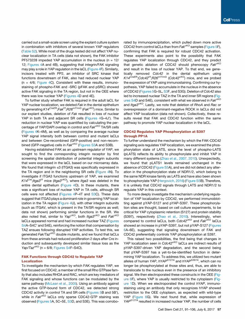
carried out a small-scale screen using the explant culture system
in combination with inhibitors of several known YAP regulators
(Table S2). While most of the drugs tested did not affect YAP nu-
clear localization in TA cells (data not shown), the FAK inhibitor
PF573228 impeded YAP accumulation in the nucleus (n = 12/
12; Figures 4A and 4B), suggesting that integrin/FAK signaling
may play a role in YAP regulation in the laCL (Figure 4F). Similarly,
incisors treated with PP2, an inhibitor of SRC kinase that
functions downstream of FAK, also had reduced nuclear YAP
(n = 4/6; Figure 4C). Consistent with these results, immuno-
staining of phospho-FAK and -SRC (pFAK and pSRC) showed
active FAK signaling in the TA region, but not in the OEE where
there was low nuclear YAP (Figures 4D and 4E).
To further study whether FAK is required in the adult laCL for
YAP nuclear localization, we deleted Fak in the dental epithelium
by generating K14CreER;Fakf/f;R26mT/mG (FakcKO) mice. Similar to
our explant studies, deletion of Fak resulted in loss of nuclear
YAP in both TA and adjacent SR cells (Figures 4G–4J’). The
reduction in nuclear YAP was quantified by calculating the per-
centage of YAP/DAPI overlap in control and FakcKO TA/SR cells
(Figures 4K–4M), as well as by comparing the average nuclear
YAP signal intensity both between control and mutant laCLs
and between Cre-recombined (GFP-positive) and non-recom-
bined (GFP-negative) cells in FakcKO (Figures S3A and S3B).
Having established FAK as an upstream regulator of YAP, we
sought to find the corresponding integrin receptor by first
screening the spatial distribution of potential integrin subunits
that were expressed in the laCL based on our microarray data.
We found that integrin a3 (ITGA3) was specifically expressed in
the TA region and in the neighboring SR cells (Figure 4N). To
investigate if ITGA3 functions upstream of YAP, we examined
K14Cre;Itga3f/f mice (Itga3cKO), where Itga3 was deleted in the
entire dental epithelium (Figure 4O). In these mutants, there
was a significant loss of nuclear YAP in TA cells, although SR
cells were not affected (Figures 4P–4T and S3C). These data
suggest that ITGA3 plays a dominant role in governing YAP local-
ization in the TA region (Figure 4U), with other integrin subunits
(such as ITGAV, which is present in the TA/SR regions as well;
data not shown) performing similar functions in the SR. We
also noted that, similar to YapcKO, both Itga3cKO and FakcKO
laCLs appeared normal and had increased nuclear TAZ (Figures
S4A–S4C’ and S4E), reinforcing the notion that compensation by
TAZ ensues following disrupted YAP activities. To test this, we
generated Fak/TazcKO double mutants, and we found that laCLs
from these animals had reduced proliferation 2 days after Cre in-
duction and subsequently developed similar tissue loss as the
Yap/TazcKO (n = 6/8; Figures S4F–S4O).
FAK Functions through CDC42 to Regulate YAPLocalizationTo investigate the mechanism by which FAK regulates YAP, we
first focused onCDC42, amember of the small RhoGTPase fam-
ily that also includes RHOA and RAC, which are key mediators of
FAK signaling and whose functions can be modulated by the
same pathway (McLean et al., 2005). Using an antibody against
the active GTP-bound form of CDC42, we detected strong
CDC42 activity in control TA and SR cells (Figures 5B and 5B’),
while in FakcKO laCLs only sparse CDC42-GTP staining was
observed (Figures 5A, 5C–5E, S3D, and S3E). This was corrobo-
rated by immunoprecipitation, which pulled down more active
CDC42 from control laCLs than from FakcKO samples (Figure 5F),
confirming that FAK is required for robust CDC42 activation.
These experiments also point to the possibility that FAK
regulates YAP localization through CDC42, and they predict
that genetic ablation of CDC42 should phenocopy FakcKO
and result in the loss of nuclear YAP. To that end, we gene-
tically removed Cdc42 in the dental epithelium using
K14CreER;Cdc42f/f;R26mT/mG (Cdc42cKO) mice, and we probed
the expression of YAP using immunostaining. Confirming our hy-
pothesis, YAP failed to accumulate in the nucleus in the absence
of CDC42 (Figures 5G–5L, S3F, and S3G). Deletion ofCdc42 also
led to increased nuclear TAZ in the TA and inner SR regions (Fig-
ures S4D and S4E), consistent with what we observed in FakcKO
and Itga3cKO. Lastly, we note that deletion of RhoA and Rac or
overexpression of a dominant-negative ROCK in laCLs did not
affect YAP localization (data not shown). Collectively, these re-
sults reveal that FAK and CDC42 function within the same
pathway to promote YAP nuclear localization in the laCL.
CDC42 Regulates YAP Phosphorylation at S397through PP1ATo further understand the mechanism by which the FAK-CDC42
signaling axis regulates YAP localization, we examined the phos-
phorylation state of LATS, since the level of phospho-LATS
(pLATS) reflects its ability to phosphorylate and inhibit YAP in
many different systems (Zhao et al., 2007, 2010). Unexpectedly,
we found that pLATS1 levels remained unchanged in the
absence of CDC42 (Figure S5A), and we did not detect any alter-
ation in the phosphorylation state of NDR1/2, which belong to
the same NDR kinase family as LATS and have also been shown
to phosphorylate YAP (Hergovich, 2016) (Figure S5B). Therefore,
it is unlikely that CDC42 signals through LATS and NDR1/2 to
regulate YAP in this context.
To more deeply investigate the mechanism underlying regula-
tion of YAP localization by CDC42, we performed immunoblot-
ting against pYAP-S127 and pYAP-S397. These phosphoryla-
tion sites are thought, based on cell culture experiments, to be
critical for YAP cytoplasmic retention (S127) and protein stability
(S397), respectively (Zhao et al., 2010). Interestingly, when
compared to control laCLs, both Cdc42cKO and FakcKO laCLs
showed an increase in pYAP-S397, but not pYAP-S127 (Figures
6A–6E), suggesting that signaling downstream of FAK and
CDC42 preferentially controls YAP phosphorylation at S397.
This raised two possibilities, the first being that changes in
YAP localization seen in Cdc42cKO laCLs are indirect results of
pYAP-S397-driven YAP degradation, and the second being
that pYAP-S397 has a yet-to-be-identified function in deter-
mining YAP localization. To address this, we utilized two mutant
alleles of human YAP, hYAPS127A and hYAPS397A, which can no
longer be phosphorylated at those sites and, thus, are able to
translocate to the nucleus even in the presence of an inhibitory
signal. We then electroporated these constructs in the OEE (Fig-
ure 6F), where YAP is usually restricted to the cytoplasm (Fig-
ure 1D). When we electroporated the control hYAP, immuno-
staining using an antibody that only recognizes hYAP showed
restriction to the OEE cytoplasm, as expected with wild-type
YAP (Figure 6G). We next found that, while expression of
YAPS127A resulted in increased nuclear YAP, the number of cells
Cell Stem Cell 21, 91–106, July 6, 2017 97

ED F
A B C
G
H I J
H’ I ’ J’
K L
K’ L’
M
N O
P Q
P’ Q’
R S
R’ S’
T U
(legend on next page)
98 Cell Stem Cell 21, 91–106, July 6, 2017

B C D
B’ C’ D’
E F
A
G H I
G’ H’ I’
J K L
J’ K’
Figure 5. CDC42 Acts Downstream of FAK to Regulate YAP Nuclear Localization
(A) Timeline depicting tamoxifen (Tam) treatment (black arrowheads) and sample collection (green and yellow arrowheads).
(B–E) Immunostaining of active CDC42 in control (B and B0) and FakcKO (C–D0) laCLs. The percentage of area with positive CDC42-GTP staining is calculated (E).
(F) Immunoprecipitation of CDC42-GTP followed by CDC42 immunoblotting. Relative expression between control and FakcKO is quantified.
(G–L) YAP immunostaining in control (G and G0) and Cdc42cKO (H–I0) laCLs. The percentage of YAP/DAPI overlap is quantified (J–L).
Representative images, cropped blots, and quantitative data are shown. Data are presented as mean ± SD (*p < 0.05, **p < 0.01, and ***p < 0.001). Dashed lines
outline laCLs. Scale bar (shown in K’) represents 50 mm in (B)–(D), (G)–(I), (J), and (K) and 9.76 mm in (B0)–(D0), (G0)–(I0), (J0), and (K0). See also Figure S4.
with nuclear YAP and the YAP signal intensity were both lower
than in hYAPS397A-electroporated cells (Figures 6G–6I), pointing
to YAP-S397 as the primary site for regulating YAP localization in
the laCL. The outcome of YAPS127A electroporation was also
similar to genetic overexpression of YAPS127A in laCLs, which
Figure 4. ITGA3/FAK Signaling Promotes YAP Nuclear Localization in
(A–C) YAP immunostaining in control (A), PF573228- and PP2-treated (B and C)
(D and E) pFAK (D) and pSRC (E) expression in laCLs.
(F) Schematic diagram of a TA cell, where FAK promotes YAP nuclear localizatio
(G) Timeline depicting tamoxifen (Tam) treatment (black arrowheads) and sample
(H–M) YAP immunostaining in control (H and H’) and FakcKO (I–J0) laCLs. The per
(N and O) ITGA3 expression in control (N) and Itga3cKO (O) laCLs.
(P–T) YAP immunostaining in control (P and P’) and Itga3cKO (Q and Q0) laCLs. T(U) Schematic diagram showing ITGA3/FAK signaling promotes nuclear YAP acc
Representative images and quantitative data are shown. All quantitative data are
outline laCLs. Scale bar (shown in S0) represents 50 mm in (A)–(E), (H)–(J), (K), (L),
also Figures S3 and S4.
was ineffective in driving YAP nuclear localization in the OEE
(Figures S6A–S6C). Finally, hYAPS127A,S397A electroporation pro-
duced the highest nuclear YAP (Figures 6G–6I), highlighting the
importance of both phosphorylation sites in controlling YAP
localization.
TA Cells
explants.
n.
collection (green arrowhead).
centage of YAP/DAPI overlap is quantified (K–M).
he percentage of YAP/DAPI overlap is quantified (R–T).
umulation in TA cells.
presented as mean ± SD (*p < 0.05, **p < 0.01, and ***p < 0.001). Dashed lines
(N)–(Q), (R), and (S) and 9.76 mm in (H0)–(J0), (K0), (L0), (P0), (Q0 ), (R0), and (S0). See
Cell Stem Cell 21, 91–106, July 6, 2017 99

B C
K L
K’ L’
D E
A
F G
H
I
J
M
(legend on next page)
100 Cell Stem Cell 21, 91–106, July 6, 2017

The preferential regulation at YAP-S397 also argues against a
LATS-dependent mechanism downstream of FAK/CDC42, as
LATS typically phosphorylates all YAP serine residues. Because
PP1A may dephosphorylate YAP primarily on S397 (Qi et al.,
2015), we tested whether PP1A binding to YAP was diminished
upon Cdc42 deletion. To that end, we performed co-immuno-
precipitation of YAP and PP1A, andwe found a significant reduc-
tion in pulled-down PP1A (but not PP2A, data not shown) in
Cdc42cKO laCL lysates (Figure 6J). This result then led to the pre-
diction that PP1A is critical for activating YAP localization in the
nucleus. Indeed, when cultured in the presence of okadaic acid,
a PP1 inhibitor, incisor explants displayed a dramatic loss of nu-
clear YAP in TA cells (Figures 6K–6M and S3H), thus establishing
an FAK/CDC42/PP1A signaling axis that governs YAP localiza-
tion in the incisor TA cells.
LATS1/2 Function in Parallel to Regulate YAPLocalizationThe results above, however, could not rule out the possibility that
LATS1/2 function in parallel to modulate YAP phosphorylation
and activity, and this hypothesis was supported by the presence
of abundant pYAP-S127 staining throughout the entire laCL (Fig-
ure S5D). To test this, we generated mice with Lats1 and Lats2
(Lats1/2cKO) double deletions in the dental epithelium, and we
observed a dramatic expansion of the dental epithelium in these
mice 1week after Cre activation (Figures 7A and 7B). Intriguingly,
deletions of Mst1 andMst2 did not result in any phenotype (Fig-
ures 7C, 7F, 7I, 7I’, and S5I–S5K), indicating that LATS1/2 activ-
ity is regulated by other kinases, which could include MAP4K
(Meng et al., 2015; Zheng et al., 2015). The hyperplasia seen in
Lats1/2cKO was limited to the TA region and the more distal am-
eloblasts, suggesting a differential response to the loss of Lats1/
2 in distinct cell types. This was corroborated by Ki67 staining,
which showed only a marginal increase in the OEE but a striking
upregulation in the more distal epithelium (Figures 7D and 7E).
The expansion of proliferating cells was indicative of an enlarged
TA region, and this was supported by thewidespread expression
of the TAmarker P-cadherin (Li et al., 2012) throughout the distal
epithelium (Figures S5E and S5F). Surprisingly, even though
Lats1/2cKO OEE was resistant to overproliferation, loss of
Lats1/2 resulted in increased nuclear YAP and the correspond-
ing RHEB expression in the entire laCL (Figures 7G–7H’, S5G,
and S5H), supporting the notion that nuclear accumulation of
YAP is not always sufficient to drive cell proliferation (Chen
et al., 2015). Thus, these experiments revealed that LATS1/2
are required in the laCL to restrain uncontrolled YAP activity
and may do so in parallel to the FAK-CDC42 signaling axis
described above.
Figure 6. CDC42 Signals through PP1A to Regulate YAP S397 Phosph
(A) Timeline indicating tamoxifen (Tam) injection (black arrowheads) and sample
(B–E) Immunoblotting and relative expression of pYAP-S397 and pYAP-S127 in
(F) Schematic diagram depicting delivery of YAP constructs to the OEE by elect
(G–I) YAP immunostaining and YAP/DAPI overlap in explants electroporated with
OEE (G). Three representative images are shown for each construct. The perce
quantified.
(J) YAP immunoprecipitation followed by PP1A detection. Relative expression of
(K–M) YAP immunostaining in control (K and K0) and okadaic acid-treated (L and
Representative images, cropped blots, and quantitative data are shown. Dashed
and (L), and 9.76 mm in (K0) and (L0 ). All data are presented as mean ± SD (*p < 0
DISCUSSION
The homeostatic maintenance of self-renewing tissues depends
on a continuous supply of differentiated cells from resident so-
matic stem cells. Using the adult mouse incisor as a model, we
have uncovered a novel signaling network regulating TA cell pro-
liferation and differentiation. Our data support a framework (Fig-
ure 7J) in which local induction of the integrin-FAK-CDC42
signaling axis modulates YAP phosphorylation at S397 to control
YAP localization and activity, which in turn govern progenitor cell
proliferation and differentiation by means of transcriptional regu-
lation of downstream effectors, such as RHEB. This pathway is
counterbalanced by LATS activity and can be compensated by
the functionally redundant TAZ. As a consequence, a robust sys-
tem is in place that can be tuned to ensure adequate production
of new cells, in order to meet the homeostatic demand of the tis-
sue, and support continuous growth of the tooth, which is critical
for the survival of the animal.
Maintenance of Progenitor Cells by YAP/TAZIn this study, we identified YAP/TAZ as key regulators of mouse
incisor renewal that promote TA cell proliferation, prevent
apoptosis, inhibit precocious differentiation in dental progenitor
cells, and maintain the overall structure of the tissue. This finding
thus provides a mechanism for regulating the expansion of pro-
genitor cells during continuous tissue renewal, and it resonates
with a growing body of work on the roles of YAP/TAZ in stem/
progenitor cells (Yu et al., 2015). Interestingly, the requirement
for YAP/TAZ in tissue homeostasis differs among organs. For
instance, while YAP is indispensable for cell proliferation in the
skin (Schlegelmilch et al., 2011), the mammary gland and intes-
tine remain relatively normal after Yap deletion (Cai et al., 2010;
Chen et al., 2014). Similarly, although Taz is essential for kidney
and lung development (Makita et al., 2008; Reginensi et al.,
2013), it is functionally redundant with YAP during heart and
craniofacial development (Wang et al., 2016; Xin et al., 2011).
Here we found that YAP/TAZ have overlapping functions in the
adult incisor, and ablation of Yap/Taz had a profound impact
on the maintenance of laCLs, especially the TA and SR regions.
The eventual loss of the entire laCL in the Yap/TazcKO is due to
either an absolute dependence of OEE cells on TA/SR cells or
on a yet-to-be-identified role of YAP in the OEE cytoplasm.
One potential cytoplasmic function of YAP/TAZ to be explored
in the future is engagement in WNT signaling (Varelas et al.,
2010), although the WNT pathway does not appear to be active
in the laCL (Suomalainen and Thesleff, 2010).
Our analysis of LatscKO laCLs also revealed differences be-
tween TA cells and DESCs/OEE cells in response to increased
orylation and Localization
collection (green and yellow arrowheads).
control, Cdc42cKO (B and C), and FakcKO (D and E) laCLs.
roporation.
hYAP, hYAP-S127A, hYAP-S397A, and hYAP-S127A,S397A (hYAP-dSA) in the
ntage of YAP/DAPI overlap (H) and average nuclear YAP pixel intensity (I) are
PP1A between control and Cdc42cKO is displayed.
L0 ) laCLs. The percentage of YAP/DAPI overlap is calculated (M).
lines outline laCLs. Scale bar (shown in L0) represents 15 mm in (G), 50 mm in (K)
.05, **p < 0.01, and ***p < 0.001). See also Figures S3, S5, and S6.
Cell Stem Cell 21, 91–106, July 6, 2017 101

A B C
D E F
G H I
G’ H’ I’
J
Figure 7. LATS1/2 Are Required to Prevent
Tissue Hyperplasia in the laCL and a Model
for YAP-Mediated Incisor Renewal
(A–C) H&E staining of control (A), Lats1/2cKO (B),
andMst1/2cKO (C) proximal incisors. Arrowhead in
(B) marks tissue hyperplasia.
(D–F) Ki67 expression in control (D), Lats1/2cKO (E),
and Mst1/2cKO (F) dental epithelium. Open
arrowhead in (E) marks the few proliferating cells in
Lats1/2cKO OEE.
(G–I0) YAP immunostaining and YAP/DAPI overlap
in control (G and G0), Lats1/2cKO (H and H0), andMst1/2cKO (I and I 0) laCLs. Arrowhead in (H) marks
increased nuclear YAP in Lats1/2cKO.
(J) Model for the regulation of incisor stem cell-
based dental renewal. ITGA3-positive TA and in-
ner SR cells are marked blue and DESCs/OEE
cells are marked brown. Red shade represents
YAP localization. In control TA cells, an ITGA3/
FAK/CDC42/PP1A signaling axis drives YAP nu-
clear localization, which promotes proliferation by
activating mTOR signaling and inhibits precocious
differentiation and apoptosis. When FAK signaling
is perturbed, loss of nuclear YAP is compensated
for by increased nuclear TAZ. In parallel, LATS1/2
fine-tune levels of nuclear YAP to prevent over-
proliferation. In contrast to TA cells, DESCs/OEE
cells are relatively inert and are resistant to YAP-
driven proliferation.
Representative images and quantitative data are
shown. Dashed lines outline the dental epithelium.
Scale bar (shown in I0) represents 130 mm in (A)–(F)
and 50 mm in (G)–(I0 ). See also Figure S5.
102 Cell Stem Cell 21, 91–106, July 6, 2017

nuclear YAP, as TA cells expanded into a multilayered structure
upon Lats1/2 deletion, and DESCs/OEE cells were resistant to
nuclear YAP-induced overproliferation. This thus points to the
possibility that nuclear YAP acts as a permissive signal and
that additional stimuli must be in place to drive proliferation.
One candidate for such signals are the FGFs secreted from the
mesenchyme overlying the TA cells. Attenuation of FGFR2b
signaling in the dental epithelium impeded TA cell proliferation,
and increased FGF signaling, due to the loss of Sprouty genes,
transformed the low-proliferating lingual CL into an laCL equiva-
lent (Klein et al., 2008; Parsa et al., 2010). Indeed, FGF signaling
has been shown to be required for YAP-induced proliferation in
other contexts (Hua et al., 2016). Alternatively, the presence of
nuclear YAP in LatscKO OEE cells is counterbalanced by a
compensatory decrease in the overall YAP protein level (Chen
et al., 2015). Finally, cells in the OEE are more densely clustered
than TA cells, and they express cell adhesion molecules, such as
E-cadherin and Claudin1, that are absent in TA cells (Li et al.,
2012) and may add further control over cell proliferation.
Regulation of YAP by Integrin/FAK Signaling inProgenitor CellsAn important question in the field of Hippo signaling and stem
cell biology is understanding howYAP activity is triggered to pro-
mote the expansion of tissue progenitors. We found that this is
achieved in the incisor by restricted expression of ITGA3 and
the corresponding activation of FAK signaling in the TA region,
which subsequently promotes YAP nuclear localization through
CDC42. Regulation of YAP by FAK signaling has been recently
observed in other stem cell systems, including skeletal and
epithelial stem cells (Elbediwy et al., 2016; Tang et al., 2013).
However, in these cases, RHOA was placed downstream of
FAK, and CDC42 was instead an inhibitory signal through its
role in apical polarity formation. The differences could be due
to the use of distinct experimental models, as previous results
were derived from cell culture studies. Along these lines,
CDC42 is an essential regulator of YAP during kidney develop-
ment and for podocyte survival, while deletion of RhoA and
Rac had little effect (Huang et al., 2016; Reginensi et al., 2013),
suggesting that CDC42 may be the predominant Rho GTPase
for YAP regulation in vivo.
We also noted that YAP and TAZ are differentially regulated in
the laCL, with TAZ being a compensatory effector when YAP or
FAK activity is disrupted. This is similar to an earlier study, where
hepatic or intestinal deletion of Yap resulted in TAZ nuclear local-
ization (Moroishi et al., 2015), demonstrating that TAZ can func-
tion as a reserve pool in vivo that becomes activated in response
to Yap loss. Indeed, deletion of both Fak and Taz phenocopies
Yap/TazcKO, although with a milder phenotype, likely due to re-
sidual nuclear YAP in some cells.
Another critical aspect of the ITGA3/FAK/CDC42 signaling
axis is that it is independent of LATS activity. Instead, we identi-
fied PP1A as an important modulator of YAP phosphorylation
downstream of CDC42. PP1A itself could potentially be acti-
vated by the CDC42 effector PAK2 (Zhang et al., 2013), and
PAK2 activity was reduced in Cdc42cKO laCLs (Figure S5C).
Interestingly, ITGA3/FAK/CDC42 signaling predominantly con-
trols YAP phosphorylation at S397, but not S127, a phenomenon
that has been previously shown in Netrin-1-induced PP1A
dephosphorylation of YAP (Qi et al., 2015). As S397 phosphory-
lation affects YAP stability in vitro (Zhao et al., 2010), it is possible
that, in the laCL, FAK signaling modulates YAP localization indi-
rectly by maintaining YAP protein levels above a certain
threshold. However, as YAP levels were comparable in control,
FakcKO and Cdc42cKO laCLs, an alternative explanation is that
pYAP-S397 directly contributes to YAP localization. This is
supported by two observations as follows: first, overexpression
of YAPS127A did not result in an efficient upregulation of
nuclear YAP in the laCL, and, second, electroporation of an
hYAPS127A,S397A construct resulted in higher nuclear YAP locali-
zation than YAPS127A. Taken together, our results indicate that
the ITGA3/FAK/CDC42 signaling axis functions in parallel to
LATS to promote nuclear YAP localization through dephosphor-
ylation at YAP-S397.
The signaling axis described here likely functions in other stem
cell settings as well, as integrin/FAK signaling is prevalent in
many different stem cell niches and is critical for maintaining
cell proliferation, preserving the stem cell population and
balancing renewal and differentiation (Prowse et al., 2011). For
instance, conditional deletion of b1 integrin in the skin results
in severe reduction of proliferation (Raghavan et al., 2000),
whereas heightened integrin signaling potentiates cancer stem
cell activities (Seguin et al., 2015). Indeed, a3b1 is crucial for pro-
moting proliferation and tumor growth in skin cancers (Sachs
et al., 2012), and it is plausible that YAP acts downstream of
the aberrant signaling, as well as in other normal or pathological
conditions where integrin signaling plays a role.
Transcriptional Outputs of YAP/TAZThe role of YAP/TAZ in transcriptional regulation has been well
characterized (Yu et al., 2015), and in the incisor we found that
YAP/TAZ facilitate TA cell expansion in part through their control
of Rheb expression and, thus, mTOR activity. As a central
effector of cell growth and proliferation, mTOR signaling has
also been shown to mediate YAP function elsewhere (Hansen
et al., 2015; Tumaneng et al., 2012), and our data add to the
growing evidence that YAP is able to induce mTOR signaling
through several different pathways.
Importantly, both Rapamycin-treated and RptorcKO laCLs did
not present any obvious loss of cell-cell adhesion analogous to
what we observed in Yap/TazcKO, suggesting that additional
downstream genes were responsible for the cell adhesion
phenotype. One potential candidate is the cell adhesion mole-
cule P-cadherin (encoded by Cdh3), which was downregulated
both at the RNA and protein levels (Figures 2B and S2O–S2R)
along with other genes, such as Serpinh1, Dpysl2, and Pfn2,
that are also important for cytoskeletal regulation (Figure 2A).
As a result, YAP/TAZ may maintain tissue integrity by controlling
the expression of these genes to modulate cellular tension and
extracellular matrix (ECM) environment, in line with a recent
finding in zebrafish (Porazinski et al., 2015).
Finally, YAP/TAZ are critical for inhibiting the expression of
genes that are associated with differentiated cells. In Yap/TazcKO
laCLs, activation of these genes primarily occurs in SR cells,
likely because these cells are further along in the differentiation
process and, therefore, more sensitive to the loss of YAP/TAZ.
It is currently unknown whether YAP/TAZ directly regulate the
expression of these genes, and it will be important to address
Cell Stem Cell 21, 91–106, July 6, 2017 103

this in future experiments and in other tissues, which may shed
light on how YAP/TAZ govern the balance between stem cell
proliferation and differentiation. Taken together, these studies
have uncovered a novel FAK-YAP-mTOR signaling pathway
that governs proliferation and differentiation in tissue progenitor
cells. This work helps to provide a framework for future research
into the roles of integrins and YAP in both normal and patholog-
ical conditions, as well as to develop strategies for stem cell-
based regeneration of dental and other mineralized tissues.
STAR+METHODS
Detailed methods are provided in the online version of this paper
and include the following:
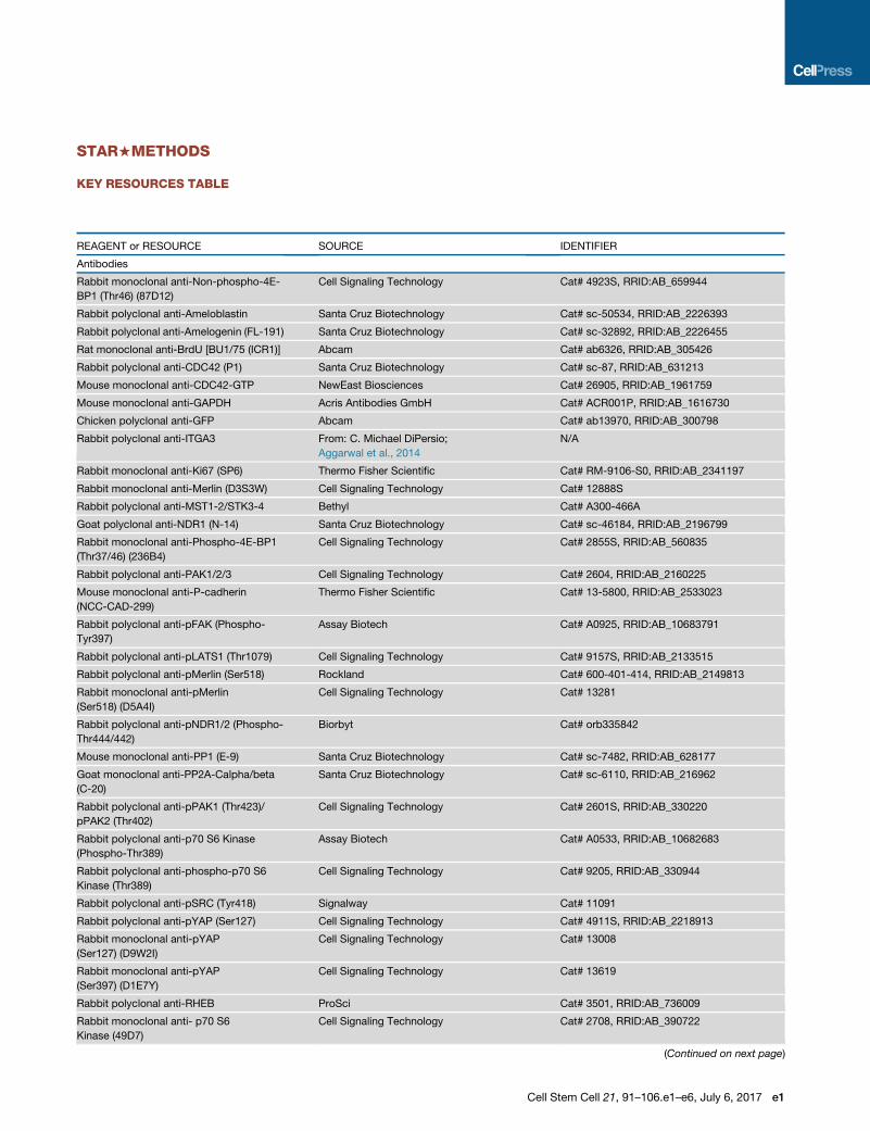
d KEY RESOURCES TABLE
d CONTACT FOR REAGENT AND RESOURCE SHARING
d EXPERIMENTAL MODEL AND SUBJECT DETAILS
104
B Mouse lines and induction of alleles
B Sample sources for laCL cells and explants
d METHOD DETAILS
B Tissue preparation and histological analysis
B Immunofluorescence staining
B In situ hybridization
B Microtomography
B Isolation of laCLs
B Explant culture
B Electroporation
B Colony formation assay
B Western blot and immunoprecipitation
B Expression profiling by microarray
B qPCR analysis
d STATISTICAL ANALYSIS
B Statistics
B ImageJ image analysis
d DATA AVAILABILITY
SUPPLEMENTAL INFORMATION
Supplemental Information includes six figures and five tables and can be found
with this article online at http://dx.doi.org/10.1016/j.stem.2017.03.023.
AUTHOR CONTRIBUTIONS
Conceptualization, J.K.-H.H. and O.D.K.; Methodology, J.K.-H.H. and O.D.K.;
Investigation, J.K.-H.H., W.D., and C.M.D.; Formal Analysis, S.J.S.; Writing –
Original Draft, J.K.-H.H. and O.D.K.; Writing – Review & Editing, J.K.-H.H.
and O.D.K.; Resources, C.M.D.; Funding Acquisition, J.K.-H.H., M.C.O.,
C.M.D., and O.D.K.; Supervision, O.D.K.
ACKNOWLEDGMENTS
We thank Sarah Alto, Rebecca d’Urso, and Nicholas Wang for assistance with
the mouse colony; Dr. Amnon Sharir for help with mCT imaging; Derek Power
for technical assistance; Drs. Anandika Aggarwal, Aaditi Mujumdar, and Chan-
dana Vundavalli for assistance with sample preparation; members of the Klein
laboratory, Dr. Jeffrey Bush, and Dr. Valerie Weaver for helpful discussions;
and Drs. Eric Olson, Randy Johnson, Louis Reichardt, Fernando Camargo,
and Cord Brakebusch and RIKEN BRC for mouse lines. This work was funded
by NIDCR R01-DE024988 and R35-DE026602 to O.D.K. and F32-DE023705
and K99-DE025874 to J.K.-H.H.
Cell Stem Cell 21, 91–106, July 6, 2017
Received: October 10, 2016
Revised: February 7, 2017
Accepted: March 26, 2017
Published: April 27, 2017
REFERENCES
Aggarwal, A., Al-Rohil, R.N., Batra, A., Feustel, P.J., Jones, D.M., andDiPersio,
C.M. (2014). Expression of integrin a3b1 and cyclooxygenase-2 (COX2) are
positively correlated in human breast cancer. BMC Cancer 14, 459.
Bai, H., Zhang, N., Xu, Y., Chen, Q., Khan, M., Potter, J.J., Nayar, S.K.,
Cornish, T., Alpini, G., Bronk, S., et al. (2012). Yes-associated protein regulates
the hepatic response after bile duct ligation. Hepatology 56, 1097–1107.
Baldi, P., and Long, A.D. (2001). A Bayesian framework for the analysis of mi-
croarray expression data: regularized t -test and statistical inferences of gene
changes. Bioinformatics 17, 509–519.
Beggs, H.E., Schahin-Reed, D., Zang, K., Goebbels, S., Nave, K.A., Gorski, J.,
Jones, K.R., Sretavan, D., and Reichardt, L.F. (2003). FAK deficiency in cells
contributing to the basal lamina results in cortical abnormalities resembling
congenital muscular dystrophies. Neuron 40, 501–514.
Benjamini, Y., and Hochberg, Y. (1995). Controlling the false discovery rate: a
practical and powerful approach to multiple testing. J. R. Stat. Soc. Series B
(Methodological) 57, 289–300.
Biehs, B., Hu, J.K., Strauli, N.B., Sangiorgi, E., Jung, H., Heber, R.P., Ho, S.,
Goodwin, A.F., Dasen, J.S., Capecchi, M.R., and Klein, O.D. (2013). BMI1 re-
presses Ink4a/Arf and Hox genes to regulate stem cells in the rodent incisor.
Nat. Cell Biol. 15, 846–852.
Cai, J., Zhang, N., Zheng, Y., deWilde, R.F., Maitra, A., and Pan, D. (2010). The
Hippo signaling pathway restricts the oncogenic potential of an intestinal
regeneration program. Genes Dev. 24, 2383–2388.
Camargo, F.D., Gokhale, S., Johnnidis, J.B., Fu, D., Bell, G.W., Jaenisch, R.,
and Brummelkamp, T.R. (2007). YAP1 increases organ size and expands un-
differentiated progenitor cells. Curr. Biol. 17, 2054–2060.
Chen, T., Heller, E., Beronja, S., Oshimori, N., Stokes, N., and Fuchs, E. (2012).
An RNA interference screen uncovers a newmolecule in stem cell self-renewal
and long-term regeneration. Nature 485, 104–108.
Chen, Q., Zhang, N., Gray, R.S., Li, H., Ewald, A.J., Zahnow, C.A., and Pan, D.
(2014). A temporal requirement for Hippo signaling in mammary gland differen-
tiation, growth, and tumorigenesis. Genes Dev. 28, 432–437.
Chen, Q., Zhang, N., Xie, R.,Wang,W., Cai, J., Choi, K.-S., David, K.K., Huang,
B., Yabuta, N., Nojima, H., et al. (2015). Homeostatic control of Hippo signaling
activity revealed by an endogenous activatingmutation in YAP. Genes Dev. 29,
1285–1297.
Damljanovi�c, V., Lagerholm, B.C., and Jacobson, K. (2005). Bulk and micro-
patterned conjugation of extracellular matrix proteins to characterized poly-
acrylamide substrates for cell mechanotransduction assays. Biotechniques
39, 847–851.
Dassule, H.R., Lewis, P., Bei, M., Maas, R., and McMahon, A.P. (2000). Sonic
hedgehog regulates growth and morphogenesis of the tooth. Development
127, 4775–4785.
Dupont, S., Morsut, L., Aragona, M., Enzo, E., Giulitti, S., Cordenonsi, M.,
Zanconato, F., Le Digabel, J., Forcato, M., Bicciato, S., et al. (2011). Role of
YAP/TAZ in mechanotransduction. Nature 474, 179–183.
Elbediwy, A., Vincent-Mistiaen, Z.I., Spencer-Dene, B., Stone, R.K., Boeing,
S., Wculek, S.K., Cordero, J., Tan, E.H., Ridgway, R., Brunton, V.G., et al.
(2016). Integrin signalling regulates YAP and TAZ to control skin homeostasis.
Development 143, 1674–1687.
Glogauer, M., Marchal, C.C., Zhu, F., Worku, A., Clausen, B.E., Foerster, I.,
Marks, P., Downey, G.P., Dinauer, M., and Kwiatkowski, D.J. (2003). Rac1
deletion in mouse neutrophils has selective effects on neutrophil functions.
J. Immunol. 170, 5652–5657.
Hansen, C.G., Ng, Y.L.D., Lam,W.-L.M., Plouffe, S.W., and Guan, K.-L. (2015).
The Hippo pathway effectors YAP and TAZ promote cell growth by modulating
amino acid signaling to mTORC1. Cell Res. 25, 1299–1313.

Hara, K., Maruki, Y., Long, X., Yoshino, K., Oshiro, N., Hidayat, S., Tokunaga,
C., Avruch, J., and Yonezawa, K. (2002). Raptor, a binding partner of target of
rapamycin (TOR), mediates TOR action. Cell 110, 177–189.
Harada, H., Kettunen, P., Jung, H.S., Mustonen, T., Wang, Y.A., and Thesleff, I.
(1999). Localization of putative stem cells in dental epithelium and their asso-
ciation with Notch and FGF signaling. J. Cell Biol. 147, 105–120.
Hay, N., and Sonenberg, N. (2004). Upstream and downstream of mTOR.
Genes Dev. 18, 1926–1945.
Heallen, T., Zhang, M., Wang, J., Bonilla-Claudio, M., Klysik, E., Johnson, R.L.,
and Martin, J.F. (2011). Hippo pathway inhibits Wnt signaling to restrain cardi-
omyocyte proliferation and heart size. Science 332, 458–461.
Hergovich, A. (2016). The Roles of NDR Protein Kinases in Hippo Signalling.
Genes (Basel) 7, E21.
Hua, G., Lv, X., He, C., Remmenga, S.W., Rodabough, K.J., Dong, J., Yang, L.,
Lele, S.M., Yang, P., Zhou, J., et al. (2016). YAP induces high-grade serous
carcinoma in fallopian tube secretory epithelial cells. Oncogene 35,
2247–2265.
Huang, Z., Zhang, L., Chen, Y., Zhang, H., Zhang, Q., Li, R., Ma, J., Li, Z., Yu,
C., Lai, Y., et al. (2016). Cdc42 deficiency induces podocyte apoptosis by in-
hibiting the Nwasp/stress fibers/YAP pathway. Cell Death Dis. 7, e2142.
Jackson, B., Peyrollier, K., Pedersen, E., Basse, A., Karlsson, R., Wang, Z.,
Lefever, T., Ochsenbein, A.M., Schmidt, G., Aktories, K., et al. (2011). RhoA
is dispensable for skin development, but crucial for contraction and directed
migration of keratinocytes. Mol. Biol. Cell 22, 593–605.
Johnson, W.E., Li, C., and Rabinovic, A. (2007). Adjusting batch effects in mi-
croarray expression data using empirical Bayes methods. Biostatistics 8,
118–127.
Juuri, E., Saito, K., Ahtiainen, L., Seidel, K., Tummers, M., Hochedlinger, K.,
Klein, O.D., Thesleff, I., and Michon, F. (2012). Sox2+ stem cells contribute
to all epithelial lineages of the tooth via Sfrp5+ progenitors. Dev. Cell 23,
317–328.
Kaukua, N., Shahidi, M.K., Konstantinidou, C., Dyachuk, V., Kaucka, M.,
Furlan, A., An, Z., Wang, L., Hultman, I., Ahrlund-Richter, L., et al. (2014).
Glial origin of mesenchymal stem cells in a tooth model system. Nature 513,
551–554.
Kayala, M.A., and Baldi, P. (2012). Cyber-T web server: differential analysis of
high-throughput data. Nucleic Acids Res. 40, W553–559.
Klein, O.D., Lyons, D.B., Balooch, G., Marshall, G.W., Basson, M.A., Peterka,
M., Boran, T., Peterkova, R., and Martin, G.R. (2008). An FGF signaling loop
sustains the generation of differentiated progeny from stem cells in mouse in-
cisors. Development 135, 377–385.
Kobayashi, K., Takahashi, M., Matsushita, N., Miyazaki, J., Koike, M.,
Yaginuma, H., Osumi, N., Kaibuchi, K., and Kobayashi, K. (2004). Survival of
developing motor neurons mediated by Rho GTPase signaling pathway
through Rho-kinase. J. Neurosci. 24, 3480–3488.
Lange, A.W., Sridharan, A., Xu, Y., Stripp, B.R., Perl, A.-K., and Whitsett, J.A.
(2015). Hippo/Yap signaling controls epithelial progenitor cell proliferation and
differentiation in the embryonic and adult lung. J. Mol. Cell Biol. 7, 35–47.
Laplante, M., and Sabatini, D.M. (2009). mTOR signaling at a glance. J. Cell
Sci. 122, 3589–3594.
Levy, D., Adamovich, Y., Reuven, N., and Shaul, Y. (2008). Yap1 phosphoryla-
tion by c-Abl is a critical step in selective activation of proapoptotic genes in
response to DNA damage. Mol. Cell 29, 350–361.
Li, M., Indra, A.K., Warot, X., Brocard, J., Messaddeq, N., Kato, S., Metzger,
D., and Chambon, P. (2000). Skin abnormalities generated by temporally
controlled RXRalpha mutations in mouse epidermis. Nature 407, 633–636.
Li, C.-Y., Cha, W., Luder, H.-U., Charles, R.-P., McMahon, M., Mitsiadis, T.A.,
and Klein, O.D. (2012). E-cadherin regulates the behavior and fate of epithelial
stem cells and their progeny in the mouse incisor. Dev. Biol. 366, 357–366.
Li, P., Silvis, M.R., Honaker, Y., Lien, W.-H., Arron, S.T., and Vasioukhin, V.
(2016). aE-catenin inhibits a Src-YAP1 oncogenic module that couples tyro-
sine kinases and the effector of Hippo signaling pathway. Genes Dev. 30,
798–811.
Livak, K.J., and Schmittgen, T.D. (2001). Analysis of relative gene expression
data using real-time quantitative PCR and the 2(-Delta Delta C(T)) Method.
Methods 25, 402–408.
Lu, L., Li, Y., Kim, S.M., Bossuyt, W., Liu, P., Qiu, Q., Wang, Y., Halder, G.,
Finegold,M.J., Lee, J.-S., and Johnson, R.L. (2010). Hippo signaling is a potent
in vivo growth and tumor suppressor pathway in the mammalian liver. Proc.
Natl. Acad. Sci. USA 107, 1437–1442.
Makita, R., Uchijima, Y., Nishiyama, K., Amano, T., Chen, Q., Takeuchi, T.,
Mitani, A., Nagase, T., Yatomi, Y., Aburatani, H., et al. (2008). Multiple renal
cysts, urinary concentration defects, and pulmonary emphysematous
changes in mice lacking TAZ. Am. J. Physiol. Renal Physiol. 294, F542–F553.
McLean, G.W., Carragher, N.O., Avizienyte, E., Evans, J., Brunton, V.G., and
Frame, M.C. (2005). The role of focal-adhesion kinase in cancer - a new ther-
apeutic opportunity. Nat. Rev. Cancer 5, 505–515.
Meng, Z., Moroishi, T., Mottier-Pavie, V., Plouffe, S.W., Hansen, C.G., Hong,
A.W., Park, H.W., Mo, J.-S., Lu, W., Lu, S., et al. (2015). MAP4K family kinases
act in parallel to MST1/2 to activate LATS1/2 in the Hippo pathway. Nat.
Commun. 6, 8357.
Mitchell, K., Szekeres, C., Milano, V., Svenson, K.B., Nilsen-Hamilton, M.,
Kreidberg, J.A., and DiPersio, C.M. (2009). Alpha3beta1 integrin in epidermis
promotes wound angiogenesis and keratinocyte-to-endothelial-cell crosstalk
through the induction of MRP3. J. Cell Sci. 122, 1778–1787.
Mo, J.-S., Meng, Z., Kim, Y.C., Park, H.W., Hansen, C.G., Kim, S., Lim, D.-S.,
and Guan, K.-L. (2015). Cellular energy stress induces AMPK-mediated regu-
lation of YAP and the Hippo pathway. Nat. Cell Biol. 17, 500–510.
Moroishi, T., Park, H.W., Qin, B., Chen, Q., Meng, Z., Plouffe, S.W., Taniguchi,
K., Yu, F.-X., Karin, M., Pan, D., and Guan, K.L. (2015). A YAP/TAZ-induced
feedback mechanism regulates Hippo pathway homeostasis. Genes Dev.
29, 1271–1284.
Muzumdar, M.D., Tasic, B., Miyamichi, K., Li, L., and Luo, L. (2007). A global
double-fluorescent Cre reporter mouse. Genesis 45, 593–605.
Oldham, M.C., Langfelder, P., and Horvath, S. (2012). Network methods
for describing sample relationships in genomic datasets: application to
Huntington’s disease. BMC Syst. Biol. 6, 63.
Parsa, S., Kuremoto, K., Seidel, K., Tabatabai, R., Mackenzie, B., Yamaza, T.,
Akiyama, K., Branch, J., Koh, C.J., Al Alam, D., et al. (2010). Signaling by
FGFR2b controls the regenerative capacity of adult mouse incisors.
Development 137, 3743–3752.
Porazinski, S., Wang, H., Asaoka, Y., Behrndt, M., Miyamoto, T., Morita, H.,
Hata, S., Sasaki, T., Krens, S.F.G., Osada, Y., et al. (2015). YAP is essential
for tissue tension to ensure vertebrate 3D body shape. Nature 521, 217–221.
Prowse, A.B.J., Chong, F., Gray, P.P., and Munro, T.P. (2011). Stem cell integ-
rins: implications for ex-vivo culture and cellular therapies. Stem Cell Res.
(Amst.) 6, 1–12.
Qi, Q., Li, D.Y., Luo, H.R., Guan, K.-L., and Ye, K. (2015). Netrin-1 exerts onco-
genic activities through enhancing Yes-associated protein stability. Proc. Natl.
Acad. Sci. USA 112, 7255–7260.
Raghavan, S., Bauer, C., Mundschau, G., Li, Q., and Fuchs, E. (2000).
Conditional ablation of beta1 integrin in skin. Severe defects in epidermal pro-
liferation, basement membrane formation, and hair follicle invagination. J. Cell
Biol. 150, 1149–1160.
Reginensi, A., Scott, R.P., Gregorieff, A., Bagherie-Lachidan, M., Chung, C.,
Lim, D.-S., Pawson, T., Wrana, J., and McNeill, H. (2013). Yap- and Cdc42-
dependent nephrogenesis and morphogenesis during mouse kidney develop-
ment. PLoS Genet. 9, e1003380.
Sachs, N., Secades, P., van Hulst, L., Kreft, M., Song, J.-Y., and Sonnenberg,
A. (2012). Loss of integrin a3 prevents skin tumor formation by promoting
epidermal turnover and depletion of slow-cycling cells. Proc. Natl. Acad.
Sci. USA 109, 21468–21473.
Schlegelmilch, K.,Mohseni,M., Kirak,O., Pruszak, J., Rodriguez, J.R., Zhou,D.,
Kreger, B.T., Vasioukhin, V., Avruch, J.,Brummelkamp, T.R., andCamargo, F.D.
(2011). Yap1 acts downstream of a-catenin to control epidermal proliferation.
Cell 144, 782–795.
Cell Stem Cell 21, 91–106, July 6, 2017 105

Seguin, L., Desgrosellier, J.S., Weis, S.M., and Cheresh, D.A. (2015). Integrins
and cancer: regulators of cancer stemness, metastasis, and drug resistance.
Trends Cell Biol. 25, 234–240.
Seidel, K., Ahn, C.P., Lyons, D., Nee, A., Ting, K., Brownell, I., Cao, T., Carano,
R.A.D., Curran, T., Schober, M., et al. (2010). Hedgehog signaling regulates the
generation of ameloblast progenitors in the continuously growing mouse
incisor. Development 137, 3753–3761.
Sengupta, S., Peterson, T.R., Laplante, M., Oh, S., and Sabatini, D.M. (2010).
mTORC1 controls fasting-induced ketogenesis and its modulation by ageing.
Nature 468, 1100–1104.
Suomalainen,M., and Thesleff, I. (2010). Patterns ofWnt pathway activity in the
mouse incisor indicate absence of Wnt/beta-catenin signaling in the epithelial
stem cells. Dev. Dyn. 239, 364–372.
Szymaniak, A.D., Mahoney, J.E., Cardoso, W.V., and Varelas, X. (2015).
Crumbs3-Mediated Polarity Directs Airway Epithelial Cell Fate through the
Hippo Pathway Effector Yap. Dev. Cell 34, 283–296.
Tang, Y., Rowe, R.G., Botvinick, E.L., Kurup, A., Putnam, A.J., Seiki, M.,
Weaver, V.M., Keller, E.T., Goldstein, S., Dai, J., et al. (2013). MT1-MMP-
dependent control of skeletal stem cell commitment via a b1-integrin/YAP/
TAZ signaling axis. Dev. Cell 25, 402–416.
Tumaneng, K., Schlegelmilch, K., Russell, R.C., Yimlamai, D., Basnet, H.,
Mahadevan, N., Fitamant, J., Bardeesy, N., Camargo, F.D., and Guan, K.-L.
(2012). YAP mediates crosstalk between the Hippo and PI(3)K–TOR pathways
by suppressing PTEN via miR-29. Nat. Cell Biol. 14, 1322–1329.
Varelas, X., Miller, B.W., Sopko, R., Song, S., Gregorieff, A., Fellouse, F.A.,
Sakuma, R., Pawson, T., Hunziker, W., McNeill, H., et al. (2010). The Hippo
pathway regulates Wnt/b-catenin signaling. Dev. Cell 18, 579–591.
Wabik, A., and Jones, P.H. (2015). Switching roles: the functional plasticity of
adult tissue stem cells. EMBO J. 34, 1164–1179.
Waikel, R.L., Kawachi, Y., Waikel, P.A., Wang, X.-J., and Roop, D.R. (2001).
Deregulated expression of c-Myc depletes epidermal stem cells. Nat. Genet.
28, 165–168.
Wang, J., Xiao, Y., Hsu, C.-W., Martinez-Traverso, I.M., Zhang, M., Bai, Y.,
Ishii, M., Maxson, R.E., Olson, E.N., Dickinson, M.E., et al. (2016). Yap and
Taz play a crucial role in neural crest-derived craniofacial development.
Development 143, 504–515.
White, A.C., Khuu, J.K., Dang, C.Y., Hu, J., Tran, K.V., Liu, A., Gomez, S.,
Zhang, Z., Yi, R., Scumpia, P., et al. (2014). Stem cell quiescence acts as a
tumour suppressor in squamous tumours. Nat. Cell Biol. 16, 99–107.
106 Cell Stem Cell 21, 91–106, July 6, 2017
Wu, X., Quondamatteo, F., Lefever, T., Czuchra, A., Meyer, H., Chrostek, A.,
Paus, R., Langbein, L., and Brakebusch, C. (2006). Cdc42 controls progenitor
cell differentiation and beta-catenin turnover in skin. Genes Dev. 20, 571–585.
Xin, M., Kim, Y., Sutherland, L.B., Qi, X., McAnally, J., Schwartz, R.J.,
Richardson, J.A., Bassel-Duby, R., and Olson, E.N. (2011). Regulation of insu-
lin-like growth factor signaling by Yap governs cardiomyocyte proliferation and
embryonic heart size. Sci. Signal. 4, ra70.
Xin, M., Kim, Y., Sutherland, L.B., Murakami, M., Qi, X., McAnally, J., Porrello,
E.R., Mahmoud, A.I., Tan, W., Shelton, J.M., et al. (2013). Hippo pathway
effector Yap promotes cardiac regeneration. Proc. Natl. Acad. Sci. USA 110,
13839–13844.
Yilmaz, O.H., Valdez, R., Theisen, B.K., Guo, W., Ferguson, D.O., Wu, H., and
Morrison, S.J. (2006). Pten dependence distinguishes haematopoietic stem
cells from leukaemia-initiating cells. Nature 441, 475–482.
Yu, F.-X., Zhao, B., Panupinthu, N., Jewell, J.L., Lian, I., Wang, L.H., Zhao, J.,
Yuan, H., Tumaneng, K., Li, H., et al. (2012). Regulation of the Hippo-YAP
pathway by G-protein-coupled receptor signaling. Cell 150, 780–791.
Yu, F.-X., Zhao, B., and Guan, K.-L. (2015). Hippo Pathway in Organ Size
Control, Tissue Homeostasis, and Cancer. Cell 163, 811–828.
Zhang, H., Wang, Z., and Zhang, Z. (2013). PP1a, PP1b and Wip-1 regulate
H4S47 phosphorylation and deposition of histone H3 variant H3.3. Nucleic
Acids Res. 41, 8085–8093.
Zhao, B., Wei, X., Li, W., Udan, R.S., Yang, Q., Kim, J., Xie, J., Ikenoue, T., Yu,
J., Li, L., et al. (2007). Inactivation of YAP oncoprotein by the Hippo pathway is
involved in cell contact inhibition and tissue growth control. Genes Dev. 21,
2747–2761.
Zhao, B., Li, L., Tumaneng, K., Wang, C.-Y., and Guan, K.-L. (2010). A coordi-
nated phosphorylation by Lats and CK1 regulates YAP stability through
SCF(beta-TRCP). Genes Dev. 24, 72–85.
Zheng, Y., Wang, W., Liu, B., Deng, H., Uster, E., and Pan, D. (2015).
Identification of Happyhour/MAP4K as Alternative Hpo/Mst-like Kinases in
the Hippo Kinase Cascade. Dev. Cell 34, 642–655.
Zhou, D., Zhang, Y., Wu, H., Barry, E., Yin, Y., Lawrence, E., Dawson, D., Willis,
J.E., Markowitz, S.D., Camargo, F.D., and Avruch, J. (2011). Mst1 and Mst2
protein kinases restrain intestinal stem cell proliferation and colonic tumori-
genesis by inhibition of Yes-associated protein (Yap) overabundance. Proc.
Natl. Acad. Sci. USA 108, E1312–E1320.
STAR+METHODS
KEY RESOURCES TABLE
REAGENT or RESOURCE SOURCE IDENTIFIER
Antibodies
Rabbit monoclonal anti-Non-phospho-4E-
BP1 (Thr46) (87D12)
Cell Signaling Technology Cat# 4923S, RRID:AB_659944
Rabbit polyclonal anti-Ameloblastin Santa Cruz Biotechnology Cat# sc-50534, RRID:AB_2226393
Rabbit polyclonal anti-Amelogenin (FL-191) Santa Cruz Biotechnology Cat# sc-32892, RRID:AB_2226455
Rat monoclonal anti-BrdU [BU1/75 (ICR1)] Abcam Cat# ab6326, RRID:AB_305426
Rabbit polyclonal anti-CDC42 (P1) Santa Cruz Biotechnology Cat# sc-87, RRID:AB_631213
Mouse monoclonal anti-CDC42-GTP NewEast Biosciences Cat# 26905, RRID:AB_1961759
Mouse monoclonal anti-GAPDH Acris Antibodies GmbH Cat# ACR001P, RRID:AB_1616730
Chicken polyclonal anti-GFP Abcam Cat# ab13970, RRID:AB_300798
Rabbit polyclonal anti-ITGA3 From: C. Michael DiPersio;
Aggarwal et al., 2014
N/A
Rabbit monoclonal anti-Ki67 (SP6) Thermo Fisher Scientific Cat# RM-9106-S0, RRID:AB_2341197
Rabbit monoclonal anti-Merlin (D3S3W) Cell Signaling Technology Cat# 12888S
Rabbit polyclonal anti-MST1-2/STK3-4 Bethyl Cat# A300-466A
Goat polyclonal anti-NDR1 (N-14) Santa Cruz Biotechnology Cat# sc-46184, RRID:AB_2196799
Rabbit monoclonal anti-Phospho-4E-BP1
(Thr37/46) (236B4)
Cell Signaling Technology Cat# 2855S, RRID:AB_560835
Rabbit polyclonal anti-PAK1/2/3 Cell Signaling Technology Cat# 2604, RRID:AB_2160225
Mouse monoclonal anti-P-cadherin
(NCC-CAD-299)
Thermo Fisher Scientific Cat# 13-5800, RRID:AB_2533023
Rabbit polyclonal anti-pFAK (Phospho-
Tyr397)
Assay Biotech Cat# A0925, RRID:AB_10683791
Rabbit polyclonal anti-pLATS1 (Thr1079) Cell Signaling Technology Cat# 9157S, RRID:AB_2133515
Rabbit polyclonal anti-pMerlin (Ser518) Rockland Cat# 600-401-414, RRID:AB_2149813
Rabbit monoclonal anti-pMerlin
(Ser518) (D5A4I)
Cell Signaling Technology Cat# 13281
Rabbit polyclonal anti-pNDR1/2 (Phospho-
Thr444/442)
Biorbyt Cat# orb335842
Mouse monoclonal anti-PP1 (E-9) Santa Cruz Biotechnology Cat# sc-7482, RRID:AB_628177
Goat monoclonal anti-PP2A-Calpha/beta
(C-20)
Santa Cruz Biotechnology Cat# sc-6110, RRID:AB_216962
Rabbit polyclonal anti-pPAK1 (Thr423)/
pPAK2 (Thr402)
Cell Signaling Technology Cat# 2601S, RRID:AB_330220
Rabbit polyclonal anti-p70 S6 Kinase
(Phospho-Thr389)
Assay Biotech Cat# A0533, RRID:AB_10682683
Rabbit polyclonal anti-phospho-p70 S6
Kinase (Thr389)
Cell Signaling Technology Cat# 9205, RRID:AB_330944
Rabbit polyclonal anti-pSRC (Tyr418) Signalway Cat# 11091
Rabbit polyclonal anti-pYAP (Ser127) Cell Signaling Technology Cat# 4911S, RRID:AB_2218913
Rabbit monoclonal anti-pYAP
(Ser127) (D9W2I)
Cell Signaling Technology Cat# 13008
Rabbit monoclonal anti-pYAP
(Ser397) (D1E7Y)
Cell Signaling Technology Cat# 13619
Rabbit polyclonal anti-RHEB ProSci Cat# 3501, RRID:AB_736009
Rabbit monoclonal anti- p70 S6
Kinase (49D7)
Cell Signaling Technology Cat# 2708, RRID:AB_390722
(Continued on next page)
Cell Stem Cell 21, 91–106.e1–e6, July 6, 2017 e1

Continued
REAGENT or RESOURCE SOURCE IDENTIFIER
Rabbit polyclonal anti-SerpinH1 ABclonal Cat# A-2517
Rabbit polyclonal anti-WWTR1 (TAZ) Sigma-Aldrich Cat# HPA007415, RRID:AB_1080602
Rabbit polyclonal anti-YAP Cell Signaling Technology Cat# 4912, RRID:AB_2218911
Mouse monoclonal anti-YAP (63.7) Santa Cruz Biotechnology Cat# sc-101199, RRID:AB_1131430
Rabbit monoclonal anti-YAP (human)
[EP1674Y]
Abcam Cat# ab52771, RRID:AB_2219141
Goat polyclonal anti-rabbit IgG (H+L), Alexa
Fluor 555 conjugated
Thermo Fisher Scientific Cat# A21428, RRID:AB_10561552
Goat polyclonal anti-rat IgG (H+L), Alexa
Fluor 555 conjugated
Thermo Fisher Scientific Cat# A-21434, RRID:AB_2535855
Goat polyclonal anti-chicken IgY (H+L),
Alexa Fluor 488 conjugate
Thermo Fisher Scientific Cat# A-11039, RRID:AB_2534096
Biotinylated goat polyclonal anti-rabbit IgG Vector Laboratories Cat# BA-1000, RRID:AB_2313606
Biotinylated goat polyclonal anti-mouse IgG Vector Laboratories Cat# BA-9200, RRID:AB_2336171
Goat polyclonal anti-rabbit IgG, HRP Cell Signaling Technology Cat# 7074, RRID:AB_2099233
Horse polyclonal anti-mouse IgG, HRP Cell Signaling Technology Cat# 7076, RRID:AB_330924
Mouse monoclonal TrueBlot anti-rabbit
IgG, HRP
Rockland Cat# 18-8816-31, RRID:AB_2610847
Rat monoclonal TrueBlot anti-mouse
IgG, HRP
Rockland Cat# 18-8817-31, RRID:AB_2610850
SmartBlot anti-goat IgG, HRP Vicgene Cat# va-6000-001
Chemicals, Peptides, and Recombinant Proteins
(-)-Blebbistatin EMD Millipore Cat# 203391; CAS: 856925-71-8
Erlotinib HCl Selleck Chemical Cat# OSI-744; CAS: 183319-69-9
Ki16425 Selleck Chemical Cat# S1315; CAS: 355025-24-0
Latrunculin A, Latrunculia magnifica EMD Millipore Cat# 428021; CAS: 76343-93-6
Okadaic acid, Prorocentrum sp. EMD Millipore Cat# 495609; CAS: 78111-17-8
PF-573228 Selleck Chemical Cat# S2013; CAS: 869288-64-2
PP2 EMD Millipore Cat# 529573; CAS: 172889-27-9
Rapamycin Selleck Chemical Cat# S1039; CAS: 53123-88-9
Recombinant Mouse EGF Protein R&D Systems Cat# 2028-EG; Accession# NP_034243
Recombinant Human FGF basic R&D Systems Cat# 233-FB; Accession# P09038
Y-27632 Selleck Chemical Cat# S1049; CAS: 129830-38-2
Critical Commercial Assays
In Situ Cell Death Detection Kit, TMR red Sigma-Aldrich Cat# 12156792910
SuperSignal West Pico Chemiluminescent
Substrate
Thermo Fisher Scientific Cat# 34080
TSA Cyanine 3 Tyramide Reagent Pack Perkin Elmer Cat# SAT704B001EA
VECTASTAIN Elite ABC HRP Kit
(Peroxidase, Standard)
Vector Laboratories Cat# PK-6100
Deposited Data
Raw and normalized microarray data This paper GEO: GSE87132
Experimental Models: Organisms/Strains
Mouse: CAT-Rho-K DN/3-1 From: Jeffrey Bush, Kobayashi et al., 2004 RBRC# RBRC01294
Mouse: Cdc42tm1Brak From: Cord Brakebusch; Wu et al., 2006 MGI: 3619134
Mouse: Col1a1tm1(tetO-YAP1*)Fcam From: Fernando Camargo; Camargo
et al., 2007
MGI: 5316453
Mouse:
Gt(ROSA)26Sortm4(ACTB-tdTomato,-EGFP)Luo
The Jackson Laboratory; Muzumdar
et al., 2007
RRID: IMSR_JAX:007676; Cat# 007676;
MGI: 3716464
Mouse: Itga3f/f From:Michael DiPersio; Mitchell et al., 2009 N/A
(Continued on next page)
e2 Cell Stem Cell 21, 91–106.e1–e6, July 6, 2017

Continued
REAGENT or RESOURCE SOURCE IDENTIFIER
Mouse: Lats1tm1.1Jfm From: Randy Johnson; Heallen et al., 2011 RRID: IMSR_JAX:024941; MGI: 5568586
Mouse: Lats2tm1.1Jfm From: Randy Johnson; Heallen et al., 2011 RRID: IMSR_JAX:025428; MGI: 5568589
Mouse: Ptk2tm1Lfr From: Valerie Weaver; Beggs et al., 2003 MGI:2684666
Mouse: Rac1tm1Djk From: Jeffrey Bush; Glogauer et al., 2003 RRID: IMSR_JAX:005550; MGI: 2663662
Mouse: Rptortm1.1Dmsa From: Ajay Chawla; Sengupta et al., 2010 RRID: IMSR_JAX:013188; MGI: 4879103
Mouse: RhoAf/f From: Cord Brakebusch; Jackson
et al., 2011
N/A
Mouse: Stk3tm1.1Rjo From: Randy Johnson; Lu et al., 2010 MGI: 4430537
Mouse: Stk4tm1.1Rjo From: Randy Johnson; Lu et al., 2010 RRID: IMSR_JAX:017635; MGI: 4430536
Mouse: Tg(KRT14-cre)1Amc Dassule et al., 2000 RRID: IMSR_JAX:004782; MGI: 2445832
Mouse: Tg(KRT14-cre/ERT2)1Ipc Li et al., 2000 MGI: 2177426
Mouse: Wwtr1tm1.1Eno From: Randy Johnson; Xin et al., 2011 MGI: 5544289
Mouse: Yap1tm1.1Eno From: Randy Johnson; Xin et al., 2011 MGI: 5446483
Recombinant DNA
pcDNA Flag Yap1 Addgene Cat# 18881
pCMV-flag S127A YAP Addgene Cat# 27370
pCMV-Flag YAP S381A Addgene Cat# 27377
pQCXIH-Flag-YAP-S127/381A Addgene Cat# 33069
Sequence-Based Reagents
Primer sequences See Table S2 N/A
Taz RNA probe This paper N/A
Yap RNA probe This paper N/A
Software and Algorithms
ImageJ https://imagej.nih.gov/ij/ N/A
ImageJ Colocalization plugin https://imagej.nih.gov/ij/plugins/
colocalization.html
N/A
SampleNetwork R function Oldham et al., 2012 N/A
ComBat R function Johnson et al., 2007 N/A
CONTACT FOR REAGENT AND RESOURCE SHARING
Further information and requests for reagents may be directed to, and will be fulfilled by, the Lead Contact, [email protected]
EXPERIMENTAL MODEL AND SUBJECT DETAILS
Mouse lines and induction of allelesK14CreER (Li et al., 2000), K14Cre (Dassule et al., 2000), YAPS127A (Camargo et al., 2007), R26mT/mG (Muzumdar et al., 2007), and con-
ditional alleles of Cdc42 (Wu et al., 2006), Fak (Beggs et al., 2003), Itga3 (Mitchell et al., 2009), Rac1 (Glogauer et al., 2003), RhoA
(Jackson et al., 2011), dominant negative Rock2 (Kobayashi et al., 2004), Rptor (Sengupta et al., 2010), Lats1 and Lats2 (Heallen
et al., 2011),Mst1 andMst2 (Lu et al., 2010), Yap (Xin et al., 2011) and Taz (Wwtr1) (Xin et al., 2013) were group housed and genotyped
as previously published (sequences provided in Table S3). The strains of these mice were the same as previously described in their
respective references at the time of acquisition but were subsequently maintained on mixed backgrounds after breeding between
different lines. All conditional lines were crossed to K14CreER, except Itga3f/f, which was bred to K14Cre. Adult mutant and Cre-nega-
tive littermate control mice, both males and females, at 8 weeks of age were selected randomly and used in all experiments.
No criteria were employed to exclude the use of any mouse for experiments. For CreER activation, 5 mg of tamoxifen dissolved
in corn oil was delivered to both mutant and control mice through intraperitoneal injection. All experiments involving mice
were approved by the Institutional Animal Care and Use Committee of the University of California, San Francisco (protocol
#AN151723).
Cell Stem Cell 21, 91–106.e1–e6, July 6, 2017 e3

Sample sources for laCL cells and explantsFor colony formation assays, dissected laCLs from K14CreER;R26mT/mG and Yap/TazcKO;R26mT/mG mice 48 hr after tamoxifen injec-
tion were used to generate dissociated cells. For explant culture studies, dissected proximal incisors from wild-type C57BL/6 mice
were used, either in conjunction with chemical inhibitors or for electroporation.
METHOD DETAILS
Tissue preparation and histological analysisEuthanizedmicewere first perfused using PBS and then 4%paraformaldehyde (PFA) in PBS.Mousemandibles were dissected away
from the rest of the cranium and fixed with 4% PFA in PBS overnight at 4�C. Mandibles were subsequently washed with PBS, dehy-
drated in 70%ethanol in water through serial ethanol washes, and equilibrated to paraffin using Leica ASP300S.Mandibleswere then
embedded in paraffin and sectioned at 6 mm. Hematoxylin & Eosin (H&E) staining was carried out using standard protocols.
Immunofluorescence stainingFor immunofluorescence, paraffin sections were rehydrated, and antigen retrieval was performed by sub-boiling slides in pH 6.2 cit-
rate buffer containing 10mMcitric acid, 2mMEDTA, 0.05%Tween 20 usingmicrowave for 15min. For YAP immunostaining, samples
were additionally washed with 2N HCl for 5 min. Primary and secondary antibodies used and corresponding dilutions were summa-
rized in Table S4. Samples were blocked in 1X animal-free blocker (Vector Laboratories), supplemented with 2.5% heat inactivated
goat serum, 0.02% SDS and 0.1% Triton-X. All antibodies were diluted in the same block without serum. For detection of CDC42-
GTP, ITGA3 (Aggarwal et al., 2014), PCAD, p4EBP, pFAK, pMerlin, pS6K1, pSRC, pYAP, RHEB, SerpinH1, TAZ, and YAP, primary
antibodies were first detected by biotinylated secondary antibodies (Table S4), and then sequentially amplified using VECTASTAIN
Elite ABC HRP Kit (Vector Laboratories) and Tyramide Signal Amplification (PerkinElmer). DAPI (Invitrogen) was used for nuclear
staining and all images were acquired using a Leica-TCS SP5 confocal microscope. TUNEL staining was performed according to
manufacturer’s instructions (Sigma-Aldrich).
In situ hybridizationSection in situ hybridization was performed as previously described (Seidel et al., 2010) on tissue sections using digoxigenin (DIG)-
labeled Yap, Taz, Amelx, and Ambn probes. Yap, Taz, Amelx, and Ambn fragments were subcloned using primers with T7
and T3 binding site-overhangs: Yap Fwd 50-ATATTAATACGACTCACTATAGGGTCAATGCCGTCATGAACCCCAAG-30, Yap Rev
50-ATATAATTAACCCTCACTAAAGGGCCAGCCAGGATGTGGTCTTGTTC-30, Taz Fwd 50-ATATTAATACGACTCACTATAGGG
CAAGTCGTGGCCACTAGCCTG-30, Taz Rev 50-ATATAATTAACCCTCACTAAAGGGTCCCGAGGTCAACATTTGTTCCTG-30, Amelx
Fwd 50-ATATTAATACGACTCACTATAGGGCAGCCGTATCCTTCCTAT-30, Amelx Rev 50-ATATAATTAACCCTCACTAAAGATGCC
CTGGTACCACTTCAT-30, Ambn Fwd 50-ATATTAATACGACTCACTATAGCACTCAGCAGCCACTGCTAC-30, Ambn Rev 50-ATATAATTAACCCTCACTAAAGCAGGGTTTTCCACCAATCAC-30 from a mouse cDNA library that was reverse transcribed using RNA ex-
tracted from an E12.5 mouse brain or adult proximal incisors. For probe synthesis, PCR products were directly used in combination
with T3 RNA polymerase (Promega) and generated probes were purified using QIAGEN RNeasy. Sections were hybridized with
probes at 70�C overnight and then washed in SSC solutions. Bound probes were detected with an alkaline phosphatase conjugated
anti-DIG antibody (Roche), followed by colorimetric development using BMPurple (Roche). Bright field images were obtained using a
Leica DFC 500 camera with a Leica DM 5000Bmicroscope. For double fluorescent in situ hybridization and immunostaining, a POD-
conjugated anti-DIG antibody (Roche) was used, followed by signal detection using Tyramide Signal Amplification (PerkinElmer). Im-
munostaining for membrane GFP was then performed as described above. Fluorescent images were obtained using a Leica-TCS
SP5 confocal microscope.
MicrotomographyMandibles were harvested and dehydrated through ethanol series to 70% ethanol in water. Samples were then soaked in phospho-
tungstic acid overnight to differentially stain soft tissues for mCT visualization using MicroXCT-200 (Xradia) with a spatial resolution of
0.5 mm. Images acquired were analyzed using Avizo (VSG).
Isolation of laCLslaCLs were isolated from incisors as previously described (Biehs et al., 2013). In brief, proximal incisors (roughly 2 mm in length) were
first collected by removing the surrounding jaw bones and being severed awaywith a pair of scissors. The dissected proximal incisors
were then incubated in 0.8% EDTA in Ca2+/Mg2+ free PBS supplemented with 30 mg/ml DNase at 37�C for 30 min to separate the
dental epithelium from the mesenchyme and periodontal tissues. laCLs that included both the bulbous portion, as well as the lateral
wing-shaped epithelium, were subsequently dissected from the rest of the epithelium and collected in cold PBS for downstream
applications.
Explant cultureDissected proximal incisors were cultured on a 0.4 mmMillicell filter (Millipore) that was rested on a metal mesh (914 mmmesh open-
ing, Spectrum Labs), such that the explants were grown at the liquid-air interface (refer to Figure 3M). The culture media used was
e4 Cell Stem Cell 21, 91–106.e1–e6, July 6, 2017

composed of BGJB media (Thermo), 15% fetal calf serum, 200 mg/ml glutamine, 250 mg/ml ascorbic acid, 1% pen/strep, and with
chemical inhibitors or DMSO control vehicle. Drugs used include Blebbistatin, Erlotinib, Ki16425, Latrunculin A, okadaic acid,
PF573228, PP2, Rapamycin, and Y27632. These chemicals and their concentrations are listed in Table S2. In most cases, explants
were cultured for 24 hr at 37�C before collection. Okadaic acid-treated explants were cultured for 12 hr and electroporated incisors
were cultured for 8 hr.
ElectroporationProximal incisors were first isolated as described above and incubated in 0.8% EDTA at 37�C for 10 min to separate the follicle layer
from the dental epithelium. 2 mg/ml of DNA in PBS, intermixed with 0.66% carboxymethylcellulose, 1mM MgCL2, and 0.66% fast
green, were mouth-pipetted into the space between the follicle layer and the outer enamel epithelium. 3 20 ms long 70V pulses
were then applied using custom made platinum electrodes and a BTX-Harvard Apparatus ECM 830 electroporator to induce DNA
uptake by OEE cells (Figure 6F). The electroporated samples were then cultured as explants for 8 hr. The following constructs
were used for electroporation: pcDNA-Flag-Yap1 (a gift from Yosef Shaul, Addgene plasmid # 18881) (Levy et al., 2008), pCMV-
flag-S127A-YAP, pCMV-Flag-YAP-S381A, and pQCXIH-Flag-YAP-S127/381A (gifts from Kunliang Guan, Addgene plasmid #
27370, 27377, and 33069) (Zhao et al., 2007, 2010).
Colony formation assayTo generate single laCL cell suspension, isolated laCLs were incubated with Accumax (Sigma) at 37�C for 30 min, spun down, and
dissociated in 10 ml of media (DMEM/F12 (GIBCO), 20 ng/ml EGF (R&D), 25 ng/ml bFGF (R&D), 1X B27 (GIBCO), and 1% pen/strep).
The cell suspension was passed through a 40 mm cell strainer (Fisher Scientific) and mixed with 80 ml of cold growth factor-reduced
Matrigel (Corning), which was then sandwiched between an activated 18 mm circular cover glass (as prepared in Damljanovi�c et al.,
2005) and a Rain-X treated cover glass. Once the Matrigel was set, the Rain-X treated cover glass was removed and cells were
cultured in media at 37�C for 72 hr before counting colonies.
Western blot and immunoprecipitationWestern blot and immunoprecipitation were performed based on manufacturer’s instructions (Thermo Fisher Scientific). For western
blot, Isolated laCLs were pooled and lysed using RIPA buffer (Thermo Fisher Scientific) supplemented with 1X Halt Protease and
Phosphatase Inhibitor (Thermo Fisher Scientific) and 0.2 mM PMSF for 30 min at 4�C. Protein extracts were loaded in 4%–12%
Bis-Tris gel (Thermo Fisher Scientific), transferred to nitrocellulose membrane, blocked with SuperBlock T20 (TBS) (Thermo Fisher
Scientific), and immunobloted with primary antibodies (Table S4) at 4�Covernight. After incubation with appropriate HRP-conjugated
secondary antibodies (Table S4) in room temperature for 1 hr, signals were detected with SuperSignal West Pico Chemiluminescent
Substrate (Thermo Fisher Scientific) and images were taken using Image Quant LAS 4000. For immunoprecipitation, laCLs were
lysed using IP Lysis Buffer (Thermo Fisher Scientific) for 2 hr at 4�C. Lysates were incubated with antibodies against YAP (Santa
Cruz) or CDC42-GTP (NewEast) that have been crosslinked with anti-mouse IgG Dynabeads (Thermo Fisher Scientific) for 2 hr at
4�C. Bound proteins were eluted in LDS Sample Buffer (Thermo Fisher Scientific) by heating at 90�C for 10 min and elutes were
analyzed by western blot as described above. Quantification was performed using ImageJ (see below). AMELX, AMBN, and
RHEBwere normalized to GAPDH, p4EBP, pLATS1, pNDR1/2, pPAK, pS6K1, pYAP-S127, and pYAP-S397 were normalized to their
respective total protein levels, and immunoprecipitated active CDC42 and PP1Awere normalized to total CDC42 input and total YAP
pulled-down respectively.
Expression profiling by microarrayExpression profiling of control and Yap/TazcKO laCLs by microarray was done in triplicates and the RNA used for each sample was
extracted from 8 laCLs (2 males and 2 females) using the QIAGEN RNeasy micro kit. Total RNA quality was assessed using a Pico
Chip on an Agilent 2100 Bioanalyzer (Agilent Technologies, Palo Alto, CA). Microarray was performed on Illumina Mouse Ref 8 v2.0
chips at the UCLA Neuroscience Genomics Core (UNGC). Raw bead-level microarray data were minimally processed by the UNGC
(no normalization or background correction) using BeadStudio software (Illumina, San Diego, CA). Preprocessing of data was per-
formed within the R statistical computing environment (R; http://cran.us.r-project.org). The SampleNetwork R function (Oldham
et al., 2012) was used to determine outlying samples, assess technical batch effects, and perform data normalization. No outlying
samples were removed. However, after quantile normalization a technical batch effect was found to be associated with ‘‘ArrayID’’
and was corrected using the ComBat R function (Johnson et al., 2007) with the type of sample (control or Yap/TazcKO) as a covariate.
Following data preprocessing, differential expression analysis was performed to identify genes that were significantly up- or down-
regulated in Yap/TazcKO laCLs (n = 4) relative to controls (n = 3). Probes on the microarray that were detected above background
levels in more than one sample (n = 17174) were included in the analysis. Differential expression analysis was performed on log2-
transformed expression data using the bayesT function from the Cyber-T R package (Baldi and Long, 2001; Kayala and Baldi,
2012). The p values were adjusted to account for multiple testing using the false discovery rate (FDR) approach (Benjamini and Hoch-
berg, 1995), and probes with FDR p < 0.05 were considered to be differentially expressed. However, as we took consideration that
Cre recombination in Yap/TazcKO was not 100%, we also verified expression of genes that were above FDR p > 0.05 using immuno-
staining or qPCR. Gene enrichment analysis was performed using Gene Set Enrichment Analysis (GSEA) (http://software.
broadinstitute.org/gsea/index.jsp).
Cell Stem Cell 21, 91–106.e1–e6, July 6, 2017 e5

qPCR analysisFor qPCR analysis, RNA from isolated Cre negative control and Yap/TazcKO laCLswas reverse-transcribed to cDNA using SensiFAST
cDNA Synthesis Kit (Bioline). qPCR was performed using iTAQ CYBR green (Biorad) and the Eppendorf Realplex2 with the following
conditions: 3min at 95�C and 40 cycles of amplification (15 s at 95�C and 30 s at 60�C). The primer sets used are listed in Table S3. All
measurements were normalized to Ppia (peptidylprolyl isomerase A) and the relative changes between control and Yap/TazcKO laCLs
were determined using the 2-DDCT method (Livak and Schmittgen, 2001).
STATISTICAL ANALYSIS
StatisticsAll bar graphs display mean ± SD (standard deviation) with the exception of the qPCR result, which is shown as mean ± SEM (stan-
dard error of mean). Numbers of animals used for each experiment are detailed in Table S5. p values were derived from unpaired two
tailed Student’s t tests, assuming unequal variance (*p < 0.05; **p < 0.01; ***p < 0.001).
ImageJ image analysisColocalization of YAP or TAZ with DAPI was measured using the ImageJ plugin ‘‘Colocalization’’ (contributed by Pierre Bourdoncle).
For determining percentage of immunostained area, positive immunofluorescence signals in TA or SR regions were first converted to
8 bit binary images andmeasured using the ‘‘Analyze Particles’’ function. The derived area was then divided by the total area of TA or
SR regions to calculate the percentage of positive immunostaining. To determine average immunostaining pixel intensity, total pixel
intensity in TA or SR regions was measured using the ImageJ ‘‘Measure’’ function and then divided by the total area of TA or SR
regions. To compare average pixel intensity of immunostaining between Cre-recombined (GFP-positive) and non-recombined
(GFP-negative) cells in mutant laCLs, membrane GFP signal was adjusted for thresholds and converted to a mask with holes filled.
Immunostaining pixel intensity was then measured within and outside the masked regions, and divided by the total area of the
masked and unmasked space respectively. When calculating nuclear YAP pixel intensity, an additional mask was created using
DAPI signals. For western blot quantification, we used the inbuilt function for ‘‘Gels’’ to first convert band intensities into histograms,
from which the area under the curve can be measured using the Wand tool and the relative expression between control and mutant
samples were calculated.
DATA AVAILABILITY
The accession number for the microarray data reported in this paper is GEO: GSE87132.
e6 Cell Stem Cell 21, 91–106.e1–e6, July 6, 2017