I. Inflammations - Med Study Group -...
41
Diseases of cervix
Transcript of I. Inflammations - Med Study Group -...
Diseases of cervix
I. Inflammations
1.Acute and Chronic Cervicitis
- At the onset of menarche, the production of
estrogens by the ovary stimulates maturation of
the cervical and vaginal squamous mucosa and
formation of intracellular glycogen vacuoles in the
squamous cells.
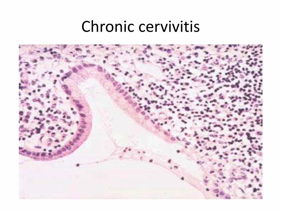
Chronic cervivitis
- As these cells are shed, the glycogen provides
a substrate for various endogenous vaginal,
particularly lactobacilli, which are the dominant
microbial species in the normal vagina.
- Lactobacilli produce lactic acid, which maintains the vaginal pH below 4.5,and this leads to
Suppression of growth of other organisms.
Because at low pH, lactobacilli produce
bacteriotoxic hydrogen peroxide (H2O2).
- If the pH becomes alkaline due to
a. bleeding,, , H2O2 production by lactobacilli
decreases.
b. Antibiotic therapy that suppress lactobacilli can
also cause the pH to rise.
- In each of these settings the altered vaginal
environment promotes the overgrowth of
other microorganisms, which may result in
cervicitis
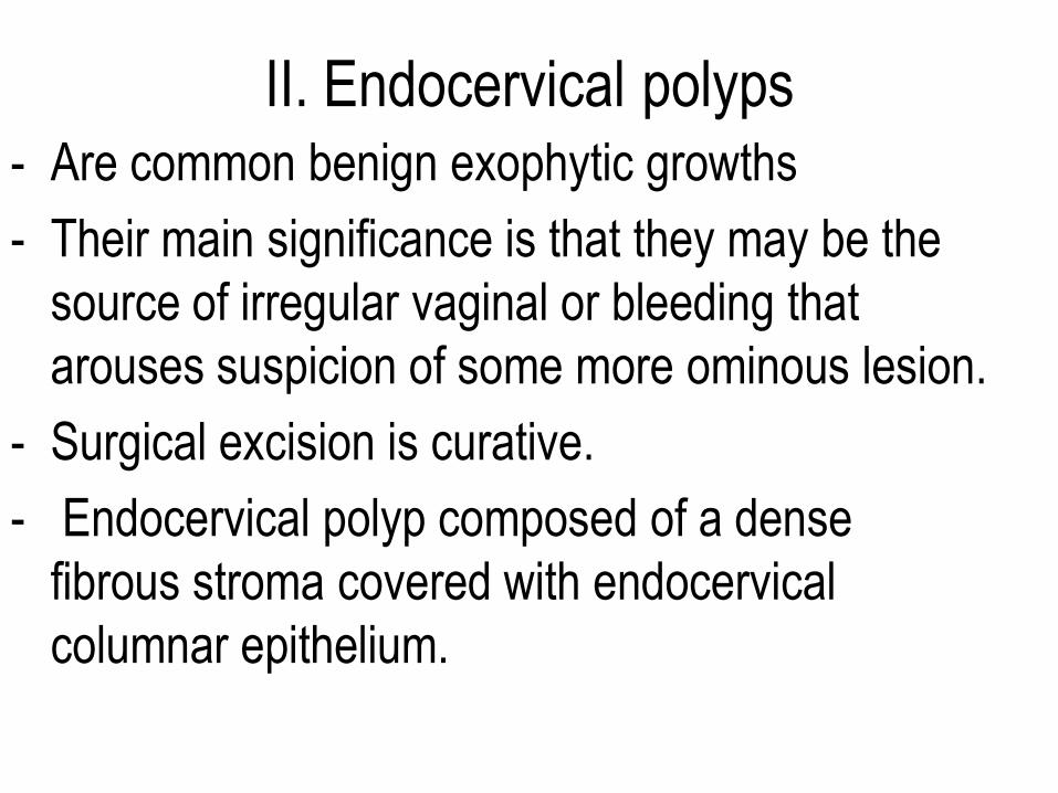
II. Endocervical polyps - Are common benign exophytic growths
- Their main significance is that they may be the
source of irregular vaginal or bleeding that
arouses suspicion of some more ominous lesion.
- Surgical excision is curative.
- Endocervical polyp composed of a dense
fibrous stroma covered with endocervical
columnar epithelium.
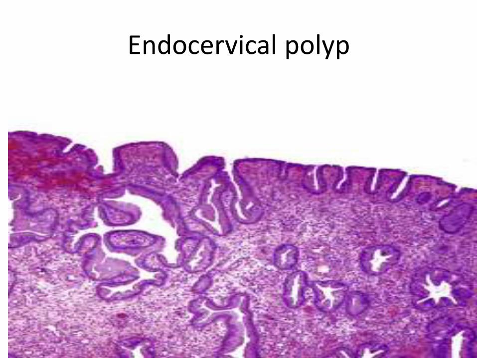
Endocervical polyp
III. Premalignant and Malignant
Neoplasms of the Cervix
- Worldwide, cervical carcinoma is the third
most common cancer in women,
- Fifty years ago, carcinoma of the cervix was
the leading cause of cancer deaths in women
in the United States, but the death rate has
declined by two thirds to its present rank as
the thirteenth cause of cancer mortality.
A. Cervical Intraepithelial Neoplasia (Squamous Intraepithelial Lesions)
- Terminology:Previously
- Cervical intraepithelial neoplasia (CIN)
classification
i. Mild dysplasia termed CIN I,
ii. Moderate dysplasia CIN II,
iii. Severe dysplasia termed CIN III.
• Recently
- The three-tier classification system has been simplified to a two-tiered system, with
1. CIN I And II renamed low-grade squamous intraepithelial lesion (LSIL)
2. and CIN III combined into one category referred to as high-grade squamous intraepithelial lesion (HSIL) .
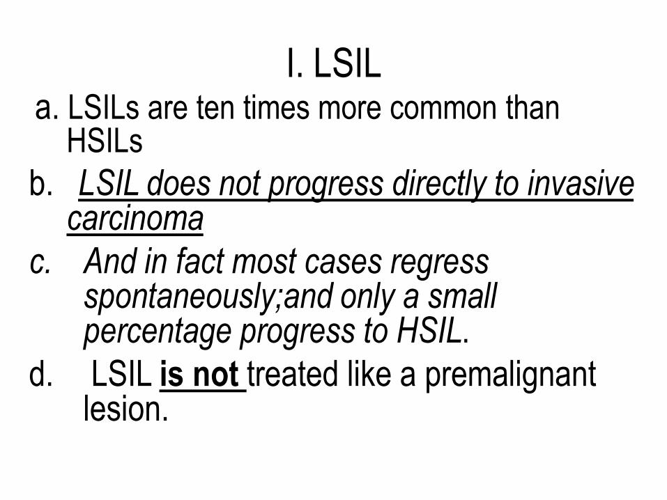
I. LSIL a. LSILs are ten times more common than
HSILs
b. LSIL does not progress directly to invasive carcinoma
c. And in fact most cases regress spontaneously;and only a small percentage progress to HSIL.
d. LSIL is not treated like a premalignant lesion.
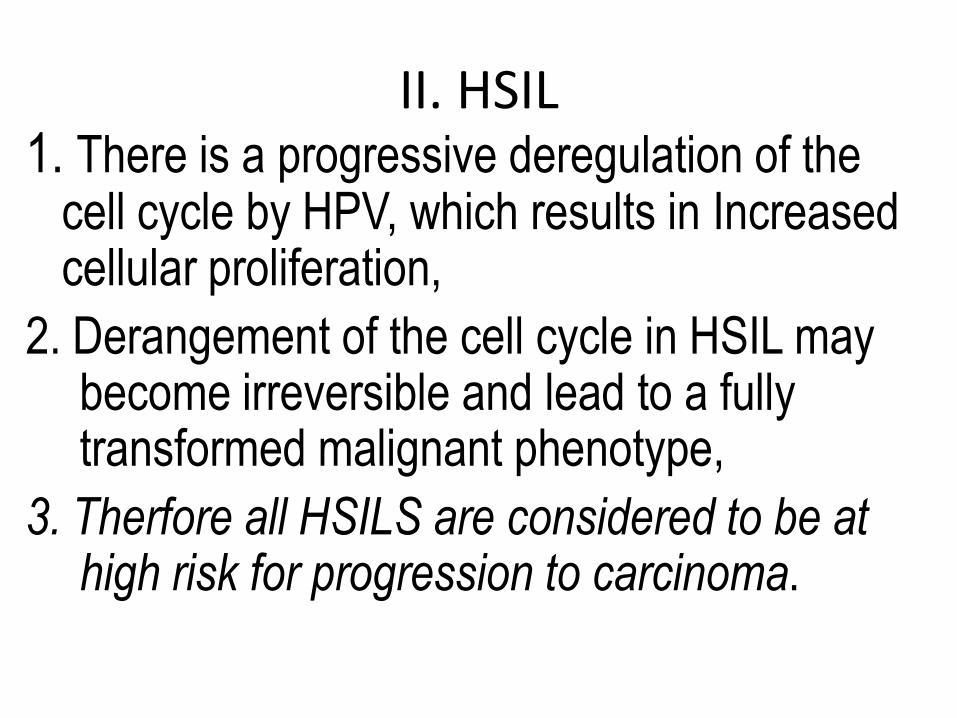
II. HSIL 1. There is a progressive deregulation of the
cell cycle by HPV, which results in Increased cellular proliferation,
2. Derangement of the cell cycle in HSIL may become irreversible and lead to a fully transformed malignant phenotype,
3. Therfore all HSILS are considered to be at high risk for progression to carcinoma.
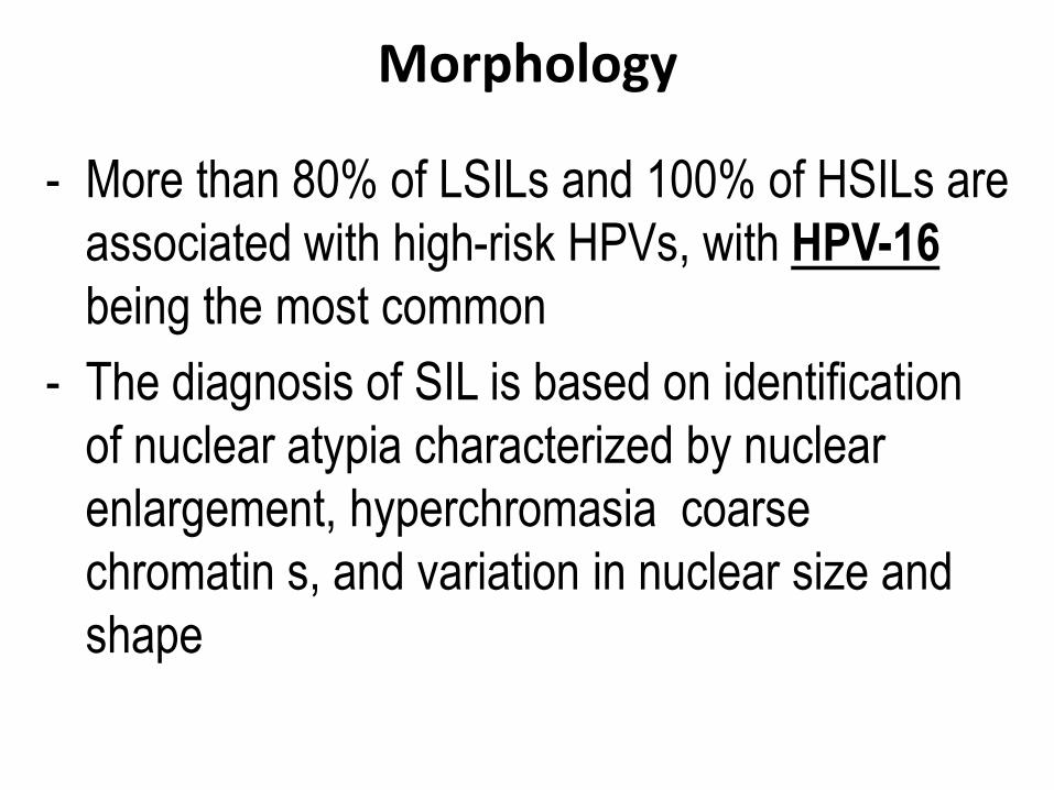
Morphology
- More than 80% of LSILs and 100% of HSILs are
associated with high-risk HPVs, with HPV-16
being the most common
- The diagnosis of SIL is based on identification
of nuclear atypia characterized by nuclear
enlargement, hyperchromasia coarse
chromatin s, and variation in nuclear size and
shape
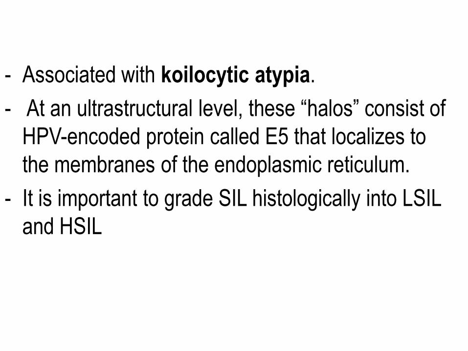
- Associated with koilocytic atypia.
- At an ultrastructural level, these “halos” consist of
HPV-encoded protein called E5 that localizes to
the membranes of the endoplasmic reticulum.
- It is important to grade SIL histologically into LSIL
and HSIL
- Progression to invasive carcinoma, when it
occurs, takes place over a period of a few
years to more than a decade
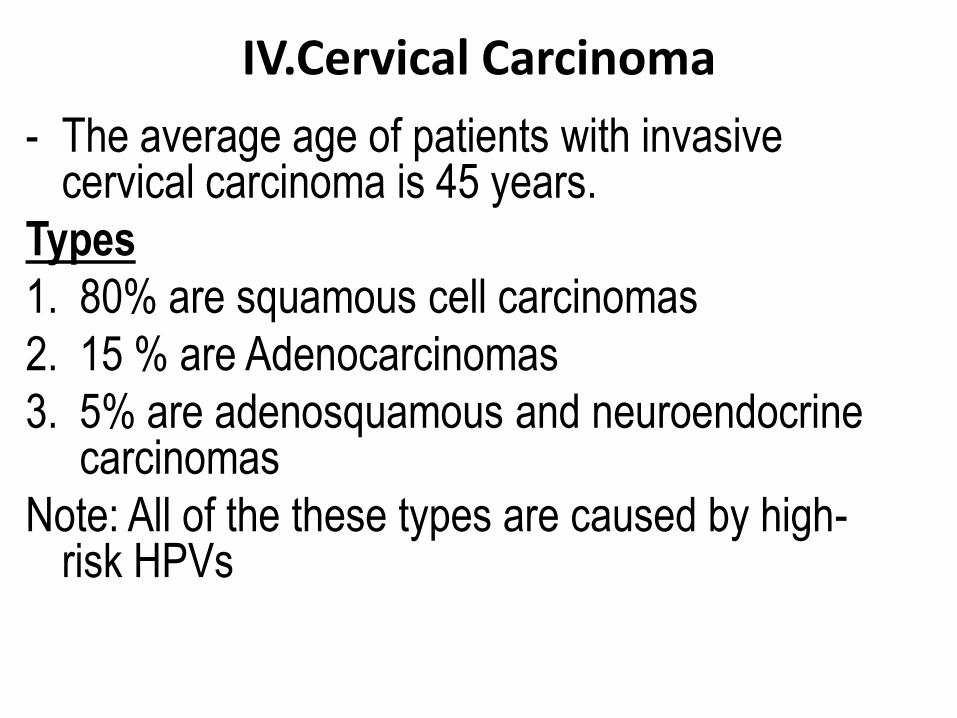
IV.Cervical Carcinoma - The average age of patients with invasive
cervical carcinoma is 45 years.
Types
1. 80% are squamous cell carcinomas
2. 15 % are Adenocarcinomas
3. 5% are adenosquamous and neuroendocrine carcinomas
Note: All of the these types are caused by high-risk HPVs
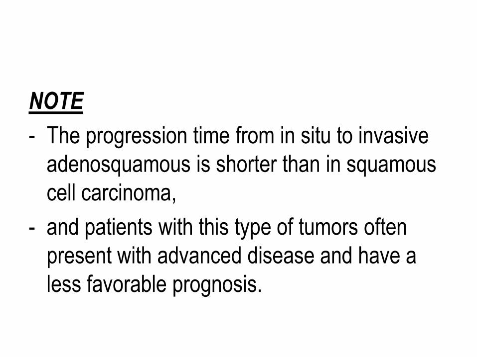
NOTE
- The progression time from in situ to invasive
adenosquamous is shorter than in squamous
cell carcinoma,
- and patients with this type of tumors often
present with advanced disease and have a
less favorable prognosis.

Morphology 1. Squamous cell carcinoma
- Is composed of nests and tongues of malignant
squamous epithelium, either keratinizing or
nonkeratinizing,
2.Adenocarcinoma
- Is characterized by proliferation of malignant
endocervical cells with large, hyperchromatic
nuclei and relatively mucin-depleted
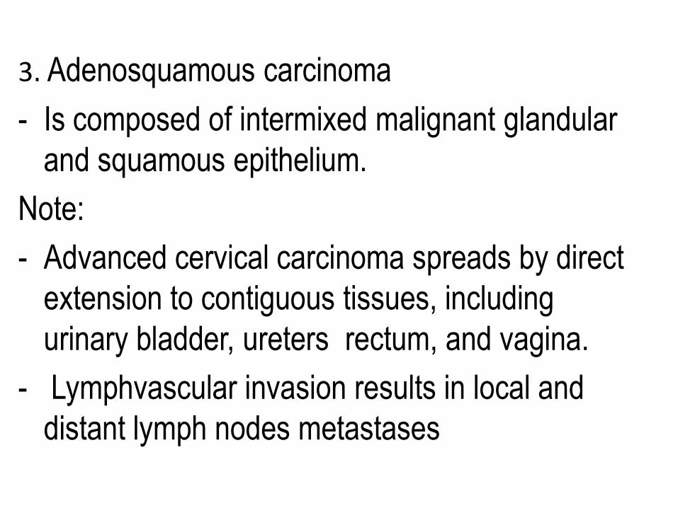
3. Adenosquamous carcinoma
- Is composed of intermixed malignant glandular
and squamous epithelium.
Note:
- Advanced cervical carcinoma spreads by direct
extension to contiguous tissues, including
urinary bladder, ureters rectum, and vagina.
- Lymphvascular invasion results in local and
distant lymph nodes metastases
- Distant metastases may also be found in the
liver, lungs, bone marrow, and other organs.
• Cervical cancer is staged as follows:
Stage 0—Carcinoma in situ (CIN III, HSIL)
Stage I—Carcinoma confined to the cervix
Stage II—Carcinoma extends beyond the cervix
but not to the pelvic wall or carcinoma that
involves the vagina but not the lower third
Stage III:Carcinoma has extended to the pelvic
wall.and the tumor involves the lower third of the
vagina
Stage IV:
a. Carcinoma has extended beyond the true pelvis
b. or has involved the mucosa of the bladder or
rectum.
c. Cancers with metastatic dissemination.
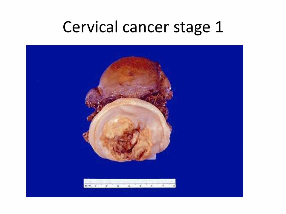
Cervical cancer stage 1
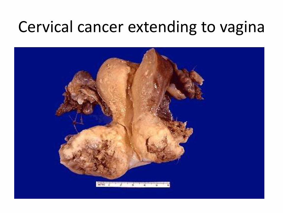
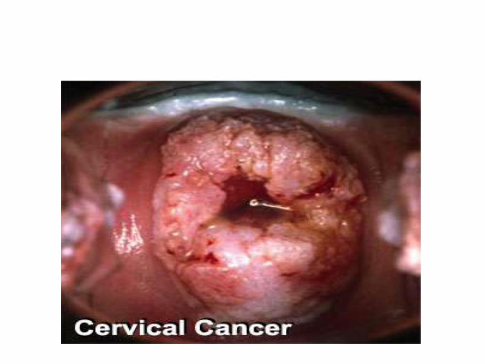
Cervical cancer extending to vagina
Clinically
- More than half of invasive cervical cancers are
detected in women who did not participate in
regular screening.(cervical smear)
- Early invasive cancers of the cervix
(microinvasive carcinomas) may be treated by
cervical cone excision alone,
- With current treatments the 5-year survival rate is
100% for microinvasive carcinomas and less than
50% for tumors extending beyond pelvis.
- Most patients with advanced cervical cancer die of
the consequences of local tumor invasion (e.g.,
ureteral obstruction, pyelonephritis, and uremia)
rather than distant metastases.
Cervical Cancer Screening and Prevention
- Cytologic cancer screening has significantly
reduced mortality from cervical cancer..
- The reason that cytologic screening is so
effective in preventing cervical cancer is that
most cancers arise from precursor lesions over
the course of years.
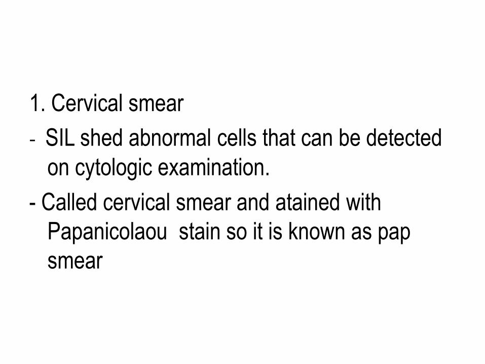
1. Cervical smear
- SIL shed abnormal cells that can be detected
on cytologic examination.
- Called cervical smear and atained with
Papanicolaou stain so it is known as pap
smear
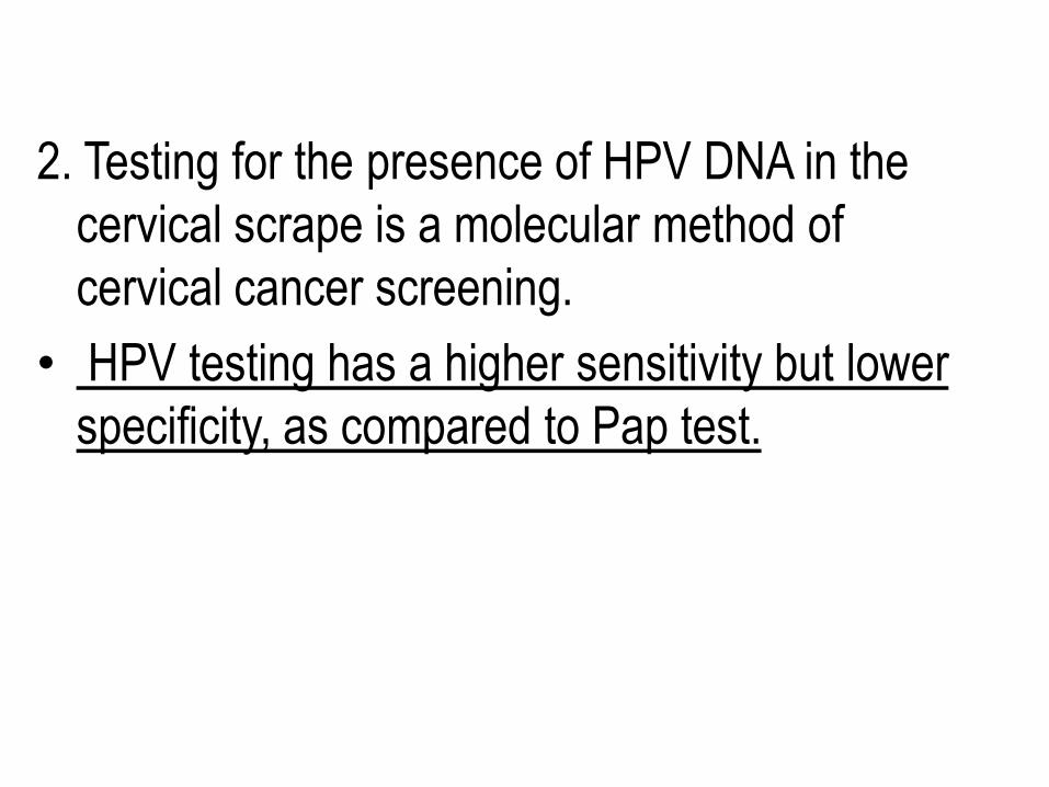
2. Testing for the presence of HPV DNA in the
cervical scrape is a molecular method of
cervical cancer screening.
• HPV testing has a higher sensitivity but lower
specificity, as compared to Pap test.
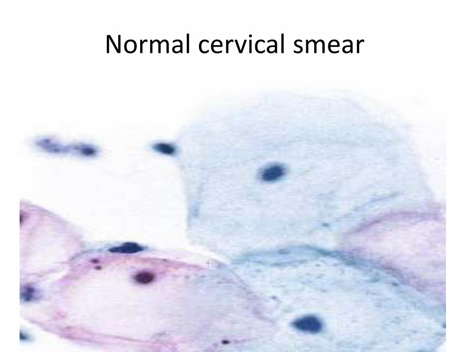
Normal cervical smear
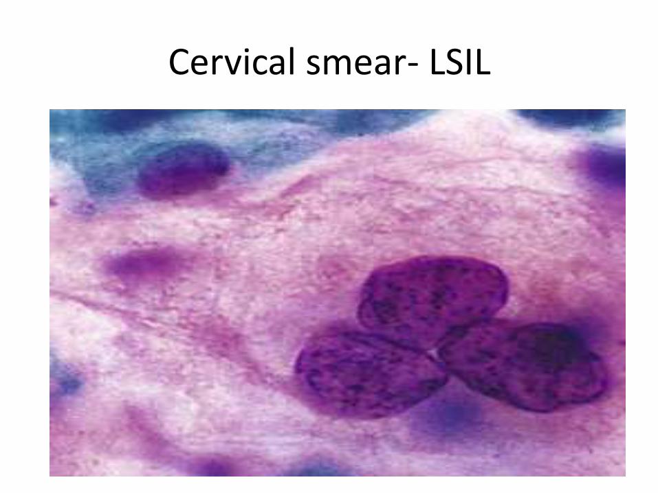
Cervical smear- LSIL
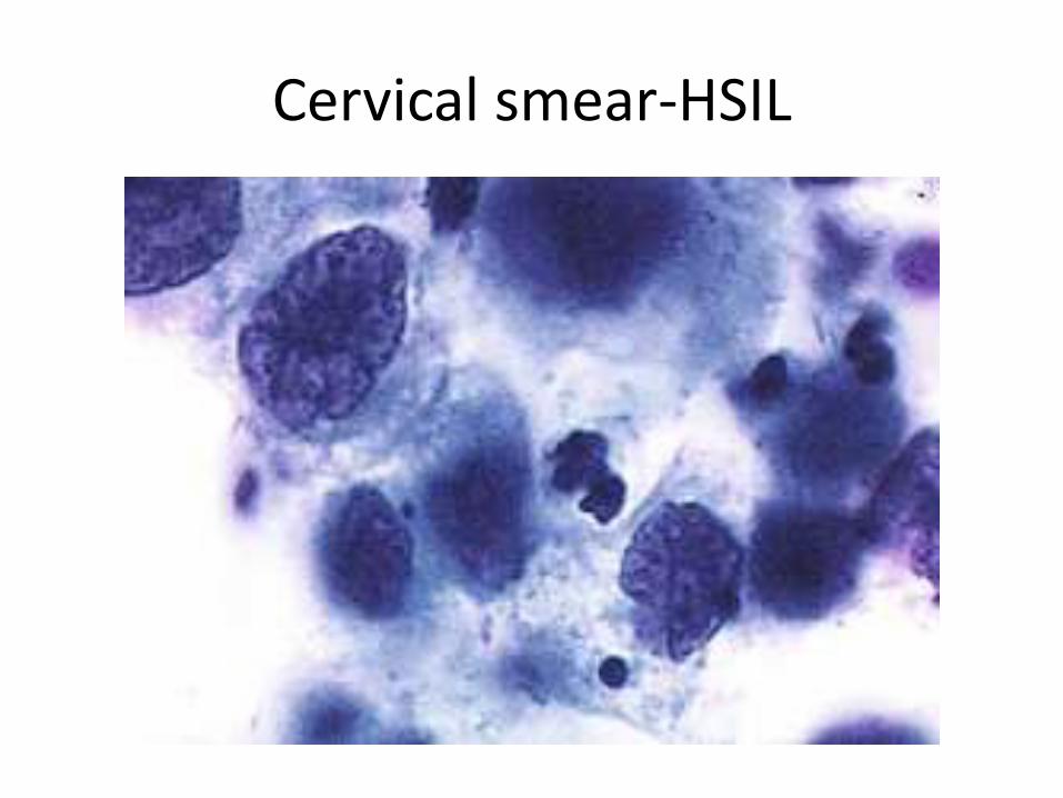
Cervical smear-HSIL
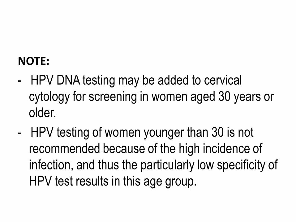
NOTE:
- HPV DNA testing may be added to cervical
cytology for screening in women aged 30 years or
older.
- HPV testing of women younger than 30 is not
recommended because of the high incidence of
infection, and thus the particularly low specificity of
HPV test results in this age group.
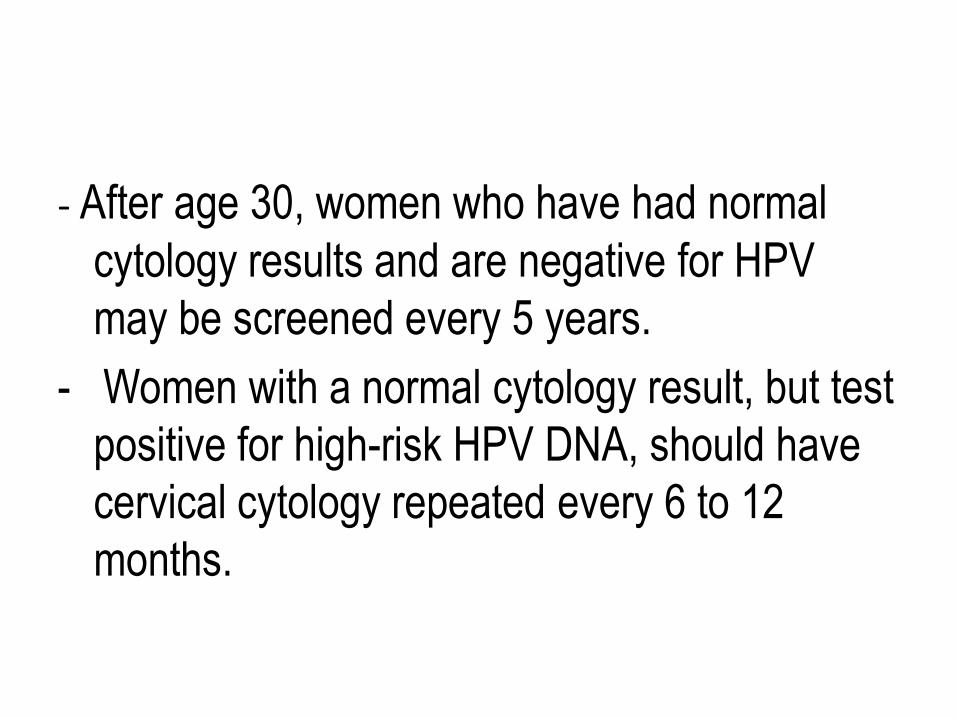
- After age 30, women who have had normal
cytology results and are negative for HPV
may be screened every 5 years.
- Women with a normal cytology result, but test
positive for high-risk HPV DNA, should have
cervical cytology repeated every 6 to 12
months.
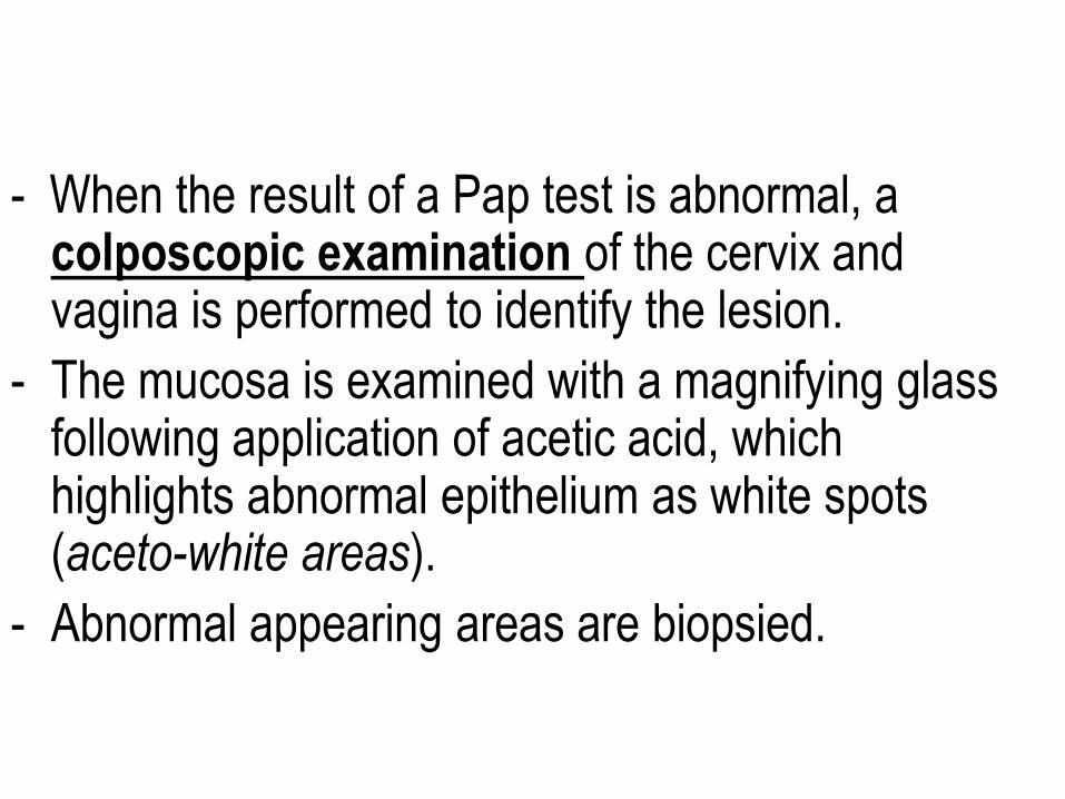
- When the result of a Pap test is abnormal, a colposcopic examination of the cervix and vagina is performed to identify the lesion.
- The mucosa is examined with a magnifying glass following application of acetic acid, which highlights abnormal epithelium as white spots (aceto-white areas).
- Abnormal appearing areas are biopsied.
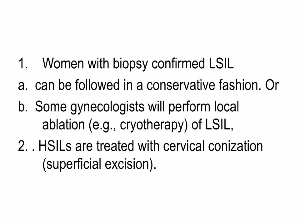
1. Women with biopsy confirmed LSIL
a. can be followed in a conservative fashion. Or
b. Some gynecologists will perform local
ablation (e.g., cryotherapy) of LSIL,
2. . HSILs are treated with cervical conization
(superficial excision).
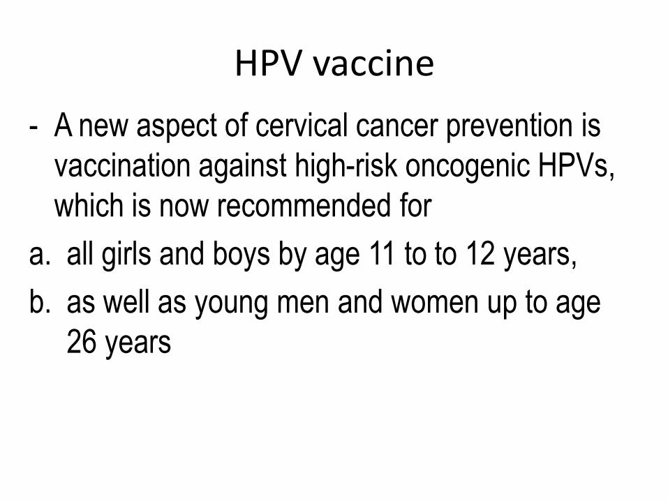
HPV vaccine
- A new aspect of cervical cancer prevention is
vaccination against high-risk oncogenic HPVs,
which is now recommended for
a. all girls and boys by age 11 to to 12 years,
b. as well as young men and women up to age
26 years
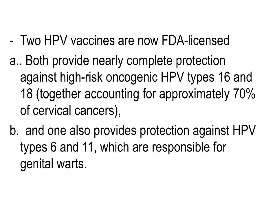
- Two HPV vaccines are now FDA-licensed
a.. Both provide nearly complete protection
against high-risk oncogenic HPV types 16 and
18 (together accounting for approximately 70%
of cervical cancers),
b. and one also provides protection against HPV
types 6 and 11, which are responsible for
genital warts.
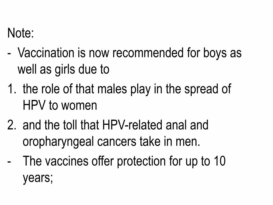
Note:
- Vaccination is now recommended for boys as
well as girls due to
1. the role of that males play in the spread of
HPV to women
2. and the toll that HPV-related anal and
oropharyngeal cancers take in men.
- The vaccines offer protection for up to 10
years;


![(Endometrial Intraepithelial Neoplasia): Improved Criteria ... · Endometrial intraepithelial neoplasia [EIN] EIN Reproducibility UsubutumA et al Modern Pathol25: 877-884, 2012. Questionaire,](https://static.fdocuments.in/doc/165x107/6053ec04465f250d537d95f4/endometrial-intraepithelial-neoplasia-improved-criteria-endometrial-intraepithelial.jpg)