High-grade Prostatic Intraepithelial Neoplasia of the · PDF fileZynger DL and Yang...
12
Int J Clin Exp Pathol (2009) 2, 327-338 www.ijcep.com/IJCEP812001 Review Article High-grade Prostatic Intraepithelial Neoplasia of the Prostate: The Precursor Lesion of Prostate Cancer Debra L Zynger 1 and Ximing Yang 2 1 Department of Pathology, University of Pittsburgh Medical Center, Pittsburgh, PA and 2 Department of Pathology, Northwestern University School of Medicine, Chicago, IL Received 01 December 2008; Accepted and available online 22 December 2008 Abstract: High-grade intraepithelial neoplasia (HGPIN) is a lesion which is widely believed to be a precursor of prostatic adenocarcinoma. Correct morphologic identification of HGPIN and an understanding of how this diagnosis affects clinical management in the research setting are necessary as HGPIN is a premalignant lesion with many genetic alterations similar to prostate cancer, but is not yet invasive cancer. As such it is critical to differentiate between benign entities, HGPIN, and prostatic adenocarcinoma for experimental design and data interpretation. This review discusses HGPIN, clarifies the terminology used in pathology reports, and describes the clinical and research implications of this entity. Keywords: High-grade prostatic intraepithelial neoplasia, HGPIN, prostate, needle biopsy Introduction Prostatic intraepithelial neoplasia (PIN), first described in 1969 [1], is a neoplastic proliferation of prostatic epithelial cells that is confined to pre-existing prostatic ducts or acini (glands). PIN was further characterized and initially termed intraductal dysplasia by McNeal and Bostwick in 1986 [2]; the currently used term "prostatic intraepithelial neoplasia" was later introduced by Bostwick and Brawer in 1987, and endorsed by consensus at a 1989 conference [3, 4]. In recent years, many studies have shown that high-grade prostatic intraepithelial neoplasia (HGPIN) is the major precursor of prostate cancer. This review aims to clarify the diagnostic terms used in pathology reports and the implications the terminology has upon clinical management. Specifically, this article focuses on HGPIN as well as diagnostic terms using the word “atypical” in the prostate. It is important to diagnose and correctly use the term HGPIN to avoid confusion with other “atypical” entities of the prostate, which may differ with respect to clinical significance. Histologic Features The term PIN encompasses morphologic changes in which prostatic epithelial cells are present in large and branched glands, with a convoluted inner contour similar to non- neoplastic glands [1-2]. Epithelial proliferation produces a layer of crowded, pseudostratified cells with cytologic atypia, such as nuclear irregularity, nucleomegaly, hyperchromasia, and prominent nucleoli (Figure 1A). These cytological features are similar to those of invasive prostate cancer. However, in contrast to adenocarcinoma, the architecture of PIN is normal. PIN glands characteristically contain basal cells around their periphery (Figure 1B), seen as a thin and occasionally discontinuous layer on hematoxylin and eosin (H&E) stained sections [3-4]. This is an important diagnostic feature because the presence of basal cells can help to differentiate PIN from prostatic adenocarcinoma in which the basal cells are absent [5-8]. PIN was originally subdivided into 3 groups which have now been combined into low-grade (PIN I) and HGPIN (PIN II and PIN III) [9]. HGPIN differs from low-grade PIN in that cytologic atypia is more apparent, particularly the presence of prominent nucleoli, as observed using a 20x-power lens (200-fold
Transcript of High-grade Prostatic Intraepithelial Neoplasia of the · PDF fileZynger DL and Yang...
Int J Clin Exp Pathol (2009) 2, 327-338 www.ijcep.com/IJCEP812001
Review Article High-grade Prostatic Intraepithelial Neoplasia of the Prostate: The Precursor Lesion of Prostate Cancer Debra L Zynger1 and Ximing Yang2 1Department of Pathology, University of Pittsburgh Medical Center, Pittsburgh, PA and 2Department of Pathology, Northwestern University School of Medicine, Chicago, IL Received 01 December 2008; Accepted and available online 22 December 2008 Abstract: High-grade intraepithelial neoplasia (HGPIN) is a lesion which is widely believed to be a precursor of prostatic adenocarcinoma. Correct morphologic identification of HGPIN and an understanding of how this diagnosis affects clinical management in the research setting are necessary as HGPIN is a premalignant lesion with many genetic alterations similar to prostate cancer, but is not yet invasive cancer. As such it is critical to differentiate between benign entities, HGPIN, and prostatic adenocarcinoma for experimental design and data interpretation. This review discusses HGPIN, clarifies the terminology used in pathology reports, and describes the clinical and research implications of this entity. Keywords: High-grade prostatic intraepithelial neoplasia, HGPIN, prostate, needle biopsy
Introduction Prostatic intraepithelial neoplasia (PIN), first described in 1969 [1], is a neoplastic proliferation of prostatic epithelial cells that is confined to pre-existing prostatic ducts or acini (glands). PIN was further characterized and initially termed intraductal dysplasia by McNeal and Bostwick in 1986 [2]; the currently used term "prostatic intraepithelial neoplasia" was later introduced by Bostwick and Brawer in 1987, and endorsed by consensus at a 1989 conference [3, 4]. In recent years, many studies have shown that high-grade prostatic intraepithelial neoplasia (HGPIN) is the major precursor of prostate cancer. This review aims to clarify the diagnostic terms used in pathology reports and the implications the terminology has upon clinical management. Specifically, this article focuses on HGPIN as well as diagnostic terms using the word “atypical” in the prostate. It is important to diagnose and correctly use the term HGPIN to avoid confusion with other “atypical” entities of the prostate, which may differ with respect to clinical significance. Histologic Features
The term PIN encompasses morphologic changes in which prostatic epithelial cells are present in large and branched glands, with a convoluted inner contour similar to non-neoplastic glands [1-2]. Epithelial proliferation produces a layer of crowded, pseudostratified cells with cytologic atypia, such as nuclear irregularity, nucleomegaly, hyperchromasia, and prominent nucleoli (Figure 1A). These cytological features are similar to those of invasive prostate cancer. However, in contrast to adenocarcinoma, the architecture of PIN is normal. PIN glands characteristically contain basal cells around their periphery (Figure 1B), seen as a thin and occasionally discontinuous layer on hematoxylin and eosin (H&E) stained sections [3-4]. This is an important diagnostic feature because the presence of basal cells can help to differentiate PIN from prostatic adenocarcinoma in which the basal cells are absent [5-8]. PIN was originally subdivided into 3 groups which have now been combined into low-grade (PIN I) and HGPIN (PIN II and PIN III) [9]. HGPIN differs from low-grade PIN in that cytologic atypia is more apparent, particularly the presence of prominent nucleoli, as observed using a 20x-power lens (200-fold
Zynger DL and Yang XJ/High-Grade Prostatic Intraepithelial Neoplasm
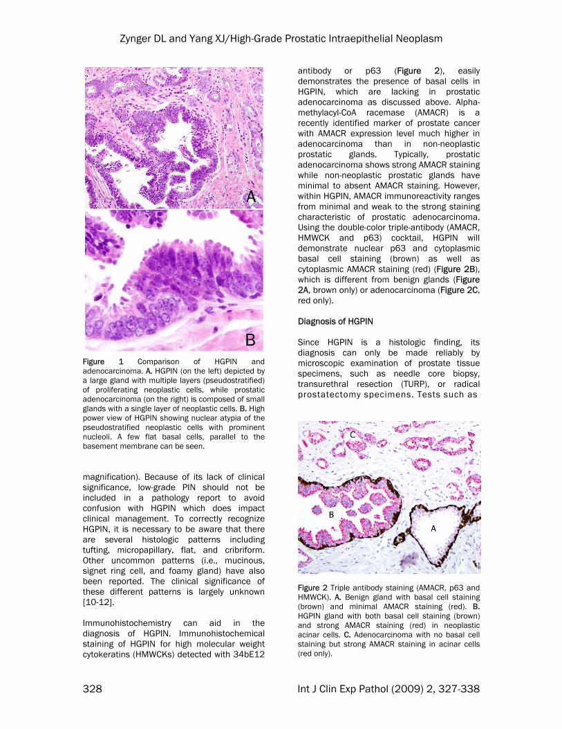
Figure 1 Comparison of HGPIN and adenocarcinoma. A. HGPIN (on the left) depicted by a large gland with multiple layers (pseudostratified) of proliferating neoplastic cells, while prostatic adenocarcinoma (on the right) is composed of small glands with a single layer of neoplastic cells. B. High power view of HGPIN showing nuclear atypia of the pseudostratified neoplastic cells with prominent nucleoli. A few flat basal cells, parallel to the basement membrane can be seen. magnification). Because of its lack of clinical significance, low-grade PIN should not be included in a pathology report to avoid confusion with HGPIN which does impact clinical management. To correctly recognize HGPIN, it is necessary to be aware that there are several histologic patterns including tufting, micropapillary, flat, and cribriform. Other uncommon patterns (i.e., mucinous, signet ring cell, and foamy gland) have also been reported. The clinical significance of these different patterns is largely unknown [10-12]. Immunohistochemistry can aid in the diagnosis of HGPIN. Immunohistochemical staining of HGPIN for high molecular weight cytokeratins (HMWCKs) detected with 34bE12
antibody or p63 (Figure 2), easily demonstrates the presence of basal cells in HGPIN, which are lacking in prostatic adenocarcinoma as discussed above. Alpha-methylacyl-CoA racemase (AMACR) is a recently identified marker of prostate cancer with AMACR expression level much higher in adenocarcinoma than in non-neoplastic prostatic glands. Typically, prostatic adenocarcinoma shows strong AMACR staining while non-neoplastic prostatic glands have minimal to absent AMACR staining. However, within HGPIN, AMACR immunoreactivity ranges from minimal and weak to the strong staining characteristic of prostatic adenocarcinoma. Using the double-color triple-antibody (AMACR, HMWCK and p63) cocktail, HGPIN will demonstrate nuclear p63 and cytoplasmic basal cell staining (brown) as well as cytoplasmic AMACR staining (red) (Figure 2B), which is different from benign glands (Figure 2A, brown only) or adenocarcinoma (Figure 2C, red only). Diagnosis of HGPIN Since HGPIN is a histologic finding, its diagnosis can only be made reliably by microscopic examination of prostate tissue specimens, such as needle core biopsy, transurethral resection (TURP), or radical prostatectomy specimens. Tests such as
Figure 2 Triple antibody staining (AMACR, p63 and HMWCK). A. Benign gland with basal cell staining (brown) and minimal AMACR staining (red). B. HGPIN gland with both basal cell staining (brown) and strong AMACR staining (red) in neoplastic acinar cells. C. Adenocarcinoma with no basal cell staining but strong AMACR staining in acinar cells (red only).
328 Int J Clin Exp Pathol (2009) 2, 327-338
Zynger DL and Yang XJ/High-Grade Prostatic Intraepithelial Neoplasm
digital examination, transrectal ultrasonography or other imaging studies are not useful for detecting HGPIN. Furthermore, fine needle aspiration (FNA), which is a popular method for diagnosis of prostate cancer in some countries, cannot distinguish HGPIN from cancer based on cytologic features alone. HGPIN is often diagnosed in a prostatic specimen obtained for a diagnostic test (such as needle core biopsy) or for the treatment of non-neoplastic prostatic pathology (such as TURP specimens for benign prostatic hyperplasia). HGPIN is a non-invasive neoplastic process, which does not form a tumor mass or cause clinical symptoms. Despite its histologic similarity to carcinoma-in-situ (CIS), a precursor to invasive cancer that arises in other organs (e.g., breast or skin), the term CIS should not be used to describe HGPIN because of the variability in its natural history and biologic behavior. HGPIN may evolve into an invasive cancer, a process that can take more than 10 years to develop [26], or remain unchanged. Therefore, aggressive treatment, such as surgery and radiation, is not warranted. Molecular Markers of HGPIN Prostate tumorigenesis is theorized to result from numerous genetic alterations. Currently, data reveals that both genotypically and phenotypically HGPIN exists in a spectrum between benign prostate and prostatic adenocarcinoma. As HGPIN is a precursor lesion, it is expected that some of the molecular abnormalities overlap with prostate cancer or benign prostate while other abnormalities will be unique to HGPIN [13-16]. Numerous changes have been found by comparing HGPIN to benign prostate tissue. Some of the aberrations which may be critical to the formation of HGPIN are increased expression of AMACR, loss of p27KIP1, PTEN, and RB activity, hypermethylation of the promoter region of GSTP1, and fusion of TMPRSS2-ERG genes [13-16]. All of these alterations are also seen in prostatic adenocarcinoma [13-16]. Ideally, specific changes would be identified to reliably classify benign prostate, HGPIN, and prostate cancer. As of yet, no molecular test has been identified that can be used for diagnostic classification, either for research purposes or clinically. While some reports suggest that HGPIN might result
in an elevation of serum total PSA [17-18] or higher values of free PSA than prostate cancer, no convincing evidence to correlate the presence of HGPIN with serum PSA has been found by others [19-23]. Therefore, if a man with an elevated serum PSA has isolated HGPIN on needle biopsy, a repeat prostate needle biopsy might be necessary to rule out other conditions causing PSA elevation, particularly prostate cancer. Newer studies using DNA microarray analysis and transgenic mouse models are promising for the development of molecular markers [24-25], and perhaps in the near future a single marker or the use of multiple molecular markers in tandem will allow for reliable discrimination between benign prostate, HGPIN, and invasive tumor. Relationship of HGPIN to Cancer The relationship between PIN and invasive cancer was first elucidated in 1986 [2, 27]. In a series of prostates obtained at autopsy, PIN was more likely to be found in men with prostate cancer than in those without cancer. Later studies have confirmed the strong association between PIN and invasive prostate cancer. The evidence linking PIN and invasive cancer is as follows: similar to prostatic adenocarcinoma, HGPIN tends to be multifocal, occurs in the peripheral zone of the prostate, and is more prevalent in prostates that harbor carcinomas than in those that do not [28]. HGPIN can be identified in 80 to 90 percent of radical prostatectomy specimens, often in close proximity to coexisting prostatic adenocarcinoma; sometimes, a direct transition from HGPIN to invasive adenocarcinoma can be seen [28-31]. As well, autopsy studies reveal that the prevalence of HGPIN (7, 12, 36, 38, 45, and 48 percent in the 3rd, 4th, 5th, 6th, 7th, and 8th decades in one series) closely follows that of prostate cancer (4, 9, 14, 24, 32, and 33 percent, respectively) [26, 32]. Prevalence Because prostate cancer is the most common visceral malignancy in men, the prevalence of HGPIN, the major premalignant lesion of the prostate, is also high. However, as HGPIN can only be diagnosed microscopically, its true prevalence in the general population may be underreported. The reported incidence of HGPIN varies greatly, which may be related to
329 Int J Clin Exp Pathol (2009) 2, 327-338
Zynger DL and Yang XJ/High-Grade Prostatic Intraepithelial Neoplasm
the population of men under study, type of specimen, or the diagnostic criteria that are applied. For example, based upon pathologic examination of prostate glands in autopsy series [2, 26] and from cystoprostatectomy specimens from men who have undergone surgery for bladder cancer [22, 33, 34], the prevalence of isolated PIN is 40 to 50 percent in men without prostate cancer with both the rate and volume of PIN increasing with age [26, 32]. In contrast, the incidence of HGPIN, as determined by prostate needle biopsies in men participating in PSA screening studies, ranges from 0.7 to 20 percent, while among those undergoing TURP, the reported incidence varies from 3 to 33 percent [35-38]. When we analyzed 87,713 men from 15 different studies using strict diagnostic criteria, we found that the average prevalence of HGPIN was 4.26 percent [35], which closely resembles the prevalence of HGPIN found in our practice. An increasing number of institutions have adopted extended biopsy schema for prostate needle core biopsies. Compared to the traditional six core (sextant) biopsy, this protocol recommends that 10 to 12 tissue cores be obtained during each prostate biopsy session. As a result, the detection rate of HGPIN on needle biopsy may increase, simply because more tissue cores are obtained [41]. Saturation biopsy methods, which may include 24 or more tissue cores, are used clinically for special cases. Clinical Significance Presence of HGPIN Men with isolated HGPIN on initial core biopsy are thought to have a higher risk of prostate cancer in the subsequent biopsy as compared to those without HGPIN. This is a controversial area and the magnitude of risk is uncertain. In early studies, the risk of finding cancer on re-biopsy ranged from 51 to 100 percent; however, cancer detection rates on repeat biopsy have declined since 1990, and range from 2.3 to 23 percent in more recent and larger series [42-46]. Perhaps the major reason for the declining rate of cancer detection on repeat biopsies for HGPIN is that the current extended prostate biopsy schema includes a higher number of cores (ranging from 8 to 12 cores) than the
previous standard sextant biopsy [47]. This leads to the detection of more prostate cancers in the initial biopsies, and reduces the positive predictive value of HGPIN in a needle biopsy for the detection of cancer upon re-biopsy. The relationship between the number of cores on initial biopsy, the number of cores on re-biopsy, and the risk of finding cancer in the re-biopsy specimen was illustrated in a series of 791 men with HGPIN on initial biopsy who underwent re-biopsy within one year of the diagnosis [46]. Cancer detection rates on re-biopsy varied based upon the biopsy schema; they were highest in men that initially had a sextant biopsy and who then underwent an extended number of cores for re-biopsy. The authors concluded that for patients diagnosed with HGPIN on extended initial core sampling, a repeat biopsy within the first year was unnecessary in the absence of other clinical indicators of cancer. Other investigators concur [47, 48]. Nevertheless, more studies are necessary in order to rationally adjust the re-biopsy strategy in men who are found to have HGPIN in an extended core biopsy. Whether such men require re-biopsy is still being researched. The most important aspect of management at this time is close clinical follow-up with serum PSA measurement. If there are clinical signs suspicious for prostate cancer, these patients should be re-biopsied. Two factors which may be particularly important in predicting the development of prostate cancer in the setting of HGPIN are elevated serum PSA and the presence of adjacent atypical small glands (acini) that are suspicious for but not diagnostic of prostate cancer, as discussed below (termed atypical small acinar proliferation or ASAP). Number of Cores with HGPIN The frequency of concurrent or subsequent diagnosis of invasive cancer may be higher when more than one biopsy specimens contain HGPIN [39, 40]. This relationship between the number of involved tissue cores and eventual diagnosis of invasive prostate cancer was illustrated in one series of 245 men with isolated HGPIN in an initial biopsy who underwent subsequent biopsies [40]. The only independent histologic predictor of risk of later invasive cancer was the number of cores involved by HGPIN: 30, 40, and 75 percent with involvement of 1 to 2, 3, or more than
330 Int J Clin Exp Pathol (2009) 2, 327-338

Zynger DL and Yang XJ/High-Grade Prostatic Intraepithelial Neoplasm
three cores. However, these data was obtained when routine six core prostate biopsies were the norm. In a more recent series that used an extended biopsy schema, the number of cores involved with HGPIN did not reliably predict the risk of finding invasive prostate cancer on rebiopsy [41]. Low-grade PIN In contrast to HGPIN, the presence of low-grade PIN is distinctly different, and has no clinical significance. As a result, men with low-grade PIN do not require a repeat biopsy unless other clinical indicators are present. In fact, it is unnecessary to even include low-grade PIN in the pathologic diagnosis, because its presence is not an indication for more aggressive follow-up or treatment. Additionally, using the term low-grade PIN in the pathology report can lead to confusion with HGPIN. Rebiopsy after a Diagnosis of HGPIN The presence of HGPIN in a prostate biopsy is not an indication for aggressive treatment, but instead, the need for close monitoring. Repeat biopsy may be necessary because of the association of HGPIN with invasive prostate cancer, although there is no defined optimal re-biopsy strategy [51]. While re-biopsy should focus on the original site of HGPIN, cancer detection rates increase with sampling of adjacent sites as well as the contralateral lobe in the standard sextant locations. Others recommend the addition of transition zone sampling [52]. Most clinicians recommend repeat biopsy within 3 to 12 months after a diagnosis of HGPIN, and perhaps sooner for men with persistently rising serum PSA levels, although this is a controversial area [47]. The management of men with no evidence of invasive prostate cancer on repeat biopsy after an initial biopsy showing isolated HGPIN is unclear. Some investigators have found a slightly higher risk of subsequent cancer (24 percent) if HGPIN is documented as compared to a totally benign biopsy [40]. More recently, some experts suggested that "men do not need routine repeat needle biopsy within the first year following the diagnosis of high-grade PIN" based upon these findings [47]. On the other hand, some investigators consider that a 24 percent rate of finding invasive cancer is not insignificant, and high enough to indicate the need for re-biopsy. When and how
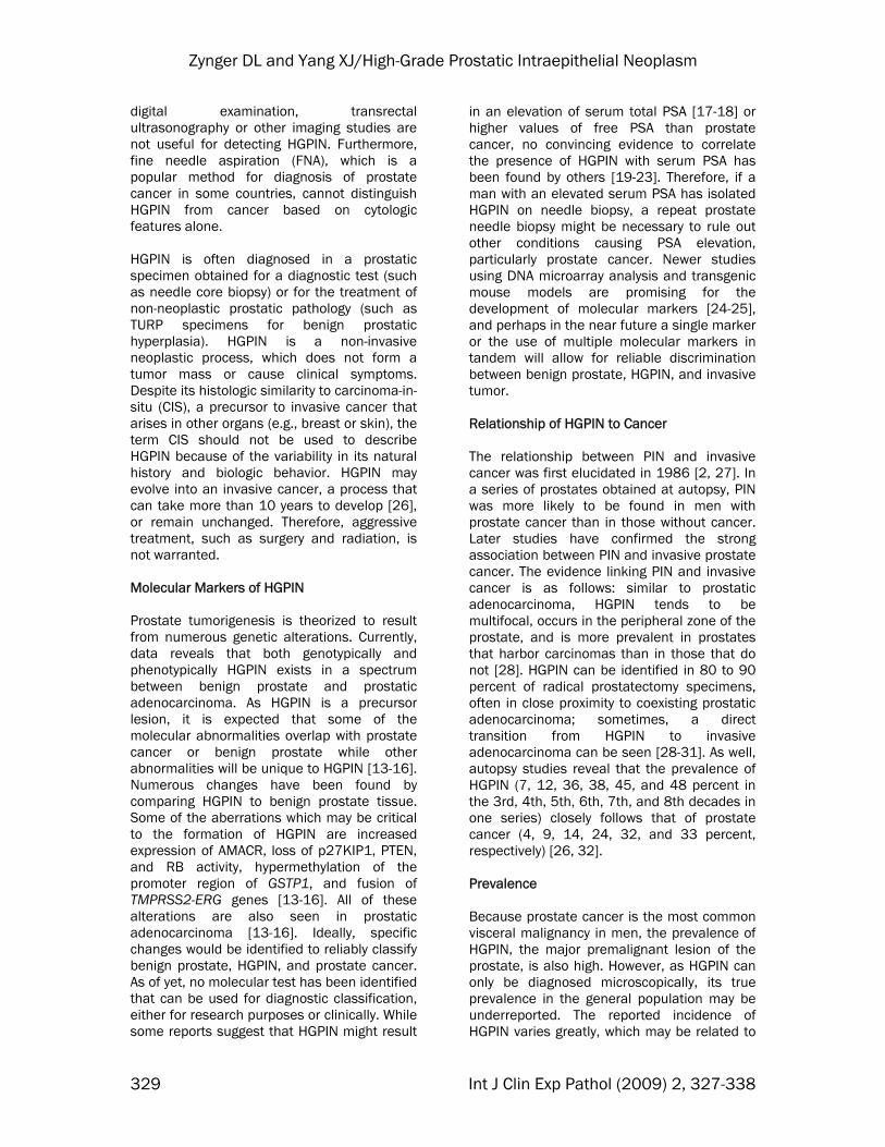
frequently perform re-biopsy on patients with isolated HGPIN should be determined based on the clinical situation of the individual until additional studies addressing these questions demonstrate more conclusive results. Impact of HGPIN on Prostate Cancer Research There are many benign and malignant conditions in the prostate mimicking HGPIN. Since HGPIN is a microscopic finding, only pathologists or investigators with substantial pathologic training can identify HGPIN microscopically. Furthermore, since HGPIN is a precursor lesion of prostate cancer, genetic and biochemical alterations developed in early prostatic tumorigenesis will be present in HGPIN. Therefore, some prostatic cancer markers reported in the literature will also be detected in HGPIN. Prostate cancer investigators need to be aware of the potentially overlapping genotype and phenotype between HGPIN and prostatic adenocarcinoma because of the implications upon experimental design and data interpretation. The following discusses theoretical examples of how a lack of understanding of HGPIN can lead a researcher astray. Difficulty Distinguishing HGPIN from Benign Conditions A study detects an elevation of the expression of gene X in HGPIN. However, because of the morphological similarity, the “HGPIN” was actually a benign prostatic condition, such as transitional cell (urothelial) metaplasia (Figure 3). Therefore, the misdiagnosis led to an erroneous conclusion. In actuality, transitional cell metaplasia, not HGPIN, shows a higher level of gene X expression. Correctly identifying non-neoplastic mimickers of HGPIN histologically prior to conducting a molecular experiment is needed to obtain usable data. Difficulty Distinguishing HGPIN from Adenocarcinoma Researchers report a higher proliferative activity in HGPIN than conventional acinar prostatic adenocarcinoma. The design flaw was that the “HGPIN” was actually ductal adenocarcinoma, which is often high-grade with a much higher proliferative activity than conventional acinar adenocarcinoma. Both ductal adenocarcinoma and cribriform
331 Int J Clin Exp Pathol (2009) 2, 327-338
Zynger DL and Yang XJ/High-Grade Prostatic Intraepithelial Neoplasm
Figure 3 Transitional cell (urothelial) metaplasia of the prostatic gland. As a benign condition, urothelial metaplasia of the prostate can mimic HGPIN. There are multiple layers of cells proliferating, but the cells do not show the cytological atypia seen in HGPIN.
adenocarcinoma of the prostate can be easily confused with HGPIN. Therefore, mistaking carcinoma for HGPIN can result in an incorrect interpretation. Failure to Recognize the Need to Assess HGPIN A highly specific prostate cancer marker Y is identified, and since this protein is present only in cancer cells not in benign prostatic glands the authors infer that the detection of mRNA and the protein specific to gene Y could replace conventional pathology diagnosis of prostate cancer. Unfortunately, later studies find that Y is also elevated in HGPIN. The positive detection of Y has resulted in unnecessary aggressive treatment because prostatic adenocarcinoma warrants aggressive treatment while HGPIN does not. As such, it is critical to determine if the marker is present in HGPIN before designating it as prostate cancer specific. Over Diagnosis of HGPIN Many patients with “HGPIN” are enrolled in a clinical trial taking medication Z to prevent development of prostate cancer. The conclusion of such a clinical trial was the medication Z reduced the prostate cancer incidence in this group of patients with HGPIN. In reality, this group had a lower incidence of cancer comparing the control groups because the majority of patients did not have HGPIN. Certain fixatives will preserve nuclear details better in biopsy specimens. Consequently, the
presence of prominent nucleoli in many benign prostatic epithelial cells may lead to a more frequent diagnosis of HGPIN. In some instances, the rate of HGPIN was reported more than 30% which is much higher than the 0.7 to 20% in most studies (see above in Prevalence of HGPIN). Reliably differentiating HGPIN from benign prostate is important to generate correct data regarding the long term prognosis of men initially diagnosed with HGPIN and to design appropriate treatment strategies. Specific Treatment for HGPIN Treatment that is designed for men with invasive prostate cancer (i.e., radical prostatectomy, radiation therapy, androgen ablation) is not suitable for those with HGPIN because of potential serious adverse effects associated with these therapies. Theoretically, such treatments could reduce the incidence of HGPIN, and potentially prevent prostate cancer. However, not all cases of HGPIN are destined to evolve into invasive cancer; no study has shown that preemptive treatment is of clinical value. Because of the known influence of neoadjuvant hormone therapy in reducing the incidence of HGPIN [53-55], it has been suggested that finasteride, an oral agent with no impact on serum testosterone levels, may be a useful agent for treating HGPIN. The Prostate Cancer Prevention Trial (PCPT) randomly assigned 18,882 men to finasteride (5 mg daily) or placebo, and followed men for seven years with annual digital rectal examination and PSA [56]. The number of men evaluable for HGPIN in the finasteride and placebo groups was 4568 and 4866, respectively. Men were evaluable for HGPIN if they had a diagnosis of HGPIN or invasive cancer on an interim or end of study biopsy, or if they had a completely negative biopsy at the study endpoint (seven years). When men diagnosed with HGPIN alone or HGPIN concurrently with invasive prostate cancer were examined jointly, those receiving finasteride had a significant 21 percent lower risk of HGPIN (9.2 versus 11.7 percent, hazard ratio 0.79, p = 0.001). Based upon a previous study, blockade of 5-alpha reductase with finasteride does not cause obvious change in the morphology of
332 Int J Clin Exp Pathol (2009) 2, 327-338
Zynger DL and Yang XJ/High-Grade Prostatic Intraepithelial Neoplasm
prostate cancer on histological evaluation [57]. Unlike other forms of androgen deprivation therapy, the effects of finasteride on the morphology of HGPIN and Gleason score are unknown [58]. In the PCPT, finasteride was effective in reducing the development of prostate cancer over a seven-year period (prostate cancer was detected in 18.4 percent of men in the finasteride group compared to 24.4 of the placebo group). However, adenocarcinomas with a high-grade appearance were more common in the finasteride group (37 versus 22 percent) [56]. The public health impact of PCPT, and particularly whether finasteride promotes high-grade prostate cancer or not remains a subject of intense ongoing debate. Another clinical trial on men with HGPIN using toremifene, which blocks estrogen receptors in the prostate, generated promising results. This randomized, double-blinded study showed a decrease of prostate cancer incidence by 6.8% in one year (24.4% for the 20 mg toremifene group versus 31.2 % for the placebo group). However, the incidence of prostate cancer in higher dose groups was similar to the placebo group (40 mg, 28.2%; 60 mg, 28.1 %). Larger studies will be necessary to substantiate these findings before toremifene can be used in clinical practice [59]. Suspicious Diagnosis: Atypical Small Acinar Proliferation (ASAP) When a prostate biopsy is undertaken, a definite pathologic diagnosis is usually needed for the purpose of clinical management. Unfortunately, definitive diagnosis is not always possible [60, 61]. For the diagnosis of invasive cancer to be made, there must be absolutely no doubt on the part of the consulting pathologist. A firm diagnosis of limited prostate cancer on needle core biopsy is one of the most difficult areas in surgical pathology. The histologic features of prostatic adenocarcinoma are complex, and may be subtle. An error in any step of tissue processing, including tissue fixation, dehydration, embedding, or even staining, may interfere with a proper diagnosis. Even if tissue processing is perfect, a definitive diagnosis of cancer may still be difficult for an experienced pathologist. One of the most common problems that interferes with the ability to make a definitive diagnosis is the size
of the suspicious lesion. Most pathologists do not feel comfortable making a diagnosis of malignancy when the focus of concern contains only a few atypical glands or acini. In the situation where the pathologist is suspicious but not totally convinced of a diagnosis of prostate cancer, the term "atypical glands suspicious for, but not diagnostic of prostatic adenocarcinoma" is often applied. Although other pathologists prefer the diagnostic term "atypical small acini suspicious for prostatic adenocarcinoma (ASAP)" [60], this phrase has been criticized because it may be confused with a definite cancer [63, 64]; however, the term has been widely accepted in clinical practice. The incidence of an uncertain atypical diagnosis can sometimes be decreased when immunohistochemical staining for HMWCK, p63, or AMACR is applied [65-67]. As previously stated, adenocarcinoma will typically be positive for AMACR and negative for HMWCK and p63, in contrast to benign glands which will be positive for HMWCK and p63 and negative for AMACR. Between 1 and 23 percent of prostate needle biopsy pathology specimens (average 5 percent) have a diagnosis of atypical foci suspicious for carcinoma [68]. This is not a pathologic entity but a diagnostic term that is used when there is suspicion for but not sufficient evidence to make a definitive diagnosis of cancer. Because the average risk of subsequently documenting cancer following an atypical or suspicious diagnosis is approximately 40 percent [64, 68], repeat biopsy is necessary. Repeat biopsy should include more sampling of the initial atypical site as well as other areas. Some urologists prefer saturated biopsy methods, in which more than 24 cores are taken. The significance of finding ASAP with HGPIN in a needle biopsy specimen is essentially the same as finding ASAP alone. The most important point is that HGPIN is totally different from a focus of ASAP suspicious for cancer on prostate needle biopsy, which carries a much higher risk (40 to 50 percent) of finding cancer at the time of re-biopsy. As a result, men with atypical foci suspicious for adenocarcinoma should be re-biopsied within 3 to 6 months, while re-biopsy within one year for men with HGPIN alone could be considered optional.
333 Int J Clin Exp Pathol (2009) 2, 327-338
Zynger DL and Yang XJ/High-Grade Prostatic Intraepithelial Neoplasm
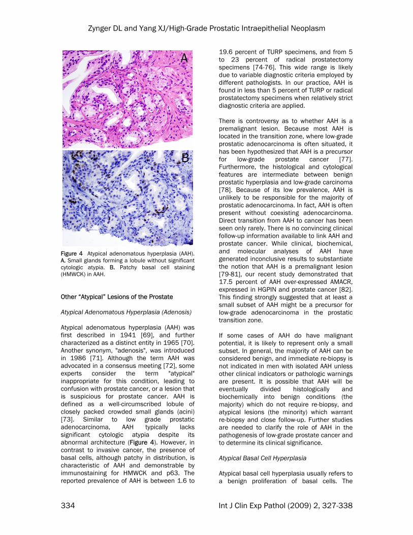
Figure 4 Atypical adenomatous hyperplasia (AAH). A. Small glands forming a lobule without significant cytologic atypia. B. Patchy basal cell staining (HMWCK) in AAH. Other “Atypical” Lesions of the Prostate Atypical Adenomatous Hyperplasia (Adenosis) Atypical adenomatous hyperplasia (AAH) was first described in 1941 [69], and further characterized as a distinct entity in 1965 [70]. Another synonym, "adenosis", was introduced in 1986 [71]. Although the term AAH was advocated in a consensus meeting [72], some experts consider the term "atypical" inappropriate for this condition, leading to confusion with prostate cancer, or a lesion that is suspicious for prostate cancer. AAH is defined as a well-circumscribed lobule of closely packed crowded small glands (acini) [73]. Similar to low grade prostatic adenocarcinoma, AAH typically lacks significant cytologic atypia despite its abnormal architecture (Figure 4). However, in contrast to invasive cancer, the presence of basal cells, although patchy in distribution, is characteristic of AAH and demonstrable by immunostaining for HMWCK and p63. The reported prevalence of AAH is between 1.6 to
19.6 percent of TURP specimens, and from 5 to 23 percent of radical prostatectomy specimens [74-76]. This wide range is likely due to variable diagnostic criteria employed by different pathologists. In our practice, AAH is found in less than 5 percent of TURP or radical prostatectomy specimens when relatively strict diagnostic criteria are applied. There is controversy as to whether AAH is a premalignant lesion. Because most AAH is located in the transition zone, where low-grade prostatic adenocarcinoma is often situated, it has been hypothesized that AAH is a precursor for low-grade prostate cancer [77]. Furthermore, the histological and cytological features are intermediate between benign prostatic hyperplasia and low-grade carcinoma [78]. Because of its low prevalence, AAH is unlikely to be responsible for the majority of prostatic adenocarcinoma. In fact, AAH is often present without coexisting adenocarcinoma. Direct transition from AAH to cancer has been seen only rarely. There is no convincing clinical follow-up information available to link AAH and prostate cancer. While clinical, biochemical, and molecular analyses of AAH have generated inconclusive results to substantiate the notion that AAH is a premalignant lesion [79-81], our recent study demonstrated that 17.5 percent of AAH over-expressed AMACR, expressed in HGPIN and prostate cancer [82]. This finding strongly suggested that at least a small subset of AAH might be a precursor for low-grade adenocarcinoma in the prostatic transition zone. If some cases of AAH do have malignant potential, it is likely to represent only a small subset. In general, the majority of AAH can be considered benign, and immediate re-biopsy is not indicated in men with isolated AAH unless other clinical indicators or pathologic warnings are present. It is possible that AAH will be eventually divided histologically and biochemically into benign conditions (the majority) which do not require re-biopsy, and atypical lesions (the minority) which warrant re-biopsy and close follow-up. Further studies are needed to clarify the role of AAH in the pathogenesis of low-grade prostate cancer and to determine its clinical significance. Atypical Basal Cell Hyperplasia Atypical basal cell hyperplasia usually refers to a benign proliferation of basal cells. The
334 Int J Clin Exp Pathol (2009) 2, 327-338
Zynger DL and Yang XJ/High-Grade Prostatic Intraepithelial Neoplasm
appropriate term is basal cell hyperplasia [83]. Atypical Cribriform Glands Atypical cribriform glands or lesions are areas that are suspicious for carcinoma. Approximately 50% of repeat biopsies will show prostate cancer. Atypical cribriform glands should be treated the same as ASAP [84]. Atypical Hyperplasia Atypical hyperplasia is not a specific term. It can refer to atypical adenomatous hyperplasia (no re-biopsy necessary), ASAP (re-biopsy necessary) or atypical basal cell hyperplasia (no re-biopsy necessary). As this term can refer to many diagnoses, it should be avoided. Summary The diagnosis of HGPIN alone on needle core biopsy has only a slightly increased risk (23-24%) of finding prostate cancer on re-biopsy compared to an initial benign prostate biopsy (20%). Therefore, an immediate re-biopsy within the first year may not be necessary for a man with HGPIN without other clinical risk factors. Men with HGPIN should understand this risk, be closely followed and be offered the chance to have re-biopsy when clinically indicated. While the diagnosis of small atypical prostatic glands suspicious but not diagnostic of adenocarcinoma, also known as ASAP, requires an immediate re-biopsy to establish a definitive diagnosis before radical treatment is initiated. The other “atypical” prostate diagnosis should be clarified and not confused with above two entities. Furthermore, failure to recognize HGPIN in prostate cancer research will lead to inaccurate conclusions, which may impede the clinical diagnosis, treatment and prevention of prostate cancer. Please address all correspondences to: Ximing J. Yang, M.D., Ph.D., Department of Pathology, Feinberg 7-334, Northwestern Memorial Hospital, Northwestern University, Feinberg School of Medicine, 251 East Huron Street, Chicago, IL 60611. Tel: 312-926-0931; Fax: 312-926-3127; Email: [email protected] References [1] McNeal JE. Origin and development of
carcinoma in the prostate. Cancer 1969;23: 24-34.
[2] McNeal JE and Bostwick DG. Intraductal dysplasia: A premalignant lesion of the prostate. Hum Pathol 1986;17:64-71.
[3] Bostwick DG and Brawer MK. Prostatic intra-epithelial neoplasia and early invasion in prostate cancer. Cancer 1987;59:788-794.
[4] Bostwick DG and Srigley J. Premalignant lesions. In: Pathology of the prostate, Bostwick DG (Ed), Churchill-Livingston, New York 1990. p37.
[5] Brawer MK, Peehl DM, Stamey TA and Bostwick DG. Keratin immunoreactivity in the benign and neoplastic human prostate. Cancer Res 1985;45:3663-3667.
[6] O'Malley FP, Grignon DJ and Shum DT. Usefulness of immunoperoxidase staining with high-molecular-weight cytokeratin in the differential diagnosis of small-acinar lesions of the prostate gland. Virchows Arch A Pathol Anat Histopathol 1990; 417:191-196.
[7] Hedrick L and Epstein JI. Use of keratin 903 as an adjunct in the diagnosis of prostate carcinoma. Am J Surg Pathol 1989;13:389-396.
[8] Yang XJ, Lecksell K, Gaudin P and Epstein JI. Rare expression of high molecular weight cytokeratin in adenocarcinoma of the prostate: A study of 110 cases of metastatic and locally advanced prostate cancer. Am J Surg Pathol 1999; 23:147-152.
[9] Drago JR, Mostofi FK and Lee F. Introductory remarks and workshop summary. Urology 1992;39:2.
[10] Bostwick DG, Amin MB, Dundore P, Marsh W and Schultz DS. Architectural patterns of high-grade prostatic intraepithelial neoplasia. Hum Pathol 1993;24:298-310.
[11] Reyes AO, Swanson PE, Carbone JM and Humphrey PA. Unusual histologic types of high-grade prostatic intraepithelial neoplasia. Am J Surg Pathol 1997;21:1215-1222.
[12] Berman DM, Yang J and Epstein JI. Foamy gland high-grade prostatic intraepithelial neoplasia. Am J Surg Pathol 2000;24:140-144.
[13] Bostwick DG, Pacelli A and Lopez-Beltran A. Molecular biology of prostatic intraepithelial neoplasia. Prostate 1996;29:117-134.
[14] Bostwick DG and Qian J. High-grade prostatic intraepithelial neoplasia. Mod Pathol 2004; 17:360-379.
[15] Alcaraz A, Barranco MA, Corral JM, Ribal MJ, Carrio A, Mallofre C, Llopis J, Cetina A and Alvarez-Vijande R. High-grade prostate intraepithelial neoplasia shares cytogenetic alterations with invasive prostate cancer. Prostate 2001;47:29-35.
[16] Zitzelsberger H, Engert D, Walch A, Kulka U, Aubele M, Hofler H, Bauchinger M and Werner M. Chromosomal changes during development and progression of prostate adenocarcinomas. Br J Cancer 2001;84:202-208.
[17] Brawer MK and Lange PH. Prostate-specific
335 Int J Clin Exp Pathol (2009) 2, 327-338
Zynger DL and Yang XJ/High-Grade Prostatic Intraepithelial Neoplasm
antigen and premalignant change: implications for early detection. CA Cancer J Clin 1989; 39:361-375.
[18] Horninger W, Volgger H, Rogatsch H, Strohmeyer D, Steiner H, Hobisch A, Klocker H and Bartsch G. Predictive value of total and percent free prostate specific antigen in high grade prostatic intraepithelial neoplasia lesions: Results of the Tyrol Prostate Specific Antigen Screening Project. J Urol 2001; 165:1143-1145.
[19] Ramos CG, Carvahal GF, Mager DE, Haberer B and Catalona WJ. The effect of high grade prostatic intraepithelial neoplasia on serum total and percentage of free prostate specific antigen levels. J Urol 1999;162:1587-1590.
[20] Yang XY and Kim HL. High grade prostatic intraepithelial neoplasia does not cause significant elevation of prostate specific antigen (abstract). Braz J Urology 2002;28: 413-417.
[21] Ronnett BM, Carmichael MJ, Carter HB and Epstein JI. Does high grade prostatic intraepithelial neoplasia result in elevated serum prostate specific antigen levels? J Urol 1993; 150:386-389.
[22] Alexander EE, Qian J, Wollan PC, Myers RP and Bostwick DG. Prostatic intraepithelial neoplasia does not appear to raise serum prostate-specific antigen concentration. Urology 1996; 47:693-698.
[23] Morote J, Raventos CX, Encabo G, Lopez M and de Torres IM. Effect of high-grade prostatic intraepithelial neoplasia on total and percent free serum prostatic-specific antigen. Eur Urol 2000;37:456-459.
[24] Bull JH, Ellison G, Patel A, Muir G, Walker M, Underwood M, Khan F and Paskins L. Identification of potential diagnostic markers of prostate cancer and prostatic intraepithelial neoplasia using cDNA microarray. Br J Cancer 2001;84:1512-1519.
[25] Abdulkadir SA, Qu Z, Garabedian E, Song SK, Peters TJ, Svaren J, Carbone JM, Naughton CK, Catalona WJ, Ackerman JJ, Gordon JI, Humphrey PA and Milbrandt J. Impaired prostate tumorigenesis in Egr1-deficient mice. Nat Med 2001;7:101-107.
[26] Sakr WA, Haas GP, Cassin BF, Pontes J and Crissman JD. The frequency of carcinoma and intraepithelial neoplasia of the prostate in young male patients. J Urol 1993;150:379-385.
[27] McNeal JE. Significance of duct-acinar dysplasia in prostatic carcinogenesis. Urology 1989; 34 (6 Suppl):9-15.
[28] Qian J, Wollan P and Bostwick DG. The extent and multicentricity of high-grade prostatic intraepithelial neoplasia in clinically localized prostatic adenocarcinoma. Hum Pathol 1997; 28:143-148.
[29] Epstein JI, Cho KR and Quinn BD. Relationship of severe dysplasia to stage A (incidental)
adenocarcinoma of the prostate. Cancer 1990; 65:2321-2327.
[30] Quinn BD, Cho KR and Epstein JI. Relationship of severe dysplasia to stage B adenocarcinoma of the prostate. Cancer 1990; 65:2328-2337.
[31] Shin M, Takayama H, Nonomura N, Wakatsuki A, Okuyama A and Aozasa K. Extent and zonal distribution of prostatic intraepithelial neoplasia in patients with prostatic carcinoma in Japan: Analysis of whole-mounted prostatectomy specimens. Prostate 2000; 42:81-87.
[32] Sanchez-Chapado M, Olmedilla G, Cabeza M, Donat E and Ruiz A. Prevalence of prostate cancer and prostatic intraepithelial neoplasia in Caucasian Mediterranean males: an autopsy study. Prostate 2003;54:238-247.
[33] Abbas F, Hochberg D, Civantos F and Soloway M. Incidental prostatic adenocarcinoma in patients undergoing radical cystoprostatectomy for bladder cancer. Eur Urol 1996;30:322-326.
[34] Troncoso P, Babaian RJ, Ro JY, Grignon DJ, von Eschenbach AC and Ayala AG. Prostatic intraepithelial neoplasia and invasive prostatic adenocarcinoma in cystoprostatectomy specimens. Urology 1989;34:52-66.
[35] Alsikafi NF, Brendler CB, Gerber GS and Yang XJ. High-grade prostatic intraepithelial neoplasia with adjacent atypia is associated with a higher incidence of cancer on subsequent needle biopsy than high-grade prostatic intraepithelial neoplasia alone. Urology 2001;57:296-300.
[36] Gaudin PB, Sesterhenn IA, Wojno KJ, Mostofi FK and Epstein JI. Incidence and clinical significance of high-grade prostatic intraepithelial neoplasia in TURP specimens. Urology 1997;49:558-563.
[37] Pacelli A and Bostwick DG. Clinical significance of high-grade prostatic intraepithelial neoplasia in transurethral resection specimens. Urology 1997;50:355-359.
[38] Hoedemaeker RF, Kranse R, Rietbergen JB, Kruger AE, Schroder FH and van der Kwast TH. Evaluation of prostate needle biopsies in a population-based screening study: the impact of borderline lesions. Cancer 1999;85:145-152.
[39] Skjorten FJ, Berner A, Harvei S, Robsahm TE and Tretli S. Prostatic intraepithelial neoplasia in surgical resections: relationship to coexistent adenocarcinoma and atypical adenomatous hyperplasia of the prostate. Cancer 1997;79:1172-1179.
[40] Kronz JD, Allan CH, Shaikh AA and Epstein JI. Predicting cancer following a diagnosis of high grade PIN on needle biopsy. Am J Surg Pathol 2001;25:1079-1085.
[41] Naya Y, Ayala AG, Tamboli P and Babaian RJ. Can the number of cores with high-grade prostate intraepithelial neoplasia predict cancer in men who undergo repeat biopsy? Urology 2004;63:503-508.
336 Int J Clin Exp Pathol (2009) 2, 327-338
Zynger DL and Yang XJ/High-Grade Prostatic Intraepithelial Neoplasm
[42] Vis AN, Hoedemaeker RF, Roobol M, Schroder FH and van der Kwast TH. The predictive value for prostate cancer of lesions that raise suspicion of concomitant carcinoma: an evaluation from a randomized, population-based study of screening for prostate cancer. Cancer 2001;92:524-534.
[43] Kamoi K, Troncoso P and Babaian RJ. Strategy for repeat biopsy in patients with high grade prostatic intraepithelial neoplasia. J Urol 2000; 163:819-823.
[44] Lefkowitz GK, Taneja SS, Brown J, Melamed J and Lepor H. Followup interval prostate biopsy 3 years after diagnosis of high grade prostatic intraepithelial neoplasia is associated with high likelihood of prostate cancer, independent of change in prostate specific antigen levels. J Urol 2002;168:1415-1418.
[45] Davidson D, Bostwick DG, Qian J, Wollan PC, Oesterling JE, Rudders RA, Siroky M and Stilmant M. Prostatic intraepithelial neoplasia is a risk factor for adenocarcinoma: Predictive accuracy in needle biopsies. J Urol 1995; 154:1295-1299.
[46] Herawi M, Kahane H, Cavallo C and Epstein JI. Risk of prostate cancer on first re-biopsy within 1 year following a diagnosis of high grade prostatic intraepithelial neoplasia is related to the number of cores sampled. J Urol 2006;175:121-124.
[47] Epstein JI and Herawi M. Prostate needle biopsies containing prostatic intraepithelial neoplasia or atypical foci suspicious for carcinoma: implications for patient care. J Urol 2006;175:820-834.
[48] Abdel-Khalek M, El-Baz M and Ibrahiem el-H. Predictors of prostate cancer on extended biopsy in patients with high-grade prostatic intraepithelial neoplasia: a multivariate analysis model. BJU Int 2004;94:528-533.
[49] Kronz JD, Shaikh AA and Epstein JI. High-grade prostatic intraepithelial neoplasia with adjacent small atypical glands on prostate biopsy. Hum Pathol 2001;32:389-395.
[50] Scattoni V, Roscigno M, Freschi M, Briganti A, Fantini GV, Bertini R, Salonia A, Montorsi F and Rigatti P. Predictors of prostate cancer after initial diagnosis of atypical small acinar proliferation at 10 to 12 core biopsies. Urology 2005;66:1043-1047.
[51] Langer JE, Rovner ES, Coleman BG, Yin D, Argr PH, Malkowicz SB, Nisenbaum HL, Rowling SE, Tomaszewski JE, Wein AJ and Jacobs JE. Strategy for repeat biopsy of patients with prostatic intraepithelial neoplasia detected by prostate needle biopsy. J Urol 1996;155:228-231.
[52] Borboroglu PG, Sur RL, Roberts JL and Amling CL. Repeat biopsy strategy in patients with atypical small acinar proliferation or high grade prostatic intraepithelial neoplasia on initial prostate needle biopsy. J Urol 2001;166:866-870.
[53] Balaji KC, Rabbani F, Tsai H, Bastar A and Fair WR. Effect of neoadjuvant hormonal therapy on prostatic intraepithelial neoplasia and its prognostic significance. J Urol 1999;162:753-757.
[54] Ferguson J, Zincke H, Ellison E, Bergstrahl E and Bostwick DG. Decrease of prostatic intraepithelial neoplasia following androgen deprivation therapy in patients with stage T3 carcinoma treated by radical prostatectomy. Urology 1994;44:91-95.
[55] Vailancourt L, Ttu B, Fradet Y, Dupont A, Gomez J, Cusan L, Suburu ER, Diamond P, Candas B and Labrie F. Effect of neoadjuvant endocrine therapy (combined androgen blockade) on normal prostate and prostatic carcinoma. A randomized study. Am J Surg Pathol 1996; 20:86-93.
[56] Thompson IM, Lucia MS, Redman MW, Darker A, La Rosa FG, Parnes HL, Lippman SM and Coltman CA. Finasteride decreases the risk of prostatic intraepithelial neoplasia. J Urol 2007; 178:107-109.
[57] Yang XJ, Lecksell K, Short K, Gottesman J, Peterson L, Bannow J, Schellhammer PF, Fitch WP, Hodge GB, Parra R, Rouse S, Waldstreicher J and Epstein JI. Does long-term finasteride therapy affect the histologic features of benign prostatic tissue and prostate cancer on needle biopsy? PLESS Study Group. Proscar Long-Term Efficacy and Safety Study. Urology 1999;53:696-700.
[58] Bostwick DG and Qian J. Effect of androgen deprivation therapy on prostatic intraepithelial neoplasia. Urology 2001;58:91-93.
[59] Price D, Stein B, Sieber P, Tutrone R, Bailen J, Goluboff E, Burzon D, Bostwick D and Steiner M. Toremifene for the prevention of prostate cancer in men with high grade prostatic intraepithelial neoplasia: results of a double-blind, placebo controlled, phase IIB clinical trial. J Urol 2006176:965-970; discussion 970-971.
[60] Iczkowski KA, MacLennan GT and Bostwick DG. Atypical small acinar proliferation suspicious for malignancy in prostate needle biopsies: clinical significance in 33 cases. Am J Surg Pathol 1997;21:1489-1495.
[61] Chan TY and Epstein JI. Follow-up of atypical prostate needle biopsies suspicious for cancer. Urology 1999;53:351-355.
[62] Iczkowski KA, Cheng L, Qian J, Shanks J, Gadaleanu V, Bostwick DG and Ramnani DM. ASAP is a valid diagnosis. Atypical small acinar proliferation. Hum Pathol 1999;30:1403-1044.
[63] Murphy WM. ASAP is a bad idea. Atypical small acinar proliferation. Hum Pathol 1999; 30:601.
[64] Epstein JI. How should atypical prostate needle biopsies be reported? Controversies regarding the term "ASAP". Hum Pathol 1999;30:1401-1402.
[65] Jiang Z, Iczkowski, KA, Woda BA, Tretiakova M
337 Int J Clin Exp Pathol (2009) 2, 327-338
Zynger DL and Yang XJ/High-Grade Prostatic Intraepithelial Neoplasm
338 Int J Clin Exp Pathol (2009) 2, 327-338
and Yang XJ. P504S immunostaining boosts diagnostic resolution of "suspicious" foci in prostatic needle biopsy specimens. Am J Clin Pathol 2004;121:99-107.
[66] Zhou M, Aydin H, Kanane H and Epstein JI. How often does alpha-methylacyl-CoA-racemase contribute to resolving an atypical diagnosis on prostate needle biopsy beyond that provided by basal cell marker? Am J Surg Pathol 2004; 28:239-243.
[67] Hameed O, Sublett J and Humphrey PA. Immunohistochemical stains for p63 and alpha-methylacyl-CoA racemase, versus a cocktail comprising both, in the diagnosis of prostatic carcinoma: a comparison of the immunohistochemical staining of 430 foci in radical prostatectomy and needle biopsy tissues. Am J Surg Pathol 2005;29:579-587.
[68] O'Dowd GJ, Miller MC, Orozco R and Veltri RW. Analysis of repeated biopsy results within 1 year after a noncancer diagnosis. Urology 2000;55:553-539.
[69] Baron E and Angrist A. Incidence of occult adenocarcinoma of the prostate after 50 years of age. Arch Pathol 1941;32:787.
[70] McNeal JE. Morphogenesis of prostatic carcinoma. Cancer 1965; 18:1659-1666.
[71] Brawn PN. Adenosis of the prostate: a dysplastic lesion that can be confused with prostate adenocarcinoma. Cancer 1982; 49:826-833.
[72] Bostwick DG, Algaba F, Amin MB, Ayala A, Eble J, Goldstein N, Helpap B, Humphrey P, Grignon D, Jones EC et al. Consensus statement on terminology: recommendation to use atypical adenomatous hyperplasia in place of adenosis of the prostate. Am J Surg Pathol 1994; 18:1069-1070.
[73] Bostwick DG, Srigley J, Grignon D, Maksem J, Humphrey P, van der Kwast TH, Bose D, Harrison J and Young RH. Atypical adenomatous hyperplasia of the prostate: Morphologic criteria for its distinction from well-differentiated carcinoma. Hum Pathol 1993;24:819-832.
[74] Gaudin PB and Epstein JI. Adenosis of the prostate. Histologic features in transurethral resection specimens. Am J Surg Pathol 1994; 18:863-870.
[75] Qian J and Bostwick DG. The extent and zonal location of prostatic intraepithelial neoplasia and atypical adenomatous hyperplasia: Relationship with carcinoma in radical prostatectomy specimens. Pathol Res Pract 1995;191:860-867.
[76] Gaudin PB and Epstein JI. Adenosis of the prostate. Histologic features in needle biopsy specimens. Am J Surg Pathol 1995;9:737-747.
[77] Grignon DJ and Sakr WA. Atypical adenomatous hyperplasia of the prostate: a critical review. Eur Urol 1996;30:206-211.
[78] Helpap B, Bonkhoff H, Cockett A, Montironi R, Troncoso P, Waters D and Bostwick D. Relationship between atypical adenomatous hyperplasia (AAH), prostatic intraepithelial neoplasia (PIN) and prostatic adenocarcinoma. Pathologica 1997;89:288-300.
[79] Cheng L, Shan A, Cheville JC, Qian J and Bostwick DG. Atypical adenomatous hyperplasia of the prostate: a premalignant lesion? Cancer Res 1998;58:389-391.
[80] Haussler O, Epstein JI, Amin MB, Heitz PU and Hailemariam S. Cell proliferation, apoptosis, oncogene, and tumor suppressor gene status in adenosis with comparison to benign prostatic hyperplasia, prostatic intraepithelial neoplasia, and cancer. Hum Pathol 1999; 30:1077-1086.
[81] Doll JA, Zhu X, Furman J, Kaleem Z, Torres C, Humphrey PA and Donis-Keller H. Genetic analysis of prostatic atypical adenomatous hyperplasia (adenosis). Am J Pathol 1999; 155:967-971.
[82] Yang XJ, Wu CL, Woda BA, Dresser K, Tretiakova M, Fanger GR and Jiang Z. Expression of alpha-Methylacyl-CoA racemase (P504S) in atypical adenomatous hyperplasia of the prostate. Am J Surg Pathol 2002; 26:921-925.
[83] Yang XJ, McEntee M and Epstein JI. The distinction of basaloid carcinoma of the prostate from benign basal cell lesions by using immunohistochemistry for Bcl-2 and Ki-67. Human Pathol 1998;29:1447-1450.
[84] Kronz JD, Shaikh AA and Epstein JI. Atypical cribriform lesions on prostate biopsy. Am J Surg Pathol 2001;25:147-155.