
I AMde.acteongroup.com/out/pictures/DownloadHandler/... · SATELEC • A Company of ACTEON Group...
22
I AM caring COMPENDIUM Ultrasonic dentistry Courtesy of : G.Gagnot, M-G.Poblete, J-F.Michel, G.Cathelineau English SATELEC • A Company of ACTEON Group • 17 av. Gustave Eiffel • BP 30216 • 33708 MERIGNAC cedex • FRANCE Tel + 33 (0) 556 340 607 • Fax + 33 (0) 556 349 292 • E-mail : [email protected] • www.acteongroup.com Non contractual document - Ref. D08399 – V1 - 05/2015 - Copyright © 2015 ACTEON – All rights reserved. No information or part of this document may be reproduced or transmitted in any form without the prior permission of ACTEON ® EQUIPMENT. www.pure-newtron.com Visualize our Youtube channel:
Transcript of I AMde.acteongroup.com/out/pictures/DownloadHandler/... · SATELEC • A Company of ACTEON Group...

I AM caring
COMPENDIUM Ultrasonic dentistry
Courtesy of : G.Gagnot, M-G.Poblete, J-F.Michel, G.Cathelineau
English
SATELEC • A Company of ACTEON Group • 17 av. Gustave Eiffel • BP 30216 • 33708 MERIGNAC cedex • FRANCETel + 33 (0) 556 340 607 • Fax + 33 (0) 556 349 292 • E-mail : [email protected] • www.acteongroup.com
Non
cont
ract
ual d
ocum
ent -
Ref
. D08
399
– V1
- 05/
2015
- Co
pyrig
ht ©
201
5 AC
TEON
– Al
l rig
hts r
eser
ved.
No
info
rmat
ion
or p
art o
f thi
s doc
umen
t may
be
repr
oduc
ed o
r tra
nsm
itted
in a
ny fo
rm w
ithou
t the
prio
r per
miss
ion
of A
CTEO
N® EQU
IPME
NT.
www.pure-newtron.com
Visualize our Youtube channel:

2 3
GENERAL INFORMATION....................................................................... p4
ULTRASONICS AND PACEMAKERS....................................................................... p7
ULTRASONICS PERFORMANCES
IN ODONTOLOGY

THE PROPER USE OF ULTRASONIC DEVICES: CONTROL OF VIBRATIONS*
G. Gagnot, M-G. PobleteRevue d’Odonto Stomatologie, 33:85-95, Mai 2004
AbstractThe aim of this article is to present the effects generated by vibrations from ultrasonic inserts used in dentistry.These effects are often not well known to practitioners and thus not applied with their entire therapeutic potential.We shall analyse the parameters that regulate the vibration of an insert: the frequency and the amplitude.The unwanted side effects are discomfort for patients and practitioners. Which can consequently lead to irreversible tissue damage. This knowledge has to lead practitioners to regulate their generators correctly and to properly manipulate the inserts in order to liberate the best vibration at its extremity thus increasing its therapeutic potential.
Treatment efficiency is bound to correct use of the instrument, which implies:
At the level of the generator:• Power regulation for adequate percussion adapted to the type of bacteria and debris on
the root surface and in the pockets.• Regulation of the irrigation liquid at the insert extremity to eliminate debris from the
pocket and to avoid heat damage.
At the level of the practitioner :• Low lateral pressure for best use of the vibration wave.• Tangential contact to the root surface at the extremity of the insert, helping also to
reduce noise.
ULTRASONICS IN DENTISTRY, THERAPEUTIC APPLICATIONS*
G. GagnotMemento, Editions CdP, 2008
AbstractThe purpose of this book is to guide the clinician in his knowledge and his daily use of ultrasonics.
The use of ultrasonic instruments revolutionizes the practice of dentistry. With this technology, dentistry becomes less invasive and more accurate.The quality of the vibrations, the knowledge of the movement and the huge choice of tips allow us to state that the ultrasonic instrumentation is essential in all aspects of surgery and dentistry.In all areas, its efficacy and safety of action complement and supplant the manual and rotary instrumentation.The devices are no longer ultrasonic scalers, they are generators of ultrasonic vibrations that are recommended for prophylaxis, periodontics, endodontics, prosthetics, conservative dentistry and oral surgery.
4 5* Original title: Du bon usage des ultrasons : la maîtrise des vibrations * Original title: Les ultrasons en odontologie, applications thérapeutiques
GEN
ERA
L IN
FORM
ATIO
N
GEN
ERA
L IN
FORM
ATIO
N
6 7
GEN
ERA
L IN
FORM
ATIO
N
ULT
RASO
NIC
S &
PAC
EMA
KERS
This is the first in vivo study to investigate electromagnetic interference (EMI) of Implanted Cardioverter Defibrillator (ICD) activity during the operation with ultrasonic dental scaler.
Ultrasonic dental scalers have been suspected of EMI with the normal functioning of ICDs and the use of this type of equipment for patients with these devices has been controversial. However, manufacturers of the ICDs state that the use of ultrasonic scalers is allowed or that there is no known risk to ICDs when using dental ultrasonic scalers. Although we encourage patients and practitionners to previously ask advice to their cardiologists, we may expect that piezo-ultrasonic generators will not affect the functionning of current ICDs during scaling procedures.
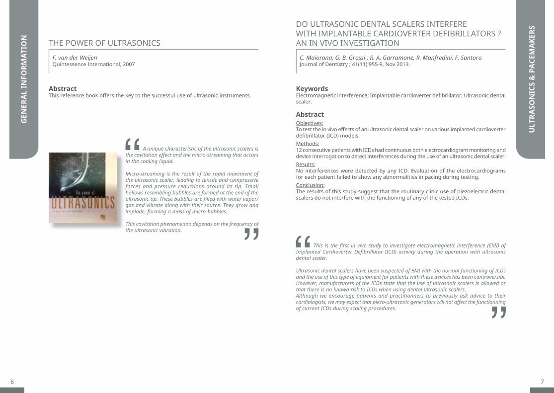
A unique characteristic of the ultrasonic scalers is the cavitation effect and the micro-streaming that occurs in the cooling liquid.
Micro-streaming is the result of the rapid movement of the ultrasonic scaler, leading to tensile and compressive forces and pressure reductions around its tip. Small hollows resembling bubbles are formed at the end of the ultrasonic tip. These bubbles are filled with water vapor/gas and vibrate along with their source. They grow and implode, forming a mass of micro-bubbles.
This cavitation phenomenon depends on the frequency of the ultrasonic vibration.
THE POWER OF ULTRASONICS
DO ULTRASONIC DENTAL SCALERS INTERFERE WITH IMPLANTABLE CARDIOVERTER DEFIBRILLATORS ? AN IN VIVO INVESTIGATION
F. van der WeijenQuintessence International, 2007
C. Maiorana, G. B. Grossi , R. A. Garramone, R. Manfredini, F. SantoroJournal of Dentistry ; 41(11):955-9, Nov 2013.
AbstractThis reference book offers the key to the successul use of ultrasonic instruments.
KeywordsElectromagnetic interference; Implantable cardioverter defibrillator; Ultrasonic dental scaler.
AbstractObjectives:To test the in vivo effects of an ultrasonic dental scaler on various implanted cardioverter defibrillator (ICD) models.Methods:12 consecutive patients with ICDs had continuous both electrocardiogram monitoring and device interrogation to detect interferences during the use of an ultrasonic dental scaler.Results:No interferences were detected by any ICD. Evaluation of the electrocardiograms for each patient failed to show any abnormalities in pacing during testing.Conclusion:The results of this study suggest that the routinary clinic use of piezoelectric dental scalers do not interfere with the functioning of any of the tested ICDs.

8 9
SCALING AND PRESERVATION..................................................................... p10
SCALING AND DENTAL PLAQUE REVEALERS..................................................................... p11
CARIES TREATMENT..................................................................... p13
PROPHYLAXIS
10 11*Original title: Évaluation clinique du détartrage supragingival assisté du révélateur de plaque F.L.A.G™ for B.LED
IN VITRO EVALUATION OF TEMPERATURE CHANGES IN THE ROOT CANAL INDUCED BY ULTRASONICS SCALERS
U. Van der Velden, TJG. Koster, AJ. Feilzer, MF. Timmerman, GA. Van der WeijdenInternational Journal of Dental Hygiene, 2014
A rise of temperature of more than 3°C in dental pulp during dental procedures can lead to vascular injuries and tissue necrosis.For all three piezo devices, it was shown that lower water supply settings than the advised full amount can be used, which may be beneficial for the operator’s visibility and to reduce risks of cross-contamination.The P-max needed the least water supply ranging from 7.9 to 12.6 ml/min. Indeed, Newtron module is very safe for dentinal tissue since the minimal irrigation flow recommended for perio tips is 13 ml/min, a drop-by-drop setting.Cavitron needed approximately the double amount of water cooling to have a constant temperature of the root canal. The Vector finally presented a risk of an increase in temperature in the root.
Keywords Cooling water, root canal, temperature, ultrasonic scaling.
AbstractObjectives:To evaluate in vitro the thermal effects induced by four different ultrasonic scalers on the temperature in the root canal during ultrasonic scaling.Methods:An extracted lower central incisor provided with a thermocouple in the root canal and a tube, entering the tooth incisally and exiting it apically to simulate an artificial bloodstream, was placed in a model of the lower jaw with soft artificial gingiva. Tested ultrasonic scaler systems included: EMS PM-600, Satelec P-max, Durr Vector and Dentsply Cavitron. The tooth was scaled with each system at full water supply of 21°C. Furthermore, the amount of water supply was determined to maintain during scaling a constant temperature in the root canal. Finally, thermal changes due to scaling without water were assessed.Results:Except for the Vector, all scaler systems showed a temperature decrease in the root canal. The Vector with water/polish suspension showed a trend towards an increase in temperature. To maintain a constant temperature in the root canal the Cavitron needed twice the amount of water compared with PM-600 and P-max. Without water, all scaling systems induced a temperature increase.Conclusion:For safe ultrasonic scaling, care should be taken that the cooling water has room temperature and that, dependent on the scaler system, the proper amount of water is supplied.
SCA
LIN
G &
PRE
SERV
ATIO
N
SCA
LIN
G &
DEN
TAL
PLAQ
UE
REVE
ALE
RS
The results of this prospective study showed that the brush application of F.L.A.G.TM for B.LED dental plaque discloser improves the subgingival scaling in terms of quality and time compared to traditional scaling.Quality of care• At the end of treatment, the plaque index of the side treated without F.L.A.G.TM for B.LED
decreased by 68%, while the plaque index of the side treated with F.L.A.G.TM for B.LED decreased by 87.5% . Thus, the use of F.L.A.G.TM for B.LED improved the dental plaque removal of 19.5%.
• After re-application of the discloser for final control, we notice that F.L.A.G.TM for B.LED improved dental plaque removal by 26.5%.
Time saving• The F.L.A.G.TM for B.LED allowed to gain about 9.7 seconds during the treatment of vestibular
surfaces of six teeth compared to conventional scaling.• The total processing time of the mouth should therefore decrease by 51 sec. This time does
not include the application of F.L.A.G.TM for B.LED on teeth and rinsing.• The time saving observed with F.L.A.G.TM for B.LED proves that treatment is optimized.
The practitioner focuses only on the affected areas and avoids over intrumentation on plaque-free tooth surfaces.
CLINICAL EVALUATION OF ASSISTED SUPRAGINGIVAL SCALING BY PLAQUE DISCLOSING AGENT F.L.A.G.™ FOR B.LED*
Timothée MarrienThèse pour l’obtention du Diplôme d’Etat de Docteur en Chirurgie dentaire - n°21 - 2015 Mention honorable (Thesis)
Keywords Dental plaque, supragingival scaling, fluorescein, F.L.A.G.TM for B.LED;
AbstractObjectives:A new supragingival scaling technique associated with a fluorescent plaque disclosing agent was developed: the F.L.A.G.TM for B.LED of ACTEON. The aim of this clinical study was to evaluate this device in terms of quality and care’s duration.Patients and Methods:Thirty volonteers (29 ± 13 years) were treated by two operators in a study like Split mouth design. The buccal surfaces of anterior teeth were divided into two groups randomly: a control group treated with ultrasonic scaling and a test group treated with the F.L.A.G.TM for B.LED. We repeatedly measured plaque indexes with a probe or the disclosing solution, and duration of the care for every patient and every sector.Results:The plaque index of the side test was on average lower than the plaque index side reference of 0.22 (Löe et Silness scale; p<0.0001). The time of scaling test sector was on average lower of 9,7 seconds than the one of the control sector (p=0.0148).Discussion:The F.L.A.G.TM for B.LED device brings a profit by eliminating more dental plaque and reducing the duration of treatment.
12 13* Original title: Ultraschall mit B.LED-Technologie: Gewinn für die Prophylaxebehandlung
SIGNIFICANT ADVANCES IN DENTAL PROPHYLAXIS: ULTRASONIC TREATMENT WITH ACTEON’S B.LED TECHNOLOGY*
V. BraunDZW Die Zahnarztwoche - Edition 37/13, 11 Sept 2013
Staining the plaque makes my work easier. Being able to see where I have to intervene makes the task much simpler. The advantage of this approach is that I avoid under or over treatment. The patients can also see the care I take with each tooth. I discuss the procedure with them and involve them in the control process using the images (pre/post) from the intraoral camera. Nine out of ten patients really appreciate the precision of my work and do not complain if it lasts longer or costs more. [...]
With the help of the SLIM B.LED handpiece and the F.L.A.G.TM plaque indicator, I can quickly and safely detect plaque and tartar, and immediately remove them with the ultrasonic handpiece. When I apply the fluid to the teeth, the colour indicator attaches itself to the plaque and becomes fluorescent yellow/green under the blue LED light of the handpiece.
Abstract“If the dental plaque is fluorescent, removing it is not a challenge.”
SCA
LIN
G &
DEN
TAL
PLAQ
UE
REVE
ALE
RS
CARI
ES T
REAT
MEN
T
The Excavus tips, with their small shape and the selective cut given by ultrasonics, can treat caries without damaging the adjacent teeth. Moreover, the regularity of their diamond coating gives them outstanding quality of grounding.Finally, the preparation of the micro-cavities with Excavus tips retains the adhesive properties with the composite materials.
EFFECT OF ULTRASONIC DIAMOND TIP ON DENTIN BONDING OF COMPOSITE
H. Takanashi, R. Kishikawa, N. Iwamoto, M. Otsuki, J. TagamiIADR/AADR/CADR - poster 1509, 2007
AbstractObjectives:The purpose of this study was to evaluate the effect of an ultrasonic diamond tip on dentin bond strength of resin composite. Methods:Twelve extracted human third molars were ground to obtain flat midcoronal dentin surface. The surfaces were polished with #600 SiC paper. The specimens were divided into four groups. The surfaces of each group were treated as follows. Group1: The surface was not ground moreover. Group 2: The surface was ground by a round-shape steel bur (016, J Morita, Japan) with a low-speed micromotor handpiece. Group3: The surface was ground by a round-shape diamond bur (M340, J Morita, Japan) with a high-speed turbine handpiece. Group4: The surface was ground by a diamond-particle tip (EX1, Excavus, Satelec, France) with ultrasonic generator (P5 Newtron, Satelec France). Each surface was treated with a self-etching primer adhesive (Clearfil SE Bond, Kuraray Medical, Japan) and a photo-cure composite (Clearfil AP-X, Kuraray Medical, Japan) was placed and photo-cured. After 24 hrs, each sample was crosscut and re-crosscut perpendicular to the resin-dentin interface to obtain 0.7x0.7 mm beam-shaped specimens. The specimens were subjected to the micro-tensile bond test at cross head speed of 1 mm/min (N=45). Results:The mean of the micro-tensile bond strengths and SD were as follows (MPa); Group1: 79.8 (21.3), Group2: 79.7 (17.6), Group3: 69.9 (13.2), Group4: 79.5 (11.2). The bond strengths of group2 and 4 were statistically higher than that of group3. There was no statistical significance among the bond strengths of group1, 2 and 4.Conclusion:The ground dentin using ultrasonic diamond tip did not reduce the dentin bond strength of resin composite.
14 15
PERIODONTICSPERIODONTAL DEBRIDEMENT..................................................................... p16
ROOT PLANING..................................................................... p18
PERIODONTAL MAINTENANCE..................................................................... p21
IMPLANT CARE..................................................................... p22

16 17* Original title: Etude comparative des instrumentations manuelle et ultrasonique sur les surfaces cémentaires, influence de
la pression latérale* Original title: Traitement ultrasonique des poches parodontales (T.U.P.). Simplification des procédures dans le traitement
étiologique des maladies parodontales
SATELEC micro perio tips facilitate maintenance treatments, making them faster and less painful. Better accepted by the patient, they are less time-consuming for the practitioner, who can adapt instrumentation to a greater number of clinical cases.
ULTRASONIC TREATMENT OF PERIODONTAL POCKETS. SIMPLIFICATION OF PROCEDURES IN THE ETIOLOGICAL TREATMENT OF PERIODONTAL DISEASES*
G. Gagnot, M-G. Poblete, J-F. MichelSwissdent (25), n°5 5-10, 2004
AbstractUltrasonic instruments have specific advantages over manual methods because they participate in the disruption and removal of subgingival plaque. We talk about periodontal debridement.Ultrasonic tips are more active due to the conjunction of five effects: hammering, sweeping, irrigation, micro-streaming, cavitation.
SATELEC micro perio tips are as effective as manual instruments to remove plaque and tartar. Used with a low lateral pressure, they are more effective on the roots and difficult to reach areas, and they allow better preservation of the periodontal tissues compared to manual instruments.
COMPARATIVE STUDY OF MANUAL AND ULTRASONIC INSTRUMENTATION ON CEMENTUM SURFACES, INFLUENCE OF LATERAL PRESSURE*
G. Gagnot, F. Mora, M-G. Poblete, J-F. Michel, G. CathelineauRevue Internationale de Parodontie et Dentisterie Restauratrice ; 24, 136-145, 2004
AbstractThe first goal of periodontal treatment is to eliminate the infection to prevent progression of inflammatory processes that destroy tooth supporting tissues. [...] These treatments should include control of supragingival plaque and sub gingival scaling. This is a mechanical treatment to remove plaque, tartar and food debris in the pockets.
PERI
OD
ON
TAL
DEB
RID
EMEN
T
PERI
OD
ON
TAL
DEB
RID
EMEN
T
18 19* Original title: Etude comparative de deux instruments ultrasonores dans le traitement des surfaces radiculaires * Original title: Traitement ultrasonique des poches parodontales: action de nouveaux inserts, indications et perspectives
dans le traitement des surfaces radiculaires et désorganisation du biofilm
H2 and H4 tips from SATELEC allow a very easy access to inter-radicular spaces and ease of use compared with manual instruments. Surfaces cleaning can be done up to the immediate proximity of the epithelial attachment.Root surfaces treated with H2 and H4 tips do not have anymore organic deposits and are close to the state of healthy tissue. They are therfore perfectly indicated for root planing and finishing of root surface.
ULTRASONIC TREATMENTS OF PERIODONTAL POCKETS: ACTION OF NEW INSERTS, INDICATIONS AND PERSPECTIVE IN THE TREATMENT OF ROOT SURFACES AND BIOFILM DISRUPTIONS*
G. Gagnot, J. Darcel, J-F. MichelJournal de Parodontologie et d’Implantologie orale, vol 19, 49-58, 1999
Keywords Scaling and root planing, diamond-coated inserts, root surface analysis, root surface treatment.
AbstractUltrasonic instrumentation is now considered an acceptable alternative to manual scaling and root planing in the initial phase of treatment of periodontal diseases. The aims of both methods are to disrupt the biofilm, to treat the root surface and the epithelial wall of the pocket.This study examines the effect of in vivo treatment with specially shaped diamond-coated inserts, on the root surfaces of teeth. The furcations of molar teeth which were due to be extracted were assessed. Following extraction, the teeth were examined with SEM. The aims if this study were: 1) to assess the nature of the root surfaces of teeth with severe or terminal alveolar
bone loss after using H2 and H4 inserts; 2) to determine the composition of the root surfaces before and after the use of these
instruments, with the aid of an Energy Dispersion Analysis Spectrum Probe (EDASP); 3) to propose a rationale for a sequence of treatments using these instruments in
the initial phase of periodontal treatment.
This study compares the two types of SATELEC micro tips H4 and BDR (TK1 et TK2), that can be recommended in the treatment of periodontal pockets. The H4L and H4R tips seems to be more efficient for elimination of bacteria and calculus, which ranks them as first-line tips for the treatment of periodontal pockets.The BDR tips are the ones recommended for the treatment of pockets with little calculus, especially during maintenance phase. The interest of BDR tips is to prompt the operator to avoid using the insert like a curette: he will no longer exert excessive pressure on the tooth that would risk to alter the cementum surface.Therefore, these two types of tips enable the long term treatment of periodontal disease, while preserving root surfaces.
A COMPARATIVE STUDY OF TWO ULTRASONIC INSTRUMENTS USED IN THE TREATMENT OF ROOT SURFACES*
G. Gagnot, A-M. Leray, V. Meuric, G. CathelineauRevue d’Odonto-Stomatologie, mai 2005
AbstractIntroduction: The objectives of this study were to compare two series of ultrasonic micro inserts used in the elimination of biofilm and root calculus and their effects on the condition of the root surface.Materials and Methods:14 single rooted teeth afflicted by terminal-stage periodontitis, indicating their extraction were used in this study. The teeth were then examined under the SEM using the 2 methods of observation: Secondary and Back-scattered electron. Three neutral examiners noted the presence of biofilm, calculus, scratches, dentin, smear layer, and the cementum surface aspect: nodular, smooth, cracked and altered.Results:Out of 111 zones observed the major significant difference (p<0.05) was the number of areas free of bacteria and calculus as well as the number of cementum alterations and smear layer. In 88 % of these zones treated by the micro insert curettes (MIC), the surfaces were unscathed or presented minute quantities of bacteria against the 72 % of teeth treated by the round section micro inserts (MIR). In 84.3 % of teeth treated by MIC does not or present very little residual calculus against 66 % of teeth treated by MIR. On the other hand, the cementum was distinctly altered by the MIC in 12.3 % of the teeth against 3.8 % using the MIR. Finally, the surfaces treated by the MIC were often times covered by smear layer(47.4 %) than those treated by the MIR (22.5 %).
ROO
T PL
AN
ING
ROO
T PL
AN
ING

20 21* Original title: Action de nouveaux inserts sur les dômes des espaces interradiculaires. Observation au MEB * Original title: L’utilisation des instruments ultrasoniques pour la maintenance parodontale
The micro perio tips from SATELEC allow the evolution of periodontal maintenance treatments to less invasive instrumentation respecting the cementum and attachment. They allow considerable time gain and offer the possibility, in addition to mechanical debridement, to irrigate with antibacterial agents. Finally, they are more efficient than the manual instruments for the elimination of calculus at the bottom of the pockets and at the furcations, especially when the depth of the pockets is superior to 6 mm.
USE OF ULTRASONIC INSTRUMENTS FOR PERIODONTAL MAINTENANCE*
I. JuzanxAlternativeS - N° 28 Nov 2005
AbstractNowadays, aggressive root planing aimed to totally eliminate the cementum that is supposed to be infiltrated by endotoxins, and obtaining a smooth and hard radicular surface, are no longer justified. The objective of periodontal debridement is to treat inflammation by cleaning and disinfection of teeth and radicular surfaces so that the surrounding tissues revert to their non-inflammatory healthy state. For maintenance, as for the other stages of periodontal treatment, the ultrasonic instruments present numerous advantages in comparison to manual instruments.
The diamond coated tips H1, H2L and H2R can increase opportunities to remove the plaque and tartar in furcation areas impossible to access with manual instruments. It is advisable to use an H4 tip after the passage of diamond tips to remove the smear layer generated by abrasion.
SEM STUDY OF THE EFFECT OF NEW ULTRASONIC INSERTS ON FURCATION DOME*
G. Gagnot, J. Darcel, J-F. Michel, G. CathelineauJournal de Parodontologie et d’Implantologie orale, vol 19, 411-417, 1999
Keywords Scaling, root planing, microtips, ultasonic scaler, root surface debridement.
AbstractThe results of long term studies show that, irrespective of the treatment used, multirooted teeth have a greater failure rate than single-rooted teeth. The reason for this is that it is more difficult or impossible to access the furcations with conventional instruments. Now, a new type of ultrasonic microtip has been developed with new ultrasonic generators. These generators provide greater control of the vibrations. This study shows the effects of diamond-coated steel microtips on molar furcations. All samples has either a Class III or IV furcation involvment. They were treated in vivo before extraction, and then observed by SEM. We have observed that 97 % of the areas were cementum. On these areas, 33.8 % were smooth, 52.3 % showed scratches and 23 % had notches. Obervations also showed that 23 % had residual organic material, 32.3 % had a smear layer, and 4.6 % had calculus and bacterial plaque on the furcation areas. These results show that this new type of instrument increases our ability to eliminate bacterial plaque and calculus furcations, which have been known to be the most difficult areas to clean.
ROO
T PL
AN
ING
PERI
OD
ON
TAL
MA
INTE
NA
NCE

22 23* Original title: Effets d’inserts ultrasoniques en composite sur des piliers implantaires. Etude in vitro
The use of composite tips Periosoft from SATELEC on implant abutments reduce surface irregularities usually observed with other instruments. Therefore, these tips allow to decontaminate implant abutments wihout damaging their surface.In maintenance treatment, the Periosoft tips also prevent the reformation of plaque and calculus on these exposed implant surfaces.
EFFECTS OF ULTRASONIC TIPS IN COMPOSITE ON IMPLANT ABUTMENTS. STUDY IN VITRO*
G. Gagnot, H. Prigent, J. Darcel, J-F. Michel, G. CathelineauJournal de Parodontologie & d’Implantologie Orale, vol 18, n° 4 p. 393-399, Jan 1999
Keywords Dental implants, maintenance, power driven instrumentation, composite tips.
AbstractThe long term success of dental implants is dependant on the plaque control of the implant surfaces above the gingival attachment. However the maintenance methods used by patients and professionals increase the roughness of the abutments. These variations influence the adhesion properties of the surfaces for bacteria and periodontal tissues. Today only plastic hand curettes are recommended, but their shape and size do not allow for the complete removal of dental plaque and calculus. Thus we have observed the effects of ultrasonic composite tips on implant abutments. The observations were carried out with a rugosimeter and scanning electron microscope (SEM). With a large magnification, the test areas have shown fewer defects than control and no increase in roughness was observed with the rugosimeter. These present observations prove that the ultrasonic composite tips do not damage the abutment surfaces and their oral use can be recommended.
Residues of materials on the implant, such as plastic or steel, are harmful and do not allow the healing of peri-implant tissues. On the contrary, Titanium leaves no residue.Implant Protect tips in Pure Titanium are therefore perfectly indicated for a safe and successful implant surfaces debridement.
OPTIMAL MATERIALS OF THE ULTRASONIC SCALERS TIP FOR DEBRIDEMENT OF MICROSTRUCTURED FIXTURE SURFACE OF DENTAL TITANIUM IMPLANT
Y. Yoda Poster EAO Roma Sept 2014
AbstractBackground:Recently, the increasing occurrence of peri-implant disease has been reported due to the rapid increase of dental implant therapy. In peri-implantitis, exposed fixture surfaces of dental implants that lose osseointegration with the supportive bone are contaminated by bacterial biofilms and/or mineralized deposits. To treat peri-implantitis, it is essential to decontaminate the exposed contaminated fixture surfaces. Ultrasonic scaling has been reported to be effective for treating peri-implant disease. However, the elements of different electron potential on the scaled titanium implant surface cause the risk of fixture erosion, and the optimal materials of ultrasonic scaler tip for debridement of microstructured surfaces of titanium implants have not been fully evaluated.Introduction: The present study seeks to evaluate morphological and elemental changes induced on microstructured surfaces of dental endosseous implants by ultrasonic scaler using pure titanium and alloyed titanium tip.Materials and Methods:4 commercially available titanium dental implants Tioblast® and TiUnite® were used in vitro experiments. Mechanical treatments were performed for approximately 30 s. The US was used at a power level of 3 in scaling mode. An ultrasonic scaler with pure titanium metal tips or alloyed titanium metal tips was employed. All treatments were performed in approximately 1 mm x 2 mm areas between the threads of an implant. In all the experiments, the treated sites were observed by optical stereomicroscopy and scanning electron microscopy (SEM). In the stereomicroscopy, the prepared specimens were observed at a magnification of 200 times. In the SEM observations, a secondary electron image was obtained at an accelerating voltage of 20 kV and a tilt angle of 50° with a magnification of 1,000 times. Elemental analysis of the treated surfaces was performed for 1,200 s using an energy-dispersive X-ray microanalyzer (EDS) at 20 kV. Results:The pure titanium and alloyed titanium treatments severely damaged the Tiunite® and Tioblast® surface, producing shiny surfaces with scratches and crumples. SEM observations revealed that all the treatments completely removed or crushed the microstructure. At the EDS analysis, there were no foreign elements on the treated sites using pure titanium tip. A small amount of Nb and Al were detected after treatment area using alloyed titanium tip. Conclusion:The treated sites of ultrasonic scaler using pure titanium tips were damaged for debridement, but not contaminated the foreign elements without titanium.
IMPL
AN
T CA
RE
IMPL
AN
T CA
RE
24 25
ENDODONTICSENDODONTIC RETREATMENT..................................................................... p26
ENDODONTIC IRRIGATION..................................................................... p33 APICAL SURGERY..................................................................... p36

2726* Original title: L’utilisation de nouveaux inserts en endodontie covnentionnelle
The EndoSuccess Retreatment kit is valuable to treat complex clinical endodontic cases, preferably under operating microscope.The ETPR tip, placed on the head of the post, with a maximum irrigation, can fragment the sealing cement and easily remove the metal post. The ET18D and ETBD tips allow the preparation of access cavity and the canal orifices localization. The ET20 and ET25 (in Titanium Niobium) tips are effective to remove residues of filling materials and fractured instruments even in apical zones. In some cases, ET25 tips can be slightly bent in order to work in root curvatures.
The mesial intercanal groove of mandibular molars is the result of the incomplete separation of the mesial root which explains the co-existence of two canals (buccal and lingual) in the same root.After the canal shaping, potentially infected pulpal tissue could persist in the groove and might compromise the treatment prognosis.This study showed that mechanical shaping of root canals with rotary files alone was insufficient.On the contrary, ET20D or ET25 tips were able to significantly deepen and increase in surface area (189.8% and 245.4% respectively) the internal and external surfaces of mesial intercanal groove.The ultrasonic treatment with ET20D or ET25 tips makes it easier to clean the area by eliminating pulp tissue and creating a reservoir for irrigation solutions. While shaping the canal system in accordance with the fundamental principles of endodontic treatments, it also seems to maximize the short- and long-term success of the endodontic treatment.
USE OF NEW ULTRASONIC TIPS IN CONVENTIONAL ENDODONTICS*
ANATOMY AND SHAPING OF THE MESIAL INTERCANAL GROOVE OF THE LOWER MOLARS
B. Khayat, J-C. MichonneauEndodontic Practice 10(4): 15-20, August 2008
A. Basso, E. Prats, F. Diemer, J-P. Mallet, P. CalasEuropean Cells and Materials Vol. 13. Suppl. 1, p.25, 2007
AbstractA new generation of ultrasonic tips made of titanium niobium by Satelec is available for conventional endodontic treatment. This material improves the quality of ultrasonic transmission and increases the mechanical resistance of the tips. These safe and reliable instruments can be valuable tools in managing complex endodontics cases.
AbstractRoot canal anatomy, especially the one of the molars, has always drawn people’s attention in endodontics.Aim:To study, on a set of first and second mandibular molars, the impact of using shaping instruments and ultrasonic tips on the depth and surface of this anatomic feature.Methodology:18 mandibular molars are used. The roots are embedded in transparent resined cubes. An isomet makes a mesio-distal cut between the mesiobuccal and mesiolingual orifices. Root canals are prepared with HeroShaper files. The groove is prepared with two ultrasonic tips (ET20D or the ET 25, Satelec, Acteon Group, Mérignac, France). Pictures are taken, for each operating stage, and analyzed with Photoshop software in order to calculate the depth and the surface of the groove. These data are analyzed (Student test, analysis of the variance) with a risk of 5%. Results:The ultrasonic preparation allows a significant surface gain of the groove on the occlusal, internal and external views. There is not any significant difference between the groove preparation done by the ET20D tip and the ET 25 one (p=0.3722 to 0.9904 depending of the views).Conclusion:The groove ultrasonic preparation favours its widening and its deepening without distorting root canal orifices. It allows the cleansing of this area by favouring the elimination of the pulpal tissue. Moreover it creates a “beam” between the mesial root canals which, prepared in this way, could be used to bond and fix coronoradicular restorations.
END
OD
ON
TIC
RETR
EATM
ENT
END
OD
ON
TIC
RETR
EATM
ENT

2928* Original title: Stratégie d’éviction des instruments brisés
The safest solution consists in redesigning the cavity access to free the broken instrument.ET25 tips, used at low power, without irrigation and intermittently around the broken instrument without touching it, allow to remove the smear layer and to release the instrument.In case of instrument broken but not stuck into the canal, a vibration of ET25 tips with irrigation dislodges it easily.
Among the possible managements of intracanal broken instrument withdrawal , ultrasonic tips represent a safe and effective solution: their success rate extends in clinical studies from 67% (Nagai et al.), to 88% (Cuje et al.) and 95% (Fu et al.).Diamond coated or Titanium-Niobium tip can be activated at low power to access the fragment and eject it through ultrasonic vibrations. The K-files can be used when the fragment is stuck deep in the canal, since the tactile sense they give allows the operator to work without visibility.The bypass of broken instrument minimizes the contact of the fragment with the canal walls and may enable the passage of ultrasonic tips to dislodge it. Finally, apical surgery may be considered if the fragment is inaccessible or in the presence of periapical lesion.
STRATEGY OF BROKEN INSTRUMENTS REMOVAL*MANAGEMENT OF INTRACANAL SEPARATED INSTRUMENTS
S. Bal, S. Bourbon Kerisit, F. BoussettaClinic, vol. 27, Fév 2006
A. A. Madarati, M. J. Hunter, P. M.H. DummerJournal of Endodontics, vol 39, n° 5, Pages 569–581, May 2013
AbstractFractures of rotative NiTi instruments into canals are frequent, and their withdrawal is delicate. It is thus recommended to work under microscope. To prevent these fractures, it is important to follow protocols indicated by manufacturers, to work without pressure in the canal, and to regularly change the instruments.
KeywordsBroken, bypassing, complications, files, fractured, instruments, management, removal, retrieval, separated.
AbstractIntroduction:Intracanal separation of endodontic instruments may hinder cleaning and shaping procedures within the root canal system, with a potential impact on the outcome of treatment. The purposes of this narrative review of separated instruments were to (1) review the literature regarding treatment options, influencing factors, and complications and (2) suggest a decision-making process for their management.Methods:An online search was conducted in peer-review journals listed in PubMed to retrieve clinical and experimental studies, case reports, and review articles by using the following key words: instruments, files, obstructions, fractured, separated, broken, removal, retrieval, management, bypassing, and complications with or without root canal and endodontic.Results:There is a lack of high-level evidence on management of separated instruments. Conventional conservative management includes removal of or bypassing the fragment or filling the root canal system to the coronal level of the fragment. A surgical intervention remains an alternative approach. These approaches are influenced by a number of factors and may be associated with complications. On the basis of current clinical evidence, a decision-making process for management is suggested.Conclusion:Guidelines for management of intracanal separated instruments have not been formulated. Decisions on management should consider the following: (1) the constraints of the root canal accommodating the fragment, (2) the stage of root canal preparation at which the instrument separated, (3) the expertise of the clinician, (4) the armamentaria available, (5) the potential complications of the treatment approach adopted, and (6) the strategic importance of the tooth involved and the presence/or absence of periapical pathosis. Clinical experience and understanding of these influencing factors as well as the ability to make a balanced decision are essential.
END
OD
ON
TIC
RETR
EATM
ENT
END
OD
ON
TIC
RETR
EATM
ENT
3130
The modified SO4 in this article is a prototype of the ET20 tip.
Introduced next to the exposed part of the obstruction and activated for 1 or 2 min to transmit vibration to the metallic obstruction, the ET20 tip will dislodge the loosened fragment and the flood of water will carry it out of the canal.
This SO4 modified in ET20 allows deeper penetration in the canal without excessive tooth structure loss while leaving sufficient space for instrument withdrawal. It is as small as ultrasonic files, but much more resistant to fracture.
The technique proposed is helpful each time an obstruction cannot be bypassed, and is safe and predictable.
The aim of this in vitro study was to determine the efficiency of the PmaxXS and its Air Active function in reducing temperature rise on the external root surface during the ultrasonic removal of fractured files, and to compare temperature rise using ET40D (Satelec, Acteon) or CPR5 (Spartan-Obtura) tips.The air flow from Pmax Xs device significantly reduced the temperature rise on the external root surface during the ultrasonic removal of fractured files. Moreover, the use of ET40D tip with this Air Active function induced significantly lower temperature rise of the external root during removal of fractured instruments compared with CPR5 tip. Indeed, the ET40D design allows to focus the air flow on the tip extremity and into the canal.
A NEW APPROACH FOR THE RETRIEVAL OF BROKEN INSTRUMENTS
EFFICIENCY OF A NEWLY DESIGNED ULTRASONIC UNIT AND TIPS IN REDUCING TEMPERATURE RISE ON ROOT SURFACE DURING THE REMOVAL OF FRACTURED FILES
W. NehmeJournal of Endodontics, vol 25, n°9, pages 633-5, September 1999
A. A. Madarati, A. J.E. Qualtrough, D. C. WattsJournal of Endodontics, vol 35, n°6, pages 896-9, June 2009
AbstractThe purpose of this article is to present a modified ultrasonic spreader (SO4 SATELEC) and a new technique that are used for the retrieval of solid obstructions that can not be bypassed by conventional methods. The technique advocated and the instruments proposed are described. A clinical case is discussed to show the possibilities and limitations of both instrument and method.
KeywordsRemoval, retreatment complications, separated endodontic instruments, temperature rise, ultrasonic tips, ultrasonic unit.
AbstractIntroduction:A potentially damaging temperature rise within the root canal and thus on the external root surface may be induced because of frictional contact of ultrasonic tips during the removal of separated instruments. The efficiency of a new ultrasonic unit, with air-spray function and ET40D (Satelec/Acteon, Merignac, France) and CPR5 (Obtura-Spartan, Fenton, MO) ultrasonic tips, in reducing temperature rise on the external root surface during the removal of fractured files was investigated.Methods:Four millimeters of F2 ProTaper files (Dentsply, Surrey, UK) were fractured 2.5 mm from the canal access of 60 lower incisor roots. Roots were randomly divided into six groups: groups CPR5/no air and ET40D/no air in which tips were used without air flow, groups CPR5/15 psi and ET40D/15 psi (tips used with 15-psi air pressure), and groups CPR5/10 psi and ET40D/10 psi (10-psi air pressure). The temperature rise was measured on the external proximal root surface, adjacent to the most coronal aspect of the fractured fragment, at 15 seconds and then at 30-second intervals up to 120 seconds.Results:After 120 seconds, the mean temperature rise (4.2 degrees C) with the air flow active was significantly lower than that with nonactive air flow (11 degrees C). At 10- and 15-psi pressures, the temperature rise after 120 seconds induced by ET40D tips was 4 degrees and 2.4 degrees C, respectively. These were significantly lower than with CPR5 tips (6.3 degrees and 4.2 degrees C, respectively).Conclusion:A new ultrasonic unit incorporating an air-flow function proved to be effective in reducing the temperature rise during removal of fractured files. ET40D ultrasonic tips were more effective than the CPR5 tip. However, both tips could be safely activated with air spray up to 120 seconds.
END
OD
ON
TIC
RETR
EATM
ENT
END
OD
ON
TIC
RETR
EATM
ENT
3332* Original title: Retraitement endodontique : réussir la deuxième fois
This study shows that ultrasonic tips, cooled with a good irrigation, is essential for the retrieval of endodontic posts to avoid the heating of roots which could drive to radicular necrosis.
Irrigation with ultrasonics enables clinicians to place an irrigation solution in the pulpar chamber and to activate it on its approach to the apical end of the root canal. IrriSafe, non-cutting ultrasonic files, is placed in each canal and is moved up and down in the canal, for three cycles of 1 minute. It has been demonstrated that ultrasonic irrigation better irrigated the lateral canals at 4.5 and 2 mm of the working length of the canal, compared to the single needle irrigation. Moreover, ultrasonic irrigation can remove debris from dentin in a canal up to 3 mm further the area reached by the tip apically in straight or curved canals. Therefore, passive irrigation with IrriSafe contributes to the success of endodontic retreatment.
INCREASE OF THE TEMPERATURE DURING THE ULTRASONIC REMOVAL OF ENDODONTIC POSTS
ENDODONTIC RETREATMENT: SUCCEED THE SECOND TIME*
N. M. Ferrarese , G. Carli, N. M. Balzano, C. Tocchio Poster ESE 2011
E. G. BrettRoots 1, spécial retraitement endodontique, 2013
AbstractIntroduction:The litterature suggests the reduction of the temperature during the removal of endodontic posts by application of ultrasonic device. The heat’s control is important to avoid damages to periodontal ligament like external gingival recession.Objective:The aim of the study is the comparison, in vitro, of different techniques and materials used in retreatment of endodontically treated teeth filled with direct and indirect posts.Methods:Several canals have been made using epoxy resin. An internal and an external thermo-probe have been connected to a thermocouple that measure the increases of the temperature when a cooled or not cooled ultrasonic tip is applied to remove the post.Three types of posts have been analyzed when removed with cooled and not cooled ultrasonic tip. Six groups have been created:
Group A1: cast metal post removed with not cooled ultrasonic tipGroup A2: cast metal post removed with cooled ultrasonic tipGroup B1: glass-fiber post removed with not cooled ultrasonic tipGroup B2: glass-fiber post removed with cooled ultrasonic tipGroup C1: retractable glass-fiber removed with not cooled ultrasonic tipGroup C2: retractable glass-fiber removed with cooled ultrasonic tip
Results:Results demonstrate that the use of a cooled tip is basically important to remove all types of posts. Retreatment of glass-fiber posts with soft core marks much less increase of temperature of all other posts. Conclusion:Conclusions, considering the limit of the study in vitro, mean that using retractable posts removed by cooled ultrasonic tips is an optimum condition of retreatment to avoid the external excessive heating of the root.
AbstractCurrent evidence showed that root retreatment of teeth having previously undergone endodontic treatment had a high success rate. The use of ultrasonic tips allows clinicians to preserve dental tissues, giving excellent visibility under dental operating microscope, which greatly improves the ability to retreat canals.Endodontic retreatment should be the first option for patients with postoperative infection.
END
OD
ON
TIC
RETR
EATM
ENT
END
OD
ON
TIC
IRRI
GAT
ION
3534
END
OD
ON
TIC
IRRI
GAT
ION
END
OD
ON
TIC
IRRI
GAT
ION
Results showed that conventional needle irrigation, EndoActivator or sonic devices, in combination with 6% sodium hypochlorite, failed to completely dissolve biofilm.Passive Ultrasonic Irrigation (Irrisafe) was more efficient to activate NaOCl thanks to the higher vibration frequency that enhances microstreaming, improving the cleaning of biofilm-infected dentine.
Passive ultrasonic irrigation was performed with Irrisafe 25 non-cutting file on a Suprasson P5 Booster and was, combined with an apical negative pressure irrigation system, the only group able to achieve the irrigant penetration both to the working lenght and into lateral canals.
Therefore, Irrisafe is a safe and effective solution for passive ultrasonic irrigation.
BIOFILM REMOVAL BY 6% SODIUM HYPOCHLORITE ACTIVATED BY DIFFERENT IRRIGATION TECHNIQUES
COMPARISON OF IRRIGANT PENETRATION UP TO WORKING LENGTH AND INTO SIMULATED LATERAL CANALS USING VARIOUS IRRIGATING TECHNIQUES
R. Ordinola-Zapata, C. M. Bramante, R. M. Aprecio, R. Handysides, D. E. JaramilloInt Endo Journal, Volume 47, Issue 7, pages 659–666, July 2014
E. Spoorthy, N. Velmurugan, S. Ballal, S. NandiniInternational Endodontic Journal, Volume 46, Issue 9, pages 815–822, Sept 2013
KeywordsBiofilms, irrigant solutions, laser-activated irrigation, photoacoustic streaming.
AbstractAim:To compare the removal of biofilm utilizing four irrigation techniques on a bovine root canal model.Methods:Fifty dentine specimens (2 × 2 mm) were infected with biofilm. The samples were then adapted to previously created cavities in the bovine model. The root canals were irrigated twice with 2 mL of 6% sodium hypochlorite for 2 min (4 min total). Following initial irrigation, the different treatment modalities were introduced for 60 s (3 × 20 s intervals). The evaluated techniques were needle irrigation, Endoactivator (Dentsply Tulsa Dental, Tulsa, OK, USA), passive ultrasonic irrigation and laser-activated irrigation (photon-induced photoacoustic streaming). The controls were irrigated with distilled water and conventional needle irrigation. Subsequently, the dentine samples were separated from the model and analysed using a scanning electron microscope (SEM). Fifteen operative fields were scanned per block, and SEM pictures were captured. Two calibrated evaluators examined the images and collected data using a four-degree scale. Nonparametric tests were used to evaluate for statistical significance amongst the groups.Results:The group undergoing laser-activated irrigation using photon-induced photoacoustic streaming exhibited the most favourable results in the removal of biofilm. Passive ultrasonic irrigation scores were significantly lower than both the Endoactivator and needle irrigation scores. Sonic and needle irrigation were not significantly different. The least favourable results were found in the control group.Conclusion:Laser activation of 6% sodium hypochlorite significantly improved the cleaning of biofilm-infected dentine followed by passive ultrasonic irrigation.
KeywordsApical negative pressure, passive ultrasonic irrigation, root canal irrigation, simulated lateral canals.
AbstractAim:To evaluate the effect of an apical negative pressure system, a passive ultrasonic irrigation system and a combination of both apical negative pressure and passive ultrasonic irrigation on the penetration of the irrigating contrast solution (ICS) up to working length and into simulated lateral canals.Methods:The root canals of 64 single-rooted teeth were instrumented using the ProTaper rotary system. In each sample, three simulated lateral canals were created at 2, 4 and 6 mm levels from the root apex using a 06-size C+ file (Dentsply Maillefer, Ballaigues, Switzerland). Samples were randomly assigned into 4 experimental groups (n = 16): group I - conventional needle irrigation, group II - passive ultrasonic irrigation, group III - apical negative irrigation system and group IV - combination of passive ultrasonic irrigation and apical negative pressure irrigation system. To examine irrigating solution penetration, Indian ink was mixed with 5.25% NaOCl and delivered into the root canals. Samples were then assessed by direct observation of the images taken using Canon EOS rebel T3. The depth of penetration of ICS up to the working length and into the simulated lateral canals was analysed using chi-squared tests.Results:The combination (ANP and PUI) and ANP group had significantly deeper ICS penetration up to the working length (P < 0.001). The combination (ANP and PUI) and the PUI group exhibited significantly greater ICS penetration into lateral canals at the 6 mm level (P < 0.001). At the 4 and 2 mm levels, the combination of ANP and PUI had significantly greater ICS penetration into the lateral canals than the other groups (P < 0.001).Conclusion:The combination of ANP and PUI was the only group able to achieve irrigating contrast solution penetration both up to the working length and into lateral canals.
3736
API
CAL
SURG
ERY
API
CAL
SURG
ERY
The use of an ultrasonic device in apical surgery showed a significant advantage over the traditional treatment, especially areas difficult to reach like the molars. The benefits are the tissues preservation (smaller cavities), a deeper preparation into the root, and a better treatment for the anatomical particularities as the isthmus.
Endodontic microsurgery has numerous indications, and the operating procedure is always performed with the aim of saving tissue. The Apical EndoSuccess Surgery kit allows to prepare a retro the canal system until 10 mm. The diamond coating is present only on the tip extremity to not enlarge too much the canal. These extremely slim ultrasonic micro-tips enable to limit the appearance of microcracks during retrograde canal preparation and to clean the intracanl isthmi and lateral canals responsible for lateroradicular lesions.
ULTRASONIC ROOT-END PREPARATION IN APICAL SURGERY
TISSUE CONSERVATION IN ENDODONTIC MICROSURGERY*
J. de Lange, T. Putters, E.M Baas, J. M. van IngenOral Surg Oral Med Oral Pathol Oral Radiol Endod, vol 104, number 6, Dec 2007
B. Khayat, J-C. MichonneauRev Odont Stomat 2008
AbstractObjective:The purpose of this study was to evaluate the potential benefit of an ultrasonic device in apical surgery on the outcome of treatment.Methods:A randomized prospective design was used in a standardized treatment protocol. Patients were allocated to treatment with an ultrasonic device (P-Max Newtron) or treatment with a bur in an otherwise similar protocol. One year after treatment the results were evaluated by 2 oral and maxillofacial surgeons who were blinded for the therapy.Results:Out of a total group of 399 patients who were included in the study, adequate follow-up could be obtained in 290 patients. The overall success rate in the ultrasonic group was 80.5% and in the group treated with a bur 70.9% (p=0.056). In molars, the difference in success rate was significant (p=0.02).Conclusion:The use of an ultrasonic device in apical surgery improved the outcome of treatment. In molars this effect was significant.
AbstractThe current evolution of all surgical techniques attempts to considerably reduce the access opening while being the least invasive possible. By using coelioscopy and microscopy, it becomes possible to access the pathologies by preserving at most surrounding tissues. These techniques, due to a very precise punctual access, present moreover much better post-operative consequences. Endodontic surgery follows this evolution by using the same types ofmicroscope used in E.N.T. and ophthalmology. This allows a permanent visual control of the working aera under a magnification up to 30 times. Each step becomes more precise, more controllable and also more conservative to bone and dental tissues. Endodontic surgery by becoming microsurgery allows a higher conservation of the root while treating the infection origin in the canal system. By approaching conventional endodontics, its prognosis has been considerably improved and its indications are now much more broadened.
* Original title: Economie tissulaire en micro-chirurgie endodontique
38 39
PROSTHESISINTRASULCULAR PROSTHETIC FINICHING..................................................................... p40
40 41* Original title: Pénétration intrasulculaire aux ultrasons : une nouvelle approche pour les préparations coronaires complètes * Original title: Précision et confort: l’apport des inserts ultrasons à la finition des préparations de prothèse fixée
ULTRASONIC SULCUS PENETRATION:A NEW APPROACH FOR FULL CROWN PREPARATIONS*
M. SousThe International Journal of Periodontics & Restorative Dentistry, volume 29, Number 3, p: 277–287, 2009
AbstractNewly developed ultrasonic diamond-coated tips (UDT) have been created for sulcus penetration and intracrevicular finish line preparation and polishing. One hundred forty-two freshly extracted premolars and molars were employed for a cutting efficiency test. Dentin surface roughness was analyzed with an optical profilometer and a scanning electron microscope. Within a reasonable cutting time, the UDTs possessed sufficient dentin-cutting capacity. The surface roughness of dentin prepared with the UDT can be improved by using a smaller grit size UDT or a smooth tip set at lower power, and the roughness approaches that of dentin prepared with a referenced red diamond bur. A clinical protocol of ultrasonic sulcus penetration with these developed UDTs is proposed.
The aim of this study was to evaluate the Perfect Margin Rounded tips in the intrasulcular penetration and dentine cutting and to compare them with the diamond bur.The Perfect Margin Rounded tips demonstrated efficient dentin cutting.They can precisely place the finishing line in the gingival sulcus without injury to the marginal periodontium, and obtain very clear and clean cervical finishing line with a sharp gingival margin.
Equipped with two diamond coated tips (PM1, PM2) to prepare the dentin and a smooth tip (PM3) to polish and perfect the surface, the Perfect Margin™ Rounded kit is dedicated to the intrasulcular finishing after supra or juxtagingvival preparation with a diamond bur.The Perfect Margin™ also consist of a conical tip PM4 perfectly suited for the preparation of the canal space and the anatomical shaping of the connection cone, while preserving a balance between the root and cameral part.
The excellent tactile sense restored by Perfect Margin™ tips give good control of the operating procedure for precise and fast positioning the finishing line. Their low amplitude of vibration allows non traumatic contact with periodontal tissues and no bleeding.
PRECISION AND COMFORT: THE CONTRIBUTION OF ULTRASONIC TIPS TO FIXED PROSTHETIC PREPARATIONS FINISHING*
M. SousClinic, Vol 29. 697-706, Dec 2008
KeywordsCoronary preparation, corono-radicular preparation, ultrasonic tip, gingival deflection.
AbstractIn fixed prosthesis, the preparation of pilar teeth is a fundamental step conditioned by the good biological and aesthetic integration of our reconstructions. The equipment used and the preparation forms have changed little. Yet it is now possible to integrate into our protocols ultrasonic tips: they offer many advantages and are therefore very complementary to diamond burs.
INTR
ASU
LCU
LAR
PRO
STH
ETIC
FIN
ISH
ING
INTR
ASU
LCU
LAR
PRO
STH
ETIC
FIN
ISH
ING

42 43
ULTRASONIC MARGIN PREPARATION FOR FIXED PROSTHODONTICS: A PILOT STUDY
P. Horne, V. Bennanni, N. Chandler, D. PurtonJournal of Esthetic and Restorative Dentistry, 2011
AbstractPurpose: Atraumatic, oscillating ultrasonic instruments have recently been developed for prosthodontic margin finishing. This in vitro observational pilot study aimed to compare the condition of crown preparation margins finished using new ultrasonic instruments with margins finished with conventional rotary instruments.Methods:Two extracted human canine teeth were prepared for crowns. A split-tooth model was used to refine the margins: half of the margin was finished with conventional rotary instruments, the other with ultrasonic instruments. The profiles of the margins were observed using scanning electron microscopy, and a quantitative comparison of surface roughness was obtained using surface roughness analysis software.Results:The margins finished with the ultrasonic instruments exhibited a better-defined axial wall/margin angle and a smoother marginal surface. Rotary instruments produced a sharper and more continuous external line angle.Two-dimensional surface roughness analysis showed that the margins produced with the ultrasonic instruments were approximately half as rough as the margins prepared with the conventional rotary instruments.Conclusion:The ultrasonic instruments produce margins in better condition than the current standard and appear to have some practical advantages.
The margins produced with Perfect Margin™ Shoulder tips were superior to those produced with the rotary instruments, and dentin surfaces prepared with PMS 3 smooth tip exhibited less debris and were cleaner with exposed dentinal tubules. Getting clear and smooth finishing lines ensures a perfect bonding and integration of the crown, and better longevity.
Preparation with Perfect Margin™ Shoulder tips allowed to prevent cervical margin lip during intrasulcular finishing thanks their the shoulder shape. Moreover, their laser marking at 1mm helped controlling the sulcular penetration to preserve the epithelial attachment.
The extremely precise preparation margin possible with ultrasonic instruments improves the quality and accuracy of crown preparations, which may lead to better impressions and closer adaptation of restorations. The use of the smooth tip PMS 3 to polish the preparation clearly improves the quality of the surface which is smoother and more regular.The complete set of three Perfect Margin Shoulder instruments is therefore recommended, which can produce comparable bond strengths to preparations with rotary instruments.
THE EFFECT OF ULTRASONIC INSTRUMENTS ON THE QUALITY OF PREPARATION MARGINS AND BONDING TO DENTIN
R. Ellis, V. Bennani, D. Purton, N. Chandler, B. LoweJournal of Esthetic and Restorative Dentistry, 2011
AbstractPurpose:The aim was to evaluate the condition of crown preparation margins finished using new ultrasonic instruments and to assess their effects on dentin bond strength.Methods:Characteristics of tooth surfaces prepared using two different ultrasonic protocols were compared; Perfect Margin Shoulder (PMS®) (PMS 3, Satelec, Merignac, France) 1, 2, and 3 (complete finishing) versus PMS 1 and 2 (partial finishing). They were assessed using scanning electron microscopy (SEM) and surface roughness analysis. Bonding of composite resin to dentin surfaces prepared with the complete PMS kit was compared with dentin surfaces prepared with finishing diamond burs, using micro-tensile testing.Results:SEM images revealed a clear difference between the two preparation sequences (PMS 1, 2 versus PMS 1, 2, and 3). Surfaces finished using the PMS tips 1, 2, and 3 appeared continuous, even, and smooth compared with PMS tips 1 and 2 only. The additional use of the PMS 3 uncoated tip enhanced smear layer removal.There was no significant difference when comparing the surface roughness obtained with the PMS 1, 2, and 3 protocol with the PMS 1 and 2 only (p > 0.05). Micro-tensile bond strength was not significantly different between the surfaces prepared with the ultrasonic instruments and the surfaces prepared with the diamond burs (p > 0.05).Conclusion:The use of the complete PMS finishing kit (PMS 1, 2, and 3) produced better quality finishing lines than PMS 1 and 2. The use of ultrasonic instruments to prepare dentin resulted in comparable bond strengths to the use of diamond burs.
INTR
ASU
LCU
LAR
PRO
STH
ETIC
FIN
ISH
ING
INTR
ASU
LCU
LAR
PRO
STH
ETIC
FIN
ISH
ING


![Lincoln Repositoryeprints.lincoln.ac.uk/33708/1/Overnight retention of... · Web viewfound to be critical for memory consolidation [42,43]. Indeed, inhibiting BDNF signalling in the](https://static.fdocuments.in/doc/165x107/60d3cea416ba275c637753eb/lincoln-retention-of-web-view-found-to-be-critical-for-memory-consolidation.jpg)
