Human uterine cervix microvascularisation: application of ... · Firstly, during excision, the...
5
Critical review Page 1 of 5 Licensee OA Publishing London 2013. Creative Commons Attribution License (CC-BY) For citation purposes: Walocha JA, Tomaszewski KA. Human uterine cervix microvascularisation: application of corrosion casting and scanning electron microscopy. OA Anatomy 2013 Jul 01;1(2):19. Compeng interests: none declared. Conflict of interests: none declared. All authors contributed to the concepon, design, and preparaon of the manuscript, as well as read and approved the final manuscript. All authors abide by the Associaon for Medical Ethics (AME) ethical rules of disclosure. Abstract Introduction The aim of this critical review is to summarise the knowledge on the subject of the entire microvascular architecture of the human uterine cervix and the way the specimens should be prepared for corrosion casting and further examinations using scanning electron microscopy. Conclusion In the authors opinion, corrosion casting combined with scanning elec- tron microscopy assessment is cur- rently the best available technique to visualise vascular architecture. Introduction The growth, development and regres- sion of blood vessels are all key fea- tures of reproduction. The vascular system of the uterus has a unique remodelling capability associated with the menstrual cycle, implantation and pregnancy 1 . It is for this reason that the study of uterine microvascu- larisation spans over several centu- ries using a variety of techniques 2 . The uterine cervix receives most of its blood supply from the uterine artery 3 . The classic approach divides the course of the uterine artery into three parts—the descending, transverse and ascending. Lateral branches originate mostly from the transverse and ascending parts. Among those branches are also ves- sels supplying the uterine cervix 3 . Palacios Jaraquemada et al. 4 reported that blood is supplied to the cervix via the uterine artery, the cervical arteries (in 67% originating from the uterine artery) and the vaginal arteries (originating from the uterine artery or the internal illiac artery). The microvascular architecture of the human uterus is well studied 5–8 , but the majority of the relevant pub- lications deals with the vasculature of the uterine corpus. In contrast, there exists a very limited number of studies on the subject of the micro- vascular architecture of the human uterine cervix 9,10 . This study primarily describes the vessels of the ectocervi- cal mucosa. Studies dealing with the microvascularisation of the endocer- vical mucosa are even more rare 11–13 . The vasculature of the female reproductive organ is especially interesting for clinicians. This is because of surgical interventions undertaken to stop bleeding that may occur in the course of cervical cancer, ectopic pregnancies or leiomyomata. The aim of this critical review is to summarise the knowledge on the subject of the entire microvascular architecture of the human uterine cervix and the way the specimens should be prepared for corrosion casting and further examinations using scanning electron microscopy (SEM). Discussion The authors have referenced some of their own studies in this review. These referenced studies have been conducted in accordance with the Declaration of Helsinki (1964) and the protocols of these studies have been approved by the relevant ethics committees related to the institution in which they were performed. All human subjects, in these referenced studies, gave informed consent to participate in these studies. A historical overview of preparing injections for corrosion casting of uterine microvasculature The idea of anatomical modelling of the internal structures of organs dates back to the Middle Ages by the way of corrosion casting 2 . The injection technique was first pro- posed and used by Mondino de Luzzi (Mundinus) (circa 1291) to aid in his anatomical teaching 2 . The uterine injection studies were prepared in the following manner. Firstly, warm water was injected into veins. This was done for two reasons—to warm the anatomical parts, and to wash out the blood. Care was taken that all the water was removed before injecting the mass. Secondly, the mass was injected. The art of creating anatomical models was sometimes considered ‘holy’ 14 . The injectable mass had to be fluid in one state, and solid in the other. The first one occurred due to heat and the latter due to cold. The inject- able material would consist of wax, resin, turpentine varnish and tallow, in proportions according to the kind of preparation. Once cooled, attempts to bend it were made—if it broke, then it was too hard. If it bent very easily, it was too soft. Ideally, it did not to bend without some force 2,14 . For the specimens to appear ‘proper’ to the eye, colour had to be added to the mass. The colours that were generally used were vermillion, king’s yellow, blue verditer and flake white 2 . The time frame between the injec- tion of the vessels and placing the specimens into an acid bath needed Human uterine cervix microvascularisation: application of corrosion casting and scanning electron microscopy JA Walocha*, KA Tomaszewski Microscopic Anatomy *Corresponding author Email: [email protected] Department of Anatomy, Jagiellonian University Medical College, Krakow, Poland
Transcript of Human uterine cervix microvascularisation: application of ... · Firstly, during excision, the...

Critical review
Page 1 of 5
Licensee OA Publishing London 2013. Creative Commons Attribution License (CC-BY)
For citation purposes: Walocha JA, Tomaszewski KA. Human uterine cervix microvascularisation: application of corrosion casting and scanning electron microscopy. OA Anatomy 2013 Jul 01;1(2):19. Co
mpe
ting
inte
rest
s: n
one
decl
ared
. Con
flict
of i
nter
ests
: non
e de
clar
ed.
All a
utho
rs c
ontr
ibut
ed to
the
conc
eptio
n, d
esig
n, a
nd p
repa
ratio
n of
the
man
uscr
ipt,
as w
ell a
s rea
d an
d ap
prov
ed th
e fin
al m
anus
crip
t.Al
l aut
hors
abi
de b
y th
e As
soci
ation
for M
edic
al E
thic
s (AM
E) e
thic
al ru
les o
f disc
losu
re.
AbstractIntroductionThe aim of this critical review is to summarise the knowledge on the subject of the entire microvascular architecture of the human uterine cervix and the way the specimens should be prepared for corrosion casting and further examinations using scanning electron microscopy.ConclusionIn the authors opinion, corrosion casting combined with scanning elec-tron microscopy assessment is cur-rently the best available technique to visualise vascular architecture.
IntroductionThe growth, development and regres-sion of blood vessels are all key fea-tures of reproduction. The vascular system of the uterus has a unique remodelling capability associated with the menstrual cycle, implantation and pregnancy1. It is for this reason that the study of uterine microvascu-larisation spans over several centu-ries using a variety of techniques2.
The uterine cervix receives most of its blood supply from the uterine artery3. The classic approach divides the course of the uterine artery into three parts—the descending, transverse and ascending. Lateral branches originate mostly from the transverse and ascending parts. Among those branches are also ves-sels supplying the uterine cervix3. Palacios Jaraquemada et al.4 reported that blood is supplied to the cervix
via the uterine artery, the cervical arteries (in 67% originating from the uterine artery) and the vaginal arteries (originating from the uterine artery or the internal illiac artery).
The microvascular architecture of the human uterus is well studied5–8, but the majority of the relevant pub-lications deals with the vasculature of the uterine corpus. In contrast, there exists a very limited number of studies on the subject of the micro-vascular architecture of the human uterine cervix9,10. This study primarily describes the vessels of the ectocervi-cal mucosa. Studies dealing with the microvascularisation of the endocer-vical mucosa are even more rare11–13.
The vasculature of the female reproductive organ is especially interesting for clinicians. This is because of surgical interventions undertaken to stop bleeding that may occur in the course of cervical cancer, ectopic pregnancies or leiomyomata.
The aim of this critical review is to summarise the knowledge on the subject of the entire microvascular architecture of the human uterine cervix and the way the specimens should be prepared for corrosion casting and further examinations using scanning electron microscopy (SEM).
DiscussionThe authors have referenced some of their own studies in this review. These referenced studies have been conducted in accordance with the Declaration of Helsinki (1964) and the protocols of these studies have been approved by the relevant ethics committees related to the institution in which they were performed. All human subjects, in these referenced
studies, gave informed consent to participate in these studies.
A historical overview of preparing injections for corrosion casting of uterine microvasculatureThe idea of anatomical modelling of the internal structures of organs dates back to the Middle Ages by the way of corrosion casting2. The injection technique was first pro-posed and used by Mondino de Luzzi (Mundinus) (circa 1291) to aid in his anatomical teaching2.
The uterine injection studies were prepared in the following manner. Firstly, warm water was injected into veins. This was done for two reasons—to warm the anatomical parts, and to wash out the blood. Care was taken that all the water was removed before injecting the mass. Secondly, the mass was injected. The art of creating anatomical models was sometimes considered ‘holy’14.
The injectable mass had to be fluid in one state, and solid in the other. The first one occurred due to heat and the latter due to cold. The inject-able material would consist of wax, resin, turpentine varnish and tallow, in proportions according to the kind of preparation. Once cooled, attempts to bend it were made—if it broke, then it was too hard. If it bent very easily, it was too soft. Ideally, it did not to bend without some force2,14.
For the specimens to appear ‘proper’ to the eye, colour had to be added to the mass. The colours that were generally used were vermillion, king’s yellow, blue verditer and flake white2.
The time frame between the injec-tion of the vessels and placing the specimens into an acid bath needed
Human uterine cervix microvascularisation: application of corrosion casting and scanning electron microscopyJA Walocha*, KA TomaszewskiM
icro
scop
ic A
nato
my
*Corresponding authorEmail: [email protected]
Department of Anatomy, Jagiellonian University Medical College, Krakow, Poland
Critical review
Page 2 of 5
Licensee OA Publishing London 2013. Creative Commons Attribution License (CC-BY)
For citation purposes: Walocha JA, Tomaszewski KA. Human uterine cervix microvascularisation: application of corrosion casting and scanning electron microscopy. OA Anatomy 2013 Jul 01;1(2):19. Co
mpe
ting
inte
rest
s: n
one
decl
ared
. Con
flict
of i
nter
ests
: non
e de
clar
ed.
All a
utho
rs c
ontr
ibut
ed to
the
conc
eptio
n, d
esig
n, a
nd p
repa
ratio
n of
the
man
uscr
ipt,
as w
ell a
s rea
d an
d ap
prov
ed th
e fin
al m
anus
crip
t.Al
l aut
hors
abi
de b
y th
e As
soci
ation
for M
edic
al E
thic
s (AM
E) e
thic
al ru
les o
f disc
losu
re.
to be after the injection mass had taken a solid form. After the soft tis-sues had sufficiently corroded, the cast would be cleaned by pouring water on it14.
Using the classic injection tech-nique, John Hunter conducted a study (circa 1754) to establish the independence of the maternal and foetal circulations in the placenta15.
Daron16 in 1936 provided one of the most important works on the subject of uterine microvasculature (in rhesus monkeys), with detailed illustrations of endometrial micro-vasculature, using intravascular col-loidal mercuric sulphide injections. These observations were the first detailed descriptions of the full-length of the endometrial arteries.
In 1938, Holmgren17 delivered important practical information stat-ing that the injection of uteri should ideally be performed directly after hysterectomy or otherwise rigor mortis of the arterial musculature and the myometrium would decrease the adequacy of injection. A major improvement regarding the injec-tion material arrived in 1944 when Faulkner18 utilised synthetic liquid latex (Neoprene). This technique produced clear and predictable arte-rial and venous casts of the uterine vasculature, however, added little to the existing body of knowledge2,18.
In 1981 Rogers and Gannon19 used corrosion casting and SEM to provide a method that was simple, rapid and reliable. It allowed, for the first time, for the three-dimensional visualisa-tion of rat uterine vasculature. This method provided a clear picture of vessel interconnections, as well as demonstrating the vessel diameter and spacing at the time of casting. It also allowed for tracking changes in endometrial vasculature throughout the various stages of the menstrual cycle.
Further improvements in corro-sion casting and SEM brought imag-ing to a new level.
Authors technique for preparing uterine cervix microvasculature corrosion casts for SEM assessmentAll of the pharmaceutical products and equipment given in this section are the ones, the authors of this man-uscript used to prepare the speci-mens during their studies.
Firstly, during excision, the uterus together with the ovaries and the cervical portion of the vagina has to be removed in such a way that rela-tively long fragments of uterine and ovarian vessels (both arteries and veins) are retained.
Immediately after removal, the uterus has to be perfused via the afferent arteries with prewarmed (37°C), heparinised saline (Heparin, Polfa, Poland, 12 IU mL–1) containing 3% dextrane (70 kDa) and 0.025% lidocaine (Lignocaine, Polfa), until the fluid outflowing via the veins will be completely transparent (about 5–10 min)20,21. Next, perfusion has to be continued using a solution of 0.66% paraformaldehyde/0.08% glutaraldehyde (Sigma) in 0.1 M caco-dylate buffer (pH 7.4) supplemented with 0.2% lidocaine. Finally, the vas-cular system has to be injected with 60–80 mL of Mercox CL-2R resin (Vilene Comp. Ltd., Japan) containing 0.0625 mg mL–1 methyl methacrylate with the polymerisation initiator (Vilene Comp. Ltd.)22,23. Then the uterus has to be left in a warm water bath (56°C) for 12 hours to allow for the polymerisation and tempering of the resin24. After completion of poly-merisation, the tissues of the uterus have to be macerated for 5–6 days by repeated soaking in 10% potas-sium hydroxide at 37°C, followed by washing with warm (50°C–55°C) running tap water. Next, the obtained vascular casts have to be washed in several changes of distilled water, cleaned in 5% trichloroacetic acid for 1–2 days, washed again in dis-tilled water for 2–3 days and freeze-dried in a lyophiliser (Liovag G2,
Aqua Fina, Germany). Later, parts of the freeze-dried casts correspond-ing to the uterine cervix and a small fragment of the uterine body have to be excised and examined macro-scopically. To facilitate sectioning of the casts, it is good to embed them at 55°C in a mixture of polyethylene glycols (PEG 2000/PEG 600, 20:1), next cool them to room temperature to solidify PEG25 and gently dissect the casts longitudinally in the plane of the endocervical canal to expose the vasculature of the cervical wall. The sectioned fragments have to be washed with stirred distilled water to remove PEG and stored in an exsicca-tor containing phosphorus pentoxide until microscopic examination. Next, the fragments have to be mounted onto copper plates using colloidal silver and ‘conductive bridges’26 and coated with gold. We examined the casts using a JEOL SEM 35-CF scanning electron microscope at 20–25 kV. We distinguished the casts of arteries and arterioles from those of veins and venules on the basis of different imprints of endothelial cell nuclei27.
Microvascular architecture of the human uterine cervix visualised using corrosion casting and SEMWhen assessing blood supply to the cervix, we did not find in our studies a ‘cervicovaginal’ artery, which was reported to exist by Chen et al.28 Most of the studies3,4,11–13 show that the cervix is supplied by several vessels originating from the uterine artery on different levels. These vessels supply both the left and right parts of the cervix and divide into a series of smaller branches, which penetrate the paracervix. The smaller branches from the left and right side communi-cate by a series of anastomoses.
It is worth mentioning that no dif-ferences between multiparas and nulliparas can be noted in the vascu-lar architecture of the human uterine cervix11–13.
Critical review
Page 3 of 5
Licensee OA Publishing London 2013. Creative Commons Attribution License (CC-BY)
For citation purposes: Walocha JA, Tomaszewski KA. Human uterine cervix microvascularisation: application of corrosion casting and scanning electron microscopy. OA Anatomy 2013 Jul 01;1(2):19. Co
mpe
ting
inte
rest
s: n
one
decl
ared
. Con
flict
of i
nter
ests
: non
e de
clar
ed.
All a
utho
rs c
ontr
ibut
ed to
the
conc
eptio
n, d
esig
n, a
nd p
repa
ratio
n of
the
man
uscr
ipt,
as w
ell a
s rea
d an
d ap
prov
ed th
e fin
al m
anus
crip
t.Al
l aut
hors
abi
de b
y th
e As
soci
ation
for M
edic
al E
thic
s (AM
E) e
thic
al ru
les o
f disc
losu
re.
Cervical supravaginal vascular architectureRight below the serous membrane one can identify densely packed arteries and veins (diameter ~500 µm), which when going towards the canal of the cervix merge into a zone of large vessels (0.8–1 mm).
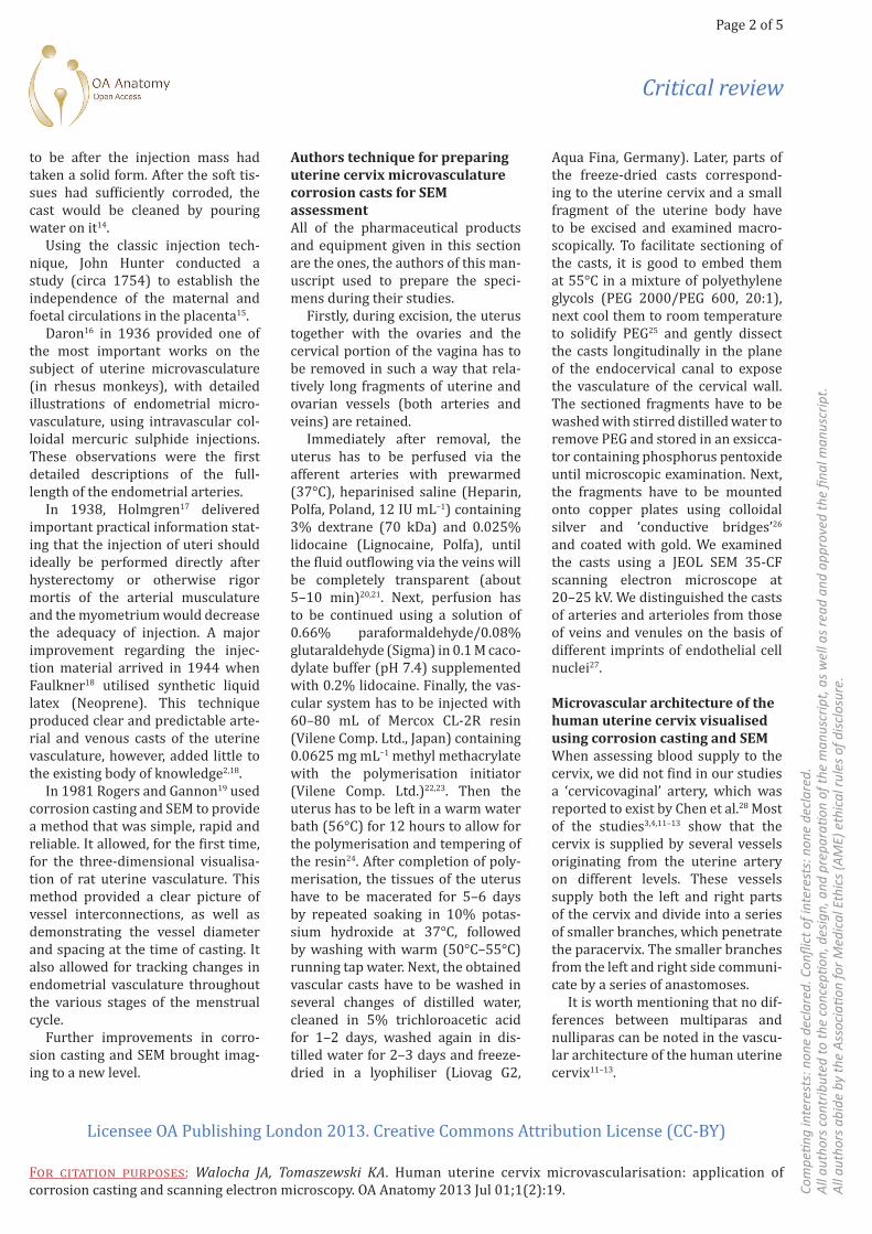
At the junction between the sup-ravaginal part of the cervix and the corpus of the uterus, four distinct vascular zones can be distinguished (Figure 1). Going from lateral to medial (towards the cervix canal) one can identify the:
• outer zone containing large arter-ies and veins;
• zone of arterioles and venules of the muscular layer, characterised by loose and irregular texture;
• zone of endocervical mucosal cap-illaries, characterised by dense texture;
• pericanalar zone containing small veins and capillaries.
The zones of the cervix are a continu-ation of the uterine body wall layers. The zone of larger blood vessels cor-responds to the vascular layer of the myometrium, whereas the other three zones belong to the mucosal layer29.
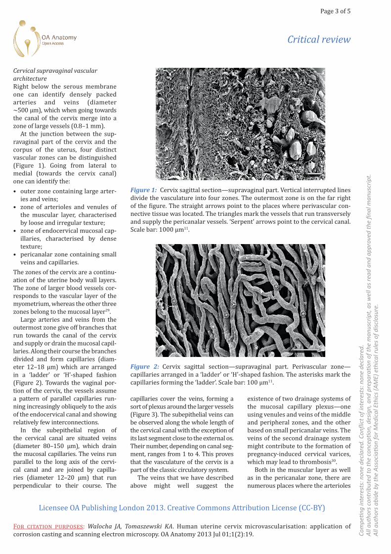
Large arteries and veins from the outermost zone give off branches that run towards the canal of the cervix and supply or drain the mucosal capil-laries. Along their course the branches divided and form capillaries (diam-eter 12–18 µm) which are arranged in a ‘ladder’ or ‘H’-shaped fashion (Figure 2). Towards the vaginal por-tion of the cervix, the vessels assume a pattern of parallel capillaries run-ning increasingly obliquely to the axis of the endocervical canal and showing relatively few interconnections.
In the subepithelial region of the cervical canal are situated veins (diame ter 80–150 µm), which drain the mucosal capillaries. The veins run parallel to the long axis of the cervi-cal canal and are joined by capilla-ries (diameter 12–20 µm) that run perpendicular to their course. The
Figure 1: Cervix sagittal section—supravaginal part. Vertical interrupted lines divide the vasculature into four zones. The outermost zone is on the far right of the figure. The straight arrows point to the places where perivascular con-nective tissue was located. The triangles mark the vessels that run transversely and supply the pericanalar vessels. ‘Serpent’ arrows point to the cervical canal. Scale bar: 1000 µm11.
Figure 2: Cervix sagittal section—supravaginal part. Perivascular zone— capillaries arranged in a ‘ladder’ or ‘H’-shaped fashion. The asterisks mark the capillaries forming the ‘ladder’. Scale bar: 100 µm11.
capillaries cover the veins, forming a sort of plexus around the larger vessels (Figure 3). The subepithelial veins can be observed along the whole length of the cervical canal with the exception of its last segment close to the external os. Their number, depending on canal seg-ment, ranges from 1 to 4. This proves that the vasculature of the cervix is a part of the classic circulatory system.
The veins that we have described above might well suggest the
existence of two drainage systems of the mucosal capillary plexus—one using venules and veins of the middle and peripheral zones, and the other based on small pericanalar veins. The veins of the second drainage system might contribute to the formation of pregnancy-induced cervical varices, which may lead to thrombosis30.
Both in the muscular layer as well as in the pericanalar zone, there are numerous places where the arterioles

Critical review
Page 4 of 5
Licensee OA Publishing London 2013. Creative Commons Attribution License (CC-BY)
For citation purposes: Walocha JA, Tomaszewski KA. Human uterine cervix microvascularisation: application of corrosion casting and scanning electron microscopy. OA Anatomy 2013 Jul 01;1(2):19. Co
mpe
ting
inte
rest
s: n
one
decl
ared
. Con
flict
of i
nter
ests
: non
e de
clar
ed.
All a
utho
rs c
ontr
ibut
ed to
the
conc
eptio
n, d
esig
n, a
nd p
repa
ratio
n of
the
man
uscr
ipt,
as w
ell a
s rea
d an
d ap
prov
ed th
e fin
al m
anus
crip
t.Al
l aut
hors
abi
de b
y th
e As
soci
ation
for M
edic
al E
thic
s (AM
E) e
thic
al ru
les o
f disc
losu
re.
and venules pass close to each other, often adjoining. This may well sug-gest that there exists a countercurrent transport between veins and arteries.
The cervical glands are sur-rounded by a dense capillary network (Figure 4). The plexuses are composed either of parallel capillaries (lower part of Figure 4) or irregularly formed vessels (upper part of Figure 4).
Cervical vaginal vascular architectureVessel arrangement in the vaginal part of the cervix is similar to the one described in the supravaginal part. However, the arteries and veins of the outer zone are of significantly smaller calibre and the veins of the pericanalar zone are much more exposed (Figure 5).
The vessels of the vaginal part of the cervix, near the external os of the uterus, form three separate layers:
• supplying vessels—characterised by relatively large diameters;
• oblique or perpendicular small arteries and veins;
• subepithelial capillaries joining the, above mentioned, oblique and perpendicular vessels.
ConclusionConcluding, in the authors opinion, corrosion casting combined with SEM assessment is currently the best available technique to visualise vas-cular architecture. It offers high reso-lution and quasi-three-dimensional images of the vessels, without inter-ference from the non-vascular tissue.
The cervix is supplied by several vessels originating from the uterine artery on different levels. These vessels supply both the left and right parts of the cervix and divide into a series of smaller branches, which penetrate the paracervix. The smaller branches from the left and right side communicate by a series of anastomoses.
No differences, between mul-tiparas and nulliparas, can be noted in the vascular architecture of the human uterine cervix.
Figure 3: Cervix sagittal section—supravaginal part. Pericanalar vein covered by capillary plexus (marked by box). Scale bar: 1000 µm11.
Figure 4: Cervix coronal section—supravaginal part. The dots mark the spaces where cervical glands were located. The glands were separated from each other by characteristically flattened veins. Scale bar: 1000 µm.
Figure 5: Cervix sagittal section—vaginal part. The asterisk mark the large, exposed, pericanalar veins. The capillaries join the pericanalar veins. Scale bar: 100 µm.
Critical review
Page 5 of 5
Licensee OA Publishing London 2013. Creative Commons Attribution License (CC-BY)
For citation purposes: Walocha JA, Tomaszewski KA. Human uterine cervix microvascularisation: application of corrosion casting and scanning electron microscopy. OA Anatomy 2013 Jul 01;1(2):19. Co
mpe
ting
inte
rest
s: n
one
decl
ared
. Con
flict
of i
nter
ests
: non
e de
clar
ed.
All a
utho
rs c
ontr
ibut
ed to
the
conc
eptio
n, d
esig
n, a
nd p
repa
ratio
n of
the
man
uscr
ipt,
as w
ell a
s rea
d an
d ap
prov
ed th
e fin
al m
anus
crip
t.Al
l aut
hors
abi
de b
y th
e As
soci
ation
for M
edic
al E
thic
s (AM
E) e
thic
al ru
les o
f disc
losu
re.
Both in the vaginal and suprav-aginal parts of the human uterine cervix, four distinct vascular zones can be distinguished—the outer zone containing large arteries and veins, the arteriole and venule zone, the endocervical mucosal capillar-ies zone and the pericanalar zone containing small veins and capil-laries. In the pericanalar zone run small veins, responsible for draining the mucosal capillaries. Both in the muscular layer, as well as in the peri-canalar zone, arterioles and venules pass close to each other, often adjoin-ing. This most probably confirms the existence of a countercurrent trans-port between adjoining veins and arteries.
References1. Osol G, Mandala M. Maternal uterine vascular remodeling during pregnancy. Physiol (Bethesda). 2009 Feb;24:58–71.2. Manconi F, Thomas GA, Fraser IS. A historical overview of the study and representation of uterine microvas-cular structures. Microvasc Res. 2010 Jan;79(1):80–9.3. Pilarczyk K, Kozik W, Czerwinski F. Arteries of the uterine cervix in repro-ductive age in microangiographic studies. Ginekol Pol. 2002 Dec;73(12):1179–83.4. Palacios Jaraquemada JM, García Mónaco R, Barbosa NE, Ferle L, Iriarte H, Conesa HA. Lower uterine blood supply: extrauterine anastomotic system and its application in surgical devascularization techniques. Acta Obstet Gynecol Scand. 2007;86(2):228–34.5. Walocha JA, Litwin JA, Miodonski AJ. Vascular system of intramural leiomy-omata revealed by corrosion casting and scanning electron microscopy. Hum Reprod. 2003 May;18(5):1088–93.6. Hamid SA, Ferguson LE, McGavigan CJ, Howe DC, Campbell S. Observing three-dimensional human microvascular and myogenic architecture using conven-tional fluorescence microscopy. Micron. 2006 Sep;37:134–8.7. Dalgaard JB. The blood vessels of the human endometrium. Acta Obstet Gynecolo Scand. 1946 Jan;26(3):342–78.8. Bulletti C, Jasonni VM, Tabanelli S, Ciotti P, Vignudelli A, Flamigni C. Changes
in the uterine vasculature during the menstrual cycle. Acta Eur Fertil. 1985 Sep–Oct;16(5):367–71.9. Sekiba K, Okuda H, Fukui H, Ishii Y, Kawaoka K, Fujimori T. A scanning elec-tron microscope study of the fine angio-architecture of the uterine cervix using a newly established cast formation tech-nique. Obstet Gynecolo Survey. 1979 Nov;34(11):823–6.10. Zinser HK, Rosenbauer KA. Untersu-chungen uber die Angioarchitektonik der normalen und pathologisch veranderten Cervix uteri. Arch Gynakol. 1960;194:73–112. German.11. Bereza T, Tomaszewski KA, Bałajewicz-Nowak M, Mizia E, Pasternak A, Walocha J. The vascular architecture of the supravaginal and vagi-nal parts of the human uterine cervix: a study using corrosion casting and scan-ning electron microscopy. J Anat. 2012 Oct;221(4):352–7.12. Bereza T, Tomaszewski KA, Walocha J, Mizia E, Bachul P, Chmielewski P. Vas-cular architecture of the human uterine cervix, as assessed in light- and scanning electron microscopy. Folia Morphol. 2012 Aug;71(3):142–7.13. Walocha JA, Litwin JA, Bereza T, Klimek-Piotrowska W, Miodonski AJ. Vascular architecture of human uterine cervix visualized by corrosion casting and scanning electron microscopy. Hum Reprod. 2012 Mar;27(3):727–32.14. Hunter J. Practical anatomy of the arrangement of anatomical preparations. In: Voorst JV, editor. Essays and observa-tions on natural history, anatomy, physi-ology, psychology and geology. London: Paternoster Row; 1861.p2.15. Ramsey EM. The story of the spiral arteries. J Reprod Med. 1981 Aug;26(8):393–9.16. Daron GH. The arterial pattern of the tunica mucosa of the uterus in Macacus rhe-sus. Am J Anat. 1936 Mar;58(2):349–419.17. Holmgren B. Some observations on the blood vessels of the uterus under nor-mal conditions and in myoma. Acta Obstet Gynecol Scand. 1938 Jan;18(2):192–213.18. Faulkner RL. The blood vessels of the myomatous uterus. Am J Obstet Gynecol. 1944;47:185–97.19. Rogers PA, Gannon BJ. The vascular and microvascular anatomy of the rat uterus during the oestrous cycle. Aust J Exp Biol Med Sci. 1981 Dec;59(6):667–79.
20. Pitynski, K, Skawina A, Polakiewicz J, Walocha J. Extraorganicvascular system of adrenal glands in human fetuses. Ann Anat. 1998 Aug;180(4):361–8.21. Maga P, Tomaszewski KA, Skrzat J, Tomaszewska IM, Iskra T, Pasternak A, et al. Microanatomical study of the recur-rent artery of Heubner. Ann Anat. 2013 Jul;195(4):342–50.22. Maga P, Tomaszewski KA, Krzyżewski RM, Golec J, Depukat P, Gregorczyk-Maga I, et al. Branches and arterial supply of the recurrent artery of Heubner. Anat Sci Int. 2013 Sep;88(4):223–9.23. Maga P, Tomaszewski KA, Pasternak A, Zawiliński J, Tomaszewska R, Gregorczyk- Maga I, et al. Extra- and intracerebral course of the recurrent artery of Heubner. Folia Morphol. 2013 May;72(2):94–9.24. Składzien J, Litwin JA, Nowogrodzka-Zagorska M, Miodonski AJ. Corrosion casting study on the vasculature of nasal mucosa in the human fetus. Anatl Rec. 1995 Jul;242(3):411–16.25. Walocha JA, Miodonski AJ, Nowogrodzka-Zagorska M, Kuciel R, Gorczyca J. Application of a mixture of glycol polyethylenes for the preparation of microcorrosion casts-an observation. Folia Morphol. 2002;61(4):313–16.26. Lametschwandtner A, Miodonski A, Simonsberger P. On the prevention of specimen charging in scanning elec-tron microscopy of vascular corrosion casts by attaching conductive bridges. Mikroskopie. 1980 Nov;36(9–10):270–3.27. Miodonski A, Hodde KC, Bakker C. Rasterelektronenmikroskopie von Plastik- Korrosions-Praparaten: morphologische Unterschiede zwischen Arterien and Venen. BEDO. 1976;9:435–42. German.28. Chen CL, Guo HX, Liu P, Huang R, Yang ZB, Tang L, et al. Three-dimensional reconstruction of the uterine vascular supply through vascular casting and thin slice computed tomography scan-ning. Minim Invasive Ther Allied Technol. 2009;18(2):98–102.29. DeSouza NM, Hawley IC, Schwieso JE, Gilderdale DJ, Soutter WP. The uterine cervix on in vitro and in vivo MR images: a study of zonal anatomy and vascular-ity using an enveloping cervical coil. Am Roentgenol. 1994 Sep;163(3):607–12.30. Sammour RN, Gonen R, Ohel G, Leibovitz Z. Cervical varices complicated by thrombosis in pregnancy. Ultrasound Obstet Gynecol. 2011 May;37(5):614–16.