Human TLR10 is an anti-inflammatory pattern-recognition ... · PDF fileHuman TLR10 is an...
7
Human TLR10 is an anti-inflammatory pattern-recognition receptor Marije Oosting a , Shih-Chin Cheng a , Judith M. Bolscher b , Rachel Vestering-Stenger a , Theo S. Plantinga a , Ineke C. Verschueren a , Peer Arts c , Anja Garritsen b , Hans van Eenennaam b , Patrick Sturm d , Bart-Jan Kullberg a , Alexander Hoischen c , Gosse J. Adema e , Jos W. M. van der Meer a , Mihai G. Netea a , and Leo A. B. Joosten a,1 a Department of Internal Medicine, c Department of Human Genetics, d Department of Microbiology, and e Department of Tumour Immunology, Radboud University Medical Center, Nijmegen 6525 GA, The Netherlands; and b Department of Immune Therapeutics, Merck Research Laboratories, Oss 5342 CC, The Netherlands Edited* by Charles A. Dinarello, University of Colorado Denver, Aurora, CO, and approved September 15, 2014 (received for review June 4, 2014) Toll-like receptor (TLR)10 is the only pattern-recognition receptor without known ligand specificity and biological function. We dem- onstrate that TLR10 is a modulatory receptor with mainly inhibitory effects. Blocking TLR10 by antagonistic antibodies enhanced proin- flammatory cytokine production, including IL-1β, specifically after ex- posure to TLR2 ligands. Blocking TLR10 after stimulation of peripheral blood mononuclear cells with pam3CSK4 (Pam3Cys) led to production of 2,065 ± 106 pg/mL IL-1β (mean ± SEM) in comparison with 1,043 ± 51 pg/mL IL-1β after addition of nonspecific IgG antibodies. Several mechanisms mediate the modulatory effects of TLR10: on the one hand, cotransfection in human cell lines showed that TLR10 acts as an inhibitory receptor when forming heterodimers with TLR2; on the other hand, cross-linking experiments showed specific induction of the anti-inflammatory cytokine IL-1 receptor antagonist (IL-1Ra, 16 ± 1.7 ng/mL, mean ± SEM). After cross-linking anti-TLR10 anti- body, no production of IL-1β and other proinflammatory cytokines could be found. Furthermore, individuals bearing TLR10 polymor- phisms displayed an increased capacity to produce IL-1β, TNF-α, and IL-6 upon ligation of TLR2, in a gene-dose–dependent manner. The modulatory effects of TLR10 are complex, involving at least several mechanisms: there is competition for ligands or for the for- mation of heterodimer receptors with TLR2, as well as PI3K/Akt- mediated induction of the anti-inflammatory cytokine IL-1Ra. Finally, transgenic mice expressing human TLR10 produced fewer cytokines when challenged with a TLR2 agonist. In conclusion, to our knowl- edge we demonstrate for the first time that TLR10 is a modulatory pattern-recognition receptor with mainly inhibitory properties. TLR10 | immunology | cytokines | SNPs H ighly conserved molecular structures of invading microor- ganisms are recognized by immune cells through pattern- recognition receptors, of which Toll-like receptors (TLRs) are the most documented family. In humans, 10 members of the TLR family have been described (1). In general, specific ligation of TLRs leads to induction of proinflammatory mediators, such as cytokines and chemokines. One member of the TLR family however, TLR10, is considered an orphan receptor because of its still-unknown ligands and function. Human TLR10 is encoded on chromosome 4 within the TLR2 gene cluster, together with TLR1, TLR2, and TLR6, and shares all structural characteristics of the TLR family (2, 3). However, TLR10 differs from other TLRs by its lack of a classic down- stream signaling pathway (4), despite its interaction with the mye- loid differentiation primary response gene 88 adaptor protein (3). TLR10 is predominantly expressed in tissues rich in immune cells, such as spleen, lymph node, thymus, tonsil, and lung (2). Expression of TLR10 can be induced in B cells, dendritic cells, eosinophils, and neutrophils (3, 5, 6), as well as on nonimmune cells, such as trophoblasts (7). TLR1 and TLR6 are known to form heterodimers with TLR2, and this was shown for TLR10 as well (3, 8). It is therefore rational to hypothesize that if TLR10 has a biological function, which it exerts through the formation of heterodimers with TLR2. In the present study we report the surprising modulatory function of human TLR10 on TLR2-driven cytokine production exerted through competition for ligands, on the one hand, and specific induction of the anti-inflammatory cytokine IL-1 receptor antagonist (IL-1Ra), on the other hand. In addition, we demon- strated that hTLR10 transgenic mice produced fewer cytokines in vivo when exposed to a potent TLR2 ligand. Results Blocking TLR10 Increases Cytokine Production by Primary Human Cells. To investigate the biological function of TLR10, freshly iso- lated human peripheral blood mononuclear cells (PBMCs) were exposed for 24 h to the TLR1/2 ligand pam3CSK4 (Pam3Cys) in the presence or absence of a specific antibody directed against TLR10. Production of IL-1β and IL-6 was increased 2-fold and 1.5- fold, respectively, in the presence of the anti-TLR10 antibody (P < 0.001) (Fig. 1 A and B). This effect of the TLR10-antibody was also observed for IL-8 production (P < 0.05), and to a somewhat lesser extent for TNF-α (Fig. 1 C and D). Similar data were obtained when the cells were exposed to viable Borrelia burgdorferi , the causative organism of Lyme disease that is mainly recognized by TLR1/TLR2 heterodimers (9, 10). No difference in IL-10 production was ob- served after blocking TLR10 with the specific antibody (Fig. S1A). Additionally, we detected no differences in mRNA expression levels of suppressor of cytokine signaling (SOCS)-1 and of SOCS-3 after blocking TLR10 molecules (Fig. S1 B and C, re- spectively). Blocking of TLR10 did not change cytokine in- duction after PBMC stimulation with ligands specific for TLR4 Significance We demonstrate the biological role of TLR10, the only member of the Toll-like receptor (TLR)-family so far without a known function. We show that TLR10 acts as an inhibitory receptor, with suppressive effects. Blocking TLR10 by specific antibodies significantly upregulated TLR2-mediated cytokine production. Additionally, we show that individuals carrying loss-of-func- tion SNPs in TLR10 display upregulation of TLR2-mediated cy- tokine production. After challenging human TLR10 transgenic mice with TLR2 ligand pam3CSK4 (Pam3Cys), less inflammation could be observed when compared with wild-type mice. Taking these data together, we show that TLR10 is the only pattern- recognition receptor within the TLR family that is able to dampen TLR2 responses, thereby suppressing immune responses through production of IL-1Ra. Author contributions: M.O., J.W.M.v.d.M., M.G.N., and L.A.B.J. designed research; M.O., S.-C.C., J.M.B., T.S.P., and I.C.V. performed research; S.-C.C., J.M.B., R.V.-S., T.S.P., I.C.V., P.A., A.G., H.v.E., P.S., B.-J.K., A.H., G.J.A., and J.W.M.v.d.M. contributed new reagents/ analytic tools; M.O., P.A., B.-J.K., A.H., M.G.N., and L.A.B.J. analyzed data; M.O., M.G.N., and L.A.B.J. wrote the paper. The authors declare no conflict of interest. *This Direct Submission article had a Prearranged Editor. 1 To whom correspondence should be addressed. Email: [email protected]. This article contains supporting information online at www.pnas.org/lookup/suppl/doi:10. 1073/pnas.1410293111/-/DCSupplemental. E4478–E4484 | PNAS | Published online October 6, 2014 www.pnas.org/cgi/doi/10.1073/pnas.1410293111
-
Upload
duonghuong -
Category
Documents
-
view
218 -
download
4
Transcript of Human TLR10 is an anti-inflammatory pattern-recognition ... · PDF fileHuman TLR10 is an...

Human TLR10 is an anti-inflammatorypattern-recognition receptorMarije Oostinga, Shih-Chin Chenga, Judith M. Bolscherb, Rachel Vestering-Stengera, Theo S. Plantingaa,Ineke C. Verschuerena, Peer Artsc, Anja Garritsenb, Hans van Eenennaamb, Patrick Sturmd, Bart-Jan Kullberga,Alexander Hoischenc, Gosse J. Ademae, Jos W. M. van der Meera, Mihai G. Neteaa, and Leo A. B. Joostena,1
aDepartment of Internal Medicine, cDepartment of Human Genetics, dDepartment of Microbiology, and eDepartment of Tumour Immunology, RadboudUniversity Medical Center, Nijmegen 6525 GA, The Netherlands; and bDepartment of Immune Therapeutics, Merck Research Laboratories, Oss 5342 CC,The Netherlands
Edited* by Charles A. Dinarello, University of Colorado Denver, Aurora, CO, and approved September 15, 2014 (received for review June 4, 2014)
Toll-like receptor (TLR)10 is the only pattern-recognition receptorwithout known ligand specificity and biological function. We dem-onstrate that TLR10 is a modulatory receptor with mainly inhibitoryeffects. Blocking TLR10 by antagonistic antibodies enhanced proin-flammatory cytokine production, including IL-1β, specifically after ex-posure to TLR2 ligands. Blocking TLR10 after stimulation of peripheralblood mononuclear cells with pam3CSK4 (Pam3Cys) led to productionof 2,065± 106 pg/mL IL-1β (mean± SEM) in comparison with 1,043±51 pg/mL IL-1β after addition of nonspecific IgG antibodies. Severalmechanisms mediate the modulatory effects of TLR10: on the onehand, cotransfection in human cell lines showed that TLR10 acts asan inhibitory receptor when forming heterodimers with TLR2; on theother hand, cross-linking experiments showed specific induction ofthe anti-inflammatory cytokine IL-1 receptor antagonist (IL-1Ra,16 ± 1.7 ng/mL, mean ± SEM). After cross-linking anti-TLR10 anti-body, no production of IL-1β and other proinflammatory cytokinescould be found. Furthermore, individuals bearing TLR10 polymor-phisms displayed an increased capacity to produce IL-1β, TNF-α,and IL-6 upon ligation of TLR2, in a gene-dose–dependent manner.The modulatory effects of TLR10 are complex, involving at leastseveral mechanisms: there is competition for ligands or for the for-mation of heterodimer receptors with TLR2, as well as PI3K/Akt-mediated induction of the anti-inflammatory cytokine IL-1Ra. Finally,transgenic mice expressing human TLR10 produced fewer cytokineswhen challenged with a TLR2 agonist. In conclusion, to our knowl-edge we demonstrate for the first time that TLR10 is a modulatorypattern-recognition receptor with mainly inhibitory properties.
TLR10 | immunology | cytokines | SNPs
Highly conserved molecular structures of invading microor-ganisms are recognized by immune cells through pattern-
recognition receptors, of which Toll-like receptors (TLRs) arethe most documented family. In humans, 10 members of theTLR family have been described (1). In general, specific ligationof TLRs leads to induction of proinflammatory mediators, suchas cytokines and chemokines. One member of the TLR familyhowever, TLR10, is considered an orphan receptor because of itsstill-unknown ligands and function.Human TLR10 is encoded on chromosome 4 within the TLR2
gene cluster, together with TLR1, TLR2, and TLR6, and sharesall structural characteristics of the TLR family (2, 3). However,TLR10 differs from other TLRs by its lack of a classic down-stream signaling pathway (4), despite its interaction with the mye-loid differentiation primary response gene 88 adaptor protein (3).TLR10 is predominantly expressed in tissues rich in immune cells,such as spleen, lymph node, thymus, tonsil, and lung (2). Expressionof TLR10 can be induced in B cells, dendritic cells, eosinophils,and neutrophils (3, 5, 6), as well as on nonimmune cells, such astrophoblasts (7). TLR1 and TLR6 are known to form heterodimerswith TLR2, and this was shown for TLR10 as well (3, 8). It istherefore rational to hypothesize that if TLR10 has a biologicalfunction, which it exerts through the formation of heterodimers withTLR2. In the present study we report the surprising modulatory
function of human TLR10 on TLR2-driven cytokine productionexerted through competition for ligands, on the one hand, andspecific induction of the anti-inflammatory cytokine IL-1 receptorantagonist (IL-1Ra), on the other hand. In addition, we demon-strated that hTLR10 transgenic mice produced fewer cytokines invivo when exposed to a potent TLR2 ligand.
ResultsBlocking TLR10 Increases Cytokine Production by Primary HumanCells. To investigate the biological function of TLR10, freshly iso-lated human peripheral blood mononuclear cells (PBMCs) wereexposed for 24 h to the TLR1/2 ligand pam3CSK4 (Pam3Cys) inthe presence or absence of a specific antibody directed againstTLR10. Production of IL-1β and IL-6 was increased 2-fold and 1.5-fold, respectively, in the presence of the anti-TLR10 antibody (P <0.001) (Fig. 1 A and B). This effect of the TLR10-antibody was alsoobserved for IL-8 production (P < 0.05), and to a somewhat lesserextent for TNF-α (Fig. 1C andD). Similar data were obtained whenthe cells were exposed to viable Borrelia burgdorferi, the causativeorganism of Lyme disease that is mainly recognized by TLR1/TLR2heterodimers (9, 10). No difference in IL-10 production was ob-served after blocking TLR10 with the specific antibody (Fig. S1A).Additionally, we detected no differences in mRNA expressionlevels of suppressor of cytokine signaling (SOCS)-1 and ofSOCS-3 after blocking TLR10 molecules (Fig. S1 B and C, re-spectively). Blocking of TLR10 did not change cytokine in-duction after PBMC stimulation with ligands specific for TLR4
Significance
We demonstrate the biological role of TLR10, the only memberof the Toll-like receptor (TLR)-family so far without a knownfunction. We show that TLR10 acts as an inhibitory receptor,with suppressive effects. Blocking TLR10 by specific antibodiessignificantly upregulated TLR2-mediated cytokine production.Additionally, we show that individuals carrying loss-of-func-tion SNPs in TLR10 display upregulation of TLR2-mediated cy-tokine production. After challenging human TLR10 transgenicmice with TLR2 ligand pam3CSK4 (Pam3Cys), less inflammationcould be observed when compared with wild-type mice. Takingthese data together, we show that TLR10 is the only pattern-recognition receptor within the TLR family that is able to dampenTLR2 responses, thereby suppressing immune responses throughproduction of IL-1Ra.
Author contributions: M.O., J.W.M.v.d.M., M.G.N., and L.A.B.J. designed research; M.O.,S.-C.C., J.M.B., T.S.P., and I.C.V. performed research; S.-C.C., J.M.B., R.V.-S., T.S.P., I.C.V.,P.A., A.G., H.v.E., P.S., B.-J.K., A.H., G.J.A., and J.W.M.v.d.M. contributed new reagents/analytic tools; M.O., P.A., B.-J.K., A.H., M.G.N., and L.A.B.J. analyzed data; M.O., M.G.N.,and L.A.B.J. wrote the paper.
The authors declare no conflict of interest.
*This Direct Submission article had a Prearranged Editor.1To whom correspondence should be addressed. Email: [email protected].
This article contains supporting information online at www.pnas.org/lookup/suppl/doi:10.1073/pnas.1410293111/-/DCSupplemental.
E4478–E4484 | PNAS | Published online October 6, 2014 www.pnas.org/cgi/doi/10.1073/pnas.1410293111

(Fig. S1D). Blocking TLR10 using a different clone of the anti-human TLR10 antibody confirmed the up-regulation of cytokinesafter TLR2 activation (Fig. S1 E–G).
TLR10 Overexpression Inhibits TLR2-Mediated Signaling. To explorethe mechanisms through which TLR10 exerts the suppressivefunction, HEK293 cells were transfected with either TLR2 alone,or TLR2 together with TLR1, TLR6, or TLR10. HEK cells werecultured in the presence of either the TLR2 ligand Pam3Cys or B.burgdorferi for 24 h. IL-8 production was observed when HEK293cells transfected with TLR2 alone were exposed to TLR2 ligands.In contrast, HEK293 cells cotransfected with both TLR2 andTLR10 showed a strong reduction of IL-8 production (Fig. 2 Aand B). The inhibitory effect of TLR10 transfection was also ob-served when cotransfected TLR2/10 HEK cells were exposed tothe TLR2/6 ligand Pam2CGDPKHPKSF (FSL-1) (Fig. 2B). Todetermine the degree of specificity for TLR2, we cotransfectedHEK cells with TLR5 and TLR10. TLR10 cotransfection withTLR5 had no effects on the IL-8 production induced by flagellin(Fig. 2C). Thus, TLR10 acts primarily as an inhibitory receptor forTLR2 ligands.
RNA Silencing of TLR10 Results in Enhanced Cytokine Production. Tocorroborate these findings, we performed a series of experimentsusing siRNA to silence the gene for TLR10 in monocyte-derivedmacrophages. To assess the efficiency of the siRNA approach,we determined the TLR10 mRNA levels in the silenced macro-phages (Fig. 2D). As commonly seen with siRNA in primarycells, we achieved a 50% inhibition of the messenger mRNAencoding TLR10. After incubation of the cells with the specificTLR10 siRNA, cells were exposed to B. burgdorferi for 24 h. Up-regulation of IL-6 production by the TLR10-silenced macro-phages was observed, which was not seen with scramble-silencingRNA (Fig. 2E). No differences in TNF-α production could beobserved (Fig. 2F).
Functional Polymorphisms of the Human TLR10 Gene. A strong ar-gument in favor of TLR10 being a functional receptor is that theTLR10 gene is under selection pressure in European populations;nonsynonymous polymorphisms within the human TLR10 gene areconsidered to influence TLR10 function (11). Two of these SNPs,N241H (rs11096957) and M326T (rs11466653), have been found tobe associated with reduced risk of prostate cancer and were de-scribed to be possibly damaging to the TLR10 gene (11, 12). Weinvestigated whether the presence of these SNPs in TLR10 affectsthe TLR2-induced cytokine response by PBMCs obtained fromhealthy individuals with different TLR10 genotypes. Within thegroup of the 112 volunteers screened, 40 were identified as bearingthe wild-type allele, 60 were heterozygous, and 12 were homozygousfor the N241H SNP. PBMCs isolated from individuals bearing thethree different genotypes were incubated with RPMI, Pam3Cys, liveB. burgdorferi, or lipopolysaccharide (LPS) for 24 h. Thereafter,cytokine concentrations were measured in the supernatants. Cells of
A B
C D
Fig. 1. Blocking TLR10 results in higher cytokine production. The 5 × 10⁵
PBMCs from 11 to 17 individuals were preincubated for 1 h at 37 °C witheither 10 μg/mL aTLR10 antibody (aTLR10, clone 3C10C5) or 10 μg/mL IgGisotype control (IgG). After preincubation, cells were stimulated for 24 hwith TLR2 ligand Pam3Cys (10 μg/mL), or live B. burgdorferi (B.b). Afterstimulation, cell supernatants were collected and proinflammatory cytokineswere measured using ELISA (A–D). Bars represent the mean and the SEM.Paired t test; *P < 0.05, **P < 0.01, ***P < 0.001.
A
B
C
D
E
F
Fig. 2. TLR10 transfected HEK cells inhibit cytokine production. HEK293 cellswere transiently transfected with TLR2, TLR1/2, TLR2/6, or TLR2/10. (A) Twenty-four hours after transfection, cells were stimulated for 24 h with differentconcentrations of live B. burgdorferi (B.b.). (B) HEK293 cells transiently trans-fected using different ratios of plasmid concentrations of TLR10 plasmid. Cellswere stimulated 24 h after transfection for 22 h with specific TLR ligandsPam3Cys or FSL-1. IL-8 production (in pg/mL) was measured in the cell-freesupernatant using ELISA. (C) HEK293 cells transiently transfected with TLR5 orTLR5/10 plasmids. After 24 h of recovery, cells were stimulated for 22 h witha dose–response of recombinant flagellin. IL-8 production was measured byELISA. Bars represent the mean of six separate experiments ± SEM. Mann–Whtiney U test, two-sided, **P < 0,01. (D) Monocyte-derived macrophages ofeight individuals were transfected with scrambled siRNA as a negative control(NC siRNA) or siRNA directed against TLR10 (TLR10 siRNA). (E) After trans-fection, monocyte-derived macrophages were stimulated with 1.106 liveB. burgdorferi per mL. (F) Twenty-four hours after stimulation, IL-6 or TNF-αprotein levels were measured in picrograms per milliliter (pg/mL) in thesupernatants using specific sandwich-ELISAs.
Oosting et al. PNAS | Published online October 6, 2014 | E4479
IMMUNOLO
GYAND
INFLAMMATION
PNASPL
US

individuals heterozygous or homozygous for the N241H poly-morphism displayed strongly increased production of IL-1β, IL-6,IL-8, and TNF-α when stimulated with TLR2 ligands comparedwith cells with wild-type TLR10 (Fig. 3 A–D). In contrast, cytokineproduction induced by stimulation of other TLR ligands (TLR3,TLR4, TLR5, TLR9) were not affected by the TLR10 poly-morphisms, again underscoring the selective interaction of TLR10with TLR2 (Fig. 3 E–H). TLR4 stimulation using LPS in a largercohort also did not lead to altered cytokine production in individ-uals bearing TLR10 polymorphisms (Fig. S2).For the M326T SNP, 105 volunteers were identified as bearing
the wild-type allele and 7 volunteers as being heterozygous for
the SNP. Despite the low prevalence of the M326T polymorphism,increased cytokine production was also observed with cells bearingthis SNP. Two other polymorphisms, rs11096955 and rs4129009,were recently found to cause structural effects in the TLR10 mol-ecule (13, 14). Additionally, these polymorphisms led to increasedcytokine production (Figs. S3 and S4). Taken together, these genedosage effects on cytokine production provide an additional proofthat TLR10 inhibits TLR2-induced cytokines, with no effect on theTLR4 signaling pathway.Our findings in these volunteers concur with the recent report
showing that variation in the TLR10/TLR1/TLR6 locus is themajor genetic determinant of interindividual differences inTLR2-mediated cytokine responses (14). This study showed thatSNPs in the TLR10 gene affect the cytokine responses elicited byspecific TLR2 ligands, determined by whole-blood assays.
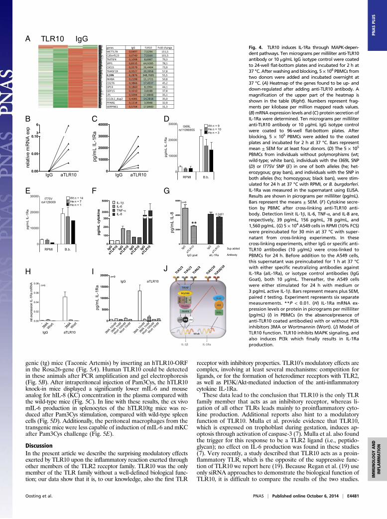
Signal Transduction. To explore whether passive competition withother stimulatory TLR2 coreceptors, such as TLR1 or TLR6, issolely responsible for the TLR10 effects or whether active sig-naling mechanisms are also involved, we assessed which signalingpathways may be induced by TLR10. Anti-TLR10 antibodieswere coated onto culture plates and freshly isolated PBMCs wereadded for 24 h, a standard method for cross-linking and stimu-lation of receptors with unknown specific ligands. Afterwards,RNA sequencing of stimulated cells was performed to obtaina comprehensive picture of TLR10-induced effects. RNA se-quencing data analysis and comparison with RNA sequencingdata of IgG–cross-linked PBMCs has identified several genesthat were up-regulated; pathway analysis performed on the genestranscribed after TLR10 ligation points to modulatory pathways,such as wound healing, chemokines, and growth factors (Fig. 4Aand Table S1). Among these genes, the gene encoding for IL-1receptor antagonist (IL-1RN) stood out as being very stronglyup-regulated (55.5-fold up-regulation) (Fig. 4A). IL-1Ra is thenatural inhibitor of excessive IL-1 signaling and it blocks bothIL-1α and IL-1β (15, 16). As a consequence, IL-1Ra down-modulates cytokine responses in several immune cells exposed tospecific ligands of pathogens.The remarkable up-regulation of IL-1Ra mRNA expression by
the PBMCs after cross-linking TLR10 was confirmed by PCR(Fig. 4B). In addition, enhanced protein levels of IL-1Ra werefound when PBMCs were incubated for 24 h with plate-boundanti-TLR10 antibody, compared with IgG isotype control (Fig.4C). Individuals bearing TLR10-dysfunctional polymorphismswere also found to produce less IL-1Ra after stimulation (Fig. 4D and E and Fig. S5 A and B). In these cross-linking experi-ments, no IL-8, IL-6, TNF-α, or IL-1β production could bedetected (Fig. 4F). After blockade with the specific anti-TLR10antibody on PBMCs, IL-1Ra levels were also up-regulatedcompared with isotype control (Fig. S5C).To prove the inhibitory role of TLR10 through IL-1Ra pro-
duction, lung epithelial cells were incubated with supernatantsfrom either IgG or anti-TLR10 antibody cross-linked PBMCs.Stimulation of A549 cells with active IL-1β led to significant in-duction of IL-8 (Fig. 4G, light gray bar), which was found to beinhibited after preincubation of A549 cells with the supernatantof aTLR10–cross-linked PBMCs (Fig. 4G, light gray dotted bar).When the supernatants of aTLR10–cross-linked PBMCs werepreincubated with a specific neutralizing antibody against IL-1Ra,this effect could no longer be observed (Fig. 4G, dark gray bars).Because it has been reported that the PI3K/Akt pathway is
needed for the induction of IL-Ra production (17, 18), weassessed whether TLR10 effects may be mediated by PI3K/Aktpathway. PI3K/Akt inhibition resulted in complete inhibition ofIL-1Ra production in the cross-linking experiments on mRNAlevel as well as protein secretion (Fig. 4 H and I).
Human TLR10 Inhibits Inflammation in Vivo. Wild-type mice do notexpress functional TLR10 because of sequence gaps and retro-viral insertions in the TLR10 gene (4). To explore the biologicaleffect of TLR10 in vivo, we generated human (h) TLR10-trans-
A B
C D
E F
G H
Fig. 3. SNP Asn241His leads to increased cytokine production and is de-pendent on TLR2-mediated responses. 5 × 10⁵ PBMCs from 40 individualswithout the N241H polymorphism (wt; wild-type; white bars), 60 individualswith the SNP in one of both alleles (he; heterozygous; gray bars), and 12homozygous individuals (ho; black bars) were stimulated for 24 h at 37 °Cwith RPMI, Pam3Cys, B. burgdorferi, or LPS. IL-1β (A), IL-6 (B), IL-8 (C), andTNF-α (D) were measured in the supernatant using ELISA. Results are shownin nanograms per milliliter (ng/mL) (A–C) or picrograms per milliliter (pg/mL)(E–H). *P < 0.05, **P < 0.01, ***P < 0.001. The 5 × 10⁵ PBMCs from indi-viduals without the Asn241His polymorphism (wt; wild-type; white bars),individuals with the SNP in one of both alleles (he; heterozygous; gray bars),and homozygous individuals (ho; black bars) were stimulated for 24 h at37 °C with either medium (RPMI), 50 μg/mL Poly IC, 5 μg/mL CpG, or 100 ng/mLflagellin. IL-1β (E ), IL-6 (F ), TNF-α (G), and IL-8 (H) were measured in thesupernatant using ELISA. Results are shown in picrograms per milliliter (pg/mL).Bars represent the means ± SEM.
E4480 | www.pnas.org/cgi/doi/10.1073/pnas.1410293111 Oosting et al.
genic (tg) mice (Taconic Artemis) by inserting an hTLR10-ORFin the Rosa26-gene (Fig. 5A). Human TLR10 could be detectedin these animals after PCR amplification and gel electrophoresis(Fig. 5B). After intraperitoneal injection of Pam3Cys, the hTLR10knock-in mice displayed a significantly lower mIL-6 and mouseanalog for hIL-8 (KC) concentration in the plasma compared withthe wild-type mice (Fig. 5C). In line with these results, the ex vivomIL-6 production in splenocytes of the hTLR10tg mice was re-duced after Pam3Cys stimulation, compared with wild-type spleencells (Fig. 5D). Additionally, the peritoneal macrophages from thetransgenic mice were less capable of induction of mIL-6 and mKCafter Pam3Cys challenge (Fig. 5E).
DiscussionIn the present article we describe the surprising modulatory effectsexerted by TLR10 upon the inflammatory reaction exerted throughother members of the TLR2 receptor family. TLR10 was the onlymember of the TLR family without a well-defined biological func-tion; our data show that it is, to our knowledge, also the first TLR
receptor with inhibitory properties. TLR10’s modulatory effects arecomplex, involving at least several mechanisms: competition forligands, or for the formation of heterodimer receptors with TLR2,as well as PI3K/Akt-mediated induction of the anti-inflammatorycytokine IL-1Ra.These data lead to the conclusion that TLR10 is the only TLR
family member that acts as an inhibitory receptor, whereas li-gation of all other TLRs leads mainly to proinflammatory cyto-kine production. Additional reports also hint to a modulatoryfunction of TLR10. Mulla et al. provide evidence that TLR10,which is expressed on trophoblast during gestation, induces ap-optosis through activation of caspase-3 (7). Mulla et al. also foundthe trigger for this response to be a TLR2 ligand (i.e., peptido-glycan); no effect on IL-6 production was found in these studies(7). Very recently, a study described that TLR10 acts as a proin-flammatory TLR, which is the opposite of the suppressive func-tion of TLR10 we report here (19). Because Regan et al. (19) useonly siRNA approaches to demonstrate the biological function ofTLR10, it is difficult to compare the results of the two studies.
A
B C D
E F G
H I J
Fig. 4. TLR10 induces IL-1Ra through MAPK-depen-dent pathways. Ten micrograms per milliliter anti-TLR10antibody or 10 μg/mL IgG isotype control were coatedto 24-well flat-bottom plates and incubated for 2 h at37 °C. After washing and blocking, 5 × 10⁶ PBMCs fromtwo donors were added and incubated overnight at37 °C. (A) Heatmap of the genes found to be up- anddown-regulated after adding anti-TLR10 antibody. Amagnification of the upper part of the heatmap isshown in the table (Right). Numbers represent frag-ments per kilobase per million mapped reads values.(B) mRNA expression levels and (C) protein secretion ofIL-1Ra were determined. Ten micrograms per milliliteranti-TLR10 antibody or 10 μg/mL IgG isotype controlwere coated to 96-well flat-bottom plates. Afterblocking, 5 × 10⁵ PBMCs were added to the coatedplates and incubated for 2 h at 37 °C. Bars representmean ± SEM for at least four donors. (D) The 5 × 10⁵
PBMCs from individuals without polymorphisms (wt;wild-type; white bars), individuals with the I369L SNP(D) or I775V SNP (E) in one of both alleles (he; het-erozygous; gray bars), and individuals with the SNP inboth alleles (ho; homozygous; black bars), were stim-ulated for 24 h at 37 °C with RPMI, or B. burgdorferi.IL-1Ra was measured in the supernatant using ELISA.Results are shown in picrograms per milliliter (pg/mL).Bars represent the means ± SEM. (F) Cytokine secre-tion by PBMC after cross-linking anti-TLR10 anti-body. Detection limit IL-1β, IL-6, TNF-α, and IL-8 are,respectively, 39 pg/mL, 156 pg/mL, 78 pg/mL, and1,560 pg/mL. (G) 5 × 10⁴ A549 cells in RPMI (10% FCS)were preincubated for 30 min at 37 °C with super-natant from cross-linking experiments. In thesecross-linking experiments, either IgG or specific anti-TLR10 antibodies (10 μg/mL) were cross-linked toPBMCs for 24 h. Before addition to the A549 cells,this supernatant was preincubated for 1 h at 37 °Cwith either specific neutralizing antibodies againstIL-1Ra (aIL-1Ra), or isotype control antibodies (IgGGoat), both 10 μg/mL. Thereafter, the A549 cellswere either stimulated for 24 h with medium or3 pg/mL active IL-1β. Bars represent means plus SEM,paired t testing. Experiment represents six separatemeasurements. **P < 0.01. (H) IL-1Ra mRNA ex-pression levels or protein in picrograms per milliliter(pg/mL) (I) in PBMCs (in the absence/presence ofanti-TLR10 coated antibodies) with or without PI3kinhibitors 3MA or Wortmannin (Wort). (J) Model ofTLR10 function. TLR10 inhibits MAPK signaling, andalso induces PI3k which finally results in IL-1Raproduction.
Oosting et al. PNAS | Published online October 6, 2014 | E4481
IMMUNOLO
GYAND
INFLAMMATION
PNASPL
US

Using a set of complementary studies, including hTLR10tg mice,we showed that TLR10 acts as an inhibitory TLR that controlsmainly TLR2-driven signals. However, we have to stress that theeffects of TLR10 observed here are not exclusively inhibitory: thetranscriptomic analysis clearly shows an important set of genesthat are strongly up-regulated upon engagement of TLR10. Thebiological relevance of this particular set of genes remains to beexplored more extensively in future studies.Within the TLR family, TLR10 is most closely related to
TLR1 and TLR6, the two TLRs that form heterodimers withTLR2 and hence function as coreceptors (2). Given the results ofour cotransfection experiments, it is highly likely that the inhibitoryeffects of TLR10 are mediated through an interaction of TLR10with TLR2 and perhaps also with TLR1 and TLR6 (Fig. 4J).Regarding the molecular mechanisms through which TLR10
exerts its inhibitory effect, broadly three different mechanisms couldbe envisaged: (i) competition for ligands with the stimulatory TLRs,(ii) competition for dimerization with the other members of theTLR2 family (TLR1, TLR6), and (iii) TLR10-specific direct in-hibitory signaling. Our cotransfection experiments plead for eitherof the first two mechanisms, whereas the experiments showing
TLR10-dependent induction of IL-1Ra suggests that the latterpathway is also operational. The in vivo experiments with thehTLR10tg mice support all three of these possible mechanisms.The link between TLR10 and IL-1Ra is corroborated by a recentpublication reporting that allelic variants of the TLR10 gene mayinfluence the susceptibility and time-course of hearing loss ofMeniere disease (13). Based on favorable clinical experience withrecombinant IL-1Ra (anakinra) treatment in Meniere disease,a controlled clinical trial this drug is underway (15). It may bespeculated that loss-of-function of the TLR10 is involved in a lackof sufficient IL-1Ra production in Meniere disease.Currently, it is unknown whether there are specific ligands for
TLR10 other than those are also binding TLR2. Because TLR10expressed on the transfected cells responded to Pam3Cys, FSL-1,and B. burgdorferi, it is likely that these TLR2 ligands also directlyinteract with TLR10. Interestingly, the effect of TLR10 occurs withboth ligation of the TLR2/TLR1 complex (by Pam3Cys) and of theTLR2/TLR6 complex (by FSL-1), indicating that TLR2 ligation iscrucial. Because transfection may have led to overexpression of thereceptors, we cannot rule out that some spontaneous hetero-dimerization of TLR10 with TLR2 has occurred.
A B
C D
E
Fig. 5. Human TLR10 inhibits TLR2-mediated re-sponses in vivo. (A) Generation of human TLR10transgenic C57BL/6 mice. Using RMCE, a CAG pro-moter cassette, the human TLR10 ORF, the hGHpolyadenylation signal, and an additional poly-adenylation signal was cloned into the ROSA26 lo-cus. (B) The humanTLR10 fragment was amplifiedusing specific hTLR10 primers. The amplification ofthe positive control fragment (585 bp) refers towild-type mice. Mice expressing the human TLR10gene have a fragment of 368 bp. (C) Plasma IL-6 andKC levels in wild-type (n = 13) and hTLR10 trans-genic (n = 13) mice, 4 h after intraperitoneal injec-tion of 50 μg Pam3Cys. IL-6 and KC were measuredusing ELISA. Data are mean ± SEM in pg/mL; *P <0.05, Mann–Whitney U test. (D) The 5 × 10⁶ spleencells of five wild-type C57BL/6 and six hTLR10tg micewere incubated with medium alone (RPMI), or10 μg/mL Pam3Cys for 24 h at 37 °C, 5% CO2.Supernatants were collected for measurement of IL-6 by ELISA. Data are mean ± SEM in picrograms permilliliter (pg/mL); five WT mice, six hTLR10tg mice,*P < 0.05, **P < 0.01, two-tailed Mann–Whitney Utest. (E) After isolation of peritoneal macrophages,IL-6 and mKC levels were measured using ELISA.Data are mean ± SEM in picrograms per milliliter(pg/mL); *P < 0.05, ***P < 0.001, Mann–Whitney Utest, seven mice per group.
E4482 | www.pnas.org/cgi/doi/10.1073/pnas.1410293111 Oosting et al.

The finding of an inhibitory TLR among the agonistic TLRs isnot entirely surprising. TLRs belong to the Toll/IL-1R family, andwithin that family other inhibitory molecules are known. Examplesare IL-1R type II and single-Ig IL-1–related receptor (SIGIRR).Such receptors may exert their suppressive effect through compe-tition with the stimulatory receptors (e.g., the decoy receptor IL-1RII), whereas others act through inhibitory intracellular signals(e.g., SIGIRR). Members of other classes of pattern-recognitionreceptor also display suppressive activities, such as the C-type lectinreceptors DC-SIGN and mannose receptor (20, 21). However,up-regulation of inhibitory cytokines (e.g., IL-36Ra, IL-38) andantagonists belonging to the IL-1 superfamily was not seen bymRNA sequencing after cross-linking anti-TLR10 antibodies.The finding of a suppressive function of TLR10 has important
consequences. First of all, this study elucidates a biological functionfor the last human TLR without a known role, and contributes toour understanding of TLR biology. Also, understanding the func-tion of TLR10 raises opportunities for better insight into pathol-ogy. Genetic variability in TLR10 may change the balance betweenpro- and anti-inflammatory responses, and hence modulate thesusceptibility to infection and to autoimmune disease. Such insightsmay be relevant for the development of novel treatment strategiesbased on modulation of the function of TLR10.
Experimental ProceduresBlood Samples. Human PBMCs were isolated by differential centrifugationusing Ficoll-Paque (GE Healthcare) from buffy-coats of healthy blood donors(Sanquin Bloodbank). For the genetic studies, DNA of a cohort of healthyvolunteers was used as described previously (22).
Reagents. RPMI 1640 (Dutch modification) was used as PBMC culture medium.Before use, RPMI was supplemented with 50mg/L gentamycin, 2 mM L-glutamin,and 1 mM pyruvate. LPS (Escherichia coli serotype 055:B5; Sigma) was repurifiedas previously described (23). Synthetic Pam3Cys was purchased from EMCMicrocollections. Borrelia spirochetes and Candida albicans were cultured andprepared as described previously (22, 24). Anti-TLR10 antibody was purchasedfrom Abcam (Clone 3C10C5, and Clone 158C1114, endotoxin-free) and mouseIgG1к monoclonal antibody was used as isotype control. SB202190 (p38 MAPKinhibitor, 0.3 μM); SP600125 (JNK1/2/3 inhibitor, 5 μM), and U0126 (MEK1/2 in-hibitor, 3 μM) were purchased from Superarray Bioscience Corporation, 3MAwas purchased from Sigma, and Wortmannin was purchased by Biolegend. Inexperiments using pharmacological inhibitors, control cells were treated with anequivalent concentration of vehicle (DMSO).
PBMC Stimulations and TLR10 Blockade. Samples of venous blood were drawnafter informed consent was obtained. Experiments were conducted accordingto the principles expressed in the Declaration of Helsinki. Venous blood wasdrawn from the cubital vein into 10-mL EDTA tubes (Monoject). PBMCs wereisolated according to standard protocols, with minor modifications. See SIExperimental Procedures for more information.
A549 Culture and Stimulations. See SI Experimental Procedures for more in-formation regarding A549 culture and stimulations.
Cytokine Measurements. IL-8 production by HEK cells was determined usingLuminex (Bio-Rad) or specific PeliKine Compact ELISA kits (Sanquin). Cyto-kines were measured using specific sandwich ELISA kits for IL-1β, TNF-α, andIL-1Ra. IL-6 and IL-10 were measured using PeliKine Compact ELISA kits(Sanquin). The detection limit of the different ELISA kits was 20 pg/mL.
Plasmid DNA Isolation. See SI Experimental Procedures for more informationregarding plasmid DNA isolation.
Transfection of HEK293 Cells with TLR10. HEK293 were cultured in DMEM F12medium (Gibco, Life Technologies), 100 U/mL Penicillin-100 μg streptomycin(Invitrogen), and 7.5% (vol/vol) nonheat inactivated FBS (HyClone, ThermoScientific). See SI Experimental Procedures for more information.
TLR10 Silencing by siRNA. Adherent monocytes were cultured into macro-phages for 6 d in the presence of 10% (vol/vol) human pool serum and 50ng/mLmacrophage-colony stimulating factor (ProSpec). See See SI ExperimentalProcedures for more information.
mRNA Extraction, DNA Isolation, and PCR Analysis. See See SI ExperimentalProcedures for more information.
RNA Sequencing. See See SI Experimental Procedures for more informationregarding RNA sequencing.
Generation of the hTLR10tg Mice. A constitutive human TLR10 knock-in mousewas generated by Taconic Artemis using targeted transgenesis. Using re-combination-mediated cassette exchange (RMCE), a CAG promoter cassette,the human TLR10 ORF, and the hGH polyadenylation signal and an additionalpolyadenylation signal was inserted into the ROSA26 locus. This obtainedRMCE vector was transfected into TaconicArtemis-C57BL/6 ES cell line. Usingpositive Neomycin selection, recombinant clones were selected. Positivelyselected blastocytes were transferred into embryos and chimeric mice wereproduced. Using a Caliper LabChip GX device, sample analysis has beenperformed. Animals were fully back-crossed to C57/Black6 background be-fore they were used for experiments. Genotyping of the mice was performedusing PCR and 1.5% agarosegel electrophoresis using primers wild-type-forward: 5′CTCTTCCCTCGTGATCTGCAACTCC; wild-type-reverse: 5′CATGT-CTTTAATCTACCTCGATGG; TLR10 cond-forward: 5′GACAGCAGAGGGTGATGC-TC; TLR10 cond-reverse: 5′CTTCCTCACAGATAGGCATGG; positive control-forward:5′GAGACTCTGGCTACTCATCC; positive control-reverse: 5′CCTTCAGCAAGAGCTG-GGGAC. PCR conditions were 95 °C 5 min; 35 cycles of 95 °C 30 s, 60 °C 30 s, 72 °C60 s, followed by 72 °C 10 min. Male wild-type and hTLR10tg mice between 12and 16 wk of age were used. The mice were fed sterilized laboratory chow (HopeFarms) and water ad libitum. The experiments were approved by the EthicsCommittee on Animal Experiments of the Radboud University Medical Centre,Nijmegen, The Netherlands. To examine the inflammatory response, both wild-type and hTLR10tg mice were injected intraperitoneally with 50 μg Pam3Cys in100 μL PBS. After 4 h, mice were killed. Cytokine concentrations in EDTA plasmaor in supernatants of ex vivo stimulated splenocytes were measured using ELISA,as described previously (10).
Statistics. Data were analyzed using nonnormally distributed two-tailedMann–Whitney U test or paired t-testing (GraphPad Prism, GraphPad Soft-ware). Values of P < 0.05 were considered statistically significant: *P < 0.05;**P < 0.01; ***P < 0.001. The means ± SEM of three or more independentexperiments are reported.
ACKNOWLEDGMENTS.We thank Carla Bartels for culturing Borrelia spirochetesand Eugène T. Verwiel for the support in the RNA sequencing analysis. Thisresearch project was supported by a grant from Top Institute Pharma(D1-101); a Vici grant from the Netherlands Organization for Scientific Re-search (to M.G.N.); and a Veni grant from the Netherlands Organization forScientific Research (to T.S.P.).
1. Akira S, Takeda K (2004) Toll-like receptor signalling. Nat Rev Immunol 4(7):499–511.2. Chuang T, Ulevitch RJ (2001) Identification of hTLR10: A novel human Toll-like re-
ceptor preferentially expressed in immune cells. Biochim Biophys Acta 1518(1-2):157–161.
3. Hasan U, et al. (2005) Human TLR10 is a functional receptor, expressed by B cells andplasmacytoid dendritic cells, which activates gene transcription through MyD88.J Immunol 174(5):2942–2950.
4. Guan Y, et al. (2010) Human TLRs 10 and 1 share common mechanisms of innateimmune sensing but not signaling. J Immunol 184(9):5094–5103.
5. Hornung V, et al. (2002) Quantitative expression of Toll-like receptor 1-10 mRNA incellular subsets of human peripheral blood mononuclear cells and sensitivity to CpGoligodeoxynucleotides. J Immunol 168(9):4531–4537.
6. Nagase H, et al. (2003) Expression and function of Toll-like receptors in eosinophils:Activation by Toll-like receptor 7 ligand. J Immunol 171(8):3977–3982.
7. Mulla MJ, et al. (2012) Cutting-edge report: TLR10 plays a role in mediating bacterialpeptidoglycan-induced trophoblast apoptosis. Am J Reprod Immunol 69(5):449–453.
8. Godfroy JI, 3rd, Roostan M, Moroz YS, Korendovych IV, Yin H (2012) Isolated Toll-likereceptor transmembrane domains are capable of oligomerization. PLoS ONE 7(11):e48875.
9. Hirschfeld M, et al. (1999) Cutting edge: Inflammatory signaling by Borreliaburgdorferi lipoproteins is mediated by Toll-like receptor 2. J Immunol 163(5):2382–2386.
10. Oosting M, et al. (2011) TLR1/TLR2 heterodimers play an important role in the rec-ognition of Borrelia spirochetes. PLoS ONE 6(10):e25998.
11. Barreiro LB, et al. (2009) Evolutionary dynamics of human Toll-like receptors and theirdifferent contributions to host defense. PLoS Genet 5(7):e1000562.
12. Stevens VL, et al. (2008) Genetic variation in the Toll-like receptor gene cluster (TLR10-TLR1-TLR6) and prostate cancer risk. Int J Cancer 123(11):2644–2650.
Oosting et al. PNAS | Published online October 6, 2014 | E4483
IMMUNOLO
GYAND
INFLAMMATION
PNASPL
US

13. Requena T, et al. (2013) Allelic variants in TLR10 gene may influence bilateral affec-
tation and clinical course of Meniere’s disease. Immunogenetics 65(5):345–355.14. Mikacenic C, Reiner AP, Holden TD, Nickerson DA, Wurfel MM (2013) Variation in the
TLR10/TLR1/TLR6 locus is the major genetic determinant of interindividual difference
in TLR1/2-mediated responses. Genes Immun 14(1):52–57.15. Dinarello CA, Simon A, van der Meer JW (2012) Treating inflammation by blocking
interleukin-1 in a broad spectrum of diseases. Nat Rev Drug Discov 11(8):633–652.16. Dinarello CA (2005) Blocking IL-1 in systemic inflammation. J Exp Med 201(9):1355–1359.17. Brandt KJ, Carpintero R, Gruaz L, Molnarfi N, Burger D (2010) A novel MEK2/PI3Kδ
pathway controls the expression of IL-1 receptor antagonist in IFN-β-activated human
monocytes. J Leukoc Biol 88(6):1191–1200.18. Molnarfi N, Hyka-Nouspikel N, Gruaz L, Dayer JM, Burger D (2005) The production of IL-1
receptor antagonist in IFN-beta-stimulated human monocytes depends on the activation
of phosphatidylinositol 3-kinase but not of STAT1. J Immunol 174(5):2974–2980.
19. Regan T, et al. (2013) Identification of TLR10 as a key mediator of the inflammatoryresponse to Listeria monocytogenes in intestinal epithelial cells and macrophages.J Immunol 191(12):6084–6092.
20. Nigou J, Zelle-Rieser C, Gilleron M, Thurnher M, Puzo G (2001) Mannosylatedlipoarabinomannans inhibit IL-12 production by human dendritic cells: Evidence fora negative signal delivered through the mannose receptor. J Immunol 166(12):7477–7485.
21. Geijtenbeek TB, et al. (2003) Mycobacteria target DC-SIGN to suppress dendritic cellfunction. J Exp Med 197(1):7–17.
22. Oosting M, et al. (2010) Recognition of Borrelia burgdorferi by NOD2 is central for theinduction of an inflammatory reaction. J Infect Dis 201(12):1849–1858.
23. Hirschfeld M, et al. (2001) Signaling by Toll-like receptor 2 and 4 agonists results indifferential gene expression in murine macrophages. Infect Immun 69(3):1477–1482.
24. van der Graaf CA, Netea MG, Verschueren I, van der Meer JW, Kullberg BJ (2005)Differential cytokine production and Toll-like receptor signaling pathways by Can-dida albicans blastoconidia and hyphae. Infect Immun 73(11):7458–7464.
E4484 | www.pnas.org/cgi/doi/10.1073/pnas.1410293111 Oosting et al.