Human nervous system
26
HUMAN NERVOUS HUMAN NERVOUS SYSTEM SYSTEM 1. 1. CENTRAL NERVOUS CENTRAL NERVOUS SYSTEM SYSTEM 2. 2. PERIPHERAL NERVOUS PERIPHERAL NERVOUS SYSTEM SYSTEM
-
Upload
muhammad-fahad-saleh -
Category
Documents
-
view
3.428 -
download
2
Transcript of Human nervous system
HUMAN NERVOUS HUMAN NERVOUS SYSTEMSYSTEM
1.1. CENTRAL NERVOUS CENTRAL NERVOUS SYSTEMSYSTEM
2.2. PERIPHERAL NERVOUS PERIPHERAL NERVOUS SYSTEMSYSTEM
CENTRAL NERVOUS SYSTEM OF CENTRAL NERVOUS SYSTEM OF HUMAN BEINGHUMAN BEING
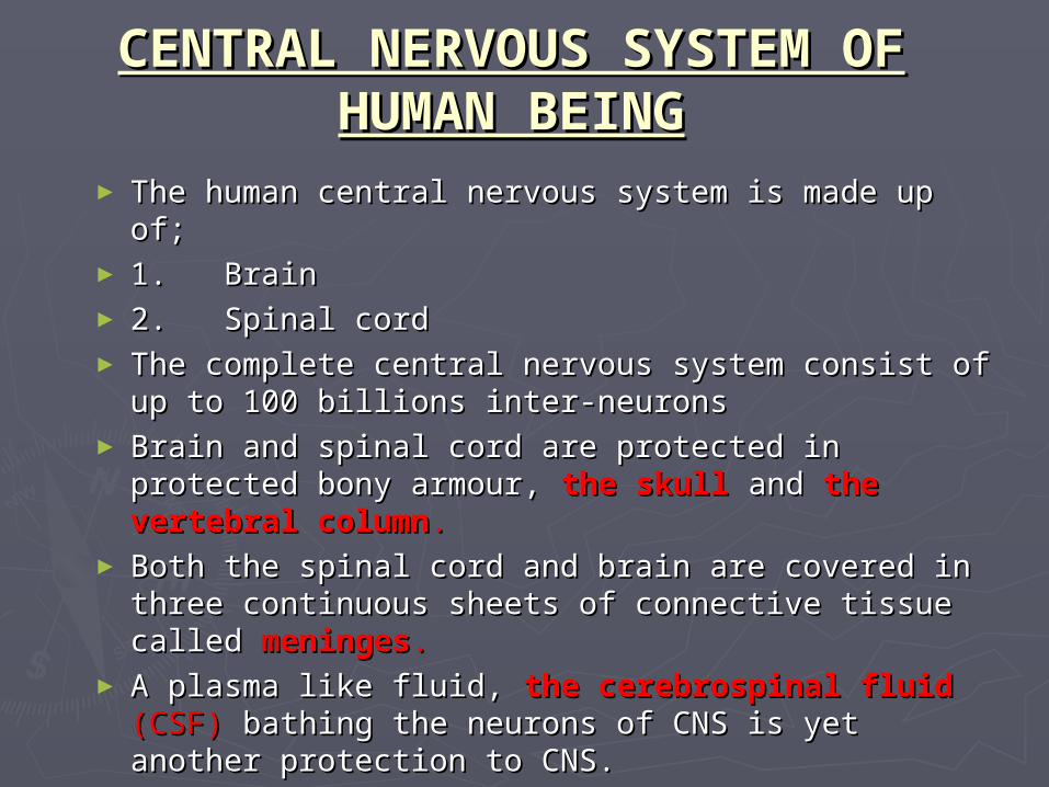
► The human central nervous system is made up of;The human central nervous system is made up of;► 1. Brain1. Brain► 2. Spinal cord2. Spinal cord► The complete central nervous system consist of up to The complete central nervous system consist of up to
100 billions inter-neurons100 billions inter-neurons► Brain and spinal cord are protected in protected bony Brain and spinal cord are protected in protected bony
armour, armour, the skull the skull and and the vertebral columnthe vertebral column..► Both the spinal cord and brain are covered in three Both the spinal cord and brain are covered in three
continuous sheets of connective tissue called continuous sheets of connective tissue called meningesmeninges..► A plasma like fluid, A plasma like fluid, the cerebrospinal fluid the cerebrospinal fluid (CSF) (CSF)
bathing the neurons of CNS is yet another protection to bathing the neurons of CNS is yet another protection to CNS.CNS.

BRAINBRAIN

BRAINBRAIN
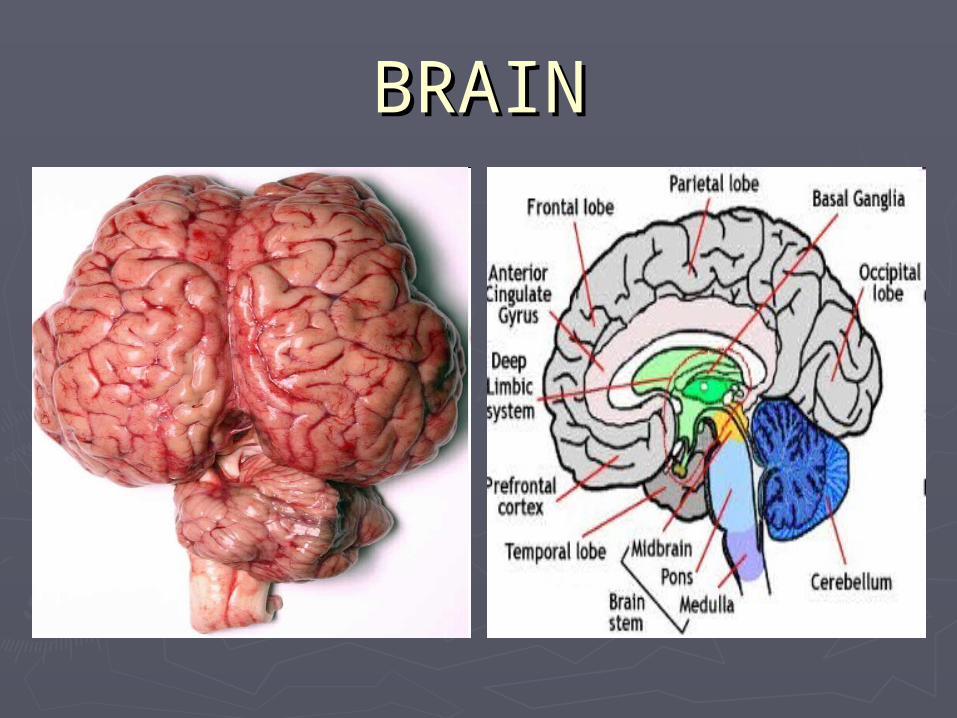
► The brain of all The brain of all vertebrates develops vertebrates develops from three swellings at from three swellings at the anterior end of the the anterior end of the neural canal of the neural canal of the embryo. embryo.
► Human brain is divided Human brain is divided into three parts;into three parts;
► Fore-BrainFore-Brain► Mid-BrainMid-Brain► Hind-BrainHind-Brain
BRAINBRAIN
Fore-Brain:Fore-Brain:
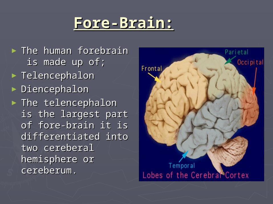
► The human forebrain is The human forebrain is made up of; made up of;
► Telencephalon Telencephalon ► Diencephalon Diencephalon ► The telencephalon is the The telencephalon is the
largest part of fore-brain largest part of fore-brain it is differentiated into it is differentiated into two cereberal two cereberal hemisphere or hemisphere or cereberum.cereberum.

FORE-BRAINFORE-BRAIN► Cereberal cortex is the largest and most complex part Cereberal cortex is the largest and most complex part
of human brain.of human brain.► By means of a prominent groove, called the By means of a prominent groove, called the
longitudinal fissure, the brain is divided into two longitudinal fissure, the brain is divided into two halves called cerebral hemispheres.halves called cerebral hemispheres.
► At the base of this fissure lies a thick bundle of nerve At the base of this fissure lies a thick bundle of nerve fibers, called the fibers, called the corpus callosum, corpus callosum, which provides a which provides a communication link between the hemispheres communication link between the hemispheres Although the right and left hemispheres seem to be a Although the right and left hemispheres seem to be a mirror image of one another, there are important mirror image of one another, there are important functional distinctions.functional distinctions.
► Each hemisphere of the cerebrum is subdivided into Each hemisphere of the cerebrum is subdivided into four lobes. four lobes.

FORE-BRAINFORE-BRAIN
► Frontal Lobe-Frontal Lobe- associated with associated with reasoning, planning, parts of speech, reasoning, planning, parts of speech, movement, emotions, and problem movement, emotions, and problem solving. solving.
► Parietal Lobe-Parietal Lobe- associated with associated with movement, orientation, recognition, movement, orientation, recognition, perception of stimuli. perception of stimuli.
► Occipital Lobe-Occipital Lobe- associated with associated with visual processing. visual processing.
► Temporal Lobe-Temporal Lobe- associated with associated with perception and recognition of auditory perception and recognition of auditory stimuli, memory, and speech.stimuli, memory, and speech.

FORE-BRAIN(DIENCEPHALON): FORE-BRAIN(DIENCEPHALON):
The diencephalonThe diencephalon consists of ; consists of ;
1.1. ThalamusThalamus2.2. Limbic system. Limbic system.
1.1. Thalamus:Thalamus:
► The thalamus is a large, dual lobed mass of grey matter cells The thalamus is a large, dual lobed mass of grey matter cells located at the top of the located at the top of the brainstem, , superior to the to the hypothalamus. The thalamus is a clearing house for sensory . The thalamus is a clearing house for sensory impulses as it receives them from different parts of brain and impulses as it receives them from different parts of brain and relays them to the appropriate part of the motor cortex.relays them to the appropriate part of the motor cortex.
► It controls the pleasure and pain. It controls the pleasure and pain.
FORE-BRAIN(DIENCEPHALON):FORE-BRAIN(DIENCEPHALON):
► Limbic System:Limbic System: ► The limbic system, often referred to as the The limbic system, often referred to as the
"emotional brain", is found buried within the "emotional brain", is found buried within the cerebrum.cerebrum.
► Parts of Limbic System:Parts of Limbic System:
► Hypothalamus Hypothalamus ► AmygdalaAmygdala► Hippocampus Hippocampus ► Some other parts of thalamusSome other parts of thalamus
FORE-BRAIN(DIENCEPHALON):FORE-BRAIN(DIENCEPHALON):
► Hypothalamus:Hypothalamus: Hypothalamus regulates the autonomic nervous system via regulates the autonomic nervous system via
hormone production and release. Affects and regulates production and release. Affects and regulates blood pressure, , heart rate, Mood and motivation, hormonal , Mood and motivation, hormonal body processes, body processes, hunger, , thirst, , sexual arousal, sexual , sexual maturation and the maturation and the sleep/wake cycle..
► Amygdala:Amygdala: It involved in signaling the cortex of motivationally significant It involved in signaling the cortex of motivationally significant
stimuli such as those related to reward, punishment and fear in stimuli such as those related to reward, punishment and fear in addition to social functions such as mating.addition to social functions such as mating.

FORE-BRAIN(DIENCEPHALON):FORE-BRAIN(DIENCEPHALON):
HIPPOCAMPUS:HIPPOCAMPUS:
It belongs to the It belongs to the limbic system and plays and plays important roles in important roles in long-term memory and and spatial spatial navigation. In . In Alzheimer's disease the the hippocampus is one of the hippocampus is one of the first regions of the brain to first regions of the brain to suffer damage; memory suffer damage; memory problems and disorientation problems and disorientation appear among the first appear among the first symptoms. symptoms.
MID-BRAINMID-BRAIN
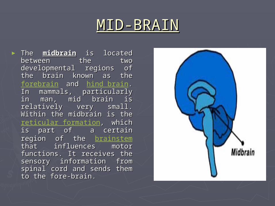
► The The midbrainmidbrain is located between is located between the two developmental regions of the two developmental regions of the brain known as the the brain known as the forebrain and and hind brain. In mammals, . In mammals, particularly in man, mid brain is particularly in man, mid brain is relatively very small. Within the relatively very small. Within the midbrain is the midbrain is the reticular formation, which is part , which is part of a certain region of the of a certain region of the brainstem that influences motor that influences motor functions. It receives the sensory functions. It receives the sensory information from spinal cord and information from spinal cord and sends them to the fore-brain.sends them to the fore-brain.

HIND-BRAINHIND-BRAIN
Cerebellum:Cerebellum:
The cerebellum, or "little The cerebellum, or "little brain", is similar to the brain", is similar to the cerebrum in that it has two cerebrum in that it has two hemispheres and has a highly hemispheres and has a highly folded surface or cortex. This folded surface or cortex. This structure is associated with structure is associated with regulation and coordination regulation and coordination of movement, posture, and of movement, posture, and balance.balance.

HIND-BRAINHIND-BRAIN
► Pons :Pons : is a structure located on the is a structure located on the brain stem. It is . It is superior to to (up from) medulla oblongata, (up from) medulla oblongata, inferior to (down from) the to (down from) the midbrain and and ventral to (in front of) the to (in front of) the cerebellum. The pons . The pons measures about 2.5 cm in length. It mainly controls with sleep, measures about 2.5 cm in length. It mainly controls with sleep, respiration, swallowing, bladder control, hearing, equilibrium, respiration, swallowing, bladder control, hearing, equilibrium, taste, eye movement, facial expressions, facial sensation, and taste, eye movement, facial expressions, facial sensation, and posture. posture.
► Reticular formation:Reticular formation: is a group of nerve fibers located inside is a group of nerve fibers located inside the the brainstem. Reticular formation important in regulating . Reticular formation important in regulating Arousal, Attention, Cardiac Reflexes, Motor Functions, Arousal, Attention, Cardiac Reflexes, Motor Functions, consciousness or wakefulness.consciousness or wakefulness.
HIND-BRAINHIND-BRAIN
Medulla oblongata:Medulla oblongata:
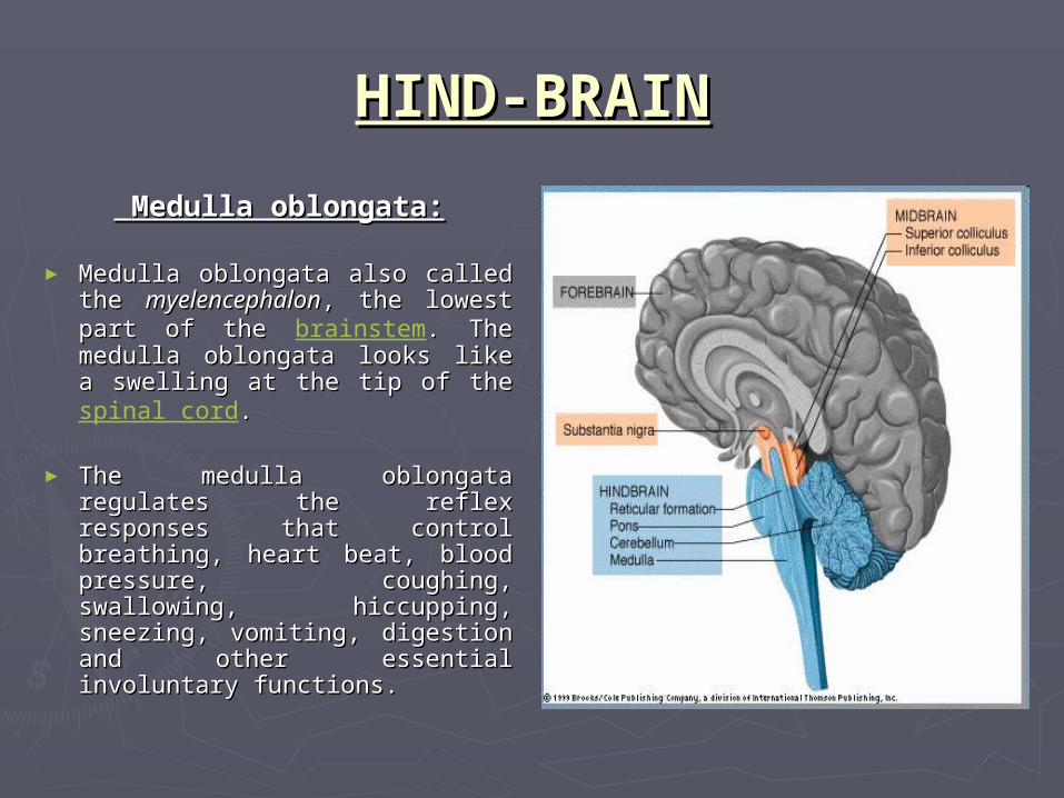
► Medulla oblongata also called the Medulla oblongata also called the myelencephalonmyelencephalon, the lowest part of , the lowest part of the the brainstem. The medulla . The medulla oblongata looks like a swelling at oblongata looks like a swelling at the tip of the the tip of the spinal cord..
► The medulla oblongata regulates The medulla oblongata regulates the reflex responses that control the reflex responses that control breathing, heart beat, blood breathing, heart beat, blood pressure, coughing, swallowing, pressure, coughing, swallowing, hiccupping, sneezing, vomiting, hiccupping, sneezing, vomiting, digestion and other essential digestion and other essential involuntary functions.involuntary functions.

BRAIN-STEMBRAIN-STEM
The lower extension of the brain where it connects to The lower extension of the brain where it connects to the spinal cord. It is formed by the combination of the spinal cord. It is formed by the combination of medulla oblongata, pons and mid–brain. Neurological medulla oblongata, pons and mid–brain. Neurological functions located in the brainstem include those functions located in the brainstem include those necessary for survival (breathing, digestion, heart rate, necessary for survival (breathing, digestion, heart rate, blood pressure) and for arousal (being awake and alert). blood pressure) and for arousal (being awake and alert). The brainstem is the pathway for all fiber tracts passing The brainstem is the pathway for all fiber tracts passing up and down from peripheral nerves and spinal cord to up and down from peripheral nerves and spinal cord to the highest parts of the brain.the highest parts of the brain.

BRAIN-STEMBRAIN-STEM
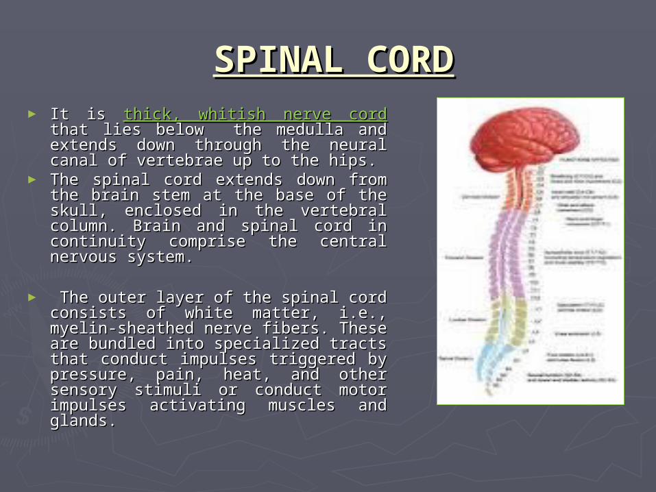
SPINAL CORDSPINAL CORD► It is It is thick, whitish nerve cord thick, whitish nerve cord that lies below that lies below
the medulla and extends down through the the medulla and extends down through the neural canal of vertebrae up to the hips.neural canal of vertebrae up to the hips.
► The spinal cord extends down from the brain The spinal cord extends down from the brain stem at the base of the skull, enclosed in the stem at the base of the skull, enclosed in the vertebral column. Brain and spinal cord in vertebral column. Brain and spinal cord in continuity comprise the central nervous continuity comprise the central nervous system.system.
► The outer layer of the spinal cord consists of The outer layer of the spinal cord consists of white matter, i.e., myelin-sheathed nerve white matter, i.e., myelin-sheathed nerve fibers. These are bundled into specialized fibers. These are bundled into specialized tracts that conduct impulses triggered by tracts that conduct impulses triggered by pressure, pain, heat, and other sensory pressure, pain, heat, and other sensory stimuli or conduct motor impulses activating stimuli or conduct motor impulses activating muscles and glands. muscles and glands.

SPINAL CORD ANATOMYSPINAL CORD ANATOMY
► The inner layer, or gray matter, has The inner layer, or gray matter, has a butterfly-shaped cross-section a butterfly-shaped cross-section and is mainly composed of nerve and is mainly composed of nerve cell bodies. Within the gray matter, cell bodies. Within the gray matter, running the length of the cord and running the length of the cord and extending into the brain, lies the extending into the brain, lies the central canal through which the central canal through which the cerebrospinal fluid circulates.cerebrospinal fluid circulates.
► The spinal cord mediates the reflex The spinal cord mediates the reflex responses to some sensory responses to some sensory impulses directly, i.e., without impulses directly, i.e., without recourse to the brain, as when a recourse to the brain, as when a person's leg is tapped producing the person's leg is tapped producing the knee jerk reflex. knee jerk reflex.
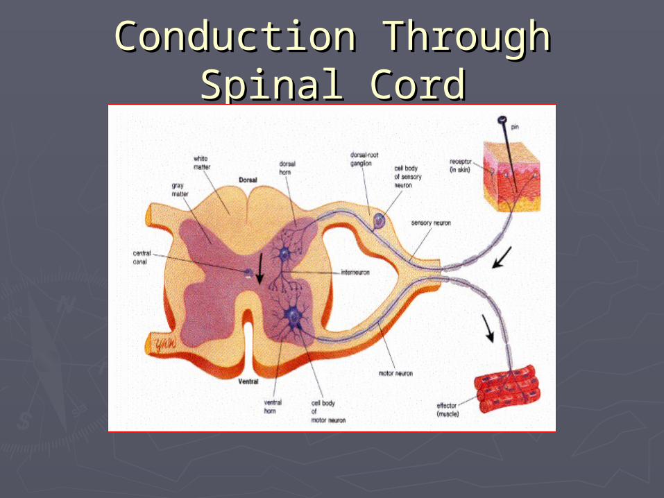
Conduction Through Spinal Conduction Through Spinal CordCord

PERIPHERAL NERVOUS PERIPHERAL NERVOUS SYSTEM (PNS)SYSTEM (PNS)
► In man, the peripheral nervous system In man, the peripheral nervous system consists ofconsists of
►12 pairs of cranial nerves12 pairs of cranial nerves►31 pairs of spinal nerves31 pairs of spinal nerves
► The PNS transmits signals between CNS and The PNS transmits signals between CNS and rest of the body.rest of the body.
► PNS is further divided into PNS is further divided into • Somatic Nervous System(Voluntary)Somatic Nervous System(Voluntary)• Autonomic Nervous System(Involuntary)Autonomic Nervous System(Involuntary)

Autonomic Nervous SystemAutonomic Nervous System
►Autonomic Nervous SystemAutonomic Nervous System is the division is the division of the peripheral nervous system that of the peripheral nervous system that controls the glands and the muscles of the controls the glands and the muscles of the internal organs.internal organs.
► The Autonomic Nervous System operates on The Autonomic Nervous System operates on its own and is involuntary. its own and is involuntary.
► The two divisions of Autonomic Nervous The two divisions of Autonomic Nervous System are System are
► Sympathetic Nervous SystemSympathetic Nervous System► Parasympathetic Nervous SystemParasympathetic Nervous System

ComparisonComparisonSYMPATHETIC SYMPATHETIC
NERVOUS SYSTEMNERVOUS SYSTEM
► It is formed by the spinal It is formed by the spinal nerves arising from thoracic nerves arising from thoracic and lumbar region.and lumbar region.
► It prepares the body for It prepares the body for highly energetic activity highly energetic activity such as such as fight or flight.fight or flight.
It is responsible forIt is responsible for► Dilated PupilDilated Pupil► Accelerated HeartbeatAccelerated Heartbeat► Slower Digestion Slower Digestion ► Stimulated glucose releaseStimulated glucose release► Accelerated breathing rateAccelerated breathing rate
PARASYMPATHETIC PARASYMPATHETIC NERVOUS SYSTEMNERVOUS SYSTEM
► It is formed by vagus It is formed by vagus nerve, some of the cranial nerve, some of the cranial nerves and spinal nerves nerves and spinal nerves arising from sacral region.arising from sacral region.
► It promotes all the internal It promotes all the internal responses associated with responses associated with a a relaxed state.relaxed state.
It is responsible forIt is responsible for► Contracted PupilContracted Pupil► Slower HeartbeatSlower Heartbeat► Stimulated DigestionStimulated Digestion