Human Mob1 proteins are required for cytokinesis by...
6
Journal of Cell Science Human Mob1 proteins are required for cytokinesis by controlling microtubule stability Claudia Florindo 1,2,3 , Joana Perdiga ˜o 1 , Didier Fesquet 4 , Elmar Schiebel 5 , Jonathon Pines 6 and A ´ lvaro A. Tavares 1,2,3, * 1 Instituto Gulbenkian de Cie ˆ ncia, P-2780-156 Oeiras, Portugal 2 Regenerative Medicine Program, Departamento de Cie ˆ ncias Biome ´dicas e Medicina, Universidade do Algarve, 8005 Faro, Portugal 3 Centre for Molecular and Structural Biomedicine, CBME/IBB, University of Algarve, Faro 8005-139, Portugal 4 Universite ´ Montpellier, Sud de France, CRBM, 34293 Montpellier, France 5 ZMBH, Universita ¨ t Heidelberg, 69120, Germany 6 The Gurdon Institute, Cambridge CB2 1QN, UK *Author for correspondence ([email protected]) Accepted 5 March 2012 Journal of Cell Science 125, 3085–3090 ß 2012. Published by The Company of Biologists Ltd doi: 10.1242/jcs.097147 Summary The completion of cytokinesis requires abscission of the midbody, a microtubule-rich cytoplasmic bridge that connects the daughter cells before their final separation. Although it has been established that both the midbody structure and membrane fusion are essential for abscission, the biochemical machinery and the cellular processes of abscission remain ill-defined. Here we report that human Mob1A and Mob1B proteins are involved in the regulation of abscission of the intercellular bridge. The Mob family is a group of highly conserved proteins in eukaryotes, described as binding partners as well as co-activators of protein kinases of the Ndr family, and as members of the Hippo pathway. We show that depletion of Mob1A and Mob1B by RNAi causes abscission failure as a consequence of hyper-stabilization of microtubules in the midbody region. Interestingly, depleting Mob1 also increases cell motility after cytokinesis, and induces prolonged centriole separation in G1 phase. In contrast, centrosomes fail to split when either Mob1A or Mob1B is overexpressed. Our findings indicate that human Mob1 proteins are involved in the regulation of microtubule stability at the midbody. We conclude that Mob1A and Mob1B are needed for cell abscission and centriole re-joining after telophase and cytokinesis. Key words: Cytokinesis, Mob1, Microtubule stability Introduction At the end of cytokinesis cells have to execute abscission, which results in the physical separation of two daughter cells, and return to interphase. It is generally accepted that a particular sequence of events are required for abscission to occur: cortical anchorage of the ingressed furrow, splitting the plasma membrane between the sister cells, and the disassembly of midbody-associated microtubules (Barr and Gruneberg, 2007; Guizetti and Gerlich, 2010). Recent work has demonstrated that endosome fusion and microtubule reorganization are essential for abscission. It was shown that disassembly of microtubules can be mediated by the microtubule-severing protein spastin (Connell et al., 2009; Guizetti et al., 2011; Schiel et al., 2011) and several proteins of the endosomal sorting complex are required for transport (ESCRT)-III localize to the midbody and are required for cytokinesis (Morita et al., 2007; Wollert et al., 2009; Guizetti et al., 2011; Schiel et al., 2011). Mob1 is a conserved protein required for cytokinesis and for the regulation of mitotic exit (McCollum and Gould, 2001; Hergovich, 2011). Mob1 proteins are binding partners and coactivators, of protein kinases of the Ndr and Lats families (reviewed in Hergovich et al., 2006; Trammell et al., 2008). In fly and human cells, Mob1 participates in the control of cell proliferation and apoptosis as part of the Hippo pathway (Lai et al., 2005; Wei et al., 2007; Praskova et al., 2008). There is also a genetic interaction between the Drosophila Mob-like genes and tricornered (trc), another Ndr protein kinase required for normal morphogenesis of a variety of polarized outgrowths (He et al., 2005). In humans, Lats1 interacts with Mob1 and this complex appears to be functionally required for cytokinesis (Bothos et al., 2005). Here we have investigated the function of the human Mob1A and Mob1B proteins to further explore their role during cytokinesis. We show that Mob1 proteins are required for abscission. Furthermore, we show that upon Mob1 downregulation, centrioles fail to rejoin at the end of cytokinesis, cells acquire increased motility, and microtubule stability is increased. Finally, we also show that depolymerization of microtubules is a prerequisite for abscission, and that Mob1 proteins are involved in the regulation of this process. Results and Discussion Human Mob1A and Mob1B proteins are essential for centriole cohesion We first examined the distribution of human Mob1A and Mob1B proteins. In HeLa cells expressing GFP-centrin, where the two centrioles were clearly distinguishable, we detected a broad single Mob1 dot in the center of the centrosome by indirect immunofluorescence microscopy (Fig. 1A). This staining persisted until the end of mitosis (Fig. 1B). However, in late telophase cells with two separated centrioles, Mob1 was detected just in the stronger GFP-centrin signal, often the one closer to the midbody Short Report 3085
Transcript of Human Mob1 proteins are required for cytokinesis by...
Journ
alof
Cell
Scie
nce
Human Mob1 proteins are required for cytokinesis bycontrolling microtubule stability
Claudia Florindo1,2,3, Joana Perdigao1, Didier Fesquet4, Elmar Schiebel5, Jonathon Pines6 andAlvaro A. Tavares1,2,3,*1Instituto Gulbenkian de Ciencia, P-2780-156 Oeiras, Portugal2Regenerative Medicine Program, Departamento de Ciencias Biomedicas e Medicina, Universidade do Algarve, 8005 Faro, Portugal3Centre for Molecular and Structural Biomedicine, CBME/IBB, University of Algarve, Faro 8005-139, Portugal4Universite Montpellier, Sud de France, CRBM, 34293 Montpellier, France5ZMBH, Universitat Heidelberg, 69120, Germany6The Gurdon Institute, Cambridge CB2 1QN, UK
*Author for correspondence ([email protected])
Accepted 5 March 2012Journal of Cell Science 125, 3085–3090� 2012. Published by The Company of Biologists Ltddoi: 10.1242/jcs.097147
SummaryThe completion of cytokinesis requires abscission of the midbody, a microtubule-rich cytoplasmic bridge that connects the daughter
cells before their final separation. Although it has been established that both the midbody structure and membrane fusion are essentialfor abscission, the biochemical machinery and the cellular processes of abscission remain ill-defined. Here we report that human Mob1Aand Mob1B proteins are involved in the regulation of abscission of the intercellular bridge. The Mob family is a group of highlyconserved proteins in eukaryotes, described as binding partners as well as co-activators of protein kinases of the Ndr family, and as
members of the Hippo pathway. We show that depletion of Mob1A and Mob1B by RNAi causes abscission failure as a consequence ofhyper-stabilization of microtubules in the midbody region. Interestingly, depleting Mob1 also increases cell motility after cytokinesis,and induces prolonged centriole separation in G1 phase. In contrast, centrosomes fail to split when either Mob1A or Mob1B is
overexpressed. Our findings indicate that human Mob1 proteins are involved in the regulation of microtubule stability at the midbody.We conclude that Mob1A and Mob1B are needed for cell abscission and centriole re-joining after telophase and cytokinesis.
Key words: Cytokinesis, Mob1, Microtubule stability
IntroductionAt the end of cytokinesis cells have to execute abscission, whichresults in the physical separation of two daughter cells, and returnto interphase. It is generally accepted that a particular sequence
of events are required for abscission to occur: cortical anchorageof the ingressed furrow, splitting the plasma membrane betweenthe sister cells, and the disassembly of midbody-associated
microtubules (Barr and Gruneberg, 2007; Guizetti and Gerlich,2010). Recent work has demonstrated that endosome fusion andmicrotubule reorganization are essential for abscission. It was
shown that disassembly of microtubules can be mediated by themicrotubule-severing protein spastin (Connell et al., 2009;Guizetti et al., 2011; Schiel et al., 2011) and several proteins ofthe endosomal sorting complex are required for transport
(ESCRT)-III localize to the midbody and are required forcytokinesis (Morita et al., 2007; Wollert et al., 2009; Guizettiet al., 2011; Schiel et al., 2011).
Mob1 is a conserved protein required for cytokinesis and forthe regulation of mitotic exit (McCollum and Gould, 2001;Hergovich, 2011). Mob1 proteins are binding partners and
coactivators, of protein kinases of the Ndr and Lats families(reviewed in Hergovich et al., 2006; Trammell et al., 2008). In flyand human cells, Mob1 participates in the control of cell
proliferation and apoptosis as part of the Hippo pathway (Laiet al., 2005; Wei et al., 2007; Praskova et al., 2008). There is alsoa genetic interaction between the Drosophila Mob-like genes and
tricornered (trc), another Ndr protein kinase required for normalmorphogenesis of a variety of polarized outgrowths (He et al.,
2005). In humans, Lats1 interacts with Mob1 and this complexappears to be functionally required for cytokinesis (Bothos et al.,2005).
Here we have investigated the function of the human Mob1A and
Mob1B proteins to further explore their role during cytokinesis. Weshow that Mob1 proteins are required for abscission. Furthermore,we show that upon Mob1 downregulation, centrioles fail to rejoin
at the end of cytokinesis, cells acquire increased motility, andmicrotubule stability is increased. Finally, we also show thatdepolymerization of microtubules is a prerequisite for abscission,
and that Mob1 proteins are involved in the regulation of thisprocess.
Results and DiscussionHuman Mob1A and Mob1B proteins are essential forcentriole cohesion
We first examined the distribution of human Mob1A and Mob1Bproteins. In HeLa cells expressing GFP-centrin, where the two
centrioles were clearly distinguishable, we detected a broadsingle Mob1 dot in the center of the centrosome by indirectimmunofluorescence microscopy (Fig. 1A). This staining persisted
until the end of mitosis (Fig. 1B). However, in late telophase cellswith two separated centrioles, Mob1 was detected just in thestronger GFP-centrin signal, often the one closer to the midbody
Short Report 3085
Journ
alof
Cell
Scie
nce
(Fig. 1B3). Mob1 was also observed at the kinetochores
(supplementary material Fig. S2) and in the central spindle and
midbody regions during late anaphase and telophase (Fig. 1). The
specificity of the Mob1 centrosomal staining was confirmed by
time-lapse video microscopy of HeLa cells expressing either GFP-
Mob1A or GFP-Mob1B (Fig. 1C).
The high degree of identity between the two Mob1 proteins
indicated a possible functional redundancy (supplementary
material Fig. S1) (Vitulo et al., 2007). Therefore, we sought to
disrupt the expression of both Mob1s by simultaneous co-
transfection of siRNA directed against each Mob1. We used three
different siRNA duplexes for each type of Mob1, and found that
Mob1 protein levels were substantially repressed (.95%) by 48 h
post-transfection (Fig. 2A; supplementary material Fig. S1C).
Identical results were obtained using any pair of different siRNAs
against either Mob1s, indicating that siRNA treatment effectively
and specifically reduced the abundance of Mob1 proteins. We
will refer to cells in which neither Mob1A nor Mob1B were
detected by immunoblotting as ‘Mob1(A+B)-depleted’.
Although Mob1(A+B)-depleted cells formed apparently
normal interphase arrays of microtubules and mitotic spindles,
downregulation of Mob1 had a striking effect upon centriole
behavior. Two distinct, widely separated GFP-centrin positive
structures were observed in .28% of interphase Mob1(A+B)-
depleted cells, compared with 5% in control cells (Fig. 2B). The
distance between separated centrioles varied considerably
(between 2 and 30 mm, Fig. 2B), compared to control cells (1.4
6 0.2 mm). This effect on centrioles was observed with all
Mob1-siRNAs, but not with the control siRNAs, and was also
observed in U2OS and RPE1 cell lines (supplementary material
Fig. S3A,B), indicating that is not cell type specific, nor linked to
the transformed state of Hela cells.
The abrogation of Mob1 function seemed to cause centrioles to
split during G1 phase, firstly because mitotic spindle poles
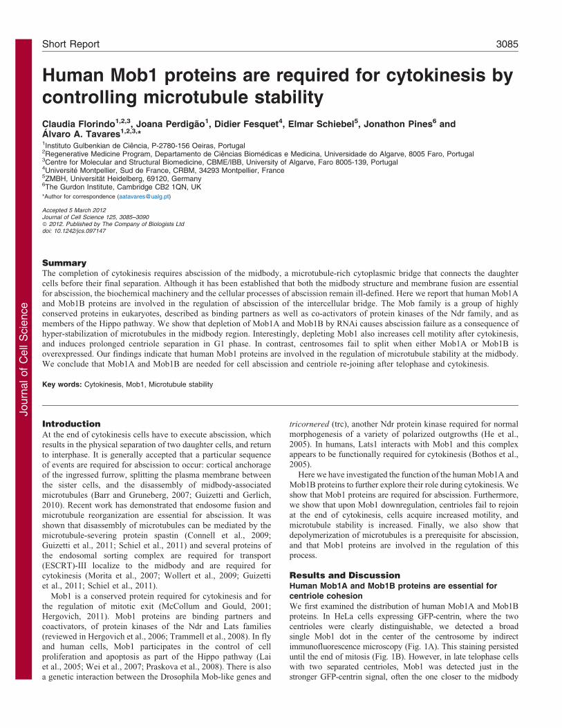
Fig. 1. Mob1 localizes to the centrosome
and to the midbody. (A) Cells were stained
for GFP-centrin (green), DAPI (blue) and
anti-Mob1 (red). Mob1 accumulates on the
centrosome but does not completely overlap
with the centrioles (two centrin dots). This
pattern remains while the centrioles duplicate
and centrosomes separate (A1 to A4).
(B) HeLa GFP-centrin cells immunostained
for a-tubulin (blue) and Mob1 (red). At the
end of telophase (3), Mob1 is detected in the
midbody and in just one centriole. (C) Time
lapse analysis of a stable cell line expressing
GFP-Mob1A. GFP-Mob1A is visible on the
centrosomes from the start of mitosis until
anaphase (arrows), and in the central spindle
and midbody (arrowheads) at the end of
mitosis. GFP-Mob1B protein has an
identical behavior.
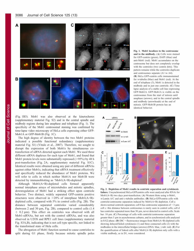
Fig. 2. Depletion of Mob1 results in centriole separation and cytokinesis
failure. Unsynchronized HeLa-GFPcentrin cells were analyzed after RNAi for
Mob1(A+B) two days post-transfection. (A) Western blots using a-Mob1,
a-Lamin A/C and anti-c-tubulin antibodies. (B) HeLa-GFPcentrin cells with
centriole/centrosome separation induced by Mob1(A+B) depletion. Cell a
shows normal centriole separation; cell b has centrosome separation of ,3 mm;
cell c: this distance between centrosomes is rarely seen in control cells; cell d
has centrioles separated more than 30 mm, never detected in control cells. Scale
bar: 10 mm. (C) Percentage of cells with centriole/centrosome separation
greater then 3 mm in asynchronous cultures, and in synchronized cells analyzed
in S/G2 phase. (D) DIC + fluorescence images from Mob1(A+B) depleted cells
showing delayed/failed cytokinesis (1) and three connected cells (2) with the
midbodies in the intercellular bridges (arrows) (DNA: blue, c-tub: red). (E) For
the quantification of linked cells after Mob1(A+B) depletion only cells with a
visible midbody, as in D1, were considered.
Journal of Cell Science 125 (13)3086
Journ
alof
Cell
Scie
nce
always had two centrioles each (even after 5 days of siRNA
treatment); secondly, because we never observed cells with four
separated centrioles (or with one pair plus two separated
centrioles) indicating that no splitting occurred after centriole
duplication. To confirm this hypothesis, we analyzed cells 3.5 h
after release from a S-phase block and we found that 32% of the
Mob1(A+B)-depleted cells had two separated pairs of centrioles
compared to ,7% in control cells (Fig. 2C), indicating that a
large proportion of Mob1-depleted cells initiated centriole
duplication with already separated centrioles.
Centrioles often split at the end telophase and re-join after
abscission (Chevrier et al., 2002; Meraldi and Nigg, 2002). In
contrast to the effect of depletion, overexpression of either Mob1
reduced the percentage of cells with split centrioles. We detected
four distinct separated centrioles in 32% of control cells undergoing
telophase/cytokinesis (supplementary material Fig. S5). On the
other hand, overexpression of Mob1A or Mob1B drastically
reduced this number to 15% and 4%, respectively (three
independent experiments, n5,100 dividing cells per exp.).
Therefore, an excess of Mob1 prevents centriole separation at the
end of cell division, and Mob1 is important for the re-association of
centrioles after mitosis (supplementary material Fig. S7).
Mob1 downregulation results in abscission failure and
increased cell motility
We found that Mob1(A+B)-depleted cells remained connected at
the end of cytokinesis by thin cytoplasmic bridges containing the
midbody (Fig. 2D; Fig. 3A; supplementary material Fig. S3C,D).
Chains of 3 or 4 connected cells were also frequently observed
(Fig. 2D2). Time-lapse imaging revealed that Mob1(A+B)-
depleted cells proceeded through mitosis with similar kinetics to
control cells but took longer to execute abscission (Fig. 3). Most
control cells took up to 4 h (80%, n560) from constriction to
complete separation of daughter cells. In contrast, abscission was
completed by 4 h in only 30% Mob1(A+B)-depleted cells
(Fig. 3B), and 38% took longer than 6 h (n570). No DAPI-
staining or anti-lamin staining was detected at the midbody region
after Mob1(A+B)-depletion (data not shown), indicating that
chromosome bridges or lagging chromosomes were unlikely to be
the major cause of abscission failure (Steigemann et al., 2009). In
addition, treatment of the cells with ZM1 (a specific Aurora B
inhibitor) did not accelerated the timing of abscission, indicating
that the delay was not caused by activation of the Aurora B-
dependent checkpoint (supplementary material Fig. S4).
Remarkably, while control cells moved slightly apart after
cytokinesis before adhering again and flattening onto the dish
(supplementary material Movie 1), Mob1-depleted cells became
highly mobile immediately after telophase/cytokinesis and
remained connected by long intercellular bridges (Fig. 3;
supplementary material Movies 2, 3). Cells moved
continuously for several hours and with frequent changes of
direction (Fig. 3C–D) even after resolution of the intercellular
bridge, indicating that movement was not dependent on
abscission failure.
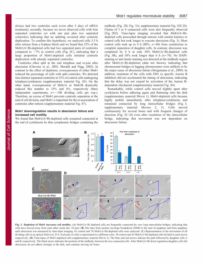
Fig. 3. Depletion of Mob1 increases cell motility. (A) Mob1(A+B) depleted cells are frequently connected by very long intercellular bridges, indicating that
cells have moved away from each other (scale bar: 10 mm). (B) The time from nuclear envelope breakdown (NEB) to the start of anaphase and from anaphase
until abscission was measured by time-lapse imaging. 65 control and 70 Mob1(A+B)-depleted cells were analyzed. (C) Representation of the movement of all
dividing cells in an optical field over 23 h. Each pair of cells is represented in a different color. 30 control and 36 Mob1(A+B)-depleted cells divided in each movie
respectively. (D) Time-lapse of Mob1-depleted cells (supplementary material Movie 2). The blue and red arrows indicate the path followed by daughter cells A
and B, respectively. The black arrow indicates the position of the midbody, between the two connected cells. After Mob1(A+B) down regulation daughter cells fail
abscission, do not adhere strongly to the dish, and continue moving for hours.
Mob1 regulates microtubule stability 3087
Journ
alof
Cell
Scie
nce
Mob1 depletion reduces dynamic instability of
microtubules
We found that Mob1(A+B)-depletion did not affect the midbody
localization of Aurora B, MKPL1, PLK1 or Cep55, in agreement
with (Wilmeth et al., 2010). Both endobrevin and syntaxin 2 also
localized correctly. indicating that abscission failure was not
caused by deficient recruitment of exocyst components to the
midbody region (not shown).
Because over-stabilization of microtubules can prevent or delay
cytokinesis (Hong et al., 2007; Manabe et al., 2002), we wondered
whether hyper-stabilization of microtubules prevented abscission
in Mob1-depleted cells. We found that Mob1-depleted cells had
increased levels of acetylated tubulin (a marker of stabilised
microtubules) (Piperno et al., 1987), and of microtubules resistant
to both nocodazole and cold treatments (Fig. 4; supplementary
material Fig. S6). In dividing cells, acetylated microtubules were
easily detected in the midbody region (Fig. 4A). But after 30min
incubation with nocodazole and cold this staining was lost in 46%
of control cells, while the majority of Mob1(A+B)-depleted cells
(.80%) retained strong acetylated tubulin staining on the
intercellular bridge (Fig. 4). Therefore down regulation of
Mob1A+B resulted in increased microtubule stability. If hyper-
stabilization of the microtubules did prevent abscission, then
treatments that reduced microtubule stability should decrease the
number of unsuccessful cell divisions. In agreement with this, we
found that Mob1(A+B)-depleted cells could execute abscission if
microtubules were artificially destabilized by nocodazole
treatment (Fig. 4C).
In summary, we have shown that Mob1(A+B) depletion
inhibits the removal of midbody-localized microtubules that is
required to allow membrane fusion and abscission (Guizetti and
Gerlich, 2010). Although poorly explored, microtubule dynamics
do have an important role in cytokinesis (D’Avino et al., 2005;
Glotzer, 2009). Cells lacking Spastin, a microtubule-severing
protein, fail abscission because they retain midbody microtubules
(Yang et al., 2008; Connell et al., 2009; Guizetti et al., 2011)
making spastin a potential candidate for regulation by Mob1.
Also, it is tempting to think that there may be a causal connection
between the unusual centriole behavior in Mob1-depleted cells
and the contribution of the mother centriole to signal abscission
observed by (Piel et al., 2001).
Populations of microtubules with different stabilities are
involved in several aspects of cytokinesis (Foe and von
Dassow, 2008; Shen et al., 2009; Connell et al., 2009).
Microtubule dynamics and reorganization are also crucial for
cell motility (Gundersen and Bulinski, 1988; Pegtel et al., 2007;
Drabek et al., 2006) and, interestingly, altered microtubule
dynamics can induce centriole separation (Jean et al., 1999). At
present it is unclear whether the centriole splitting, abscission
failure and hypermotility of Mob1(A+B)-depleted cells are all
consequences of altered microtubule stability or reflect different
functions of Mob1A and Mob1B (supplementary material Fig.
S7).
The only Mob-like protein in Tetrahymena (TtMob1) is a cell
polarity marker and is essential for division plane placement and
cytokinesis (Tavares et al., 2012), therefore, since directed cell
movement is dependent on cell polarization, the effect on
cytoskeletal organization may to be the ancestral function of
Mob-like proteins. The underlying molecular mechanism of
Mob1 activity likely involves binding and activating Dbf2-like
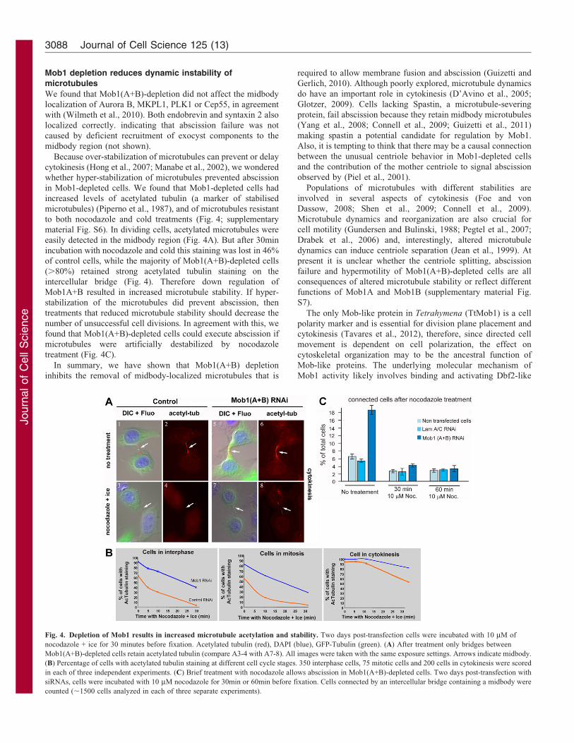
Fig. 4. Depletion of Mob1 results in increased microtubule acetylation and stability. Two days post-transfection cells were incubated with 10 mM of
nocodazole + ice for 30 minutes before fixation. Acetylated tubulin (red), DAPI (blue), GFP-Tubulin (green). (A) After treatment only bridges between
Mob1(A+B)-depleted cells retain acetylated tubulin (compare A3-4 with A7-8). All images were taken with the same exposure settings. Arrows indicate midbody.
(B) Percentage of cells with acetylated tubulin staining at different cell cycle stages. 350 interphase cells, 75 mitotic cells and 200 cells in cytokinesis were scored
in each of three independent experiments. (C) Brief treatment with nocodazole allows abscission in Mob1(A+B)-depleted cells. Two days post-transfection with
siRNAs, cells were incubated with 10 mM nocodazole for 30min or 60min before fixation. Cells connected by an intercellular bridge containing a midbody were
counted (,1500 cells analyzed in each of three separate experiments).
Journal of Cell Science 125 (13)3088
Journ
alof
Cell
Scie
nce
kinases, since both Lats1/2 and NDR kinases contribute to the
regulation of the cytoskeleton (Justice et al., 1995; Yang et al.,
2004; Hergovich et al., 2006). Consistent with this idea,
components of the Hippo pathway cooperate with the Nek2
kinase to regulate centrosome disjunction (Mardin et al., 2010).
Although other centrosome components are required to prevent
premature centriole/centrosome splitting (Bahmanyar et al.,
2008; Wang et al., 2008; Nakamura et al., 2009), Mob1 is
unique in the sense that it affects centriole re-joining.
In conclusion, we have shown that membrane recruitment to
the midbody area is not in itself enough, and that microtubules
have to be reorganised before membrane vesicles can fuse. Our
results open the door to explore the regulation of microtubule
dynamics by the Mob1-dependant kinase(s). Moreover, they
indicate the exciting possibility that the Hippo pathway, which
has a key role in regulating cell proliferation and apoptosis
(reviewed in Pan, 2010), may influence microtubule dynamics in
response to extracellular and intracellular signals and thereby
influence cell polarity.
Materials and MethodsAntibodies and siRNAs
The a-Mob1 serum (Ponchon et al., 2004), recognized both Mob1A and Mob1B in
HeLa extracts. The signal could be competed with either Mob1A or Mob1Bexpressed bacterially, and was suppressed upon Mob1A+Mob1B RNAi depletion
(supplementary material Fig. S1). Primary antibodies: c-tubulin (GTU–88,Sigma), a-tubulin (DM1a, Sigma), acetylated tubulin (C3B9). Three different
siRNAs were used per gene with identical results: 59-AAACACGCAGAA-GCCACACUU-39 and Ambion’s #s30511 and #s30510 for Mob1A, oligos 59-
AACACCUCCUUUAAGCACUUU-39, and Ambion’s #s40977 and #s40978 forMob1B. As controls Lamin A/C oligo (Elbashir et al., 2001) and Dharmachon
Scramble II oligo were used. GenBank sequences: Human Mob1A, AJ577473;Human Mob1B, AJ577474.
Cell culture and synchronization
HeLa and U2OS cells were cultured in DMEM supplemented with 10% heat-
inactivated fetal calf serum (FCS) and 2 mM glutamine. HeLa-GFP-centrin cellswere cultured in RPMI 1640 with GLUTAMAX-I, supplemented with 10% FCS.
RPE-I cells were cultured using D-MEM/F12 media supplemented with 10% FCS,2 mM glutamine and 0.348% sodium bicarbonate. HeLa cells were synchronized
in S phase using a double thymidine (2.5 mM) block of 24 h, with a 12 h intervalbetween the blocks. Cells entered mitosis 8–10 h after release of the second block.
Transfections
(A) fixed cell analysis: Cells were seeded on sterile coverslips at a density of 2–
36104 cells per well in a 24 well plate. Next day cells were transfected using 1ulLipofectamine2000 (Life technologies) with siRNAs at a final concentration of
100 nM. Two days after transfection the cells were analyzed. For siRNA-DNA co-transfections we used LF2000 and 200 ng of DNA per reaction.
(B) live imaging analysis: 26105 HeLa and HeLa GFPcentrin cells were seededon 0.17 Dmm dish (Bioptechs). Cells were synchronized in S-phase and
transfections were done between the two blocks. Cells were filmed 8–10 hoursafter released from the second block. Images were acquired every 3 minutes.
Nocodazole and cold treatment assays
Two days post-transfection, cells were treated with ice cold D-MEM + Nocodazole
(10 mM) for 5, 10 and 30 minutes, on top of an ice bed. Non-treated cells wereused as controls. Cells were fixed with methanol/acetone and processed for
immunofluorescence. Only YFP positive cells were scored: 350 interphase cells,75 mitotic cells and 75 linked cells were scored per experiment in three
independent experiments.
For Aurora B inihibition we used an adapted protocol from (Steigemann et al.,2009). Two days after RNAi treatment, ZM1 (an Aurora B inhibitor) (Tocris) was
added to a final concentration of 2 mM and incubated for 3 h. After this time cellswere washed, fixed and processed for immunofluorescence.
AcknowledgementsWe would like to thank Michel Bornens, Thomas Meyer, ThomasWeimbs, Isabel Martinez Garay and Iain Cheeseman for reagents.
FundingC. F. and J. P. were recipients of FCT fellowships [grant numbersPRAXIS XXI/DB/19823/99 to C. F. and SFRH/BPD/20698/2004 to J.Perdigao]. Work in the Tavares lab was financed by the following FCTgrants: [grant numbers POCTI/BCI/34405/99, POCTI/CBO/39099/2001, POCTI/BIA/PRO/60337/2004, PTDC/SAU-OBD/105234-2008and PEst-OE/EQB/LA0023/2011], and in the Fesquet lab by ContractARC N˚ 5479.
Supplementary material available online at
http://jcs.biologists.org/lookup/suppl/doi:10.1242/jcs.097147/-/DC1
ReferencesBahmanyar, S., Kaplan, D. D., Deluca, J. G., Giddings, T. H., Jr, O’Toole, E. T.,
Winey, M., Salmon, E. D., Casey, P. J., Nelson, W. J. and Barth, A. I. (2008). beta-Catenin is a Nek2 substrate involved in centrosome separation. Genes Dev. 22, 91-105.
Barr, F. A. and Gruneberg, U. (2007). Cytokinesis: placing and making the final cut.Cell 131, 847-860.
Bothos, J., Tuttle, R. L., Ottey, M., Luca, F. C. and Halazonetis, T. D. (2005). HumanLATS1 is a mitotic exit network kinase. Cancer Res. 65, 6568-6575.
Chevrier, V., Piel, M., Collomb, N., Saoudi, Y., Frank, R., Paintrand, M.,Narumiya, S., Bornens, M. and Job, D. (2002). The Rho-associated protein kinasep160ROCK is required for centrosome positioning. J. Cell Biol. 157, 807-817.
Connell, J. W., Lindon, C., Luzio, J. P. and Reid, E. (2009). Spastin couplesmicrotubule severing to membrane traffic in completion of cytokinesis and secretion.Traffic 10, 42-56.
D’Avino, P. P., Savoian, M. S. and Glover, D. M. (2005). Cleavage furrow formationand ingression during animal cytokinesis: a microtubule legacy. J. Cell Sci. 118,1549-1558.
Drabek, K., van Ham, M., Stepanova, T., Draegestein, K., van Horssen, R., Sayas,C. L., Akhmanova, A., Ten Hagen, T., Smits, R., Fodde, R. et al. (2006). Role ofCLASP2 in microtubule stabilization and the regulation of persistent motility. Curr.
Biol. 16, 2259-2264.Elbashir, S. M., Harborth, J., Lendeckel, W., Yalcin, A., Weber, K. and Tuschl, T.
(2001). Duplexes of 21-nucleotide RNAs mediate RNA interference in culturedmammalian cells. Nature 411, 494-498.
Foe, V. E. and von Dassow, G. (2008). Stable and dynamic microtubules coordinatelyshape the myosin activation zone during cytokinetic furrow formation. J. Cell Biol.
183, 457-470.Glotzer, M. (2009). The 3Ms of central spindle assembly: microtubules, motors and
MAPs. Nat. Rev. Mol. Cell Biol. 10, 9-20.Guizetti, J. and Gerlich, D. W. (2010). Cytokinetic abscission in animal cells. Semin.
Cell Dev. Biol. 21, 909-916.
Guizetti, J., Schermelleh, L., Mantler, J., Maar, S., Poser, I., Leonhardt, H., Muller-Reichert, T. and Gerlich, D. W. (2011). Cortical constriction during abscissioninvolves helices of ESCRT-III-dependent filaments. Science 331, 1616-1620.
Gundersen, G. G. and Bulinski, J. C. (1988). Selective stabilization of microtubulesoriented toward the direction of cell migration. Proc. Natl. Acad. Sci. USA 85, 5946-5950.
He, Y., Fang, X., Emoto, K., Jan, Y. N. and Adler, P. N. (2005). The tricornered Ser/Thr protein kinase is regulated by phosphorylation and interacts with furry duringDrosophila wing hair development. Mol. Biol. Cell 16, 689-700.
Hergovich, A. (2011). MOB control: reviewing a conserved family of kinase regulators.Cell. Signal. 23, 1433-1440.
Hergovich, A., Stegert, M. R., Schmitz, D. and Hemmings, B. A. (2006). NDRkinases regulate essential cell processes from yeast to humans. Nat. Rev. Mol. Cell
Biol. 7, 253-264.Hong, K. U., Park, Y. S., Seong, Y. S., Kang, D., Bae, C. D. and Park, J. (2007).
Functional importance of the anaphase-promoting complex-Cdh1-mediated degrada-tion of TMAP/CKAP2 in regulation of spindle function and cytokinesis. Mol. Cell.
Biol. 27, 3667-3681.
Jean, C., Tollon, Y., Raynaud-Messina, B. and Wright, M. (1999). The mammalianinterphase centrosome: two independent units maintained together by the dynamics ofthe microtubule cytoskeleton. Eur. J. Cell Biol. 78, 549-560.
Justice, R. W., Zilian, O., Woods, D. F., Noll, M. and Bryant, P. J. (1995). TheDrosophila tumor suppressor gene warts encodes a homolog of human myotonicdystrophy kinase and is required for the control of cell shape and proliferation. Genes
Dev. 9, 534-546.Lai, Z. C., Wei, X., Shimizu, T., Ramos, E., Rohrbaugh, M., Nikolaidis, N., Ho, L. L.
and Li, Y. (2005). Control of cell proliferation and apoptosis by mob as tumorsuppressor, mats. Cell 120, 675-685.
Manabe, R., Whitmore, L., Weiss, J. M. and Horwitz, A. R. (2002). Identification of anovel microtubule-associated protein that regulates microtubule organization andcytokinesis by using a GFP-screening strategy. Curr. Biol. 12, 1946-1951.
Mardin, B. R., Lange, C., Baxter, J. E., Hardy, T., Scholz, S. R., Fry, A. M. andSchiebel, E. (2010). Components of the Hippo pathway cooperate with Nek2 kinaseto regulate centrosome disjunction. Nat. Cell Biol. 12, 1166-1176.
McCollum, D. and Gould, K. L. (2001). Timing is everything: regulation of mitoticexit and cytokinesis by the MEN and SIN. Trends Cell Biol. 11, 89-95.
Mob1 regulates microtubule stability 3089
Journ
alof
Cell
Scie
nce
Meraldi, P. and Nigg, E. A. (2002). The centrosome cycle. FEBS Lett. 521, 9-13.
Morita, E., Sandrin, V., Chung, H. Y., Morham, S. G., Gygi, S. P., Rodesch, C. K.
and Sundquist, W. I. (2007). Human ESCRT and ALIX proteins interact with
proteins of the midbody and function in cytokinesis. EMBO J. 26, 4215-4227.
Nakamura, A., Arai, H. and Fujita, N. (2009). Centrosomal Aki1 and cohesin function
in separase-regulated centriole disengagement. J. Cell Biol. 187, 607-614.
Pan, D. (2010). The hippo signaling pathway in development and cancer. Dev. Cell 19,
491-505.
Pegtel, D. M., Ellenbroek, S. I., Mertens, A. E., van der Kammen, R. A., de Rooij, J.
and Collard, J. G. (2007). The Par-Tiam1 complex controls persistent migration by
stabilizing microtubule-dependent front-rear polarity. Curr. Biol. 17, 1623-1634.
Piel, M., Nordberg, J., Euteneuer, U. and Bornens, M. (2001). Centrosome-dependent
exit of cytokinesis in animal cells. Science 291, 1550-1553.
Piperno, G., LeDizet, M. and Chang, X. J. (1987). Microtubules containing acetylated
alpha-tubulin in mammalian cells in culture. J. Cell Biol. 104, 289-302.
Ponchon, L., Dumas, C., Kajava, A. V., Fesquet, D. and Padilla, A. (2004). NMR
solution structure of Mob1, a mitotic exit network protein and its interaction with an
NDR kinase peptide. J. Mol. Biol. 337, 167-182.
Praskova, M., Xia, F. and Avruch, J. (2008). MOBKL1A/MOBKL1B phosphorylation
by MST1 and MST2 inhibits cell proliferation. Curr. Biol. 18, 311-321.
Schiel, J. A., Park, K., Morphew, M. K., Reid, E., Hoenger, A. and Prekeris, R.
(2011). Endocytic membrane fusion and buckling-induced microtubule severing
mediate cell abscission. J. Cell Sci. 124, 1411-1424.
Shen, Q., Zheng, X., McNutt, M. A., Guang, L., Sun, Y., Wang, J., Gong, Y., Hou, L.
and Zhang, B. (2009). NAT10, a nucleolar protein, localizes to the midbody and
regulates cytokinesis and acetylation of microtubules. Exp. Cell Res. 315, 1653-1667.
Steigemann, P., Wurzenberger, C., Schmitz, M. H., Held, M., Guizetti, J., Maar, S.and Gerlich, D. W. (2009). Aurora B-mediated abscission checkpoint protectsagainst tetraploidization. Cell 136, 473-484.
Tavares, A., Goncalves, J., Florindo, C, Tavares, A. A. and Soares, H. (2012). Mob1:defining cell polarity for proper cell division. J. Cell Sci. 125, 516-527.
Trammell, M. A., Mahoney, N. M., Agard, D. A. and Vale, R. D. (2008). Mob4 playsa role in spindle focusing in Drosophila S2 cells. J. Cell Sci. 121, 1284-1292.
Vitulo, N., Vezzi, A., Galla, G., Citterio, S., Marino, G., Ruperti, B., Zermiani, M.,Albertini, E., Valle, G. and Barcaccia, G. (2007). Characterization and evolution ofthe cell cycle-associated Mob domain-containing proteins in eukaryotes. Evolut.
Bioinf. Online 3, 121-158.Wang, X., Yang, Y., Duan, Q., Jiang, N., Huang, Y., Darzynkiewicz, Z. and Dai, W.
(2008). sSgo1, a major splice variant of Sgo1, functions in centriole cohesion where itis regulated by Plk1. Dev. Cell 14, 331-341.
Wei, X., Shimizu, T. and Lai, Z. C. (2007). Mob as tumor suppressor is activated byHippo kinase for growth inhibition in Drosophila. EMBO J. 26, 1772-1781.
Wilmeth, L. J., Shrestha, S., Montano, G., Rashe, J. and Shuster, C. B. (2010).Mutual dependence of Mob1 and the chromosomal passenger complex forlocalization during mitosis. Mol. Biol. Cell 21, 380-392.
Wollert, T., Wunder, C., Lippincott-Schwartz, J. and Hurley, J. H. (2009).Membrane scission by the ESCRT-III complex. Nature 458, 172-177.
Yang, D., Rismanchi, N., Renvoise, B., Lippincott-Schwartz, J., Blackstone, C. and
Hurley, J. H. (2008). Structural basis for midbody targeting of spastin by theESCRT-III protein CHMP1B. Nat. Struct. Mol. Biol. 15, 1278-1286.
Yang, X., Yu, K., Hao, Y., Li, D. M., Stewart, R., Insogna, K. L. and Xu, T. (2004).LATS1 tumour suppressor affects cytokinesis by inhibiting LIMK1. Nat. Cell Biol. 6,609-617.
Journal of Cell Science 125 (13)3090