Human Mesenchymal Stem Cells - bioind.com · Human mesenchymal stem cells (hMSC) are multipotent...
24
Human Mesenchymal Stem Cells Serum-free, xeno-free and animal component-free systems for the culture and differentiation of human mesenchymal stem cells
Transcript of Human Mesenchymal Stem Cells - bioind.com · Human mesenchymal stem cells (hMSC) are multipotent...
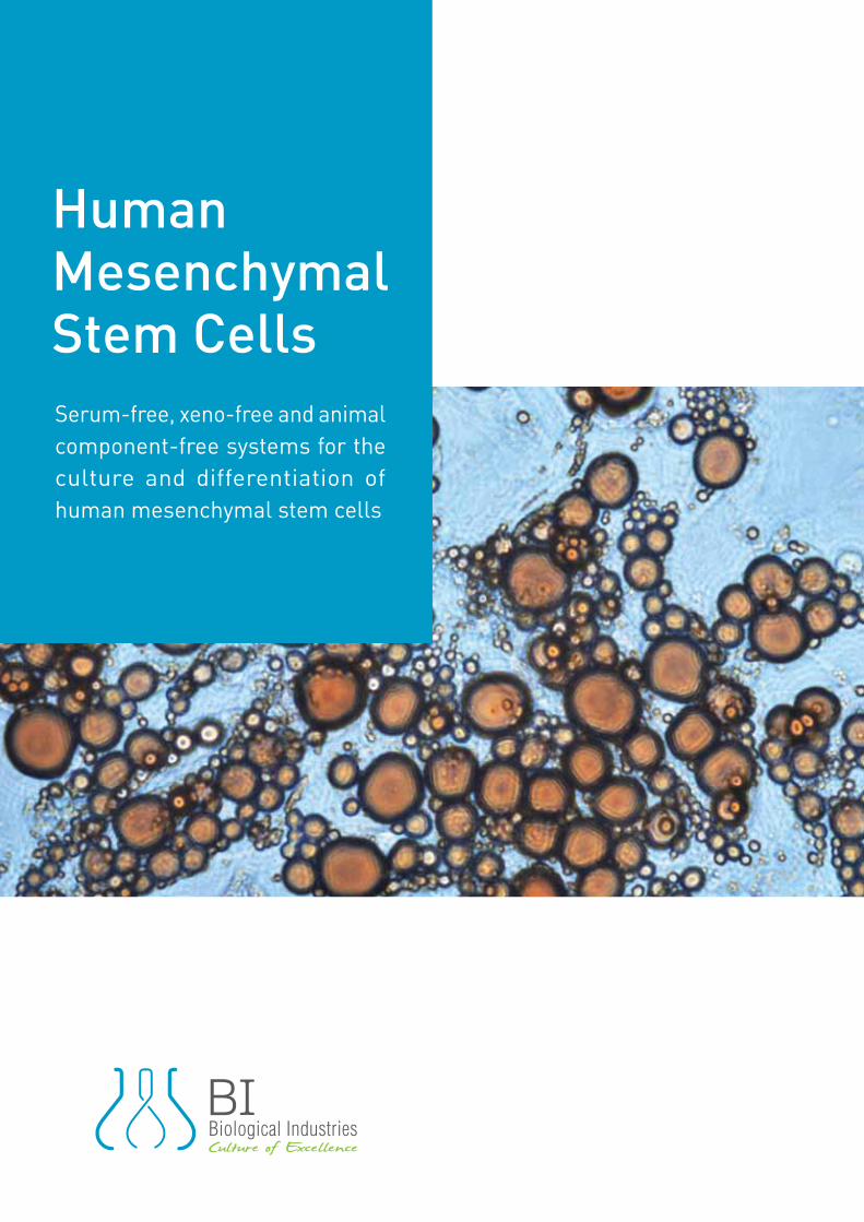
Human Mesenchymal Stem CellsSerum-free, xeno-free and animal component-free systems for the culture and differentiation of human mesenchymal stem cells
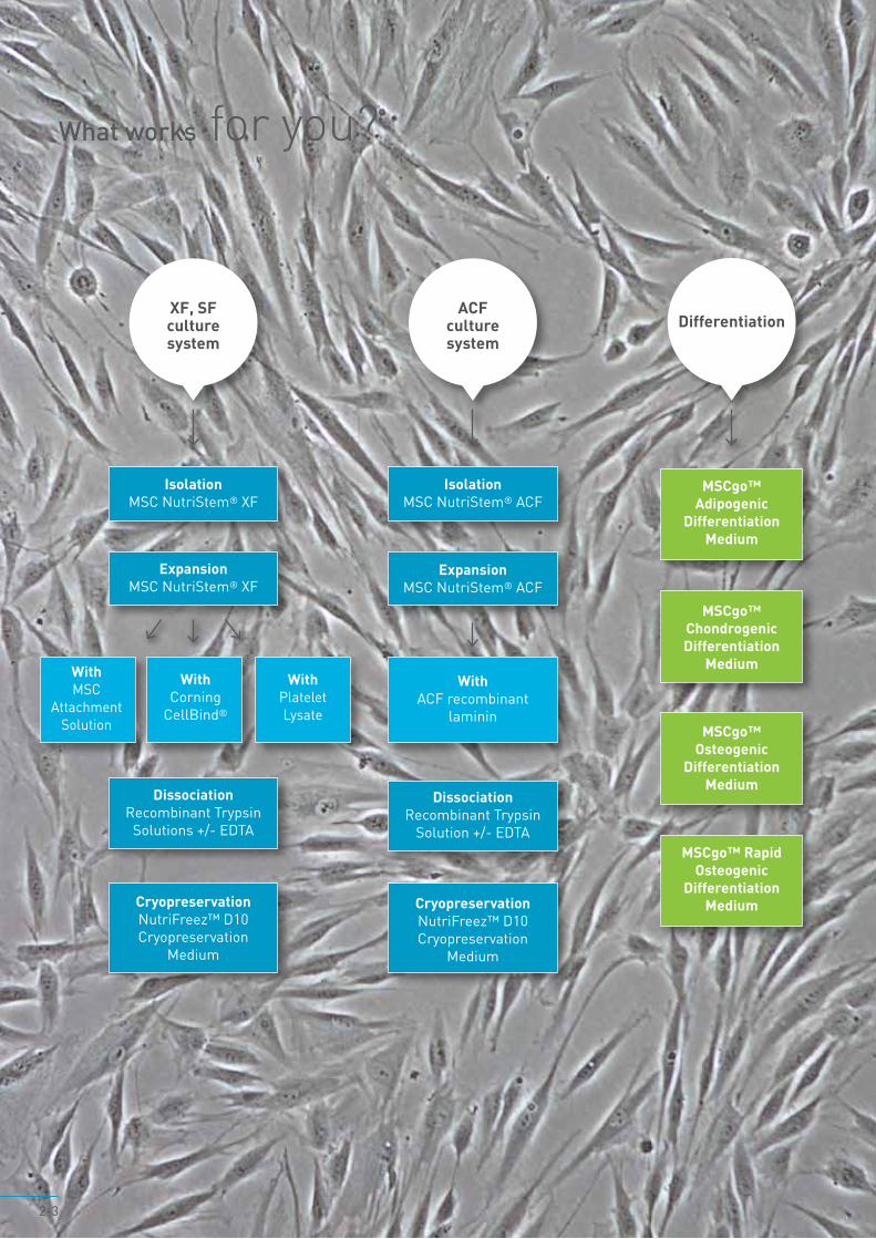
What works for you?
WithMSCgo™ Adipogenic
Differentiation Medium
XF, SF culture system
ExpansionMSC NutriStem® XF
WithMSC
Attachment Solution
WithCorning
CellBind®
WithPlatelet Lysate
DissociationRecombinant Trypsin
Solutions +/- EDTA
Differentiation
MSCgo™ Chondrogenic Differentiation
Medium
MSCgo™ Osteogenic
Differentiation Medium
MSCgo™ Rapid Osteogenic
Differentiation Medium
IsolationMSC NutriStem® XF
CryopreservationNutriFreez™ D10 Cryopreservation
Medium
2-3
ACFculture system
IsolationMSC NutriStem® ACF
CryopreservationNutriFreez™ D10Cryopreservation
Medium
ExpansionMSC NutriStem® ACF
DissociationRecombinant Trypsin
Solution +/- EDTA
WithACF recombinant
laminin
Human mesenchymal stem cells (hMSC) are multipotent adult stem cells present in a variety of tissue niches
in the human body. hMSC have advantages over other stem cell types due to the broad variety of their tissue
sources, for being immunoprivileged, and for their ability to specifically migrate to tumors and wounds in vivo.
Due to these traits hMSC have become desirable tools in tissue engineering and cell therapy. In most clinical
applications hMSC are expanded in vitro before use. The quality of the culture medium and its performance
are particularly crucial with regard to therapeutic applications, since hMSC properties can be significantly
affected by medium components and culture conditions. A defined serum-free, xeno-free culture system
optimized for hMSC isolation and expansion greatly facilitates the development of robust, clinically acceptable
culture processes, reproducibility and generating quality-assured cells.
BI offers two novel culture systems, a serum-free (SF) and xeno-free (XF) option, and an animal component-
free (ACF) option. The systems include specially developed solutions for the attachment, dissociation and
cryopreservation, as well as MSC NutriStem® media, which enable long-term growth of hMSC from various
sources while retaining self-renewal and multi-lineage differentiation potential.
In addition to the culture system, BI offers serum-free, xeno-free media for the direct differentiation of hMSC
from various sources into adipocytes, chondrocytes and osteocytes. The differentiation media contain all the
growth factors and supplements necessary for the directed differentiation of hMSC.
Seamless transition from research to the clinic
4-5
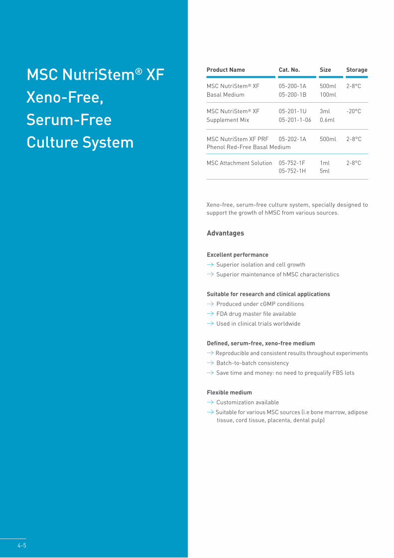
MSC NutriStem® XF Xeno-Free, Serum-Free Culture System
Xeno-free, serum-free culture system, specially designed to support the growth of hMSC from various sources.
Product Name Cat. No. Size Storage
MSC NutriStem® XF 05-200-1A 500ml 2-8°CBasal Medium 05-200-1B 100ml
MSC NutriStem® XF 05-201-1U 3ml -20°CSupplement Mix 05-201-1-06 0.6ml
MSC NutriStem XF PRF 05-202-1A 500ml 2-8°CPhenol Red-Free Basal Medium
MSC Attachment Solution 05-752-1F 1ml 2-8°C 05-752-1H 5ml
Advantages
Excellent performance
Superior isolation and cell growth
Superior maintenance of hMSC characteristics
Suitable for research and clinical applications
Produced under cGMP conditions
FDA drug master file available
Used in clinical trials worldwide
Defined, serum-free, xeno-free medium
Reproducible and consistent results throughout experiments
Batch-to-batch consistency
Save time and money: no need to prequalify FBS lots
Flexible medium
Customization available
Suitable for various MSC sources (i.e bone marrow, adipose tissue, cord tissue, placenta, dental pulp)
Isolation
hMSC from various sources (hMSC-PL, hMSC-AT, hMSC-WJ, hMSC-BM) can be efficiently isolated using MSC NutriStem® XF on pre-coated dishes. Addition of 2-2.5% human AB serum may be required for certain tissues.Using MSC NutriStem® XF for isolation of hMSC enhances purity of MSC populations in earlier passages and increases the number of hMSC in comparison to FBS-containing medium.
hMSC-PL
Figure 1: hMSC were isolated from frozen crude placenta under SF, XF culture conditions (MSC NutriStem® XF on pre-coated plates with MSC Attachment Solution, without supplementation of human AB serum) and in medium containing FBS. Representative images (x40) taken 11 days post initial isolation (P0).
MSC NutriStem® XF Serum-containg medium
Figure 2: Comparison of hMSC-PL isolation from crude placenta 17 days post initial seeding (P0) in each medium. Quantity of viable cells, measured by trypan blue exclusion assay.
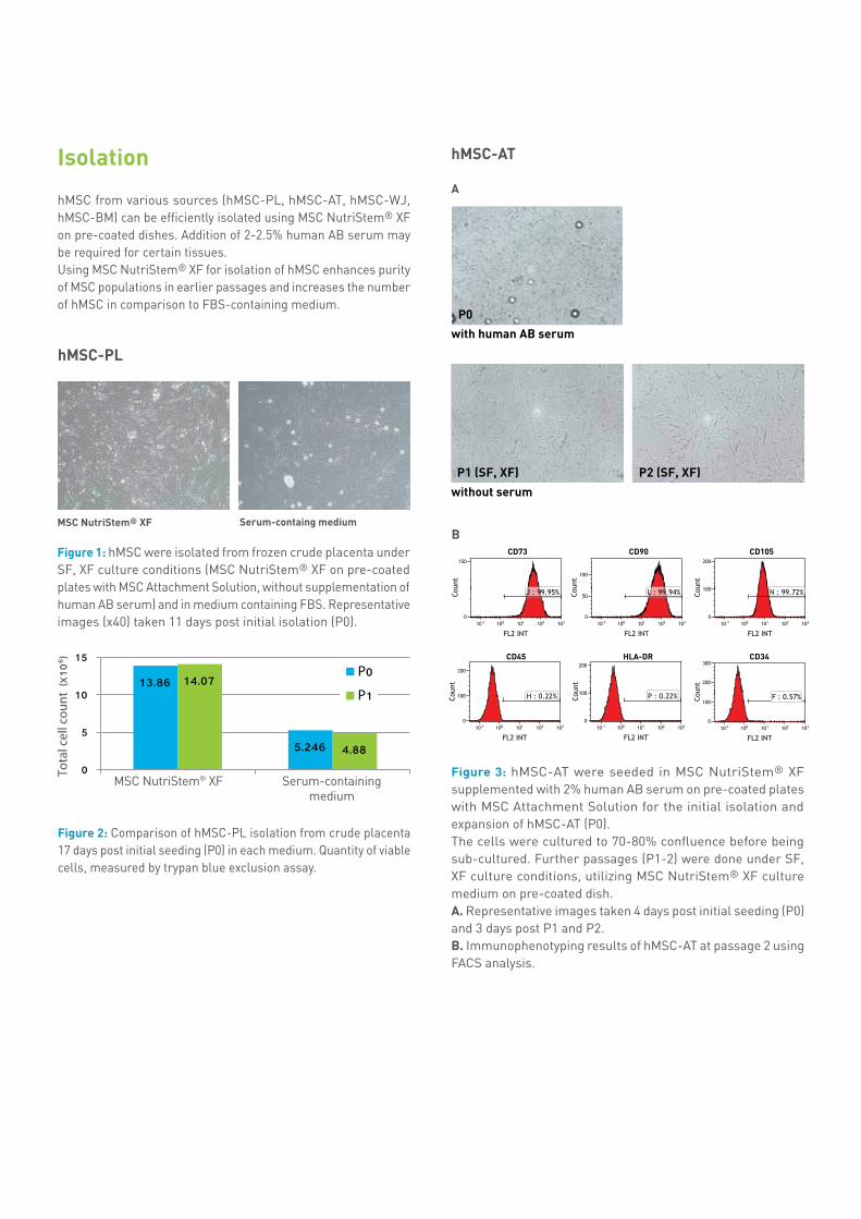
hMSC-AT
A
with human AB serum
without serum
Figure 3: hMSC-AT were seeded in MSC NutriStem® XF supplemented with 2% human AB serum on pre-coated plates with MSC Attachment Solution for the initial isolation and expansion of hMSC-AT (P0). The cells were cultured to 70-80% confluence before being sub-cultured. Further passages (P1-2) were done under SF, XF culture conditions, utilizing MSC NutriStem® XF culture medium on pre-coated dish. A. Representative images taken 4 days post initial seeding (P0) and 3 days post P1 and P2. B. Immunophenotyping results of hMSC-AT at passage 2 using FACS analysis.
CD73
CD45
CD90
HLA-DR
CD105
CD34
B
MSC NutriStem® XF Serum-containing medium

6-7
B Day 2 post initial isolation
MSC NutriStem® XF(supplemented with 2% human AB serum)
Weiss medium (composed of 2% FBS)
Representative images
Day 7 post initial isolation
Total cell countLive
395,000
295,000
15,000
75,000
Dead
A
MSC NutriStem® XF +2% human AB serum
Weiss medium (composed of 2% FBS)
cord1 cord2 cord3 cord4
Viab
le is
olat
ed c
ells
(x10
6 )
hMSC-WJ
Figure 4: hMSC were initially isolated from 4 independent human umbilical cords utilizing MSC NutriStem® XF supplemented with 2% human AB serum on pre-coated plates with MSC Attachment Solution in comparison to serum-containing medium. A. Comparing the amount of viable cells – passage 0. Cell count was measured by trypan blue exclusion assay. B. Representative images (x40) of cord 4 taken on day 2 post initial isolation in each medium, and cell count results of day 7 post initial isolation.
A
B
MSC NutriStem® XF Serum-containing medium
MSC NutriStem® XF Serum-containing medium
90.8%
CD90
CD45+CD34+CD14
90.9%
98.2%
10.3%
CD73
CD90
CD105
CD45+CD34+CD14
98.9%
99%
99.2%
1.5%
Figure 5: Comparison of hMSC-BM isolation from fresh BM utilizing MSC NutriStem® XF and serum-containing medium (11-day assay) A. Cell count was measured by trypan blue exclusion assay. B. Immunophenotype using FACS analysis.
hMSC-BM
Key References
• L. Berger et al. Tumor Specific Recruitment and Reprogramming of Mesenchymal Stem Cells in Tumor igenesis. STEM CELLS Volume 34, Issue 4, Version of Record online: 31 DEC 2015
• Cai, Zhen, et al. Chondrogenesis of Human Adipose-Derived Stem Cells by In Vivo Co-graft with Auricular Chondrocytes from Microtia. Aesthetic plastic surgery 39.3 (2015): 431-439.
• S.H. Mei, et al. Isolation and large-scale expansion of bone marrow-derived mesenchymal stem cells with serum-free media under GMP-compliance. Cytotherapy, Volume 16, Issue 4, Supplement , Page S111, April 2014
• Y. Lopez, M. Weiss, et al. Identification of Optimal Conditions for Generating MSCs for Preclinical Testing: Comparison of Three Commercial Serum-Free Media and Low-Serum Growth Medium. From 18th ISCT Annual Meeting, Seattle, USA, 2012.

Expansion
Figure 7: Expansion of hMSC-BM in MSC NutriStem® XF and FBS-containing medium. Initial seeding was 5000 cells/cm2 for each of the tested media (day 0). Images were taken at day 3 post seeding.
Serum-containing medium MSC NutriStem® XF
Figure 6: hMSC-BM were cultured in MSC NutriStem® XF in comparison to commercial SF and serum-containing media. Initial seeding was 5000 cells/cm2 for each of the tested media (day 0). Cells were counted daily by trypan blue exclusion assay.
hMSC-BM
hMSC-AT
Superior proliferation of hMSC
hMSC cultured in MSC NutriStem® XF exhibit higher proliferation rate and long term growth in comparison to competitors’ media.
x40
x40
cells
/cm
2 (x 1
03)
Days post seeding
MSC NutriStem® XF
Commercial SF medium
Serum-containing medium
MSC NutriStem® XF mediumSerum-containing medium
Figure 9: Expansion of hMSC-AT in MSC NutriStem® XF medium in comparison to serum-containing medium. Initial seeding was 6000 cells/cm2 for each of the tested media (day 0). Images were taken 3 days post initial culture.
(x100) (x100)
Figure 8: Expansion of hMSC-AT in MSC NutriStem® XF and commercially available XF, SF, and serum-containing media.Cells were cultured in plates, pre-coated with MSC Attachment Solution. Initial seeding was 5000 cells/cm2 for each of the tested media (day 0). Cells were counted at day 3 in each passage.
MSC NutriStem® XF
Commercial XF medium
Commercial SF medium
Commercial serum-containing medium
P0 P1 P3P2 P4
Figure 10: hMSC-WJ from 9 different donors expanded for 4 passages in MSC NutriStem® XF in comparison to serum-containing medium and commercial SF and XF media. Cell proliferation was assessed by cell count using a trypan blue exclusion assay.
Cel
l Num
ber
Passage
MSC NutriStem® XF Serum-containing medium (Weiss)Commercial SF mediumCommercial XF medium
hMSC-WJ
8-9
normal morphology
Typical fibroblast-like cells morphology was obtained when using MSC NutriStem® XF.
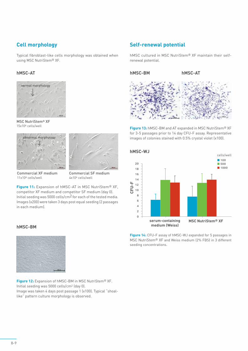
Cell morphology
hMSC-AT
hMSC-BM
Figure 11: Expansion of hMSC-AT in MSC NutriStem® XF, competitor XF medium and competitor SF medium (day 0). Initial seeding was 5000 cells/cm2 for each of the tested media. Images (x200) were taken 3 days post equal seeding (2 passages in each medium).
15x104 cells/wellMSC NutriStem® XF
11x104 cells/wellCommercial XF medium
4x10⁴ cells/wellCommercial SF medium
abnormal morphology
Figure 12: Expansion of hMSC-BM in MSC NutriStem® XF. Initial seeding was 5000 cells/cm2 (day 0). Image was taken 4 days post passage 1 (x100). Typical “shoal-like” pattern culture morphology is observed.
Figure 14: CFU-F assay of hMSC-WJ expanded for 5 passages in MSC NutriStem® XF and Weiss medium (2% FBS) in 3 different seeding concentrations.
Self-renewal potential
hMSC cultured in MSC NutriStem® XF maintain their self-renewal potential.
Figure 13: hMSC-BM and AT expanded in MSC NutriStem® XF for 3-5 passages prior to 14 day CFU-F assay. Representative images of colonies stained with 0.5% crystal violet (x100).
hMSC-BM hMSC-AT
MSC NutriStem® XFserum-containing medium (Weiss)
CFU
-F
cells/wellhMSC-WJ
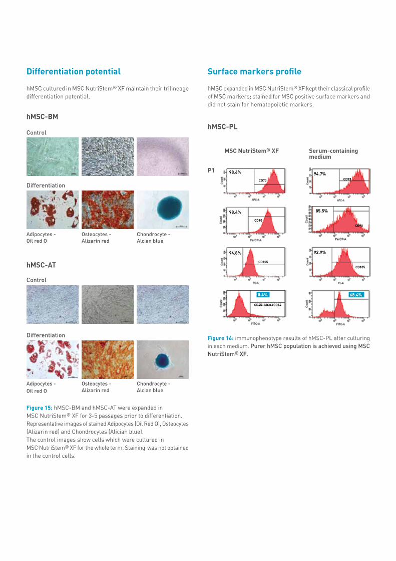
hMSC cultured in MSC NutriStem® XF maintain their trilineage differentiation potential.
Osteocytes - Alizarin red
Osteocytes - Alizarin red
Adipocytes - Oil red O
Adipocytes - Oil red O
Chondrocyte - Alcian blue
Chondrocyte - Alcian blue
Figure 15: hMSC-BM and hMSC-AT were expanded inMSC NutriStem® XF for 3-5 passages prior to differentiation. Representative images of stained Adipocytes (Oil Red O), Osteocytes (Alizarin red) and Chondrocytes (Alician blue). The control images show cells which were cultured in MSC NutriStem® XF for the whole term. Staining was not obtained in the control cells.
Differentiation potential
Control
Control
Differentiation
Differentiation
hMSC-BM
hMSC-AT
MSC NutriStem® XF
hMSC-PL
Surface markers profile
Figure 16: immunophenotype results of hMSC-PL after culturing in each medium. Purer hMSC population is achieved using MSC NutriStem® XF.
hMSC expanded in MSC NutriStem® XF kept their classical profile of MSC markers; stained for MSC positive surface markers and did not stain for hematopoietic markers.
CD45+CD34+CD14
CD73
CD90
CD105
98.6%
98.4%
94.8%
8.4%
CD105
CD73
CD90
94.7%
92.9%
85.5%
P1
68.4%
Serum-containing medium
10-11
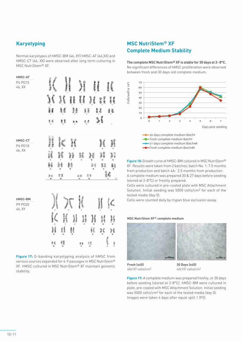
Normal karyotypes of hMSC-BM (46, XY) hMSC-AT (46,XX) and hMSC-CT (46, XX) were observed after long term culturing in MSC NutriStem® XF.
Karyotyping
Figure 17: G-banding karyotyping analysis of hMSC from various sources expanded for 4-9 passages in MSC NutriStem® XF. hMSC cultured in MSC NutriStem® XF maintain genomic stability.
hMSC-AT
P4 PD1546, XX
hMSC-CT
P6 PD1846, XX
hMSC-BM
P9 PD2046, XY
Figure 19: A complete medium was prepared freshly, or 30 days before seeding (stored at 2-8°C). hMSC-BM were cultured in plate, pre-coated with MSC Attachment Solution. Initial seeding was 5000 cells/cm2 for each of the tested media (day 0). Images were taken 4 days after equal split 1 (P2).
Fresh ( x40 )40x103 cells/cm2
MSC NutriStem XF® complete medium
30 Days ( x40 ) 42x103 cells/cm2
The complete MSC NutriStem® XF is stable for 30 days at 2- 8°C. No significant differences of hMSC proliferation were observed between fresh and 30 days old complete medium.
Figure 18: Growth curve of hMSC-BM cultured in MSC NutriStem® XF. Results were taken from 2 batches: batch No. 1: 7.5 months from production and batch 4A: 2.5 months from production. A complete medium was prepared 30 & 27 days before seeding (stored at 2-8°C) or freshly prepared. Cells were cultured in pre-coated plate with MSC Attachment Solution. Initial seeding was 5000 cells/cm2 for each of the tested media (day 0). Cells were counted daily by trypan blue exclusion assay.
MSC NutriStem® XF Complete Medium Stability
Key References
• C. Elseberg et al. The Challenge of Human Mesenchymal Stromal Cell Expansion: Current and Prospective Answers. New Insights into Cell Culture Technology, Dr. Sivakumar Joghi Thatha Gowder (Ed.), InTech. 2017
• S. Bobis-Wozowicz et al. Diverse impact of xeno-free conditions on biological and regenerative properties of hUC-MSCs and their extracellular vesicles. Journal of Molecular Medicine, 2016
• K.Y. Tan et al. Serum-free media formulations are cell line–specific and require optimization for microcarrier culture. Cytotherapy, 2015 • M.Pokrywczynska et al., Transdifferentiation of Bone Marrow Mesenchymal Stem Cells into the Islet-Like Cells: the Role of Extracellular Matrix Proteins.
Archivum Immunologiae et Therapiae Experimentalis, May 2015 • U Pivorait et. al., Exosomes from Human Dental Pulp Stem Cells Suppress Carrageenan-Induced Acute Inflammation in Mice. Inflammation, April 2015• Mira Genser-Nir et al. Toward a serum-free, xeno-free culture system for optimal growth and expansion of hMSC suited to therapeutic applications. From
23rd European Society for Animal Cell Technology (ESACT), 2013• McVey, Mark John, et al. Microparticles as biomarkers of lung disease-enumeration in biological fluids using lipid bilayer microspheres. American Journal
of Physiology-Lung Cellular and Molecular Physiology (2016): ajplung-00369.
Clinical Applications• D. Boruczkowski et al.Third-party Wharton’s jelly mesenchymal stem cells for treatment of steroid-resistant acute and chronic graft-versus-host
disease: a report of 10 cases. Turkish Journal of Biology, 40: 493-500, 2016• Jianxia H. et al. Long term effect and safety of Wharton’s jelly-derived mesenchymal stem cells on type 2 diabetes. Experimental and Therapeutic
Medicine, Volume 12 Issue 3, 2016
12-13
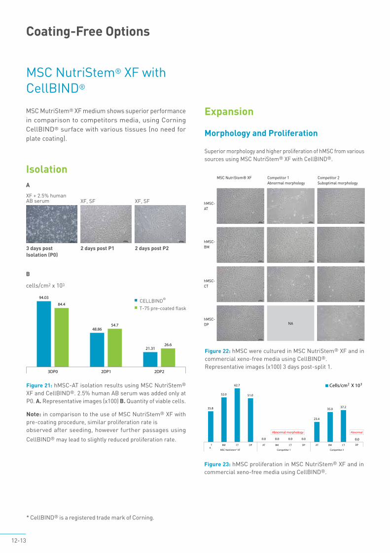
MSC MutriStem® XF medium shows superior performance in comparison to competitors media, using Corning CellBIND® surface with various tissues (no need for plate coating).
Figure 21: hMSC-AT isolation results using MSC NutriStem® XF and CellBIND®. 2.5% human AB serum was added only at P0. A. Representative images (x100) B. Quantity of viable cells.
Note: in comparison to the use of MSC NutriStem® XF with pre-coating procedure, similar proliferation rate isobserved after seeding, however further passages using
CellBIND® may lead to slightly reduced proliferation rate.
* CellBIND® is a registered trade mark of Corning.
MSC NutriStem® XF with CellBIND®
Coating-Free Options
Isolation
Expansion
Morphology and Proliferation
Superior morphology and higher proliferation of hMSC from various sources using MSC NutriStem® XF with CellBIND®.
2 days post P1
XF, SF
3 days post Isolation (P0)
XF + 2.5% human AB serum
2 days post P2
XF, SF
CELLBIND®
T-75 pre-coated flask
Figure 22: hMSC were cultured in MSC NutriStem® XF and in commercial xeno-free media using CellBIND®. Representative images (x100) 3 days post-split 1.
MSC NutriStem® XF Competitor 1Abnormal morphology
Competitor 2Suboptimal morphology
hMSC-AT
hMSC-CT
hMSC-BM
hMSC-DP
hMSC-DPhMSC-DPNA
Figure 23: hMSC proliferation in MSC NutriStem® XF and in commercial xeno-free media using CellBIND®.
A
B
cells/cm2 x 103
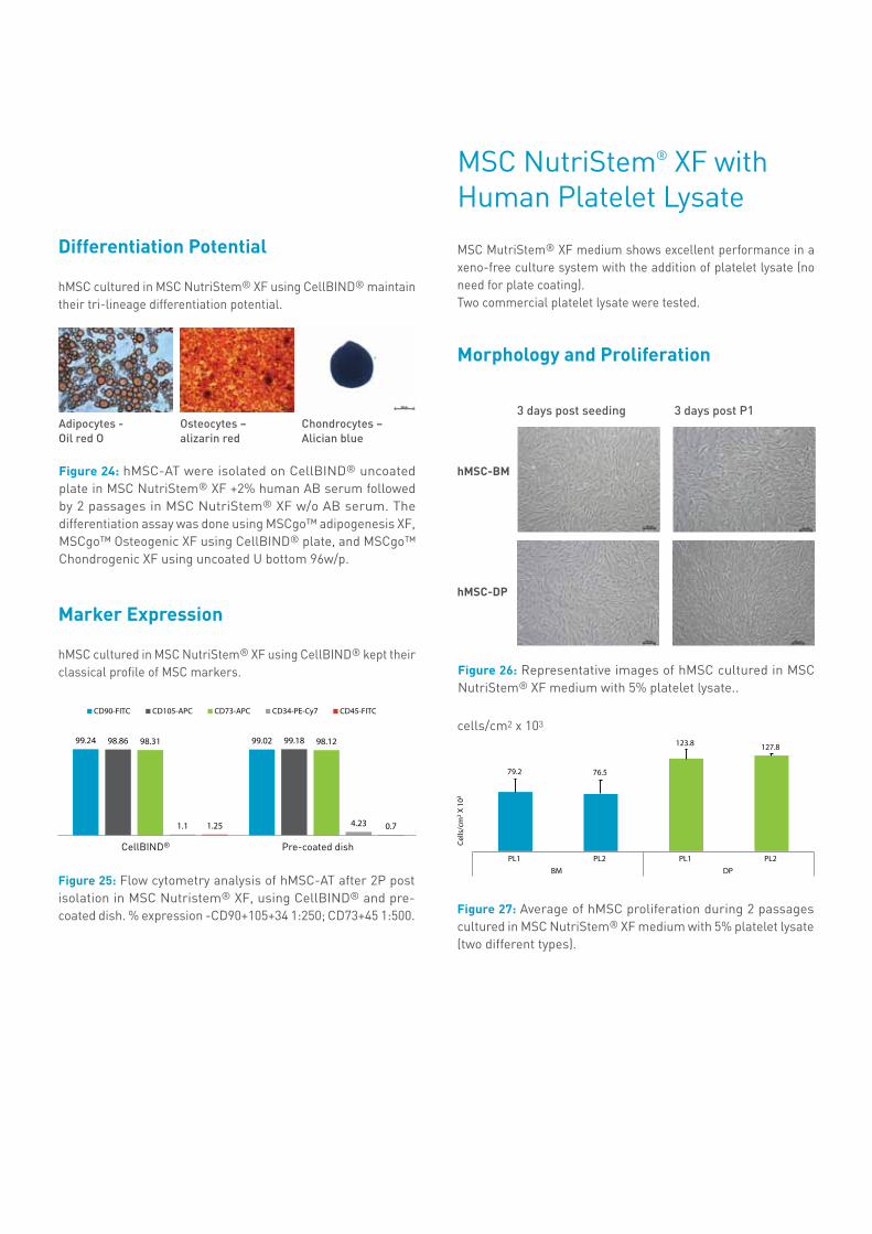
Figure 25: Flow cytometry analysis of hMSC-AT after 2P post isolation in MSC Nutristem® XF, using CellBIND® and pre-coated dish. % expression -CD90+105+34 1:250; CD73+45 1:500.
CellBIND® Pre-coated dish
Figure 24: hMSC-AT were isolated on CellBIND® uncoated plate in MSC NutriStem® XF +2% human AB serum followed by 2 passages in MSC NutriStem® XF w/o AB serum. The differentiation assay was done using MSCgo™ adipogenesis XF, MSCgo™ Osteogenic XF using CellBIND® plate, and MSCgo™ Chondrogenic XF using uncoated U bottom 96w/p.
Differentiation Potential
hMSC cultured in MSC NutriStem® XF using CellBIND® maintain their tri-lineage differentiation potential.
Marker Expression
hMSC cultured in MSC NutriStem® XF using CellBIND® kept their classical profile of MSC markers.
Osteocytes – alizarin red
Adipocytes - Oil red O
Chondrocytes – Alician blue
Figure 26: Representative images of hMSC cultured in MSC NutriStem® XF medium with 5% platelet lysate..
MSC NutriStem® XF with Human Platelet Lysate
Morphology and Proliferation
MSC MutriStem® XF medium shows excellent performance in a xeno-free culture system with the addition of platelet lysate (no need for plate coating).Two commercial platelet lysate were tested.
Figure 27: Average of hMSC proliferation during 2 passages cultured in MSC NutriStem® XF medium with 5% platelet lysate (two different types).
hMSC-BM
hMSC-DP
3 days post P13 days post seeding
cells/cm2 x 103
14-15
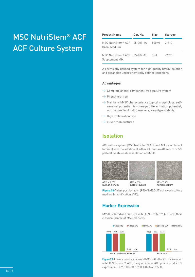
MSC NutriStem® ACF ACF Culture System
Product Name Cat. No. Size Storage
MSC NutriStem® ACF 05-203-1A 500ml 2-8°CBasal Medium
MSC NutriStem® ACF 05-204-1U 3ml -20°CSupplement Mix
Advantages
Complete animal component-free culture system
Phenol red-free
Maintains hMSC characteristics (typical morphology, self-renewal potential, tri-lineage differentiation potential, normal profile of hMSC markers, karyotype stability)
High proliferation rate
cGMP-manufactured
Figure 28: 3 days post Isolation (P0) of hMSC-AT using each culture medium (magnification x100).
Figure 29: Flow cytometry analysis of hMSC-AT after 2P post isolation in MSC Nutristem® ACF, using a Laminin ACF precoated dish. % expression -CD90+105+34 1:250; CD73+45 1:500.
ACF + 5% platelet lysate
ACF + 2.5% human serum
XF + 2.5% human serum
Isolation
ACF culture system (MSC NutriStem® ACF and ACF recombinant laminin) with the addition of either 2% human AB serum or 5% platelet lysate enables isolation of hMSC.
A chemically defined system for high quality hMSC isolation and expansion under chemically defined conditions.
Marker Expression
hMSC isolated and cultured in MSC NutriStem® ACF kept theirclassical profile of MSC markers.
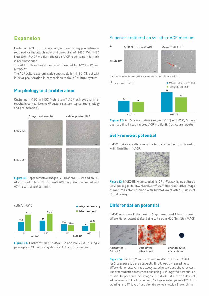
Figure 30: Representative images (x100) of hMSC-BM and hMSC-AT cultured in MSC NutriStem® ACF on plate pre-coated with ACF recombinant laminin.
Figure 33: hMSC-BM were seeded for CFU-F assay being cultured for 2 passages in MSC NutriStem® ACF. Representative image of matured colony stained with Crystal violet after 13 days of CFU-F assay.
* Arrow represents precipitants observed in the culture medium.
Figure 34: hMSC-BM were cultured in MSC NutriStem® ACFfor 2 passages (3 days post-split 1) followed by reseeding todifferentiation assays (into osteocytes, adipocytes and chondrocytes).The differentiation assay was done using BI MSCgo™ differentiationmedia. Representative images of hMSC-BM after 17 days of adipogenesis (Oil red O staining), 14 days of osteogenesis (2% ARS staining) and 17 days of and chondrogenesis (Alcian Blue staining).
Self-renewal potential
hMSC maintain self-renewal potential after being cultured in MSC NutriStem® ACF.
Differentiation potential
hMSC maintain Osteogenic, Adipogenic and Chondrogenic differentiation potential after being cultured in MSC NutriStem® ACF.
Superior proliferation vs. other ACF medium
hMSC-BM
hMSC-AT
4 days post-split 12 days post seeding
Osteocytes – alizarin red
Adipocytes - Oil red O
Chondrocytes – Alician blue
Expansion
Under an ACF culture system, a pre-coating procedure is required for the attachment and spreading of hMSC. With MSCNutriStem® ACF medium the use of ACF recombinant laminin is recommended.The ACF culture system is recommended for hMSC-BM and hMSC-AT. The ACF culture system is also applicable for hMSC-CT, but with inferior proliferation in comparison to the XF culture system.
Morphology and proliferation
Culturing hMSC in MSC NutriStem® ACF achieved similar results in comparison to XF culture system (typical morphology and proliferation).
Figure 31: Proliferation of hMSC-BM and hMSC-AT during 2 passages in XF culture system vs. ACF culture system.
cells/cm2x103
hMSC-BM
MesenCult ACFMSC NutriStem® ACFA
B
Figure 32: A. Representative images (x100) of hMSC, 3 days post seeding in each tested ACF media. B. Cell count results
MSC NutriStem® ACFMesenCult ACF
cells/cm2x103

Figure 36: hMSC-BM were analyzed for karyotype after 3 passages in MSC NutriStem® ACF medium.
Figure 37: A complete medium was prepared freshly, or 30 days before seeding (stored at 2-8°C). hMSC-BM were cultured in pre-coated plates with ACF recombinant Laminin. A. representative images 3 days post seeding (x100). B. Average proliferation during 2 passages.Similar culture morphology, confluence and cells count results were observed.
Karyotyping
Normal karyotype of hMSC-BM (46, XY) was observed after culturing in MSC NutriStem® ACF.
MSC NutriStem® ACF complete medium stability
The complete MSC NutriStem® ACF medium is stable for 30 days (2-8°C).
Fresh 30 days
16-17
Figure 35: After 3 passages in MSC NutriStem® ACF medium hMSC were harvested and labeled with fluorochrome conjugated antibodies against CD90, CD105, CD73, CD34 and CD45 and analyzed by FACS. A. Histograms results (markers expression in red and IgG expression in black) B. Profile markers expression results (% of positive marker expression- % of specific IgG positive expression).
Surface markers profile
hMSC expanded in MSC NutriStem® ACF kept their classical profile of MSC markers.
A
A
BB
cells/cm2x103
Dissociation
Recombinant Trypsin Solution is an ACF cell dissociation solution, designed as an alternative to porcine/bovine trypsin.The addition of EDTA usually accelerates the dissociation phase.The solutions do not contain any chymotrypsin, carboxypeptidase A, or other protease contaminant.Recombinant Trypsin Solution formulations were developed for efficient dissociation of adherent cell types from surfaces and tissues and were optimized for sensitive cells, such as hMSC.
Advantages:• Ready-to-use• Non-animal or human origin• Optimized for hMSC (from a variety of sources), cultured in both
SF and serum-containing systems• Free from undesirable proteases such as carboxypeptidase A
and chymotrypsin• Eliminates contaminating activities found in bulk production
of enzymes• Storage: room temperature
Neutralization of Recombinant Trypsin is achieved with MSC NutriStem® XF or Soybean Trypsin Inhibitor (SBTI) Cat. No.: 03-048-1.The use of recombinant trypsin, rather than crude trypsin, is often essential for successful, long term growth of cells under SF culture conditions.
Figure 38: Recovery of hMSC-BM after dissociation with both Recombinant Trypsin Solution and the common Trypsin EDTA Solution (porcine) following re-seeding in MSC NutriStem® XF on pre-coated plates. Representative images were taken on day 5 post-dissociation (x100).
Recombinant Trypsin Solution Crude Trypsin EDTA Solution
Product Name Cat. No. Size Storage
Recombinant Trypsin 03-078-1A 500ml RTSolution without EDTA 03-078-1B 100ml
Recombinant Trypsin 03-079-1A 500ml RTSolution with EDTA 03-079-1B 100ml
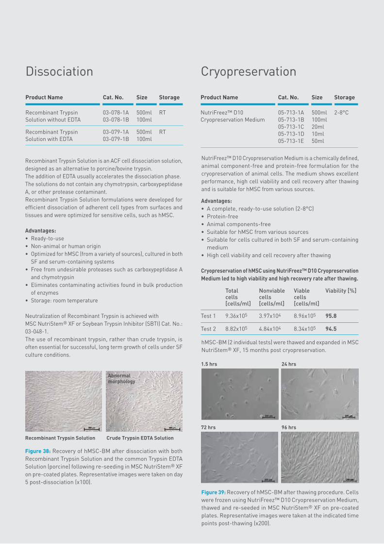
Cryopreservation
NutriFreez™ D10 Cryopreservation Medium is a chemically defined, animal component-free and protein-free formulation for the cryopreservation of animal cells. The medium shows excellent performance, high cell viability and cell recovery after thawing and is suitable for hMSC from various sources.
hMSC-BM (2 individual tests) were thawed and expanded in MSC NutriStem® XF, 15 months post cryopreservation.
Figure 39: Recovery of hMSC-BM after thawing procedure. Cells were frozen using NutriFreez™ D10 Cryopreservation Medium, thawed and re-seeded in MSC NutriStem® XF on pre-coated plates. Representative images were taken at the indicated time points post-thawing (x200).
Advantages:• A complete, ready-to-use solution (2-8°C)• Protein-free• Animal components-free• Suitable for hMSC from various sources• Suitable for cells cultured in both SF and serum-containing
medium• High cell viability and cell recovery after thawing
Cryopreservation of hMSC using NutriFreez™ D10 Cryopreservation Medium led to high viability and high recovery rate after thawing.
Total Nonviable Viable Viability [%] cells cells cells [cells/ml] [cells/ml] [cells/ml] Test 1 9.36x10⁵ 3.97x10⁴ 8.96x10⁵ 95.8
Test 2 8.82x10⁵ 4.84x10⁴ 8.34x10⁵ 94.5
1.5 hrs 24 hrs
72 hrs 96 hrs
Product Name Cat. No. Size Storage
NutriFreez™ D10 05-713-1A 500ml 2-8°CCryopreservation Medium 05-713-1B 100ml 05-713-1C 20ml 05-713-1D 10ml 05-713-1E 50ml
Abnormal morphology
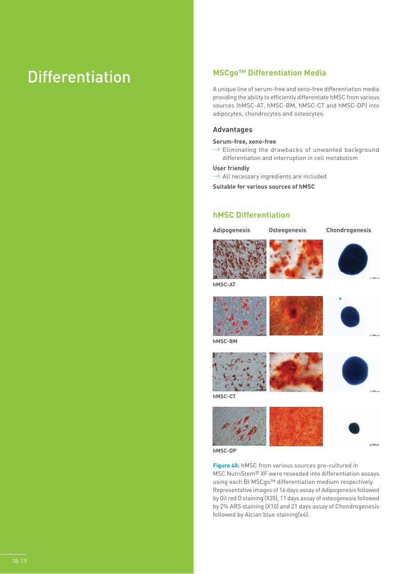
DifferentiationA unique line of serum-free and xeno-free differentiation media providing the ability to efficiently differentiate hMSC from various sources (hMSC-AT, hMSC-BM, hMSC-CT and hMSC-DP) into adipocytes, chondrocytes and osteocytes.
Advantages
Serum-free, xeno-free Eliminating the drawbacks of unwanted background differentiation and interruption in cell metabolism
User friendly All necessary ingredients are included
Suitable for various sources of hMSC
Figure 40: hMSC from various sources pre-cultured in MSC NutriStem® XF were reseeded into differentiation assays using each BI MSCgo™ differentiation medium respectively. Representative images of 16 days assay of Adipogenesis followed by Oil red O staining (X20), 11 days assay of osteogenesis followed by 2% ARS staining (X10) and 21 days assay of Chondrogenesis followed by Alcian blue staining(x4).
hMSC-AT
hMSC-BM
hMSC-CT
hMSC-DP
Adipogenesis Osteogenesis Chondrogenesis
18-19
MSCgo™ Differentiation Media
hMSC Differentiation
B
A
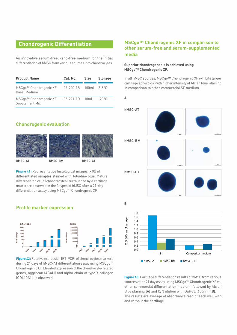
MSCgo™ Chondrogenic XF in comparison to other serum-free and serum-supplemented media
In all hMSC sources, MSCgo™ Chondrogenic XF exhibits larger cartilage spheroids with higher intensity of Alcian blue staining in comparison to other commercial SF medium.
Superior chondrogenesis is achieved using MSCgo™ Chondrogenic XF.
Figure 43: Cartilage differentiation results of hMSC from various sources after 21 day assay using MSCgo™ Chondrogenic XF vs. other commercial differentiation medium, followed by Alcian blue staining (A) and O/N elution with GuHCL (600nm) (B). The results are average of absorbance read of each well with and without the cartilage.
hMSC-AT
hMSC-BM
hMSC-CT
Chondrogenic evaluation
An innovative serum-free, xeno-free medium for the initial differentiation of hMSC from various sources into chondrocytes.
Chondrogenic Differentiation
Product Name Cat. No. Size Storage
MSCgo™ Chondrogenic XF 05-220-1B 100ml 2-8°CBasal Medium MSCgo™ Chondrogenic XF 05-221-1D 10ml -20°CSupplement Mix
Profile marker expression
Figure 42: Relative expression (RT-PCR) of chondrocytes markers during 21 days of hMSC-AT differentiation assay using MSCgo™ Chondrogenic XF. Elevated expression of the chondrocyte-related genes, aggrecan (ACAN) and alpha chain of type X collagen (COL10A1), is observed.
Figure 41: Representative histological images (x40) of differentiated samples stained with Toluidine blue. Mature differentiated cells (chondrocytes) surrounded by a cartilage matrix are observed in the 3 types of hMSC after a 21-day differentiation assay using MSCgo™ Chondrogenic XF.
hMSC-AT hMSC-BM hMSC-CT
20-21
Adipogenic Differentiation
Figure 45: Elevate expression of the adipocyte-related genes, (FABP4) and alpha chain of type X collagen (PPARG), is observed during 14 days adipogenesis of hMSC using MSCgo™ Adipogenic XF.
Figure 44: Typical expression of FABP4 is observed post 11 days adipogenesis of hMSC using MSCgo™ Adipogenic XF.
hMSC-BM
Light FABP4 Merged (Dapi)
An Innovative serum-free, xeno-free medium for the differentiation of hMSC into adipocytes.
MSCgo™ Adipogenic XF in comparison to other serum-free and serum-supplemented media
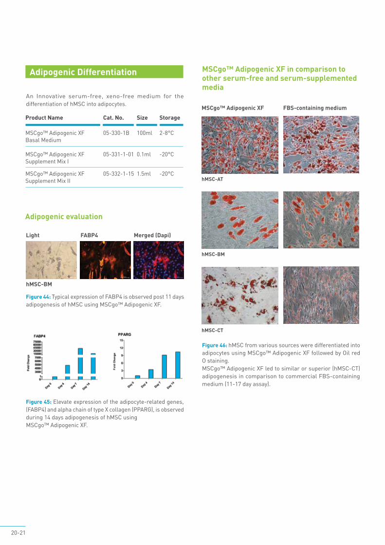
Adipogenic evaluation
hMSC-AT
hMSC-BM
hMSC-CT
MSCgo™ Adipogenic XF FBS-containing medium
Figure 46: hMSC from various sources were differentiated into adipocytes using MSCgo™ Adipogenic XF followed by Oil red O staining.MSCgo™ Adipogenic XF led to similar or superior (hMSC-CT) adipogenesis in comparison to commercial FBS-containing medium (11-17 day assay).
Product Name Cat. No. Size Storage
MSCgo™ Adipogenic XF 05-330-1B 100ml 2-8°CBasal Medium MSCgo™ Adipogenic XF 05-331-1-01 0.1ml -20°CSupplement Mix I MSCgo™ Adipogenic XF 05-332-1-15 1.5ml -20°CSupplement Mix II
MSCgo™ rapid Osteogenic XF will lead to faster osteogenesis (less than 10 days) in comparison to the MSCgo™ Osteogenic XF (10-21days).
Complete, ready-to-use, xeno-free and serum-free media for the differentiation of hMSC from various sources into osteocytes.
Osteogenic Differentiation
Figure 47: Calcified nodules observed using both hMSC-BM and hMSC-AT after a 10 day MSC differentiation assay using MSCgo™ Osteogenic XF.
Osteogenic evaluation
hMSC-BM hMSC-AT
Figure 48: Relative expression (RT-PCR) of osteocyte markers after 10 days of osteogenesis of hMSC using MSCgo™ Osteogenic XF. Osteogenic markers were upregulated whereas an undifferentiated hMSC marker (CD-105) was downregulated. BGLAP represents a maturation state of osteogenesis.
Product Name Cat. No. Size Storage
MSCgo™ Osteogenic XF 05-440-1B 100ml 2-8°C
MSCgo™ rapid 05-442-1B 100ml 2-8°COsteogenic XF
Profile marker expression
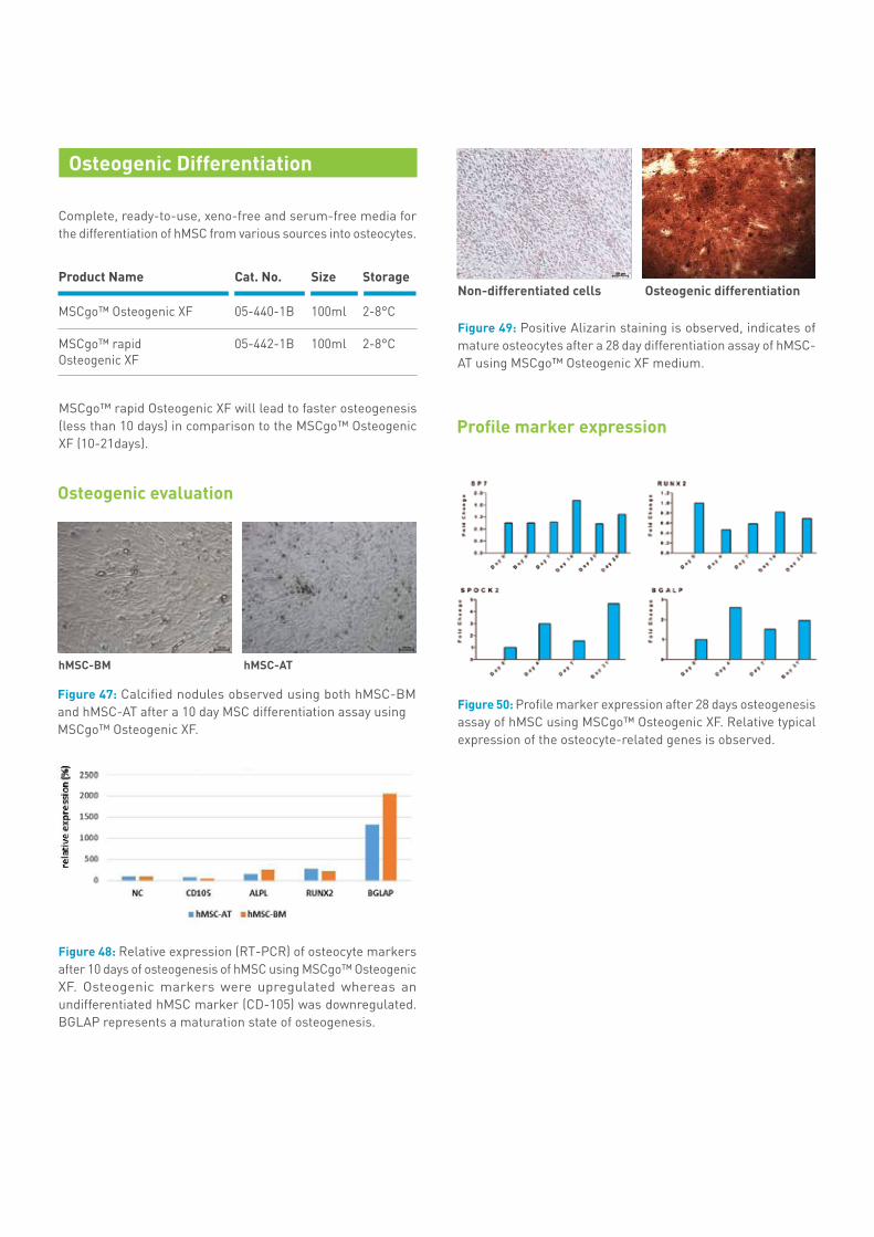
Figure 49: Positive Alizarin staining is observed, indicates of mature osteocytes after a 28 day differentiation assay of hMSC-AT using MSCgo™ Osteogenic XF medium.
Figure 50: Profile marker expression after 28 days osteogenesis assay of hMSC using MSCgo™ Osteogenic XF. Relative typical expression of the osteocyte-related genes is observed.
Osteogenic differentiationNon-differentiated cells
22-23
Key References
• L. Leber et. al. Microcarrier choice and bead-to-bead transfer for human mesenchymal stem cells in serum-containing and chemically defined media, Process BiochemistryVolume 59, Part B, August 2017, Pages 255-265
• L. Pu et. al. Compared to the amniotic membrane, Wharton’s jelly may be a more suitable source of mesenchymal stem cells for cardiovascular tissue engineering and clinical regeneration, Stem Cell Research & Therapy20178:72
• M. Meng et al., Umbilical cord mesenchymal stem cell transplantation in the treatment of multiple sclerosis. American Journal of Translational Research, 2018;10(1):212-223
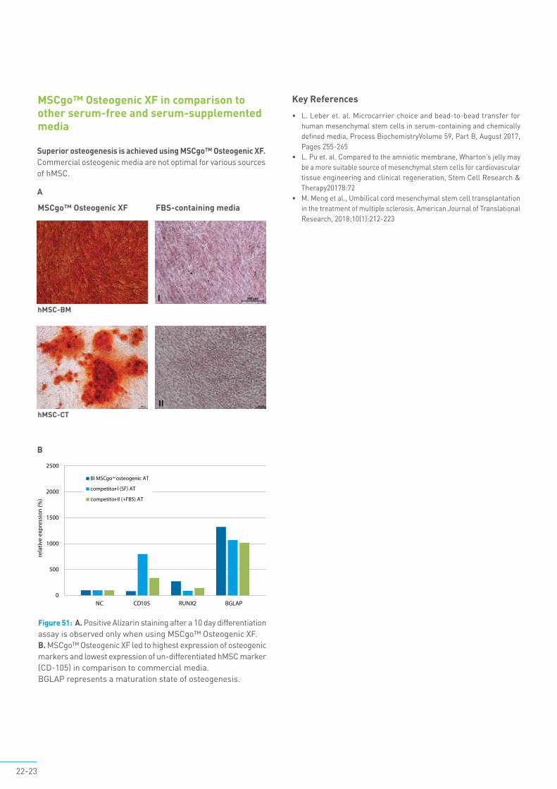
Figure 51: A. Positive Alizarin staining after a 10 day differentiation assay is observed only when using MSCgo™ Osteogenic XF.B. MSCgo™ Osteogenic XF led to highest expression of osteogenic markers and lowest expression of un-differentiated hMSC marker (CD-105) in comparison to commercial media. BGLAP represents a maturation state of osteogenesis.
MSCgo™ Osteogenic XF in comparison to other serum-free and serum-supplemented media
B
Superior osteogenesis is achieved using MSCgo™ Osteogenic XF.Commercial osteogenic media are not optimal for various sources of hMSC.
FBS-containing media
hMSC-BM
MSCgo™ Osteogenic XF
A
hMSC-CT
I
II
™
Ordering information
Product Name Cat. No. Size Storage
MSC NutriStem® XF Basal Medium 05-200-1A 500ml 2-8°C 05-200-1B 100ml
MSC NutriStem® XF Supplement Mix 05-201-1U 3ml -20°C 05-201-1-06 0.6ml MSC NutriStem® XF Phenol Red-Free 05-202-1A 500ml 2-8°C
MSC Attachment Solution 05-752-1F 1ml 2-8°C 05-752-1H 5ml 2-8°C
Recombinant Trypsin Solution 03-078-1A 500ml RT 03-078-1B 100ml
Recombinant Trypsin Solution with EDTA 03-079-1A 500ml RT 03-079-1B 100ml
NutriFreez™ D10 Cryopreservation Medium 05-713-1A 500ml 05-713-1B 100ml 05-713-1C 20ml 05-713-1D 10ml 2-8°C 05-713-1E 50ml
MSCgo™ Osteogenic XF 05-440-1A 500ml 2-8ºC
MSCgo™ Rapid Osteogenic XF 05-442-1A 500ml 2-8ºC 05-442-1B 100ml
MSCgo™ Chondrogenic XF 05-220-1A 500ml 2-8ºC
MSCgo™ Chondrogenic XF Supplement Mix 05-221-1D 10ml -20°C
MSCgo™ Adipogenic XF 05-330-1A 500ml 2-8ºC
MSCgo™ Adipogenic XF Supplement Mix I 05-331-1-01 0.1ml -20°C
MSCgo™ Adipogenic XF Supplement Mix II 05-332-1-15 1.5ml -20°C
Biological Industries Israel Beit Haemek Ltd.Kibbutz Beit Haemek 2511500, Israel T.+972.4.9960595 F.+972.4.9968896 Email: [email protected]
Biological Industries USA100 Sebethe Drive . Cromwell, CT, 06416-1073T. 860-316-2702 F. 860-269-0596E-mail: [email protected]
E44/2 02/18
Biological Industries (BI) has been committed for over 30 years to provide optimal and innovative solutions for cell culture practice. BI manufactures and supplies life science products to biopharmaceutical, academic and government research facilities, as well as to biopharma companies.
Our diverse portfolio of products and services includes all of the following:• Liquid and powdered cell culture media • Sterile sera (foetal bovine serum, newborn calf serum, donor horse, etc.) • Novel serum-free and animal component-free media and supplements • Products for stem cell research and cell-based therapies • Products for cytogenetics • Products for mycoplasma detection and treatment • Disinfectants • Products for molecular biology • custom formulations and contract manufacturing services
All BI’s products are manufactured via a quality management system ISO 9001:2015 and in regards of medical devices ISO 13485:2016. All aspects of the products life cycle fall under the QMS procedures. The set-up of clean zone and clean room facilities for manufacturing are following ISO 14644, whereas the production rooms are ISO 8, storage of sterile accessories ISO 7 and filling rooms ISO 5. Aseptic filling and validation is performed according to ISO 13408.
BI exports its products to more than 50 countries worldwide, via a network of exclusive distributors. Over the years we have established a reputation for fast delivery, and excellent technical support. From the outset, the policy of BI has been based on the need to maintain an active Research and Development program in all facets of company activities. The company has its own in-house R&D department, and in addition, maintains active contact with science-based companies and research institutions in Israel and abroad, including know-how agreements with several such institutions. These ongoing efforts have led to the introduction of a series of serum-free medium products, as well as many other products for cell culture and molecular biology.
dis
trib
utors in ov er50c o u n t r i e s
w w w . b i o i n d . c o m
ISO 134852016
ISO 90012015