Stem cells in Myopahies Farzad Fatehi. Stem Cells in Muscles.
56
Stem cells in Myopahies Farzad Fatehi
-
Upload
clare-holt -
Category
Documents
-
view
221 -
download
1
Transcript of Stem cells in Myopahies Farzad Fatehi. Stem Cells in Muscles.
Stem cells in MyopahiesStem cells in Myopahies
Farzad Fatehi
Stem Cells in Muscles
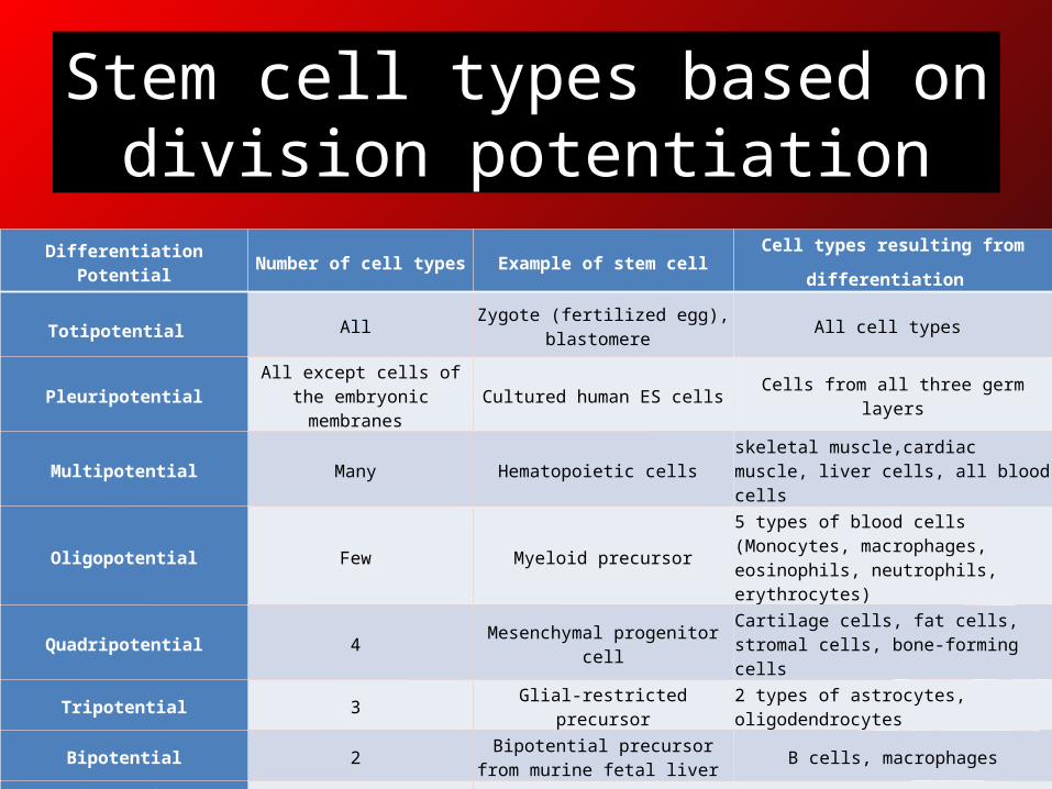
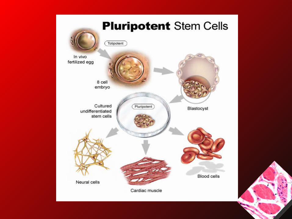
Stem cell types based on division potentiation
Differentiation Potential Number of cell types Example of stem cell Cell types resulting from differentiation
Totipotential All Zygote (fertilized egg), blastomere All cell types
PleuripotentialAll except cells of the
embryonic membranes Cultured human ES cells Cells from all three germ layers
Multipotential Many Hematopoietic cells skeletal muscle,cardiac muscle, liver cells, all blood cells
Oligopotential Few Myeloid precursor5 types of blood cells (Monocytes, macrophages, eosinophils, neutrophils, erythrocytes)
Quadripotential 4 Mesenchymal progenitor cellCartilage cells, fat cells, stromal cells, bone-forming cells
Tripotential 3 Glial-restricted precursor 2 types of astrocytes, oligodendrocytes
Bipotential 2 Bipotential precursor from murine
fetal liver B cells, macrophages
Unipotential 1 Mast cell precursor Mast cells
Nullipotential NoneTerminally differentiated cell e.g.
Red blood cellNo cell division
Stem Cells in Muscles
• Role of stem cells in muscle diseases: – Deliver normal genomes to myofibers
• When a donor myogenic cell fuses with a host myofiber:– This myofiber will express proteins coded by the
donor and host nuclei, now being called a hybrid myofiber.
Stem Cells in Muscles• This allows the expression of a protein whose
deficiency in the myofiber cause a disease, as observed for dystrophin in patients with Duchenne muscular dystrophy and in mdx mice (a model of DMD), for merosin in dy/d mice (a model of congenital muscle dystrophy), and for dysferlin in SJL mice (a model of dysferlinopathies; Jackson Laboratory, Maine).– Mendell JR, Kissel JT, Amato AA, et al.: Myoblast transfer in the treatment of Duchenne's muscular
dystrophy. N Engl J Med 1995, 333:832-838. – Partridge TA, Morgan JE, Coulton GR, et al.: Conversion of mdx myofibres from dystrophin-negative
to -positive by injection of normal myoblasts. Nature 1989, 337:176-179. – Vilquin JT, Kinoshita I, Roy B, et al.: Partial laminin alpha2 chain restoration in alpha2 chain-deficient
dy/dy mouse by primary muscle cell culture transplantation. J Cell Biol 1996, 133:185-197. – Leriche-Guerin K, Anderson LV, Wrogemann K, et al.: Dysferlin expression after normal myoblast
transplantation in SCID and in SJL mice. Neuromuscul Disord 2002, 12:167-173.
Stem Cells in Muscles
• Some mouse experiments suggested that, in addition, donor myoblasts can provide a permanent source of precursor cells in the host muscles.
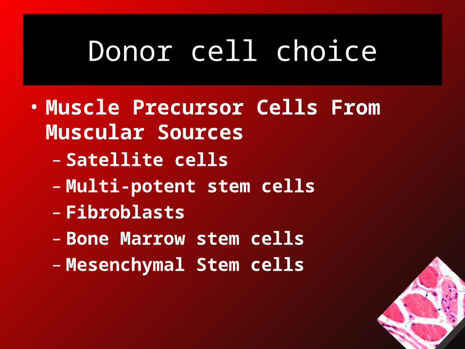
Donor cell choice
• Muscle Precursor Cells From Muscular Sources– Satellite cells– Multi-potent stem cells– Fibroblasts– Bone Marrow stem cells– Mesenchymal Stem cells
Satellite cells
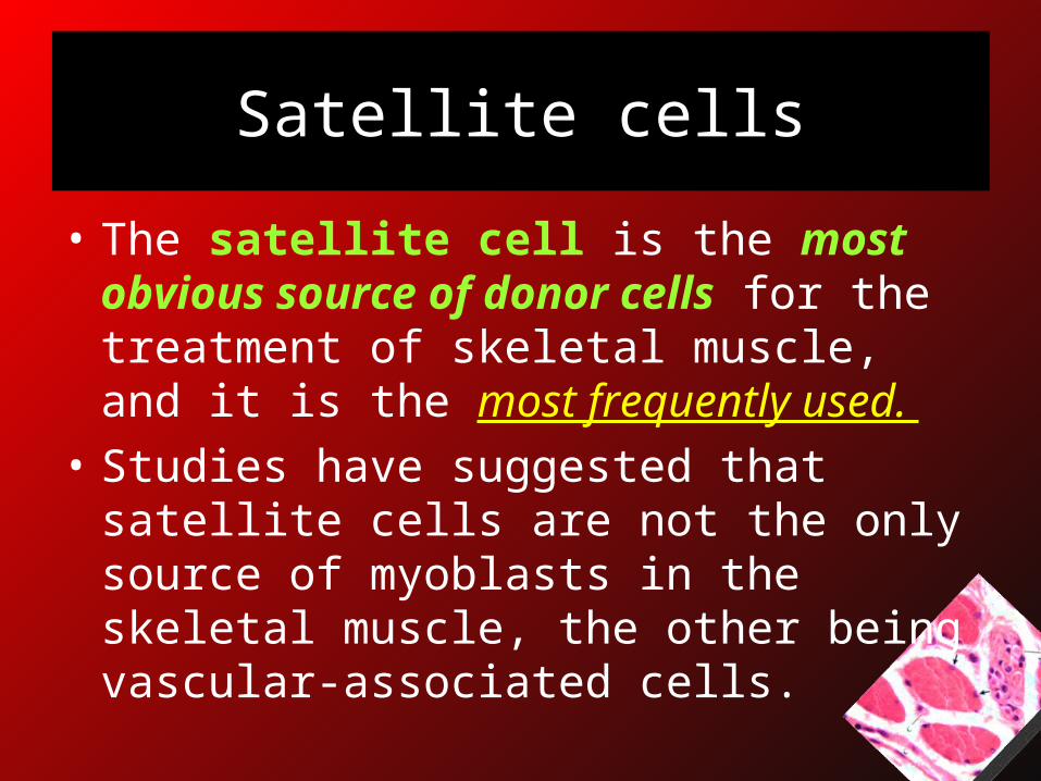
• The satellite cell is the most obvious source of donor cells for the treatment of skeletal muscle, and it is the most frequently used.
• Studies have suggested that satellite cells are not the only source of myoblasts in the skeletal muscle, the other being vascular-associated cells.
Satellite cells
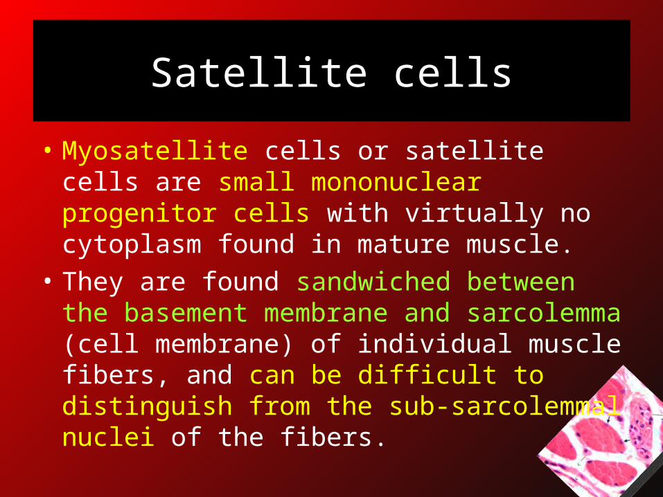
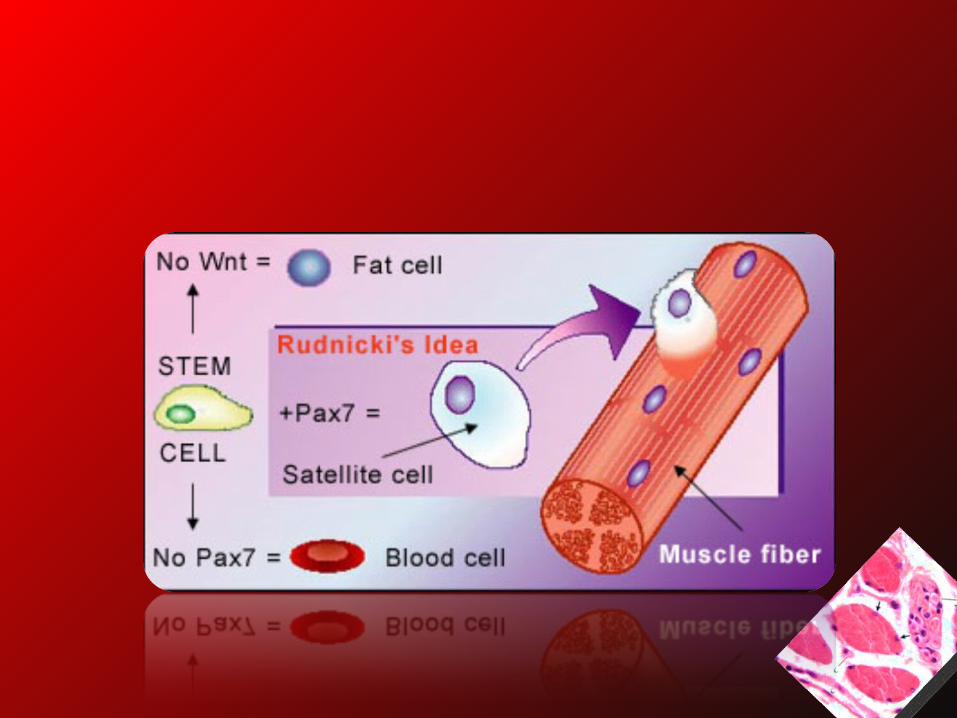
• Myosatellite cells or satellite cells are small mononuclear progenitor cells with virtually no cytoplasm found in mature muscle.
• They are found sandwiched between the basement membrane and sarcolemma (cell membrane) of individual muscle fibers, and can be difficult to distinguish from the sub-sarcolemmal nuclei of the fibers.
Satellite cells
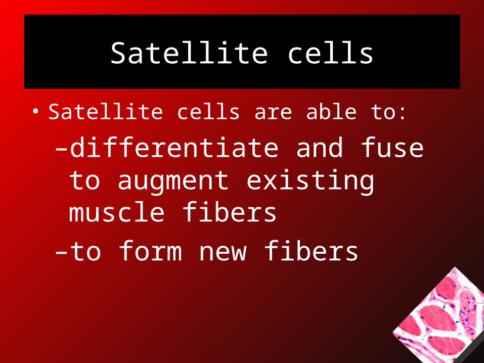
• Satellite cells are able to:
–differentiate and fuse to augment existing muscle fibers–to form new fibers
Satellite cells
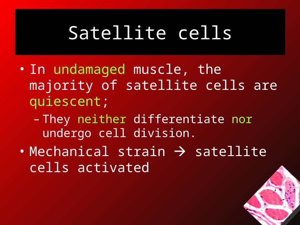
• In undamaged muscle, the majority of satellite cells are quiescent; – They neither differentiate nor undergo cell
division.
• Mechanical strain satellite cells activated
Satellite cells
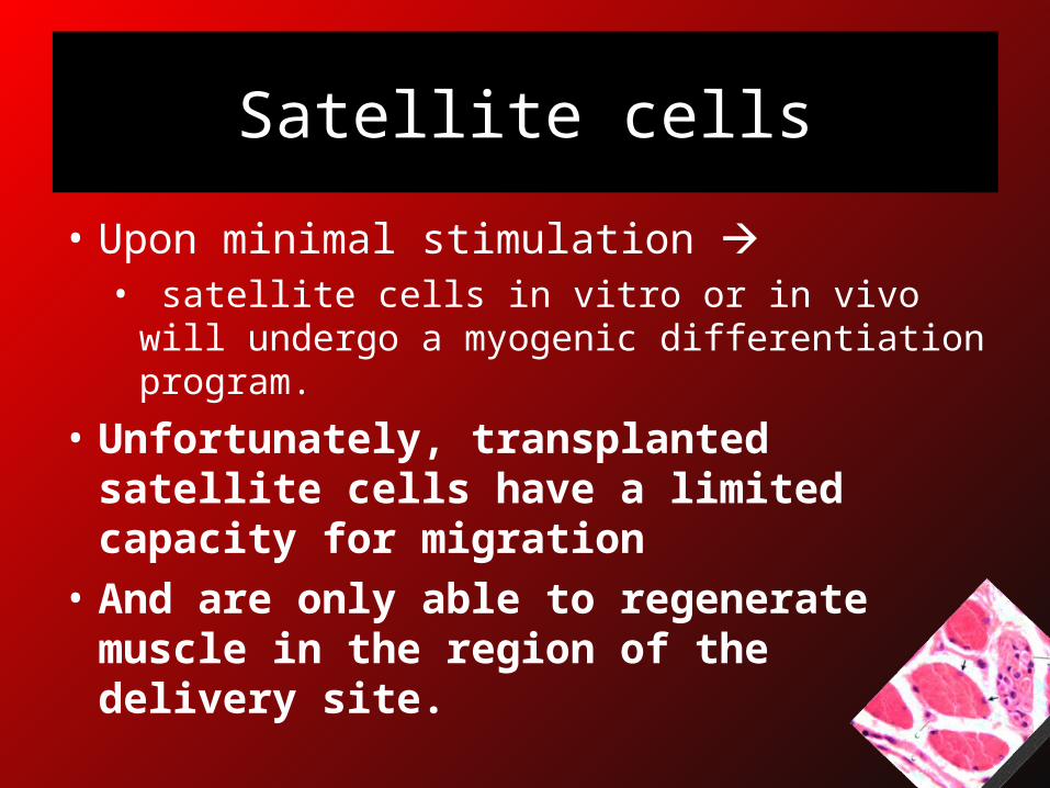
• Upon minimal stimulation • satellite cells in vitro or in vivo will undergo a
myogenic differentiation program.
• Unfortunately, transplanted satellite cells have a limited capacity for migration
• And are only able to regenerate muscle in the region of the delivery site.
Satellite cells
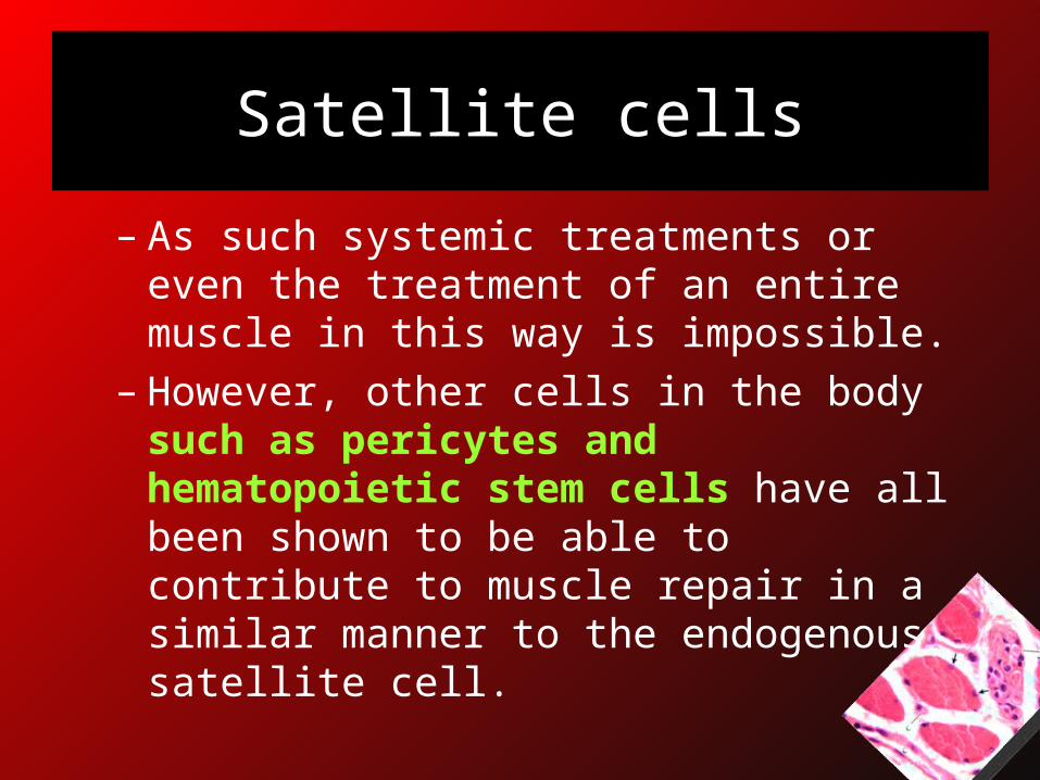
– As such systemic treatments or even the treatment of an entire muscle in this way is impossible.
– However, other cells in the body such as pericytes and hematopoietic stem cells have all been shown to be able to contribute to muscle repair in a similar manner to the endogenous satellite cell.
Mesoangioblasts
• The advantage of using these cell types (pericytes, mesoangioblasts) for therapy in muscle diseases is that they can be systemically delivered, autonomously migrating to the site of injury.
• Particularly successful recently has been the delivery of mesoangioblast cells into the Golden Retriever dog model of Duchenne muscular dystrophy, which effectively cured the disease.– Amini-Nik S, Glancy D et al. (2011). "Pax7 expressing cells contribute
to dermal wound repair, regulating scar size through a β-catenin mediated process". Stem Cells 9 (29): 1371-9.
Multipotent stem cells
• They have been considered by some authors to be precursors of the satellite cells.
• They have:• A high proliferative capacity• A potential advantage to be used for
autotransplantation• Some of their properties may also favor a
potential delivery through the circulatory system.
Multipotent stem cells
• It was reported that the iM injection of normal muscle-derived stem cells in mdx mice produced 10-fold more dystrophin-positive myofibers than the injection of myoblasts derived from satellite cells.
• Qu-Petersen Z, Deasy B, Jankowski R, et al.: Identification of a novel population of muscle stem cells in mice: potential for muscle regeneration. J Cell Biol 2002, 157:851-864.
Muscle Precursor Cells of Non-muscular Sources
• Fibroblasts were transformed into myoblasts either by introducing MyoD1, a master regulator gene for myogenesis, and by treating the cells with galectin-1, a lectin secreted by myoblasts and myotubes.
Bone Marrow-Derived Circulating Cells
• Recent interest in these cells has been based not only on the search for an alternative source of myogenic cells but on the hope of developing a systemic treatment for DMD, ie, a bone marrow transfusion producing the passage of myogenic precursors from the blood to the myofibers in a sufficient number to be therapeutic.
• Previous evidence in mice indicated that the role of non-muscular cells for muscle regeneration is null or negligible.
• However, some hope was raised after dystrophin expression was observed in some myofibers after the intravenous infusion of normal hematopoietic cells in mdx mice and some participation of donor cells in muscle regeneration after bone marrow transplantation in normal mice.
Mesenchymal Stem Cells
• The stromal cells that participate in the supporting structures of bone marrow were reported to differentiate in vitro into a mesodermal phenotype, such as skeletal muscle, and for this reason were called mesenchymal stem cells.
• Preferential differentiation into skeletal muscle was reported under specific conditions in mice.
Mesenchymal stem cells
• It was reported that mesenchymal stem cells isolated from the synovial membrane of adult humans have the potentiality to fuse with myofibers and to remain as satellite cells after the intramuscular implantation in mice.
Mesenchymal stem cells
• It was also suggested that they have the capacity to participate in muscle regeneration after IV injection in the same model.
Route of Donor Cell Delivery
Donor Cell Delivery
• Two routes have been tested for the skeletal muscle: – Local– Systemic
• The challenge is important in myopathies because the target tissue accounts for more than half of the body.
Local Delivery
• iM injection is the most frequent method of delivering donor cells to skeletal muscles.
–It ensures sufficient numbers of donor cells into the muscle.–It produces tissue damage •↑ uptake of the donor myoblasts by
regenerating myofibers.
Local Delivery
• However, donor cells do not diffuse significantly.
cells fuse mainly with the myofibers reached by the injection A narrow track of hybrid myofibers.

iM injection
• For better fusion multiple injections close to one another.
Increasing the Fusion of Donor Myoblasts With Host Fibers
• Increasing the number of regenerating myofibers increases the uptake of the donor myoblasts into hybrid myofibers, and
inhibiting the participation of host satellite cells further favors donor myoblasts.
Ways to inhibit host satellite cells
• Ionizing radiation at high doses: the most method used
• iM injection of myotoxins from snake venom in mice and in monkey experiments.
• Local anesthetics in mouse experiments.• Cryodamage muscle necrosis killing the
host satellite cells
Systemic delivery
Systemic Delivery
• Donor cell delivery by the bloodstream it
would be ideal– Myogenic cells would be distributed through all
skeletal muscles, including those not appropriate for local injections, such as the diaphragm.
Systemic Delivery
– However, IV and intraperitoneal injections of myoblasts were unsuccessful, even after extensive muscle injury.
• Intraarterial injection of a myoblast was more successful, but only after mechanical injury of the muscle.
Systemic Delivery
• The systemic delivery of hematopoietic stem cells, unfractionated bone marrow also produced fusion with host myofibers, mainly after muscle injury.– Ferrari G, Cusella-De Angelis G, Coletta M, et al.: Muscle regeneration by bone
marrow-derived myogenic progenitors. Science 1998, 279:1528-1530.– Ferrari G, Stornaiuolo A, Mavilio F: Failure to correct murine muscular
dystrophy. Nature 2001, 411:1014-1015.
Donor Cell Survival

Donor Cell Survival
• If an optimal donor cell is appropriately delivered to the target organ, it remains to ensure the posttransplantation survival of those cells. – Early survival• During the immediate hours or days after
transplantation, before the acute rejection reaction.– Long-term survival– Defining long-term as the period needed for a
clinical benefit, which ideally must be lifelong.
Early Survival
• Many donor cells die rapidly after transplantation– During the first 1 to 3 days
• This does not prevent the success of myoblast transplantation– Because the phenomenon is limited and the
proliferation of the surviving cells compensates for the cell death.
Long-Term Survival
• The long-term success of normal myoblast transplantation is threatened by acute rejection.
• Acute rejection is the mechanism by which donor alloantigen-reactive cytolytic T lymphocytes destroy the allograft by a response implicating recognition of MHC I on the donor cells.
• Acute rejection can be clinically controlled by immunosuppressants and experimentally avoided by the development of immune tolerance or by autotransplantation.
Stem cells in Inherited Myopathies
Primary Myopathies
• Primary myopathies are characterized by – Progressive wasting of skeletal muscle that leads to
deterioration of movements and, in the most severe cases, such as in Duchenne’s muscular dystrophy (DMD), to complete paralysis and death.
• Most myopathies in which the molecular defect has been identified are due to mutations affecting proteins that form a supramolecular link between the cytoskeleton and the extracellular matrix, such as dystrophin, the mutated protein in DMD.
Current treatment
• The current therapeutic approaches to DMD involve pharmacological suppression of the inflammatory and immune responses, which usually provides only modest and temporary beneficial effects.
Future treatment
• Future approaches depend on:– Cell therapy– Gene therapy
• These range from the design of efficient, nonantigenic gene transfer vectors for in vivo gene therapy, to pharmacological upregulation of the synthesis of utrophin, a related protein that compensates for the loss of dystrophin , to myoblast transplantation, the focus of this Perspective.
In Duchenne:
• In 1989, Partridge and his collaborators showed that iM injection of C2C12 cells, an immortal myogenic cell line derived from adult satellite cells, could efficiently reconstitute dystrophin-positive, apparently normal fibers in dystrophic mdx mice. – Yao SN, Kurachi K: Implanted myoblasts not only fuse with myofibers but also survive as
muscle precursor cells. J Cell Sci 1993, 105:957-963.
In Duchenne:
• This finding inspired a number of problematic attempts in the early 1990s to apply this strategy clinically, but using non-immortal myoblasts in patients.
• Myogenic cells isolated from immune-compatible donors were expanded in vitro and injected into specific muscles of DMD patients.
• All trials failed, for a number of reasons, some of which could have been predicted.
• C2C12 cells have unlimited life-spans and are syngeneic with mdx mice, features that are lacking in normal human donor cells.
In Duchenne: cells succumb soon
• Other difficulties :• We now know that most myoblasts (up to
99%) succumb soon after injection, due first to an inflammatory and then to a cell-mediated immune response.
In Duchenne: Not further migration
• We also know that the cells that survive this initial catastrophe do not migrate more than a few millimeters away from the injection site, – countless injections would be required to provide
a significant distribution of donor cells.
These protocols Better designed clinical trials on the
horizon Clearer definitions of clinically relevant
endpoints
Inflammatory Myopathies
Polymyositis (PM) and Dermatomyositis (DM)
• Polymyositis (PM) and dermatomyositis (DM) are idiopathic inflammatory muscle disorders characterised by muscle weakness and fatigue and by skin involvement in DM.
• Complications can be life-threatening.• Corticosteroids and immunosuppressant
agents remain the mainstay of treatment, but there are still a significant percentage of non-responders and clinical relapses.
PM and DM
• Haematopoietic stem cell transplantation is performed in patients with refractory PM with satisfactory clinical efficacy, but the conditioning regimen for the procedure has many side effects. So newer and less toxic treatments are urgently needed for patients with refractory DM/PM.
• Henes JC, Heinzelmann F, Wacker A, et al. Antisignal recognition particle-positive polymyositis successfully treated with myeloablative autologous stem cell transplantation. Ann Rheum Dis 2009;68:447–8 .
PM and DM
• In a recent study, the efficacy of mesenchymal stem cell was checked.
• Dandan Wang; Huayong Zhang; Mengshu Ca. Efficacy of Allogeneic Mesenchymal Stem Cell Transplantation in Patients with Drug-resistant Polymyositis and Dermatomyositis. Ann Rheum Dis. 2011;70(7):1285-1288.
Methods
• A single-arm trial involving 10 patients with DM/PM who were either refractory to standard treatment, or had severe systemic involvement.
• All patients consented and underwent allogeneic MSCT.
• Clinical and laboratory manifestations were compared before and after MSCT.
Methods• Baseline observations included a complete medical history
and detailed physical and laboratory examination. • Follow-up evaluation was conducted at 1, 2, 3 and 6
months after transplantation, then 6 monthly thereafter. • Transplantation-related mortality included all deaths
associated with transplantation of MSC, except those related to recurrence of underlying disease.
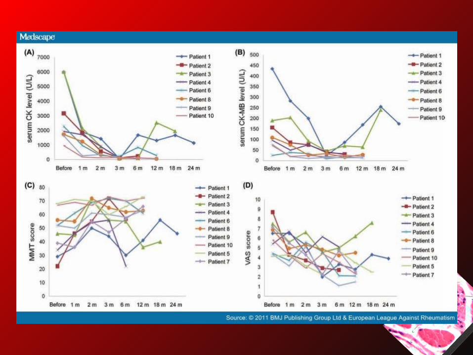
• Disease activity was measured by CK, CK-MB, manual muscle test of eight muscle groups as performed previously by the same rheumatologist.
• Patient's global assessment of the overall impact of disease on well-being was rated on a 0–10 visual analogue scale (VAS).
Conclusion
• Stem cell therapy may be considered as an option for refractory inflammatory myopathies.


![STEM CELLS EMBRYONIC STEM CELLS/INDUCED PLURIPOTENT STEM CELLS Stem Cells.pdf · germ cell production [2]. Human embryonic stem cells (hESCs) offer the means to further understand](https://static.fdocuments.in/doc/165x107/6014b11f8ab8967916363675/stem-cells-embryonic-stem-cellsinduced-pluripotent-stem-cells-stem-cellspdf.jpg)