Podocyte-specific Nox4 deletion affords renoprotection … · Podocyte-specific Nox4 deletion...
11
ARTICLE Podocyte-specific Nox4 deletion affords renoprotection in a mouse model of diabetic nephropathy Jay C. Jha 1,2 & Vicki Thallas-Bonke 1 & Claudine Banal 1 & Stephen P. Gray 1 & Bryna S. M. Chow 1 & Georg Ramm 3 & Susan E. Quaggin 4 & Mark E. Cooper 1,2 & Harald H. H. W. Schmidt 5 & Karin A. Jandeleit-Dahm 1,2 Received: 6 July 2015 /Accepted: 1 October 2015 /Published online: 28 October 2015 # Springer-Verlag Berlin Heidelberg 2015 Abstract Aims/hypothesis Changes in podocyte morphology and func- tion are associated with albuminuria and progression of dia- betic nephropathy. NADPH oxidase 4 (NOX4) is the main source of reactive oxygen species (ROS) in the kidney and Nox4 is upregulated in podocytes in response to high glucose. We assessed the role of NOX4-derived ROS in podocytes in vivo in a model of diabetic nephropathy using a podocyte-specific NOX4-deficient mouse, with a major focus on the development of albuminuria and ultra-glomerular structural damage. Methods Streptozotocin-induced diabetes-associated changes in renal structure and function were studied in male floxed Nox4 and podocyte-specific, NOX4 knockout (podNox4KO) mice. We assessed albuminuria, glomerular ex- tracellular matrix accumulation and glomerulosclerosis, and markers of ROS and inflammation, as well as glomerular basement membrane thickness, effacement of podocytes and expression of the podocyte-specific protein nephrin. Results Podocyte-specific Nox4 deletion in streptozotocin- induced diabetic mice attenuated albuminuria in association with reduced vascular endothelial growth factor (VEGF) expression and prevention of the diabetes-induced reduc- tion in nephrin expression. In addition, podocyte-specific Nox4 deletion reduced glomerular accumulation of collagen IV and fibronectin, glomerulosclerosis and mesangial expansion, as well as glomerular basement mem- brane thickness. Furthermore, diabetes-induced increases in renal ROS, glomerular monocyte chemoattractant protein-1 (MCP-1) and protein kinase C alpha (PKC-α) were attenuated in podocyte-specific NOX4-deficient mice. Conclusions/interpretation Collectively, this study shows the deleterious effect of Nox4 expression in podocytes by promot- ing podocytopathy in association with albuminuria and extra- cellular matrix accumulation in experimental diabetes, emphasising the role of NOX4 as a target for new renoprotective agents. Keywords Albuminuria . Diabetic nephropathy . Glomerular basement membrane . NADPH oxidase 4 . Podocyte . Reactive oxygen species Abbreviations ACR Albumin/creatinine ratio DN Diabetic nephropathy ECM Extracellular matrix GBM Glomerular basement membrane 8-OHdG 8-Hydroxy-2′-deoxyguanosine Harald H. H. W. Schmidt and Karin A. Jandeleit-Dahm contributed equally as senior authors. Electronic supplementary material The online version of this article (doi:10.1007/s00125-015-3796-0) contains peer-reviewed but unedited supplementary material, which is available to authorised users. * Karin A. Jandeleit-Dahm [email protected] 1 Diabetes Complications Division, Baker IDI Heart & Diabetes Research Institute, PO Box 6492, St Kilda Rd, Melbourne, VIC 8008, Australia 2 Department of Medicine, Monash University, Melbourne, VIC, Australia 3 Monash Micro-imaging, Monash University, Melbourne, VIC, Australia 4 Robert H. Lurie Medical Research Center, Chicago, IL, USA 5 Department of Pharmacology, Cardiovascular Research Institute Maastricht (CARIM), Faculty of Medicine, Health & Life Science, Maastricht University, Maastricht, the Netherlands Diabetologia (2016) 59:379–389 DOI 10.1007/s00125-015-3796-0
Transcript of Podocyte-specific Nox4 deletion affords renoprotection … · Podocyte-specific Nox4 deletion...
ARTICLE
Podocyte-specific Nox4 deletion affords renoprotection in a mousemodel of diabetic nephropathy
Jay C. Jha1,2 & Vicki Thallas-Bonke1 & Claudine Banal1 & Stephen P. Gray1 &
Bryna S. M. Chow1& Georg Ramm3
& Susan E. Quaggin4& Mark E. Cooper1,2 &
Harald H. H. W. Schmidt5 & Karin A. Jandeleit-Dahm1,2
Received: 6 July 2015 /Accepted: 1 October 2015 /Published online: 28 October 2015# Springer-Verlag Berlin Heidelberg 2015
AbstractAims/hypothesis Changes in podocyte morphology and func-tion are associated with albuminuria and progression of dia-betic nephropathy. NADPH oxidase 4 (NOX4) is the mainsource of reactive oxygen species (ROS) in the kidney andNox4 is upregulated in podocytes in response to high glucose.We assessed the role of NOX4-derived ROS in podocytesin vivo in a model of diabetic nephropathy using apodocyte-specific NOX4-deficient mouse, with a major focuson the development of albuminuria and ultra-glomerularstructural damage.Methods Streptozotocin-induced diabetes-associated changesin renal structure and function were studied in malefloxedNox4 and podocyte-specific, NOX4 knockout
(podNox4KO) mice. We assessed albuminuria, glomerular ex-tracellular matrix accumulation and glomerulosclerosis, andmarkers of ROS and inflammation, as well as glomerularbasement membrane thickness, effacement of podocytes andexpression of the podocyte-specific protein nephrin.Results Podocyte-specific Nox4 deletion in streptozotocin-induced diabetic mice attenuated albuminuria in associationwith reduced vascular endothelial growth factor (VEGF)expression and prevention of the diabetes-induced reduc-tion in nephrin expression. In addition, podocyte-specificNox4 deletion reduced glomerular accumulation ofcollagen IV and fibronectin, glomerulosclerosis andmesangial expansion, as well as glomerular basement mem-brane thickness. Furthermore, diabetes-induced increasesin renal ROS, glomerular monocyte chemoattractantprotein-1 (MCP-1) and protein kinase C alpha (PKC-α)were attenuated in podocyte-specific NOX4-deficient mice.Conclusions/interpretation Collectively, this study shows thedeleterious effect ofNox4 expression in podocytes by promot-ing podocytopathy in association with albuminuria and extra-cellular matrix accumulation in experimental diabetes,emphasising the role of NOX4 as a target for newrenoprotective agents.
Keywords Albuminuria . Diabetic nephropathy . Glomerularbasement membrane . NADPH oxidase 4 . Podocyte .
Reactive oxygen species
AbbreviationsACR Albumin/creatinine ratioDN Diabetic nephropathyECM Extracellular matrixGBM Glomerular basement membrane8-OHdG 8-Hydroxy-2′-deoxyguanosine
Harald H. H. W. Schmidt and Karin A. Jandeleit-Dahm contributedequally as senior authors.
Electronic supplementary material The online version of this article(doi:10.1007/s00125-015-3796-0) contains peer-reviewed but uneditedsupplementary material, which is available to authorised users.
* Karin A. [email protected]
1 Diabetes Complications Division, Baker IDI Heart & DiabetesResearch Institute, PO Box 6492, St Kilda Rd,Melbourne, VIC 8008, Australia
2 Department of Medicine, Monash University, Melbourne, VIC,Australia
3 Monash Micro-imaging, Monash University, Melbourne, VIC,Australia
4 Robert H. Lurie Medical Research Center, Chicago, IL, USA5 Department of Pharmacology, Cardiovascular Research Institute
Maastricht (CARIM), Faculty of Medicine, Health & Life Science,Maastricht University, Maastricht, the Netherlands
Diabetologia (2016) 59:379–389DOI 10.1007/s00125-015-3796-0
MCP Monocyte chemoattractant proteinNOX NADPH oxidasePKC Protein kinase CROS Reactive oxygen speciesVEGF Vascular endothelial growth factor
Introduction
Diabetic nephropathy (DN) is the leading cause of renalfailure in the world, with patients often requiring dialysis orkidney transplantation [1, 2]. Podocyte damage and loss havebeen suggested to play pivotal roles in the pathogenesis of DNincluding albuminuria, which is a key feature of this condition[3, 4]. Podocytes are glomerular visceral epithelial cells con-nected by the slit diaphragm, which functions as a size-selective filtration barrier [5]. A number of studies have iden-tified ‘oxidative stress’ as a crucial mediator in the develop-ment and progression of DN [6–8]. It has been postulated thatthis enhanced oxidative stress via the pro-oxidant enzymeNADPH oxidase (NOX), particularly NOX4 [7, 9, 10] andmore recently NOX5 [11], in the diabetic kidney causes injuryto podocytes and is associated with deleterious changes inrenal function and structure [4, 12–14].
In a recent study, we found that global genetic deletion orpharmacological inhibition of NOX4 significantly attenuateddiabetes-induced increases in albuminuria, glomerulosclerosisand accumulation of extracellular matrix (ECM) proteins via areduction in reactive oxygen species (ROS) production in anexperimental model of DN [9]. It has been suggested thatstimuli such as high glucose, TGF-β and angiotensin IIincrease ROS production and lead to injury and apoptosis inpodocytes, and are associated with increased excretion ofalbumin into the urine [4, 15–17]. Another study has reportedthat systemic administration of a nonspecific NOX inhibitor,apocynin, ameliorated urinary albumin excretion in a diabeticmouse model [18, 19]. Recently, we found that silencingNOX4 using short hairpin RNA or inhibiting NOX4 using anovel, specific NOX1/NOX4 inhibitor (GKT137831) in hu-man podocytes significantly reduced the high glucose-induced increase in ROS production, as well as gene expres-sion of profibrotic and proinflammatory markers [9]. In thepresent study, we aimed to examine whether the diabetes-induced activation of NOX4-derived ROS specifically inpodocytes affects glomerular structure and function in vivoin an animal model of DN, using mice with specific deletionof Nox4 within podocytes.
Methods
Animals Homozygous floxedNox4 (floxed NADPH oxidase4) mice (provided by H. H. H. W. Schmidt, CARIM,
Maastricht, the Netherlands) and heterozygous podocyte-specific Cre recombinase, podocin-Cre (NPHS2-Cre(podCre)) mice (provided by S. E. Quaggin, Robert H. LurieMedical Research Center, Chicago, IL, USA) were crossed togenerate podocyte-specific, NOX4 knockout (podNox4KO)mice. Cre-mediated recombination resulted in deletion of theNox4 flanked sequence in the podocyte. We confirmed Nox4deletion in the podocytes by genotyping (see electronic sup-plementary material [ESM] Fig. 1a) and qPCR (ESM Fig. 1b),as well as by immunostaining (ESM Fig. 1c). Detailedmethods for the isolation of podocytes and glomerular andtubular fractions, as well as confirmation of Nox4 deletion,are provided in ESM Methods. All of the mouse lines weremaintained on a C57BL/6 background. Control mice werelittermates of floxedNox4 and podCre mouse crosses.
All animal studies were approved by the Alfred MedicalResearch & Education Precinct Animal Ethics Committee un-der guidelines laid down by the National Health and MedicalResearch Council of Australia. All animals were housed at thePrecinct Animal Centre of the Baker IDI Heart & DiabetesInstitute. During the study, animals had unrestricted access towater and food and were maintained on a 12 h light/dark cyclein a pathogen-free environment on standard mouse chow(Specialty Feeds, Glen Forest, Perth, WA, Australia).
Induction of diabetes Diabetes was induced in 6-week-oldmale floxedNox4, podNox4KO and podCremice by five dailyi.p. injections of streptozotocin (Sigma-Aldrich, St Louis,MO, USA), at a dose of 55 mg/kg body weight in citratebuffer, with control mice receiving citrate buffer alone. Onlymice with blood glucose ≥15 mmol/l after injection ofstreptozotocin have been included in experiments; mice withblood glucose <15 mmol/l were excluded from the study(<10% of the total number of mice). After 10 and 20 weeks,the animals were anaesthetised using sodium pentobarbitonei.p. (100 mg/kg body weight; Euthatal, Sigma-Aldrich, CastleHill, NSW, Australia). The kidneys were rapidly dissected,weighed and snap-frozen or processed in paraffin for subse-quent analysis.
Measurement of metabolic variables At 10 and 20 weeksafter induction of diabetes, mice were individually placed intometabolic cages (Iffa Credo, L’Arbresele, France) for 24 h.Urine was collected for subsequent analysis. Blood glucoseand glycated haemoglobin were measured, as previously de-scribed [9, 20]. Systolic BP was assessed using acomputerised non-invasive tail-cuff method [21]. Urinaryalbumin concentration was measured at 10 and 20 weeks afterthe induction of diabetes, using a mouse albumin ELISAquantification kit (Bethyl Laboratories, Montgomery, TX,USA). Urinary creatinine was determined using a commer-cially available creatinine assay kit (Abcam, Cambridge,UK). The urinary albumin/creatinine ratio (ACR) was
380 Diabetologia (2016) 59:379–389
calculated. A mouse cystatin C ELISA kit (BioVendor, Brno,Czech Republic) was used to determine serum cystatin C ac-cording to the manufacturer’s instructions.
Histological assessmentKidney sections (3 μm)were stainedwith periodic acid–Schiff for the measurement ofglomerulosclerotic injury and mesangial expansion, as wellas with Masson’s trichrome for the assessment of glomerularECM accumulation [22]. Mesangial area and ECM accumu-lation were analysed (percentage of glomerular area) fromdigital pictures of glomeruli (20 glomeruli per kidney peranimal) using Image-Pro Plus 6.0 software (Media Cybernetics,Bethesda, MD, USA), as previously described [9, 20].Glomerulosclerotic injury was graded based on the severity ofglomerular damage, as previously described [23]. Twentyglomeruli per kidney were assessed in a masked fashion.
In vivo transmission electron microscopy Kidney sectionswere fixed, embedded, cut and visualised using a Hitachi 7500transmission electron microscope (Hitachi, Tokyo, Japan).Electron micrographs were used to determine the glomerularbasement membrane (GBM) thickness, and the number offiltration slit pores was counted as previously described [24,25]. For further details, see the ESM Methods.
Immunohistochemistry Immunostaining for collagen IV,fibronectin, nitrotyrosine, nephrin and protein kinase C(PKC)-α was performed and the proportional area of stainingwas quantified as previously described [9, 20]. For furtherdetails, see the ESM Methods.
Western blot The glomerular fraction was obtained from thefrozen renal cortex of the respective control and diabetic mice,as described in ESM Methods. Protein extracts (5 μg) fromeach sample were electrophoresed on 7.5% acrylamide gelsunder non-reducing conditions, as previously described [26].Western blot analysis was then performed with a primary anti-body to collagen IV (1:1000, Abcam, Cambridge, MA, USA)
and assessed with goat anti-rabbit (Dako, Carpinteria, CA,USA) secondary antibody. Membranes were subsequentlyprobed for α-tubulin (Sigma-Aldrich) for determination ofequal loading of samples. Blots were detected using the ECLdetection kit (Sigma-Aldrich) and densitometry was performedusing Quantity One software (Bio-Rad, Richmond, CA, USA).
In vivo glomerular gene expression analysisTotal RNAwasextracted from isolated glomeruli (Polytron PT-MR2100;Kinematica, Littau/Lucerne, Switzerland) in TRIzol reagent(Invitrogen Australia, Mt Waverely, VIC, Australia), as previ-ously described [9, 20]. Probes and primer sequences for theRT-PCR of nephrin, vascular endothelial growth factor(VEGF) and monocyte chemoattractant protein (MCP)-1 aredescribed in ESM Table 1. Expression of the genes encodingnephrin, VEGF and MCP-1 was quantified and determinedrelative to expression of the housekeeping gene 18S (18Sribosomal RNA TaqMan Control Reagent kit) using theTaqMan system (ABI Prism 7500; Perkin-Elmer, Poster City,CA, USA). Results are expressed relative to non-diabeticfloxedNox4mice, which were arbitrarily assigned a value of 1.
Urinary VEGF ELISA The Quantikine Mouse ELISA kit(R&D Systems, Minneapolis, MN, USA) was used to mea-sure VEGF in the urine, as per the kit instructions. UrinaryVEGF is expressed as picograms per 24 h.
Urinary 8-isoprostanes ELISA An 8-isoprostanes enzymeimmunoassay kit (Cayman Chemical Company, Ann Arbor,MI, USA) was used to measure 8-isoprostanes in urine, asdescribed by the manufacturer. Urinary 8-isoprostanes isexpressed as picograms per 24 h.
Urinary 8-hydroxy-2′-deoxyguanosine ELISA A urinary8-hydroxy-2′-deoxyguanosine (8-OHdG) enzyme immunoassaykit (StressMarq Biosciences Victoria, BC, Canada) was used tomeasure 8-OHdG in urine, as described by the manufacturer.Urinary 8-OHdG is expressed as nanograms per 24 h.
Table 1 General andmetabolic variables after 10 and 20weeks of study in control and diabetic floxedNox4 and podNox4KOmice (n=8–15 per group)
Variable 10 weeks 20 weeks
floxedNox4 podNox4KO floxedNox4 podNox4KO
Control Diabetes Control Diabetes Control Diabetes Control Diabetes
Body weight (g) 26±0.8 23±0.9* 29±1.1 25±0.7* 33±0.7 26±0.9* 34±1.1 27±1.1*
Kidney weight/body weight (%) 0.54±0.02 0.70±0.06* 0.57±0.01 0.70±0.03* 0.56±0.01 0.90±0.04* 0.57±0.01 0.83±0.03*
Systolic BP (mmHg) 97±2 99±1 103±2 107±1 98±3 99±2 104±2 105±2
Plasma glucose (mmol/l) 9.3±0.7 26.5±3.6* 11.6±0.7 27.2±3.4* 9.4±0.7 28.7±2.2* 11.3±0.6 24.8±2.3*
Total glycated haemoglobin (%) 6.2±0.1 10.6±1.3* 4.5±0.1 9.1±0.3* 5.9±0.8 9.9±1.1* 6.1±0.9 10.6±1.5*
Data are as means ± SEM
*p<0.05 vs the respective control
Diabetologia (2016) 59:379–389 381
Lucigenin assaysGlomerular fractions obtained from the fro-zen renal cortices of the different experimental groups wereharvested in 100 μl ice-cold phosphate buffer (50 mmol/l
KH2PO4, 1 mmol/l EGTA and 150 mmol/l sucrose; pH 7.4)with protease inhibitors, as previously described [11]. Base-line activity was measured by adding 50 μl glomerularextract to 175 μl buffer and 2.5 μl 1 mmol/l lucigenin(Sigma-Aldrich). NADPH-dependent superoxide wasmeasured by the addition of 25 μl 1 mmol/l NADPH(Sigma-Aldrich). Baseline activity was subtracted andnormalised to the protein concentration.
Statistical analysis All variables were analysed by one-wayANOVA using GraphPad Prism 6 (GraphPad, SanDiego, CA,USA) for multiple comparison of the means or by thetwo-tailed unpaired Mann–Whitney U test, when required.p<0.05 was considered to be statistically significant. Resultsare expressed as mean ± SEM unless otherwise specified.
Results
Metabolic variables Induction of diabetes was associatedwith reduced body weight, elevated blood glucose and in-creased glycated haemoglobin in both groups when comparedwith their respective non-diabetic controls (Table 1). Further-more, no differences in metabolic variables were seen indiabetic podNox4KO mice compared with diabeticfloxedNox4 mice (Table 1). In addition, systolic blood
floxe
dNox
4po
dNox
4KO
Control Diabeticc
b
Cont Diab Cont DiabfloxedNox4 podNox4KO
Cont Diab Cont DiabfloxedNox4 podNox4KO
Cont Diab Cont DiabfloxedNox4 podNox4KO
*
§
a
Cont Diab Cont DiabfloxedNox4 podNox4KO
*
§
d*
‡
*†§
e
40
30
20
10
0
1.5
2.0
1.0
0.5
0
Nep
hrin
(fol
d in
duct
ion/
18S
)
2.5
1.5
2.0
1.0
0.5
0
Nep
hrin
are
a (%
)
Glo
mer
ular
Veg
f(f
old
indu
ctio
n/18
S)
Urin
ary
VE
GF
(pg/
24 h
)
500
400
300
200
100
0
Fig. 2 Podocyte-specific Nox4deletion attenuated increasedVEGF expression and preservednephrin expression in diabeticmice. Glomerular gene (a) (n=5–6 per group) and protein (b, c)(n=7–8 per group) expression ofnephrin (magnification ×40) inrespective control (Cont) anddiabetic (Diab) floxedNox4 andpodNox4KO mice after 20 weeksof diabetes. Glomerular Vegf geneexpression (d) (n=5–6 per group)and urinary VEGF excretion (e)(n=9–10 per group) in respectivecontrol and diabetic floxedNox4and podNox4KO mice after20 weeks of diabetes. Data aremeans ± SEM. *p<0.05 vscontrol floxedNox4 mice; ‡p=0.05 vs diabetic floxedNox4mice;†p<0.05 vs control podNox4KO;§p<0.05 vs diabetic floxedNox4mice
Cont Diab Cont DiabfloxedNox4 podNox4KO
Cont Diab Cont DiabfloxedNox4 podNox4KO
a
*†§
b
c d
Cont Diab Cont DiabfloxedNox4 podNox4KO
**
†§
Cont Diab Cont DiabfloxedNox4 podNox4KO
*
†§
*
†
500
400
300
200
100
0
400
300
200
100
0
400
300
200
100
0
500
400
300
200
100
0Alb
umin
uria
(μg
/24
h)A
CR
(μg
/μm
ol)
Ser
um c
ysta
tin C
(ng
/ml)
Alb
umin
uria
(μg
/24
h)
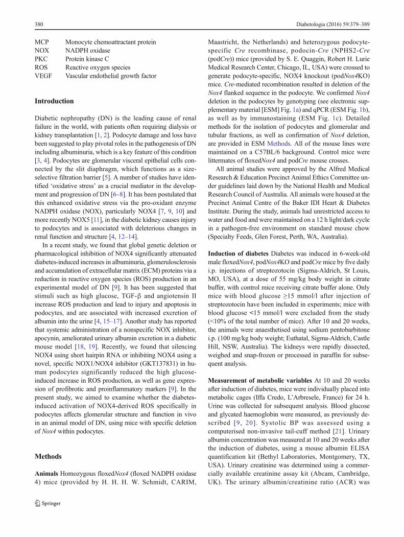
Fig. 1 Podocyte-specific Nox4 deletion attenuated albuminuria in dia-betic mice. Urinary albumin excretion at 10 (a) and 20 (b) weeks ofdiabetes, and ACR (c) and serum cystatin C (d) at 20 weeks of diabetesin floxedNox4 and podNox4KO mice (n=10–15 per group). Data aremeans ± SEM. *p<0.05, **p<0.01 vs control (Cont) floxedNox4 mice;†p<0.05 vs control (Cont) podNox4KOmice; §p<0.05 vs diabetic (Diab)floxedNox4 mice
382 Diabetologia (2016) 59:379–389

pressure was similar in all groups. The kidney weight/bodyweight ratio was significantly increased in diabetic mice, withsimilar ratios in diabetic floxedNox4 and podNox4KO mice.Furthermore, metabolic variables were unchanged in podCremice when compared with floxedNox4mice after 20 weeks ofdiabetes (ESM Table 2).
Renal functional variables Albuminuria was significantlyattenuated after 10 and 20 weeks of diabetes in podNox4KOmice when compared with diabetic floxedNox4mice (Fig. 1a,b). Similar effects were observed when the data wereexpressed as urinary ACR after 20 weeks of diabetes(Fig. 1c). Furthermore, no difference in albuminuria wasfound in podCre mice when compared with floxedNox4miceafter 20 weeks of diabetes (ESM Table 2). The serum cystatinC level was reduced in both diabetic floxedNox4 andpodNox4KO mice when compared with their non-diabeticcounterparts (Fig. 1d), suggesting that the glomerular filtrationrate was not reduced by Nox4 deletion in podocytes and thatboth groups of diabetic mice exhibited hyperfiltration.
Gene and protein expression of nephrin As previouslydemonstrated in another model of DN [27], we found
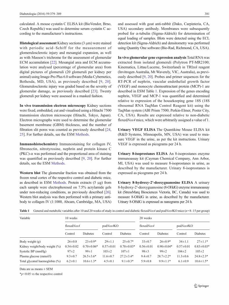
decreased gene and protein expression of glomerular nephrinin diabetic floxedNox4 mice (Fig. 2a–c). Interestingly, thediabetes-induced decrease in nephrin expression was pre-served in diabetic podNox4KO mice (Fig. 2a–c).
Gene expression and urinary excretion of VEGF Weobserved that glomerular Vegf (also known as Vegfa) geneexpression and urinary excretion of VEGF were higher indiabetic floxedNox4 mice when compared with non-diabetic controls, and these variables were attenuated indiabetic podNox4KO mice (Fig. 2d, e).
Glomerular structural assessment Glomerulosclerosis andmesangial area (Fig. 3a–c) as well as ECM accumulation(Fig. 3d, e) were increased in floxedNox4mice after 20 weeksof diabetes when compared with non-diabetic floxedNox4mice. Increased glomerulosclerosis and mesangial area(p<0.05) as well as ECM accumulation (p=0.05) wereattenuated in diabetic podNox4KO mice (Fig. 3a–e).
GBM thickness and podocyte foot process effacementUsing quantifying histomorphometric techniques, both thick-ening of GBM as well as irregularity in podocyte foot process
Control Diabetic
Control Diabetic
floxe
dNox
4po
dNox
4KO
floxe
dNox
4po
dNox
4KO
Cont Diab Cont Diab
floxedNox4 podNox4KO
floxedNox4 podNox4KO
floxedNox4 podNox4KO
Cont Diab Cont Diab
Cont Diab Cont Diab
2.5
2.0
1.5
1.0
0.5
0
40
30
20
10
0
40
30
20
10
0
‡
†§
†§
†
Glo
mer
ulos
cler
otic
inde
xM
esan
gial
are
a (%
)G
lom
erul
ar E
CM
area
(%
)
*
*
*
a b
c
e
d
Fig. 3 Podocyte-specific Nox4deletion attenuated glomerularinjury in diabetic mice. Periodicacid–Schiff staining(magnification ×40) (a),glomerulosclerotic index (b) andmesangial area expansion (c), aswell as Masson’s trichromestaining (magnification ×40) (d)and glomerular ECMaccumulation (e) in control (Cont)and diabetic (Diab) floxedNox4and podNox4KO mice (n=7–10per group) after 20 weeks ofdiabetes. Data are means ± SEM.*p<0.05 vs control floxedNox4mice; ‡p=0.05 vs diabeticfloxedNox4 mice; †p<0.05 vscontrol podNox4KO; §p<0.05 vsdiabetic floxedNox4 mice
Diabetologia (2016) 59:379–389 383
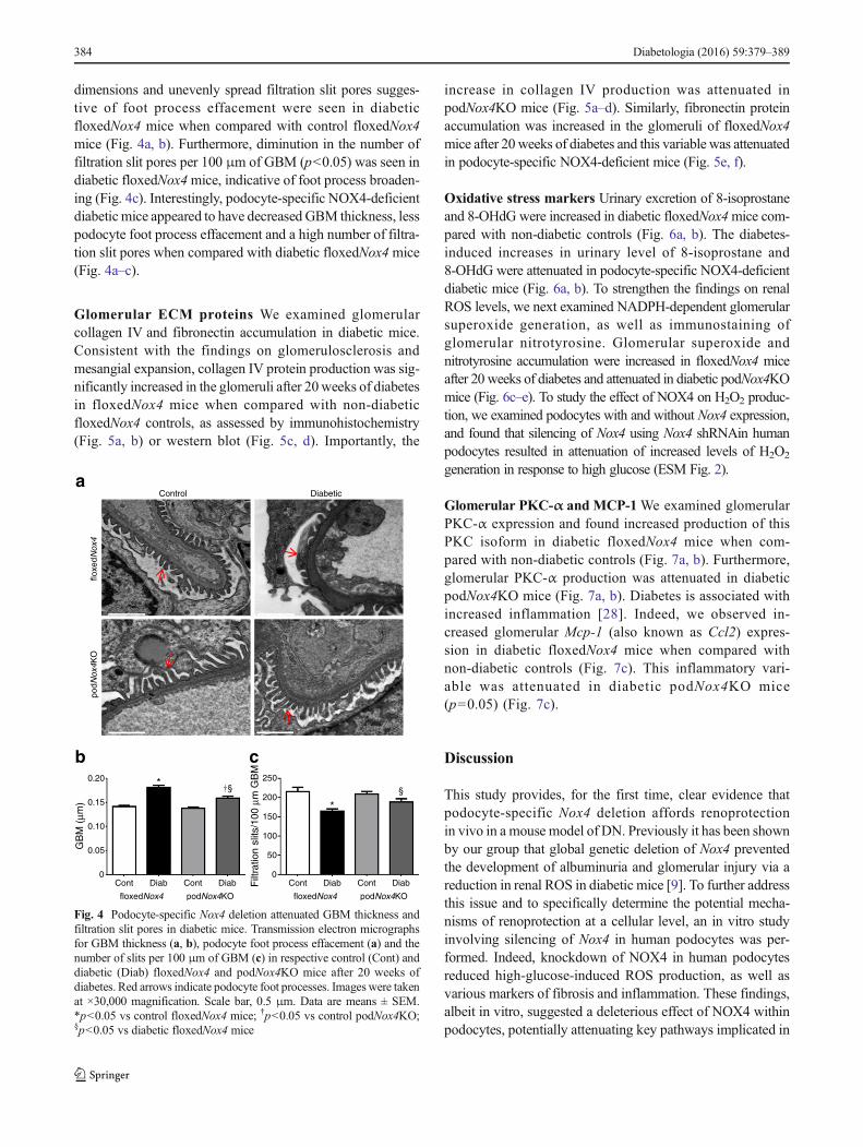
dimensions and unevenly spread filtration slit pores sugges-tive of foot process effacement were seen in diabeticfloxedNox4 mice when compared with control floxedNox4mice (Fig. 4a, b). Furthermore, diminution in the number offiltration slit pores per 100 μm of GBM (p<0.05) was seen indiabetic floxedNox4mice, indicative of foot process broaden-ing (Fig. 4c). Interestingly, podocyte-specific NOX4-deficientdiabetic mice appeared to have decreased GBM thickness, lesspodocyte foot process effacement and a high number of filtra-tion slit pores when compared with diabetic floxedNox4mice(Fig. 4a–c).
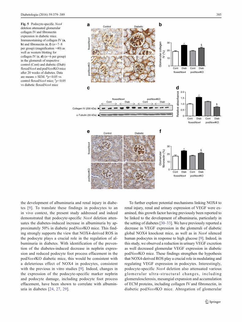
Glomerular ECM proteins We examined glomerularcollagen IV and fibronectin accumulation in diabetic mice.Consistent with the findings on glomerulosclerosis andmesangial expansion, collagen IV protein production was sig-nificantly increased in the glomeruli after 20weeks of diabetesin floxedNox4 mice when compared with non-diabeticfloxedNox4 controls, as assessed by immunohistochemistry(Fig. 5a, b) or western blot (Fig. 5c, d). Importantly, the
increase in collagen IV production was attenuated inpodNox4KO mice (Fig. 5a–d). Similarly, fibronectin proteinaccumulation was increased in the glomeruli of floxedNox4mice after 20weeks of diabetes and this variable was attenuatedin podocyte-specific NOX4-deficient mice (Fig. 5e, f).
Oxidative stress markers Urinary excretion of 8-isoprostaneand 8-OHdG were increased in diabetic floxedNox4mice com-pared with non-diabetic controls (Fig. 6a, b). The diabetes-induced increases in urinary level of 8-isoprostane and8-OHdG were attenuated in podocyte-specific NOX4-deficientdiabetic mice (Fig. 6a, b). To strengthen the findings on renalROS levels, we next examined NADPH-dependent glomerularsuperoxide generation, as well as immunostaining ofglomerular nitrotyrosine. Glomerular superoxide andnitrotyrosine accumulation were increased in floxedNox4 miceafter 20 weeks of diabetes and attenuated in diabetic podNox4KOmice (Fig. 6c–e). To study the effect of NOX4 on H2O2 produc-tion, we examined podocytes with and without Nox4 expression,and found that silencing of Nox4 using Nox4 shRNAin humanpodocytes resulted in attenuation of increased levels of H2O2
generation in response to high glucose (ESM Fig. 2).
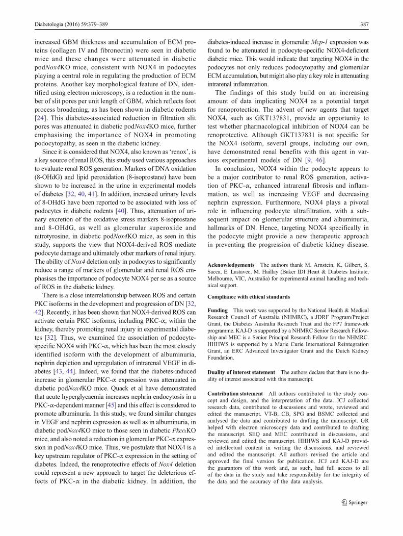
Glomerular PKC-α and MCP-1We examined glomerularPKC-α expression and found increased production of thisPKC isoform in diabetic floxedNox4 mice when com-pared with non-diabetic controls (Fig. 7a, b). Furthermore,glomerular PKC-α production was attenuated in diabeticpodNox4KO mice (Fig. 7a, b). Diabetes is associated withincreased inflammation [28]. Indeed, we observed in-creased glomerular Mcp-1 (also known as Ccl2) expres-sion in diabetic floxedNox4 mice when compared withnon-diabetic controls (Fig. 7c). This inflammatory vari-able was attenuated in diabetic podNox4KO mice(p=0.05) (Fig. 7c).
Discussion
This study provides, for the first time, clear evidence thatpodocyte-specific Nox4 deletion affords renoprotectionin vivo in a mouse model of DN. Previously it has been shownby our group that global genetic deletion of Nox4 preventedthe development of albuminuria and glomerular injury via areduction in renal ROS in diabetic mice [9]. To further addressthis issue and to specifically determine the potential mecha-nisms of renoprotection at a cellular level, an in vitro studyinvolving silencing of Nox4 in human podocytes was per-formed. Indeed, knockdown of NOX4 in human podocytesreduced high-glucose-induced ROS production, as well asvarious markers of fibrosis and inflammation. These findings,albeit in vitro, suggested a deleterious effect of NOX4 withinpodocytes, potentially attenuating key pathways implicated in
floxe
dNox
4po
dNox
4KO
Control Diabetic
*
*
floxedNox4 podNox4KO
Cont Diab Cont Diab
floxedNox4 podNox4KO
Cont Diab Cont Diab
†§ §
0.20
0.15
0.10
0.05
0
250
200
150
100
50
0
GB
M (
μm)
Filt
ratio
n sl
its/1
00 μ
m G
BM
a
b c
Fig. 4 Podocyte-specific Nox4 deletion attenuated GBM thickness andfiltration slit pores in diabetic mice. Transmission electron micrographsfor GBM thickness (a, b), podocyte foot process effacement (a) and thenumber of slits per 100 μm of GBM (c) in respective control (Cont) anddiabetic (Diab) floxedNox4 and podNox4KO mice after 20 weeks ofdiabetes. Red arrows indicate podocyte foot processes. Imageswere takenat ×30,000 magnification. Scale bar, 0.5 μm. Data are means ± SEM.*p<0.05 vs control floxedNox4 mice; †p<0.05 vs control podNox4KO;§p<0.05 vs diabetic floxedNox4 mice
384 Diabetologia (2016) 59:379–389
the development of albuminuria and renal injury in diabe-tes [9]. To translate these findings in podocytes to anin vivo context, the present study addressed and indeeddemonstrated that podocyte-specific Nox4 deletion atten-uates the diabetes-induced increase in albuminuria by ap-proximately 50% in diabetic podNox4KO mice. This find-ing strongly supports the view that NOX4-derived ROS inthe podocyte plays a crucial role in the regulation of al-buminuria in diabetes. With identification of the preven-tion of the diabetes-induced decrease in nephrin expres-sion and reduced podocyte foot process effacement in thepodNox4KO diabetic mice, this would be consistent witha deleterious effect of NOX4 in podocytes, consistentwith the previous in vitro studies [9]. Indeed, changes inthe expression of the podocyte-specific marker nephrinand podocyte damage, including podocyte foot processeffacement, have been shown to correlate with albumin-uria in diabetes [24, 27, 29].
To further explore potential mechanisms linking NOX4 torenal injury, renal and urinary expression of VEGF were ex-amined, this growth factor having previously been reported tobe linked to the development of albuminuria, particularly inthe setting of diabetes [30–33]. We have previously reported adecrease in VEGF expression in the glomeruli of diabeticglobal NOX4 knockout mice, as well as in Nox4 silencedhuman podocytes in response to high glucose [9]. Indeed, inthis study, we observed a reduction in urinary VEGF excretionas well decreased glomerular VEGF expression in diabeticpodNox4KO mice. These findings strengthen the hypothesisthat NOX4-derived ROS play a crucial role in modulating andregulating VEGF expression in podocytes. Interestingly,podocyte-specific Nox4 deletion also attenuated variousg lomeru l a r u l t r a - s t ruc tu ra l changes , i nc lud ingglomerulosclerosis, mesangial expansion and accumulationof ECM proteins, including collagen IV and fibronectin, indiabetic podNox4KO mice. Abrogation of glomerular
podN
ox4K
Oflo
xedN
ox4
podN
ox4K
Oflo
xedN
ox4
* §
* §
Cont Diab Cont Diab
floxedNox4 podNox4KO
Cont Diab Cont Diab
floxedNox4 podNox4KO
Control Diabetic
Control Diabetic
30
20
10
0
30
20
10
0
Collagen IV (200 kDa)
α-Tubulin (55 kDa)
Cont DiabfloxedNox4
Cont DiabpodNox4KO
Glo
mer
ular
col
lage
nIV
(%
)G
lom
erul
arfib
rone
ctin
(%
)
*
§
Cont Diab Cont Diab
floxedNox4 podNox4KO
2.0
1.5
1.0
0.5
0Rel
ativ
e ab
sorb
ance
a b
e f
c d
Fig. 5 Podocyte-specific Nox4deletion attenuated glomerularcollagen IVand fibronectinexpression in diabetic mice.Immunostaining of collagen IV (a,b) and fibronectin (e, f) (n=7–8per group) (magnification ×40) aswell as western blotting forcollagen IV (c, d) (n=6 per group)in the glomeruli of respectivecontrol (Cont) and diabetic (Diab)floxedNox4 and podNox4KOmiceafter 20 weeks of diabetes. Dataare means ± SEM. *p<0.05 vscontrol floxedNox4mice; §p<0.05vs diabetic floxedNox4 mice
Diabetologia (2016) 59:379–389 385
structural damage in diabetic podNox4KOmice would indicatepotential cross-talk between podocytes and other glomerularcells that are involved in the process of glomerulosclerosis. Arecent study, albeit in the heart, supports our findings thatNOX4 plays a role in fibrosis and hypertrophy [34]. In thatstudy, NOX4 was shown to induce cardiac fibrosis andhypertrophy via activation of the Akt/mTOR and nuclearfactor-κB signalling pathways [34].
A correlation between albuminuria and GBM thickness hasbeen reported previously [35, 36]. Thickening of the GBM isconsidered to be one of the characteristic lesions in diabeticpatients with albuminuria [35, 37]. It has been shown thatGBM thickening in diabetes occurs as a consequence of ac-cumulation of increased ECM components [38, 39]. The ac-cumulation of ECM proteins results from both increased pro-duction and decreased degradation of these proteins. Indeed,
*‡
*§
Cont Diab Cont Diab
floxedNox4 podNox4KO
Cont Diab Cont Diab
floxedNox4 podNox4KO
floxe
dNox
4po
dNox
4KO
Control Diabetic
140
120
100
80
60
40
20
0
20
15
10
5
0
NA
DP
H-d
epen
dent
supe
roxi
de p
rodu
ctio
n(R
LU m
in-1
[mg
prot
ein]
-1)
Glo
mer
ular
nitr
otyr
osin
e (%
)
*
§
*
Cont Diab Cont Diab
floxedNox4 podNox4KO
Cont Diab Cont Diab
floxedNox4 podNox4KO
4,000
3,000
2,000
1,000
0
10,000
8,000
6,000
4,000
2,000
0Urin
ary
8-is
opro
stan
e(p
g/24
h)
Urin
ary
8-O
HdG
(ng/
24 h
)†§
a
c d
e
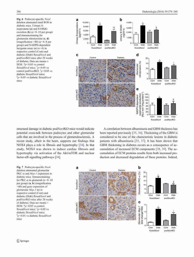
bFig. 6 Podocyte-specific Nox4deletion attenuated renal ROS indiabetic mice. Urinary 8-isoprostane (a) and 8-OHdGexcretion (b) (n=8–10 per group)and immunostaining forglomerular nitrotyrosine (c, d)(magnification ×40) (n=6–8 pergroup) and NADPH-dependentlucigenin assay (e) (n=6) inrespective control (Cont) anddiabetic (Diab) floxedNox4 andpodNox4KO mice after 20 weeksof diabetes. Data are means ±SEM. *p<0.05 vs controlfloxedNox4 mice; †p<0.05 vscontrol podNox4KO; ‡p=0.05 vsdiabetic floxedNox4 mice;§p<0.05 vs diabetic floxedNox4mice
25
20
15
10
5
0
6
4
2
0
*
§
*‡
Cont Diab Cont Diab
floxedNox4 podNox4KO
Cont Diab Cont Diab
floxedNox4 podNox4KO
Control Diabetic
floxe
dNox
4po
dNox
4KO
Glo
mer
ular
PK
C-α
(%
)G
lom
erul
ar M
cp-1
(fol
d ch
ange
/18S
)
a b
c
Fig. 7 Podocyte-specific Nox4deletion attenuated glomerularPKC-α and Mcp-1 expression indiabetic mice. Immunostainingfor PKC-α in glomeruli (n=8–10per group) (a, b) (magnification×40) and gene expression ofglomerular Mcp-1 (c) inrespective control (Cont) anddiabetic (Diab) floxedNox4 andpodNox4KO mice after 20 weeksof diabetes. Data are means ±SEM. *p<0.05 vs controlfloxedNox4 mice; ‡p=0.05 vsdiabetic floxedNox4 mice;§p<0.01 vs diabetic floxedNox4mice
386 Diabetologia (2016) 59:379–389
increased GBM thickness and accumulation of ECM pro-teins (collagen IV and fibronectin) were seen in diabeticmice and these changes were attenuated in diabeticpodNox4KO mice, consistent with NOX4 in podocytesplaying a central role in regulating the production of ECMproteins. Another key morphological feature of DN, iden-tified using electron microscopy, is a reduction in the num-ber of slit pores per unit length of GBM, which reflects footprocess broadening, as has been shown in diabetic rodents[24]. This diabetes-associated reduction in filtration slitpores was attenuated in diabetic podNox4KO mice, furtheremphasising the importance of NOX4 in promotingpodocytopathy, as seen in the diabetic kidney.
Since it is considered that NOX4, also known as ‘renox’, isa key source of renal ROS, this study used various approachesto evaluate renal ROS generation. Markers of DNA oxidation(8-OHdG) and lipid peroxidation (8-isoprostane) have beenshown to be increased in the urine in experimental modelsof diabetes [32, 40, 41]. In addition, increased urinary levelsof 8-OHdG have been reported to be associated with loss ofpodocytes in diabetic rodents [40]. Thus, attenuation of uri-nary excretion of the oxidative stress markers 8-isoprostaneand 8-OHdG, as well as glomerular superoxide andnitrotyrosine, in diabetic podNox4KO mice, as seen in thisstudy, supports the view that NOX4-derived ROS mediatepodocyte damage and ultimately other markers of renal injury.The ability ofNox4 deletion only in podocytes to significantlyreduce a range of markers of glomerular and renal ROS em-phasises the importance of podocyte NOX4 per se as a sourceof ROS in the diabetic kidney.
There is a close interrelationship between ROS and certainPKC isoforms in the development and progression of DN [32,42]. Recently, it has been shown that NOX4-derived ROS canactivate certain PKC isoforms, including PKC-α, within thekidney, thereby promoting renal injury in experimental diabe-tes [32]. Thus, we examined the association of podocyte-specific NOX4 with PKC-α, which has been the most closelyidentified isoform with the development of albuminuria,nephrin depletion and upregulation of intrarenal VEGF in di-abetes [43, 44]. Indeed, we found that the diabetes-inducedincrease in glomerular PKC-α expression was attenuated indiabetic podNox4KO mice. Quack et al have demonstratedthat acute hyperglycaemia increases nephrin endocytosis in aPKC-α-dependent manner [45] and this effect is considered topromote albuminuria. In this study, we found similar changesin VEGF and nephrin expression as well as in albuminuria, indiabetic podNox4KO mice to those seen in diabetic PkcαKOmice, and also noted a reduction in glomerular PKC-α expres-sion in podNox4KO mice. Thus, we postulate that NOX4 is akey upstream regulator of PKC-α expression in the setting ofdiabetes. Indeed, the renoprotective effects of Nox4 deletioncould represent a new approach to target the deleterious ef-fects of PKC-α in the diabetic kidney. In addition, the
diabetes-induced increase in glomerular Mcp-1 expression wasfound to be attenuated in podocyte-specific NOX4-deficientdiabetic mice. This would indicate that targeting NOX4 in thepodocytes not only reduces podocytopathy and glomerularECMaccumulation, butmight also play a key role in attenuatingintrarenal inflammation.
The findings of this study build on an increasingamount of data implicating NOX4 as a potential targetfor renoprotection. The advent of new agents that targetNOX4, such as GKT137831, provide an opportunity totest whether pharmacological inhibition of NOX4 can berenoprotective. Although GKT137831 is not specific forthe NOX4 isoform, several groups, including our own,have demonstrated renal benefits with this agent in var-ious experimental models of DN [9, 46].
In conclusion, NOX4 within the podocyte appears tobe a major contributor to renal ROS generation, activa-tion of PKC-α, enhanced intrarenal fibrosis and inflam-mation, as well as increasing VEGF and decreasingnephrin expression. Furthermore, NOX4 plays a pivotalrole in influencing podocyte ultrafiltration, with a sub-sequent impact on glomerular structure and albuminuria,hallmarks of DN. Hence, targeting NOX4 specifically inthe podocyte might provide a new therapeutic approachin preventing the progression of diabetic kidney disease.
Acknowledgements The authors thank M. Arnstein, K. Gilbert, S.Sacca, E. Lastavec, M. Haillay (Baker IDI Heart & Diabetes Institute,Melbourne, VIC, Australia) for experimental animal handling and tech-nical support.
Compliance with ethical standards
Funding This work was supported by the National Health & MedicalResearch Council of Australia (NHMRC), a JDRF Program/ProjectGrant, the Diabetes Australia Research Trust and the FP7 frameworkprogramme. KAJ-D is supported by a NHMRC Senior Research Fellow-ship and MEC is a Senior Principal Research Fellow for the NHMRC.HHHWS is supported by a Marie Curie International ReintegrationGrant, an ERC Advanced Investigator Grant and the Dutch KidneyFoundation.
Duality of interest statement The authors declare that there is no du-ality of interest associated with this manuscript.
Contribution statement All authors contributed to the study con-cept and design, and the interpretation of the data. JCJ collectedresearch data, contributed to discussions and wrote, reviewed andedited the manuscript. VT-B, CB, SPG and BSMC collected andanalysed the data and contributed to drafting the manuscript. GRhelped with electron microscopy data and contributed to draftingthe manuscript. SEQ and MEC contributed in discussions, andreviewed and edited the manuscript. HHHWS and KAJ-D provid-ed intellectual content in writing the discussions, and reviewedand edited the manuscript. All authors revised the article andapproved the final version for publication. JCJ and KAJ-D arethe guarantors of this work and, as such, had full access to allof the data in the study and take responsibility for the integrity ofthe data and the accuracy of the data analysis.
Diabetologia (2016) 59:379–389 387
References
1. Roglic G, Unwin N, Bennett PH et al (2005) The burden of mor-tality attributable to diabetes: realistic estimates for the year 2000.Diabetes Care 28:2130–2135
2. Molitch ME, DeFronzo RA, Franz MJ et al (2004) Nephropathy indiabetes. Diabetes Care 27(Suppl 1):S79–s83
3. Bjorn SF, Bangstad HJ, Hanssen KF et al (1995) Glomerular epi-thelial foot processes and filtration slits in IDDM patients.Diabetologia 38:1197–1204
4. Eid AA, Gorin Y, Fagg BM et al (2009) Mechanisms of podocyteinjury in diabetes: role of cytochrome P450 and NADPH oxidases.Diabetes 58:1201–1211
5. Tryggvason K, Patrakka J,Wartiovaara J (2006) Hereditary protein-uria syndromes and mechanisms of proteinuria. N Engl J Med 354:1387–1401
6. Calcutt NA, Cooper ME, Kern TS, Schmidt AM (2009) Therapiesfor hyperglycaemia-induced diabetic complications: from animalmodels to clinical trials. Nat Rev Drug Discov 8:417–429
7. Gill PS, Wilcox CS (2006) NADPH oxidases in the kidney.Antioxid Redox Signal 8:1597–1607
8. Kaneto H, Katakami N, Kawamori D et al (2007) Involvement ofoxidative stress in the pathogenesis of diabetes. Antioxid RedoxSignal 9:355–366
9. Jha JC, Gray SP, Barit D et al (2014) Genetic targeting or pharma-cologic inhibition of NADPH oxidase nox4 providesrenoprotection in long-term diabetic nephropathy. J Am SocNephrol 25:1237–1254
10. Gorin Y, Block K, Hernandez J et al (2005) Nox4 NAD(P)H oxi-dase mediates hypertrophy and fibronectin expression in the diabet-ic kidney. J Biol Chem 280:39616–39626
11. Holterman CE, Thibodeau JF, Towaij C et al (2014) Nephropathyand elevated BP in mice with podocyte-specific NADPH oxidase 5expression. J Am Soc Nephrol 25:784–797
12. Brenner BM, Hostetter TH, Humes HD (1978) Molecular basis ofproteinuria of glomerular origin. N Engl J Med 298:826–833
13. Palicz A, Foubert TR, Jesaitis AJ, Marodi L, McPhail LC (2001)Phosphatidic acid and diacylglycerol directly activate NADPH ox-idase by interacting with enzyme components. J Biol Chem 276:3090–3097
14. Satoh M, Fujimoto S, Haruna Yet al (2005) NAD(P)H oxidase anduncoupled nitric oxide synthase are major sources of glomerularsuperoxide in rats with experimental diabetic nephropathy. Am JPhysiol Ren Physiol 288:F1144–F1152
15. Ding G, Reddy K, Kapasi AA et al (2002) Angiotensin II inducesapoptosis in rat glomerular epithelial cells. Am J Physiol RenPhysiol 283:F173–F180
16. Schiffer M, Bitzer M, Roberts IS et al (2001) Apoptosis inpodocytes induced by TGF-β and Smad7. J Clin Invest 108:807–816
17. Wolf G, Chen S, Ziyadeh FN (2005) From the periphery of theglomerular capillary wall toward the center of disease: podocyteinjury comes of age in diabetic nephropathy. Diabetes 54:1626–1634
18. Asaba K, Tojo A, Onozato ML et al (2005) Effects of NADPHoxidase inhibitor in diabetic nephropathy. Kidney Int 67:1890–1898
19. Susztak K, Raff AC, Schiffer M, Bottinger EP (2006) Glucose-induced reactive oxygen species cause apoptosis of podocytes andpodocyte depletion at the onset of diabetic nephropathy. Diabetes55:225–233
20. Watson AM, Li J, Schumacher C et al (2010) The endothelin re-ceptor antagonist avosentan ameliorates nephropathy and athero-sclerosis in diabetic apolipoprotein E knockout mice.Diabetologia 53:192–203
21. Krege JH, Hodgin JB, Hagaman JR, Smithies O (1995) A nonin-vasive computerized tail-cuff system for measuring blood pressurein mice. Hypertension 25:1111–1115
22. Chai Z, Dai A, Tu Y et al (2013) Genetic deletion of cell divisionautoantigen 1 retards diabetes-associated renal injury. J Am SocNephrol 24:1782–1792
23. Lassila M, Seah KK, Allen TJ et al (2004) Accelerated nephropathyin diabetic apolipoprotein e-knockout mouse: role of advancedglycation end products. J Am Soc Nephrol 15:2125–2138
24. Mifsud SA, Allen TJ, Bertram JF et al (2001) Podocyte foot processbroadening in experimental diabetic nephropathy: ameliorationwith renin-angiotensin blockade. Diabetologia 44:878–882
25. Sato S, Sasaki Y, Adachi A, Ghazizadeh M (2010) Validation ofglomerular basement membrane thickness changes with aging inminimal change disease. Pathobiology 77:315–319
26. Koulis C, Chow BS, McKelvey M et al (2015) AT2R agonist,compound 21, is reno-protective against type 1 diabetic nephropa-thy. Hypertension 65:1073–1081
27. Doublier S, Salvidio G, Lupia E et al (2003) Nephrin expression isreduced in human diabetic nephropathy: evidence for a distinct rolefor glycated albumin and angiotensin II. Diabetes 52:1023–1030
28. Chow FY, Nikolic-Paterson DJ, Ozols E, Atkins RC, Rollin BJ,Tesch GH (2006) Monocyte chemoattractant protein-1 promotesthe development of diabetic renal injury in streptozotocin-treatedmice. Kidney Int 69:73–80
29. Cooper ME, Mundel P, Boner G (2002) Role of nephrin in renaldisease including diabetic nephropathy. Semin Nephrol 22:393–398
30. Cooper ME, Vranes D, Youssef S et al (1999) Increased renal ex-pression of vascular endothelial growth factor (VEGF) and its re-ceptor VEGFR-2 in experimental diabetes. Diabetes 48:2229–2239
31. Sung SH, Ziyadeh FN, Wang A, Pyagay PE, Kanwar YS, Chen S(2006) Blockade of vascular endothelial growth factor signalingameliorates diabetic albuminuria in mice. J Am Soc Nephrol 17:3093–3104
32. Thallas-Bonke V, Jha JC, Gray SP et al (2014) Nox-4 deletionreduces oxidative stress and injury by PKC-α-associated mecha-nisms in diabetic nephropathy. Physiol Rep 2, e12192
33. Ziyadeh FN,Wolf G (2008) Pathogenesis of the podocytopathy andproteinuria in diabetic glomerulopathy. Curr Diabetes Rev 4:39–45
34. Zhao QD, Viswanadhapalli S, Williams P et al (2015) NADPHoxidase 4 induces cardiac fibrosis and hypertrophy through activat-ing Akt/mTOR and NFκB signaling pathways. Circulation 131:643–655
35. Bangstad HJ, Osterby R, Dahl-Jorgensen K, Berg KJ, Hartmann A,Hanssen KF (1994) Improvement of blood glucose control inIDDM patients retards the progression of morphological changesin early diabetic nephropathy. Diabetologia 37:483–490
36. Tervaert TW, Mooyaart AL, Amann K et al (2010) Pathologic clas-sification of diabetic nephropathy. J Am Soc Nephrol 21:556–563
37. Mac-Moune Lai F, Szeto CC, Choi PC et al (2004) Isolate diffusethickening of glomerular capillary basement membrane: a renallesion in prediabetes? Mod Pathol 17:1506–1512
38. Falk RJ, Scheinman JI, Mauer SM, Michael AF (1983)Polyantigenic expansion of basement membrane constituents indiabetic nephropathy. Diabetes 32(Suppl 2):34–39
39. Kim Y, Kleppel MM, Butkowski R, Mauer SM, Wieslander J,Michael AF (1991) Differential expression of basement membranecollagen chains in diabetic nephropathy. Am J Pathol 138:413–420
40. Kim J, Shon E, Kim CS, Kim JS (2012) Renal podocyte injury in arat model of type 2 diabetes is prevented by metformin. ExpDiabetes Res 2012:210821
41. Montero A, Munger KA, Khan RZ et al (2000) F2-isoprostanesmediate high glucose-induced TGF-β synthesis and glomerularproteinuria in experimental type I diabetes. Kidney Int 58:1963–1972
388 Diabetologia (2016) 59:379–389
42. Giorgi C, Agnoletto C, Baldini C et al (2010) Redox control ofprotein kinase C: cell- and disease-specific aspects. AntioxidRedox Signal 13:1051–1085
43. Menne J, Meier M, Park JK et al (2006) Nephrin loss in experimen-tal diabetic nephropathy is prevented by deletion of protein kinaseC alpha signaling in-vivo. Kidney Int 70:1456–1462
44. Menne J, Shushakova N, Bartels J et al (2013) Dual inhibition ofclassical protein kinase C-α and protein kinase C-β isoforms
protects against experimental murine diabetic nephropathy.Diabetes 62:1167–1174
45. Quack I, Woznowski M, Potthoff SA et al (2011) PKCα mediatesβ-arrestin2-dependent nephrin endocytosis in hyperglycemia. JBiol Chem 286:12959–12970
46. Gorin Y, Cavaglieri RC, Khazim K et al (2015) Targeting NADPHoxidase with a novel dual Nox1/Nox4 inhibitor attenuates renalpathology in type 1 diabetes. Am J Physiol Ren Physiol 308:F1276–F1287
Diabetologia (2016) 59:379–389 389