Human Carbonic Anhydrase IV:In VitroActivation and Purification of Disulfide-Bonded Enzyme Following...
9
PROTEIN EXPRESSION AND PURIFICATION 9, 279–287 (1997) ARTICLE NO. PT960691 Human Carbonic Anhydrase IV: In Vitro Activation and Purification of Disulfide-Bonded Enzyme Following Expression in Escherichia coli Abdul Waheed, Thanh Pham, Michelle Won, Torayuki Okuyama, and William S. Sly 1 Edward A. Doisy Department of Biochemistry and Molecular Biology, St. Louis University School of Medicine, St. Louis, Missouri 63104 Received June 10, 1996, and in revised form October 14, 1996 plasma membranes of epithelial cells and endothelial Human carbonic anhydrase IV (CA IV) expressed in cells of microcapillaries of several tissues (4 – 10). The Escherichia coli was refolded and activated in cell ex- carbohydrate content of CA IV in mammals varies with tracts with the help of endogenous periplasmic protein species (2,11). Although CA IV is a glycoprotein in ev- disulfide isomerase, DsbA, in the presence of oxidized ery other species so far examined, including rat, mouse, glutathione. The refolding and activation were inhib- cow, sheep, and rabbit (2,11), human CA IV contains ited by bacitracin but not affected by known cofactors no carbohydrate, suggesting that carbohydrate is not or activators of other chaperones. Although the yield important for CA IV enzymatic activity. CA IV has been of the purified CA IV recovered from cell extracts was purified from tissues of several mammalian species maximal when activated at 47C in the presence of 2 mM (1,2,11,12). The stability of CA IV activity in SDS-con- oxidized glutathione, the rate of refolding and activa- taining solutions distinguishes CA IV from soluble iso- tion was much more rapid at 25 and 377C. The enzyme zymes of CA and simplifies isolation of CA IV for tissues purified from the E. coli cell extracts following activa- containing multiple CAs (1,11). Resistance to SDS has tion in vitro showed similar structural stability and been attributed to the disulfide bonds of the CA IV functional properties as CA IV purified from secretion (1,11,16). medium from a stably transfected CHO cell line. These CA IV cDNAs have been cloned from human, rat, studies suggest that the soluble truncated form of hu- man CA IV expressed in E. coli, which is a disulfide- and mouse tissues (13,6,14) and the human CA IV gene bonded zinc metalloenzyme, can provide a useful has been localized to chromosome 17 (15). The amino model enzyme for studies of protein folding and en- acid sequence deduced from the cDNAs contains se- zyme activation in vitro. Furthermore, the procedure quence for a C-terminal hydrophobic domain typical of described for recovery of CA IV following expression precursors of GPI-anchored proteins. The CA IV cDNA in E. coli may be useful for in vitro activation and sub- also predicts four cysteine residues (6,13). Transfection sequent purification of other disulfide-containing pro- of COS-7 cells with a cDNA encoding a truncated CA teins. q 1997 Academic Press IV, which has the C-terminal hydrophobic domain de- leted, results in the synthesis and secretion of a soluble, functionally active CA IV (16). Human carbonic anhydrase IV (CA IV 2 ) is a highly active, GPI-anchored protein (1 – 4) expressed on the In order to prepare milligram quantities of CA IV for structure – function studies, for X-ray crystallography, and for studying the sensitivity of CA IV to new CA 1 To whom correspondence should be addressed at Edward A. Doisy Department of Biochemistry and Molecular Biology, St. Louis Uni- inhibitors of pharmacological significance, we consid- versity School of Medicine, 1402 South Grand Blvd., St. Louis, MO ered expressing recombinant CA IV in Escherichia coli. 63104. Fax: (314) 776-1183. Several recombinant proteins with and without disul- 2 Abbreviations used: CA, carbonic anhydrase; GPI, glycosylphos- phatidylinositol; DsbA, protein disulfide isomerase A; PDI, protein fide bonds have been expressed in E. coli and purified disulfide isomerase; SDS, sodium dodecyl sulfate; IPTG, isopropyl- from cell lysates or from inclusion bodies containing 1-thio-b-D-galactopyranoside; GSSG, oxidized glutathione; GSH, re- insoluble enzyme isolated from cell lysates (17 – 21). duced glutathione; DTT, dithiothreitol; PCR, polymerase chain reac- Although most procedures for solubilizing enzyme from tion; EDTA, ethylenediaminetetraacetic acid; PMSF, phenylmethyl- sulfonyl fluoride; PAGE, polyacrylamide gel electrophoresis. inclusion bodies by denaturation and refolding of the 279 1046-5928/97 $25.00 Copyright q 1997 by Academic Press All rights of reproduction in any form reserved.
-
Upload
abdul-waheed -
Category
Documents
-
view
212 -
download
0
Transcript of Human Carbonic Anhydrase IV:In VitroActivation and Purification of Disulfide-Bonded Enzyme Following...

PROTEIN EXPRESSION AND PURIFICATION 9, 279–287 (1997)ARTICLE NO. PT960691
Human Carbonic Anhydrase IV: In Vitro Activation andPurification of Disulfide-Bonded Enzyme FollowingExpression in Escherichia coli
Abdul Waheed, Thanh Pham, Michelle Won, Torayuki Okuyama, and William S. Sly1
Edward A. Doisy Department of Biochemistry and Molecular Biology, St. Louis University School of Medicine,St. Louis, Missouri 63104
Received June 10, 1996, and in revised form October 14, 1996
plasma membranes of epithelial cells and endothelialHuman carbonic anhydrase IV (CA IV) expressed in cells of microcapillaries of several tissues (4–10). The
Escherichia coli was refolded and activated in cell ex- carbohydrate content of CA IV in mammals varies withtracts with the help of endogenous periplasmic protein species (2,11). Although CA IV is a glycoprotein in ev-disulfide isomerase, DsbA, in the presence of oxidized ery other species so far examined, including rat, mouse,glutathione. The refolding and activation were inhib- cow, sheep, and rabbit (2,11), human CA IV containsited by bacitracin but not affected by known cofactors no carbohydrate, suggesting that carbohydrate is notor activators of other chaperones. Although the yield important for CA IV enzymatic activity. CA IV has beenof the purified CA IV recovered from cell extracts was purified from tissues of several mammalian speciesmaximal when activated at 47C in the presence of 2 mM (1,2,11,12). The stability of CA IV activity in SDS-con-oxidized glutathione, the rate of refolding and activa-
taining solutions distinguishes CA IV from soluble iso-tion was much more rapid at 25 and 377C. The enzymezymes of CA and simplifies isolation of CA IV for tissuespurified from the E. coli cell extracts following activa-containing multiple CAs (1,11). Resistance to SDS hastion in vitro showed similar structural stability andbeen attributed to the disulfide bonds of the CA IVfunctional properties as CA IV purified from secretion(1,11,16).medium from a stably transfected CHO cell line. These
CA IV cDNAs have been cloned from human, rat,studies suggest that the soluble truncated form of hu-man CA IV expressed in E. coli, which is a disulfide- and mouse tissues (13,6,14) and the human CA IV genebonded zinc metalloenzyme, can provide a useful has been localized to chromosome 17 (15). The aminomodel enzyme for studies of protein folding and en- acid sequence deduced from the cDNAs contains se-zyme activation in vitro. Furthermore, the procedure quence for a C-terminal hydrophobic domain typical ofdescribed for recovery of CA IV following expression precursors of GPI-anchored proteins. The CA IV cDNAin E. coli may be useful for in vitro activation and sub- also predicts four cysteine residues (6,13). Transfectionsequent purification of other disulfide-containing pro- of COS-7 cells with a cDNA encoding a truncated CAteins. q 1997 Academic Press IV, which has the C-terminal hydrophobic domain de-
leted, results in the synthesis and secretion of a soluble,functionally active CA IV (16).Human carbonic anhydrase IV (CA IV2) is a highly
active, GPI-anchored protein (1–4) expressed on the In order to prepare milligram quantities of CA IV forstructure–function studies, for X-ray crystallography,and for studying the sensitivity of CA IV to new CA1 To whom correspondence should be addressed at Edward A. Doisy
Department of Biochemistry and Molecular Biology, St. Louis Uni- inhibitors of pharmacological significance, we consid-versity School of Medicine, 1402 South Grand Blvd., St. Louis, MO
ered expressing recombinant CA IV in Escherichia coli.63104. Fax: (314) 776-1183.Several recombinant proteins with and without disul-2 Abbreviations used: CA, carbonic anhydrase; GPI, glycosylphos-
phatidylinositol; DsbA, protein disulfide isomerase A; PDI, protein fide bonds have been expressed in E. coli and purifieddisulfide isomerase; SDS, sodium dodecyl sulfate; IPTG, isopropyl- from cell lysates or from inclusion bodies containing1-thio-b-D-galactopyranoside; GSSG, oxidized glutathione; GSH, re- insoluble enzyme isolated from cell lysates (17–21).duced glutathione; DTT, dithiothreitol; PCR, polymerase chain reac-
Although most procedures for solubilizing enzyme fromtion; EDTA, ethylenediaminetetraacetic acid; PMSF, phenylmethyl-sulfonyl fluoride; PAGE, polyacrylamide gel electrophoresis. inclusion bodies by denaturation and refolding of the
2791046-5928/97 $25.00Copyright q 1997 by Academic PressAll rights of reproduction in any form reserved.
AID PEP 0691 / 6q0e$$$$61 01-31-97 22:17:45 pepa AP: PEP
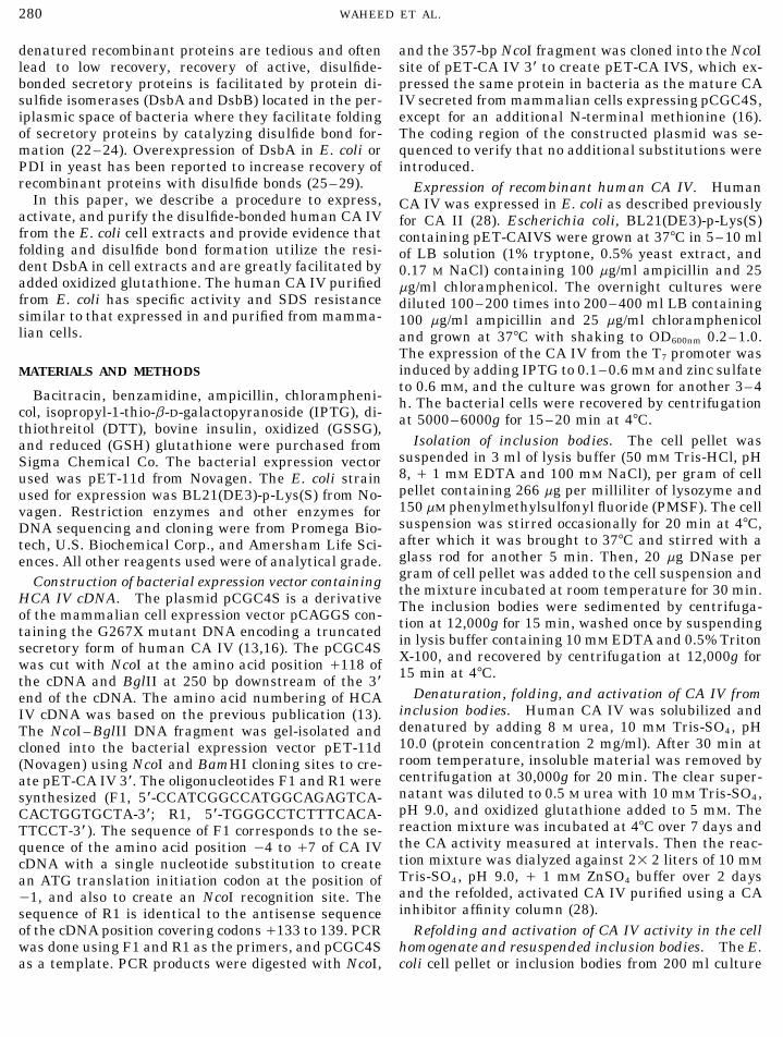
WAHEED ET AL.280
denatured recombinant proteins are tedious and often and the 357-bp NcoI fragment was cloned into the NcoIsite of pET-CA IV 3* to create pET-CA IVS, which ex-lead to low recovery, recovery of active, disulfide-
bonded secretory proteins is facilitated by protein di- pressed the same protein in bacteria as the mature CAIV secreted from mammalian cells expressing pCGC4S,sulfide isomerases (DsbA and DsbB) located in the per-
iplasmic space of bacteria where they facilitate folding except for an additional N-terminal methionine (16).The coding region of the constructed plasmid was se-of secretory proteins by catalyzing disulfide bond for-
mation (22–24). Overexpression of DsbA in E. coli or quenced to verify that no additional substitutions wereintroduced.PDI in yeast has been reported to increase recovery of
recombinant proteins with disulfide bonds (25–29). Expression of recombinant human CA IV. HumanIn this paper, we describe a procedure to express, CA IV was expressed in E. coli as described previously
activate, and purify the disulfide-bonded human CA IV for CA II (28). Escherichia coli, BL21(DE3)-p-Lys(S)from the E. coli cell extracts and provide evidence that containing pET-CAIVS were grown at 377C in 5–10 mlfolding and disulfide bond formation utilize the resi- of LB solution (1% tryptone, 0.5% yeast extract, anddent DsbA in cell extracts and are greatly facilitated by 0.17 M NaCl) containing 100 mg/ml ampicillin and 25added oxidized glutathione. The human CA IV purified mg/ml chloramphenicol. The overnight cultures werefrom E. coli has specific activity and SDS resistance diluted 100–200 times into 200–400 ml LB containingsimilar to that expressed in and purified from mamma- 100 mg/ml ampicillin and 25 mg/ml chloramphenicollian cells. and grown at 377C with shaking to OD600nm 0.2–1.0.
The expression of the CA IV from the T7 promoter wasinduced by adding IPTG to 0.1–0.6 mM and zinc sulfateMATERIALS AND METHODSto 0.6 mM, and the culture was grown for another 3–4
Bacitracin, benzamidine, ampicillin, chlorampheni- h. The bacterial cells were recovered by centrifugationcol, isopropyl-1-thio-b-D-galactopyranoside (IPTG), di- at 5000–6000g for 15–20 min at 47C.thiothreitol (DTT), bovine insulin, oxidized (GSSG),
Isolation of inclusion bodies. The cell pellet wasand reduced (GSH) glutathione were purchased fromsuspended in 3 ml of lysis buffer (50 mM Tris-HCl, pHSigma Chemical Co. The bacterial expression vector8, / 1 mM EDTA and 100 mM NaCl), per gram of cellused was pET-11d from Novagen. The E. coli strainpellet containing 266 mg per milliliter of lysozyme andused for expression was BL21(DE3)-p-Lys(S) from No-150 mM phenylmethylsulfonyl fluoride (PMSF). The cellvagen. Restriction enzymes and other enzymes forsuspension was stirred occasionally for 20 min at 47C,DNA sequencing and cloning were from Promega Bio-after which it was brought to 377C and stirred with atech, U.S. Biochemical Corp., and Amersham Life Sci-glass rod for another 5 min. Then, 20 mg DNase perences. All other reagents used were of analytical grade.gram of cell pellet was added to the cell suspension andConstruction of bacterial expression vector containing the mixture incubated at room temperature for 30 min.HCA IV cDNA. The plasmid pCGC4S is a derivative The inclusion bodies were sedimented by centrifuga-of the mammalian cell expression vector pCAGGS con- tion at 12,000g for 15 min, washed once by suspendingtaining the G267X mutant DNA encoding a truncated in lysis buffer containing 10 mM EDTA and 0.5% Tritonsecretory form of human CA IV (13,16). The pCGC4S X-100, and recovered by centrifugation at 12,000g forwas cut with NcoI at the amino acid position /118 of 15 min at 47C.the cDNA and BglII at 250 bp downstream of the 3*
Denaturation, folding, and activation of CA IV fromend of the cDNA. The amino acid numbering of HCAinclusion bodies. Human CA IV was solubilized andIV cDNA was based on the previous publication (13).denatured by adding 8 M urea, 10 mM Tris-SO4, pHThe NcoI–BglII DNA fragment was gel-isolated and10.0 (protein concentration 2 mg/ml). After 30 min atcloned into the bacterial expression vector pET-11droom temperature, insoluble material was removed by(Novagen) using NcoI and BamHI cloning sites to cre-centrifugation at 30,000g for 20 min. The clear super-ate pET-CA IV 3*. The oligonucleotides F1 and R1 werenatant was diluted to 0.5 M urea with 10 mM Tris-SO4,synthesized (F1, 5*-CCATCGGCCATGGCAGAGTCA-pH 9.0, and oxidized glutathione added to 5 mM. TheCACTGGTGCTA-3*; R1, 5*-TGGGCCTCTTTCACA-reaction mixture was incubated at 47C over 7 days andTTCCT-3* ). The sequence of F1 corresponds to the se-the CA activity measured at intervals. Then the reac-quence of the amino acid position 04 to /7 of CA IVtion mixture was dialyzed against 21 2 liters of 10 mMcDNA with a single nucleotide substitution to createTris-SO4, pH 9.0, / 1 mM ZnSO4 buffer over 2 daysan ATG translation initiation codon at the position ofand the refolded, activated CA IV purified using a CA01, and also to create an NcoI recognition site. Theinhibitor affinity column (28).sequence of R1 is identical to the antisense sequence
of the cDNA position covering codons/133 to 139. PCR Refolding and activation of CA IV activity in the cellhomogenate and resuspended inclusion bodies. The E.was done using F1 and R1 as the primers, and pCGC4S
as a template. PCR products were digested with NcoI, coli cell pellet or inclusion bodies from 200 ml culture
AID PEP 0691 / 6q0e$$$$61 01-31-97 22:17:45 pepa AP: PEP

FOLDING OF DISULFIDE-BONDED PROTEIN IN E. coli 281
expressing CA IV was homogenized with 10 ml of lysate a stepwise gradient of NaCl. The absorbance at 280nm and insulin reduction activity of the fractions werebuffer, 50 mM Tris-SO4 (pH 8) / 0.1% Triton X-100 and
1 mM benzamidine using Brinckman Polytron at room followed. The fractions eluted with 25 and 50 mM NaClcontained most of the DsbA activity. These fractionstemperature. Since E. coli cytosol is reducing, the re-
combinant proteins expressed inside the bacteria are were concentrated using an Amicon PM-10 membraneand dialyzed against 100 mM Tris-HCl buffer, pH 7.5.assumed to have cysteine residues in the reduced state.
However, the periplasma of E. coli contains a protein The enzyme preparation was applied to a Sephadex G-75 (2.51 100 cm) column. Five-milliliter fractions weredisulfide isomerase, DsbA, capable of catalyzing the
formation of disulfide bonds in the presence of oxidized collected and monitored for OD280 nm and insulin reduc-ing activity (DsbA activity). The fractions containingglutathione (25). Therefore, the cell homogenates and
resuspended inclusion bodies were incubated at 47C in DsbA activity were saved and checked for homogeneityon SDS–PAGE.the presence and absence of 10 mM GSSG. The CA IV
activity was measured at different times to assess the DsbA activity determination. DsbA activity was de-rate of refolding and activation of the enzyme. termined by the turbidometric procedure of Holmgren
In order to study the effect of temperature and con- (33) using bovine insulin at 1.0 mg/ml in 100 mM Tris-centrations of oxidized glutathione and chaperone acti- HCl, pH 7.5, 2 mM DsbA, and 300 mM DTT. The tur-vators (29,30) on the activation rate, the cell homoge- bidity induced by reduction of insulin chains was mea-nates were incubated at 4, 25, and 377C or incubated sured at 650 nm. In negative controls, assay buffer waswith 0.5–10 mM GSSG and 2 mM ATP, 10 mM MgCl2 present in the place of enzyme sample.and KCl each at 257C. Aliquots were removed at inter- Activation of CA IV by DsbA. The inclusion bodiesvals and assayed for CA IV activity. The amount (mg) of were incubated in lysate buffer alone or with purifiedactivated enzyme was calculated from the CA activity DsbA, 2 mM, in the presence of 10 mM GSSG with andusing 4000 U/mg as the specific activity of CA IV. without 10 mM bacitracin at 257C. An aliquot of the
Effect of bacitracin on the activation of CA IV activity. reaction mixture was removed and assayed for the CAThe soluble supernatant obtained following centrifuga- activity.tion of homogenates of uninduced E. coli was used as Purification of recombinant CA IV from E. coli. Hu-a source of crude protein disulfide isomerase. The unin- man CA IV was purified from the E. coli cell extractsduced E. coli cell homogenate in 50 mM Tris-SO4 (pH or isolated inclusion body extracts using a sulfon-8) / 0.1% Triton X-100 and 1 mM benzamidine buffer amide–Affi-Gel-10 column as described for human CAwas centrifuged at 30,000g for 20 min to produce this II (28). Cell homogenates or inclusion bodies from 200-supernatant. The homogenates of isolated inclusion ml cell cultures were activated with 10 mM GSSG inbodies in lysate buffer were incubated alone or in the lysate buffer at 4 or 257C. After maximum activation,presence of 10 mM GSSG, with and without the protein the reaction mixture was centrifuged at 30,000g for 20disulfide isomerase preparation at 257C. An aliquot of min and the clear supernatant was applied to the affin-the reaction mixture was removed at indicated times ity column. The unbound proteins were removed byand assayed for CA activity. Under these conditions, washing the column sequentially with 10 mM Tris-SO4,the crude protein disulfide isomerase preparation con- pH 9.0, 100 mM Tris-SO4 (pH 7) / 0.2 M Na2SO4, andtained no CA activity and none was produced by adding 100 mM Tris-SO4, pH 7, buffers. The bound enzymeGSSG. To assess the role of this crude protein disulfide was eluted from the column with 100 mM sodium ace-isomerase in the folding and activation of CA IV, the tate (pH 5.6) / 0.5 M sodium perchlorate. The CA IVinclusion body homogenate was incubated in the pres- recovered was concentrated by Amicon filtration usingence of GSSG and an aliquot of the crude protein disul- a YM-10 membrane and extensively dialyzed againstfide isomerase preparation, with and without 10 mM of 50 mM Tris-SO4, pH 8.0 buffer. The protein concentra-bacitracin, an inhibitor of protein disulfide isomerase tion and the CA activity were determined.(31,32). Carbonic anhydrase measurement. Carbonic anhy-
Purification of DsbA from E. coli. The resident drase activity was assayed according to Maren (34) asDsbA from E. coli, BL21(DE3)-p-lys(S), was purified described (35). To test for SDS sensitivity, purified CAto homogeneity using DEAE-Sepharose and Sephadex IV (1 EU) in the presence of 1 mg/ml bovine serumG75 column chromatographies. The cell pellet of a 5- albumin was incubated with 0.2% SDS at room temper-liter culture was homogenized in 100 ml of 10 mM Tris- ature for 30 min before CA assay.HCl, pH 7.5, and centrifuged at 30,000g. The clear su- Protein concentration was determined by the proce-pernatant was applied to a 15-ml DEAE-Sepharose col- dure of Lowry using bovine serum albumin as a stan-umn equilibrated with 10 mM Tris-HCl buffer, pH 7.5, dard protein (36). Sodium dodecylsulfate–polyacryl-at 47C. Unbound proteins were washed out with equili- amide gel electrophoresis (SDS–PAGE) was performed
under reducing conditions (37).bration buffer and the bound proteins were eluted with
AID PEP 0691 / 6q0e$$$$61 01-31-97 22:17:45 pepa AP: PEP

WAHEED ET AL.282
RESULTS
Previous studies in several laboratories have shownthat the human cytoplasmic isozymes CA I, CA II, CAIII, and the mitochondrial isozyme CA V can all beproduced in E. coli in high yields (18,28). We recentlyreported that human CA IV, the normally GPI-an-chored, membrane-associated isozyme, is secreted as asoluble isozyme and in large amounts, when a trunca-tion mutant which lacks the C-terminal transmem-brane domain and site of GPI attachment is expressedin mammalian cells (16). In an effort to produce thesoluble form of CA IV in E. coli, we engineered thecDNA to remove both the C-terminal domain men-tioned above and also the N-terminal 23-amino-acidsignal sequence, and subcloned the shortened cDNAinto the bacterial expression vector PET11d. Unlike
FIG. 2. Activation of CA IV in the crude cell homogenates and thethe results seen following induction of CA II in this inclusion bodies at 47C. The total cell homogenates (CH) without (m)vector, which produced large amounts of soluble CA II and with (l) 10 mM GSSG, and inclusion bodies (IB) (j) and cytosol
(s) with 10 mM GSSG were incubated and analyzed for CA IV activi-activity in bacterial lysates, there was 0.001% the levelties which are expressed in mg/ml active enzyme based on a specificof activity seen with CA II in extracts of cells preparedactivity of 4000 units/mg.following induction of the CA IV cDNA in PET11d.
However, as shown in Fig. 1, the expected 33-kDa pro-tein was quite abundant in homogenates of cells in-
vitro by oxidized glutathione (GSSG) (19). To test thisduced at both 0.2 and 0.6 OD units of the cell culture.hypothesis, the isolated inclusion bodies were dena-Figure 1 also shows that more than 99% of this proteintured in urea as described (19), and then dialyzed tosedimented with membranes in ‘‘inclusion bodies’’ fol-remove urea in a buffer containing ZnCl and oxidizedlowing sedimentation of the homogenates from cellsglutathione. After 1 week of incubation at 47C, onlyinduced at 0.6 OD units.100–200 enzyme units per liter were observed, which
Activation of CA IV in vitro. Since the active form is 0.01% of the CA IV activated under what were laterof CA IV produced by mammalian cells contains two found to be optimal conditions. Although the yield wasdisulfide bridges, we suspected that the aggregated en- disappointing, this experiment demonstrated that thezyme was reduced in E. coli and might be activated in lysates from induced bacteria contain a latent CA IV
that can be activated. A variety of modifications of lysisconditions, sedimentation conditions, and reactivationconditions were tried in an effort to improve the recov-ery of active enzyme. Denaturation of the sedimentableenzyme in urea proved unnecessary. The experimentsummarized in Fig. 2 shows that GSSG can promotethe activation of CA IV in crude cell homogenates aswell as in resuspended pellets. There was no activationof CA IV in the 30,000g supernatant (cytosol), sug-gesting that all CA IV is present in sedimentable pellet.Thus, initially inactive enzyme, nearly all of which issedimentable, can be activated by GSSG without ureadenaturation.
Next, we observed the effects of temperature on acti-vation of the latent CA IV activity. Figure 3 comparesthe rates and extent of activation of CA IV by GSSG atFIG. 1. Expression of human CA IV in E. coli. The E. coli cells25 and 377C in crude homogenates and in resuspendedcontaining CA IV cDNA were grown to a density of 0.2 and 0.6inclusion bodies. Activation was more rapid at 37 thanOD600nm before induction with 0.6 mM IPTG. The cell homogenates
(lanes 2–4), supernatant (lane 5), and inclusion bodies (lane 6), each at 257C, which in turn was more rapid than at 47C.equivalent to 50 mg protein, were analyzed by SDS–PAGE. The gel However, the extent of activation was greater at 25was stained with silver and the polypeptide for CA IV is marked. than at 377C, and greater for enzyme in crude lysatesThe apparent molecular weights of the standard proteins (lane 1)
than in inclusion bodies. These data suggested that aare indicated. Cell homogenates were from uninduced cells (lane 4)and cells induced at OD 0.2 (lane 2) and OD 0.6 (lane 3). protein in the cell homogenate which might be temper-
AID PEP 0691 / 6q0e$$$$61 01-31-97 22:17:45 pepa AP: PEP

FOLDING OF DISULFIDE-BONDED PROTEIN IN E. coli 283
FIG. 5. Effect of concentration of oxidized glutathione on the activa-FIG. 3. Activation of CA IV activity in the total cell homogenates tion of CA IV activity. The total cell homogenates were incubatedand the inclusion bodies at 25 and 377C. The cell homogenates at with 5 mM reduced glutathione (GSH) or with oxidized glutathione257C (m) and 377C (j) and the inclusion bodies at 257C (l) and 377C (GSSG) at 0.5–10 mM concentrations for 7 h at 257C and analyzed(l) were incubated with 10 mM GSSG. An aliquot of the reaction for CA IV activity.mixtures was analyzed for CA activity at indicated times.
Optimal GSSG concentration for activation of CA IV.To define the optimum concentration of GSSG, the ho-
ature-sensitive (inactivated at 377C) might be promot- mogenate of cells expressing CA IV was incubated ating the activation. To test this hypothesis, an experi- 257C with reduced glutathione (GSH) or with varyingment was carried out in which an induced cell homoge- concentrations of oxidized glutathione for 7 h beforenate was activated for 4 h at 377C, divided, and the extent of activation was measured (Fig. 5). No acti-incubated an additional 4 h with or without added solu- vation was seen with GSH. The maximum activationble supernatant from an uninduced cell homogenate. with GSSG was seen at 2 mM concentration. No furtherAs shown in Fig. 4, the added soluble supernatant pro- activation was produced by 5 or 10 mM GSSG.duces further activation while little additional activa-
Effects of cofactors for other chaperones on activationtion is seen in the absence of additional supernatant.of CA IV. Chaperone-dependent folding of certain pro-teins is enhanced by Mg2/, ATP, and K/ (29,30). Anexperiment testing the ability of these agents to influ-ence activation is summarized in Fig. 6. None of these
FIG. 4. Effect of added uninduced E. coli cell homogenates on acti-vation of CA IV at 377C. CA IV activities were determined on aninduced cell homogenate incubated for 4 h at 377C with (j) or without FIG. 6. Effect of other chaperone activators on the activation of CA
IV activity. The total cell homogenates alone as a control (first bar),(l) 5 mM GSSG, after which the sample was divided in two parts,one of which was further incubated at 377C without other additives, with 10 mM GSSG (second bar), 10 mM MgCl2 (third bar), 10 mM
KCl (fourth bar), and 2 mM ATP (fifth bar) were incubated at 257Cand one of which was further incubated after adding an equal volumeof uninduced cell homogenate (U-CH) which contains no CA IV. for 16 and 32 h.
AID PEP 0691 / 6q0e$$$$61 01-31-97 22:17:45 pepa AP: PEP

WAHEED ET AL.284
iplasmic disulfide isomerase DsbA seemed a likely can-didate for this activity in the extract (39).
Effect of purified DsbA on the activation of CA IV.In order to confirm the role of DsbA in the activationof CA IV, we purified DsbA using ion exchange andsizing column chromatography. However, after the firststep, the E. coli DsbA was still contaminated with afew other proteins (Fig. 8, inset, lane 1). As shown inFig. 8 (inset, lane 2), it was homogeneous after thesizing column step and showed an apparent molecularweight of 22 kDa on SDS–PAGE after the exchangecolumn. The purified 22-kDa DsbA was able to catalyzethe reduction of insulin in the presence of DTT as ob-served earlier (22).
To examine the ability of DsbA to activate CA IV,isolated inclusion bodies were suspended with 10 mMFIG. 7. Effect of uninduced E. coli extract (U-CH) and bacitracin,
a DsbA inhibitor, on the activation of CA IV in the sedimentable GSSG in the presence and absence of 2 mM purified DsbAinclusion bodies. The inclusion bodies alone (m), U-CH / GSSG (h), and the CA IV activity was determined. The results ininclusion bodies / GSSG (l), inclusion bodies / GSSG / U-CH Fig. 9 show that the inactive CA IV in the inclusion(j), and inclusion bodies / GSSG / U-CH / bacitracin (l) were
bodies was activated by DsbA at a rate of 50–60 mg/incubated at 257C; the CA IV activity was determined as described.ml/h over the first 3 h at 257C, and without DsbA theAddition of bacitracin to inclusion bodies alone blocked the low level
of activation seen in the absence of bacitracin (m) (data not shown). activation rate was 3–4 mg/ml/h. These results sug-gested that DsbA stimulated the activation of CA IV by15- to 20-fold. However, when the inclusion bodies were
agents significantly affected the rate or extent of acti- suspended with 10 mM GSSG, 2 mM DsbA, and 10 mMvation of CA IV in the absence of GSSG, which was bacitracin, the activation rate dropped to 10 mg/ml/h.only 20% of that produced by GSSG. Furthermore, com- Purification of CA IV from E. coli homogenates. Webinations of these agents did not accelerate activation next sought to determine whether the soluble trun-in the presence or absence of added GSSG (data not cated form of CA IV expressed in E. coli and activatedshown). in vitro in cell homogenates could be purified in quanti-
Bacitracin inhibits GSSG-dependent activation of CA ties sufficient for structure–function studies and forIV by the supernatant. We next determined whether crystallization. Cell homogenates from 200-ml culturesthe factor in the supernatant from uninduced cell ho- were induced at OD 0.6, following which both unsedi-mogenate which enhanced the activation of CA IV in mented cell homogenates and resuspended inclusionhomogenates (Fig. 4) would enhance activation of CA bodies sedimented from cell homogenates were acti-IV in inclusion bodies. Figure 7 demonstrates the stim-ulation of activation by GSSG, the enhancement of thisstimulation by additional supernatant, and theblocking of this stimulation by the protein disulfideisomerase inhibitor bacitracin. In this experiment, in-clusion bodies were isolated from induced cells, resus-pended, and incubated with or without GSSG at 257C.As seen earlier, GSSG markedly stimulated the rateand extent of activation. Addition of the 30,000g 1 20min supernatant from the noninduced cell lysate,which had no CA activity itself and no latent CA activ-ity that could be activated by GSSG, further stimulatedthe GSSG-dependent activation of CA IV in inclusionbodies. The addition of bacitracin inhibited the activa-tion of CA IV in inclusion bodies, whether or not addi-tional supernatant was added. These results suggested
FIG. 8. Purification of DsbA from E. coli. DsbA preparations con-that maximal activation was dependent on an activitytaining 2 mg protein after DEAE-sepharose and Sephadex G-75 col-in the bacterial extract that was present in less thanumn chromatographies were analyzed by SDS–PAGE and stainedsaturating amounts in the membrane-associated inclu- with silver stain (inset, lanes 1 and 2, respectively). DsbA activity
sion bodies containing sedimentable CA IV, and that was determined by measuring the rate of reduction of insulin, fol-lowed turbidimetrically at 650 nm.this activity could be inhibited by bacitracin. The per-
AID PEP 0691 / 6q0e$$$$61 01-31-97 22:17:45 pepa AP: PEP

FOLDING OF DISULFIDE-BONDED PROTEIN IN E. coli 285
bic domain, which is normally cleaved and removed inthe process of GPI anchoring, the CA IV is synthesizedat a greater rate and as a secretory form with full activ-ity (16). The desirability of producing large amounts ofthe secretory form of CA IV for use in characterizingits sensitivity to pharmacologically active CA inhibitorsas well as for crystallization and for structure–functionstudies prompted us to attempt to prepare the recombi-nant, soluble, truncated form of human CA IV from E.coli.
Following conventional procedures for the purifica-tion of recombinant proteins in E. coli, where purifiedinclusion bodies containing CA IV were first denaturedand the denatured, solubilized enzyme was then re-folded and activated in the presence of GSSG andZnSO4 before purification, we obtained disappointingyields of purified enzyme. Similar results have beenreported for several other recombinant proteins con-
FIG. 9. Effects of purified DsbA on the activation of CA IV in the taining disulfide bonds (17,19). These observationssedimentable inclusion bodies and the inhibitors of activation by probably reflect the fact that disulfide bond formationbacitracin. The inclusion bodies were incubated at 257C with 10 mM in eukaryotic (38) and prokaryotic (25) cells is catalyzedGSSG alone (h), with DsbA / GSSG (s), and DsbA / GSSG /
by protein disulfide isomerases to which the enzyme inbacitracin (n). The CA activity was determined at hourly intervals.inclusion bodies has not been exposed, and which maybe inactivated by denaturants used to release enzymefrom inclusion bodies.
vated by incubation with GSSG at 4 or 257C. When Several lines of evidence suggest that DsbA, a pro-maximum activation was achieved, the lysates were tein disulfide isomerase in the periplasmic space of E.applied to sulfonamide–Affi-Gel 10 columns to deter- coli (22), which is known to play an important role inmine which condition gave the best yield of human the formation of disulfide bonds in E. coli proteins (22–CA IV from induced E. coli. The results indicated that 25), mediates the folding and activation of CA IV inenzyme activated in unsedimented homogenates at 47C vitro.gave the best yield (1.3 mg of CA IV per 200 ml ofinduced cell culture). Activation at 257C gave only 0.3–0.5 mg/200-ml culture. Activation of enzyme and resus-pended inclusion bodies (without added cytosol) gaveyields of 0.6 and 0.5 mg/200 ml culture at 4 and 257C,respectively. Figure 10 shows the analysis by SDS–PAGE of the activated crude homogenate, theflowthrough, and the enzyme which was retained onthe affinity column and eluted with sodium perchlorate(1). The 33-kDa soluble truncated form of CA IV wasthe only protein eluted. Its specific activity (3000–5000U/mg) in three preparations was comparable to thatof recombinant CA IV purified from mammalian cellsecretions (3000 U/mg). The homogeneous enzymepreparation was resistant to 0.2% sodium dodecyl sul-fate, which distinguishes CA IV from other CAs. TheCA IV preparation did not contain proteolyticallynicked forms of the CA IV that have been seen in en-zyme produced in mammalian cells (16).
DISCUSSION FIG. 10. SDS–polyacrylamide gel electrophoresis of CA IV beforeand after purification. The cell extract (lane 1) from the total cellThe human carbonic anhydrase IV precursor pro-homogenates activated at 257C with 10 mM GSSG was applied toduced in mammalian cells is processed to a nonglyco- the sulfonamide–Affigel-10 affinity column. The flowthrough (lane
sylated, GPI-anchored, mature enzyme with two disul- 2) and the CA IV (lane 3) eluted with sodium perchlorate were ana-lyzed by SDS–PAGE. The polypeptide of the CA IV is marked.fide bonds. After deletion of the C-terminal hydropho-
AID PEP 0691 / 6q0e$$$$61 01-31-97 22:17:45 pepa AP: PEP

WAHEED ET AL.286
First, optimal refolding and activation require GSSG ACKNOWLEDGMENTS(as reported for DsbA) and were completely inhibited
We thank Elizabeth Torno for editorial assistance. This work wasby reduced glutathione (GSH). Second, optimal activa- supported by National Institutes of Health Grants DK40163 andtion also required a component of the cell homogenate GM34182.which appears to be present in limiting amounts inresuspended inclusion bodies, but can be supplemented REFERENCESby added enzyme in the supernatant of uninduced cell
1. Zhu, X. L., and Sly, W. S. (1990) Carbonic anhydrase IV fromlysate or cell homogenate. Third, the rate of enzymehuman lung: Purification, characterization, and comparison with
activation was temperature-dependent. However, the membrane carbonic anhydrase from human kidney. J. Biol.lower extent of activation at higher temperatures (37 Chem. 265, 8795–8801.õ 25 õ 47C) suggested that the protein disulfide iso- 2. Waheed, A., Zhu, X. L., and Sly, W. S. (1992) Membrane-associ-
ated carbonic anhydrase from rat lung: Purification, character-merase activity in extracts, presumably DsbA, is inacti-ization, tissue distribution, and comparison with CA IV’s of othervated at the higher temperature before the refoldingmammals. J. Biol. Chem. 267, 3308–3311.and activation are complete. Further evidence that the
3. Waheed, A., Zhu, X. L., Sly, W. S., Wetzel, P., and Gros, G. (1992)enzyme catalyzing the activation is DsbA (or a closely Rat skeletal muscle membrane associated carbonic anhydraserelated protein disulfide isomerase) was provided by is 39-kDa glycosylated, GPI-anchored CA IV. Arch. Biochem.
Biophys. 294, 550–556.the inhibitory effects of bacitracin, which is known to4. Sender, G., Gros, G., Waheed, A., Hageman, G. S., and Sly, W. S.inhibit the activity of protein disulfide isomerase (32).
(1994) Immunohistochemical localization of carbonic anhydraseFinally, purified DsbA was shown to activate CA IVIV in capillaries of rat and human skeletal muscle. J. Histochem.in inclusion bodies and its inactivation was similarly Cytochem. 42, 1229–1236.
inhibited by bacitracin. No evidence was found to impli- 5. Brown, D., Zhu, X. L., and Sly, W. S. (1990) Localization of mem-cate other chaperones in the in vitro activation process brane-associated, carbonic anhydrase type IV in kidney epithe-by experiments measuring the effects of added MgCl2, lial cells. Proc. Natl. Acad. Sci. USA 87, 7457–7461.KCl, or ATP, all of which were without effect on the 6. Fleming, R. E., Crouch, E. C., Ruzicka, C. A., and Sly, W. S.
(1993) Pulmonary carbonic anhydrase IV: Developmental regu-rate or extent of activation.lation and cell-specific expression in the capillary endothelium.Discovery that the sedimentable enzyme could be re- Am. J. Physiol. 265, L627–L635.
leased from inclusion bodies without denaturation and 7. Ghandour, M. S., Langley, O. K., Zhu, X. L., Waheed, A., andactivated in vitro allowed us to develop a simpler proce- Sly, W. S. (1992) Carbonic anhydrase IV on brain capillary endo-dure for activation and refolding of the human CA IV thelial cells: A marker associated with the blood-brain barrier.
Proc. Natl. Acad. Sci. USA 89, 6823–6827.in E. coli cell homogenates (or inclusion bodies) that8. Hageman, G. S., Zhu, X. L., Waheed, A., and Sly, W. S. (1991)takes advantage of the resident protein disulfide iso-
Localization of carbonic anhydrase IV in a specific capillary bedmerase in homogenates.of the human eye. Proc. Natl. Acad. Sci. USA 88, 2716–2720.
Having determined the conditions for optimizing the9. Parkkila, S., Parkkila, A.-K., Kaunisto, K., Waheed, A., Sly,
in vitro activation of CA IV in homogenates of E. coli W. S., and Rajaniemi, H. (1993) Location of membrane-boundfollowing induction of the recombinant enzyme, we ex- carbonic anhydrase isozyme (CA IV) in the human male repro-
ductive tract. J. Histochem. Cytochem. 41, 751–757.plored the purification of the enzyme by inhibitor affin-10. Sly, W. S., and Hu, P. Y. (1995) Human carbonic anhydrases andity chromatography. The activated soluble truncated
carbonic anhydrase deficiencies. Annu. Rev. Biochem. 64, 375–form of CA IV was readily purified from cell homoge-401, 1995.nates by affinity chromatography on sulfonamide-in-
11. Whitney, P. L., and Briggle, T. V. (1982) Membrane-associatedhibitor affinity columns. The properties of the soluble carbonic anhydrase purified from bovine lung. J. Biol. Chem.truncated CA IV purified from in vitro activation in 257, 12056–12059.extracts of E. coli were not distinguishable from those 12. Wistrand, P. J., and Knuuttila, K.-G. (1989) Renal membrane
bound carbonic anhydrase. Purification and properties. Kidneyof the secretory form of CA IV produced following over-Int. 35, 851–859.expression of the CA IV in mammalian cells. The spe-
13. Okuyama, T., Sato, S., Zhu, X. L., Waheed, A., and Sly, W. S.cific activity was comparable, as was the characteristic(1992) Human carbonic anhydrase IV. cDNA cloning, sequenceresistance to 0.2% SDS. comparison, and expression in COS cell membranes. Proc. Natl.
In conclusion, these studies demonstrate that CA IV, Acad. Sci. USA 89, 1315–1319.a disulfide-bond-containing protein, can easily be re- 14. Tamai, S., Cody, L. B., and Sly, W. S. (1996) Molecular cloning
of the mouse gene coding for carbonic anhydrase IV. Biochem.folded and activated in E. coli cell extracts. The activa-Genet. 34, 31–43, 1996.tion requires a resident protein, assumed to be the pro-
15. Okuyama, T., Batanian, J. R., and Sly, W. S. (1993) Genomictein disulfide isomerase, DsbA, and is promoted byorganization and localization of gene for human carbonic anhy-added GSSG. We suggest also that the soluble trun- drase IV to chromosome 17q. Genomics 16, 678–684.
cated form of human CA IV, which is a disulfide-bonded 16. Okuyama, T., Waheed, A., Kusumoto, W., Zhu, X. L., and Sly,metalloenzyme, can serve as a useful model protein to W. S. (1995) Carbonic anhydrase IV: Role of removal of C-termi-
nal domain in glycosyl phosphatidyl-inositol anchoring and real-study protein folding and activation.
AID PEP 0691 / 6q0e$$$$61 01-31-97 22:17:45 pepa AP: PEP

FOLDING OF DISULFIDE-BONDED PROTEIN IN E. coli 287
ization of enzyme activity. Arch. Biochem. Biophys. 320, 315– 28. Hu, P. Y., Waheed, A., and Sly, W. S. (1995) Partial rescue ofhuman carbonic anhydrase II frameshift mutation by ribosomal322, 1995.frameshift. Proc. Natl. Acad. Sci. USA 92, 2136–2140.17. Marston, F. A. O. (1986) The purification of eukaryotic polypep-
29. Viitanen, P. V., Lubben, T. H., Reed, J., Goloubinoff, P., O’Keefe,tides synthesized in Escherichia coli. Biochem. J. 240, 1–12.D. P., and Lorimer, G. H. (1990) Chaperonin-facilitated refolding18. Shatzman, A. R., and Rosenberg, M. (1987) Expression, identifi-in ribulose bisphosphate carboxylase and ATP hydrolysis bycation, and characterization of recombinant gene products inchaperonin 60 (groEL) are K/ dependent. Biochemistry 29,Escherichia coli. Methods Enzymol. 152, 661–673.5665–5671.
19. Conner, G. E., and Richo, G. (1992) Isolation and characteriza- 30. Buchner, J., Schmidt, M., Fuchs, M., Jaenicke, R., Rudolph, R.,tion of a stable activation intermediate of the lysosomal aspartyl Schmidt, F. X., and Kiefhaber, T. (1991) GroE facilitates refold-protease cathepsin D. Biochemistry 31, 1142–1147. ing of citrate synthase by suppressing aggregation. Biochemistry
20. Kwon, K.-S., Lee, S., and Yu, M.-H. (1995) Refolding and a1- 30, 1586–1591.antitrypsin expressed as inclusion bodies in Escherichia coli: 31. Mandel, R., Ryser, H. J.-P., Ghani, F., Wu, M., and Peak, D.Characterization of aggregation. Biochim. Biophys. Acta 1247, (1993) Inhibition of a reductive function of the plasma membrane179–184. by bacitracin and antibodies against protein disulfide-isomerase.
Proc. Natl. Acad. Sci. USA 90, 4112–4116.21. DiBella, E. E., Maurer, M. C., and Scheraga, H. A. (1995) Ex-pression and folding of recombinant bovine prothrombin-2 and 32. Mizunaga, T., Katakura, Y., Miura, T., and Maruyama, Y. (1990)its activation to thrombin. J. Biol. Chem. 270, 163–169. Purification and characterization of yeast protein disulfide iso-
merase. J. Biochem. 108, 846–851.22. Bardwell, J. C. A., McGovern, K., and Beckwith, J. (1991) Identi-33. Holmgren, A. (1979) Thioredoxin catalyzes the reduction of insu-fication of a protein required for disulfide bond formation in vivo.
lin disulfides by dithioerythritol and dihydrolipoamide. J. Biol.Cell 67, 581–589.Chem. 254, 9627–9632.23. Bardwell, J. C. A., Lee, J.-H., Jander, G., Martin, N., Belin, D.,
34. Maren, T. H. (1960) A simplified micromethod for the determina-and Beckwith, J. (1993) A pathway for disulfide bond formationtion of carbonic anhydrase and its inhibitors. J. Pharmacol. Exp.in vivo. Proc. Natl. Acad. Sci. USA 90, 1038–1042.Ther. 130, 26–29.
24. Jander, G., Martin, N. L., and Beckwith, J. (1994) Two cysteines 35. Sundaram, V., Rumbolo, P., Grubb, J. H., Strisciuglio, P., andin each periplasmic domain of the membrane protein DsbB are Sly, W. S. (1986) Carbonic anhydrase II deficiency: Diagnosisrequired for its function in protein disulfide bond formation. and carrier detection using differential enzyme inhibition andEMBO J. 13, 5121–5127. inactivation. Am. J. Hum. Genet. 38, 125–136.
25. Wunderlich, M., and Glockshuber, R. (1993) In vivo control of 36. Peterson, G. L. (1979) A simplification of the protein assayredox potential during protein folding catalyzed by bacterial pro- method of Lowry et al. which is more generally applicable. Anal.tein disulfide-isomerase (DsbA). J. Biol. Chem. 268, 24547– Biochem. 100, 201–220.24550. 37. Laemmli, U. K. (1970) Cleavage of structural proteins during
26. Knappik, A., Krebber, C., and Pluckthum, A. (1993) The effect the assembly of the head of bacteriophage T4. Nature 227, 680–of folding catalysts on the in vivo folding process of different 685.antibody fragments expressed in Escherichia coli. Biotechnology 38. Bardwell, J. C. A., and Beckwith, J. (1993) The bonds that tie:11, 77–83. Catalyzed disulfide bond formation. Cell 74, 769–771.
39. Humphreys, D. P., Wier, N., Mountain, A., and Lund, P. A.27. Robinson, A. S., Hines, V., and Wittrup, K. D. (1994) Proteindisulfide isomerase overexpression increases secretion of foreign (1995) Human protein disulfide isomerase functionally comple-
ments a dsbA mutation and enhances the yield of pectate lyaseproteins in Saccharomyces cerevisiae. Biotechnology 12, 381–384. C in Escherichia coli. J. Biol. Chem. 270, 28210–28215.
AID PEP 0691 / 6q0e$$$$61 01-31-97 22:17:45 pepa AP: PEP