Azobenzene-based inhibitors of human carbonic anhydrase II · 1129 Azobenzene-based inhibitors of...
7
1129 Azobenzene-based inhibitors of human carbonic anhydrase II Leander Simon Runtsch 1 , David Michael Barber 1 , Peter Mayer 1 , Michael Groll 2 , Dirk Trauner *1 and Johannes Broichhagen *1 Full Research Paper Open Access Address: 1 Department of Chemistry, Ludwig-Maximilians-University Munich and Munich Center for Integrated Protein Science, Butenandtstrasse 5–13, 81377 Munich, Germany and 2 Department of Biochemistry, Technical University Munich and Munich Center for Integrated Protein Science, Lichtenbergstr. 4, 85748 Garching, Germany Email: Dirk Trauner * - [email protected]; Johannes Broichhagen * - [email protected] * Corresponding author Keywords: azobenzene chemistry; enzyme inhibitors; human carbonic anhydrase II; sulfonamide; X-ray crystallography Beilstein J. Org. Chem. 2015, 11, 1129–1135. doi:10.3762/bjoc.11.127 Received: 02 May 2015 Accepted: 19 June 2015 Published: 07 July 2015 Associate Editor: D. Spring © 2015 Runtsch et al; licensee Beilstein-Institut. License and terms: see end of document. Abstract Aryl sulfonamides are a widely used drug class for the inhibition of carbonic anhydrases. In the context of our program of photochromic pharmacophores we were interested in the exploration of azobenzene-containing sulfonamides to block the catalytic activity of human carbonic anhydrase II (hCAII). Herein, we report the synthesis and in vitro evaluation of a small library of nine photochromic sulfonamides towards hCAII. All molecules are azobenzene-4-sulfonamides, which are substituted by different func- tional groups in the 4´-position and were characterized by X-ray crystallography. We aimed to investigate the influence of electron- donating or electron-withdrawing substituents on the inhibitory constant K i . With the aid of an hCAII crystal structure bound to one of the synthesized azobenzenes, we found that the electronic structure does not strongly affect inhibition. Taken together, all com- pounds are strong blockers of hCAII with K i = 25–65 nM that are potentially photochromic and thus combine studies from chem- ical synthesis, crystallography and enzyme kinetics. 1129 Introduction Carbonic anhydrase (CA) is an ubiquitously found zinc- containing metalloenzyme with many isoforms, which all catalyze the conversion of carbon dioxide and water to bicar- bonate and a proton (Figure 1a, left) [1]. Despite its native purpose of pH and pressure regulation, its intrinsic esterase activity can be utilized to measure the catalytic activity by hydrolysis of p-nitrophenyl actetate (pNPA) to a phenolate, of which the product appearance can be observed colorimetrically (Figure 1a, right) [2]. In humans, isoform II (human carbonic anhydrase II; hCAII) is found in many tissues and is respon- sible for maintaining the inner eye pressure among other regula- tory tasks [1]. Consequently, its failure is associated with glau- coma [1,3]. Treatment of this severe disease, that leads to blind- ness, is achieved with the application of aryl sulfonamides [3].
Transcript of Azobenzene-based inhibitors of human carbonic anhydrase II · 1129 Azobenzene-based inhibitors of...
1129
Azobenzene-based inhibitors of human carbonic anhydrase IILeander Simon Runtsch1, David Michael Barber1, Peter Mayer1, Michael Groll2,Dirk Trauner*1 and Johannes Broichhagen*1
Full Research Paper Open Access
Address:1Department of Chemistry, Ludwig-Maximilians-University Munich andMunich Center for Integrated Protein Science, Butenandtstrasse5–13, 81377 Munich, Germany and 2Department of Biochemistry,Technical University Munich and Munich Center for Integrated ProteinScience, Lichtenbergstr. 4, 85748 Garching, Germany
Email:Dirk Trauner* - [email protected]; Johannes Broichhagen* [email protected]
* Corresponding author
Keywords:azobenzene chemistry; enzyme inhibitors; human carbonic anhydraseII; sulfonamide; X-ray crystallography
Beilstein J. Org. Chem. 2015, 11, 1129–1135.doi:10.3762/bjoc.11.127
Received: 02 May 2015Accepted: 19 June 2015Published: 07 July 2015
Associate Editor: D. Spring
© 2015 Runtsch et al; licensee Beilstein-Institut.License and terms: see end of document.
AbstractAryl sulfonamides are a widely used drug class for the inhibition of carbonic anhydrases. In the context of our program of
photochromic pharmacophores we were interested in the exploration of azobenzene-containing sulfonamides to block the catalytic
activity of human carbonic anhydrase II (hCAII). Herein, we report the synthesis and in vitro evaluation of a small library of nine
photochromic sulfonamides towards hCAII. All molecules are azobenzene-4-sulfonamides, which are substituted by different func-
tional groups in the 4´-position and were characterized by X-ray crystallography. We aimed to investigate the influence of electron-
donating or electron-withdrawing substituents on the inhibitory constant Ki. With the aid of an hCAII crystal structure bound to one
of the synthesized azobenzenes, we found that the electronic structure does not strongly affect inhibition. Taken together, all com-
pounds are strong blockers of hCAII with Ki = 25–65 nM that are potentially photochromic and thus combine studies from chem-
ical synthesis, crystallography and enzyme kinetics.
1129
IntroductionCarbonic anhydrase (CA) is an ubiquitously found zinc-
containing metalloenzyme with many isoforms, which all
catalyze the conversion of carbon dioxide and water to bicar-
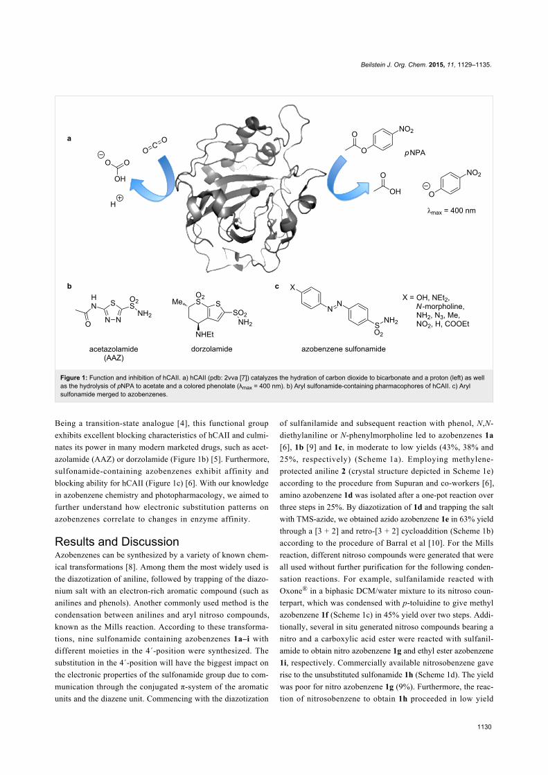
bonate and a proton (Figure 1a, left) [1]. Despite its native
purpose of pH and pressure regulation, its intrinsic esterase
activity can be utilized to measure the catalytic activity by
hydrolysis of p-nitrophenyl actetate (pNPA) to a phenolate, of
which the product appearance can be observed colorimetrically
(Figure 1a, right) [2]. In humans, isoform II (human carbonic
anhydrase II; hCAII) is found in many tissues and is respon-
sible for maintaining the inner eye pressure among other regula-
tory tasks [1]. Consequently, its failure is associated with glau-
coma [1,3]. Treatment of this severe disease, that leads to blind-
ness, is achieved with the application of aryl sulfonamides [3].
Beilstein J. Org. Chem. 2015, 11, 1129–1135.
1130
Figure 1: Function and inhibition of hCAII. a) hCAII (pdb: 2vva [7]) catalyzes the hydration of carbon dioxide to bicarbonate and a proton (left) as wellas the hydrolysis of pNPA to acetate and a colored phenolate (λmax = 400 nm). b) Aryl sulfonamide-containing pharmacophores of hCAII. c) Arylsulfonamide merged to azobenzenes.
Being a transition-state analogue [4], this functional group
exhibits excellent blocking characteristics of hCAII and culmi-
nates its power in many modern marketed drugs, such as acet-
azolamide (AAZ) or dorzolamide (Figure 1b) [5]. Furthermore,
sulfonamide-containing azobenzenes exhibit affinity and
blocking ability for hCAII (Figure 1c) [6]. With our knowledge
in azobenzene chemistry and photopharmacology, we aimed to
further understand how electronic substitution patterns on
azobenzenes correlate to changes in enzyme affinity.
Results and DiscussionAzobenzenes can be synthesized by a variety of known chem-
ical transformations [8]. Among them the most widely used is
the diazotization of aniline, followed by trapping of the diazo-
nium salt with an electron-rich aromatic compound (such as
anilines and phenols). Another commonly used method is the
condensation between anilines and aryl nitroso compounds,
known as the Mills reaction. According to these transforma-
tions, nine sulfonamide containing azobenzenes 1a–i with
different moieties in the 4´-position were synthesized. The
substitution in the 4´-position will have the biggest impact on
the electronic properties of the sulfonamide group due to com-
munication through the conjugated π-system of the aromatic
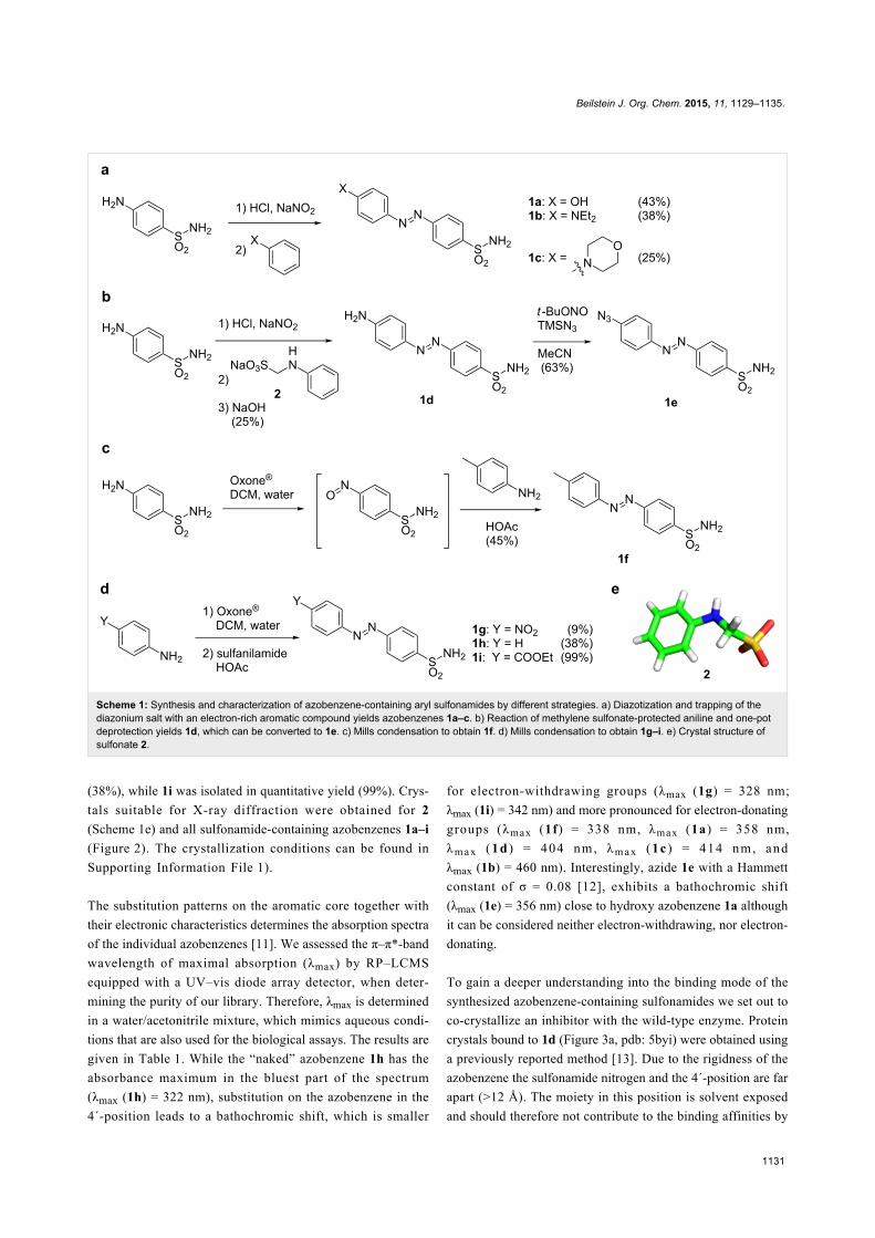
units and the diazene unit. Commencing with the diazotization
of sulfanilamide and subsequent reaction with phenol, N,N-
diethylaniline or N-phenylmorpholine led to azobenzenes 1a
[6], 1b [9] and 1c, in moderate to low yields (43%, 38% and
25%, respectively) (Scheme 1a). Employing methylene-
protected aniline 2 (crystal structure depicted in Scheme 1e)
according to the procedure from Supuran and co-workers [6],
amino azobenzene 1d was isolated after a one-pot reaction over
three steps in 25%. By diazotization of 1d and trapping the salt
with TMS-azide, we obtained azido azobenzene 1e in 63% yield
through a [3 + 2] and retro-[3 + 2] cycloaddition (Scheme 1b)
according to the procedure of Barral et al [10]. For the Mills
reaction, different nitroso compounds were generated that were
all used without further purification for the following conden-
sation reactions. For example, sulfanilamide reacted with
Oxone® in a biphasic DCM/water mixture to its nitroso coun-
terpart, which was condensed with p-toluidine to give methyl
azobenzene 1f (Scheme 1c) in 45% yield over two steps. Addi-
tionally, several in situ generated nitroso compounds bearing a
nitro and a carboxylic acid ester were reacted with sulfanil-
amide to obtain nitro azobenzene 1g and ethyl ester azobenzene
1i, respectively. Commercially available nitrosobenzene gave
rise to the unsubstituted sulfonamide 1h (Scheme 1d). The yield
was poor for nitro azobenzene 1g (9%). Furthermore, the reac-
tion of nitrosobenzene to obtain 1h proceeded in low yield
Beilstein J. Org. Chem. 2015, 11, 1129–1135.
1131
Scheme 1: Synthesis and characterization of azobenzene-containing aryl sulfonamides by different strategies. a) Diazotization and trapping of thediazonium salt with an electron-rich aromatic compound yields azobenzenes 1a–c. b) Reaction of methylene sulfonate-protected aniline and one-potdeprotection yields 1d, which can be converted to 1e. c) Mills condensation to obtain 1f. d) Mills condensation to obtain 1g–i. e) Crystal structure ofsulfonate 2.
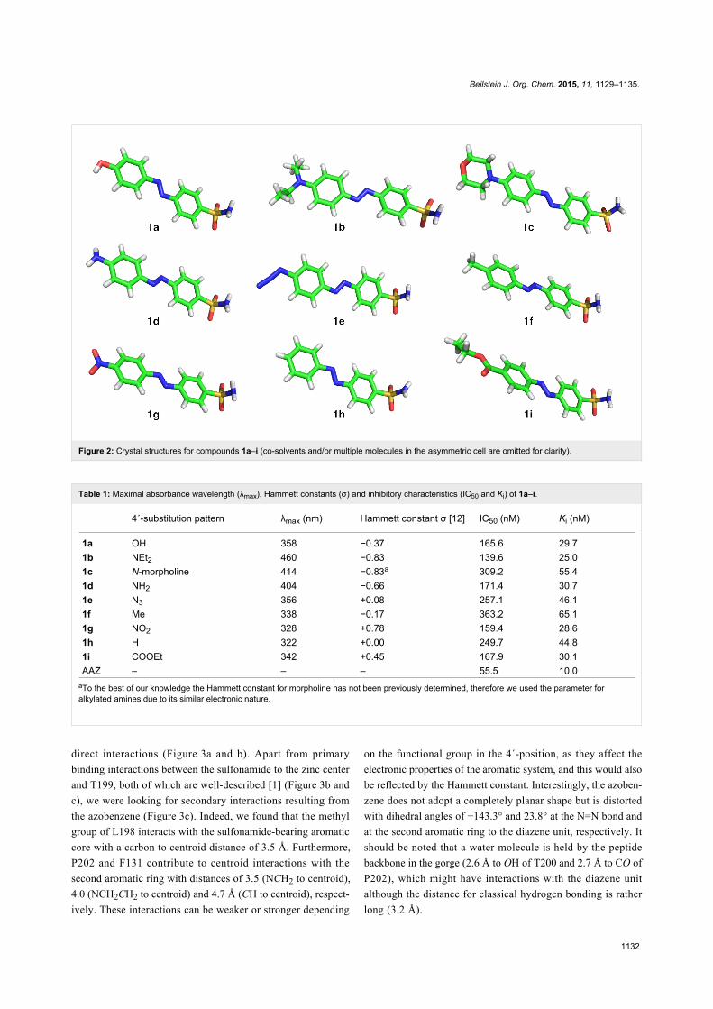
(38%), while 1i was isolated in quantitative yield (99%). Crys-
tals suitable for X-ray diffraction were obtained for 2
(Scheme 1e) and all sulfonamide-containing azobenzenes 1a–i
(Figure 2). The crystallization conditions can be found in
Supporting Information File 1).
The substitution patterns on the aromatic core together with
their electronic characteristics determines the absorption spectra
of the individual azobenzenes [11]. We assessed the π–π*-band
wavelength of maximal absorption (λmax) by RP–LCMS
equipped with a UV–vis diode array detector, when deter-
mining the purity of our library. Therefore, λmax is determined
in a water/acetonitrile mixture, which mimics aqueous condi-
tions that are also used for the biological assays. The results are
given in Table 1. While the “naked” azobenzene 1h has the
absorbance maximum in the bluest part of the spectrum
(λmax (1h) = 322 nm), substitution on the azobenzene in the
4´-position leads to a bathochromic shift, which is smaller
for electron-withdrawing groups (λmax (1g) = 328 nm;
λmax (1i) = 342 nm) and more pronounced for electron-donating
groups (λmax (1f) = 338 nm, λmax (1a) = 358 nm,
λm a x (1d ) = 404 nm, λm a x (1c ) = 414 nm, and
λmax (1b) = 460 nm). Interestingly, azide 1e with a Hammett
constant of σ = 0.08 [12], exhibits a bathochromic shift
(λmax (1e) = 356 nm) close to hydroxy azobenzene 1a although
it can be considered neither electron-withdrawing, nor electron-
donating.
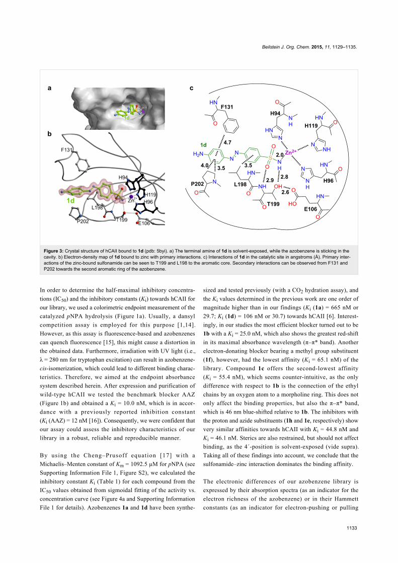
To gain a deeper understanding into the binding mode of the
synthesized azobenzene-containing sulfonamides we set out to
co-crystallize an inhibitor with the wild-type enzyme. Protein
crystals bound to 1d (Figure 3a, pdb: 5byi) were obtained using
a previously reported method [13]. Due to the rigidness of the
azobenzene the sulfonamide nitrogen and the 4´-position are far
apart (>12 Å). The moiety in this position is solvent exposed
and should therefore not contribute to the binding affinities by
Beilstein J. Org. Chem. 2015, 11, 1129–1135.
1132
Figure 2: Crystal structures for compounds 1a–i (co-solvents and/or multiple molecules in the asymmetric cell are omitted for clarity).
Table 1: Maximal absorbance wavelength (λmax), Hammett constants (σ) and inhibitory characteristics (IC50 and Ki) of 1a–i.
4´-substitution pattern λmax (nm) Hammett constant σ [12] IC50 (nM) Ki (nM)
1a OH 358 −0.37 165.6 29.71b NEt2 460 −0.83 139.6 25.01c N-morpholine 414 −0.83a 309.2 55.41d NH2 404 −0.66 171.4 30.71e N3 356 +0.08 257.1 46.11f Me 338 −0.17 363.2 65.11g NO2 328 +0.78 159.4 28.61h H 322 +0.00 249.7 44.81i COOEt 342 +0.45 167.9 30.1AAZ – – – 55.5 10.0
aTo the best of our knowledge the Hammett constant for morpholine has not been previously determined, therefore we used the parameter foralkylated amines due to its similar electronic nature.
direct interactions (Figure 3a and b). Apart from primary
binding interactions between the sulfonamide to the zinc center
and T199, both of which are well-described [1] (Figure 3b and
c), we were looking for secondary interactions resulting from
the azobenzene (Figure 3c). Indeed, we found that the methyl
group of L198 interacts with the sulfonamide-bearing aromatic
core with a carbon to centroid distance of 3.5 Å. Furthermore,
P202 and F131 contribute to centroid interactions with the
second aromatic ring with distances of 3.5 (NCH2 to centroid),
4.0 (NCH2CH2 to centroid) and 4.7 Å (CH to centroid), respect-
ively. These interactions can be weaker or stronger depending
on the functional group in the 4´-position, as they affect the
electronic properties of the aromatic system, and this would also
be reflected by the Hammett constant. Interestingly, the azoben-
zene does not adopt a completely planar shape but is distorted
with dihedral angles of −143.3° and 23.8° at the N=N bond and
at the second aromatic ring to the diazene unit, respectively. It
should be noted that a water molecule is held by the peptide
backbone in the gorge (2.6 Å to OH of T200 and 2.7 Å to CO of
P202), which might have interactions with the diazene unit
although the distance for classical hydrogen bonding is rather
long (3.2 Å).
Beilstein J. Org. Chem. 2015, 11, 1129–1135.
1133
Figure 3: Crystal structure of hCAII bound to 1d (pdb: 5byi). a) The terminal amine of 1d is solvent-exposed, while the azobenzene is sticking in thecavity. b) Electron-density map of 1d bound to zinc with primary interactions. c) Interactions of 1d in the catalytic site in angstroms (Å). Primary inter-actions of the zinc-bound sulfonamide can be seen to T199 and L198 to the aromatic core. Secondary interactions can be observed from F131 andP202 towards the second aromatic ring of the azobenzene.
In order to determine the half-maximal inhibitory concentra-
tions (IC50) and the inhibitory constants (Ki) towards hCAII for
our library, we used a colorimetric endpoint measurement of the
catalyzed pNPA hydrolysis (Figure 1a). Usually, a dansyl
competition assay is employed for this purpose [1,14].
However, as this assay is fluorescence-based and azobenzenes
can quench fluorescence [15], this might cause a distortion in
the obtained data. Furthermore, irradiation with UV light (i.e.,
λ = 280 nm for tryptophan excitation) can result in azobenzene-
cis-isomerization, which could lead to different binding charac-
teristics. Therefore, we aimed at the endpoint absorbance
system described herein. After expression and purification of
wild-type hCAII we tested the benchmark blocker AAZ
(Figure 1b) and obtained a Ki = 10.0 nM, which is in accor-
dance with a previously reported inhibition constant
(Ki (AAZ) = 12 nM [16]). Consequently, we were confident that
our assay could assess the inhibitory characteristics of our
library in a robust, reliable and reproducible manner.
By using the Cheng–Prusoff equation [17] with a
Michaelis–Menten constant of Km = 1092.5 µM for pNPA (see
Supporting Information File 1, Figure S2), we calculated the
inhibitory constant Ki (Table 1) for each compound from the
IC50 values obtained from sigmoidal fitting of the activity vs.
concentration curve (see Figure 4a and Supporting Information
File 1 for details). Azobenzenes 1a and 1d have been synthe-
sized and tested previously (with a CO2 hydration assay), and
the Ki values determined in the previous work are one order of
magnitude higher than in our findings (Ki (1a) = 665 nM or
29.7; Ki (1d) = 106 nM or 30.7) towards hCAII [6]. Interest-
ingly, in our studies the most efficient blocker turned out to be
1b with a Ki = 25.0 nM, which also shows the greatest red-shift
in its maximal absorbance wavelength (π–π* band). Another
electron-donating blocker bearing a methyl group substituent
(1f), however, had the lowest affinity (Ki = 65.1 nM) of the
library. Compound 1c offers the second-lowest affinity
(Ki = 55.4 nM), which seems counter-intuitive, as the only
difference with respect to 1b is the connection of the ethyl
chains by an oxygen atom to a morpholine ring. This does not
only affect the binding properties, but also the π–π* band,
which is 46 nm blue-shifted relative to 1b. The inhibitors with
the proton and azide substituents (1h and 1e, respectively) show
very similar affinities towards hCAII with Ki = 44.8 nM and
Ki = 46.1 nM. Sterics are also restrained, but should not affect
binding, as the 4´-position is solvent-exposed (vide supra).
Taking all of these findings into account, we conclude that the
sulfonamide–zinc interaction dominates the binding affinity.
The electronic differences of our azobenzene library is
expressed by their absorption spectra (as an indicator for the
electron richness of the azobenzene) or in their Hammett
constants (as an indicator for electron-pushing or pulling
Beilstein J. Org. Chem. 2015, 11, 1129–1135.
1134
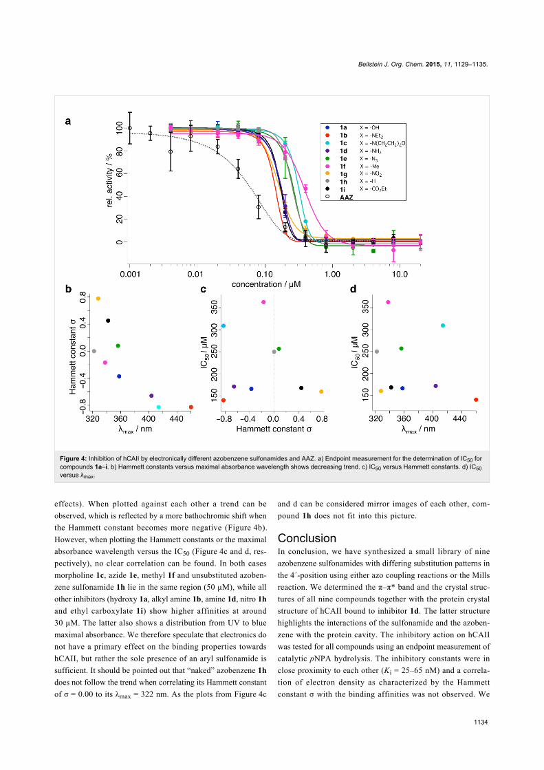
Figure 4: Inhibition of hCAII by electronically different azobenzene sulfonamides and AAZ. a) Endpoint measurement for the determination of IC50 forcompounds 1a–i. b) Hammett constants versus maximal absorbance wavelength shows decreasing trend. c) IC50 versus Hammett constants. d) IC50versus λmax.
effects). When plotted against each other a trend can be
observed, which is reflected by a more bathochromic shift when
the Hammett constant becomes more negative (Figure 4b).
However, when plotting the Hammett constants or the maximal
absorbance wavelength versus the IC50 (Figure 4c and d, res-
pectively), no clear correlation can be found. In both cases
morpholine 1c, azide 1e, methyl 1f and unsubstituted azoben-
zene sulfonamide 1h lie in the same region (50 µM), while all
other inhibitors (hydroxy 1a, alkyl amine 1b, amine 1d, nitro 1h
and ethyl carboxylate 1i) show higher affinities at around
30 µM. The latter also shows a distribution from UV to blue
maximal absorbance. We therefore speculate that electronics do
not have a primary effect on the binding properties towards
hCAII, but rather the sole presence of an aryl sulfonamide is
sufficient. It should be pointed out that “naked” azobenzene 1h
does not follow the trend when correlating its Hammett constant
of σ = 0.00 to its λmax = 322 nm. As the plots from Figure 4c
and d can be considered mirror images of each other, com-
pound 1h does not fit into this picture.
ConclusionIn conclusion, we have synthesized a small library of nine
azobenzene sulfonamides with differing substitution patterns in
the 4´-position using either azo coupling reactions or the Mills
reaction. We determined the π–π* band and the crystal struc-
tures of all nine compounds together with the protein crystal
structure of hCAII bound to inhibitor 1d. The latter structure
highlights the interactions of the sulfonamide and the azoben-
zene with the protein cavity. The inhibitory action on hCAII
was tested for all compounds using an endpoint measurement of
catalytic pNPA hydrolysis. The inhibitory constants were in
close proximity to each other (Ki = 25–65 nM) and a correla-
tion of electron density as characterized by the Hammett
constant σ with the binding affinities was not observed. We
Beilstein J. Org. Chem. 2015, 11, 1129–1135.
1135
have expanded the repertoire of sulfonamide blockers of hCAII
and described synthetic routes to potentially photochromic
representatives. Furthermore, the protein crystal structure of
hCAII bound to 1d described herein can be used as a template
for the rational design of novel hCAII blockers. The biological
activity of these blockers is currently under investigation and
the results will be published in due course.
Supporting InformationSupporting Information File 1Chemical procedures, spectral data and X-ray
crystallographic tables. Protein purification, crystallization
conditions and measurement of Michaelis–Menten
constant.
[http://www.beilstein-journals.org/bjoc/content/
supplementary/1860-5397-11-127-S1.pdf]
AcknowledgementsD.M.B is grateful to the European Comission for a Marie
Skłodowska-Curie Intra-European fellowship (PIEF-GA-2103-
627990). J.B. is grateful to the Studienstiftung des deutschen
Volkes for a Ph.D. fellowship. M.G. and D.T. thank the Munich
Centre for Integrated Protein Science (CIPSM) as well as the
Deutsche Forschungsgemeinschaft SFB749 for financial
support. D.T. acknowledges support of the European Research
Council for an Advanced Grant (268795). We are grateful to the
staff of the beamline X06SA at the Paul Scherrer Institute,
Swiss Light Source, Villigen (Switzerland) for assistance during
data collection. Dr. David H. Woodmansee is acknowledged for
helpful discussion and advice together with providing ethyl
4-nitrosobenzoate as well as Eric P. Trautman and Martin Maier
for excellent synthetic assistance.
Author ContributionsD.T. supervised the research. J.B. and D.T. conceived and
designed the study. J.B. and L.S.R. performed chemical, bio-
logical and small molecule crystallization experiments. J.B.
performed protein purification, crystallization and binding data
analysis. P.M. collected X-ray datasets of 1a–i and solved the
structures. M.G. collected hCAII X-ray dataset and solved the
structure. J.B., D.M.B and D.T. wrote the manuscript with input
from all authors.
References1. Krishnamurthy, V. M.; Kaufman, G. K.; Urbach, A. R.; Gitlin, I.;
Gudiksen, K. L.; Weibel, D. B.; Whitesides, G. M. Chem. Rev. 2008,108, 946–1051. doi:10.1021/cr050262p
2. Pocker, Y.; Stone, J. T. Biochemistry 1968, 7, 2936–2945.doi:10.1021/bi00848a034
3. Supuran, C. T. Nat. Rev. Drug Discovery 2008, 7, 168–181.doi:10.1038/nrd2467
4. Boriack-Sjodin, P. A.; Zeitlin, S.; Chen, H.-H.; Crenshaw, L.; Gross, S.;Dantanarayana, A.; Delgado, P.; May, J. A.; Dean, T.;Christianson, D. W. Protein Sci. 1998, 7, 2483–2489.doi:10.1002/pro.5560071201
5. Masini, E.; Carta, F.; Scozzafava, A.; Supuran, C. T.Expert Opin. Ther. Pat. 2013, 23, 705–716.doi:10.1517/13543776.2013.794788
6. Maresca, A.; Carta, F.; Vullo, D.; Scozzafava, A.; Supuran, C. T.Bioorg. Med. Chem. Lett. 2009, 19, 4929–4932.doi:10.1016/j.bmcl.2009.07.088
7. Sjöblom, B.; Polentarutti, M.; Djinović-Carugo, K.Proc. Natl. Acad. Sci. U. S. A. 2009, 106, 10609–10613.doi:10.1073/pnas.0904184106
8. Merino, E. Chem. Soc. Rev. 2011, 40, 3835–3853.doi:10.1039/c0cs00183j
9. Broichhagen, J.; Schönberger, M.; Cork, S. C.; Frank, J. A.;Marchetti, P.; Bugliani, M.; Shapiro, A. M. J.; Trapp, S.; Rutter, G. A.;Hodson, D. J.; Trauner, D. Nat. Commun. 2014, 5, No. 5116.doi:10.1038/ncomms6116
10. Barral, K.; Moorhouse, A. D.; Moses, J. E. Org. Lett. 2007, 9,1809–1811. doi:10.1021/ol070527h
11. Birnbaum, P. P.; Linford, J. H.; Style, D. W. G. Trans. Faraday Soc.1953, 49, 735–744. doi:10.1039/tf9534900735
12. Leffler, J. E.; Grunwald, E. Rates and equilibria of organic reactions astreated by statistical, thermodynamic, and extrathermodynamicmethods; Wiley: New York, 1963; p 458.
13. Lesburg, C. A.; Huang, C.; Christianson, D. W.; Fierke, C. A.Biochemistry 1997, 36, 15780–15791. doi:10.1021/bi971296x
14. Chen, R. F.; Kernohan, J. C. J. Biol. Chem. 1967, 242, 5813–5823.15. Vögtle, F.; Gorka, M.; Hesse, R.; Ceroni, P.; Maestri, M.; Balzani, V.
Photochem. Photobiol. Sci. 2002, 1, 45–51. doi:10.1039/b106813j16. D'Ambrosio, K.; Smaine, F.-Z.; Carta, F.; De Simone, G.; Winum, J.-Y.;
Supuran, C. T. J. Med. Chem. 2012, 55, 6776–6783.doi:10.1021/jm300818k
17. Cheng, Y.-C.; Prusoff, W. H. Biochem. Pharmacol. 1973, 22,3099–3108. doi:10.1016/0006-2952(73)90196-2
License and TermsThis is an Open Access article under the terms of the
Creative Commons Attribution License
(http://creativecommons.org/licenses/by/2.0), which
permits unrestricted use, distribution, and reproduction in
any medium, provided the original work is properly cited.
The license is subject to the Beilstein Journal of Organic
Chemistry terms and conditions:
(http://www.beilstein-journals.org/bjoc)
The definitive version of this article is the electronic one
which can be found at:
doi:10.3762/bjoc.11.127