Hsp70 induction in Drosophila melanogasterjeb.biologists.org/content/jexbio/205/3/345.full.pdf ·...
14
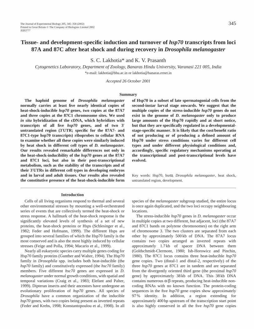
Cells of all living organisms respond to thermal and several other environmental stresses by mounting a well-orchestrated series of events that are collectively termed the heat-shock or stress response. A hallmark of the heat-shock response is the significantly elevated levels of synthesis of a set of new proteins, the heat-shock proteins or Hsps (Schlesinger et al., 1982; Feder and Hofmann, 1999). The different Hsps are grouped into several families of which the Hsp70 family is the most conserved and is also the most highly induced by cellular stresses (Feige and Polla, 1994; Macario et al., 1999). Nearly all eukaryotic species carry multiple genes coding for Hsp70 family proteins (Gunther and Walter, 1994). The Hsp70 family in Drosophila spp. includes both heat-inducible (the hsp70 family) and constitutively expressed (the hsc70 family) members. Five different hsc70 genes are expressed in D. melanogaster under normal growth conditions, with spatial and temporal variations (Craig et al., 1983; Elefant and Palter, 1999). Dipteran insects and their ancestors have undergone an evolutionary proliferation of hsp70 genes. All species of Drosophila have a common organization of the inducible hsp70 genes, with two copies being present as inverted repeats (Feder and Krebs, 1998; Konstantopoulou et al., 1998). In all species of the melanogaster subgroup studied, the entire locus is once again duplicated, and the two loci occupy neighbouring locations. The stress-inducible hsp70 genes in D. melanogaster occur in multiple copies at two different, but adjacent, loci (the 87A7 and 87C1 bands on polytene chromosomes) on the right arm of chromosome 3. The two clusters are separated from each other by approximately 500 kb of DNA. The 87A7 locus contains two copies arranged as inverted repeats with approximately 1.7 kb of spacer DNA between them (Goldschmidt-Clermont, 1980; Ish-Horowicz and Pinchin, 1980). The 87C1 locus contains three heat-inducible hsp70 gene copies. Two (distal-1 and distal-2, respectively) of the three hsp70 genes at 87C1 are in tandem and are separated from the divergently oriented third gene (the proximal hsp70 gene) by approximately 38 kb of DNA. This 38 kb DNA contains numerous α/β repeats, producing heat-inducible non- coding RNAs with no known function. The protein-coding sequences in the five hsp70 gene copies show approximately 97 % identity. In addition, a region extending for approximately 400 bp upstream of the transcription start point is also highly conserved in all the five hsp70 gene copies 345 The Journal of Experimental Biology 205, 345–358 (2002) Printed in Great Britain © The Company of Biologists Limited 2002 JEB3777 The haploid genome of Drosophila melanogaster normally carries at least five nearly identical copies of heat-shock-inducible hsp70 genes, two copies at the 87A7 and three copies at the 87C1 chromosome sites. We used in situ hybridization of the cDNA, which hybridizes with transcripts of all five hsp70 genes, and of two 3′ untranslated region (3′UTR; specific for the 87A7- and 87C1-type hsp70 transcripts) riboprobes to cellular RNA to examine whether all these copies were similarly induced by heat shock in different cell types of D. melanogaster. Our results revealed remarkable differences not only in the heat-shock-inducibility of the hsp70 genes at the 87A7 and 87C1 loci, but also in their post-transcriptional metabolism, such as the stability of the transcripts and of their 3′UTRs in different cell types in developing embryos and in larval and adult tissues. Our results also revealed the constitutive presence of the heat-shock-inducible form of Hsp70 in a subset of late spermatogonial cells from the second-instar larval stage onwards. We suggest that the multiple copies of the stress-inducible hsp70 genes do not exist in the genome of D. melanogaster only to produce large amounts of the Hsp70 rapidly and at short notice, but that they are specifically regulated in a developmental- stage-specific manner. It is likely that the cost/benefit ratio of not producing or of producing a defined amount of Hsp70 under stress conditions varies for different cell types and under different physiological conditions and, accordingly, specific regulatory mechanisms operating at the transcriptional and post-transcriptional levels have evolved. Key words: Hsp70, hsrϖ, Drosophila melanogaster, heat shock, untranslated region, development. Summary Introduction Tissue- and development-specific induction and turnover of hsp70 transcripts from loci 87A and 87C after heat shock and during recovery in Drosophila melanogaster S. C. Lakhotia* and K. V. Prasanth Cytogenetics Laboratory, Department of Zoology, Banaras Hindu University, Varanasi 221 005, India *e-mail: [email protected] or [email protected] Accepted 26 October 2001
Transcript of Hsp70 induction in Drosophila melanogasterjeb.biologists.org/content/jexbio/205/3/345.full.pdf ·...
Cells of all living organisms respond to thermal and severalother environmental stresses by mounting a well-orchestratedseries of events that are collectively termed the heat-shock orstress response. A hallmark of the heat-shock response is thesignificantly elevated levels of synthesis of a set of newproteins, the heat-shock proteins or Hsps (Schlesinger et al.,1982; Feder and Hofmann, 1999). The different Hsps aregrouped into several families of which the Hsp70 family is themost conserved and is also the most highly induced by cellularstresses (Feige and Polla, 1994; Macario et al., 1999).
Nearly all eukaryotic species carry multiple genes coding forHsp70 family proteins (Gunther and Walter, 1994). The Hsp70family in Drosophila spp. includes both heat-inducible (thehsp70family) and constitutively expressed (the hsc70family)members. Five different hsc70genes are expressed in D.melanogasterunder normal growth conditions, with spatial andtemporal variations (Craig et al., 1983; Elefant and Palter,1999). Dipteran insects and their ancestors have undergone anevolutionary proliferation of hsp70genes. All species ofDrosophila have a common organization of the induciblehsp70genes, with two copies being present as inverted repeats(Feder and Krebs, 1998; Konstantopoulou et al., 1998). In all
species of the melanogastersubgroup studied, the entire locusis once again duplicated, and the two loci occupy neighbouringlocations.
The stress-inducible hsp70genes in D. melanogasteroccurin multiple copies at two different, but adjacent, loci (the 87A7and 87C1 bands on polytene chromosomes) on the right armof chromosome 3. The two clusters are separated from eachother by approximately 500 kb of DNA. The 87A7 locuscontains two copies arranged as inverted repeats withapproximately 1.7 kb of spacer DNA between them(Goldschmidt-Clermont, 1980; Ish-Horowicz and Pinchin,1980). The 87C1 locus contains three heat-inducible hsp70gene copies. Two (distal-1 and distal-2, respectively) of thethree hsp70genes at 87C1 are in tandem and are separatedfrom the divergently oriented third gene (the proximal hsp70gene) by approximately 38 kb of DNA. This 38 kb DNAcontains numerous α/β repeats, producing heat-inducible non-coding RNAs with no known function. The protein-codingsequences in the five hsp70gene copies show approximately97 % identity. In addition, a region extending forapproximately 400 bp upstream of the transcription start pointis also highly conserved in all the five hsp70 gene copies
345The Journal of Experimental Biology 205, 345–358 (2002)Printed in Great Britain © The Company of Biologists Limited 2002JEB3777
The haploid genome of Drosophila melanogasternormally carries at least five nearly identical copies ofheat-shock-inducible hsp70genes, two copies at the 87A7and three copies at the 87C1 chromosome sites. We usedin situ hybridization of the cDNA, which hybridizes withtranscripts of all five hsp70 genes, and of two 3′untranslated region (3′UTR; specific for the 87A7- and87C1-type hsp70 transcripts) riboprobes to cellular RNAto examine whether all these copies were similarly inducedby heat shock in different cell types of D. melanogaster.Our results revealed remarkable differences not only inthe heat-shock-inducibility of the hsp70genes at the 87A7and 87C1 loci, but also in their post-transcriptionalmetabolism, such as the stability of the transcripts and oftheir 3′UTRs in different cell types in developing embryosand in larval and adult tissues. Our results also revealedthe constitutive presence of the heat-shock-inducible form
of Hsp70 in a subset of late spermatogonial cells from thesecond-instar larval stage onwards. We suggest that themultiple copies of the stress-inducible hsp70genes do notexist in the genome of D. melanogasteronly to producelarge amounts of the Hsp70 rapidly and at short notice,but that they are specifically regulated in a developmental-stage-specific manner. It is likely that the cost/benefit ratioof not producing or of producing a defined amount ofHsp70 under stress conditions varies for different celltypes and under different physiological conditions and,accordingly, specific regulatory mechanisms operating atthe transcriptional and post-transcriptional levels haveevolved.
Key words: Hsp70, hsrω, Drosophila melanogaster, heat shock,untranslated region, development.
Summary
Introduction
Tissue- and development-specific induction and turnover of hsp70transcripts from loci87A and 87C after heat shock and during recovery in Drosophila melanogaster
S. C. Lakhotia* and K. V. PrasanthCytogenetics Laboratory, Department of Zoology, Banaras Hindu University, Varanasi 221 005, India
*e-mail: [email protected] or [email protected]
Accepted 26 October 2001
346
(Ingolia et al., 1980; Karch et al., 1981; Leigh Brown and Ish-Horowicz, 1981).
Although the protein-coding sequences and the 5′ regulatorysequences of the five hsp70genes show very high homology,their 3′ untranslated region (3′UTR) shows significantdivergences. The 3′UTRs [approximately 250 bp upstreamof the poly(A)+ site] of the two hsp70genes at the 87A7locus are similar to each other, but show high divergence(approximately 68 %) compared with those of the hsp70genecopies at the 87C1 locus. Interestingly, the 3′UTRs of theproximal and the distal-1 hsp70genes at the 87C1 locus showhigh homology, but these are moderately divergent from thatof the distal-2 (57 % homology) hsp70gene at the same locus(Torok et al., 1982).
The biological need for the presence of five copies of a genecoding for nearly identical heat-inducible Hsp70 in the D.melanogastergenome is intriguing. One possibility is that themultiple copies of hsp70genes help produce more Hsp70rapidly in response to heat shock or other stress (Feder andKrebs, 1998; Feder and Hofmann, 1999). In addition oralternatively, copies of hsp70genes at the two loci may bedifferentially regulated during development for some cell-specific requirement. Earlier studies in this laboratory haveindicated that transcription at the 87A and 87C loci could beregulated differently under certain conditions of heat shock.For example, when new transcription at the non-coding 93Dlocus was inhibited during heat shock, either because ofcombined treatment with another inducer such as benzamideor colchicine or because of the deletion of the 93D locus, therelative transcriptional or puffing activity of the hsp70genesat the 87A and 87C loci in larval salivary glands was alteredin a treatment-specific manner (for reviews, see Lakhotia andSharma, 1996; Lakhotia et al., 1999). In view of the differentialinduction of 87A and 87C puffs under these conditions and thedivergence in the 3′UTR sequences, which are known to haveimportant roles in the stability and localization of thetranscripts (Sachs, 1993; Curtis et al., 1995; Ross, 1996), it ispossible that the individual members of this multigene hsp70family in D. melanogaster, although coding for similarproteins, may indeed be regulated differentially in other celltypes also. The present study examines this possibility.
Transcription of the hsp70genes from the 87A and 87C lociin embryos, in different larval tissues and in adult testesfollowing heat shock and during recovery was analyzed byRNA:RNA in situ hybridization using the non-radioactivedigoxigenin-labelled coding region or 3′UTR-specificriboprobes. The results showed that, although the hsp70transcripts were induced after heat shock in most (but not all)tissues, their quantity and properties varied. In certain cells andtissues, transcripts from the 87A and 87C loci showeddifferential induction and stability. Immunostaining withantibody specific for heat-shock-induced Hsp70 revealed anunexpected constitutive presence of Hsp70 in mitoticallydividing spermatogonial cells. Our results clearly showed thatthe heat-shock-inducibility of the hsp70genes from the twoclusters is differentially regulated in different cell types and
that the induced transcripts are metabolized in a cell- anddevelopment-specific manner. It appears that the multiplecopies of hsp70genes are not just a consequence of duplicationto produce more Hsp70 during heat shock, but that the multiplecopies exert functions in a cell- and developmental-stage-specific pattern.
Materials and methodsFly stocks and rearing conditions
The Oregon R+ wild-type strain of Drosophila melanogasterMeigen was reared at 22±1 °C on standard food containingagar, maize powder, yeast and sugar.
TreatmentsHeat shock and recovery
Embryos or different tissues from late third-instar larvae ortestes from different larval and adult stages were heat-shockedfor 40 min at 37 °C in Poels’ salt solution (PSS) (Lakhotia andTapadia, 1998). As parallel controls, the tissues were dissectedout at room temperature (22 °C) directly from unstressed larvaeand flies and incubated in PSS at room temperature for 40 min.In experiments requiring recovery from heat shock, wholelarvae or adult flies were heat-shocked for 40 min at 37 °C invials with moist filter paper and then returned to normal foodvials and allowed to recover at room temperature for thedesired period (see Results). Following recovery, the larvae oradults were quickly dissected in PSS, and the required organswere removed for processing for in situlocalization of hsp70transcripts or Hsp70 (see below). Pilot experiments revealedthat heat shock to isolated organs or to intact organismsresulted in comparable levels of induction of the hsp70genes.In one set of experiments, testes from different unstressedlarval, pupal and adult stages were quickly dissected out inPSS at room temperature and immediately processed forimmunostaining with Hsp70 antibody (see below).
RNA:RNA in situhybridization (RISH) and immunostaining
Three recombinant clones, i.e. pPW18(containing the5′UTR and the coding region of hsp70but not the 3′UTR)(Sharma and Lakhotia, 1995), pVZ-70-3′#1 (containing the3′UTR sequence of the proximal hsp70gene at the 87A7 locus)(Dellavalle et al., 1994) and pVZ-70-3′ (containing the 3′UTRsequence of the proximal hsp70gene at the 87C1 locus)(Dellavalle et al., 1994), were used to generate digoxigenin(DIG)-labelled antisense riboprobes. pPW18was linearizedwith HindIII, while pVZ-70-3′#1 and pVZ-70-3′ UTR cloneswere linearized with SalI.The linearized plasmids weretranscribed in vitro using DIG-UTP (Roche, Germany) as thelabelled substrate, following the manufacturer’s instructions, togenerate anti-sense riboprobes. The pPW18riboprobe detectsall the hsp70 transcripts from either of the two clusters,whether with or without the 3′UTRs. In contrast, the pVZ-70-3′#1 riboprobe detects only the 3′UTR-carrying hsp70transcripts from either of two hsp70gene copies at the 87A7site, while the pVZ-70-3′ riboprobe detects the 3′UTR-carrying
S. C. Lakhotia and K. V. Prasanth
347Hsp70 induction in Drosophila melanogaster
hsp70 transcripts from the 87C1 locus (excluding the distal-2hsp70gene). The three probes will be referred to as cDNA,87A3′UTR and 87C3′UTR, respectively, in the following. Thespecificity of each of the riboprobes was checked by RISH withpolytene chromosome preparations from mildly heat-shockedsalivary glands of wild-type late third-instar larvae.
The desired riboprobes were hybridized in situ to cellularRNA in embryos and tissues from larvae, pupae and adults, asdescribed previously (Prasanth et al., 2000; Lakhotia et al.,2001).
The 7Fb rat monoclonal antibody, which detects only theheat-inducible form of Hsp70 in D. melanogaster(Velazquezand Lindquist, 1984), was used at a dilution of 1:400 forimmunostaining of different tissues, as described previously(Prasanth et al., 2000; Lakhotia et al., 2001). In some cases,wild-type embryos were immunostained with the nervous-system-specific antibody BP104 (Hortsch et al., 1990) toidentify the neural cells. In each case, antibody bindingwas detected colorimetrically using appropriate secondaryantibodies conjugated with alkaline phosphatase, as describedpreviously (Prasanth et al., 2000; Lakhotia et al., 2001).
Microscopy and analysis
All RISH and immunostained preparations were examinedunder a Nikon E800 microscope, and the images wereassembled using Adobe Photoshop 5.0 software.
ResultsTo analyze the heat-shock inducibility of hsp70genes from
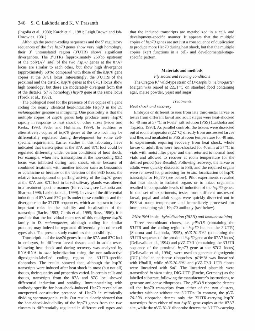
the 87A and 87C loci and the localization and turnover of theirtranscripts in different cell types from embryo to adult, RISHwas carried out using DIG-labelled antisense riboprobesderived either from the 3′UTRs of hsp70genes from loci 87Aand 87C, which specifically detect the transcripts of therespective locus, or from cDNA without the 3′UTR, whichdetects transcripts of all five hsp70genes. To confirm thespecificity of these probes, RISH was performed on mildlyheat-shocked wild-type larval salivary gland polytenechromosomes using the three probes separately. As expected,the cDNA probe hybridized to hsp70 transcripts at both the87A and 87C loci (Fig. 1A). The 87A3′UTR probe hybridizedonly to hsp70 transcripts at the 87A locus, but not at the 87Clocus (Fig. 1B). The 87C3′UTR probe hybridized only to the87C locus (Fig. 1C) and, as expected, gave two hybridizationbands at the 87C1 locus since the 3′UTR of the third gene atthis site is considerably different (Torok et al., 1982). None ofthe probes hybridized to any other chromosomal sites.
Differential expression and stability of heat-shock-inducibletranscripts from hsp70 genes at the 87A and 87C loci in
different cell typesEmbryonic cells
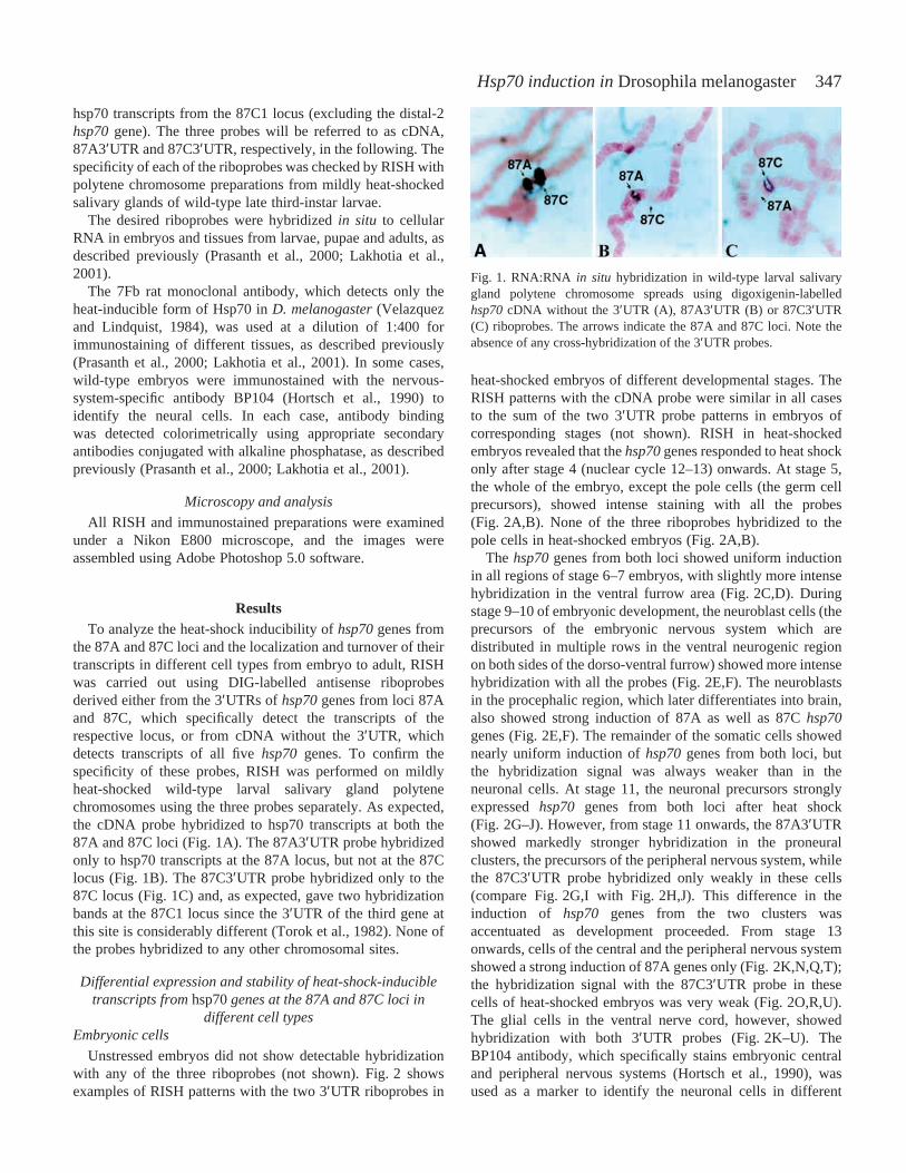
Unstressed embryos did not show detectable hybridizationwith any of the three riboprobes (not shown). Fig. 2 showsexamples of RISH patterns with the two 3′UTR riboprobes in
heat-shocked embryos of different developmental stages. TheRISH patterns with the cDNA probe were similar in all casesto the sum of the two 3′UTR probe patterns in embryos ofcorresponding stages (not shown). RISH in heat-shockedembryos revealed that the hsp70genes responded to heat shockonly after stage 4 (nuclear cycle 12–13) onwards. At stage 5,the whole of the embryo, except the pole cells (the germ cellprecursors), showed intense staining with all the probes(Fig. 2A,B). None of the three riboprobes hybridized to thepole cells in heat-shocked embryos (Fig. 2A,B).
The hsp70genes from both loci showed uniform inductionin all regions of stage 6–7 embryos, with slightly more intensehybridization in the ventral furrow area (Fig. 2C,D). Duringstage 9–10 of embryonic development, the neuroblast cells (theprecursors of the embryonic nervous system which aredistributed in multiple rows in the ventral neurogenic regionon both sides of the dorso-ventral furrow) showed more intensehybridization with all the probes (Fig. 2E,F). The neuroblastsin the procephalic region, which later differentiates into brain,also showed strong induction of 87A as well as 87C hsp70genes (Fig. 2E,F). The remainder of the somatic cells showednearly uniform induction of hsp70genes from both loci, butthe hybridization signal was always weaker than in theneuronal cells. At stage 11, the neuronal precursors stronglyexpressed hsp70 genes from both loci after heat shock(Fig. 2G–J). However, from stage 11 onwards, the 87A3′UTRshowed markedly stronger hybridization in the proneuralclusters, the precursors of the peripheral nervous system, whilethe 87C3′UTR probe hybridized only weakly in these cells(compare Fig. 2G,I with Fig. 2H,J). This difference in theinduction of hsp70 genes from the two clusters wasaccentuated as development proceeded. From stage 13onwards, cells of the central and the peripheral nervous systemshowed a strong induction of 87A genes only (Fig. 2K,N,Q,T);the hybridization signal with the 87C3′UTR probe in thesecells of heat-shocked embryos was very weak (Fig. 2O,R,U).The glial cells in the ventral nerve cord, however, showedhybridization with both 3′UTR probes (Fig. 2K–U). TheBP104 antibody, which specifically stains embryonic centraland peripheral nervous systems (Hortsch et al., 1990), wasused as a marker to identify the neuronal cells in different
Fig. 1. RNA:RNA in situ hybridization in wild-type larval salivarygland polytene chromosome spreads using digoxigenin-labelledhsp70cDNA without the 3′UTR (A), 87A3′UTR (B) or 87C3′UTR(C) riboprobes. The arrows indicate the 87A and 87C loci. Note theabsence of any cross-hybridization of the 3′UTR probes.
348 S. C. Lakhotia and K. V. Prasanth
Fig. 2. Heat-shock-induced expression of hsp70genes from the 87A and 87C loci in wild-type embryos of developmental stages 5–16 asrevealed by RNA:RNA in situhybridization (RISH) using 3′UTR riboprobes of hsp70genes from the 87A (A,C,E,G,I,K,N,Q,T,W) or the 87C(B,D,F,H,J,L,O,R,U,X) locus. Stages of the embryos are indicated on the right (D, L and V in this column refer to the dorsal, lateral or ventralviews, respectively, of the embryo). The arrow in A points to the pole cells. The black arrowheads in E and F and the small white arrowheadsin F, G and H indicate the neuroblasts in the procephalic region and ventral nerve cord, respectively. The white arrows in I and J indicate theproneural clusters in stage 11 embryos. The white arrowheads in K, R and U refer to the glial cells. The small black arrows in N, O and P pointto the tracheal pits, while the long black arrows in Q, R, S and T show the peripheral nervous system cells. Insets in T, U and V show theclusters of neuroblasts (T and V) and glial cells (U) from the respective embryos. Embryos in M, P, S and V are immunostained with theBP104 antibody to show the embryonic central and peripheral nervous systems for comparison with RISH patterns in K–U. Scale bar(applicable to all parts of the figure, except the insets in T, U and V, which are at higher magnification), 200µm.
349Hsp70 induction in Drosophila melanogaster
embryonic stages (Fig. 2M,P,S,V). In very late embryos(Fig. 2W,X), the tracheal system also showed stronghybridization with all three riboprobes. The central nervoussystem continued to show stronger hybridization with the87A3′UTR (Fig. 2W). The non-neuronal somatic cells in lateembryonic stages showed uniform induction of transcriptsfrom both the loci after heat shock but at a much lower levelthan in the neural cells and tracheal network (Fig. 2W,X).RISH with the cDNA probe generated a more-or-less uniformsignal throughout all embryonic stages, with strongerhybridization in the nervous system (not shown).
In all cells of the various embryonic stages examined, hsp70transcripts were localized in the cytoplasm (see insets inFig. 2T,U).
RISH patterns in embryos recovering from heat shock werenot examined.
Larval gut
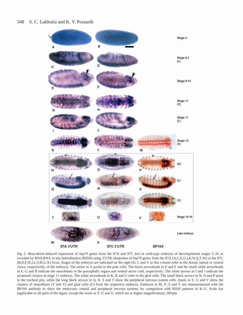
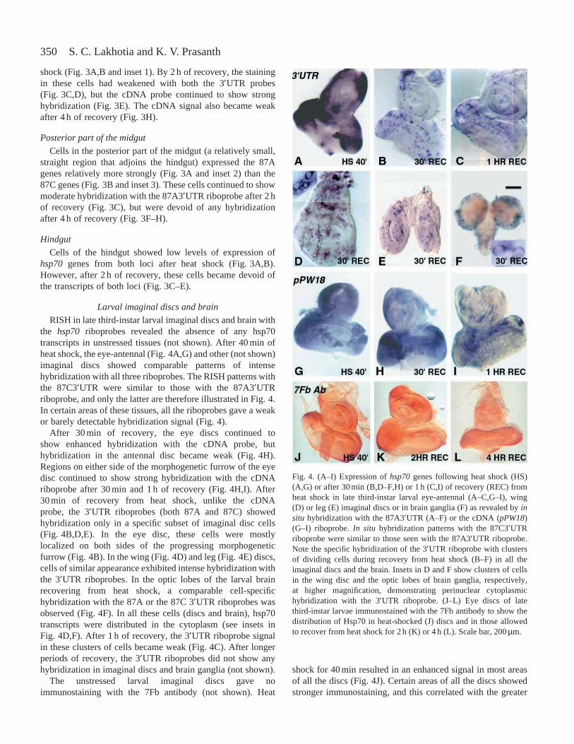
No hsp70 transcripts were detectable in any of the gut cellsin unstressed late third-instar larvae. After heat shock, thehsp70 transcripts were localized in the cytoplasm of larvalgut cells. It is interesting to note that the transcripts weremostly restricted to the area immediately adjacent to theouter face of the nuclear envelope (see insets 1, 2 and 3 inFig. 3A,B).
Proventriculus
Cells in the proventriculus did not show any appreciablehybridization with any of the three riboprobes, eitherimmediately after heat shock or during recovery (Fig. 3).
Gastric caeca
Cells in the gastric caeca (two pairs of finger-like projectionsfrom the anterior midgut or ‘stomach’) showed stronginduction of hsp70genes from both loci after heat shock(Fig. 3A,B). After 2 h of recovery, both the 87A and the 87Ctranscripts continued to be present in these cells (Fig. 3C–E),but no hsp70 transcripts were detectable in the gastric caecaafter 4 h of recovery (Fig. 3F–H).
Anterior part of the midgut (‘stomach’)
Soon after heat shock, cells in the enlarged anterior part ofthe midgut or ‘stomach’ exhibited intense expression of hsp70genes from both the loci (Fig. 3A,B). After 2 h of recovery,cells in the ‘stomach’ region showed positive hybridizationonly with 87C3′UTR (Fig. 3D) and cDNA (Fig. 3E), but notwith the 87A 3′UTR riboprobe (Fig. 3C). After 4 h of recovery,none the 3′UTR probes showed any hybridization (Fig. 3F,G),but a group of cells in the anteriormost enlarged part of the‘stomach’ continued to show hybridization with the cDNAprobe (Fig. 3H).
Immediately after the heat shock, the narrower loopedsegment that joins the anterior and middle parts of the midgutshowed weak hybridization with the 87A3′UTR riboprobebut moderate hybridization with the 87C3′UTR riboprobe(Fig. 3A,B). After 2 h of recovery, all three riboprobes gave a
weak signal, but none showed any detectable hybridization inthis part of the midgut after 4 h of recovery (Fig. 3F,G).
Middle part of the midgut
Cells in the long coiled middle part of the midgut, whichincludes the region with larger polytene cells, showedpronounced hybridization with all the probes soon after heat
Fig. 3. Differential expression of hsp70genes in larval gut asrevealed by RNA:RNA in situhybridization (RISH) using the87A3′UTR (A,C,F), the 87C3′UTR (B,D,G) or the cDNA (E,H)riboprobe after 40 min of heat shock (HS) (A,B) or after 2 h (C,D,E)or 4 h (F,G H) of recovery (rec) from a 40 min heat shock. Insets in Aand B show cells from the gut region at a higher magnification: inset1 corresponds to cells from the middle part of the midgut (the two3′UTR probes give a similar perinuclear signal in the cytoplasm);inset 2 shows a cell from the distal part of the midgut with strong87A3′UTR hybridization; inset 3 is a cell from a similar position tothat in inset 2 showing weak hybridization with the 87C3′UTRriboprobe. Scale bars (which apply to all images in a given row,except the insets), 200µm.
350
shock (Fig. 3A,B and inset 1). By 2 h of recovery, the stainingin these cells had weakened with both the 3′UTR probes(Fig. 3C,D), but the cDNA probe continued to show stronghybridization (Fig. 3E). The cDNA signal also became weakafter 4 h of recovery (Fig. 3H).
Posterior part of the midgut
Cells in the posterior part of the midgut (a relatively small,straight region that adjoins the hindgut) expressed the 87Agenes relatively more strongly (Fig. 3A and inset 2) than the87C genes (Fig. 3B and inset 3). These cells continued to showmoderate hybridization with the 87A3′UTR riboprobe after 2 hof recovery (Fig. 3C), but were devoid of any hybridizationafter 4 h of recovery (Fig. 3F–H).
Hindgut
Cells of the hindgut showed low levels of expression ofhsp70 genes from both loci after heat shock (Fig. 3A,B).However, after 2 h of recovery, these cells became devoid ofthe transcripts of both loci (Fig. 3C–E).
Larval imaginal discs and brain
RISH in late third-instar larval imaginal discs and brain withthe hsp70 riboprobes revealed the absence of any hsp70transcripts in unstressed tissues (not shown). After 40 min ofheat shock, the eye-antennal (Fig. 4A,G) and other (not shown)imaginal discs showed comparable patterns of intensehybridization with all three riboprobes. The RISH patterns withthe 87C3′UTR were similar to those with the 87A3′UTRriboprobe, and only the latter are therefore illustrated in Fig. 4.In certain areas of these tissues, all the riboprobes gave a weakor barely detectable hybridization signal (Fig. 4).
After 30 min of recovery, the eye discs continued toshow enhanced hybridization with the cDNA probe, buthybridization in the antennal disc became weak (Fig. 4H).Regions on either side of the morphogenetic furrow of the eyedisc continued to show strong hybridization with the cDNAriboprobe after 30 min and 1 h of recovery (Fig. 4H,I). After30 min of recovery from heat shock, unlike the cDNAprobe, the 3′UTR riboprobes (both 87A and 87C) showedhybridization only in a specific subset of imaginal disc cells(Fig. 4B,D,E). In the eye disc, these cells were mostlylocalized on both sides of the progressing morphogeneticfurrow (Fig. 4B). In the wing (Fig. 4D) and leg (Fig. 4E) discs,cells of similar appearance exhibited intense hybridization withthe 3′UTR riboprobes. In the optic lobes of the larval brainrecovering from heat shock, a comparable cell-specifichybridization with the 87A or the 87C 3′UTR riboprobes wasobserved (Fig. 4F). In all these cells (discs and brain), hsp70transcripts were distributed in the cytoplasm (see insets inFig. 4D,F). After 1 h of recovery, the 3′UTR riboprobe signalin these clusters of cells became weak (Fig. 4C). After longerperiods of recovery, the 3′UTR riboprobes did not show anyhybridization in imaginal discs and brain ganglia (not shown).
The unstressed larval imaginal discs gave noimmunostaining with the 7Fb antibody (not shown). Heat
shock for 40 min resulted in an enhanced signal in most areasof all the discs (Fig. 4J). Certain areas of all the discs showedstronger immunostaining, and this correlated with the greater
S. C. Lakhotia and K. V. Prasanth
Fig. 4. (A–I) Expression of hsp70genes following heat shock (HS)(A,G) or after 30 min (B,D–F,H) or 1 h (C,I) of recovery (REC) fromheat shock in late third-instar larval eye-antennal (A–C,G–I), wing(D) or leg (E) imaginal discs or in brain ganglia (F) as revealed by insitu hybridization with the 87A3′UTR (A–F) or the cDNA (pPW18)(G–I) riboprobe. In situhybridization patterns with the 87C3′UTRriboprobe were similar to those seen with the 87A3′UTR riboprobe.Note the specific hybridization of the 3′UTR riboprobe with clustersof dividing cells during recovery from heat shock (B–F) in all theimaginal discs and the brain. Insets in D and F show clusters of cellsin the wing disc and the optic lobes of brain ganglia, respectively,at higher magnification, demonstrating perinuclear cytoplasmichybridization with the 3′UTR riboprobe. (J–L) Eye discs of latethird-instar larvae immunostained with the 7Fb antibody to show thedistribution of Hsp70 in heat-shocked (J) discs and in those allowedto recover from heat shock for 2 h (K) or 4 h (L). Scale bar, 200µm.
351Hsp70 induction in Drosophila melanogaster
levels of hsp70 transcripts in these regions (e.g. compareFig. 4A,G,J). The levels of Hsp70 in various imaginal discsdeclined as the recovery period increased (Fig. 4K,L).
Testes
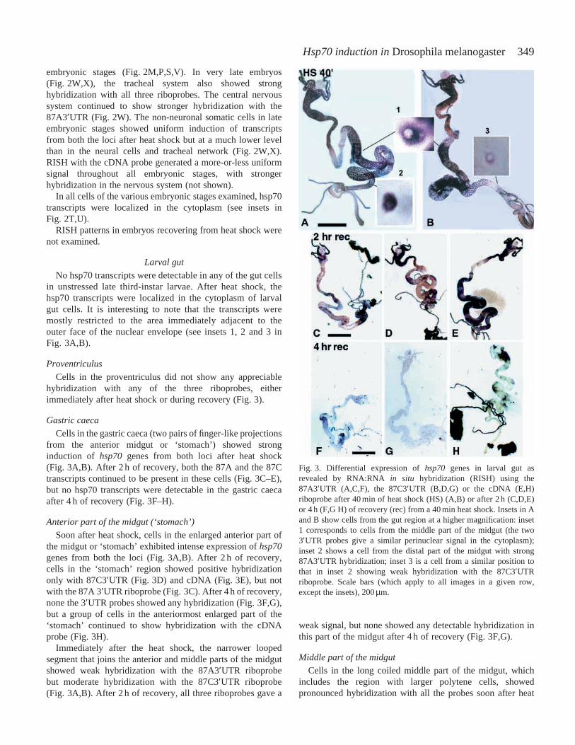
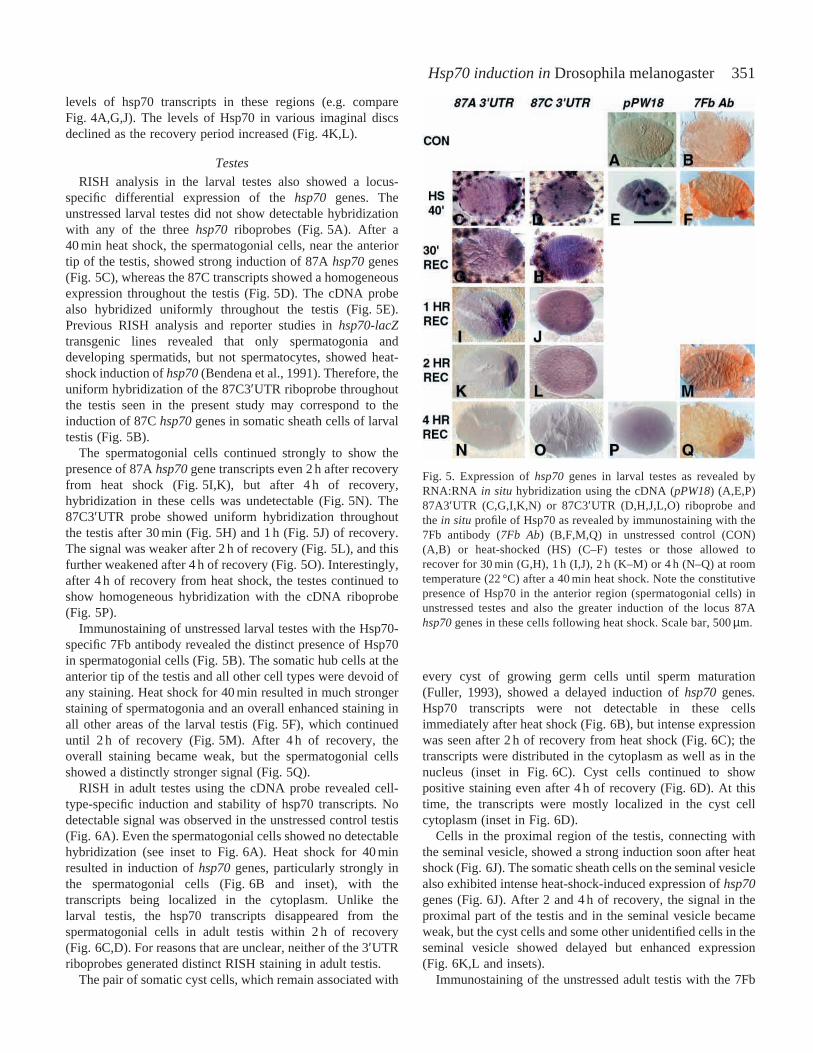
RISH analysis in the larval testes also showed a locus-specific differential expression of the hsp70genes. Theunstressed larval testes did not show detectable hybridizationwith any of the three hsp70 riboprobes (Fig. 5A). After a40 min heat shock, the spermatogonial cells, near the anteriortip of the testis, showed strong induction of 87A hsp70genes(Fig. 5C), whereas the 87C transcripts showed a homogeneousexpression throughout the testis (Fig. 5D). The cDNA probealso hybridized uniformly throughout the testis (Fig. 5E).Previous RISH analysis and reporter studies in hsp70-lacZtransgenic lines revealed that only spermatogonia anddeveloping spermatids, but not spermatocytes, showed heat-shock induction of hsp70(Bendena et al., 1991). Therefore, theuniform hybridization of the 87C3′UTR riboprobe throughoutthe testis seen in the present study may correspond to theinduction of 87C hsp70genes in somatic sheath cells of larvaltestis (Fig. 5B).
The spermatogonial cells continued strongly to show thepresence of 87A hsp70gene transcripts even 2 h after recoveryfrom heat shock (Fig. 5I,K), but after 4 h of recovery,hybridization in these cells was undetectable (Fig. 5N). The87C3′UTR probe showed uniform hybridization throughoutthe testis after 30 min (Fig. 5H) and 1 h (Fig. 5J) of recovery.The signal was weaker after 2 h of recovery (Fig. 5L), and thisfurther weakened after 4 h of recovery (Fig. 5O). Interestingly,after 4 h of recovery from heat shock, the testes continued toshow homogeneous hybridization with the cDNA riboprobe(Fig. 5P).
Immunostaining of unstressed larval testes with the Hsp70-specific 7Fb antibody revealed the distinct presence of Hsp70in spermatogonial cells (Fig. 5B). The somatic hub cells at theanterior tip of the testis and all other cell types were devoid ofany staining. Heat shock for 40 min resulted in much strongerstaining of spermatogonia and an overall enhanced staining inall other areas of the larval testis (Fig. 5F), which continueduntil 2 h of recovery (Fig. 5M). After 4 h of recovery, theoverall staining became weak, but the spermatogonial cellsshowed a distinctly stronger signal (Fig. 5Q).
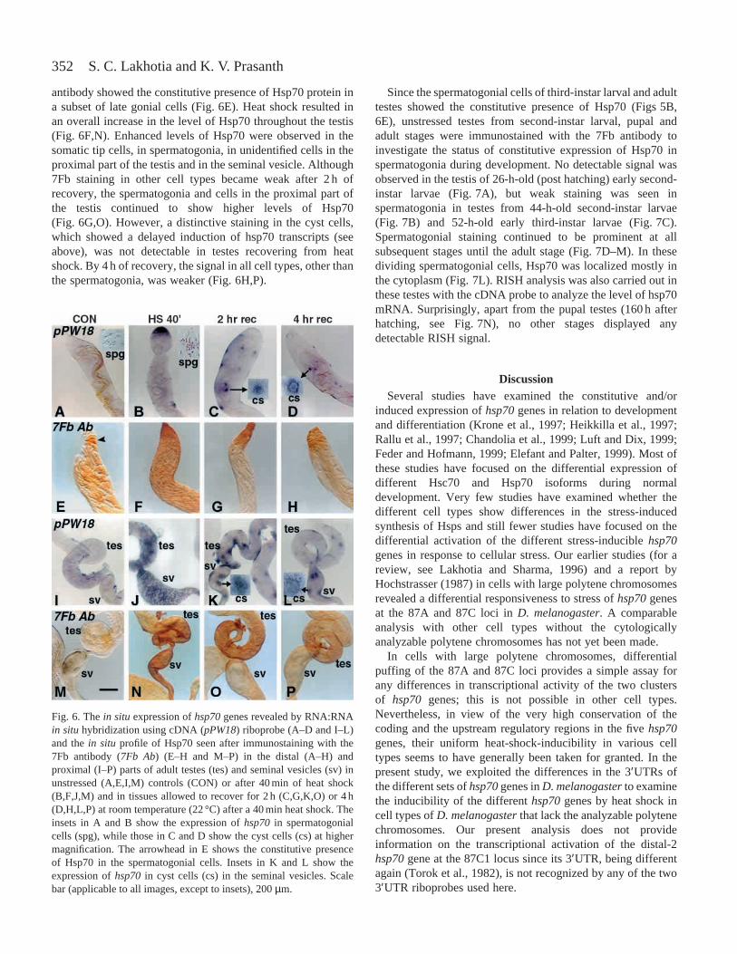
RISH in adult testes using the cDNA probe revealed cell-type-specific induction and stability of hsp70 transcripts. Nodetectable signal was observed in the unstressed control testis(Fig. 6A). Even the spermatogonial cells showed no detectablehybridization (see inset to Fig. 6A). Heat shock for 40 minresulted in induction of hsp70genes, particularly strongly inthe spermatogonial cells (Fig. 6B and inset), with thetranscripts being localized in the cytoplasm. Unlike thelarval testis, the hsp70 transcripts disappeared from thespermatogonial cells in adult testis within 2 h of recovery(Fig. 6C,D). For reasons that are unclear, neither of the 3′UTRriboprobes generated distinct RISH staining in adult testis.
The pair of somatic cyst cells, which remain associated with
every cyst of growing germ cells until sperm maturation(Fuller, 1993), showed a delayed induction of hsp70genes.Hsp70 transcripts were not detectable in these cellsimmediately after heat shock (Fig. 6B), but intense expressionwas seen after 2 h of recovery from heat shock (Fig. 6C); thetranscripts were distributed in the cytoplasm as well as in thenucleus (inset in Fig. 6C). Cyst cells continued to showpositive staining even after 4 h of recovery (Fig. 6D). At thistime, the transcripts were mostly localized in the cyst cellcytoplasm (inset in Fig. 6D).
Cells in the proximal region of the testis, connecting withthe seminal vesicle, showed a strong induction soon after heatshock (Fig. 6J). The somatic sheath cells on the seminal vesiclealso exhibited intense heat-shock-induced expression of hsp70genes (Fig. 6J). After 2 and 4 h of recovery, the signal in theproximal part of the testis and in the seminal vesicle becameweak, but the cyst cells and some other unidentified cells in theseminal vesicle showed delayed but enhanced expression(Fig. 6K,L and insets).
Immunostaining of the unstressed adult testis with the 7Fb
Fig. 5. Expression of hsp70genes in larval testes as revealed byRNA:RNA in situ hybridization using the cDNA (pPW18) (A,E,P)87A3′UTR (C,G,I,K,N) or 87C3′UTR (D,H,J,L,O) riboprobe andthe in situ profile of Hsp70 as revealed by immunostaining with the7Fb antibody (7Fb Ab) (B,F,M,Q) in unstressed control (CON)(A,B) or heat-shocked (HS) (C–F) testes or those allowed torecover for 30 min (G,H), 1 h (I,J), 2 h (K–M) or 4 h (N–Q) at roomtemperature (22 °C) after a 40 min heat shock. Note the constitutivepresence of Hsp70 in the anterior region (spermatogonial cells) inunstressed testes and also the greater induction of the locus 87Ahsp70genes in these cells following heat shock. Scale bar, 500µm.
352
antibody showed the constitutive presence of Hsp70 protein ina subset of late gonial cells (Fig. 6E). Heat shock resulted inan overall increase in the level of Hsp70 throughout the testis(Fig. 6F,N). Enhanced levels of Hsp70 were observed in thesomatic tip cells, in spermatogonia, in unidentified cells in theproximal part of the testis and in the seminal vesicle. Although7Fb staining in other cell types became weak after 2 h ofrecovery, the spermatogonia and cells in the proximal part ofthe testis continued to show higher levels of Hsp70(Fig. 6G,O). However, a distinctive staining in the cyst cells,which showed a delayed induction of hsp70 transcripts (seeabove), was not detectable in testes recovering from heatshock. By 4 h of recovery, the signal in all cell types, other thanthe spermatogonia, was weaker (Fig. 6H,P).
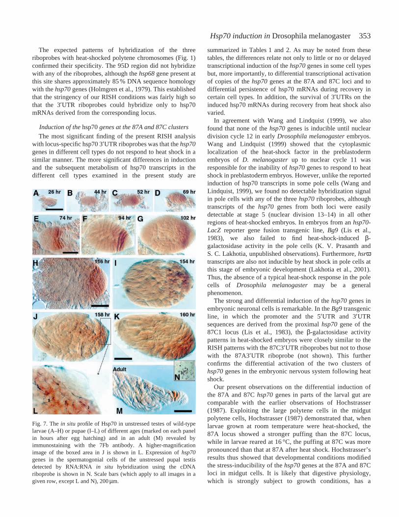
Since the spermatogonial cells of third-instar larval and adulttestes showed the constitutive presence of Hsp70 (Figs 5B,6E), unstressed testes from second-instar larval, pupal andadult stages were immunostained with the 7Fb antibody toinvestigate the status of constitutive expression of Hsp70 inspermatogonia during development. No detectable signal wasobserved in the testis of 26-h-old (post hatching) early second-instar larvae (Fig. 7A), but weak staining was seen inspermatogonia in testes from 44-h-old second-instar larvae(Fig. 7B) and 52-h-old early third-instar larvae (Fig. 7C).Spermatogonial staining continued to be prominent at allsubsequent stages until the adult stage (Fig. 7D–M). In thesedividing spermatogonial cells, Hsp70 was localized mostly inthe cytoplasm (Fig. 7L). RISH analysis was also carried out inthese testes with the cDNA probe to analyze the level of hsp70mRNA. Surprisingly, apart from the pupal testes (160 h afterhatching, see Fig. 7N), no other stages displayed anydetectable RISH signal.
DiscussionSeveral studies have examined the constitutive and/or
induced expression of hsp70genes in relation to developmentand differentiation (Krone et al., 1997; Heikkilla et al., 1997;Rallu et al., 1997; Chandolia et al., 1999; Luft and Dix, 1999;Feder and Hofmann, 1999; Elefant and Palter, 1999). Most ofthese studies have focused on the differential expression ofdifferent Hsc70 and Hsp70 isoforms during normaldevelopment. Very few studies have examined whether thedifferent cell types show differences in the stress-inducedsynthesis of Hsps and still fewer studies have focused on thedifferential activation of the different stress-inducible hsp70genes in response to cellular stress. Our earlier studies (for areview, see Lakhotia and Sharma, 1996) and a report byHochstrasser (1987) in cells with large polytene chromosomesrevealed a differential responsiveness to stress of hsp70genesat the 87A and 87C loci in D. melanogaster. A comparableanalysis with other cell types without the cytologicallyanalyzable polytene chromosomes has not yet been made.
In cells with large polytene chromosomes, differentialpuffing of the 87A and 87C loci provides a simple assay forany differences in transcriptional activity of the two clustersof hsp70 genes; this is not possible in other cell types.Nevertheless, in view of the very high conservation of thecoding and the upstream regulatory regions in the five hsp70genes, their uniform heat-shock-inducibility in various celltypes seems to have generally been taken for granted. In thepresent study, we exploited the differences in the 3′UTRs ofthe different sets of hsp70genes in D. melanogasterto examinethe inducibility of the different hsp70genes by heat shock incell types of D. melanogasterthat lack the analyzable polytenechromosomes. Our present analysis does not provideinformation on the transcriptional activation of the distal-2hsp70gene at the 87C1 locus since its 3′UTR, being differentagain (Torok et al., 1982), is not recognized by any of the two3′UTR riboprobes used here.
S. C. Lakhotia and K. V. Prasanth
Fig. 6. The in situexpression of hsp70genes revealed by RNA:RNAin situhybridization using cDNA (pPW18) riboprobe (A–D and I–L)and the in situprofile of Hsp70 seen after immunostaining with the7Fb antibody (7Fb Ab) (E–H and M–P) in the distal (A–H) andproximal (I–P) parts of adult testes (tes) and seminal vesicles (sv) inunstressed (A,E,I,M) controls (CON) or after 40 min of heat shock(B,F,J,M) and in tissues allowed to recover for 2 h (C,G,K,O) or 4 h(D,H,L,P) at room temperature (22 °C) after a 40 min heat shock. Theinsets in A and B show the expression of hsp70in spermatogonialcells (spg), while those in C and D show the cyst cells (cs) at highermagnification. The arrowhead in E shows the constitutive presenceof Hsp70 in the spermatogonial cells. Insets in K and L show theexpression of hsp70in cyst cells (cs) in the seminal vesicles. Scalebar (applicable to all images, except to insets), 200 µm.
353Hsp70 induction in Drosophila melanogaster
The expected patterns of hybridization of the threeriboprobes with heat-shocked polytene chromosomes (Fig. 1)confirmed their specificity. The 95D region did not hybridizewith any of the riboprobes, although the hsp68gene present atthis site shares approximately 85 % DNA sequence homologywith the hsp70genes (Holmgren et al., 1979). This establishedthat the stringency of our RISH conditions was fairly high sothat the 3′UTR riboprobes could hybridize only to hsp70mRNAs derived from the corresponding locus.
Induction of the hsp70genes at the 87A and 87C clusters
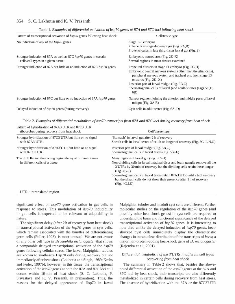
The most significant finding of the present RISH analysiswith locus-specific hsp70 3′UTR riboprobes was that the hsp70genes in different cell types do not respond to heat shock in asimilar manner. The more significant differences in inductionand the subsequent metabolism of hsp70 transcripts in thedifferent cell types examined in the present study are
summarized in Tables 1 and 2. As may be noted from thesetables, the differences relate not only to little or no or delayedtranscriptional induction of the hsp70genes in some cell typesbut, more importantly, to differential transcriptional activationof copies of the hsp70genes at the 87A and 87C loci and todifferential persistence of hsp70 mRNAs during recovery incertain cell types. In addition, the survival of 3′UTRs on theinduced hsp70 mRNAs during recovery from heat shock alsovaried.
In agreement with Wang and Lindquist (1999), we alsofound that none of the hsp70genes is inducible until nucleardivision cycle 12 in early Drosophila melanogasterembryos.Wang and Lindquist (1999) showed that the cytoplasmiclocalization of the heat-shock factor in the preblastodermembryos of D. melanogasterup to nuclear cycle 11 wasresponsible for the inability of hsp70genes to respond to heatshock in preblastoderm embryos. However, unlike the reportedinduction of hsp70 transcripts in some pole cells (Wang andLindquist, 1999), we found no detectable hybridization signalin pole cells with any of the three hsp70riboprobes, althoughtranscripts of the hsp70genes from both loci were easilydetectable at stage 5 (nuclear division 13–14) in all otherregions of heat-shocked embryos. In embryos from an hsp70-LacZ reporter gene fusion transgenic line, Bg9 (Lis et al.,1983), we also failed to find heat-shock-induced β-galactosidase activity in the pole cells (K. V. Prasanth andS. C. Lakhotia, unpublished observations). Furthermore, hsrωtranscripts are also not inducible by heat shock in pole cells atthis stage of embryonic development (Lakhotia et al., 2001).Thus, the absence of a typical heat-shock response in the polecells of Drosophila melanogastermay be a generalphenomenon.
The strong and differential induction of the hsp70genes inembryonic neuronal cells is remarkable. In the Bg9transgenicline, in which the promoter and the 5′UTR and 3′UTRsequences are derived from the proximal hsp70gene of the87C1 locus (Lis et al., 1983), the β-galactosidase activitypatterns in heat-shocked embryos were closely similar to theRISH patterns with the 87C3′UTR riboprobes but not to thosewith the 87A3′UTR riboprobe (not shown). This furtherconfirms the differential activation of the two clusters ofhsp70genes in the embryonic nervous system following heatshock.
Our present observations on the differential induction ofthe 87A and 87C hsp70genes in parts of the larval gut arecomparable with the earlier observations of Hochstrasser(1987). Exploiting the large polytene cells in the midgutpolytene cells, Hochstrasser (1987) demonstrated that, whenlarvae grown at room temperature were heat-shocked, the87A locus showed a stronger puffing than the 87C locus,while in larvae reared at 16 °C, the puffing at 87C was morepronounced than that at 87A after heat shock. Hochstrasser’sresults thus showed that developmental conditions modifiedthe stress-inducibility of the hsp70genes at the 87A and 87Cloci in midgut cells. It is likely that digestive physiology,which is strongly subject to growth conditions, has a
Fig. 7. The in situprofile of Hsp70 in unstressed testes of wild-typelarvae (A–H) or pupae (I–L) of different ages (marked on each panelin hours after egg hatching) and in an adult (M) revealed byimmunostaining with the 7Fb antibody. A higher-magnificationimage of the boxed area in J is shown in L. Expression of hsp70genes in the spermatogonial cells of the unstressed pupal testisdetected by RNA:RNA in situ hybridization using the cDNAriboprobe is shown in N. Scale bars (which apply to all images in agiven row, except L and N), 200µm.
354
significant effect on hsp70gene activation in gut cells inresponse to stress. This modulation of hsp70inducibilityin gut cells is expected to be relevant to adaptability innature.
The significant delay (after 2 h of recovery from heat shock)in transcriptional activation of the hsp70genes in cyst cells,which remain associated with the bundles of differentiatinggerm cells (Fuller, 1993), is most unusual. We are not awareof any other cell type in Drosophila melanogasterthat showsa comparable delayed transcriptional activation of the hsp70genes following cellular stress. The larval Malpighian tubulesare known to synthesize Hsp70 only during recovery but notimmediately after heat shock (Lakhotia and Singh, 1989; Krebsand Feder, 1997b); however, in this tissue, the transcriptionalactivation of the hsp70genes at both the 87A and 87C loci stilloccurs within 10 min of heat shock (S. C. Lakhotia, P.Srivastava and K. V. Prasanth, in preparation). Thus, thereasons for the delayed appearance of Hsp70 in larval
Malpighian tubules and in adult cyst cells are different. Furthermolecular studies on the regulation of the hsp70genes (andpossibly other heat-shock genes) in cyst cells are required tounderstand the basis and functional significance of the delayedtranscriptional activation of hsp70genes. It is interesting tonote that, unlike the delayed induction of hsp70genes, heat-shocked cyst cells immediately display the characteristicchanges in intranuclear distribution of the transcripts of hsrω, amajor non-protein-coding heat-shock gene of D. melanogaster(Rajendra et al., 2001).
Differential metabolism of the 3′UTRs in different cell typesrecovering from heat shock
The summary in Table 2 shows that, besides the above-noted differential activation of the hsp70genes at the 87A and87C loci by heat shock, their transcripts are also differentlymetabolized in certain cells during recovery from heat stress.The absence of hybridization with the 87A or the 87C3′UTR
S. C. Lakhotia and K. V. Prasanth
Table 1.Examples of differential activation of hsp70genes at 87A and 87C loci following heat shock
Pattern of transcriptional activation of hsp70genes following heat shock Cell/tissue type
No induction of any of the hsp70genes Stage 1–3 embryos Pole cells in stage 4–5 embryos (Fig. 2A,B)Proventriculus in late third-instar larval gut (Fig. 3)
Stronger induction of 87A as well as 87C hsp70genes in certain Embryonic neuroblasts (Fig. 2E–X)cells/cell types in a given tissue Several regions in most tissues examined
Stronger induction of 87A but little or no induction of 87C hsp70genes Proneural clusters in stage 11 embryos (Fig. 2G,H)Embryonic central nervous system (other than the glial cells),
peripheral nervous system and tracheal pits from stage 13 onwards (Fig. 2K–X)
Posterior part of larval midgut (Fig. 3B,C)Spermatogonial cells of larval (and adult?) testes (Figs 5C,D,
6B)
Stronger induction of 87C but little or no induction of 87A hsp70genes Narrow segment joining the anterior and middle parts of larval midgut (Fig. 3A,B)
Delayed induction of hsp70genes (during recovery) Cyst cells in adult testes (Fig. 6A–D)
Table 2. Examples of differential metabolism of hsp70 transcripts from 87A and 87C loci during recovery from heat shock
Pattern of hybridization of 87A3′UTR and 87C3′UTR riboprobes during recovery from heat shock Cell/tissue type
Stronger hybridization of 87C3′UTR but little or no signal ‘Stomach’ in larval gut after 2 h of recoverywith 87A3′UTR Sheath cells in larval testes after 1 h or longer of recovery (Fig. 5G–L,N,O)
Stronger hybridization of 87A3′UTR but little or no signal Posterior part of larval midgut (Fig. 3B,C)with 87C3′UTR Spermatogonial cells in larval testes (Fig. 5G–L)
The 3′UTRs and the coding region decay at different times Many regions of larval gut (Fig. 3C–H)in different cells of a tissue Non-dividing cells in larval imaginal discs and brain ganglia remove all the
3′UTRs by 30 min of recovery but the dividing cells retain these longer (Fig. 4B–I)
Spermatogonial cells in larval testes retain 87A3′UTR until 2 h of recovery but the sheath cells do not show their presence after 1 h of recovery (Fig. 4G,I,K)
UTR, untranslated region.
355Hsp70 induction in Drosophila melanogaster
probe during recovery in certain cell types (Table 2) could bedue either to the loss of the 3′UTR sequence alone or tocomplete degradation of the mRNA from the particular locus.The present study does not allow a distinction between thesepossibilities.
In several instances, different cell types in the same tissueshowed differences in the time of removal of the 3′UTRs onhsp70 mRNAs. Thus, in larval imaginal discs and brain, whilethe cDNA probe continued to give an intense signal in mostcells after 30 min of recovery from heat shock, the two 3′UTRriboprobes hybridized only to a subset of cells that werealways in characteristically organized clusters. Thedistribution of these clusters of cells matched that of theactively proliferating cells detected by anti-phospho-histoneantibody (Hendzel et al., 1997; Brodsky et al., 2000). Itappears, therefore, that, while the actively dividing cells inthese tissues retained the 3′UTRs, other cells quickly removedthem. This persistence of the 3′UTRs on the hsp70 mRNAmay be related to the need for rapid degradation of thesetranscripts in actively dividing cells since high levels of Hsps,especially Hsp70, have been reported to be detrimental torapidly dividing cells (Feder et al., 1992; Feder and Hofmann,1999). The 3′UTRs of hsp70 transcripts contain AU-richinstability elements that cause their rapid degradation duringrecovery (Yost et al., 1990; Sachs, 1993). Although, in larvalspermatogonial cells, the 3′UTRs were also removed soonerthan the coding region, it is intriguing that the dividingspermatogonial cells continued to show the strong presence ofHsp70 4 h after heat shock and even in the absence of thermalstress (see below). Apparently, the presence of Hsp70 does notinterfere with the division process (Feder et al., 1992) inspermatogonia.
The reasons for our failure to detect hybridization of 3′UTRprobes with cellular RNA in adult testes, when the cDNAprobe readily hybridized, are not clear. Whether this was atechnical failure or whether the 3′UTRs are not transcribed orare very quickly removed from the adult spermatogonial cellsis not known. In the absence of hybridization of the 3′UTRriboprobes, we cannot define the origin of the hsp70 transcriptsin heat-shocked spermatogonial cells in adult testes. However,in view of the results with larval testes, it is tempting tospeculate that these transcripts in the adult testes are alsoderived from the 87A hsp70genes.
Constitutive presence of Hsp70 in spermatogonia
A novel observation of the present study was theconstitutive presence of Hsp70 in spermatogonia from thesecond-instar larval stage onwards. The 7Fb antibody is wellknown to react only with the heat-inducible form ofDrosophila melanogasterHsp70 and does not recognize anyof the Hsc70 proteins or the heat-inducible Hsp68 (Velazquezand Lindquist, 1984). That the presence of Hsp70 inspermatogonial cells was not due to inadvertent stress issupported by the following considerations. The somatic hubcells at the tip of the testis, which showed elevated levels ofHsp70 soon after heat shock, displayed no constitutive
immunostaining. In the unstressed spermatogonia, Hsp70 waspresent in the cytoplasm rather than in the nucleus, as seen instressed cells (Velazquez and Lindquist, 1984).
In the context of the constitutive presence of Hsp70 inspermatogonia, the absence of detectable hybridization of thecDNA riboprobe in most of the unstressed testes, except insome pupal testis samples, is intriguing. Perhaps some or allof the hsp70genes in unstressed spermatogonia are transcribedonly intermittently and/or at a very low level so that RISH failsto detect these transcripts in most cases. Alternatively or inaddition, the Hsp70 in spermatogonial cells may be more stablethan in other cell types so that, once synthesized, it persistseven in the absence of fresh transcription. This may findsupport in our observation that, after 2 or 4 h of recoveryfrom heat shock, Hsp70 was abundantly present in thespermatogonial cells of the testes, although the riboprobesshowed no hybridization at this time (compareFig. 6C,D,G,H). Further studies are needed to determinewhether the other Hsps also show a comparable constitutivepresence in the spermatogonial cells and if this involves theheat-shock transcription factor.
The present observation on the constitutive presence ofHsp70 in spermatogonia is the first report of the developmentalexpression of heat-inducible Hsp70 in Drosophilamelanogaster. Developmental expression of Hsp70 familyproteins in mammalian male germ cells is well documented(Eddy, 1999). Two members of the Hsp70 family, HSP70-2and HSC70T, are regulated to express specifically in meioticand post-meiotic speramatogenic cells in mice (Matsumoto andFujimoto, 1990; Dix et al., 1997). Interestingly, homologuesof Hsp70-2 are reported in the testes of many other animals(Eddy, 1999), suggesting that such spermatogenic cellchaperones are conserved across phyla. However, unlike thesituation in the mouse, the meiotic stages in Drosophilamelanogaster testes do not express Hsp70 constitutively nor isthis protein heat-shock-inducible at this stage. Thus, theconstitutively present Hsp70 in spermatogonial cells inDrosophila melanogaster may have different function(s). Thisneeds further analysis.
Multiple levels of regulation of the hsp70 genes in Drosophilamelanogaster
The strong conservation of the 5′UTRs and the immediatelyupstream regulatory regions in the five hsp70genes in D.melanogaster(see Introduction) have generally been acceptedas the cause for the more-or-less uniform induction of Hsp70observed in most previous studies utilizing [35S]methioninelabelling and/or western blotting or immunostaining. However,the present in-depth in situanalysis of induction of individualgroups of hsp70genes has revealed remarkable differences inthe induction of the different hsp70genes by heat shock. It isobvious from the summaries in Tables 1 and 2 that the heat-shock (and presumably other stress)-induced expression ofHsp70 is regulated not only at the level of transcription (noactivation versusvarying levels of activation, and selectiveactivation of only some of the hsp70genes or delayed
356
activation) but also at the post-transcriptional (export from thenucleus) and translational (e.g. delayed translation andturnover of the hsp70 mRNAs) levels.
The mechanisms underlying this differential regulation arenot known. The 3′UTRs, being different in the two sets ofgenes (Torok et al., 1982), obviously have a role in post-transcriptional regulation since these regions are known toaffect the stability and transport of the mRNAs (Lakhotia,1995; Whittaker et al., 1999; Lipshitz and Smibert, 2000). Ourresults imply that widely accepted ideas, e.g. that the hsp70gene promoter remains in an ‘open’ conformation in readinessfor immediate and rapid transcriptional activation followingcellular stress (Parsell and Lindquist, 1994; Feder andHofmann, 1999; Wang and Lindquist, 1999) and that this geneis autoregulated (DiDomenico et al., 1982), may be true formost but not every cell type of Drosophila melanogaster.Regulation of the hsp70genes, the key players in the cellularstress response in Drosophila melanogaster, is obviously muchmore complex, and many additional features of their regulationare yet to be understood. Recent studies (Sorensen et al., 1999;Andrulis et al., 2000; Tang et al., 2000; Lerman and Feder,2001; Marchler and Wu, 2001; Wu et al., 2001) are, in fact,uncovering some of these features.
The significance of the differential regulation of the fivehsp70 genes is not clear, especially since the amino acidsequences encoded by these five gene copies show very littledivergence and since not all species of Drosophila containduplicated sets of hsp70genes. However, we believe that thecell-type-specific patterns of activation of the different hsp70genes observed by us have functional and evolutionarysignificance. The fact that this evolutionary duplicationoccurred in the melanogastergroup of species (Feder andKrebs, 1998; Konstantopoulou et al., 1998), and has beenretained through the species diversification, itself suggests itsadaptive value. In this context, two other possibilities also needto be examined further: (i) that the few amino acid differencesin the Hsp70 produced by these duplicated sets of genes havea significant effect on chaperoning activity and, therefore, theirdifferential inducibility may ensure fine-tuning of the stressresponse, and (ii) that the differences in the 3′UTRs providedifferential targeting of the hsp70 mRNAs to subcellularcompartments and that this again is relevant for specificcellular needs.
It is apparent that the multiple copies of the hsp70genes inDrosophilaspp. are not just to provide abundant Hsp70 at ashort notice. Studies of hsp70genes in natural populations ofD. melanogaster(Krebs and Feder, 1997a; Feder and Krebs,1998; Feder and Hofmann, 1999; Lansing et al., 2000) havealso revealed significant variations in Hsp70 expression andtheir adaptive significance. It appears that the specificphysiology of a cell/tissue type affects the cost/benefit ratio ofthe Hsp70 under stress conditions and, therefore, thatelaborate regulatory mechanisms have evolved to fine-tune theproduction and level of Hsp70 in a given cell. It is interestingthat much of the polymorphism in the hsp70 genes in D.melanogasterpopulations seems to be in the UTR and
promoter sequences (M. E. Feder, personal communication).More intensive studies on the induction of the hsp70genes indifferent tissues of natural populations of Drosophilaspp.would be instructive.
The present observations on the tissue/stage-specificresponse of the different hsp70genes to heat shock haveimplications for many developmental genetic studies that utilizethe heat-shock promoter to drive a reporter or some other geneactivity in Drosophilaspp. The hsp70promoter has been widelyused by the fly community to conditionally express any gene inall cell types for experimental purposes. On the basis of ourpresent results, we point out that, depending upon the specifichsp70 gene promoter and the UTR sequences used in theconstruction of the transgene that is desired to be expressed inresponse to heat shock, its expression may vary qualitativelyand quantitatively in different tissues. This needs to be takeninto consideration in interpreting the experimental results.
Studies on the heat-shock response have mostly beenrestricted to a few easily amenable cell/tissue types in modelsystems maintained under controlled laboratory conditions,and this has led to the development of paradigms that do notreflect the dynamically different requirements of heat-shockgene activities under different ecological and physiologicalconditions (Lakhotia, 2001; Feder, 2001). Further studies onthe regulation of heat-shock genes in more diverse cell typesand under different environmental conditions in naturalpopulations would provide interesting and exciting informationon the complex regulation and biological relevance of this mostancient gene programme.
We thank Professor Susan Lindquist for providing the 7Fbantibody used in this study. This work was supported by aresearch grant from the Department of Biotechnology,Government of India, New Delhi to S.C.L. K.V.P. wassupported by a fellowship from the Council of Scientific andIndustrial Research (CSIR), New Delhi, India.
ReferencesAndrulis, E. D., Guzman, E., Doring, P., Werner, J. and Lis, J. T.(2000).
High-resolution localization of DrosophilaSpt5 and Spt6 at heat shockgenes in vivo: roles in promoter proximal pausing and transcriptionelongation.Genes Dev. 14, 2635–2649.
Bendena, W. J., Southgate, A. A., Garbe, J. C. and Pardue, M. L.(1991). Expression of the heat shock locus hsr omegain non-stressedcells during development in Drosophila melanogaster. Dev. Biol. 144,65–77.
Brodsky, M. H., Sekelsky, J. J., Tsang, G., Hawley, R. S. and Rubin, G.M. (2000). Mus304 encodes a novel DNA damage checkpoint proteinrequired during Drosophiladevelopment.Genes Dev.14, 666–678.
Chandolia, R. K., Peltier, M. R., Tian, W. and Hansen, P. J.(1999).Transcriptional control of development, protein synthesis and heat-shock-induced heat shock protein 70 synthesis in 2-cell bovine embryos.Biol.Reprod.61, 1644–1648.
Craig, E. A., Ingolia, T. D. and Manseau, L. J.(1983). Expression ofDrosophilaheat shock cognate genes during heat shock and development.Dev. Biol. 99, 418–426.
Curtis, D., Lehmann, R. and Zamore, P. D.(1995). Translational regulationin development.Cell 81, 171–178.
Dellavalle, R. P., Petersen, R. and Lindquist, S.(1994). Preferentialdeadenylation of hsp70 mRNA plays a key role in regulating hsp70expression in Drosophila melanogaster. Mol. Cell. Biol. 14, 3646–3659.
S. C. Lakhotia and K. V. Prasanth
357Hsp70 induction in Drosophila melanogaster
DiDomenico, B., Bugaisky, G. E. and Lindquist, S.(1982). The heat shockresponse is self regulated at both the transcriptional and post-transcriptionallevels.Cell 31, 593–603.
Dix, D. J., Allen, J. W., Collins, B. W., Mori, C., Nakamura, N., Poorman-Allen, P., Goulding, E. H. and Eddy, E. M. (1997). Targeted genedisruption of Hsp70-2results in failed meiosis, germ cell apoptosis and maleinfertility. Proc. Natl. Acad. Sci. USA 93, 3264–3268.
Eddy, E. M. (1999). Role of heat shock protein hsp70-2 in spermatogenesis.Rev. Reprod.4, 23–30.
Elefant, F. and Palter, K. B.(1999). Tissue-specific expression of dominantnegative mutant DrosophilaHSC70 causes developmental defects andlethality. Dev. Genet. 25, 31–39.
Feder, M. E. (2001). Ecological and evolutionary functional genomics ofmolecular chaperones.Biol. Int. 40, 20–23.
Feder, M. E. and Hofmann, G. E.(1999). Heat shock proteins, molecularchaperones and the stress response: Evolutionary and ecological physiology.Annu. Rev. Physiol. 61, 243–282.
Feder, M. E. and Krebs, R. A.(1998). Natural and genetic engineering ofthermotolerance in Drosophila melanogaster: consequence forthermotolerance.Am. Zool. 38, 503–517.
Feder, M. E., Rossi, J. M., Solomon, J., Solomon, N. and Lindquist, S.(1992). The consequences of expressing hsp70 in Drosophilacells at normaltemperatures.Genes Dev.6, 1402–1413.
Feige, U. and Polla, S.(1994). Heat shock proteins: the hsp70 family.Experientia50, 979–986.
Fuller, M. (1993). Spermatogenesis. In The Development of Drosophilamelanogaster (ed. M. Bate and A. Martinez), pp. 71–147. New York: ColdSpring Harbor Laboratory Press.
Goldschmidt-Clermont, M. (1980). Two genes for the major heat shockprotein of Drosophila melanogasterarranged as an inverted repeat.NucleicAcids Res. 8, 235–252.
Gunther, E. and Walter, L. (1994). Genetic aspects of the Hsp70 multigenefamily in vertebrates.Experientia50, 987–1001.
Heikkilla, J. J., Ohan, N., Tam, Y. and Ali, A. (1997). Heat shock proteingene expression during Xenopusdevelopment.Cell Mol. Life Sci.53,114–121.
Hendzel, M. J., Wei, Y., Mancini, M. A., Hooser, A. V., Ranalli, T.,Brinkley, B. R., Bazett-Jones, D. P. and Allis, C. D.(1997). Mitosis-specific phosphorylation of histone H3 initiates primarily withinpericentromeric heterochromatin during G2 and spreads in an orderedfashion coincident with mitotic chromosome condensation.Chromosoma106, 348–360.
Hochstrasser, M. (1987). Chromosome structure in 4 wild type polytenetissues of D. melanogaster. The 87A and 87C heat shock loci are expressedunequally in the midgut in a manner dependent on growth temperature.Chromosoma95, 197–208.
Holmgren, R., Livak, K., Morimoto, R. I., Freund, R. and Messelson, M.(1979). Studies of cloned sequences from four Drosophilaheat shock loci.Cell 18, 1359–1370.
Hortsch, M., Bieber, A. J., Patel, N. H. and Goodman, C. S.(1990).Alternative splicing generates a nervous system specific form of neuroglian.Neuron4, 697–709.
Ingolia, T. D., Craig, E. A. and McCarthy, N. J.(1980). Sequence of threecopies of the gene for the major Drosophilaheat-shock-induced protein andtheir flanking regions.Cell 21, 669–679.
Ish-Horowicz, D. and Pinchin, S. M.(1980). Genetic organization of the87A7 and 87C1 heat induced loci of Drosophila melanogaster. J. Mol. Biol.142, 231–245.
Karch, F., Torok, I. and Tissieres, A.(1981). Extensive regions of homologyin front of the two hsp70 heat shock variant genes in Drosophilamelanogaster.J. Mol. Biol. 148, 219–230.
Konstantopoulou, I., Nikolaidis, N. and Scouras, Z. G.(1998). The hsp70locus of Drosophila auraria(monitor subgroup) is single and containscopies in conserved arrangement.Chromosoma107, 577–586.
Krebs, R. A. and Feder, M. E.(1997a). Natural variation in the expressionof the heat-shock protein Hsp70 in a population of Drosophila melanogasterand its correlation with tolerance of ecologically relevant thermal stress.Evolution51, 173–179.
Krebs, R. A. and Feder, M. E.(1997b). Tissue-specific variation in hsp70expression and thermal damage in Drosophila melanogasterlarvae.J. Exp.Biol. 200, 2007–2015.
Krone, P. H., Lele, Z. and Sass, J. B.(1997). Heat shock genes and the heatshock response in zebrafish embryos.Biochem. Cell Biol.75, 487–497.
Lakhotia, S. C. (1995). Targeting RNA in cell.Curr. Sci.69, 219–221.
Lakhotia, S. C. (2001). Stress response – ecological and developmentalconnections, Biol. Int. 40, 24–27
Lakhotia, S. C., Rajendra, T. K. and Prasanth, K. V. (2001).Developmental regulation and complex organization of the promoter of thenon-coding hsrωgene of Drosophila melanogaster. J. Biosci.21, 25–38.
Lakhotia, S. C., Ray, P., Rajendra, T. K. and Prasanth, K. V.(1999). Thenon-coding transcripts of hsrωgene in Drosophila: do they regulatetrafficking and availability of nuclear RNA-processing factors? Curr. Sci.77, 553–563.
Lakhotia, S. C. and Sharma, A.(1996). The 93D (hsr-omega) locus ofDrosophila: non-coding gene with house keeping function.Genetica97,339–348.
Lakhotia, S. C. and Singh, A. K. (1989). A novel set of heat shockpolypeptides in Malpighian tubules of Drosophila melanogaster. J. Genet.68, 129–137.
Lakhotia, S. C. and Tapadia, M. G.(1998). Genetic mapping of the amideresponse element/s of the hsrωlocus of Drosophila melanogaster.Chromosoma107, 127–135.
Lansing, E., Justesen, J. and Loeschcke, V. V.(2000). Variation in theexpression of Hsp70, the major heat-shock protein and thermotolerance inlarval and adult selection lines of Drosophila melanogaster. J. Therm. Biol.25, 443–450.
Leigh-Brown, A. J. and Ish-Horowicz, D.(1981). Evolution of the 87A and87C heat shock loci in Drosophila.Nature290, 677–682.
Lerman, D. N. and Feder, M. E.(2001). Laboratory selection at differenttemperatures modifies heat-shock transcription factor (HSF) activation inDrosophila melanogaster. J. Exp. Biol. 204, 315–323.
Lipshitz, H. D. and Smibert, C. A.(2000). Mechanisms of RNA localizationand translational regulation.Curr. Opin. Genet. Dev.10, 476–488.
Lis, J. T., Simon, J. A. and Sutton, C. A.(1983). New heat shock puffs andβ-galactosidaseactivity resulting from transformation of Drosophilawithan hsp70-lacZhybrid gene.Cell 35, 403–410.
Luft, J. C. and Dix, D. J. (1999). Hsp70 expression and function duringembryogenesis.Cell Stress Chaperones4, 162–170.
Macario, A. J. L., Lange, M., Ahring, B. K. and De Macario, E. Y. C.(1999). Stress genes and proteins in the Archaea.Microbiol. Mol. Biol. Rev.63, 923–967.
Marchler, G. and Wu, C. (2001). Modulation of Drosophila heat shocktranscription factor activity by the molecular chaperone DROJ1.EMBO J.20, 499–509.
Matsumoto, M. and Fujimoto, H. (1990). Cloning of a hsp70-related geneexpressed in mouse spermatids.Biochem. Biophys. Res. Commun. 166,43–49.
Parsell, D. A. and Lindquist, S. (1994). Heat shock proteins and stresstolerance. In The Biology of Heat Shock Proteins and MolecularChaperones(ed. R. I. Morimoto, A. Tissieres and C. Georgopoulos), pp.457–493. New York: Cold Spring Harbor Laboratory Press.
Prasanth, K. V., Rajendra, T. K., Lal, A. K. and Lakhotia, S. C.(2000).Omega speckles – a novel class of nuclear speckles containing hnRNPsassociated with non-coding hsr-omega RNA in Drosophila.J. Cell Sci. 113,3485–3497.
Rajendra, T. K., Prasanth, K. V. and Lakhotia, S. C.(2001). Male sterilityassociated with over-expression of the non-coding hsrωgene in cyst cellsof testis of Drosophila melanogaster. J. Genet.80 (in press).
Rallu, M., Loones, M. T., Lallemand, Y., Morimoto, R., Morange, M. andMezger, V. (1997). Function and regulation of heat shock factor 2 duringmouse embryogenesis.Proc. Natl. Acad. Sci. USA94, 2392–2397.
Ross, J.(1996). Control of messenger RNA stability in higher eukaryotes.Trends Genet. 12, 171–175.
Sachs, A. B.(1993). Messenger RNA degradation in eukaryotes.Cell 74,413–421.
Schlesinger, M. J., Ashburner, M. and Tissieres, A.(1982). (ed). HeatShock Proteins: From Bacteria to Man. New York: Cold Spring HarborLaboratory Press.
Sharma, A. and Lakhotia, S. C.(1995). In situquantification of hsp70 andalpha-beta transcripts at 87A and 87C loci in relation to hsr-omegageneactivity in polytene cells of D. melanogaster. Chromosome Res. 3, 386–393.
Sorensen, J. G., Michalak, P., Justesen, J. and Loeschcke, V.(1999).Expression of the heat-shock protein HSP70 in Drosophila buzzatiilinesselected for thermal resistance.Hereditas131, 155–164.
Tang, H., Liu, Y., Madabusi, L. and Gilmour, D. S. (2000). Promoter-proximal pausing on the hsp70 promoter in Drosophila melanogasterdepends on the upstream regulator.Mol. Cell Biol.20, 2569–2580.
Torok, I., Mason, P. J., Karch, F., Kiss, I. and Udvardy, A.(1982).

358
Extensive regions of homology associated with heat-induced genes at loci87A7 and 87C1 in D. melanogaster. In Heat Shock Proteins: From Bacteriato Man (ed M. J. Schlesinger, M. Ashburner and A. Tissieres), pp. 19–25.New York: Cold Spring Harbor Laboratory Press.
Velazquez, J. M. and Lindquist, S.(1984). Hsp70: nuclear concentrationduring environmental stress and cytoplasmic storage during recovery.Cell36, 655–662.
Wang, Z. and Lindquist, S. (1999). Developmentally regulated nucleartransport of transcription factors in Drosophilaembryos enable the heatshock response.Development125, 4841–4850.
Whittaker, K. L., Ding, D., Fisher, W. W. and Lipshitz, H. D. (1999).
Different 3′untranslated regions target alternatively processed hu-li tai shao(hts) transcripts to distinct cytoplasmic locations during Drosophilaoogenesis.J. Cell Sci. 112, 3385–3398.
Wu, C. H., Madabusi, L., Nishioka, H., Emanuel, P., Sypes,M., Arkhipova, I. and Gilmour, D. S. (2001). Analysis of corepromoter sequences located downstream from the TATA element in thehsp70 promoter from Drosophila melanogaster. Mol. Cell Biol. 21,1593–1602.
Yost, H. J., Petersen, R. B. and Lindquist, S.(1990). RNA metabolism:strategies for regulation in the heat shock response.Trends Genet. 6,223–227.
S. C. Lakhotia and K. V. Prasanth