HIV-1 Vpr links proteasomal degradation and checkpoint activation ...
31
DeHart and Planelles, page 1 HIV-1 Vpr links proteasomal degradation and checkpoint activation Jason L. DeHart and Vicente Planelles* Division of Cell Biology and Immunology, Department of Pathology, University of Utah School of Medicine, Salt Lake City, UT 84112 Corresponding author: Vicente Planelles Mailing address: Department of Pathology, University of Utah School of Medicine 15 North Medical Drive East #2100 – Room 2520 Salt Lake City, Utah 84112 Phone 801-581-8655 Fax 801-587-7799 email: [email protected] ACCEPTED Copyright © 2007, American Society for Microbiology and/or the Listed Authors/Institutions. All Rights Reserved. J. Virol. doi:10.1128/JVI.01628-07 JVI Accepts, published online ahead of print on 12 September 2007 on February 16, 2018 by guest http://jvi.asm.org/ Downloaded from
-
Upload
phamnguyet -
Category
Documents
-
view
230 -
download
0
Transcript of HIV-1 Vpr links proteasomal degradation and checkpoint activation ...

DeHart and Planelles, page 1
HIV-1 Vpr links proteasomal degradation and checkpoint activation
Jason L. DeHart and Vicente Planelles*
Division of Cell Biology and Immunology, Department of Pathology, University of Utah School
of Medicine, Salt Lake City, UT 84112
Corresponding author: Vicente Planelles
Mailing address: Department of Pathology,
University of Utah School of Medicine
15 North Medical Drive East #2100 – Room 2520
Salt Lake City, Utah 84112
Phone 801-581-8655
Fax 801-587-7799
email: [email protected]
ACCEPTED
Copyright © 2007, American Society for Microbiology and/or the Listed Authors/Institutions. All Rights Reserved.J. Virol. doi:10.1128/JVI.01628-07 JVI Accepts, published online ahead of print on 12 September 2007
on February 16, 2018 by guest
http://jvi.asm.org/
Dow
nloaded from

DeHart and Planelles, page 2
Regulated protein degradation is a process that controls many cellular functions,
including cell cycle progression, checkpoint activation and apoptosis induction, and has
also been implicated in development, cancer and neurodegenerative diseases (45, 47, 48).
Most of the regulated protein destruction is accomplished by the ubiquitin proteasome
system (UPS). Proteins that are destined for degradation are first marked by chains of
poly-ubiquitin that are appended to the epsilon amino groups of lysine residues in the
target protein. Poly-ubiquitin chains target the substrate for degradation in the 26S
proteasome.
Ubiquitin is a small protein that is highly conserved in eukaryotes. Covalent
attachment of ubiquitin to the target protein requires the sequential activity of three
enzymes (reviewed in (41, 45)). In the first step, a ubiquitin-activating enzyme (E1)
forms a thioester bond with ubiquitin. The ubiquitin molecule is then transferred to a
ubiquitin-conjugating enzyme (E2). The E2 enzyme then interacts with a ubiquitin ligase
(E3), which acts as a bridge to bring the E2 and the target protein in the vicinity of each
other. The ubiquitin chain is then transferred to a lysine residue on the target protein.
The specificity of this process is ultimately determined by the E3 ligase complex. There
are three types of E3 ligase complexes (see Figure 1, panel A) for schematic
representation): HECT ubiquitin ligases, single RING finger ubiquitin ligases, and multi-
subunit RING-finger ubiquitin ligases, referred to as CRUL (Cullin RING ubiquitin
ligases; Figure 1, panel A shows a Cul4-DDB1-based CRUL). CRUL ligases are also
referred to as SCF (Skp1/Cullin1/F-box) -type E3 ligases.
Viruses can manipulate many aspects of the infected cell biology, including the
UPS. Manipulation of the UPS by a viral protein was first demonstrated in a landmark
ACCEPTED
on February 16, 2018 by guest
http://jvi.asm.org/
Dow
nloaded from
DeHart and Planelles, page 3
paper in 1993 by Scheffner and colleagues (52), in which they reported that the E6
proteins from the human papilloma virus (HPV) types 16 and 18 induced the
polyubiquitination and degradation of the tumor suppressor, p53. Degradation of p53
allows HPV-infected cells to circumvent cell cycle arrest and induction of apoptosis,
effects that arise from viral infection, thereby providing a permissive cellular
environment where HPV can efficiently replicate. Together with the ability of HPV E7
to block the function of the retinoblastoma protein (Rb), E6-mediated destruction of p53
is a key activity responsible for HPV’s tumorigenic capacity (20).
After the discovery of HPV E6’s function, it has become increasingly apparent
that manipulation of the UPS is a shared strategy used by unrelated viruses (Table I).
Recent reports have demonstrated that the paramyxovirus V proteins, the hepatitis B
virus protein X, and the human immunodeficiency virus type 1 (HIV-1) Vpu and Vif
proteins are all able to manipulate E3 ubiquitin ligase complexes (24, 36, 40, 43, 55, 56,
64).
The most recent addition to this list of viral proteins is HIV-1 Vpr (12, 25, 34, 53,
54, 62). This review will focus on the newly identified interaction of Vpr with the
ubiquitin proteasome system (UPS) and the role it plays in the biology of HIV-1 Vpr.
Vpr induces replication stress
HIV-1 vpr encodes a 96-amino acid, 14 kDa protein. Research from a number of
laboratories in the last decade has shown that Vpr performs multiple functions, including
the induction of cell cycle arrest in the G2 phase, transactivation of the viral promoter,
nuclear import of preintegration complexes in macrophages, induction of apoptosis and
ACCEPTED
on February 16, 2018 by guest
http://jvi.asm.org/
Dow
nloaded from
DeHart and Planelles, page 4
enhancement of the fidelity of reverse transcription (reviewed in (2, 35)). The molecular
structure of Vpr has been resolved using nuclear magnetic resonance (44) and shown to
consist of a core with 3 interacting !-helices, flanked by flexible, unstructured N- and C-
terminal domains (see Figure 1, panel B for schematic representation).
Vpr induces G2 arrest by activating the ataxia telagiectasia-mutated and Rad3-
related protein (ATR). ATR is a sensor of replication stress. Replication stress is a
cellular condition that results from the stalling of replication forks as a consequence of
various cellular insults, such as deoxyribonucleotide depletion, topoisomerase inhibition
and ultraviolet light-induced DNA damage (reviewed in (42)). Through its serine-
threonine kinase activity, ATR, in response to replication stress, phosphorylates a number
of well-known protein targets and also leads to the formation of DNA-damage nuclear
foci. The physiological roles of this response are to halt DNA replication and to recruit
DNA repair machinery. Expression of Vpr, either alone or in the context of full-length
HIV-1, results in phosphorylation of the following ATR targets: histone 2A variant-X
(H2A-X), replication protein A 32Kd subunit (RPA32), checkpoint kinase-1 (Chk1),
breast cancer associated protein-1 (BRCA1) and p53-binding protein-1 (53BP1) (33, 38,
51, 68).
There is strong selection in favor of the HIV-1 vpr gene in vivo (16). In a vaccine
study conducted in chimpanzees (14), two animals were challenged with a virus stock
derived from HIVIIIB, which encodes a truncated, non-functional Vpr protein. A
retrospective analysis of the vpr gene sequence in this animals’ virus populations
revealed restoration of the truncated open reading frame in both chimpanzees (16). In
addition, in an accidental infection of a laboratory worker with a stock of HIVIIIB, which
ACCEPTED
on February 16, 2018 by guest
http://jvi.asm.org/
Dow
nloaded from

DeHart and Planelles, page 5
also contained the above inactivating mutation (50, 61), the vpr gene reverted to full-
length within two years (16). In further support of a role for Vpr in viral pathogenesis, an
abnormal accumulation of infected cells in G2 has recently been demonstrated in cells
from HIV-1-infected patients (69).
Vpr-induced G2 arrest has two downstream effects with important consequences
for both the infected cell and the virus. First, the transcriptional activity of the viral
promoter is increased in G2/M, when compared to that of cells in G1/S (16). Accordingly,
production of viral particles is also enhanced during the G2 phase (16). It has also been
suggested that accumulation of infected cells in G2 may favor selective translation of
viral products owing to the presence of an internal ribosome entry site in the HIV-1
genome (9).
The second consequence of G2 arrest is the subsequent onset of apoptosis in the
infected cells. Induction of apoptosis by Vpr is linked to induction of G2 arrest, as both
effects depend on the presence and function of ATR (1).
Vpr interacts with an CRUL E3 ubiquitin ligase containing Cullin 4A, DDB1 and
DCAF1
The most intriguing question regarding the biology of Vpr is how it induces ATR
activation leading to induction of G2 arrest. The answer to this question has remained a
mystery until very recently. Plausible mechanisms that have been proposed include the
ability of Vpr to directly bind to DNA (11, 66); the potential ability to bind to members
of the ATR complex (68); activation of Wee1 kinase (65); the ability of Vpr to bind to
Cdc25 (15), and the ability of Vpr to influence DNA repair (30). Ironically, a most
ACCEPTED
on February 16, 2018 by guest
http://jvi.asm.org/
Dow
nloaded from
DeHart and Planelles, page 6
revealing clue to Vpr’s mechanism of action was obtained in 1994, when a novel cellular
protein of unknown function was isolated as a co-precipitation partner of Vpr, initially
named Vpr-interacting protein (RIP) and later renamed Vpr binding protein (VprBP)
(67).
The function of RIP/VprBP remained enigmatic until 2006, when several groups
identified a family of proteins that were associated with the damaged-DNA specific
binding protein 1 (DDB1), a Cullin 4 adaptor (3, 19, 22, 28). This novel family of
proteins, which include VprBP, act as the substrate specificity modules in a Cullin 4 and
DDB1-based E3 ubiquitin ligase complex (3, 19, 22, 28). VprBP was, accordingly,
renamed DDB1- and Cullin 4A-associated factor (DCAF) -1.
Through co-immunoprecipitation experiments, several groups have very recently
confirmed the physical association of Vpr with DCAF1, and extended this notion to show
that Vpr is capable of binding a larger complex consisting of Cul4A, DDB1 and DCAF1
(12, 25, 34, 53, 62). In addition, several lines of evidence from the above groups strongly
suggest that activation of the G2 checkpoint by Vpr is induced via binding and, possibly,
activation of the Cul4A-DDB1-DCAF1 ligase.
DDB1 is a Cul4 adaptor
DDB1 is the only known Cul4 adaptor, and it links Cul4 to a number of possible
substrate specificity subunits, which constitute a family of WD repeat proteins
collectively referred to as DCAFs. The ubiquitination targets for several DCAFs have
been identified. For example, CDT2 (DCAF2) recruits the origin of replication licensing
factor, CDT1 (21, 26) to prevent re-replication of DNA. Cockayne syndrome protein A
ACCEPTED
on February 16, 2018 by guest
http://jvi.asm.org/
Dow
nloaded from

DeHart and Planelles, page 7
(CSA; another WD repeat protein that associates with DDB1; not updated to the “DCAF”
nomenclature) targets Cockayne syndrome-B (CSB) for destruction, as part of the
recovery phase of transcription-coupled DNA repair (18). The damaged DNA-binding
2/xeroderma pigmentosum complementation group E protein (DDB2/XPE) is another
DCAF that interacts with DDB1-Cul4A to promote degradation of XPC (57), and the
histones 3 and 4 (59), as part of the response to DNA damage. Cul4A- and Cul4B-
containing E3 ligases are also responsible for destruction of the cyclin-dependent kinase
inhibitor, p27, and cyclin E, respectively (23). A recent report has also found MDM2 and
p53 to be degraded by Cul4-DDB1 complexes (5). Thus, the general roles of Cul4-
DDB1 E3 ligases appear to consistently involve genome stability, DNA replication and
cell cycle checkpoint control. Cul4-based E3 ligases have also been implicated in the
formation of heterochromatin via recruitment of methyl transferases to the chromatin (22,
27). It is not clear, at the moment, whether the ubiquitin transferase activity of the Cul4
complex is required for its role in heterochromatin formation.
DCAF1 links Vpr to DDB1 and is required for Vpr-induced G2 arrest
Five laboratories recently detected the association of Vpr with a Cul4A-
containing E3 ligase complex. While most of the evidence came from the observation
that Vpr co-immunoprecipitated (co-IP) with DCAF1 and/or DDB1 (12, 25, 34, 53, 62).
Dissection of the interactions through mutational analysis of both DCAF1 and Vpr
revealed that DCAF1 acts to bridge Vpr to DDB1 and the larger E3 ligase complex. Le
Rouzic et al., in an effort to ablate the DDB1-interacting domains in DCAF1 (these are
the WDXR repeats, which are conserved in most DCAFs) by mutagenesis, demonstrated
ACCEPTED
on February 16, 2018 by guest
http://jvi.asm.org/
Dow
nloaded from

DeHart and Planelles, page 8
that the WDXR motifs are not only required for binding to DDB1 but also to Vpr (34).
Furthermore, overexpression of a DCAF1 fragment encompassing the WDXR repeats
increased Vpr-mediated G2 arrest, indicating that the ability of DCAF1 to bind – and,
possibly, bridge DDB1 and Vpr is central to Vpr function (34). A small caveat to the
above mutagenesis experiments the WDXR motifs could be essential elements of the
protein structure of DCAF1 (and not necessarily the binding regions for DDB1 and Vpr),
and, therefore, their disruption precludes binding to both DDB1 and Vpr in a non-specific
fashion.
Experiments performed by DeHart et al., Wen et al., and Hrecka et al., revealed
that depletion of DCAF1 by RNA interference eliminated the ability of Vpr to co-
precipitate with DDB1 (12, 25, 62) and indicated that DCAF1 bridges Vpr onto DDB1-
Cul4A.
Based on the above evidence, a model has emerged in which Vpr binds to a Cul4-
DDB1-DCAF1 E3 ligase, to trigger polyubiquitination and subsequent degradation of a
putative cellular protein (Figure 1, panel C), resulting in activation of the G2 checkpoint
(12, 25, 34, 62). We will refer to the previous model as the Vpr-UPS model (Figure 1,
panel C). An alternative model has been proposed (53), which is based on the ability of
DDB1 to function in detection of DNA damage as part of the UV-DDB complex. We
will refer to this model as the Vpr-UV-DDB model (Figure 1, panel D; discussed below).
The Vpr-UPS model predicts that Vpr simultaneously interacts with two cellular
proteins: DCAF1 and a putative ubiquitination substrate (Figure 1, panel C). Thus, Vpr
would be using two different interfaces, which could be independently mutated to
generate two types of mutants: those that disrupt DCAF1 binding and those that disrupt
ACCEPTED
on February 16, 2018 by guest
http://jvi.asm.org/
Dow
nloaded from
DeHart and Planelles, page 9
substrate binding. While both types of Vpr mutants would be predicted to be inactive,
those that retain the ability to bind to DCAF1 but are unable to recruit substrate should
act as dominant-negative (DN) proteins.
The above predictions were confirmed as follows. The domain of Vpr that binds
to DCAF1 was mapped to the leucine-rich (LR) motif 60
LIRILQQLL68
within the third !-
helix of HIV-189.6 Vpr (67). The first type of mutant (disrupting DCAF1 interaction) is
exemplified by the Q65R substitution described by Le Rouzic et al. (34) (Q65 is shown in
red in Figure 1, panel B). Consistent with the hypothesis that DCAF1-Vpr interaction is
required for Vpr function, Vpr(Q65R) failed to induce G2 arrest (34). Truncation of the
last 18 residues of Vpr [Vpr(1-78)] or replacement of arginine at position 80 by alanine,
Vpr(R80A), resulted in proteins with intact binding to DCAF1, but also unable to induce
G2 arrest (12, 34). Co-expression of either Vpr(1-78) or Vpr(R80A) with wild-type Vpr
resulted in a DN effect by the mutant on the induction of G2 arrest by wild-type Vpr (12,
34). Mutation of the LR domain (Q65R) in the context of Vpr(R80A) ablated the DN
character of Vpr(R80A), indicating that the DN character of Vpr(R80A) depended on its
ability to bind DCAF1. Together, these experiments indicate that (a) binding of Vpr to
DCAF1 is necessary but not sufficient for induction of G2 arrest; and (b) the carboxy-
terminal domain of Vpr is likely required for the recruitment of a cellular protein, whose
ubiquitination and degradation leads to G2 arrest.
A functional UPS is required for Vpr function
The interaction of Vpr with an E3 ubiquitin ligase could result in two theoretical
outcomes: inhibition or activation of the enzymatic activity. In the first case, one would
ACCEPTED
on February 16, 2018 by guest
http://jvi.asm.org/
Dow
nloaded from
DeHart and Planelles, page 10
predict that RNAi mediated depletion of DCAF1 would mimic the activity of Vpr. On
the other hand, if Vpr promoted activation of the E3 ligase, then depletion of DCAF1
should counteract the effect of Vpr. The evidence overwhelmingly (albeit indirectly)
points in the second direction, as depletion of DCAF1 uniformly restores a normal cell
cycle profile in the presence of Vpr (12, 25, 34, 62).
DeHart et al. provide two additional lines of evidence to support that a functional
UPS is required for Vpr function (12). First, incubation with epoxomicin, a
pharmacologic inhibitor of the proteasome, overcomes the ability of Vpr to induce G2
arrest. Secondly, overexpression of a dominant-negative ubiquitin mutant, Ub(K48R),
which blocks polyubiquitination (43) restores a normal cell cycle profile in the presence
of Vpr.
The amino-terminal domain of Cul4A is responsible for binding to the adaptor,
DDB1, whereas the c-terminus interacts with the ring of cullins-1 (ROC1) and E2. The
c-terminal domain of Cul4A also contains the site for neddylation, where a ubiquitin-like
molecule, Nedd8, needs to be covalently linked in order for CRUL ligases to become
active. Thus, truncation of the c-terminal domain of Cul4A should lead to a DN mutant
that is unable to recruit E2 or to be neddylated, as was previously demonstrated for a
similar mutant in Cul1 (63). Wen et al. constructed a DN Cul4A, and observed that its
overexpression relieved Vpr induced G2 arrest (62). This observation further supports the
requirement for an active Cul4A-based E3 ubiquitin ligase in Vpr-induced G2 arrest. It is
noteworthy that overexpression of a DN-Cul1 construct also alleviated G2 arrest to a
similar degree (62). Although this result may appear to undermine the specificity of DN
Cullin reagents, one must bear in mind that activation of the G2 checkpoint has been
ACCEPTED
on February 16, 2018 by guest
http://jvi.asm.org/
Dow
nloaded from
DeHart and Planelles, page 11
shown to require, at a downstream step, degradation of the Cdc25A phosphatase via a
Cul1-Skp1-"TRCP E3 ligase (10, 29). Degradation of Cdc25A through this pathway
requires phosphorylation of a phosphodegron domain that is a known target of Chk1
kinase. Since Chk1 is a target of ATR upon activation by Vpr, this seems like a plausible
explanation for the involvement of Cul1 in Vpr-induced G2 arrest. The relative roles of
Cul1 and Cul4 (whether redundant or sequential) in the signaling of Vpr induced G2
arrest will obviously require further clarification.
Vpr and other viral manipulators of the UPS
The details of the interaction of Vpr with the E3 complex are important in the
context of similar interactions of viral proteins with DDB1-Cul4A. DDB1 contains three
beta propeller domains, A, B and C (BPA, BPB and BPC; see Figure 1, panel E).
Proteins V and X from the simian virus 5 (SV5), a paramyxovirus, and HBV,
respectively, interact directly with DDB1, by positioning themselves in the cleft that lies
between BPA and BPC. The interaction of proteins V and X with DDB1 is mutually
exclusive with DCAF-DDB1 interaction, at list for several DCAFs tested so far (3, 37).
However, proteins V and X lack WDXR motifs (3). In this fashion, protein V recruits
STAT2, which is then used as an adaptor to recruit STAT1. STAT1, a protein that is not
normally turned over via proteasome-dependent proteolysis, is then polyubiquitinated in
a DCAF-independent manner and targeted for degradation (24). The target for HBV pX
remains unknown.
In contrast to proteins V and X, Vpr appears to use the WD repeat motifs to
interact with DCAF1 and, importantly, Vpr binding to DCAF1 does not exclude DCAF1
ACCEPTED
on February 16, 2018 by guest
http://jvi.asm.org/
Dow
nloaded from
DeHart and Planelles, page 12
from the E3 ligase complex. Vpr, thus, behaves like Vpu, which does not directly bind to
Skp1 (the Cul1 adaptor that is equivalent to DDB1 in Cul4) but, instead, uses the "-
TRCP subunit. Interestingly, in addition to promoting degradation of CD4, Vpu acts as
an inhibitor of "-TRCP-mediated ubiquitination of I#B, a natural substrate for "-TRCP
(8).
Since DCAF1 is part of the Vpr-associated E3 ligase, it is formally possible that
the substrate being recruited to the E3 complex by Vpr is a natural substrate for DCAF1.
If this were the case, Vpr would represent a departure from its precedents, wherein the
viral proteins recruit non-cognate targets to the E3 ligase (Table I). Unfortunately, since
the identity of the protein(s) being polyubiquitinated under the influence of Vpr is still
unknown, one can only speculate on this point. The observation that DCAF1 depletion
relieves low-dose aphidicolin (like Vpr, an inducer of replication stress)-mediated G2
arrest (12) lends support to the idea that Vpr may enhance degradation of a normal
DCAF1 substrate. How could Vpr enhance the E3 ligase activity toward a normal
DCAF1 substrate? The results published by Hrecka et al. suggest that Vpr may induce
activating neddylation of Cul4A-DDB1 (25) and, therefore, raise the possibility that Vpr
regulates the activity and, perhaps not the specificity of the complex.
DDB1 also participates in NER
In addition to its role as Cul4 adaptor, DDB1 also plays a role in nucleotide
excision repair (NER). DDB1, in concert with DDB2/XPE, forms the UV-damaged
DNA binding protein complex (UV-DDB) (32). In this complex, DDB2 has intrinsic
DNA-binding ability. Upon UV-induced DNA damage, DDB1 translocates to the
ACCEPTED
on February 16, 2018 by guest
http://jvi.asm.org/
Dow
nloaded from

DeHart and Planelles, page 13
nucleus, where it is tethered to the damaged DNA via a direct interaction with DDB2,
initiating repair of the damaged DNA by recruiting NER factors (32).
Schrofelbauer et al. reported that Vpr, by directly binding to DDB1, inhibited the
DDB1-DDB2 interaction (53). As a consequence of this interference, Vpr impaired the
ability of DDB1 to translocate to the nucleus in response to UV irradiation, and Vpr-
expressing cells had a decreased ability to repair DNA following UV irradiation. Taken
together, these data suggest that Vpr inhibits repair of DNA damage by blocking the
interaction between DDB1 and DDB2 (Vpr-UV-DDB model; Figure 1, panel D).
Damaged DNA accumulates in the presence of Vpr, and this damage is ultimately the
trigger of the G2 checkpoint.
This is an attractive model of how Vpr may act and, in principle, is not mutually
exclusive with the Vpr-UPS model. However, a close look at the evidence raises some
potential contradictions between both models. First, the function of the DDB1-DDB2
complex in recognizing DNA damage does not require the presence of DCAF1, whereas
Vpr induced G2 arrest does. Second, binding of Vpr to DDB1 is not direct, and is,
instead mediated by DCAF1. Third, certain carboxy-terminal mutants of Vpr, such as
Vpr(1-78) and Vpr(R80A), efficiently co-IP with DDB1 but are unable to induce G2
arrest, suggesting that binding to DDB1-DCAF1 is not sufficient for checkpoint
activation.
Xeroderma pigmentosum complementation group E cells lack DDB2/XPE
function due to mutations in DDB2, but appear to express normal DDB1. DeHart et al.
examined the role of DDB2/XPE by introducing Vpr in XPE cells (12). Expression of
Vpr arrested XPE cells in G2 in a manner that was indistinguishable from that of control
ACCEPTED
on February 16, 2018 by guest
http://jvi.asm.org/
Dow
nloaded from
DeHart and Planelles, page 14
fibroblasts. This finding is also inconsistent with the idea that Vpr activates the G2
checkpoint by disrupting UV-DDB activity.
It is tempting to conclude, from the above findings, that the actions of Vpr on an
E3 ubiquitin ligase and on the UV-DDB complex represent independent effects of Vpr
and, perhaps, the disruption of UV-DDB is yet another way in which Vpr exerts
cytopathicity in the infected cell.
Does Vpr modify the intrinsic ubiquitin ligase activity of Cul4A-DDB1-DCAF1?
The activity of CRUL ligases is regulated via neddylation (Figure 1, panel A).
Neddylation results in enhanced recruitment of E2 to the Cullin complex, and increases
E3 ligase activity (60). The COP1 signalosome, which associates with cullins, de-
neddylates the complex, rendering it inactive by allowing interaction with CAND1 (60).
CAND1 is a repressor of CRUL ligases because it triggers dissociation of the substrate
receptor subunits. Hrecka et al. examined the neddylation status of Cul4A in the
presence of Vpr and found that overexpression of Vpr and DCAF1 led to increased
neddylation of Cul4A (25). While the activity of Cul4A-DDB1-DCAF1 could not be
measured against its normal target(s) simply because none are known, Hrecka et al., used
auto ubiquitination of Cul4A as a surrogate marker. They found that in the presence Vpr,
Cullin 4A was extensively polyubiquitinated (25).
Degradation target(s) of Vpr and DCAF1
As mentioned above, natural substrates for DCAF1 are not known, nor is the one
targeted by Vpr in order to induce G2 arrest. Schrofelbauer observed that Vpr induced
ACCEPTED
on February 16, 2018 by guest
http://jvi.asm.org/
Dow
nloaded from

DeHart and Planelles, page 15
proteasomal degradation of uracyl de-glycosylase (UDG), an effect also mediated by a
Cul4-DDB1 E3 ligase (53, 54). Wen et al., in contrast, observed that UDG was
constitutively (i.e., in a manner that was independent of Vpr) targeted by a Cul4A-DDB1
ligase, and also reported that DCAF1 knockdown did not affect UNG stability, whether
in the presence of Vpr or in its absence (62). Both groups concurred that degradation of
UNG is independent of Vpr-induced G2 arrest (53, 62). The relevance of UNG
degradation to HIV-1 replication has recently been put in question (31). Therefore,
understanding whether and how UPS-mediated degradation of UNG may play a role in
HIV-1 biology will require further investigation.
Several substrates have been identified for DCAFs other than DCAF1. Of
relevance here, Cdt1, an important regulator of replication origin licensing, is subjected
to proteasomal degradation following polyubiquitination by the Cul4A-DDB1 E3 ligase
(4, 46). Enforced inhibition of DDB1 can lead to the stabilization of Cdt1 resulting in
endoreduplication and activation of ATR and the G2 checkpoint (39). Endoreduplication,
in fact, was found to occur in the presence of Vpr, leading to the formation of cells
containing 8N chromosomes (6). DeHart et al. tested the hypothesis that Vpr, by re-
directing the specificity of the E3 ligase to a particular substrate, might in turn hinder
ubiquitination of Cdt1 (12). Cdt1 steady-state levels, however, did not change in the
presence or absence of Vpr and, therefore, it was concluded that Vpr does not activate the
G2 checkpoint via inhibition of the Cul4A-DDB1 E3 ligase towards degradation of Cdt1.
ACCEPTED
on February 16, 2018 by guest
http://jvi.asm.org/
Dow
nloaded from
DeHart and Planelles, page 16
Conservation of DCAF1 binding in SIVmac/HIV-2 Vpx
Vpr is structurally and functionally conserved in five of the primate lentiviral
lineages, including HIV-1/SIVcpz, HIV-2/SIVmac/SIVsm, SIVagm, SIVsyk, and
SIVmnd (13, 49, 58). The HIV-2/SIVmac/SIVsm group encodes two highly related
genes termed vpr and vpx. Vpr and Vpx share significant sequence identity with HIV-1
Vpr. Two functions ascribed to HIV-1 Vpr have segregated in HIV-2/SIVmac/SIVsm,
such that HIV-2/SIVmac/SIVsm Vpr induces G2 arrest whereas Vpx participates in
nuclear transport of pre-integration complexes (PIC) in non-dividing cells (13, 49). In
view of this, one would predict that HIV-2/SIVmac/SIVsm Vpr proteins would be able to
bind to DCAF1, while the Vpx counterparts would not. In line with this expectation, it
was shown that SIVmac and HIV-2 Vpr interacted with DCAF1 (34, 62). Surprisingly,
however, Le Rouzic et al. found that the SIVmac and HIV-2 Vpx proteins, which are
unable to manipulate the cell cycle, also bind to DCAF1, using a LR motif that is
conserved with that of HIV-1 and SIVmac Vpr (34).
Thus, for SIVmac, both Vpr and Vpx are able to bind DCAF1 but only Vpr has an
effect on the cell cycle. This raises the question of whether or not the ability of Vpx to
interact with DCAF1 is necessary for Vpx to promote infection of non-dividing cells (7,
13, 17). And if so, is this function exerted through manipulation of the corresponding E3
ubiquitin ligase? Clearly, the recent findings about Vpr and the UPS have generated
many new questions that should be the subject of intense investigation in the near future.
It should be pointed out that Wen et al., in contrast, found that HIV-2 Vpx failed
to bind to DCAF1 (62). It appears that this discrepancy could be explained by the
different HIV-2 isolates used for the cloning of the vpx gene, HIV-2ROD (62) and HIV-
ACCEPTED
on February 16, 2018 by guest
http://jvi.asm.org/
Dow
nloaded from
DeHart and Planelles, page 17
2GH-1 (34), although further studies will be required to clarify this issue (Drs. Le Rouzic
and de Noronha, personal communication).
Concluding remarks
Vpr is the third accessory protein from HIV-1 to be identified as a manipulator of
E3 ubiquitin ligases. And, pending further confirmation, the same could apply to
lentiviruses in the HIV-2/SIVmac/SIVsm group (Vpr, Vif and Vpx). It is striking that
Vpr, Vpu and Vif (and, by extension, HPV E6, SV5 protein V and HBV protein X),
without evidence for common ancestry, have evolved to use the same strategy for very
different purposes. Vpu uses Cul1-Skp1-"TRCP to degrade CD4 and allow optimal virus
production. Vif uses Cul5-ElonginB-C to degrade APOBEC3G and F and overcome
cytidine deamination leading to hypermutation. And now we know that Vpr manipulates
Cul4-DDB1-DCAF1 to induce G2 arrest and apoptosis.
It is also intriguing how the degradation of a putative cellular factor instigated by
Vpr leads to replication stress and G2 arrest. Although documented instances exist for
how the UPS controls cell cycle progression and checkpoint activation, how Vpr
connects both sets of machinery appears to be different and therefore its mechanism
represents a missing link.
Outside of a moderate transactivation effect associated with the G2 phase, it is not
yet clear how HIV-1 profits from manipulation of the UPS. We anticipate that the
discovery of the ubiquitination target(s) for Vpr will not only present us with the missing
link mentioned above, but it may also reveal previously unsuspected ways in which
lentiviral Vpr manipulates the biology of host cells.
ACCEPTED
on February 16, 2018 by guest
http://jvi.asm.org/
Dow
nloaded from

DeHart and Planelles, page 18
Acknowledgments
We are grateful to Drs. Catherine Transy, Florence Margottin-Goguet and Erwann
Le Rouzic (Institut Cochin, Paris, France), Dr. Carlos de Noronha (Albany Medical
College, Albany, NY) and Dr. Erik Zimmerman (University of Utah, Salt Lake City, UT)
for enlightening discussions and valuable help with preparation of the manuscript. This
work was supported by an NIH research grant AI49057 to VP.
ACCEPTED
on February 16, 2018 by guest
http://jvi.asm.org/
Dow
nloaded from
DeHart and Planelles, page 19
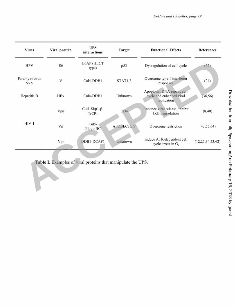
Table I. Examples of viral proteins that manipulate the UPS.
Virus
Viral protein
UPS
interactions
Target
Functional Effects
References
HPV
E6
E6AP (HECT
type)
p53
Dysregulation of cell cycle
(52)
Paramyxovirus
SV5
V
Cul4-DDB1
STAT1,2
Overcome type-I interferon
responses
(24)
Hepatitis B
HBx
Cul4-DDB1
Unknown
Apoptosis, DNA repair, cell
cycle and enhanced viral
replication
(36,56)
Vpu
Cul1-Skp1-"-
TrCP1
CD4
Enhance viral release, inhibit
I!B degradation
(8,40)
Vif
Cul5-
EloginBC
APOBEC3G/F
Overcome restriction
(43,55,64)
HIV-1
Vpr
DDB1-DCAF1
Unknown
Induce ATR-dependent cell
cycle arrest in G2
(12,25,34,53,62)
ACCEPTED
on February 16, 2018 by guest
http://jvi.asm.org/
Dow
nloaded from
DeHart and Planelles, page 20
References
1. Andersen, J. L., J. L. Dehart, E. S. Zimmerman, O. Ardon, B. Kim, G.
Jacquot, S. Benichou, and V. Planelles. 2006. HIV-1 Vpr-Induced Apoptosis Is
Cell Cycle Dependent and Requires Bax but Not ANT. PLoS Pathog 2:e127.
2. Andersen, J. L., and V. Planelles. 2005. The role of Vpr in HIV-1 pathogenesis.
Curr HIV Res 3:43-51.
3. Angers, S., T. Li, X. Yi, M. J. MacCoss, R. T. Moon, and N. Zheng. 2006.
Molecular architecture and assembly of the DDB1-CUL4A ubiquitin ligase
machinery. Nature 443:590-3.
4. Arias, E. E., and J. C. Walter. 2006. PCNA functions as a molecular platform to
trigger Cdt1 destruction and prevent re-replication. Nat Cell Biol 8:84-90.
5. Banks, D., M. Wu, L. A. Higa, N. Gavrilova, J. Quan, T. Ye, R. Kobayashi,
H. Sun, and H. Zhang. 2006. L2DTL/CDT2 and PCNA interact with p53 and
regulate p53 polyubiquitination and protein stability through MDM2 and
CUL4A/DDB1 complexes. Cell Cycle 5:1719-29.
6. Bartz, S. R., M. E. Rogel, and M. Emerman. 1996. Human immunodeficiency
virus type 1 cell cycle control: Vpr is cytostatic and mediates G2 accumulation by
a mechanism which differs from DNA damage checkpoint control. J Virol
70:2324-31.
7. Belshan, M., L. A. Mahnke, and L. Ratner. 2006. Conserved amino acids of the
human immunodeficiency virus type 2 Vpx nuclear localization signal are critical
for nuclear targeting of the viral preintegration complex in non-dividing cells.
Virology 346:118-26.
ACCEPTED
on February 16, 2018 by guest
http://jvi.asm.org/
Dow
nloaded from

DeHart and Planelles, page 21
8. Bour, S., C. Perrin, H. Akari, and K. Strebel. 2001. The human
immunodeficiency virus type 1 Vpu protein inhibits NF-kappa B activation by
interfering with beta TrCP-mediated degradation of Ikappa B. J Biol Chem
276:15920-8.
9. Brasey, A., M. Lopez-Lastra, T. Ohlmann, N. Beerens, B. Berkhout, J. L.
Darlix, and N. Sonenberg. 2003. The leader of human immunodeficiency virus
type 1 genomic RNA harbors an internal ribosome entry segment that is active
during the G2/M phase of the cell cycle. J Virol 77:3939-49.
10. Busino, L., M. Donzelli, M. Chiesa, D. Guardavaccaro, D. Ganoth, N. V.
Dorrello, A. Hershko, M. Pagano, and G. F. Draetta. 2003. Degradation of
Cdc25A by beta-TrCP during S phase and in response to DNA damage. Nature
426:87-91.
11. de Rocquigny, H., A. Caneparo, T. Delaunay, J. Bischerour, J. F. Mouscadet,
and B. P. Roques. 2000. Interactions of the C-terminus of viral protein R with
nucleic acids are modulated by its N-terminus. Eur J Biochem 267:3654-60.
12. Dehart, J. L., E. S. Zimmerman, O. Ardon, C. M. Monteiro-Filho, E. R.
Arganaraz, and V. Planelles. 2007. HIV-1 Vpr activates the G2 checkpoint
through manipulation of the ubiquitin proteasome system. Virol J 4:57.
13. Fletcher, T. M., B. Brichacek, N. Sharova, M. A. Newman, G. Stivahtis, P. M.
Sharp, M. Emerman, B. H. Hahn, and M. Stevenson. 1996. Nuclear import
and cell cycle arrest functions of the HIV-1 Vpr protein are encoded by two
separate genes in HIV-2/SIV(SM). Embo J 15:6155-65.
ACCEPTED
on February 16, 2018 by guest
http://jvi.asm.org/
Dow
nloaded from

DeHart and Planelles, page 22
14. Fultz, P. N., P. Nara, F. Barre-Sinoussi, A. Chaput, M. L. Greenberg, E.
Muchmore, M. P. Kieny, and M. Girard. 1992. Vaccine protection of
chimpanzees against challenge with HIV-1-infected peripheral blood
mononuclear cells. Science 256:1687-90.
15. Goh, W. C., N. Manel, and M. Emerman. 2004. The human immunodeficiency
virus Vpr protein binds Cdc25C: implications for G2 arrest. Virology 318:337-49.
16. Goh, W. C., M. E. Rogel, C. M. Kinsey, S. F. Michael, P. N. Fultz, M. A.
Nowak, B. H. Hahn, and M. Emerman. 1998. HIV-1 Vpr increases viral
expression by manipulation of the cell cycle: a mechanism for selection of Vpr in
vivo. Nat Med 4:65-71.
17. Goujon, C., L. Riviere, L. Jarrosson-Wuilleme, J. Bernaud, D. Rigal, J. L.
Darlix, and A. Cimarelli. 2007. SIVSM/HIV-2 Vpx proteins promote retroviral
escape from a proteasome-dependent restriction pathway present in human
dendritic cells. Retrovirology 4:2.
18. Groisman, R., I. Kuraoka, O. Chevallier, N. Gaye, T. Magnaldo, K. Tanaka,
A. F. Kisselev, A. Harel-Bellan, and Y. Nakatani. 2006. CSA-dependent
degradation of CSB by the ubiquitin-proteasome pathway establishes a link
between complementation factors of the Cockayne syndrome. Genes Dev
20:1429-34.
19. He, Y. J., C. M. McCall, J. Hu, Y. Zeng, and Y. Xiong. 2006. DDB1 functions
as a linker to recruit receptor WD40 proteins to CUL4-ROC1 ubiquitin ligases.
Genes Dev 20:2949-54.
ACCEPTED
on February 16, 2018 by guest
http://jvi.asm.org/
Dow
nloaded from
DeHart and Planelles, page 23
20. Hebner, C. M., and L. A. Laimins. 2006. Human papillomaviruses: basic
mechanisms of pathogenesis and oncogenicity. Rev Med Virol 16:83-97.
21. Higa, L. A., I. S. Mihaylov, D. P. Banks, J. Zheng, and H. Zhang. 2003.
Radiation-mediated proteolysis of CDT1 by CUL4-ROC1 and CSN complexes
constitutes a new checkpoint. Nat Cell Biol 5:1008-15.
22. Higa, L. A., M. Wu, T. Ye, R. Kobayashi, H. Sun, and H. Zhang. 2006.
CUL4-DDB1 ubiquitin ligase interacts with multiple WD40-repeat proteins and
regulates histone methylation. Nat Cell Biol 8:1277-83.
23. Higa, L. A., X. Yang, J. Zheng, D. Banks, M. Wu, P. Ghosh, H. Sun, and H.
Zhang. 2006. Involvement of CUL4 ubiquitin E3 ligases in regulating CDK
inhibitors Dacapo/p27Kip1 and cyclin E degradation. Cell Cycle 5:71-7.
24. Horvath, C. M. 2004. Weapons of STAT destruction. Interferon evasion by
paramyxovirus V protein. Eur J Biochem 271:4621-8.
25. Hrecka, K., M. Gierszewska, S. Srivastava, L. Kozaczkiewicz, S. K. Swanson,
L. Florens, M. P. Washburn, and J. Skowronski. 2007. Lentiviral Vpr usurps
Cul4-DDB1[VprBP] E3 ubiquitin ligase to modulate cell cycle. Proc Natl Acad
Sci U S A.
26. Hu, J., C. M. McCall, T. Ohta, and Y. Xiong. 2004. Targeted ubiquitination of
CDT1 by the DDB1-CUL4A-ROC1 ligase in response to DNA damage. Nat Cell
Biol 6:1003-9.
27. Jia, S., R. Kobayashi, and S. I. Grewal. 2005. Ubiquitin ligase component Cul4
associates with Clr4 histone methyltransferase to assemble heterochromatin. Nat
Cell Biol 7:1007-13.
ACCEPTED
on February 16, 2018 by guest
http://jvi.asm.org/
Dow
nloaded from
DeHart and Planelles, page 24
28. Jin, J., E. E. Arias, J. Chen, J. W. Harper, and J. C. Walter. 2006. A family of
diverse Cul4-Ddb1-interacting proteins includes Cdt2, which is required for S
phase destruction of the replication factor Cdt1. Mol Cell 23:709-21.
29. Jin, J., T. Shirogane, L. Xu, G. Nalepa, J. Qin, S. J. Elledge, and J. W.
Harper. 2003. SCFbeta-TRCP links Chk1 signaling to degradation of the
Cdc25A protein phosphatase. Genes Dev 17:3062-74.
30. Jowett, J. B., Y. M. Xie, and I. S. Chen. 1999. The presence of human
immunodeficiency virus type 1 Vpr correlates with a decrease in the frequency of
mutations in a plasmid shuttle vector. J Virol 73:7132-7.
31. Kaiser, S. M., and M. Emerman. 2006. Uracil DNA glycosylase is dispensable
for human immunodeficiency virus type 1 replication and does not contribute to
the antiviral effects of the cytidine deaminase Apobec3G. J Virol 80:875-82.
32. Kulaksiz, G., J. T. Reardon, and A. Sancar. 2005. Xeroderma pigmentosum
complementation group E protein (XPE/DDB2): purification of various
complexes of XPE and analyses of their damaged DNA binding and putative
DNA repair properties. Mol Cell Biol 25:9784-92.
33. Lai, M., E. S. Zimmerman, V. Planelles, and J. Chen. 2005. Activation of the
ATR Pathway by Human Immunodeficiency Virus Type 1 Vpr Involves Its Direct
Binding to Chromatin In Vivo. J Virol 79:15443-15451.
34. Le Rouzic, E., N. Belaidouni, E. Estrabaud, M. Morel, J. C. Rain, C. Transy,
and F. Margottin-Goguet. 2007. HIV1 Vpr Arrests the Cell Cycle by Recruiting
DCAF1/VprBP, a Receptor of the Cul4-DDB1 Ubiquitin Ligase. Cell Cycle
6:182-188.
ACCEPTED
on February 16, 2018 by guest
http://jvi.asm.org/
Dow
nloaded from
DeHart and Planelles, page 25
35. Le Rouzic, E., and S. Benichou. 2005. The Vpr protein from HIV-1: distinct
roles along the viral life cycle. Retrovirology 2:11.
36. Leupin, O., S. Bontron, C. Schaeffer, and M. Strubin. 2005. Hepatitis B virus
X protein stimulates viral genome replication via a DDB1-dependent pathway
distinct from that leading to cell death. J Virol 79:4238-45.
37. Leupin, O., S. Bontron, and M. Strubin. 2003. Hepatitis B virus X protein and
simian virus 5 V protein exhibit similar UV-DDB1 binding properties to mediate
distinct activities. J Virol 77:6274-83.
38. Li, G., R. T. Elder, K. Qin, H. U. Park, D. Liang, and R. Y. Zhao. 2007. PP2A
dependent and independent pathways for ATR phosphorylation of Chk1. J Biol
Chem.
39. Lovejoy, C. A., K. Lock, A. Yenamandra, and D. Cortez. 2006. DDB1
maintains genome integrity through regulation of Cdt1. Mol Cell Biol 26:7977-
90.
40. Margottin, F., S. P. Bour, H. Durand, L. Selig, S. Benichou, V. Richard, D.
Thomas, K. Strebel, and R. Benarous. 1998. A novel human WD protein, h-
beta TrCp, that interacts with HIV-1 Vpu connects CD4 to the ER degradation
pathway through an F-box motif. Mol Cell 1:565-74.
41. McCall, C. M., J. Hu, and Y. Xiong. 2005. Recruiting substrates to cullin 4-
dependent ubiquitin ligases by DDB1. Cell Cycle 4:27-9.
42. McGowan, C. H., and P. Russell. 2004. The DNA damage response: sensing
and signaling. Curr Opin Cell Biol 16:629-33.
ACCEPTED
on February 16, 2018 by guest
http://jvi.asm.org/
Dow
nloaded from

DeHart and Planelles, page 26
43. Mehle, A., B. Strack, P. Ancuta, C. Zhang, M. McPike, and D. Gabuzda.
2004. Vif overcomes the innate antiviral activity of APOBEC3G by promoting its
degradation in the ubiquitin-proteasome pathway. J Biol Chem 279:7792-8.
44. Morellet, N., S. Bouaziz, P. Petitjean, and B. P. Roques. 2003. NMR structure
of the HIV-1 regulatory protein VPR. J Mol Biol 327:215-27.
45. Nalepa, G., M. Rolfe, and J. W. Harper. 2006. Drug discovery in the ubiquitin-
proteasome system. Nat Rev Drug Discov 5:596-613.
46. Nishitani, H., and Z. Lygerou. 2002. Control of DNA replication licensing in a
cell cycle. Genes Cells 7:523-34.
47. O'Connell, B. C., and J. W. Harper. 2007. Ubiquitin proteasome system (UPS):
what can chromatin do for you? Curr Opin Cell Biol 19:206-14.
48. Petroski, M. D., and R. J. Deshaies. 2005. Function and regulation of cullin-
RING ubiquitin ligases. Nat Rev Mol Cell Biol 6:9-20.
49. Planelles, V., J. B. Jowett, Q. X. Li, Y. Xie, B. Hahn, and I. S. Chen. 1996.
Vpr-induced cell cycle arrest is conserved among primate lentiviruses. J Virol
70:2516-24.
50. Reitz, M. S., Jr., L. Hall, M. Robert-Guroff, J. Lautenberger, B. M. Hahn, G.
M. Shaw, L. I. Kong, S. H. Weiss, D. Waters, R. C. Gallo, and et al. 1994.
Viral variability and serum antibody response in a laboratory worker infected with
HIV type 1 (HTLV type IIIB). AIDS Res Hum Retroviruses 10:1143-55.
51. Roshal, M., B. Kim, Y. Zhu, P. Nghiem, and V. Planelles. 2003. Activation of
the ATR-mediated DNA damage response by the HIV-1 viral protein R. J Biol
Chem 278:25879-86.
ACCEPTED
on February 16, 2018 by guest
http://jvi.asm.org/
Dow
nloaded from
DeHart and Planelles, page 27
52. Scheffner, M., J. M. Huibregtse, R. D. Vierstra, and P. M. Howley. 1993. The
HPV-16 E6 and E6-AP complex functions as a ubiquitin-protein ligase in the
ubiquitination of p53. Cell 75:495-505.
53. Schrofelbauer, B., Y. Hakata, and N. R. Landau. 2007. HIV-1 Vpr function is
mediated by interaction with the damage-specific DNA-binding protein DDB1.
Proc Natl Acad Sci U S A 104:4130-5.
54. Schrofelbauer, B., Q. Yu, S. G. Zeitlin, and N. R. Landau. 2005. Human
immunodeficiency virus type 1 Vpr induces the degradation of the UNG and
SMUG uracil-DNA glycosylases. J Virol 79:10978-87.
55. Sheehy, A. M., N. C. Gaddis, and M. H. Malim. 2003. The antiretroviral
enzyme APOBEC3G is degraded by the proteasome in response to HIV-1 Vif.
Nat Med 9:1404-7.
56. Sitterlin, D., F. Bergametti, and C. Transy. 2000. UVDDB p127-binding
modulates activities and intracellular distribution of hepatitis B virus X protein.
Oncogene 19:4417-26.
57. Sugasawa, K., Y. Okuda, M. Saijo, R. Nishi, N. Matsuda, G. Chu, T. Mori, S.
Iwai, K. Tanaka, K. Tanaka, and F. Hanaoka. 2005. UV-induced
ubiquitylation of XPC protein mediated by UV-DDB-ubiquitin ligase complex.
Cell 121:387-400.
58. Tristem, M., A. Purvis, and D. L. Quicke. 1998. Complex evolutionary history
of primate lentiviral vpr genes. Virology 240:232-7.
59. Wang, H., L. Zhai, J. Xu, H. Y. Joo, S. Jackson, H. Erdjument-Bromage, P.
Tempst, Y. Xiong, and Y. Zhang. 2006. Histone H3 and H4 ubiquitylation by
ACCEPTED
on February 16, 2018 by guest
http://jvi.asm.org/
Dow
nloaded from
DeHart and Planelles, page 28
the CUL4-DDB-ROC1 ubiquitin ligase facilitates cellular response to DNA
damage. Mol Cell 22:383-94.
60. Wei, N., and X. W. Deng. 2003. The COP9 signalosome. Annu Rev Cell Dev
Biol 19:261-86.
61. Weiss, S. H., J. J. Goedert, S. Gartner, M. Popovic, D. Waters, P. Markham,
F. di Marzo Veronese, M. H. Gail, W. E. Barkley, J. Gibbons, and et al. 1988.
Risk of human immunodeficiency virus (HIV-1) infection among laboratory
workers. Science 239:68-71.
62. Wen, X., K. M. Duus, T. D. Friedrich, and C. M. de Noronha. 2007. The HIV1
protein Vpr acts to promote G2 cell cycle arrest by engaging a DDB1 and
Cullin4A-containing ubiquitin ligase complex using VprBP/DCAF1 as an
adaptor. In press. J Biol Chem (In Press).
63. Wu, K., A. Chen, and Z. Q. Pan. 2000. Conjugation of Nedd8 to CUL1
enhances the ability of the ROC1-CUL1 complex to promote ubiquitin
polymerization. J Biol Chem 275:32317-24.
64. Yu, X., Y. Yu, B. Liu, K. Luo, W. Kong, P. Mao, and X. F. Yu. 2003.
Induction of APOBEC3G ubiquitination and degradation by an HIV-1 Vif-Cul5-
SCF complex. Science 302:1056-60.
65. Yuan, H., M. Kamata, Y. M. Xie, and I. S. Chen. 2004. Increased levels of
Wee-1 kinase in G(2) are necessary for Vpr- and gamma irradiation-induced G(2)
arrest. J Virol 78:8183-90.
ACCEPTED
on February 16, 2018 by guest
http://jvi.asm.org/
Dow
nloaded from
DeHart and Planelles, page 29
66. Zhang, S., D. Pointer, G. Singer, Y. Feng, K. Park, and L. J. Zhao. 1998.
Direct binding to nucleic acids by Vpr of human immunodeficiency virus type 1.
Gene 212:157-66.
67. Zhao, L. J., S. Mukherjee, and O. Narayan. 1994. Biochemical mechanism of
HIV-I Vpr function. Specific interaction with a cellular protein. J Biol Chem
269:15577-82.
68. Zimmerman, E. S., J. Chen, J. L. Andersen, O. Ardon, J. L. Dehart, J.
Blackett, S. K. Choudhary, D. Camerini, P. Nghiem, and V. Planelles. 2004.
Human Immunodeficiency Virus Type 1 Vpr-Mediated G2 Arrest Requires
Rad17 and Hus1 and Induces Nuclear BRCA1 and {gamma}-H2AX Focus
Formation. Mol Cell Biol 24:9286-9294.
69. Zimmerman, E. S., M. P. Sherman, J. L. Blackett, J. A. Neidleman, C. Kreis,
P. Mundt, S. A. Williams, M. Warmerdam, J. Kahn, F. M. Hecht, R. M.
Grant, C. M. de Noronha, A. S. Weyrich, W. C. Greene, and V. Planelles.
2006. Human immunodeficiency virus type 1 Vpr induces DNA replication stress
in vitro and in vivo. J Virol 80:10407-18.
ACCEPTED
on February 16, 2018 by guest
http://jvi.asm.org/
Dow
nloaded from

DeHart and Planelles, page 30
Figure Legend
Figure 1. E3 ubiquitin ligases and their interactions with viral proteins. A. The three
main types of E3 ligases. B. The three-dimensional structure of HIV-1 Vpr. Adapted
from (44). Cylinders represent !-helices and lines are unstructured regions. C. The Vpr-
UPS model predicts the interaction of Vpr with DCAF1 and a putative degradation
substrate, “S”. BPA, BPC and BPC denote the three beta-propeller domains of DDB1. Le
Rouzic et al. (34) found that the WDXR motifs of DCAF1 are required for the interaction
with Vpr; for simplicity, we have represented Vpr interacting with the WDXR motif on
BPC; there is no evidence to support which of the two WDXR motifs is required for Vpr
interaction. D. The Vpr-UV-DDB model predicts that the interaction of Vpr with DDB1
displaces DDB2 and prevents the DDB1-DDB2 complex, also known as UV-DDB, from
recognizing damaged DNA. E. Simian virus protein V interacts with DDB1 and displaces
DCAF, creating a new interface that recruits STAT2 as an adaptor, which then recruits
STAT1 for ubiquitination.
ACCEPTED
on February 16, 2018 by guest
http://jvi.asm.org/
Dow
nloaded from