Species-specific Differences in Proteasomal Processing and ...
13
Species-specific Differences in Proteasomal Processing and Tapasin-mediated Loading Influence Peptide Presentation by HLA-B27 in Murine Cells* Received for publication, August 11, 2003 Published, JBC Papers in Press, September 8, 2003, DOI 10.1074/jbc.M308816200 Laura Sesma, In ˜ aki Alvarez, Miguel Marcilla, Alberto Paradela, and Jose ´ A. Lo ´ pez de Castro‡ From the Centro de Biologı´a Molecular Severo Ochoa (Consejo Superior de Investigaciones Cientı´ficas and Universidad Auto ´noma de Madrid), Facultad de Ciencias, Universidad Auto ´noma, 28049 Madrid, Spain Expression of HLA-B27 in murine cells has been used to establish animal models for human spondyloarthritis and for antigen presentation studies, but the effects of xenogeneic HLA-B27 expression on peptide presenta- tion are little known. The issue was addressed in this study. HLA-B27-bound peptide repertoires from human and murine cells overlapped by 75– 85%, indicating that many endogenous HLA-B27 ligands are generated and presented in both species. Of 20 differentially presented peptides that were sequenced, only 40% arose from ob- vious inter-species protein polymorphism, suggesting that differences in antigen processing-loading ac- counted for many species-specific ligands. Digestion of synthetic substrates with human and murine 20 S pro- teasomes revealed cleavage differences that accounted for or correlated with differential expression of partic- ular peptides. One HLA-B27 ligand found only in human cells was similarly generated in vitro by human and murine proteasomes. Differential presentation corre- lated with significantly decreased amounts of this li- gand in human tapasin-deficient cells reconstituted with murine tapasin, indicating that species-specific in- teractions between HLA-B27, tapasin, and/or other pro- teins in the peptide-loading complex influenced presen- tation of this peptide. Our results indicate that differences in proteasomal specificity and in interac- tions involving tapasin determine differential process- ing and presentation of a significant number of HLA-B27 ligands in human and murine cells. The xenogeneic expression of HLA-B27, an MHC 1 class I molecule, strongly associated with spondyloarthritis (1, 2), has been used to establish transgenic animal disease models and to study the antigen-presenting and other properties of this mol- ecule. HLA-B27 transgenic rats develop a spontaneous disease with many similarities to human spondyloarthropathy (3). Dis- ease manifestations are dependent on the genetic background and transgene copy number (4) and are modulated by alter- ations of the HLA-B27-bound peptide repertoire (5). Trans- genic mice have also been used as a possible model for human HLA-B27-associated disease. Development of spontaneous in- flammatory arthritis in HLA-B27 transgenic mice lacking 2m (6) may be related to absence of this polypeptide rather than to presence of the HLA-B27 heavy chain (7). In contrast, HLA- B27 transgenic mice expressing 2m are being used in reactive arthritis studies (8, 9) and have increased susceptibility to develop ankylosing enthesopathy (10, 11). Other HLA class I molecules have also been expressed on murine cells for antigen presentation, epitope identification, and T-cell recognition studies (12–16). However, antigen presentation by HLA-B27 or any other HLA class I molecule expressed on murine cells implies some inherent differences, relative to human cells, that have not been sufficiently characterized at a molecular level. Thus, spe- cies-related differences in the proteome, in proteasome cleav- age specificity, in the peptide specificity of the transporter associated with antigen processing (TAP), and in the interac- tion of the human class I molecule with human or murine tapasin or other proteins in the peptide-loading complex, all might influence the HLA-B27-bound peptide repertoire and antigen presentation upon expression on murine cells. Numer- ous studies have addressed the peptide-transporting prefer- ences of human and murine TAP (reviewed in Refs. 17 and 18), but the actual influence of species-related differences in this and other steps of the processing-loading pathway on HLA class I-mediated antigen presentation in murine cells is little known. Knowledge of such differences is critical for assessing the validity of HLA class I transgenic models for antigen pres- entation and human disease. In this study, we have comparatively analyzed the HLA-B27- bound peptide repertoires expressed on human and murine cells and have characterized the origin of the differential ex- pression of multiple HLA-B27 ligands in only one cell type. The results indicate a substantial lack of overlap between both peptide repertoires, which is only partially explained by spe- cies- or cell type-related protein differences. Both proteasome specificity differences and heterologous interactions in the pep- tide-loading complex contribute to differential peptide presen- tation by HLA-B27 on either human or murine cells. Our re- sults have general implications for human MHC class I-mediated antigen presentation in murine systems. MATERIALS AND METHODS Cell Lines and Monoclonal Antibodies—HMy2.C1R (C1R) is a human lymphoid cell line with low expression of its endogenous class I antigens (19, 20). B*2705-C1R transfectant cells were described elsewhere (21). P815-HTR (P815) is a murine mastocytome cell line. B*2705-P815 * This work was supported by grants SAF99/0055, PM99-0098, and SAF2002/00125 from the Ministry of Science and Technology, and 08.3/ 0005/2001.1 from the Comunidad Auto ´noma de Madrid. The costs of publication of this article were defrayed in part by the payment of page charges. This article must therefore be hereby marked “advertisement” in accordance with 18 U.S.C. Section 1734 solely to indicate this fact. ‡ To whom correspondence should be addressed. Tel.: 34-91-397- 8050; Fax: 34-91-397-8087; E-mail: [email protected]. 1 The abbreviations used are: MHC, major histocompatibility com- plex; TAP, transporter associated with antigen processing; mAb, mono- clonal antibody; PBS, phosphate-buffered saline; HPLC, high-perfor- mance liquid chromatography; MS, mass spectrometry; MALDI-TOF, matrix-assisted laser desorption ionization time-of-flight; PSD, post- source decay; IPG, immobilized pH gradient; CHAPS, 3-[(3-cholamido- propyl)dimethylammonio]-1-propanesulfonic acid; IEF, isoelectric focusing. THE JOURNAL OF BIOLOGICAL CHEMISTRY Vol. 278, No. 47, Issue of November 21, pp. 46461–46472, 2003 © 2003 by The American Society for Biochemistry and Molecular Biology, Inc. Printed in U.S.A. This paper is available on line at http://www.jbc.org 46461 by guest on February 17, 2018 http://www.jbc.org/ Downloaded from
-
Upload
nguyenquynh -
Category
Documents
-
view
216 -
download
1
Transcript of Species-specific Differences in Proteasomal Processing and ...
Species-specific Differences in Proteasomal Processing andTapasin-mediated Loading Influence Peptide Presentation byHLA-B27 in Murine Cells*
Received for publication, August 11, 2003Published, JBC Papers in Press, September 8, 2003, DOI 10.1074/jbc.M308816200
Laura Sesma, Inaki Alvarez, Miguel Marcilla, Alberto Paradela, and Jose A. Lopez de Castro‡
From the Centro de Biologıa Molecular Severo Ochoa (Consejo Superior de Investigaciones Cientıficas and UniversidadAutonoma de Madrid), Facultad de Ciencias, Universidad Autonoma, 28049 Madrid, Spain
Expression of HLA-B27 in murine cells has been usedto establish animal models for human spondyloarthritisand for antigen presentation studies, but the effects ofxenogeneic HLA-B27 expression on peptide presenta-tion are little known. The issue was addressed in thisstudy. HLA-B27-bound peptide repertoires from humanand murine cells overlapped by 75–85%, indicating thatmany endogenous HLA-B27 ligands are generated andpresented in both species. Of 20 differentially presentedpeptides that were sequenced, only 40% arose from ob-vious inter-species protein polymorphism, suggestingthat differences in antigen processing-loading ac-counted for many species-specific ligands. Digestion ofsynthetic substrates with human and murine 20 S pro-teasomes revealed cleavage differences that accountedfor or correlated with differential expression of partic-ular peptides. One HLA-B27 ligand found only in humancells was similarly generated in vitro by human andmurine proteasomes. Differential presentation corre-lated with significantly decreased amounts of this li-gand in human tapasin-deficient cells reconstitutedwith murine tapasin, indicating that species-specific in-teractions between HLA-B27, tapasin, and/or other pro-teins in the peptide-loading complex influenced presen-tation of this peptide. Our results indicate thatdifferences in proteasomal specificity and in interac-tions involving tapasin determine differential process-ing and presentation of a significant number of HLA-B27ligands in human and murine cells.
The xenogeneic expression of HLA-B27, an MHC1 class Imolecule, strongly associated with spondyloarthritis (1, 2), hasbeen used to establish transgenic animal disease models and tostudy the antigen-presenting and other properties of this mol-ecule. HLA-B27 transgenic rats develop a spontaneous diseasewith many similarities to human spondyloarthropathy (3). Dis-
ease manifestations are dependent on the genetic backgroundand transgene copy number (4) and are modulated by alter-ations of the HLA-B27-bound peptide repertoire (5). Trans-genic mice have also been used as a possible model for humanHLA-B27-associated disease. Development of spontaneous in-flammatory arthritis in HLA-B27 transgenic mice lacking �2m(6) may be related to absence of this polypeptide rather than topresence of the HLA-B27 heavy chain (7). In contrast, HLA-B27 transgenic mice expressing �2m are being used in reactivearthritis studies (8, 9) and have increased susceptibility todevelop ankylosing enthesopathy (10, 11). Other HLA class Imolecules have also been expressed on murine cells for antigenpresentation, epitope identification, and T-cell recognitionstudies (12–16).
However, antigen presentation by HLA-B27 or any otherHLA class I molecule expressed on murine cells implies someinherent differences, relative to human cells, that have notbeen sufficiently characterized at a molecular level. Thus, spe-cies-related differences in the proteome, in proteasome cleav-age specificity, in the peptide specificity of the transporterassociated with antigen processing (TAP), and in the interac-tion of the human class I molecule with human or murinetapasin or other proteins in the peptide-loading complex, allmight influence the HLA-B27-bound peptide repertoire andantigen presentation upon expression on murine cells. Numer-ous studies have addressed the peptide-transporting prefer-ences of human and murine TAP (reviewed in Refs. 17 and 18),but the actual influence of species-related differences in thisand other steps of the processing-loading pathway on HLAclass I-mediated antigen presentation in murine cells is littleknown. Knowledge of such differences is critical for assessingthe validity of HLA class I transgenic models for antigen pres-entation and human disease.
In this study, we have comparatively analyzed the HLA-B27-bound peptide repertoires expressed on human and murinecells and have characterized the origin of the differential ex-pression of multiple HLA-B27 ligands in only one cell type. Theresults indicate a substantial lack of overlap between bothpeptide repertoires, which is only partially explained by spe-cies- or cell type-related protein differences. Both proteasomespecificity differences and heterologous interactions in the pep-tide-loading complex contribute to differential peptide presen-tation by HLA-B27 on either human or murine cells. Our re-sults have general implications for human MHC classI-mediated antigen presentation in murine systems.
MATERIALS AND METHODS
Cell Lines and Monoclonal Antibodies—HMy2.C1R (C1R) is a humanlymphoid cell line with low expression of its endogenous class I antigens(19, 20). B*2705-C1R transfectant cells were described elsewhere (21).P815-HTR (P815) is a murine mastocytome cell line. B*2705-P815
* This work was supported by grants SAF99/0055, PM99-0098, andSAF2002/00125 from the Ministry of Science and Technology, and 08.3/0005/2001.1 from the Comunidad Autonoma de Madrid. The costs ofpublication of this article were defrayed in part by the payment of pagecharges. This article must therefore be hereby marked “advertisement”in accordance with 18 U.S.C. Section 1734 solely to indicate this fact.
‡ To whom correspondence should be addressed. Tel.: 34-91-397-8050; Fax: 34-91-397-8087; E-mail: [email protected].
1 The abbreviations used are: MHC, major histocompatibility com-plex; TAP, transporter associated with antigen processing; mAb, mono-clonal antibody; PBS, phosphate-buffered saline; HPLC, high-perfor-mance liquid chromatography; MS, mass spectrometry; MALDI-TOF,matrix-assisted laser desorption ionization time-of-flight; PSD, post-source decay; IPG, immobilized pH gradient; CHAPS, 3-[(3-cholamido-propyl)dimethylammonio]-1-propanesulfonic acid; IEF, isoelectricfocusing.
THE JOURNAL OF BIOLOGICAL CHEMISTRY Vol. 278, No. 47, Issue of November 21, pp. 46461–46472, 2003© 2003 by The American Society for Biochemistry and Molecular Biology, Inc. Printed in U.S.A.
This paper is available on line at http://www.jbc.org 46461
by guest on February 17, 2018http://w
ww
.jbc.org/D
ownloaded from
transfectant cells were previously described (22). Both the human andmurine transfectants express high and similar HLA-B27 levels (23).These were periodically checked by flow cytometry to ensure stableexpression of this molecule. The C1R and P815 cell lines were culturedin Dulbecco’s modified Eagle’s medium supplemented with 7.5% fetalbovine serum (both from Invitrogen, Paisley, UK). 721.220 is a humanlymphoblastoid cell line (a gift from Dr. James McCluskey, Universityof Melbourne, Australia) in which HLA-A and HLA-B genes have beendeleted and a non-functional tapasin protein is expressed (24, 25). Thiscell line expresses low levels of endogenous HLA-Cw*0102. Transfec-tions of HLA-B*2705 and wild type human or murine tapasin into721.220 have been previously described (26). These cells were culturedin RPMI 1640 medium supplemented with 10% fetal bovine serum. ThemAb used in this study were W6/32 (IgG2a, specific for a monomorphicHLA-A, -B, and -C determinant) (27) and ME1 (IgG1, specific for HLA-B27, -B7, and -B22) (28).
Flow Cytometry—About 6 � 104 cells were washed twice in 200 �l ofPBS and resuspended in 50 �l of undiluted mAb supernatant. Afterincubating 30 min, cells were washed twice in 200 �l of PBS andresuspended in 50 �l of fluorescein isothiocyanate-conjugated anti-mouse IgG rabbit antiserum (Calbiochem-Novabiochem GmbH,Schwalbach, Germany), incubated for 30 min, and washed two times in200 �l of PBS. All operations were done at 4 °C. Flow cytometry wascarried out on a BD Biosciences FACSCalibur instrument usingCellQuest software.
Isolation of B*2705-bound Peptides—This was carried out from 1010
C1R or P815 transfectant cells lysed in 1% Nonidet P-40 in the presenceof a mixture of protease inhibitors, after immunopurification of HLA-B27 with the W6/32 mAb and acid extraction, exactly as describedelsewhere (29). HLA-B27-bound peptide pools were fractionated byHPLC at a flow rate of 100 �l/min as previously described (30), and50-�l fractions were collected.
Mass Spectrometry Analysis and Sequencing—The peptide composi-tion of HPLC fractions was analyzed by matrix-assisted desorptionionization time-of-flight (MALDI-TOF) MS using a calibrated KompactProbe instrument (Kratos-Shimadzu) operating in the positive linearmode, as previously described (30). Alternatively, a Bruker ReflexTM IIIMALDI-TOF mass spectrometer (Bruker-Franzen Analytic GmbH, Bre-men, Germany) equipped with the SCOUTTM source in positive ionreflector mode was also used, as described elsewhere (31).
Peptide sequencing was carried out by quadrupole ion trap nanoelec-trospray MS/MS in an LCQ instrument (Finnigan ThermoQuest, SanJose, CA), as previously described (32, 33). In a few cases microelectro-spray MS/MS was used, using the same procedure, except that sampleswere injected, through an HPLC equipped with a C18 capillary column(150 � 0.18 mm) connected online, at a flow rate of 1.5 �l/min. In somecases, peptide sequencing was also done by post-source decay (PSD)-MALDI-TOF MS, as previously described (31).
In all cases peptide-containing HPLC fractions were dried and re-suspended in 5 �l of methanol/water (1:1) containing 0.1% formic acid.Aliquots of 0.5 or 1 �l were used for MALDI-TOF or nanoelectrosprayMS analyses, respectively. For microelectrospray MS/MS-dried sampleswere resuspended in 0.5% acetic acid.
Synthetic Peptides—Peptides were synthesized using the standardsolid-phase Fmoc (N-(9-fluorenyl)methoxycarbonyl) chemistry andwere purified by HPLC. The correct composition and molecular mass ofpurified peptides were confirmed by amino acid analysis using a 6300Amino Acid Analyzer (Beckman Coulter, Palo Alto, CA), which alsoallowed their quantification, and MALDI-TOF MS, respectively.
Purification of 20 S Proteasome—The 20 S proteasome was purifiedfrom 3 � 109 B*2705-C1R or B*2705-P815 cell lysates by ion-exchangechromatography and centrifugation in a glycerol gradient as previouslydescribed (30) with the following modifications. Proteasome-containingfractions from the previous purification step were identified by 12%SDS-PAGE and further subjected to anion-exchange chromatographyby fast protein liquid chromatography using a Mono-Q SR5/5 column(Amersham Biosciences, Uppsala, Sweden), at a flow rate of 1 ml/min,as follows: isocratic conditions with buffer A (50 mM Tris/HCl, 50 mM
KCl, pH 8) for 1 h, followed by a linear gradient of 0–100% buffer B (50mM Tris/HCl, 0.5 M KCl, pH 8) for 1 h. Purity of the fractions wasassessed by SDS-PAGE. Aliquots of purified proteasome were stored at�80 °C. Absence of contaminant proteases in the 20 S proteasomesamples was assessed by inhibition of proteolytic cleavage of a syntheticpeptide substrate, histone 2A-(77–105), with the irreversible protea-some inhibitors lactacystine (50 �g/ml) and epoxomicin (1 �g/ml).
Two-dimensional Gel Electrophoresis of 20 S Proteasomes—Samplesof purified 20 S proteasomes were loaded by hydration of immobilizedpH gradient (IPG) strips, non-linear pH 3–10, of 18-cm length (Amer-
sham Biosciences), previously diluted to a total volume of 350 �l in 6 M
urea, 2 M thiourea, 2% CHAPS, IPG non-linear pH 3–10, 1 mM Tris-(2-carboxymethyl)phosphine-HCl, and bromphenol blue. In the first di-mension, IEF was performed in a IPGphor (Amersham Biosciences)under the following conditions: 30 V for 6 h, 60 V for 6 h, 500 V for 30min, 1,000 V for 30 min, a gradient of 1,000–8,000 V for 30 min, and8,000 V up to 32,000 Vh. After IEF, strips were equilibrated in 6 M urea,30% glycerol, 2% SDS, and bromphenol blue, twice for 20 min. Dithio-threitol (2%) and 4% iodoacetamide were added in the first and secondequilibration steps, respectively. The second dimension was performedusing 12.5% SDS-PAGE. Gels were stained with silver nitrate andanalyzed using the software ImageMaster (Amersham Biosciences).Spots were assigned by tryptic digestion followed by MS fingerprinting.
Digestion of Synthetic Substrates—Peptide substrates (125 �g/ml)were incubated at 37 °C with purified 20 S proteasome at an enzyme/substrate ratio of 1:10 (w/w) in 20 mM Hepes buffer, pH 7.6. Digestionwas stopped by adding 1/5 volume of 0.4% aqueous trifluoroacetic acid.Digestion mixtures were dried down to 100 �l in a Speed-Vac andfractionated by HPLC using the same conditions as for HLA-B27-boundpeptides. Individual digestion products were identified on the basis oftheir molecular mass by MALDI-TOF MS and, when necessary forunambiguous assignment, by PSD-MALDI-TOF or electrospray MS/MSsequencing.
RESULTS
HLA-B27 Presents Distinct Peptide Repertoires on Humanand Murine Cells—Peptide pools were isolated by acid extrac-tion from HLA-B*2705 immunopurified from C1R and P815transfectant cells and fractionated by HPLC under identicalconditions and consecutive runs. The peptide composition ofcorrelative HPLC fractions from both peptide pools were sys-tematically compared by MALDI-TOF MS, using the samestrategy as previously used to compare HLA-B27 subtype-bound peptide repertoires (34, 35). In short, the MS spectrum ofany given HPLC fraction from one peptide pool was comparedwith the MS spectra of the correlative, previous, and followingHPLC fractions from the other peptide pool. This was done toaccount for slight shifts in retention time that may occur be-tween consecutive chromatographic runs. Ion peaks of thesame (�1) mass-to-charge (m/z) ratio and retention time wereconsidered to reflect shared peptides on human and murinecells. Identity of retention time and m/z does not necessarilyindicate peptide identity, because in very complex mixturesunrelated peptides sharing these features might eventuallyco-elute. However, it is reasonable to assume that the over-whelming majority of identical peptide masses compared cor-respond to identical peptides. Indeed, in four of four cases inwhich ion peaks of the same m/z and retention time weresequenced from both the human and murine peptide pools theycorresponded to identical peptides (Fig. 1). In nine additionalcases a peptide sequenced from the murine peptide pool showedan identical counterpart in the human pool known from previ-ous sequencing studies of HLA-B27-bound peptides from C1Rcells to be the same peptide (Fig. 1). Ion peaks found in only onecell type in two independent experiments were considered asdifferentially expressed peptides. A total of 1372 and 1551molecular species were compared from C1R and P815, respec-tively (Table I). Of these, 211 (15%) and 390 (25%) peptideswere found only in human or in murine cells, respectively. Inaddition, of 351 shared peptides that showed particularlystrong intensity signals in the MALDI-TOF spectra of at leastone cell line, 54 (15%) and 82 (23%) showed 10-fold or higherintensity in the human or murine cells in two independentexperiments, respectively. This is consistent with substantiallyhigher expression of the shared ligand in the corresponding cellline. No significant differences in the average size of peptidesexpressed on either cell type were found (Table I).
The reproducibility of the MALDI-TOF spectra was assessedin two ways: by obtaining independent spectra from the samesample and by performing two independent comparisons with
Differential Processing of HLA-B27 Ligands46462
by guest on February 17, 2018http://w
ww
.jbc.org/D
ownloaded from

different peptide preparations. In both cases, the MS spectra ofthe same, or equivalent, HPLC fraction were in general veryreproducible both in the nature of the ion peaks detected and intheir relative intensities, although occasionally some variation
was found. For this reason, assignment of both qualitative andquantitative differences was always done on the basis of repro-ducibility in two independent experiments. These results indi-cate that the HLA-B27-bound peptide repertoires on humanand murine cells, although highly overlapping, contain a sig-nificant number of differentially bound ligands, as well asshared ones presented at substantially different amounts
Murine TAP Does Not Impair Presentation of B*2705 Li-gands with C-terminal Basic Residues—A total of 27 sharedligands, including 3 octamers, 13 nonamers, 7 decamers, 3undecamers, and 1 dodecamer, were sequenced by MS (Fig. 1).In addition, the sequence of 20 B*2705 ligands found only inhuman (9 peptides) or murine cells (11 peptides) was alsodetermined (Fig. 2). All shared peptides corresponded to con-served sequences between both species. All the peptides se-quenced contained the canonic anchor motif of HLA-B27, Arg2.Shared ligands also presented the same variety of C-terminalpeptide residues previously defined for HLA-B*2705 ligandsfrom human cells, including aliphatic, aromatic, and basic res-idues. The number of shared ligands with C-terminal basic
FIG. 1. Amino acid sequence of HLA-B*2705 ligands present in both human (C1R) and murine (P815) cells. All sequences weredetermined by quadrupole/ion trap electrospray MS/MS. Isobaric residues (Ile/Leu and Lys/Gln) were assigned on the basis of unambiguousmatching with sequences in the protein data base. The putative parental protein, with which full match was obtained, and the correspondingSwissProt accession number (www.ebi.ac.uk/swissprot/access.htm), is indicated. The one or more cell lines from which the sequence wasdetermined are also indicated. When a peptide was sequenced from only one cell line, its presence in the other one was deduced from the findingof an ion peak of equal m/z and retention time. References are given for those peptides sequenced in this study that were previously reported asHLA-B27 ligands.
TABLE IComparison of HLA-B*2705-bound peptides from
human and murine cells
C1R-B*2705 P815-B*2705
Total peptides compared 1372 1551Mass range 860-1667 Da 860-1667 DaAverage mass 1154 Da 1154 DaShared peptides 1161 (85%) 1161 (75%)Specific peptides 211 (15%) 390 (25%)Average mass of shared peptides 1144 Da 1144 DaAverage mass of specific peptides 1205 Da 1193 DaMajor peaks counteda 351 351Quantitative differencesb 54 (15%) 82 (23%)
a Ion peaks that showed particularly strong intensity in the MALDI-TOF spectrum from one or both cell lines.
b Ion peaks that showed 10-fold or more intensity in the MALDI-TOFspectrum from one cell line.
Differential Processing of HLA-B27 Ligands 46463
by guest on February 17, 2018http://w
ww
.jbc.org/D
ownloaded from

residues was 6 of 27. Among differential peptides (Fig. 2), 5 of11 found only in C1R cells and 2 of 11 found only in P815 cellsshowed a C-terminal basic motif. Thus, of a total of 36 peptidesfrom human cells and 38 peptides from murine cells, 11 (31%)and 8 (21%), respectively, contained C-terminal basic residues.The percentage from human cells is nearly the same as that(32%) previously reported in an independent compilation (36)and is only moderately higher than the percentage of B*2705ligands with C-terminal basic residues found in murine cells.These results strongly suggest that the low preference of mu-rine TAP for C-terminal basic residues reported from in vitrotransport studies (37) introduces little bias against presenta-tion of peptides with these motifs by HLA-B27 on murine cells.
Differential Presentation of HLA-B27 Ligands Is Only Par-tially Due to Protein Polymorphism—The 20 B*2705 ligandsfound only in either human or murine cells that were se-quenced could be classified in three subsets (Fig. 2). Group 1,which included two peptides (10%), consisted of ligands arisingfrom species- or cell type-specific proteins. Group 2, whichincluded six peptides (30%), consisted of ligands arising fromproteins present on both cell types but differing in one or moreresidues within the sequence of the peptide. Group 3, which
included 12 peptides (60%) consisted of ligands arising fromproteins that are either identical in both cell types (i.e. theHLA-B27 and human �2m transgene products) or identical inthe region corresponding to the peptide ligand and its neigh-borhood. Thus, less than half of the species- or cell type-relateddifferences in the HLA-B27-bound peptide repertoire are ex-plained by obvious differences in the parental proteins (groups1 and 2). These results imply that differential processing,transport, and/or loading have a significant influence on differ-ential peptide presentation by HLA-B27 on human or murinecells.
Distinct Proteasomal Cleavage Contributes to DifferentialPresentation of HLA-B27 Ligands—To assess the contributionof proteasomal processing to differential expression of particu-lar HLA-B27 ligands in one cell type, we used synthetic pre-cursors of four differentially expressed ligands from group 3(Fig. 2), including two peptides found only in C1R cells and twoothers found only in P815 cells, with the sequence of the pa-rental proteins in and around the sequence of the ligand. Eachof these substrates was digested in vitro, in parallel experi-ments, with 20 S proteasomes purified from C1R and fromP815 cells. Two-dimensional gel electrophoresis analysis indi-
FIG. 2. Amino acid sequence of HLA-B*2705 ligands found only in either human (C1R) or murine (P815) cells. Conventions are as inFig. 1. Peptides were classified in three groups. Group 1 includes peptides derived from species- or cell type-specific proteins, with no counterpartin the other cell. Group 2 includes peptides arising from proteins present in both cells but differing in one or more amino acid residues within thesequence of the ligand. Group 3 includes peptides arising from proteins that are identical totally (i.e. the HLA-B27 heavy chain and human �2mtransgene products) or within and around the sequence of the ligand.
Differential Processing of HLA-B27 Ligands46464
by guest on February 17, 2018http://w
ww
.jbc.org/D
ownloaded from

cated that both the human and murine proteasome prepara-tions contained a mixture of proteasome and immunoprotea-some. The proteasome/immunoproteasome ratio was similar inboth cases, within the limits of the technique used, as judgedfrom the relative intensities of the spots corresponding to �1/�1i and �2/�2i subunits (the murine �5 subunit did not appearin the two-dimensional gel due to its very basic pI) in thehuman and murine samples (Fig. 3). This technique does notallow us to rule out putative small differences in proteasomecomposition between cell lines, due to inaccuracies inherent tosilver staining, whose intensity is variable for different pro-teins. However, given the similarity in proteasome/immunopro-teasome composition between both cell lines, it is very unlikelythat the cleavage differences observed (described below: Figs.4–7), in particular differential cleavage of specific peptide bondsby the 20 S proteasome of only one cell line, can be attributed tocell-dependent variation in proteasome composition.
Each digestion mixture was fractionated by HPLC, and pep-tide-containing fractions were analyzed by MALDI-TOF MS.The yield of individual products was estimated on the basis ofthe corresponding chromatographic absorbance peaks at 210nm, normalized to take into account peptidic length differ-ences. When several peptides co-eluted, the contribution ofeach one to the absorbance of the corresponding peak wasestimated on the basis of their intensities in the MALDI-TOFMS spectra. This is only an approximation, because ion peakintensity may not strictly correlate with peptide abundance.This approach has been used in previous studies from ourlaboratory (30, 31, 35, 38).
Of the four ligands analyzed, three different situations werefound. The first one corresponded to the C1R-specific peptideIRNDEELNK, arising from residues 87–95 of histone 2A. Di-gestion of a synthetic precursor spanning residues 77–105 fromthis protein, which is identical in this region between mouse
and human subjects, with human and murine proteasomeshowed that (Fig. 4): 1) cleavage of the same substrate by thehuman or murine proteasome was not identical, revealing re-producible quantitative and qualitative differences in thecleavage of certain peptide bonds, 2) cleavage occurred at theexact N and C termini of the IRNDEELNK peptide (afterAla-86 and after Lys-95) with human proteasome, leading torecovery of the peptide ligand in the digest, albeit with lowyield (0.1% of the total digest), 3) the murine proteasome failedto cleave after K95; this fact alone can explain the absence ofthe IRNDEELNK ligand in P815 cells, 4) cleavage at the exactN terminus of the peptide, after Ala-86, was less efficient withthe murine than with the human proteasome (6 and 30%,respectively), and 5) cleavage within the sequence of the pep-tide ligand was significantly higher with the murine than withthe human proteasome (39 and 9%, respectively). This wasmainly due to increased cleavage after Asn-89 and Asn-94 bythe murine proteasome.
These results indicate that the 20 S proteasome from C1Rand P815 cells have distinct cleavage specificities. For theparticular peptide analyzed, this explains its presentation byHLA-B27 on the human but not in the murine cell line. Diges-tion of this substrate by the human or murine proteasomes wasinhibited with lactacystine (50 �g/ml) and epoxomicin (1 �g/ml), indicating that the differential cleavage observed was notdue to contaminant proteases in the 20 S proteasome samples(data not shown).
A second situation was observed with two other peptidesfound only in P815, but not in C1R cells: KRAYLQAR, corre-sponding to residues 1699–1706 of the fatty acid synthase, andQRTPKIQVY, corresponding to residues 2–10 of human �2m(Figs. 5 and 6). As shown below, absence of these ligands inthe human cells correlated with failure or lower efficiency ofthe human proteasome to cleave at the exact N terminus of the
FIG. 3. Two-dimensional-gel electrophoresis of 20 S proteasomes from C1R (A) and P815 (B) cells. About 10 �g of purified proteasomeswere analyzed. Identification of spots was performed by trypsin digestion and MS fingerprinting. Immunoproteasome subunits are indicated by thename of the corresponding constitutive subunit followed by the subscript “i.” The �5 subunit of the murine proteasome did not appear in the gel,because its pI is outside the range focused in the experimental IEF conditions used.
Differential Processing of HLA-B27 Ligands 46465
by guest on February 17, 2018http://w
ww
.jbc.org/D
ownloaded from

ligand. In the first example (Fig. 5) two slightly different pre-cursor substrates, differing only by the Ser to Asp change atposition 1712, were used, to reflect the polymorphism of theparental human and murine proteins in this region. The fol-lowing cleavage differences were found: 1) the murine protea-some cleaved at the exact N and C termini of KRAYLQAR(after Glu-1698 and after Arg-1706) and directly generated thisligand in the digestion mixture (0.3% of the total digest), 2) thehuman proteasome failed to cleave after Glu-1698 and, there-fore, to directly generate the peptide ligand, and 3) the humanproteasome cleaved at the C terminus of the ligand, afterArg-1706, with somewhat lower efficiency than the murineproteasome (8 and 16%, respectively). N-terminal precursors ofthe natural ligand were generated by both proteasomes.
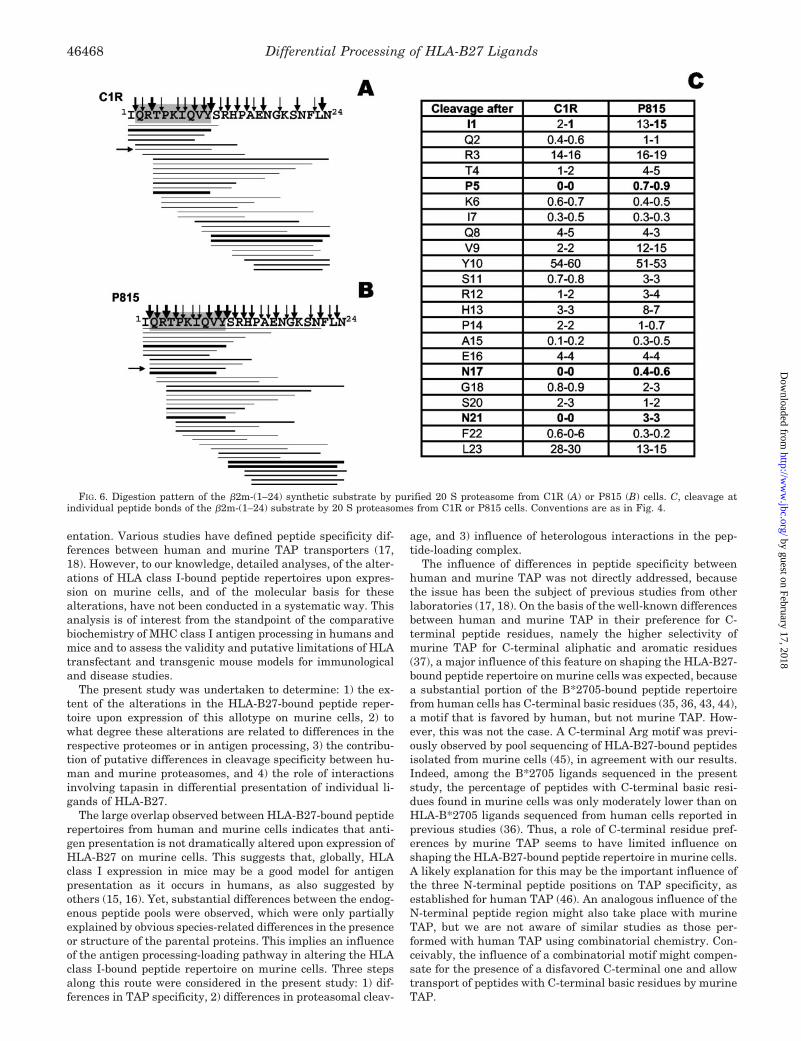
The second example (Fig. 6) corresponded to the QRTP-KIQVY peptide, arising from residues 2–10 of human �2m. Itsselective presence in murine cells correlated with the following:1) significantly higher cleavage efficiency at the exact N termi-nus of the peptide (after Ile-1) by the murine proteasome,relative to the human one (14% versus 1.5%), 2) about 4-foldhigher yield of the QRTPKIQVY ligand with the murine pro-teasome than with the human one (2% and 0.5%, respectively),and 3) presumably, a higher intracellular expression of thehuman �2m in murine cells, because the corresponding genewas introduced by transfection. However, this higher expres-sion does not, by itself, explain the absence of the ligand inhuman cells, because �2m is a very abundant protein also inthe CIR cell line.
A third situation was observed with the RRYLENGKETLQRpeptide, arising from residues 169–181 of the HLA-B27 heavychain, and found only in C1R cells. In this case cleavage at theexact N and C termini (after Leu-168 and Arg-181) by thehuman and murine proteasomes occurred with comparableefficiency (Fig. 7). In addition, cleavage within the sequence ofthe ligand was globally similar, although differences in cleav-age efficiency at particular peptide bonds (i.e.: after Arg-169and Leu-179) were observed between both proteasomes. As aresult, the peptide ligand was generated in vitro by the humanand murine proteasomes with similar yields: 0.5% and 0.3%,respectively. Thus, absence of the B27-(169–181) peptide inP815 transfectant cells cannot be explained by differences inproteasomal processing, as judged from in vitro digestions with20 S proteasomes.
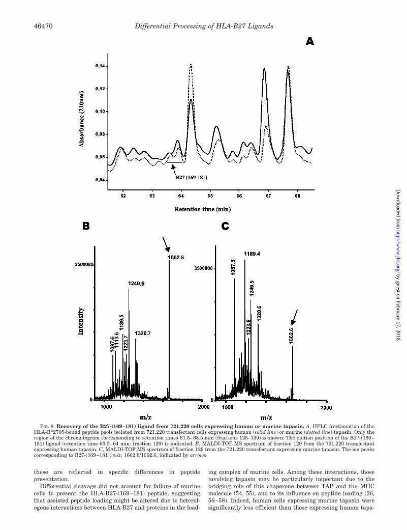
Species-specific Interactions with Tapasin Influence Presen-tation of a Natural Ligand by HLA-B27—We next examinedwhether absence of the B27-(169–181) ligand in the HLA-B27-bound peptide pool from P815 cells could be due to heterologousinteractions involving the human MHC molecule, murine ta-pasin, and/or other proteins in the peptide-loading complex.Thus, we isolated the HLA-B27-bound peptide pools fromtransfectants of the tapasin-deficient human cell line 721.220,which had been reconstituted with either human or murinetapasin. Peptide pools were fractionated by HPLC, and theB27-(169–181) peptide was searched in the chromatographicfractions around its corresponding retention time usingMALDI-TOF MS (Fig. 8). The peptide, which showed up as an
FIG. 4. Digestion pattern of the histone 2A-(77–105) synthetic substrate by purified 20 S proteasome from C1R (A) or P815 (B) cells. The sequenceof the HLA-B27 ligand is shaded. Thick, medium, and thin lines indicate peptide products recovered with �5%, 1–5%, and �1% yield of the totaldigest, respectively. Only peptides recovered with �0.1% yield are indicated. The IRNDEELNK peptide is indicated by a horizontal arrow. Thick,medium, and thin vertical arrows indicate cleavage sites that generated peptides with total yields �10%, 1–10%, and �1% of the total digest,respectively. C, cleavage at individual peptide bonds of the histone 2A-(77–105) substrate by 20 S proteasomes from C1R or P815 cells; figures arecleavage yields estimated as the total yield of peptides whose N-terminal or C-terminal ends corresponded to that peptide bond. The resultsobtained in two independent digestion experiments using the same preparations of the human or murine proteasome are shown in each column.Peptide bonds cleaved only with the proteasome of one species, or cleaved by both proteasomes with a 10-fold or larger difference, are in boldface.
Differential Processing of HLA-B27 Ligands46466
by guest on February 17, 2018http://w
ww
.jbc.org/D
ownloaded from

ion peak at m/z 1662.6/1662.8, eluted at HPLC fraction #129from both transfectants.
The amount of this peptide in each pool was estimated asfollows. First, the absorbance of HPLC fraction #129 relative tothe total absorbance of each peptide pool was calculated. Onthis basis, the peptide amount in this fraction was 1.1% of thetotal peptide pool from each transfectant. Second, the per-centage of the B27-(169–181) peptide, relative to the totalpeptide amount in fraction #129 from each peptide pool, wasestimated on the basis of the intensity of the ion peaks in thecorresponding MALDI-TOF MS spectra (Fig. 8, B and C).This is only an approximate estimation, because MALDI-TOFMS is not quantitative. Nevertheless, the difference was re-producible both in two independent spectra obtained from thesame sample, and in two independent preparations (data notshown). The intensity corresponding to the B27-(169–181)peptide in the MALDI-TOF MS spectrum of #129 from721.220 cells transfected with human (m/z: 1662.8) or murinetapasin (m/z: 1662.6) was 25.9% and 4.5%, respectively, of theadded intensity of all ion peaks in each fraction. Therefore,the estimated abundance of this peptide in the HLA-B27-bound peptide pools was: 1.1 � 0.259 � 0.28% and 1.1 �0.045 � 0.05% in the human and murine tapasin transfec-tants, respectively. Thus, in the presence of human tapasin,loading of the B27-(169–181) peptide into HLA-B27 was5.6-fold higher.
The reliability of this estimation was confirmed in a secondindependent experiment. In this one, the estimated abundanceof the B27-(169–181) peptide in the 721.220 cell transfectants
with human or murine tapasin was 0.4% and 0.04%, respec-tively, or a 10-fold increase in the presence of human tapasin.In turn, B27-(169–181) was previously estimated to represent0.4% of the HLA-B27-bound peptide pool from B*2705-C1Rtransfectants cells, which are human lymphoid cells with fullyfunctional tapasin (31).
These results indicate that the heterologous interaction be-tween B*2705 and/or other components of the peptide-loadingcomplex and murine tapasin significantly decreases the load-ing efficiency of the B27-(169–181) ligand, and strongly sug-gest that this influence contribute to impairing presentation ofthis ligand in murine cells.
Because 721.220 cells are of human origin, they do not fullyreproduce the situation of an HLA-B27 transfectant in a mu-rine cell. Thus, it is likely that additional species-related inter-actions in the peptide-loading complex, besides those involvingHLA-B27 and tapasin, further contribute to impairing presen-tation of B27-(169–181) in murine cells. This is strongly sug-gested from a comparison of the estimated abundance of thisligand in various cell types, as summarized in Table II.
DISCUSSION
Expression of human MHC class I molecules, including HLA-B27, in murine cells and transgenic mice has been widely usedto study antigen presentation to CTL (8, 9, 12, 13, 15, 16, 22,23, 39–42) and to establish animal models for human disease(6, 10, 11). However, species-related differences both in theproteome and in the specificity of the antigen processing-load-ing pathway may influence HLA class I-mediated antigen pres-
FIG. 5. Digestion pattern of the fatty acid synthase (1689–1718) synthetic substrate by purified 20 S proteasome from C1R (A) or P815 (B) cells.Both substrates differ by the S1712D change, corresponding to the polymorphism of the human and murine proteins at this position. C, cleavageat individual peptide bonds of the fatty acid synthase (1689–1718) substrate by 20 S proteasomes from C1R or P815 cells. Conventions are as inFig. 4.
Differential Processing of HLA-B27 Ligands 46467
by guest on February 17, 2018http://w
ww
.jbc.org/D
ownloaded from
entation. Various studies have defined peptide specificity dif-ferences between human and murine TAP transporters (17,18). However, to our knowledge, detailed analyses, of the alter-ations of HLA class I-bound peptide repertoires upon expres-sion on murine cells, and of the molecular basis for thesealterations, have not been conducted in a systematic way. Thisanalysis is of interest from the standpoint of the comparativebiochemistry of MHC class I antigen processing in humans andmice and to assess the validity and putative limitations of HLAtransfectant and transgenic mouse models for immunologicaland disease studies.
The present study was undertaken to determine: 1) the ex-tent of the alterations in the HLA-B27-bound peptide reper-toire upon expression of this allotype on murine cells, 2) towhat degree these alterations are related to differences in therespective proteomes or in antigen processing, 3) the contribu-tion of putative differences in cleavage specificity between hu-man and murine proteasomes, and 4) the role of interactionsinvolving tapasin in differential presentation of individual li-gands of HLA-B27.
The large overlap observed between HLA-B27-bound peptiderepertoires from human and murine cells indicates that anti-gen presentation is not dramatically altered upon expression ofHLA-B27 on murine cells. This suggests that, globally, HLAclass I expression in mice may be a good model for antigenpresentation as it occurs in humans, as also suggested byothers (15, 16). Yet, substantial differences between the endog-enous peptide pools were observed, which were only partiallyexplained by obvious species-related differences in the presenceor structure of the parental proteins. This implies an influenceof the antigen processing-loading pathway in altering the HLAclass I-bound peptide repertoire on murine cells. Three stepsalong this route were considered in the present study: 1) dif-ferences in TAP specificity, 2) differences in proteasomal cleav-
age, and 3) influence of heterologous interactions in the pep-tide-loading complex.
The influence of differences in peptide specificity betweenhuman and murine TAP was not directly addressed, becausethe issue has been the subject of previous studies from otherlaboratories (17, 18). On the basis of the well-known differencesbetween human and murine TAP in their preference for C-terminal peptide residues, namely the higher selectivity ofmurine TAP for C-terminal aliphatic and aromatic residues(37), a major influence of this feature on shaping the HLA-B27-bound peptide repertoire on murine cells was expected, becausea substantial portion of the B*2705-bound peptide repertoirefrom human cells has C-terminal basic residues (35, 36, 43, 44),a motif that is favored by human, but not murine TAP. How-ever, this was not the case. A C-terminal Arg motif was previ-ously observed by pool sequencing of HLA-B27-bound peptidesisolated from murine cells (45), in agreement with our results.Indeed, among the B*2705 ligands sequenced in the presentstudy, the percentage of peptides with C-terminal basic resi-dues found in murine cells was only moderately lower than onHLA-B*2705 ligands sequenced from human cells reported inprevious studies (36). Thus, a role of C-terminal residue pref-erences by murine TAP seems to have limited influence onshaping the HLA-B27-bound peptide repertoire in murine cells.A likely explanation for this may be the important influence ofthe three N-terminal peptide positions on TAP specificity, asestablished for human TAP (46). An analogous influence of theN-terminal peptide region might also take place with murineTAP, but we are not aware of similar studies as those per-formed with human TAP using combinatorial chemistry. Con-ceivably, the influence of a combinatorial motif might compen-sate for the presence of a disfavored C-terminal one and allowtransport of peptides with C-terminal basic residues by murineTAP.
FIG. 6. Digestion pattern of the �2m-(1–24) synthetic substrate by purified 20 S proteasome from C1R (A) or P815 (B) cells. C, cleavage atindividual peptide bonds of the �2m-(1–24) substrate by 20 S proteasomes from C1R or P815 cells. Conventions are as in Fig. 4.
Differential Processing of HLA-B27 Ligands46468
by guest on February 17, 2018http://w
ww
.jbc.org/D
ownloaded from

High conservation of the proteasome in mammals wouldsuggest that differences in proteasomal cleavage specificitybetween human and mouse subjects are unlikely to account forany significant variability in HLA class I-bound peptide reper-toires. However, differential processing of an influenza nucle-oprotein epitope (14) provided indirect evidence compatiblewith a role of the proteasome in generating this particularepitope only in human cells, although proteasomal cleavagewas not analyzed in that study. Our results here demonstratequalitative and quantitative differences in the cleavage pat-terns of synthetic peptide substrates between human and mu-rine 20 S proteasomes. These differences accounted for differ-ential presentation of one of four ligands analyzed, due to lackof cleavage at the C-terminal Lys residue of the peptide ligandby murine proteasomes. This result indirectly suggests thatpresentation of this ligand depends only on proteasomal cleav-age at its exact C terminus and that the endopeptidase activityof the tripeptidyl peptidase II at Lys residues (47) is not in-volved. In two other instances there was correlation betweenlack of presentation of the peptide ligand and lack of cleavageat its exact N terminus or lower generation in vitro by thecorresponding 20 S proteasome. MHC class I ligands can beproduced after trimming of N-terminally extended precursorsby ER aminopeptidases (48–52). The extent to which trimmingaccounts for generation of the constitutive MHC class I peptiderepertoires is significant, but far from absolute, and it isconceivable that many ligands may require direct generationby the proteasome. Our data with KRAYLQAR and QRTP-KIQVY are compatible with the possibility that, for theseparticular ligands, direct generation by the proteasome maydetermine presentation by HLA-B27, with little or no in-volvement of aminopeptidase-mediated trimming. The possi-bility that the observed differences in proteasome cleavagespecificity might be due to a different proteasome/immuno-proteasome ratio in the 20 S proteasomes isolated from C1Rand P815 cells seems unlikely, because both cells showed a
similar proteasome/immunoproteasome composition, withinthe limits of the analytical technique used. Moreover, be-cause both cells contained a mixture of constitutive protea-some and immunoproteasome, small differences in the ratioof both forms would hardly explain that cleavage of certainpeptide bonds occurred only with proteasomes from one celltype. Inhibition of proteolytic cleavage of a synthetic sub-strate by the proteasome inhibitors lactacystine andepoxomicin ruled out that the observed differences might bedue to contaminant proteases in the 20 S proteasome sam-ples. Thus, our results indicate that there are differences inthe cleavage specificity of the 20 S proteasome betweenmouse and human subjects. In addition, although these ex-periments reflect in a rather crude and not quantitative wayproteasomal processing in vivo (53), they strongly suggestthat these differences have a significant influence on differ-ential processing of particular peptides and, through this, onthe shaping of HLA-B27-bound peptide repertoire differencesbetween human and mouse subjects. Obviously this conclu-sion can be generalized to other HLA class I moleculesexpressed on murine cells.
In our study only one cell type from either humans or micewas compared, and it can be argued that cell- or organ-depend-ent variations in proteasome composition may influence pro-teasomal cleavage. Indeed, within a given species, proteasomalprocessing may vary depending on the exact proteasome/im-munoproteasome balance in the cell or other factors, so that thewhole spectrum of MHC class I ligands in vivo, and thereforeinter-species differences, may be more complex than outlined inthis study for HLA-B27. However, our data demonstrate thatin two cell lines with similar 20 S proteasome/immunoprotea-some composition there are distinct HLA-B27-bound peptiderepertoires, and that some of the differences correlate withdistinct cleavage patterns of the 20 S proteasomes from thesecells. Therefore, our data indicate that human and murine 20 Sproteasomes have differences in cleavage specificity and that
FIG. 7. Digestion pattern of the HLA-B27-(165–194) synthetic substrate by purified 20 S proteasome from C1R (A) or P815 (B) cells. C, cleavageat individual peptide bonds of the HLA-B27-(165–194) substrate by 20 S proteasomes from C1R or P815 cells. Conventions are as in Fig. 4.
Differential Processing of HLA-B27 Ligands 46469
by guest on February 17, 2018http://w
ww
.jbc.org/D
ownloaded from
these are reflected in specific differences in peptidepresentation.
Differential cleavage did not account for failure of murinecells to present the HLA-B27-(169–181) peptide, suggestingthat assisted peptide loading might be altered due to heterol-ogous interactions between HLA-B27 and proteins in the load-
ing complex of murine cells. Among these interactions, thoseinvolving tapasin may be particularly important due to thebridging role of this chaperone between TAP and the MHCmolecule (54, 55), and to its influence on peptide loading (26,56–58). Indeed, human cells expressing murine tapasin weresignificantly less efficient than those expressing human tapa-
FIG. 8. Recovery of the B27-(169–181) ligand from 721.220 cells expressing human or murine tapasin. A, HPLC fractionation of theHLA-B*2705-bound peptide pools isolated from 721.220 transfectant cells expressing human (solid line) or murine (dotted line) tapasin. Only theregion of the chromatogram corresponding to retention times 61.5–68.5 min (fractions 125–139) is shown. The elution position of the B27-(169–181) ligand (retention time 63.5–64 min: fraction 129) is indicated. B, MALDI-TOF MS spectrum of fraction 129 from the 721.220 transfectantexpressing human tapasin. C, MALDI-TOF MS spectrum of fraction 129 from the 721.220 transfectant expressing murine tapasin. The ion peakscorresponding to B27-(169–181); m/z: 1662.8/1662.6, indicated by arrows.
Differential Processing of HLA-B27 Ligands46470
by guest on February 17, 2018http://w
ww
.jbc.org/D
ownloaded from

sin in presenting this peptide in the context of HLA-B27. Thiswas not due to poor incorporation of murine tapasin into theloading complex of the human 721.220 cells, or other nonspe-cific phenomena, because the overwhelming majority (�95%) ofthe HLA-B27-bound peptide repertoire on 721.220 cells trans-fected with human tapasin was conserved in the correspondingtransfectant with murine tapasin.2 Thus, the effect of murinetapasin on decreasing loading of B27-(169–181) was relativelyselective and presumably results from the heterologous inter-action of this protein with one or more components of thehuman loading complex, including HLA-B27, TAP, calreticulin,and ERP57. It is likely that decreased loading of this peptide isexacerbated in murine cells, where the only human protein inthe loading complex is HLA-B27, explaining that the peptidewas not presented on murine cells.
It has been shown that HLA-B*4402 has poor surface expres-sion and antigen presentation when expressed on murine cells,but this was overcome by overexpression of murine tapasin(59). In that study it was shown that tapasin bridging ofB*4402 and TAP was not sufficient for efficient peptide load-ing, but this required a distinct function of tapasin that wasdependent on compatibility of components within the peptideloading complex. The situation is clearly different in HLA-B27,because both surface expression and constitutive peptide pres-entation was globally similar in P815 and C1R cells, but it ispossible that modulation of B27-(169–181) loading might berelated to species-specific interactions of tapasin with othercomponents of the peptide loading complex, besides HLA-B27and TAP.
In conclusion, expression of HLA-B27 on murine cells resultsin substantial alterations of the endogenous peptide repertoire.These alterations arise from at least the following: 1) species-or cell type-specific proteins, 2) homologous proteins withamino acid differences within the sequence of the peptide li-gand, 3) differences in proteasomal cleavage between mouseand human subjects, and 4) differential interaction of HLA-B27and/or other proteins in the loading complex with human ormurine tapasin.
The influence of differential processing/loading of HLA-B27ligands in murine cells has a potential influence and should betaken into account, in cytolytic T lymphocytes and diseasestudies carried out in transgenic models, because presentationof relevant antigens might be drastically affected. Yet, a ma-jority of the endogenous HLA-B27-bound peptide repertoirewas conserved in the two cell types analyzed, suggesting thatpresentation of many HLA-B27 ligands is not significantlyaffected by expression of this molecule on murine cells.
Acknowledgments—We thank James McKluskey and Anthony Pur-cell (University of Melbourne, Australia) for 221.220 transfectant celllines; Juan A. Lopez, Emilio Camafeita, and Juan P. Albar (CentroNacional de Biotecnologıa, Madrid) for help in two-dimensional gels andPSD-MALDI-TOF MS; Rosana Rogado, Anabel Marina, and Jesus
Vazquez (Centro de Biologıa Molecular Severo Ochoa, Madrid) for as-sistance in MALDI-TOF and electrospray MS; and Luis Anton forcritical reading of the manuscript. We thank the Fundacion RamonAreces for an institutional grant to the Centro de Biologıa MolecularSevero Ochoa.
REFERENCES
1. Brewerton, D. A., Hart, F. D., Nicholls, A., Caffrey, M., James, D. C., andSturrock, R. D. (1973) Lancet 1, 904–907
2. Brewerton, D. A., Caffrey, M., Nicholls, A., Walters, D., Oates, J. K., andJames, D. C. (1973) Lancet 2, 996–998
3. Hammer, R. E., Maika, S. D., Richardson, J. A., Tang, J. P., and Taurog, J. D.(1990) Cell 63, 1099–1112
4. Taurog, J. D., Maika, S. D., Simmons, W. A., Breban, M., and Hammer, R. E.(1993) J. Immunol. 150, 4168–4178
5. Zhou, M., Sayad, A., Simmons, W. A., Jones, R. C., Maika, S. D., Satumtira, N.,Dorris, M. L., Gaskell, S. J., Bordoli, R. S., Sartor, R. B., Slaughter, C. A.,Richardson, J. A., Hammer, R. E., and Taurog, J. D. (1998) J. Exp. Med.188, 877–886
6. Khare, S. D., Luthra, H. S., and David, C. S. (1995) J. Exp. Med. 182,1153–1158
7. Kingsbury, D. J., Mear, J. P., Witte, D. P., Taurog, J. D., Roopenian, D. C., andColbert, R. A. (2000) Arthritis Rheum. 43, 2290–2296
8. Kuon, W., Holzhutter, H. G., Appel, H., Grolms, M., Kollnberger, S., Traeder,A., Henklein, P., Weiss, E., Thiel, A., Lauster, R., Bowness, P., Radbruch,A., Kloetzel, P. M., and Sieper, J. (2001) J. Immunol. 167, 4738–4746
9. Kuon, W., Lauster, R., Bottcher, U., Koroknay, A., Ulbrecht, M., Hartmann,M., Grolms, M., Ugrinovic, S., Braun, J., Weiss, E. H., and Sieper, J. (1997)Arthritis Rheum. 40, 945–954
10. Weinreich, S., Eulderink, F., Capkova, J., Pla, M., Gaede, K., Heesemann, J.,van Alphen, L., Zurcher, C., Hoebe Hewryk, B., Kievits, F., and Ivanyi, P.(1995) Hum. Immunol. 42, 103–115
11. Weinreich, S. S., Hoebe-Hewryk, B., van der Horst, A. R., Boog, C. J. P., andIvanyi, P. (1997) Immunogenetics 46, 35–40
12. Koller, T. D., Clayberger, C., Maryanski, J. L., and Krensky, A. M. (1987)J. Immunol. 138, 2044–2049
13. Bernhard, E. J., Le, A. X., Yannelli, J. R., Holterman, M. J., Hogan, K. T.,Parham, P., and Engelhard, V. H. (1987) J. Immunol. 139, 3614–3621
14. Braud, V. M., McMichael, A. J., and Cerundolo, V. (1998) Eur. J. Immunol. 28,625–635
15. Tishon, A., LaFace, D. M., Lewicki, H., van Binnendijk, R. S., Osterhaus, A.,and Oldstone, M. B. (2000) Virology 275, 286–293
16. Cheuk, E., D’Souza, C., Hu, N., Liu, Y., Lang, H., and Chamberlain, J. W.(2002) J. Immunol. 169, 5571–5580
17. Schmitt, L., and Tampe, R. (2000) Chembiochemistry 1, 16–3518. Lankat-Buttgereit, B., and Tampe, R. (2002) Physiol. Rev. 82, 187–20419. Storkus, W. J., Howell, D. N., Salter, R. D., Dawson, J. R., and Cresswell, P.
(1987) J. Immunol. 138, 1657–165920. Zemmour, J., Little, A. M., Schendel, D. J., and Parham, P. (1992) J. Immunol.
148, 1941–194821. Calvo, V., Rojo, S., Lopez, D., Galocha, B., and Lopez de Castro, J. A. (1990)
J. Immunol. 144, 4038–404522. Rojo, S., Lopez, D., Calvo, V., and Lopez de Castro, J. A. (1991) J. Immunol.
146, 634–64223. Galocha, B., Lopez, D., and Lopez de Castro, J. A. (1993) J. Immunol. 150,
1653–166224. Greenwood, R., Shimizu, Y., Sekhon, G. S., and DeMars, R. (1994) J. Immunol.
153, 5525–553625. Copeman, J., Bangia, N., Cross, J. C., and Cresswell, P. (1998) Eur. J. Immu-
nol. 28, 3783–379126. Peh, C. A., Burrows, S. R., Barnden, M., Khanna, R., Cresswell, P., Moss, D. J.,
and McCluskey, J. (1998) Immunity 8, 531–54227. Barnstable, C. J., Bodmer, W. F., Brown, G., Galfre, G., Milstein, C., Williams,
A. F., and Ziegler, A. (1978) Cell 14, 9–2028. Ellis, S. A., Taylor, C., and McMichael, A. (1982) Hum. Immunol. 5, 49–5929. Paradela, A., Garcia-Peydro, M., Vazquez, J., Rognan, D., and Lopez de Castro,
J. A. (1998) J. Immunol. 161, 5481–549030. Paradela, A., Alvarez, I., Garcia-Peydro, M., Sesma, L., Ramos, M., Vazquez,
J., and Lopez de Castro, J. A. (2000) J. Immunol. 164, 329–33731. Alvarez, I., Sesma, L., Marcilla, M., Ramos, M., Martı, M., Camafeita, E., and
Lopez de Castro, J. A. (2001) J. Biol. Chem. 276, 32729–3273732. Yague, J., Vazquez, J., and Lopez de Castro, J. A. (1998) Tissue Antigens 52,
416–42133. Marina, A., Garcia, M. A., Albar, J. P., Yague, J., Lopez de Castro, J. A., and
Vazquez, J. (1999) J. Mass Spectrom. 34, 17–2734. Sesma, L., Montserrat, V., Lamas, J. R., Marina, A., Vazquez, J., and Lopez de
2 L. Sesma, I. Alvarez, M. Marcilla, A. Paradela, and J. A. Lopez deCastro, unpublished observations.
TABLE IIInfluence of species-specific interactions in the loading complex on presentation of the B27 (169-181) ligand by B*2705
The table shows the origin of HLA-B27, TAP, and tapasin in four types of B*2705 transfectant cell lines, and the estimated percentage of theB27-(169-181) ligand relative to the total B*2705-bound peptide pool in each cell line.
C1R 721.220 P815
HLA-B27 Human Human Human HumanTAP Human Human Human MouseTapasin Human Human Mouse MouseB27-(169–181) 0.4%a 0.3–0.4%b 0.05–0.04%b Not found
a Data from Ref. 31.b Results from two independent experiments.
Differential Processing of HLA-B27 Ligands 46471
by guest on February 17, 2018http://w
ww
.jbc.org/D
ownloaded from
Castro, J. A. (2002) J. Biol. Chem. 277, 16744–1674935. Ramos, M., Paradela, A., Vazquez, M., Marina, A., Vazquez, J., and Lopez de
Castro, J. A. (2002) J. Biol. Chem. 277, 28749–2875636. Lamas, J. R., Paradela, A., Roncal, F., and Lopez de Castro, J. A. (1999)
Arthritis Rheum. 42, 1975–198537. Momburg, F., Roelse, J., Howard, J. C., Butcher, G. W., Hammerling, G. J.,
and Neefjes, J. J. (1994) Nature 367, 648–65138. Yague, J., Alvarez, I., Rognan, D., Ramos, M., Vazquez, J., and Lopez de
Castro, J. A. (2000) J. Exp. Med. 191, 2083–209239. Kievits, F., Ivanyi, P., Krimpenfort, P., Berns, A., and Ploegh, H. L. (1987)
Nature 329, 447–44940. Weiss, E. H., Schliesser, G., Botteron, C., McMichael, A., Riethmuller, G.,
Kievits, F., Ivanyi, P., and Brem, G. (1990) Scand. J. Rheumatol. Suppl. 87,91–96
41. Kalinke, U., Arnold, B., and Hammerling, G. J. (1990) Nature 348, 642–64442. Huarte, E., Sarobe, P., Lasarte, J. J., Brem, G., Weiss, E. H., Prieto, J., and
Borras-Cuesta, F. (2002) Int. J. Cancer 97, 58–6343. Jardetzky, T. S., Lane, W. S., Robinson, R. A., Madden, D. R., and Wiley, D. C.
(1991) Nature 353, 326–32944. Rotzschke, O., Falk, K., Stevanovic, S., Gnau, V., Jung, G., and Rammensee,
H. G. (1994) Immunogenetics 39, 74–7745. Boisgerault, F., Mounier, J., Tieng, V., Stolzenberg, M. C., Khalil-Daher, I.,
Schmid, M., Sansonetti, P., Charron, D., and Toubert, A. (1998) Infect.Immun. 66, 4484–4490
46. Uebel, S., Kraas, W., Kienle, S., Wiesmuller, K. H., Jung, G., and Tampe, R.(1997) Proc. Natl. Acad. Sci. U. S. A. 94, 8976–8981
47. Seifert, U., Maranon, C., Shmueli, A., Desoutter, J. F., Wesoloski, L., Janek,K., Henklein, P., Diescher, S., Andrieu, M., de la Salle, H., Weinschenk, T.,Schild, H., Laderach, D., Galy, A., Haas, G., Kloetzel, P. M., Reiss, Y., andHosmalin, A. (2003) Nat. Immunol. 4, 375–379
48. Komlosh, A., Momburg, F., Weinschenk, T., Emmerich, N., Schild, H., Nadav,E., Shaked, I., and Reiss, Y. (2001) J. Biol. Chem. 276, 30050–30056
49. York, I. A., Chang, S. C., Saric, T., Keys, J. A., Favreau, J. M., Goldberg, A. L.,and Rock, K. L. (2002) Nat. Immunol. 3, 1177–1184
50. Saric, T., Chang, S. C., Hattori, A., York, I. A., Markant, S., Rock, K. L.,Tsujimoto, M., and Goldberg, A. L. (2002) Nat. Immunol. 3, 1169–1176
51. Serwold, T., Gonzalez, F., Kim, J., Jacob, R., and Shastri, N. (2002) Nature419, 480–483
52. Tanioka, T., Hattori, A., Masuda, S., Nomura, Y., Nakayama, H., Mizutani, S.,and Tsujimoto, M. (2003) J. Biol. Chem. 278, 32275–32283
53. Rock, K. L., and Goldberg, A. (1999) Annu. Rev. Immunol. 17, 739–77954. Sadasivan, B., Lehner, P. J., Ortmann, B., Spies, T., and Cresswell, P. (1996)
Immunity 5, 103–11455. Solheim, J. C., Harris, M. R., Kindle, C. S., and Hansen, T. H. (1997) J. Im-
munol. 158, 2236–224156. Li, S., Paulsson, K. M., Chen, S., Sjogren, H. O., and Wang, P. (2000) J. Biol.
Chem. 275, 1581–158657. Barnden, M. J., Purcell, A. W., Gorman, J. J., and McCluskey, J. (2000)
J. Immunol. 165, 322–33058. Purcell, A. W., Gorman, J. J., Garcia-Peydro, M., Paradela, A., Burrows, S. R.,
Talbo, G. H., Laham, N., Peh, C. A., Reynolds, E. C., Lopez de Castro, J. A.,and McCluskey, J. (2001) J. Immunol. 166, 1016–1027
59. Peh, C. A., Laham, N., Burrows, S. R., Zhu, N. Z., and McCluskey, J. (2000)J. Immunol. 164, 292–299
60. Luckey, C. J., Marto, J. A., Partridge, M., Hall, E., White, F. M., Lippolis, J. D.,Shabanowitz, J., Hunt, D. F., and Engelhard, V. H. (2001) J. Immunol. 167,1212–1221
61. Alvarez, I., Martı, M., Vazquez, J., Camafeita, E., Ogueta, S., and Lopez deCastro, J. A. (2001) J. Biol. Chem. 276, 48740–48747
62. Garcia, F., Marina, A., Albar, J. P., and Lopez de Castro, J. A. (1997) TissueAntigens 49, 23–28
63. Ramos, M., Alvarez, I., Sesma, L., Logean, A., Rognan, D., and Lopez deCastro, J. A. (2002) J. Biol. Chem. 277, 37573–37581
Differential Processing of HLA-B27 Ligands46472
by guest on February 17, 2018http://w
ww
.jbc.org/D
ownloaded from

CastroLaura Sesma, Iñaki Alvarez, Miguel Marcilla, Alberto Paradela and José A. López de
Loading Influence Peptide Presentation by HLA-B27 in Murine CellsSpecies-specific Differences in Proteasomal Processing and Tapasin-mediated
doi: 10.1074/jbc.M308816200 originally published online September 8, 20032003, 278:46461-46472.J. Biol. Chem.
10.1074/jbc.M308816200Access the most updated version of this article at doi:
Alerts:
When a correction for this article is posted•
When this article is cited•
to choose from all of JBC's e-mail alertsClick here
http://www.jbc.org/content/278/47/46461.full.html#ref-list-1
This article cites 62 references, 31 of which can be accessed free at
by guest on February 17, 2018http://w
ww
.jbc.org/D
ownloaded from