Histology of the Esophagus & Stomach · PDF fileHistology of the Esophagus & Stomach 1 S l i d...
13
Histology of the Esophagus & Stomach 1 S l i d e 1 HISTOLOGY OF THE ESOPHAGUS AND STOMACH Upper middle & Lower Esophagus, Cardiac, Fundic and Pyloric Stomach Robert W. Ogilvie, Ph.D. Professor Emeritus Medical University of South Carolina Visiting Professor University of South Carolina Roger H. Sawyer, Ph.D. Professor , Biological Sciences Executive Dean & Senior Associate Dean for Graduate Education, College of Arts & Sciences University of South Carolina This lecture will present the histology of the esophagus and stomach. S l i d e 2 Relevant Resources Text PDF containing slides and narrative text of this lecture WebMic Program WebMic Study Guide Gastrointestinal Tract: Unit 14, pages 167 - 188 These are the resources for text reading and practical lab exercises that will complement this lecture. S l i d e 3 Esophagus Stomach Vocabulary Gastric pits Surface mucous cells Simple tubular glands Mucous neck cells Chief cells Parietal cells Neuroendocrine cells Meissner’s (submucosal) Plexus Auerbach’s (myenteric) Plexus GALT Mucosa Submucosa Muscularis Mucosa Muscularis Externa Adventitia Mucosal Glands Submucosal Glands Intracellular Canaliculus This is the vocabulary of terms related to the content of this lecture.
-
Upload
trinhhuong -
Category
Documents
-
view
223 -
download
1
Transcript of Histology of the Esophagus & Stomach · PDF fileHistology of the Esophagus & Stomach 1 S l i d...

Histology of the Esophagus & Stomach
1
Slide 1
HISTOLOGY OF THEESOPHAGUS AND STOMACH
Upper middle & Lower Esophagus, Cardiac, Fundic and Pyloric Stomach
Robert W. Ogilvie, Ph.D.Professor EmeritusMedical University of South CarolinaVisiting ProfessorUniversity of South Carolina
Roger H. Sawyer, Ph.D.Professor , Biological SciencesExecutive Dean & Senior Associate Dean for Graduate Education, College of Arts & SciencesUniversity of South Carolina
This lecture will present the histology of the esophagus and stomach.
Slide 2
Relevant Resources
Text PDF containing slides and narrative text of this lecture
WebMic Program WebMic Study Guide Gastrointestinal Tract: Unit 14, pages 167 - 188
These are the resources for text reading and practical lab exercises that will complement this lecture.
Slide 3
Esophagus Stomach Vocabulary
Gastric pits Surface mucous cells Simple tubular glands Mucous neck cells Chief cells Parietal cells Neuroendocrine cells Meissner’s (submucosal)
Plexus Auerbach’s (myenteric) Plexus GALT
Mucosa Submucosa Muscularis Mucosa Muscularis Externa Adventitia Mucosal Glands Submucosal Glands Intracellular Canaliculus
This is the vocabulary of terms related to the content of this lecture.

Histology of the Esophagus & Stomach
2
Slide 4
Learning Outcomes
After completing a study of this lecture and working through the related laboratory exercise, you should be able to: describe and distinguish between mucosal and submucosal glands describe, distinguish between and give the function of the four layers of
the wall of the esophagus describe and distinguish between the basal, suprabasal, and
differentiated cell layers of the epithelium of the esophagus. describe, distinguish between and give the function of the four layers of
the wall of the stomach. name and differentiate between gastric glands of the stomach. classify the fundic glands of the stomach. define, describe, distinguish between and give the function of each of
the cell types in a fundic gland. explain how the stomach mucosa is protected from the effects of the
hydrochloric acid it produces.
These are the learning outcomes of this lecture. At the end of the lecture you will be presented with a few questions to aid in assessing your understanding and retention of the important concepts in this lecture.
Slide 5
Lecture Outline Mucosal vs. Submucosal Glands Esophagus
Organization Histology overview Mucosa Epithelium Nerve Plexi and Ganglia Esophagus-Stomach Junction Esophageal Cancer
Stomach Gross AnatomicalRegions
Organization Histology overview Gastric Glands Fundic Glands
Quiz
This is the outline of topics presented in this lecture. Each topic listed on this slide is hyperlinked to the first slide in that topic sequence with return hyperlinks back to this lecture outline slide.
Slide 6
Important ConceptMucosal Vs. Submucosal Gland
Submucosal Gland
muscularis mucosa
lamina propria
Mucosal Gland
muscularis mucosa
Laminapropria
Mucosal Glands
Return to outline
submucosasubmucosa
First, an important concept. All glands are composed of epithelial cells, specifically, glandular epithelial cells. Glands may be one cell or multicellular. Salivary glands are complex multicellular glands organized with a series of ducts and secretory acini. In the remainder of the GI tract we will encounter glands that are just merely tubes, or glands that have one or two ducts that branch at the end of which is a collection of secretory cells (e.g. mucous cells). A very important component of the esophagus, stomach and intestines are glands that reside within the wall of these segments of the gastrointestinal tract. The glands are located in either the mucosa or the submucosa or both. A mucosal gland is always located within the mucosa and is a part of the epithelial lining. Recall that the mucosa consists of the epithelial lining, the lamina propria (loose connective tissue) and the muscularis mucosa. Illustrated here in the left image are

Histology of the Esophagus & Stomach
3
mucosal glands that are tubes. The secretory cells make up the wall of the tube and the product moves through the tube lumen to the lumen of the GI tract. The specimen on the left is from the colon. Observe the muscularis mucosa noting that the simple tubular glands are located in the epithelial layer surrounded by the lamina propria. The glandular cells here are goblet cells. The specimen on the right illustrates the submucosal gland. A submucosal gland is located in the submucosa and therefore by definition is not a part of the mucosa as it is located beneath or external to the muscularis mucosa. Observe the muscularis mucosa noting that the gland is located beneath or external to the muscularis mucosa. Although not shown in this specimen as the plane of section did not pass through it, all submucosal glands are connected to the lumen by a duct. Submucosal glands are found only in the esophagus and duodenum (first part of the small intestine). As you learn the histology of the GI tract stay alert as to whether the glands are mucosal or submucosal in the particular segment you are studying.
Slide 7
The esophagus connects the pharynx with the stomach. It is lined with stratified squamous non-keratinized epithelium that provides a smooth surface for passage swallowed food. The technical term for swallowed food is ‘bolus’.
Esophagus
Return to outline

Histology of the Esophagus & Stomach
4
Slide 8
Adventitia
Esophagus Organization
Muscularis externa• inner circular,outer longitudinal & oblique• upper 1/3 skeletal muscle• middle 1/3 mixed• lower 1/3 smooth muscle
Submucosa – contains mucous glands
Mucosa:lining epithelium –nonkeratinized st squamous lamina propria – loose CT ( mucous glands at
distal end near the stomach)muscularis mucosa – smooth m. at distal end
lumen of esophagus
Return to outline
The esophagus is a hollow organ consisting of 4 layers. If the tube is cut across, the first layer seen surrounding the lumen is the mucosa composed of epithelium, lamina propria and a smooth muscle layer called the muscularis mucosa. The next layer is composed of loose connective tissue that may contain glands and adipose tissue and it is termed the submucosa. The next layer is composed of muscle organized in two layers, an inner circular oriented and an outer longitudinal oriented layer. The muscularis externa of the upper 1/3 of the esophagus is striated skeletal muscle, in the middle 1/3 it is a mixture of striated skeletal and smooth muscle while in the lower 1/3 it is all smooth muscle. The outermost layer is composed of loose connective tissue and is named the adventitia.
Slide 9
Esophagus Histology Overview9
mucous glands
mucosamuscularis externa
innercircular
outerlongitudinal
adventitia
MM
Upper 1/3Striated Skeletal Muscle
Middle 1/3Striated Skeletal Muscle
Smooth Muscle
Lower 1/3Smooth Muscle
Return to outline
This is a low magnification view of the entire thickness of a histological cross-section of the esophagus stained with hematoxylin & eosin. Observe the layers: mucosa, the submucosa with mucous glands, the muscularis externa composed of an inner layer of circularly arranged muscle and an outer layer of longitudinally oriented muscle. Wrapping the outside of the esophagus is the adventitia. The upper one third of the esophagus has a large amount of striated skeletal muscle that functions to aid in voluntarily moving the food bolus. The middle third is a mixture of striated and smooth muscle and at this point the voluntary act of swallowing is beginning to be taken over by involuntary control by the autonomic nervous system. The composition of the muscularis externa in the bottom third is all smooth muscle so that the food bolus is moved through that segment and into the stomach by the action of peristalsis – a wave like contraction controlled by the nerve plexi in the esophagus. The mucous glands secrete mucus into the lumen to lubricate the bolus of food thereby reducing the friction between the food and the epithelial lining of the esophagus.

Histology of the Esophagus & Stomach
5
Slide 10
Esophageal Lining Mucosa
Mucosa appears white opaque because the epithelium consists of so many layers of cells
Autopsy Specimenlumen view of esophagus mucosa
Histological SpecimenEsophagus Mucosa & Submucosa
muscularis mucosa
mucousglands
lamina propria
Return to outline
In the right image we see how the esophagus looks if it is opened at an autopsy. The mucosa is white opaque because there are so many layers of epithelial cells in the stratified squamous non-keratinized epithelium that there is no possibility for the color of blood to be reflected. The left image is a histological specimen of the esophageal mucosa and submucosa. Observe the very thick epithelium that consists of nearly 40 layers of cells. Observe the muscularis mucosa composed of bundles of smooth muscle cells between the dotted lines. In the submucosa are two clumps of mucous type glands indicated by the arrows.
Slide 11
Esophagus Epithelium
SSNK* Stratum Basale (SB) Stem cells
Stratum Spinosum Suprabasal layer (SL)
Proliferating cells Differentiated Layer (DL)
Lightly stained cells Cells contain glycogen
Stratum Superficial (SS) Several layers at the surface
that are about to ‘slough off’
*Stratified Squamous Non-Keratinized
SB
SL
DL
Return to outline
SS
Lymphocytes & Langerhans cells
(circled)
The lumen of the normal esophagus is lined by a tough, nonkeratinizing, stratified, squamous epithelium. The lower border of the squamous epithelium is irregular due to the presence of transitory folds of the lamina propria and more particularly, due to the presence of high conical papillae of connective tissue. These papillae are highly vascularized. On routine microscopy the squamous epithelium can be divided into the stratum basale (SB) where stem cells are located next to the basal lamina, the stratum spinosum and the stratum superficial (superificiale). The stratum spinosum can be divided into an intermediate or suprabasal layer (SL) and a differentiated layer (DL) that is more superficial. Most of the proliferating cells in this epithelium are in the supra basal layer. In the differentiated layer (DL) layer the cells contain glycogen, a mark of maturity. The most superficial layer is composed of several layers of cells that are flattened and show signs of dying. These cells are soon to be lost into the lumen (a process known as desquamation). These cells with their glycogen content will add to the mucus in the lumen. Langerhans cells (recall these from the epidermis as antigen presenting cells) and cytotoxic T-cells are commonly seen in the epithelium of the esophagus. Several are encircled in the inset from the boxed in region. Throughout the GI tract cells of the immune system are constantly monitoring for antigen and respond with either a humoral or cellular response.

Histology of the Esophagus & Stomach
6
Slide 12
Esophagus Nerve Plexi & Ganglia
Enteric Nervous System Meissner’s plexi
Located in the submucosa Sympathetic terminal nerve
fibers Parasympathetic ganglia and
nerve fibers
Auerbach’s (myenteric) Plexi Located between muscularis
externa layers Sympathetic terminal nerve
fibers Parasympathetic ganglia and
nerve fibers
Return to outline
Nerves and ganglia of the autonomic nervous system function in the GI tract to cause muscle contraction to move food and to control blood flow through action on the smooth muscle of arteries and arterioles. The autonomic nerves and ganglion found in the GI tract are known as the enteric nervous system. In this slide two ganglia and nerve plexi of the enteric nervous system will be presented. Meissner’s plexi are located in the submucosa. If an area as enclosed by the rectangle is enlarged the cell bodies of parasympathetic neurons can be seen. The nerves present would be both sympathetic and parasympathetic. The sympathetic nerve terminals would have arisen from neuron cell bodies alongside the spinal cord in paraganglia. The parasympathetic nerve fibers would be connected to the cell bodies of parasympathetic neurons as seen here. The other plexi are Auerbach’s (also myenteric) located between the inner and outer layers of the muscularis externa. If an area as outlined is enlarged the plexi with neuron cell bodies would look like this. In both the submucosal Meissner’s and the Auerbach’s (myenteric) plexi, the sympathetic innervation is inhibitory to peristalsis (the wave like contraction of muscle down the GI tract to propel the food bolus and digesting food). The parasympathetic component in both plexi facilitates peristalsis.
Slide 13
Esophagus-Stomach Junction
Endoscopic View
esophagus
stomach
View at Autopsy
stratified squamous non-keratinized
epithelium
simple columnar epithelium with mucosal glands
Return to outline
When viewed in the living state using an endoscope a transition in color at the esophagus-stomach junction can be observed. This endoscopic view obtained from the atlas at www.endoatlas.com is looking at the junction from the esophagus looking into the stomach. Observe the transition in color from whitish surface of the esophagus the red surface of the stomach. The border between the esophagus and stomach zig-zags and that is why it is named the Z line. In other words it is an interdigitating border. The stomach surface is red because the color of blood in the blood vessels is seen beneath the thin simple columnar epithelium. The esophagus whitish because the stratified squamous non-keratinized epithelium is so thick. In this opened esophagus as seen during an autopsy, the opaque whitish pink color of the esophagus changes abruptly at the stomach which is reddish. A histological section taken along the dotted line reveals this junction and confirms that it does change abruptly from the stratified squamous non-keratinized to simple columnar epithelium with mucosal glands.

Histology of the Esophagus & Stomach
7
Slide 14
normal epitheliumsquamous cell
carcinoma
invasivecancer
Esophageal CancerReturn to outline
normal
carcinoma
Esophageal cancers are typically carcinomas which arise from the epithelium of the esophagus. Difficulty in swallowing is one of the symptoms of esophageal cancer. Most esophageal cancers fall into one of two classes: squamous cell carcinomas and adenocarcinomas. Squamous cell carcinomas are similar to head and neck cancer in their appearance are usually associated with tobacco and alcohol consumption. Adenocarcinomas are often associated with a history of gastroesophageal reflux. Gastroesophageal reflux is the backing up of acid from the stomach into the esophagus. Continuous reflux on a regular basis irritates the esophagus causing it to undergo first metaplasia to a columnar epithelial lining to dysplasia (abnormal pattern of cells) of the epithelium when it no longer resembles any normal epithelium to finally cancerous cells. This slide illustrates squamous cell carcinoma. Observe the normal on the left compared to the carcinoma on the right. This carcinoma has invaded the lamina propria with masses of abnormal squamous cells and therefore it is malignant. The enlargement of this boxed in region shows normal epithelium on the left and carcinoma of the epithelium on the right. This smaller boxed in area shows a close-up of a small amount of normal epithelium together with the carcinoma. Observe that the cancerous epithelium bears no resemblance to the normal epithelium.
Slide 15
StomachThe stomach begins at the esophageal-stomach junction and ends at the stomach-duodenal junction. The stomach has mucosal glands but does not have any submucosal glands
Return to outline

Histology of the Esophagus & Stomach
8
Slide 16
Stomach is J shaped 2 surfaces
anterior & posterior
2 curvatures lesser & greater
4 regions cardiac fundus body pylorus
Stomach Regions
Cardiac
Fundus
Body
Pyloric
Rohen, 282A
anterior surface
Return to outline
The stomach is J shaped. It has two curvatures, a lesser and a greater. It has two surfaces, anterior (facing you) and posterior. It has 4 parts: 1) the cardiac region where the esophagus joins the stomach 2) the fundus that is the blind top of the stomach 3) the body region that makes up most of the mass of the stomach and 3) the pyloric region the end of which connects to the duodenum.
Slide 17
Stomach Histology Organization
Mucosa:epithelium – simple columnar epithelium lining surface, gastric pits & gastric glandslamina propria – loose connective tissue muscularis mucosa – smooth muscle
Submucosa – loose connective tissue, nerves, blood vessels and no glands
Muscularis externa• 3 layers of smooth muscle
Serosa
Return to outline
The stomach is a hollow organ with four distinct layers making up its wall. The mucosa is composed of simple columnar epithelium the cells lining the surface, gastric pits and glands the cells of which are continuous with the lining epithelial cells, a lamina propria of loose connective tissue that surrounds the gastric mucosal glands, the submucosa composed of loose connective tissue with blood vessels, nerves and adipose tissue, the muscularis externa that, unlike the esophagus, the smooth muscle of the stomach muscularis externa is arranged in three layers. The outermost layer is a serosa.
Slide 18 Mucosa
Submucosa
muscularis externa
Stomach – Histology Overviewmuscularis mucosa
Return to outline
Here we see a histological section at low magnification of the wall of the stomach. Note the mucosa, muscularis mucosa - one of the components of the mucosa, the submucosa where you can observe loose connective tissue, adipose tissue, but no glands and the muscularis externa, that unlike the esophagus, it is organized in 3 layers. The outer layer is longitudinal, the middle layer is circular and the internal layer is oblique. At the pylorus, the middle layer is greatly thickened to form the pyloric sphincter.

Histology of the Esophagus & Stomach
9
Slide 19
Stomach Muscle
3 layers of smooth muscle Outer longitudinal Middle circular Inner oblique
Pyloric sphincter Anatomic and
physiologic Parasympathetics
inhibit (relax) Sympathetics
constrictNetter, 260
The stomach has three layers of smooth muscle; an outer arranged longitudinally, a middle layer arranged in a circular direction around the stomach, and an inner layer that has fibers that run obliquely to the long axis of the stomach. At the pyloric end just before the stomach joins the duodenum the circular muscle layer is dominate forming a sphincter that is under the control of the autonomic nervous system. Parasympathetic fibers cause the sphincter muscles to relax thereby allowing the stomach contents to flow into the duodenum. Sympathetic fibers constrict the sphincter muscles preventing entrance of stomach contents into the duodenum. Parasympathetic action facilitates digestion. Sympathetic action inhibits digestion.
Slide 20
Cardiac Glands:•Short pits•Coiled glands•Secrete mucusand lysozyme
Pyloric Glands:•Deep pits•Shorter, coiled glands•Secrete mucus, lysozyme, gastrin, and somatostatin
Gastric glands
Fundic Glands:
•Short pits
•Long,branched glands (3-7 per pit) containing
•Chief cells
•Parietal cells
•Mucus cells
•Neuro-endocrine cells
• Secrete HCl, pepsinogen, intrinsic factor, mucus
Return to outline
The three types of gastric mucosal glands- cardiac, fundic & pyloric- are illustrated here. They are classified as simple tubular glands. Short epithelial lined pits project inward and to each of these pits one, two or three tubular glands may be attached and empty their products into the pit and from there into the lumen of the stomach. The cardiac glands have short pits with coiled tubes that have a wide lumen. They produce mucus and lysozyme. Fundic glands have relatively short pits with very long tubes which are usually branched, up to 3-7 per pit. They produce the gastric juice of the stomach that includes HCl, pepsinogen (converted to pepsin by HCL), intrinsic factor, mucus. The secretions are produced by specific cell types. Hydrochloric acid secreted by parietal cells begins the digestion of dietary protein by hydrolyzing the peptide bonds breaking the protein down into various sized polypeptides. Parietal cells also secrete intrinsic factor, a glycoprotein that binds to vitamin B12 that is essential for B12 to be absorbed later in the ileum of the small intestine. Pepsin secreted by chief cells (converted from a precursor, pepsinogen by HCL) is a potent proteolytic enzyme that breaks polypeptides into smaller units, peptides. In the small intestine other enzymes break the peptides down into amino acids. Pyloric glands have long, deep pits that connect to coiled tubes. Pyloric glands produce mucus, lysozyme, and gastrin by G cells and somatostatin by D cells. All three glands have mucous cells that produce mucus. Mucus provides an acid protecting coating of the cells lining the stomach. Both G and D cells are neuroendocrine cells and their secretions are classified as hormones. Gastrin stimulates parietal cells located in the fundic glands that secrete HCL and intrinsic factor. Although gastrin and somatostatin hormones are secreted by neuroendocrine cells in all three gastric glands, most of the cells secreting these are located in the pyloric glands.

Histology of the Esophagus & Stomach
10
Slide 21
Fundic Gland Cell TypesAll parts•surface
All parts•neck
Body•Neck to Mid-gland
Body•base
Body / pyloricregions•base
All parts•neck
Return to outline
The largest distribution in the stomach of the three types of glands – cardiac, fundic and pyloric – is of the fundic type of gland containing cells that secrete hydrochloric acid and pepsin, the main components of gastric juice. Although the glands are traditionally named Fundic Glands, they are just in the Fundic region. They are present throughout the body as well as the fundic regions of the stomach. Starting at the bottom of the fundic gland, the enteroendocrine cells produce specific stimulatory/inhibitory substances such as gastrin and somatostatin. Chief cells (also called zymogenic cells) are primarily responsible for producing pepsinogen which will be converted into the highly active proteolytic enzyme pepsin. These cells also produce lipase. Parietal (also called Oxyntic from a greek word meaning to make acid) are involved primarily in producing the components of hydrochloric acid through the intermediary enzyme carbonic anhydrase (details of this process in a subsequent slide). These cells also produce gastric intrinsic factor, a glycoprotein that binds to vitamin B12 so that it can be absorbed in the small intestine. A deficiency of vitamin B12 results in the development of abnormal erythrocytes that are too large but do not have an increased amount of hemoglobin. The result is that the average erythrocyte has a lower than normal concentration of hemoglobin, the oxygen carrying molecule. The name for the disease was first termed pernicious anemia because, before the cause was discovered, the disease was often fatal. Now it is treatable with the oral administration of vitamin B12 (it is absorbed by the oral mucosa) or, in difficult cases, intravenous administration of vitamin B12. Now the common name for the condition is megaloblastic anemia (large red blood cells with an inadequate amount of hemoglobin). Mucous neck cells have a different type of mucus from the surface cells. The vesicles are at the apical end of the cell & they stain intensely with PAS. The surface lining cells produce mucus continuously. Regenerative cells provide renewal cells both up & down the gastric gland. Unlike stem cells of the small intestine which are located in the base of the gland, these reside in the neck region. Surface lining cells produce a thick mucus covering which becomes infiltrated with carbonate, the other dissociative product of carbonic acid, to aid in the prevention of self digestion.

Histology of the Esophagus & Stomach
11
Slide 22
HCl production & intrinsic factor for Vitamin B 12 complex
Parietal Cells
The parietal cell gives the appearance of “sunny side up” eggs with its prominent acidophilic cytoplasm & centrally located nucleus. This is due to the large amount of membranous material in the cell referred to as an intracellular canaliculus when at rest. The membranous system consists of an intracelluar canalicular and tubulovesicular system that increases the membrane surface area for the molecules that participate in the formation of hydrochloric acid. The cells also have a very high density of mitochondria that provide the energy required to actively transport hydrogen ions across the plasma membrane into the lumen of the stomach. Recall that cell membranes, cytoplasmic membranes and mitochondria have a dominant population of basic proteins that react with the acid dye eosin. That is the reason why these cells are acidophilic staining intensely pink with eosin.
Slide 23
Parietal CellFunction
Observe this drawing of a parietal cell noting the intracellular canaliculus, the carbonic anhydrase enzyme that facilitates combining of water and carbon dioxide, the active transport of hydrogen and chloride ion and the many mitochondria. To make hydrochloric acid, the parietal cell ribosomes synthesize an enzyme called carbonic anhydrase. Carbonic anhydrase facilitates the combination of carbon dioxide & water to make carbonic acid, a highly unstable molecule which breaks down almost immediately into the hydrogen ion & the bicarbonate ion. The H+ is actively transported across the membrane in the numerous intracellular canaliculi present in the parietal cell. An intracellular canaliculus is a deep invagination of the cell membrane with folds that greatly increase the surface area of the plasma membrane. In this membrane there are proteins that literally and actively pump or carry hydrogen ions from the cytoplasm into the lumen of the fundic gland. There, hydrogen ions complex with chloride ions to make hydrochloric acid. The chloride ions, as the drawing indicates, are actively transported from the interstitium on the capillary side of the parietal cell, across the parietal cell, again by a membrane protein pump into the lumen of the fundic gland. The bicarbonate ion diffuses into the interstitium and is picked up by blood capillaries. The two pumps require energy to function and that energy is supplied by the many mitochondria present in a parietal cell. The more hydrochloric acid is produced the more bicarbonate is formed. The result is acid for digestion and the alkaline bicarbonate for buffering against the acid digestion of the lumen lining epithelial cells as we shall see in the next slide.
Histology of the Esophagus & Stomach
12
Slide 24
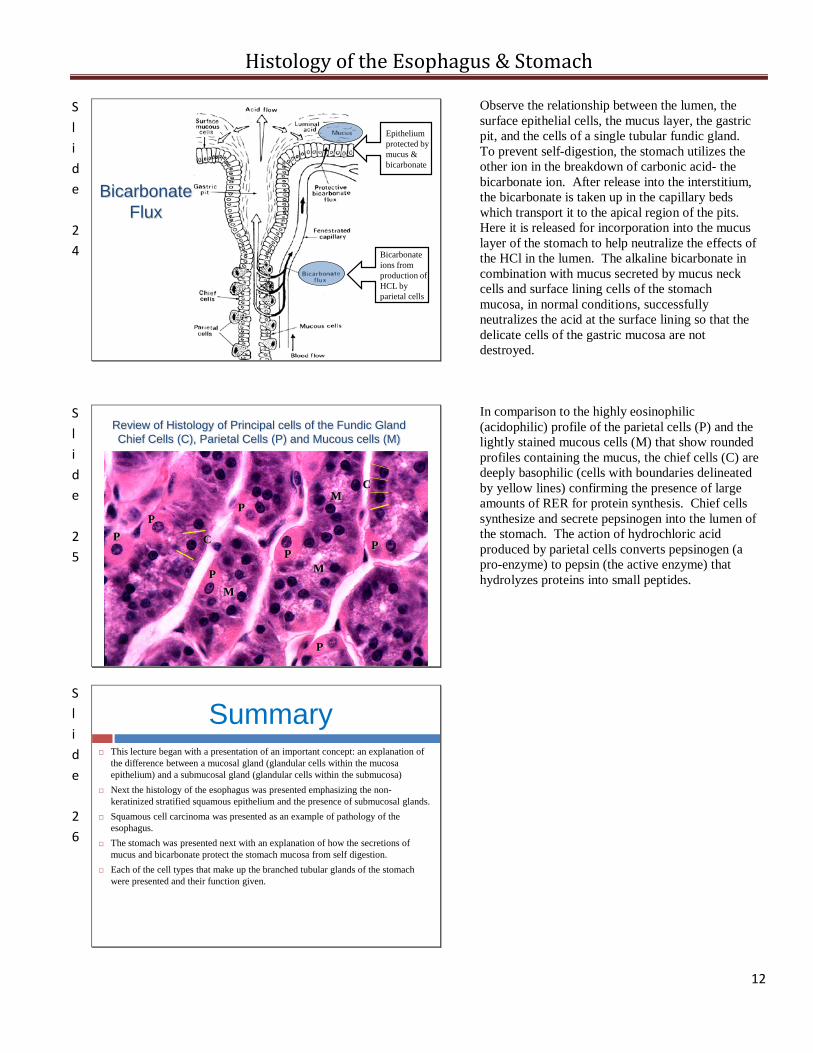
Bicarbonate Flux
Bicarbonate ions from production of HCL by parietal cells
Epithelium protected by mucus & bicarbonate
Observe the relationship between the lumen, the surface epithelial cells, the mucus layer, the gastric pit, and the cells of a single tubular fundic gland. To prevent self-digestion, the stomach utilizes the other ion in the breakdown of carbonic acid- the bicarbonate ion. After release into the interstitium, the bicarbonate is taken up in the capillary beds which transport it to the apical region of the pits. Here it is released for incorporation into the mucus layer of the stomach to help neutralize the effects of the HCl in the lumen. The alkaline bicarbonate in combination with mucus secreted by mucus neck cells and surface lining cells of the stomach mucosa, in normal conditions, successfully neutralizes the acid at the surface lining so that the delicate cells of the gastric mucosa are not destroyed.
Slide 25
Review of Histology of Principal cells of the Fundic GlandChief Cells (C), Parietal Cells (P) and Mucous cells (M)
P
PP
P
PP
P
M
C
C
M
M
In comparison to the highly eosinophilic (acidophilic) profile of the parietal cells (P) and the lightly stained mucous cells (M) that show rounded profiles containing the mucus, the chief cells (C) are deeply basophilic (cells with boundaries delineated by yellow lines) confirming the presence of large amounts of RER for protein synthesis. Chief cells synthesize and secrete pepsinogen into the lumen of the stomach. The action of hydrochloric acid produced by parietal cells converts pepsinogen (a pro-enzyme) to pepsin (the active enzyme) that hydrolyzes proteins into small peptides.
Slide 26
Summary This lecture began with a presentation of an important concept: an explanation of
the difference between a mucosal gland (glandular cells within the mucosa epithelium) and a submucosal gland (glandular cells within the submucosa)
Next the histology of the esophagus was presented emphasizing the non-keratinized stratified squamous epithelium and the presence of submucosal glands.
Squamous cell carcinoma was presented as an example of pathology of the esophagus.
The stomach was presented next with an explanation of how the secretions of mucus and bicarbonate protect the stomach mucosa from self digestion.
Each of the cell types that make up the branched tubular glands of the stomach were presented and their function given.

Histology of the Esophagus & Stomach
13