Histology of cns.dk.2014
51
Histology of Central Nervous System Dr..Deepak N.Khedekar. Asst. professor Dept of Anatomy LTMMC & GH,MUMBAI 2014
-
Upload
deepak-khedekar -
Category
Health & Medicine
-
view
273 -
download
0
Transcript of Histology of cns.dk.2014
Histology of
Central Nervous System
Dr..Deepak N.Khedekar.Asst. professor
Dept of AnatomyLTMMC & GH,MUMBAI
2014

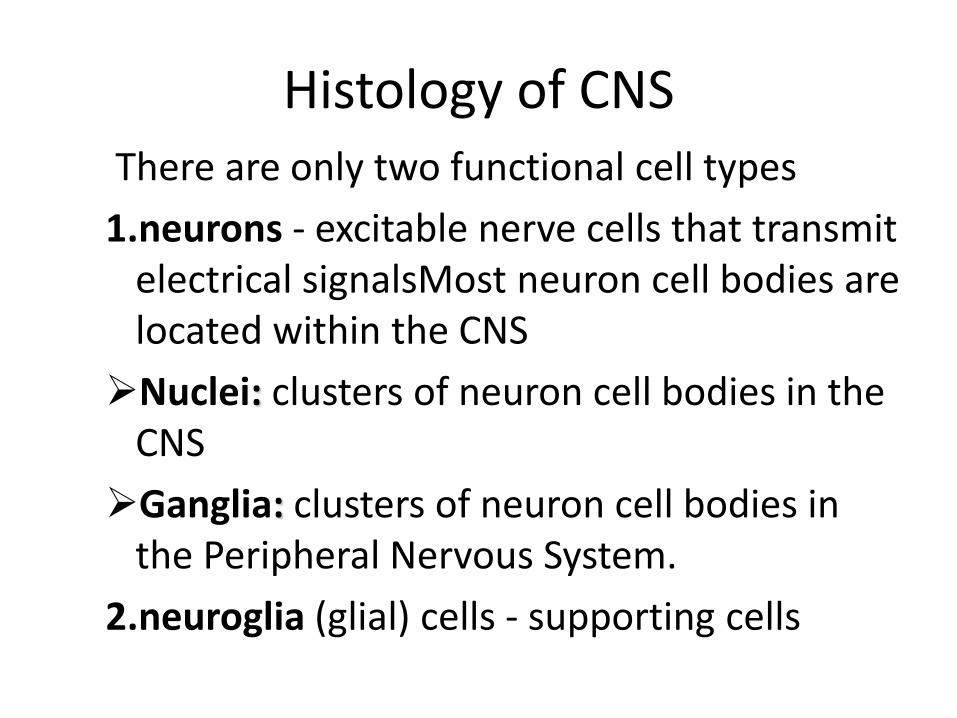
Histology of CNSThere are only two functional cell types
1.neurons - excitable nerve cells that transmit electrical signalsMost neuron cell bodies are located within the CNS
Nuclei: clusters of neuron cell bodies in the CNS
Ganglia: clusters of neuron cell bodies in the Peripheral Nervous System.
2.neuroglia (glial) cells - supporting cells
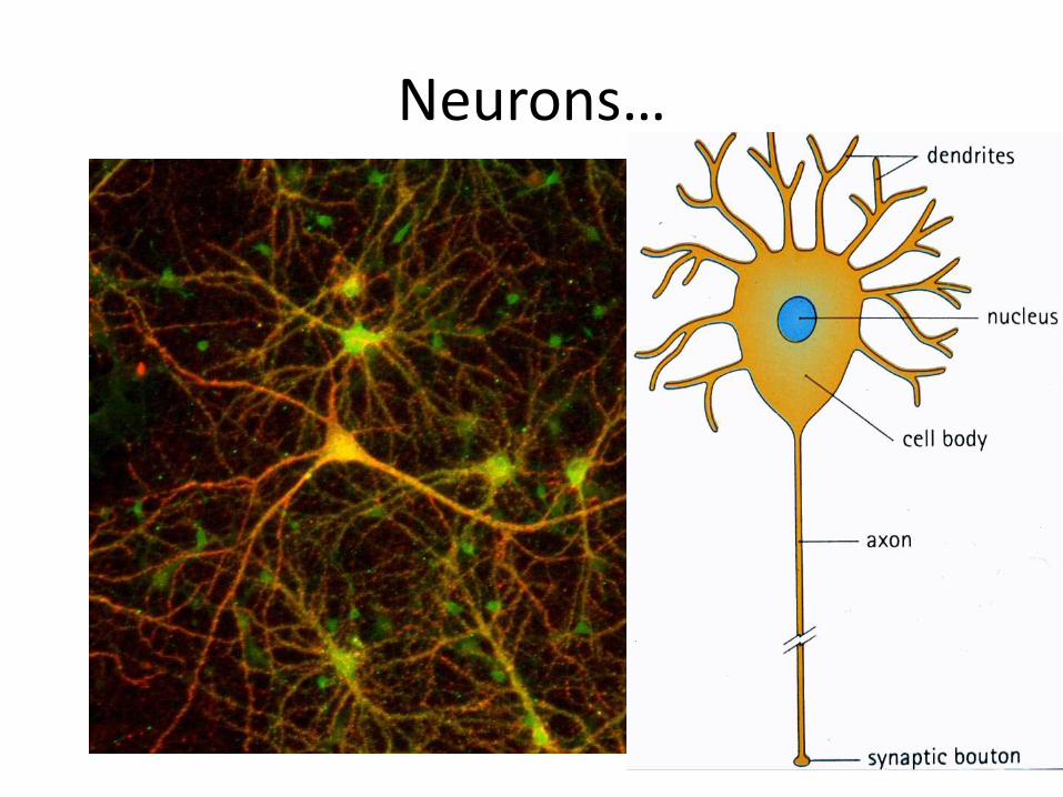
Neurons…

Motor neuron
NUN
D
DA
AH
NB
NB
D
V
A-axon D-dendrite N-nucleus NB-Nissl bodyAH-axon hillock V-blood vessel NU-nucleolus
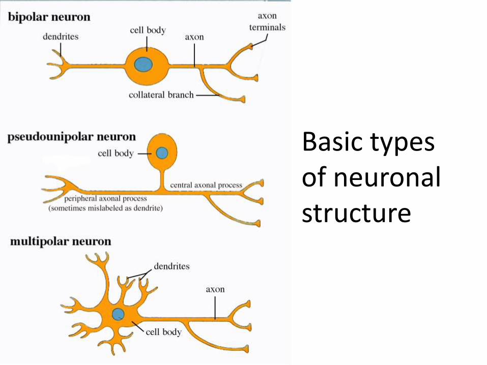
Basic types of neuronal structure

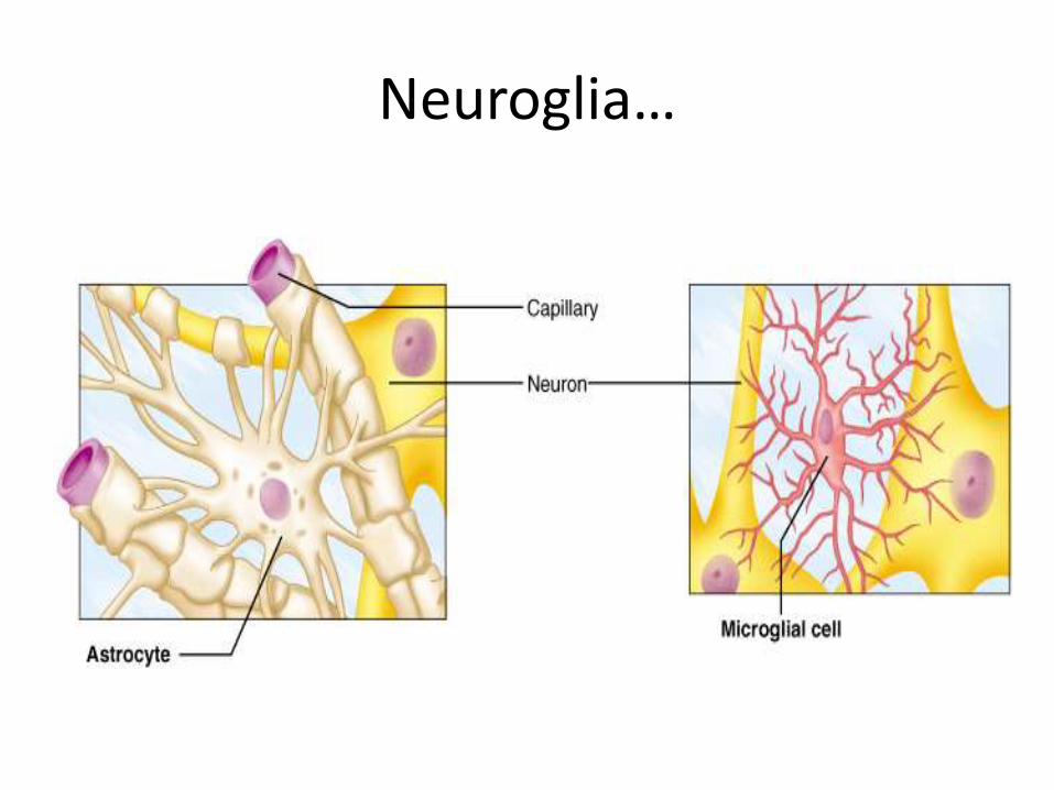
Neuroglia-Supporting cells…
Neuroglia - 4 types in the CNS
Astrocytes
• star shaped with many processes,connect to neurons; help anchor them to nearby blood capillaries
• control the chemical environment of the neurons
Microglia
• oval with thorny projections
• monitor the health of neurons, if infection occurs, they change into macrophages
Neuroglia…
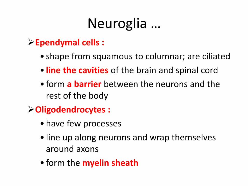
Neuroglia …Ependymal cells :
• shape from squamous to columnar; are ciliated
• line the cavities of the brain and spinal cord
• form a barrier between the neurons and the rest of the body
Oligodendrocytes :
• have few processes
• line up along neurons and wrap themselves around axons
• form the myelin sheath

Neuroglia…

Neuroglia in the Peripheral NS
Satellite cells
• surround neuron cell bodies in the periphery
• maintain the extracellular environment
Neurolemmocytes (Schwann cells)
• surround axons/dendrites and form the myelin sheath around larger nerve fibers
• similar to oligodendrocytes in function –insulators

Slide 1.Spinal cord…

Spinal cord…

Spinal cord-Gray matter…
Multipolar neurons:
• Vesicular nucleus with distinct nucleolus
• Nissl subastance (basophilic)
Neuroglia :small light staining supportive cells
small basophilic nuclei

Spinal cord…

Spinal cord- Gray mater…

Spinal cord- White matter
Closely packed groups of myelinated axons
Myelin sheaths appear as clear spaces around dark staining axons

Anterior horn of spinal cord

Slide 2.Cerebral Cortex
• The grey matter consists of neuron cell bodies and their dendritic interconnections & glialcells. The cortex contains 30 billion neurons.
• The evolved cortex in mammals called neocortex consists of 6 layers of neurons.

Cerebral Cortex…Neocortex
• >90 % of our total cortical area.
• 6 layered structured, Refered to as homogenic cortex.
Paleocortex
• Covers some parts of the base of the telencephalon.(olfactory area)
-Forms heterogenic cortex.
Archicortex
• The hippocampal formation.

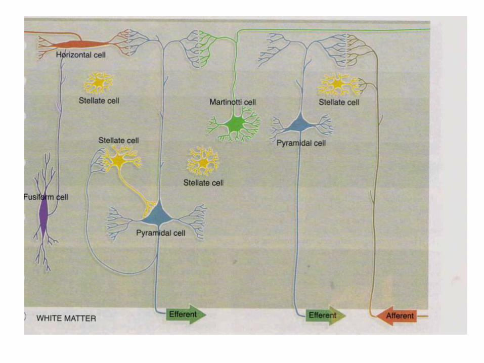
Neuron Cell types
• Two principal cell types are present in neo cortex. (PSM FuC)
– 1.Pyramidal cell .
– 2.Stellate cell .
• Other cells are …
– 3. Cells of Martinotti
– 4.Fusiform cells
– 5.Horizontal cells of Cajal
.
.

Pyramidal Cells…
• Pyramid shaped 10 micr. to 70 micr.).
• Axon arises from the base and the dendrite from the apex.
• The largest of the pyramidal cells are called the BETZ cells.
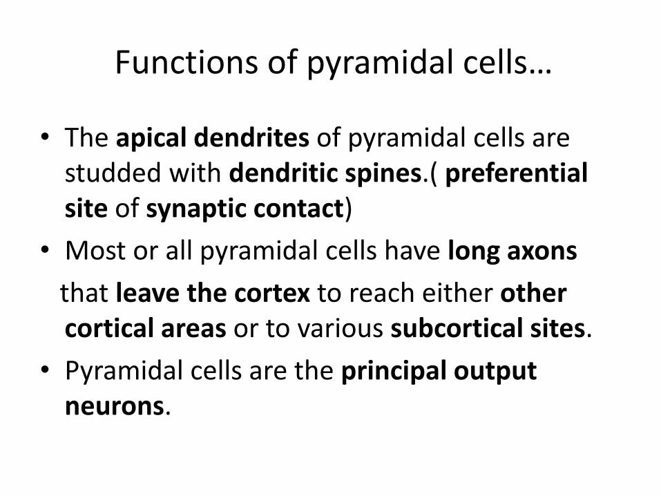
Functions of pyramidal cells…
• The apical dendrites of pyramidal cells are studded with dendritic spines.( preferential site of synaptic contact)
• Most or all pyramidal cells have long axons
that leave the cortex to reach either other cortical areas or to various subcortical sites.
• Pyramidal cells are the principal output neurons.

Stellate Cells…
Also known as granular cells.
-They are the principal interneurons of cortex .
--They are typically small (< 10 micrometres) multipolarneurons.
• principal interneurons

Cells Of Martinotti…
• Small polygonal cells.
• Forms synapses with the pyramidal cells.
• Have very few short dendrites.
• axon extends towards the surface and bifurcate to run horizontally in most superficial layers.

Fusiform Cells…
• Spindle shaped cells, are oriented at right angles to the cortex.
• Axon arises from the side of the cell body and passes superficially.
• Dendrites extend from each end of the cell body branching into deeper and more superficial layers.
• Functions are similar to that of pyramidal cells.

Horizontal Cells Of Cajal (Or) RetziusCajal Cells…
• Small ,spindle shaped, oriented parallel to the surface.
• Least common cell type, found only in most superficial layer.
• Axons pass laterally to synapse with dendrites of pyramidal cells.
• prominent during development, but disappear after birth.

6 Layers Of Neocortex (MGP-PGM)
1.Plexiform or molecular layer 2.outer granular layer3.outer pyramidal cell layer4.inner granular layer 5.inner pyramidal cell layer/ganglion cell layer6.multiform cell layer
Layers are not equally prominent everywhere. They form granular & agranular layers.
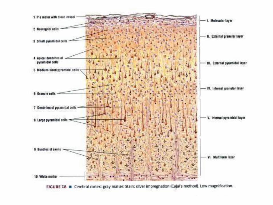

6 cortical layers …

1.Plexiform layer or molecular layer
• Most superficial layer,
• contains many dendritic and axonal synapses with one another.
• Sparse nuclei of neuroglia are seen
• Occasional horizontal cells of cajal are seen.
• 2.OUTER GRANULAR LAYER
• Dense population of small pyramidal cells and stellate cells.
• Different types of neuroglial cells

3.Pyramidal cell layer
• Moderate sized pyramidal cells predominate.
• Large pyramidal cells are present in further deeper layers.Martinotti cells are also present.
• 4.INNER GRANULAR LAYER
• Consists of densely packed granule cells /stellatecells
5.GANGLIONIC LAYER
• Large pyramidal cells,Stellate cells (few),Cells of martinotti
• Huge pyramidal Betz cells of motor cortex are present. Hence the name ganglion cell layer.


Slide 3.Cerebellum…
Gray matter (Cerebellar cortex) consist of 3 layers
Outer molecular layer
middle purkinje layer
Inner granular layer
White matter consist of myelinated nerve fibers or axons

Cerebellum Gray matter(10x)…

Cerebellum(40x)…

Cerebellum…
Outer molecular layer
• Few, small neurons (basket cells and stellate cells)
• Many fibers extending parallel to length of folium
middle purkinje layer
• Purkinje cells few in no., pyriform in shape, dendrites in molecular layer
Inner granular layer
• Numerous neurons with prominent nuclei
• Glomeruli-empty spaces in granular layer

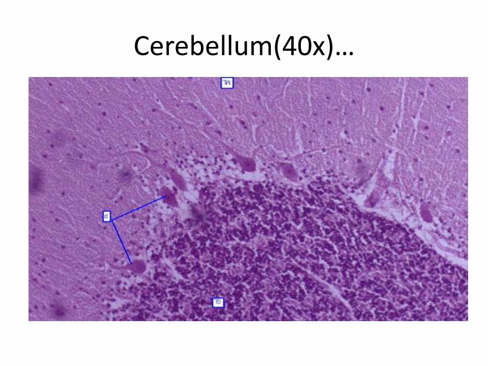
Cerebellum(40x)…

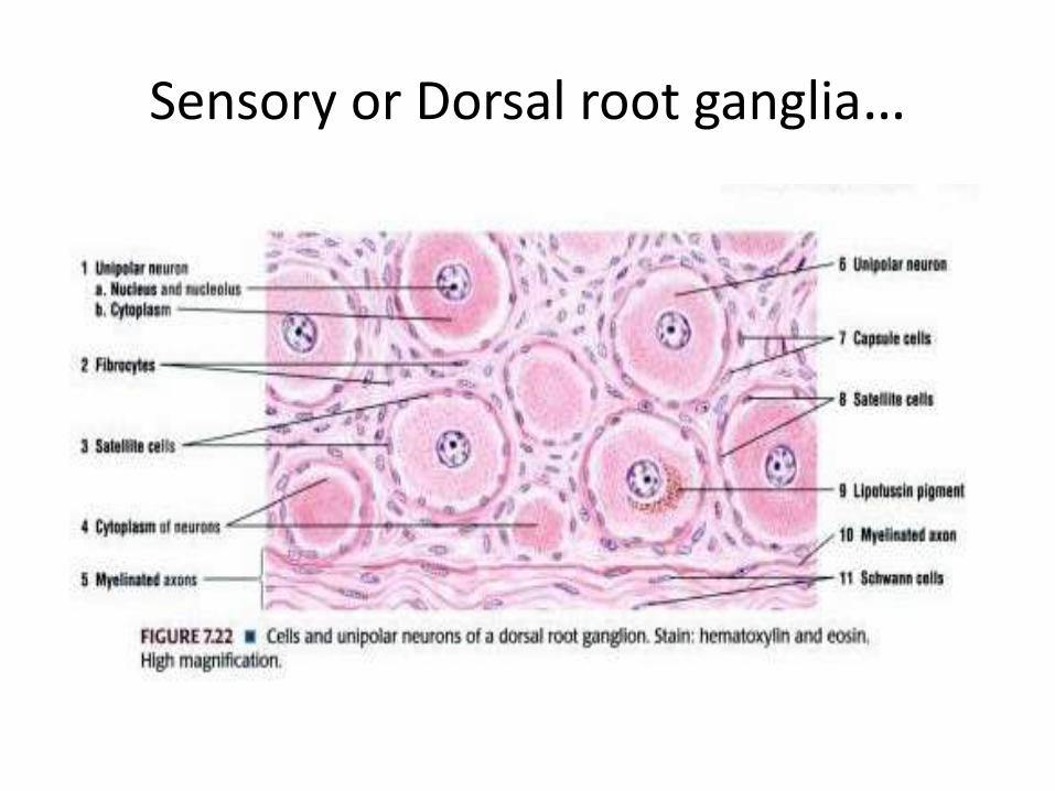
Slide 4.Sensory Ganglia…
• Two types: spinal (dorsal root) and cranial ganglia associated with spinal and cranial nerves, respectively
• Contain large sensory neurons and abundant small glial cells, called satellite cells
• Sensory neurons are pseudounipolar

Sensory or Dorsal root ganglia(10x)
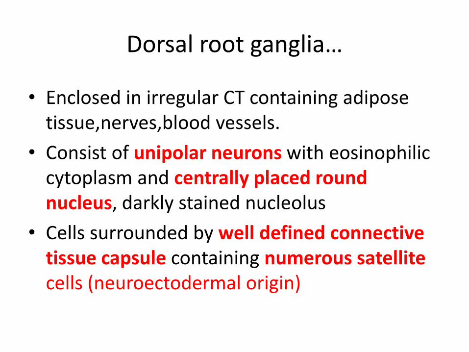
Dorsal root ganglia…
• Enclosed in irregular CT containing adipose tissue,nerves,blood vessels.
• Consist of unipolar neurons with eosinophilic cytoplasm and centrally placed round nucleus, darkly stained nucleolus
• Cells surrounded by well defined connective tissue capsule containing numerous satellite cells (neuroectodermal origin)

Sensory or dorsal root ganglion…
Sensory or Dorsal root ganglia…

Sensory or Dorsal root ganglion…

Autonomic-Sympathetic ganglia…

Autonomic-Sympathetic ganglia…
• Enclosed in irregular CT containing adipose tissue,nerves,blood vessels.
• Consist of multipolar neurons with eosinophilic cytoplasm and ecccentrallyplaced round nucleus, darkly stained nucleolus
• Cells surrounded by ill defined connective tissue capsule containing few satellite cells(neuroectodermal origin)

Sensory Ganglia vs ANS Ganglia

At the end of the every lecture…..